evaluation of wound healing properties of dryopteris filix
TRANSCRIPT

Iranian Journal of Pharmaceutical Sciences 2021: 17 (1): 79-90
www.ijps.ir
Original Article
Evaluation of Wound Healing Properties of Dryopteris filix-mas Leaf and
Root Extracts on Albino Rats
Earnest Oghenesuvwe Erhirhiea,b
*, Cathrine Odinakachi Anyima, Ugochukwu Moses Okezie
c, Sabastine Obiora
Igboemea, Emmanuel Emeka Ilodigwe
a
aDepartment of Pharmacology and Toxicology, Faculty of Pharmaceutical Sciences, Nnamdi Azikiwe University,
Awka, Nigeria, bDepartment of Pharmacology and Toxicology, Faculty of Pharmaceutical Sciences, Chukwuemeka
Odumegwu Ojukwu University, Igbariam, Nigeria, cDepartment of Pharmaceutical Biology and Biotechnology,
Faculty of Pharmaceutical Sciences, Nnamdi Azikiwe University, Awka, Nigeria.
Abstract
Dryopteris filix-mas (D. filix-mas), belonging to the family of Dryopteridacea is a swampy fern that is popularly
used by the Southern Nigerian dwellers in the treatment of wounds, hemorrhages, boil and other diseases. In order to
authenticate its folkloric benefits in wounds, this study evaluated its wound healing activity using excision model. A
total of fifty (50) Wistar rats were randomized into ten groups of five animals each. After creation of surface wounds,
group 1 received paraffin base (control). Group 2 received Povidon iodine (standard). Groups 3, 4, 5 and 6 received
1.25, 2.5, 5 and 10% (w/w) of an ethanol leaf extract of D. filix-mas formulated with paraffin base respectively.
Groups 7, 8, 9, 10 were treated with 1.25, 2.5, 5 and 10% (w/w) of ethanol root extract D. filix-mas formulated with
paraffin base respectively. Treatments were topically applied to wounds once daily and healing rate was monitored
every 3 days for 21 days. Wound swaps were taken on day 10th and day 20
th for bacteria load determination. In-vitro
antimicrobial activities of the leaf and root extract were tested against Staphylococcus aureus, Pseudomonas
aeruginosa and Escherichia coli using agar-well diffusion method. Higher concentrations (5 and 10%) of the leaf and
root extracts exhibited better wound healing activities more than lower concentrations. The leaf extract produced a
better healing rate (wound contraction), antimicrobial activity and body weight regaining activities more than the root
extract. This study validates the traditional use of D filix-mas in the treatment of wounds.
Keywords: Antimicrobial activity, Dryopteris filix-mas, Leaf extract, Root extract, Wound healing, Wistar rats.
1. Introduction
Wounds are physical injuries to any part of
the body. They may result from fire accident,
chemicals, radiation, electricity or surgery [1].
Wounds involve laceration or a break of the
skin that causes distortion in the normal skin
structure and function [2]. Complicated
wounds, including burns, chronic wound
Corresponding Authors: Erhirhie Earnest Oghenesuvwe,
Department of Pharmacology and Toxicology, Faculty of
Pharmaceutical Sciences, Nnamdi Azikiwe University,
Awka, Nigeria, Department of Pharmacology and
Toxicology, Faculty of Pharmaceutical Sciences,
Chukwuemeka Odumegwu Ojukwu University, Igbariam,
Nigeria.
Tel: +234-7060434974
Email: [email protected].
Cite this article as: Erhirhie E. O., Anyim C. O., Okezie U.
M., Igboeme S. O., Ilodigwe E. E., Evaluation of Wound
Healing Properties of Dryopteris filix-mas Leaf and Root
Extracts on Albino Rats, 2021, 17 (1): 79-90.

Erhirhie E. O, et al. / IJPS 2021; 17 (1): 79-90
80
ulcers, and orthopedic wounds play a
significant part in morbidity and mortality.
Statistics revealed that about 34% of diseases
such as boils, diabetes, chicken pox, tumors,
athlete‟s foot, sporotrichosis, and necrotizing
ascitis that occur worldwide are associated
with the wound pathogenesis [3-5]. A study
carried out by Adigun and co-workers in a
tertiary hospital in Kwara State, Nigeria
revealed that 31.55% of total hospital patients
suffered from various wounds and the cost
spent on wound care was quite burdensome
[6].
Several synthetic drugs such as povidone
iodine, methoxamine, silver sulfadiazine,
polyhexamethylene biguanide are useful
agents in the treatment of wound [7].
However, some limitations such as high cost,
allergic reactions, bacterial resistance are
associated with the use of these conventional
agents in the treatment of wounds [7]. Thus,
efforts have been made to discover alternative
approaches in the treatment and management
of various wounds.
Among alternative remedies, medicinal plants
have played significant role in the treatment of
wounds since prehistoric times [8]. Plant
extracts have been used as poultices for the
purpose of stopping hemorrhage and
promoting wound healing [9]. They are
effective, accessible, affordable, and have no
or less side effects than some conventional
wound healing drugs [10].
For example,
Hibiscus sabdariff, Aloe vera, honey,
ageratum conyzoides, Azadirachta indica,
Hyptis suaveolans, Moringa oleifera and other
medicinal plants have been validated and
documented to have wound healing properties
based on their folkloric information [2, 5, 8,
11].
D. filix-mas (Linn.) Schott), also known as
male fern, wild fern, bear‟s paw is a medicinal
plant used traditionally in the treatment of
wound [12]. It is a strong ornamental fern that
is commonly found in damp, shady and
swampy environments. It is widely distributed
in various parts of North and South America,
Europe, Asia, United States of America and
Africa including Nigeria [13]. D. filix-mas is
an evergreen plant that reaches a maximum
height of 1.5 m, with a single crown on each
rootstock. The leaves consist of 20-35 pinnae
on each side of the rachis. The leaves taper at
both ends, with the basal pinnae about half the
length of the middle pinnae [14]. It is the male
version of the widespread lady fern, Athyrium
filix-femina, which is considered as a
traditional vegetable [15]. Its decoction is
applied topically in the treatment of mumps,
carbuncles, abscesses, boils and sores
provoked by severe burns. An infusion of the
rhizome is applied topically in rheumatism,
septic wounds, hemorrhoids and ulcers in the
Republic of Azerbaijan [16]. It is used among
rural dwellers in Lebanon in the treatment of
neuralgia and rheumatic disorders [17].
In Southern parts of Nigeria the root and
leaf decoctions are used topically for wounds
and various hemorrhage, such as epistaxis,
menorrhagia and postpartum hemorrhage,
inflammation, rheumatoid arthritis and worm
infestations. Recent studies have reported its
antidiarrheal [18], tocolytics [19], teratogenic
[20], and anti-inflammatory [21] effects.
Wound Healing Potentials of Dryopteris filix-mas on Albino Rats
81
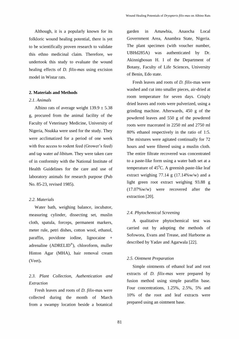
Although, it is a popularly known for its
folkloric wound healing potential, there is yet
to be scientifically proven research to validate
this ethno medicinal claim. Therefore, we
undertook this study to evaluate the wound
healing effects of D. filix-mas using excision
model in Wistar rats.
2. Materials and Methods
2.1. Animals
Albino rats of average weight 139.9 ± 5.38
g, procured from the animal facility of the
Faculty of Veterinary Medicine, University of
Nigeria, Nsukka were used for the study. They
were acclimatized for a period of one week
with free access to rodent feed (Grower‟s feed)
and tap water ad libitum. They were taken care
of in conformity with the National Institute of
Health Guidelines for the care and use of
laboratory animals for research purpose (Pub
No. 85-23, revised 1985).
2.2. Materials
Water bath, weighing balance, incubator,
measuring cylinder, dissecting set, muslin
cloth, spatula, forceps, permanent markers,
meter rule, petri dishes, cotton wool, ethanol,
paraffin, povidone iodine, lignocaine +
adrenaline (ADRELID®), chloroform, muller
Hinton Agar (MHA), hair removal cream
(Veet).
2.3. Plant Collection, Authentication and
Extraction
Fresh leaves and roots of D. filix-mas were
collected during the month of March
from a swampy location beside a botanical
garden in Amawbia, Anaocha Local
Government Area, Anambra State, Nigeria.
The plant specimen (with voucher number,
UBHd285A) was authenticated by Dr.
Akinnigbosun H. I of the Department of
Botany, Faculty of Life Sciences, University
of Benin, Edo state.
Fresh leaves and roots of D. filix-mas were
washed and cut into smaller pieces, air-dried at
room temperature for seven days. Crisply
dried leaves and roots were pulverized, using a
grinding machine. Afterwards, 450 g of the
powdered leaves and 550 g of the powdered
roots were macerated in 2250 ml and 2750 ml
80% ethanol respectively in the ratio of 1:5.
The mixtures were agitated continually for 72
hours and were filtered using a muslin cloth.
The entire filtrate recovered was concentrated
to a paste-like form using a water bath set at a
temperature of 450C. A greenish paste-like leaf
extract weighing 77.14 g (17.14%w/w) and a
light green root extract weighing 93.88 g
(17.07%w/w) were recovered after the
extraction [20].
2.4. Phytochemical Screening
A qualitative phytochemical test was
carried out by adopting the methods of
Sofowora, Evans and Trease, and Harborne as
described by Yadav and Agarwala [22].
2.5. Ointment Preparation
Simple ointments of ethanol leaf and root
extracts of D. filix-mas were prepared by
fusion method using simple paraffin base.
Four concentrations, 1.25%, 2.5%, 5% and
10% of the root and leaf extracts were
prepared using an ointment base.

Erhirhie E. O, et al. / IJPS 2021; 17 (1): 79-90
82
2.6. Test Microorganism
Staphylococcus aureus, Pseudomonas
aeruginosa and Escherichia coli strains
implicated in wound infection were gotten
from Pharmaceutical Microbiology and
Biotechnology Laboratory, Nnamdi Azikiwe
University, Agulu, Nigeria. These isolates
were reconfirmed by subjecting them to gram
staining as well as specific biochemical test.
2.7. Preparation of Media
Manitol salt agar (111 g/L) and
MacConkey agar (48.5 g/L) were prepared
following the manufacturer's specification by
dispersing the required quantity of agar in
distilled water. It was homogenized and then
sterilized by autoclave at 121oC for 15
minutes.
2.8. Determination of Antimicrobial Activity
(Agar-well diffusion assay) on Root and Leaf
Extracts
The antibacterial activity of the extracts
was evaluated by cup plate agar diffusion
method described by Okezie et al [23]. The
microorganisms used in this test were from the
human pathogenic bacteria, Staphylococcus
aureus, Pseudomonas aeruginosa and
Escherichia coli. The bacterial cultures were
adjusted to 0.5 McFarland turbidity standards.
Thereafter, each of the test organisms was
seeded onto sterile Mueller-Hinton Agar MHA
(Oxoid, Difco USA) and Sabouraud dextrose
agar plates using sterile swab, (diameter: 90
mm). A sterile cork borer was used to make
wells (6 mm in diameter) on each MHA. Stock
concentrations of the extracts were made using
sterile distilled water as diluent. Here, 800 mg
of each of the extract was dissolved in 2 ml of
sterile distilled water. Thereafter, serial
dilutions were made using the same diluents to
get graded concentrations (400, 200, 100, 50,
25, and 12.5 mg/ml). Aliquot (80 µl) of each
concentration was applied to each well
previously seeded with the test organism. The
cultures were incubated at 37oC for 24 hrs.
Antimicrobial activity was determined by
measuring the zone of inhibition (ZOI) around
each well (excluding the diameter of the well).
For each concentration, replicate trials were
conducted against test organisms.
2.9. Wound Healing Studies
Evaluation of wound healing activity of the
root and leaf extracts was carried out using
wound excision model in Wister albino rats,
according to the method described by Vinay et
al. [24]. Prior to the creation of the wound,
hair on the mid-back of animals was carefully
shaved using a pair of scissors and tiny hair
remaining were completely removed using
hair removal cream (Veet). Thereafter, animals
were anesthetized by injection of 0.3 ml of a
local anesthetic (Lignocaine + Adrenaline)
into the shaved portion. Circular wounds of
approximately 1.5 to 2.0 cm diameter were
created along a circumference that was
outlined with the orifice of a test tube stained
with methylene blue. The wounds were left
undressed for 24 hours.

Wound Healing Potentials of Dryopteris filix-mas on Albino Rats
83
Thereafter, animals were divided into ten
groups of five animals each. Animals in group
1 were treated with paraffin base and they
served as the control. Animals in group 2,
which served as standard received Povidon
iodine. Groups 3, 4, 5 and 6 were treated with
1.25, 2.5, 5 and 10% (w/w) of ethanol leaf
extract-ointment of D. filix-mas respectively.
Animals in groups 7, 8, 9, 10 were treated with
1.25, 2.5, 5 and 10% (w/w) of ethanol root
extract-ointment of D. filix-mas respectively.
Treatments were topically applied once daily
and wound closure rate was assessed every 3
days by measuring the longitudinal and
transverse diameter of the wound, using a pair
of dividers. Bacterial load was assessed on day
10th and day 20
th, while healing rate, body
weight gain and inhibition in wound bacteria
count were calculated using the formulae
below.
2.10. Statistical Analyses
Data were presented as Mean ± standard
error of mean (SEM), n = 5 and were analyzed
by one way analysis of variance (ANOVA)
using SPSS version 20. Differences between
mean were considered statistically significant
at p<0.05.
3. Results and Discussion
3.1. Phytochemical Composition
Reducing sugar, proteins, were present in
similar proportion in both extracts. Tannins,
flavonoids and alkaloids were more abundant
in the leaf extract. Anthraquinones were absent
in both extracts while saponins and steroids
were absent in the root extract (Table 1). More
phytoconstituents, saponins, alkaloids, sterols,
terpenoids, glycosides, tannins, flavonoids,
reducing sugar and proteins were present in
the leaf extract than the root extract, which is
an indication that the leaf extract may retain
more healing potentials against wounds. Jian
et al, [25]
reported that saponins and
flavonoids show evidence of wound healing
activity. Terpenoids, due to their acerbic and
antimicrobial properties have been reported to
promote wound contraction and increased rate
of epithelialization [26].
Total phenolic
contents and antioxidant activity of the leaf
extract previously reported by Sekender et al
[14] also supports the healing properties
exhibited by the leaf extract. Suppression of
inflammatory process, oxidative stress
processes occurring in wounds have been
attributed to polyphenols, which have
potentials to terminate chain reactions
associated with oxidative damage in biological
system [5].
Polyphenols have also been
reported to prevent edema and itching
associated with wounds via inhibition of
allergic mediators such as histamine and
Healing rate =
Wound diameter at the baseline−Wound diameter at any day
Wound diameter at baseline ×
100
1
Body weight gains =
Final body weight on day 20th−Initial body weight before treatment
Final body weight on day 20th
× 100
1
Reduction in wound bacteria count =
Bacteria count on day 10th − Bacteria count day 20th
BactBacteria count on day 10th ×
100
1

Erhirhie E. O, et al. / IJPS 2021; 17 (1): 79-90
84
serotonin [27, 28]. To this end, we hypothesize
that flavonoid, quercetrin which was an abundant
phyto-compound responsible for the anti-
inflammatory activity of the leaf extract in our
earlier study [21] could be strongly connected
with the wound healing activity of D. filix mas.
3.2. Antimicrobial Activity
The root extract did not show activity
against E. coli and P. aeruginosa, except little
activity against S. aureus. On the other hand,
the leaf extract produced better activity against
S. aureus when compared to the root extract.
Also, the leaf extract inhibited the growth of
P. aeruginosa at higher concentrations, 200
and 400 mg/ml with minimum inhibitory
concentrations of 200 and 50 mg/mL for P.
aeruginosa and S. aureus respectively (Table
2). Microorganism, P. aeruginosa and S.
aureus are implicated in the pathogenesis of
wound. In this study, we found that the leaf
extract of D. filix-mas showed a concentration
dependent inhibition against P. aeruginosa
and S. aureus more than the root extract which
only showed less inhibition against S. aureus.
This inhibitory activity of the leaf extracts
could have contributed to its fast healing rate
by minimizing super infections that are
implicated in wounds. This is supported by the
antimicrobial activity of methanol leaf extract
of D- flix-mas against the growth of E. coli,
S.ureus, P. aeruginosa as well as S. abony and
and E. faecalis [29]. Some polyphenols have
been shown to reduce the growth of
microorganisms such as S. aureus and E. coli,
which are implicated in wound progression
[30].
3.3. Wound healing Effect
There was a time dependent healing rate in
group 1 treated with soft paraffin (negative
control), Povidone iodine (positive control)
and various concentrations of plant extract.
An exception to these were reduced healing
rates observed on day 3 in control, 2.5% leaf
extract, 1.25 and 2.5% root extract treated
groups (Table 3).
Progressive healing rate observed in this
study suggests that healing is a gradual and
continuous process which involves several
phases such as hemostasis, inflammation,
leukocyte migration, proliferation and
maturation as substantiated by the study of
Boateng et al [1] who reported that wound
healing is a progresses process. Faster rate of
wound closure and contraction following
topical application of higher concentrations, 5
and 10% of ethanol leaf and root extracts of D.
filix-mas suggests that substantial healing
effect could be attained at higher
concentration. The healing effect could be
attributed to flavonoids, and other
phytoconstituents in the extracts.
There was reduced healing rate in groups
treated with Povidone iodine, 1.25 and 2.5%
leaf extract as well as 1.25 and 2.5% root
extract when compared to healing rate in the
negative control group on various days.
Although, healing rate in Povidone iodine
group was slightly higher than 1.25 and 2.5%
in leaf extract as well as 1.25 and 2.5% in root
extract treated groups on day 21st (Table 3).
There was time dependent closure in wounds
treated with the negative control (soft paraffin)
and positive control (povidone iodine).

Wound Healing Potentials of Dryopteris filix-mas on Albino Rats
85
Healing rates in 5.0 and 10.0% leaf and root
extracts were higher than healing rates in both
negative and positive control groups as well as
1.25 and 2.5% treated groups. This was
consistent from day 3 through day 21 (Table 3).
Wound healing is a natural process which
takes place by regeneration of dermal and
epidermal tissue. When tissue damages are not
treated and properly managed, it could lead to
chronic inflammation and secondary
infections, thus causing more damage to
surrounding tissues [10]. Whenever there is an
injury, a set of overlapping procedures take
place in a predictable manner to restore the
damage [25]. Effective remedies in the
treatment of wounds have potentials of
minimizing secondary infections,
inflammation and promotion of tissue
regeneration [10].
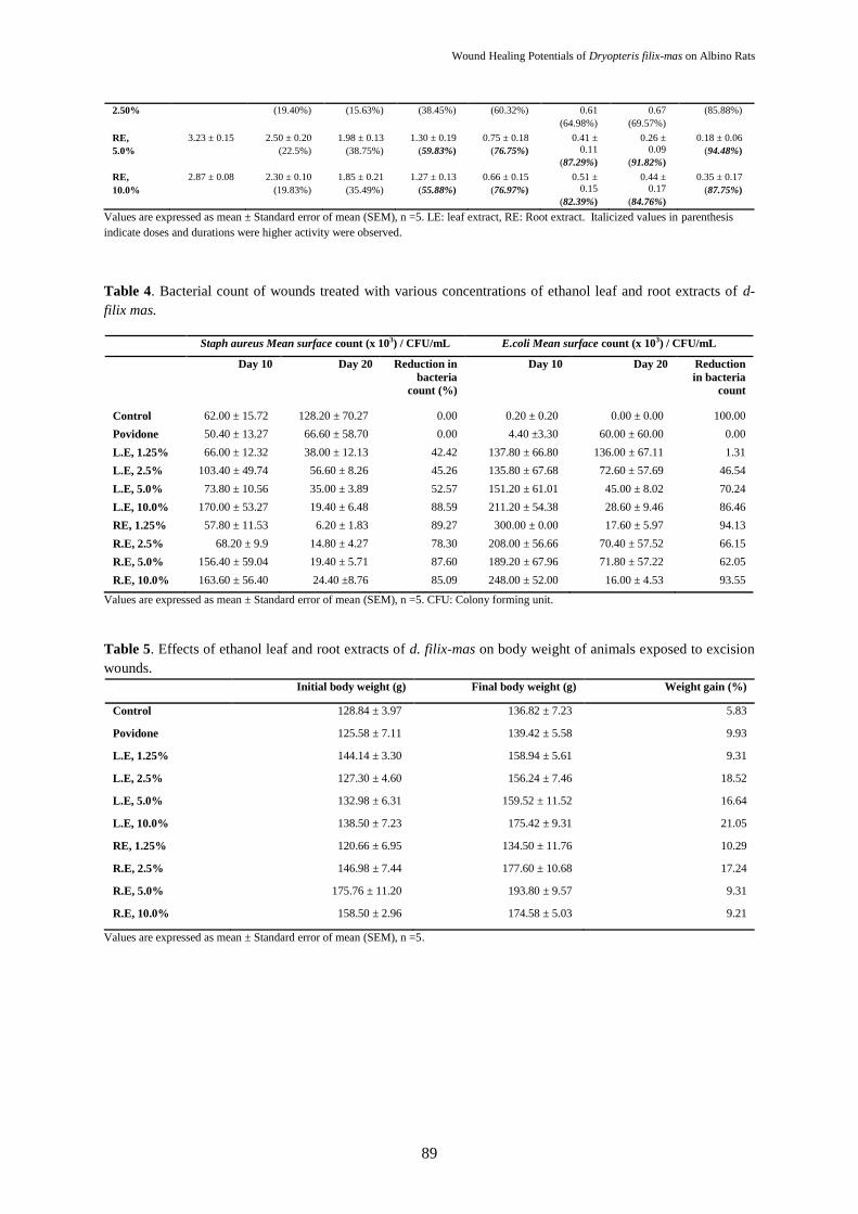
3.3.1 Effects of Treatments on Wound
Microbial Load
There was no reduction in Staph aureus
count of wounds treated with soft paraffin and
Povidone iodine between day 10th and day
20th. However, there were reductions in Staph
aureus counts in wound treated with various
concentrations of D. filix-mas leaf and root
extracts (Table 4). Reduction in S. aureus
count (42.42, 45.26, 52.57 and 88.59%) was
dose dependent in the leaf extract treated
groups, but the reduction (89.27, 78.30, 87.60
and 85.09%) was not dose dependent in the
root extract treated groups (Table 4). E. coli
was very minimal in wounds treated with soft
paraffin (control group) on day 20th (100.00%
reduction in bacteria count) when compared to
other groups. This may be attributed to the
ability of E. coli to have degraded the extract
thereby allowing more proliferation. On the
other hand, paraffin alone, being an organic
substance may not have been easily degraded
by E. coli. There was a dose dependent
reduction rate of E. coli in wounds treated with
1.25, 2.5, 5.0 and 10.0% of leaf extract. E. coli
count in wounds treated with 1.25, 2.5, 5.0 and
10.0% of root extract was reduced on day 20th,
but was not dose dependent (Table 4). Groups
treated with povidone iodine and soft paraffin
showed no reduction in E. coli when compared
to a concentration dependent reduction in the
group treated with different concentrations of
leaf extract of D. filix-mas. This suggests that
povidone iodine and soft paraffin wax do not
possess antimicrobial properties against E. coli
associated wound infections.
3.3.2. Effects on Body Weight
Higher body weight gain was observed in
animals treated with povidone iodine, 1.25,
2.5, 5.0 and 10.0% leaf and root extracts of D.
filix-mas when compared to body weight gain
of control group (5.83%). Body weight gain of
animals in the leaf extract treated groups was
higher than body weight gain of animals in
root extract treated groups, although body
weight was not dose dependent (Table 5).
Higher weight gain observed in the leaf extract
groups could be as a result of its wound
healing effect and presence of
phytoconstituents which could promote feed
intake and weight gain (Erhirhie and Ilodigwe,
2019).

Erhirhie E. O, et al. / IJPS 2021; 17 (1): 79-90
86
4. Conclusion
This study showed that 5 and 10% ethanol
leaf extract-ointments of D. filix-mas
facilitated wound healing more than root
extract and standard drug, povidone iodine.
Possible mechanisms include antioxidant,
antimicrobial and anti-inflammatory processes
which could be attributed to flavonoids, the
most abundant phytochemicals in the leaf
extract. From the foregoing, high
concentrations of D. filix-mas could be
effective in the treatment of surface wounds.
This study therefore validates the folkloric use
of D. filix-mas in the treatment of wounds,
boils and hemorrhages.
Acknowledgments
We are thankful to Dr. H.A. Akinnibosun
who assisted with the plant specimen
identification.
References
[1] Boateng JS, Kerr HM, Howard NES and Gillian
ME. Wound Healing Dressings and Drug Delivery
Systems: A Review. J. Pharm. Sci. (2008) 97:2892–
923.
[2] Agyare C, Boakye YD, Bekoe EO, Hensel A,
Dapaah SO and Appiah T. Review: African
medicinal plants with wound healing properties. J.
Ethnopharmacol. (2016) 177:85–100.
[3] Abbasi AM, Khan MA, Ahmad M, Zafar M, Jahan
S and Sultana S. Ethnopharmacological application of
medicinal plants to cure skin diseases and in folk
cosmetics among the tribal communities of north-west
Frontier province. Pakistan J. Ethnopharmacol.
(2010) 128: 322–35.
[4] Solanki R and Nagori BP. A review on
microorganisms causing wound infections on skin.
Asian J. Pharm. Tech. (2013) 3(3): 119–22.
[5] Builders PF and Builders MI. Wound Care:
Traditional African Medicine Approach. Intech open
(2016) http://dx.doi.org/10.5772/65521. Accessed on
June 5th, 2020.
[6] Adigun IA, Rahman GA, Yusuf IF and Ofoegbu
CKF. The point prevalence and cost of
wound management in a Nigerian teaching hospital.
Nig. Med. J. (2010) 51(1): 23–25.
[7] Zhu JJ, Yao S, Guo X, Yue BS, Ma XY and Li J.
Bioactivity-Guided Screening of Wound-Healing
Active Constituents from American Cockroach
(Periplaneta americana). Molecules. (2018) 23(1):
101.
[8] Wilson RW and Francis KW. Evaluation of
Wound Healing Activity of Ethanolic Extract of
Leaves of Croton Megalocarpus Using Excision
Wound Model on Wister Albino Rats. IJSR. (2016)
4(3): 182-94.
[9] Piriz MA, Lima CAB, Jardim VMR, Mesquita
MK, Souza ADZ and Hecck RM. Medicinal plants in
the wound healing process: a literature review. Rev.
Bras. Plantas Med. (2014) 16(3): 628-36.
[10] Marume A, Matope G, Katsande S and Khoza S.
Wound Healing Properties of Selected Plants Used in
Ethnoveterinary Medicine. 2017; 8(September), 1–10.
https://doi.org/10.3389/fphar.2017.00544.
[11] Hadagali MD and Chua LS. The anti-
inflammatory and wound healing properties of honey.
Eur. Food. Res. Tech. (2014) 239(6): 1003–14.
[12] Nwosu MO. „Ethnobotanical studies on some
pteridophytes of Southern Nigeria‟, Economic Botany
(2002) 56(3): 255–59.
[13] Duke JA. Handbook of medicinal herbs, Herbal
Reference Library. 2nd
ed. CRC Press, Florida USA
(2001) 677.
[14] Sekendar AM, Mostafa K, Raihan MO, Rahman
MK, Hossain MA and Alam MS. Antioxidant and
cytotoxic activities of methanolic extract of D. filix-
mas (L.) Schott leaves. Int. J. Drug Dev. Res. (2012)
4(2): 223-29.
[15] Mohammed ASM, Khan MMRL, Jabin SA,
Abedin N, Islam MF and Shaha B. „Nutritional

Wound Healing Potentials of Dryopteris filix-mas on Albino Rats
87
quality and safety aspects of wild vegetables consume
in Bangladesh. Asian Pac. J. Trop. Biomed. (2016)
6(2): 125–31.
[16] Batsatsashvili K, Mehdiyeva N, Kikvidze Z,
Khutsishvili M, Sikharulidze S, Tchelidze D, Alizade
V, Narel Y, Zambrana P and Rainer W. Dryopteris
filix – mas (L) Schott Dryopteridaceae. Bussmann
Springer International Publishing AG. R.W.
Bussmann (ed.), Ethnobotany of the Caucasus,
European Ethnobotany. (2016), DOI 10.1007/978- 3-
319-50009-6_87-1. Accessed on June 5th, 2020.
[17] Marc EB, Nelly A, Annick DD and Frederic D.
Plants used as remedies antirheumatic and
antineuralgic in the traditional medicine of Lebanon.
J. Ethnopharmacol. (2008): 120: 315–34.
[18] Uwumarongie HO, Enike MA and Bafor EE.
Pharmacognostic Evaluation and Gastrointestinal
Activity of Dryopteris filix-mas (L.) Schott
(Dryopteridaceae). EJHCPR. (2016): 2: 19 – 25.
[19] Bafor EE, Omokaro WO, Uwumarongie OH,
Elvis-Offiah UB, Omoruyi O, Viegelmann CV and
Edrada-Ebel R. D. filix-mas (Dryopteridaceae) leaves
inhibit mouse uterine activity. JOMPED. (2017)
1(1):1-12.
[20] Erhirhie EO, Ezemokwe ON and Ilodigwe E.
Teratogenic Effects of Ethanol Leaf Extract of
Dryopteris filix –mas (L.) Schott. AJNP. (2018) 6(1):
573-83.
[21] Erhirhie EO, Emeghebo CN, Ilodigwe EE,
Ajaghaku DL, Umeokoli BO, Eze PM, Ngwoke KG
and Okoye FBC. Dryopteris filix-mas (L.) Schott
ethanolic leaf extract and fractions exhibited profound
anti-inflammatory activity. Avicenna J. Phytomed.
(2019) 9(4): 396-409.
[22] Yadav RNS, Agarwala M. Phytochemical
analysis of some medicinal plants. J. Phytol. (2011)
3(12): 10-14.
[23] Okezie UM, Eze PM, Okoye FBC, Ikegbunam
MN, Ugwu MC and Esimone CO.
Biologically active metabolites of an endophytic
fungus isolated from Vernonia amygdalin. AJOPRED
(2017) 9(1): 24-26.
[24] Vinay K, Abdullah AK and Nagarajan K. Animal
Models for the Evaluation of Wound Healing
Activity. IBDR (2013): 3(5): 93-107.
https://www.researchgate.net/publication/274010528_
animal_models_for_the_evaluation_of_wound_healin
g_activity.
[25] Jian PS and Bari SB. Evaluation of wound
healing effect of petroleum ether and methanolic
extract of Abemoschus manihot, Medikik malvaceae,
Wrightia tinctoria R. Br. Apocyanacae in rats. Braz. J.
Pharmacogn. (2010) 20:156-271.
[26] Umeh VN, Ilodigwe EE., Ajaghaku DL, Erhirhie
EO, Moke GE and Akah PA. Wound-healing Activity
of the Aqueous Leaf Extract and Fractions of Ficus
exasperata (Moraceae) and its Safety Evaluation on
Albino Rats. J. Tradit. Complement. Med. (2014)
1(4): 246-52.
[27] Juríková T, Mlček J, Sochor J and Hegedűsová
A. Polyphenols and their Mechanism of Action in
Allergic Immune Response. Glob. J. Allergy. (2015)
1(2): 37-39.
[28] Iba Y, Shibata A, Kato M and Masukawa T.
Possible involvement of mast cells in collagen
remodeling in the late phase of cutaneous wound
healing in mice. Int. Immunopharmacol. (2014)
4:1873–80.
[29] Soare LC, Ferdes M, Stefanov S, Denkova Z,
Nicolova R, Denev P, Bejan C and Paunescu A.
Antioxidant activity, polyphenols content and
antimicrobial activity of several native Pteridophytes
of Romania. Notulae Botanicae. Horti Agrobotanici
Cluj Napoca. (2012) 40(1): 53-57.
[30] Atawodi SE, Atawodi JC, Idakwo
GA, Pfundstein B, Haubner R, Wurtele G, Bartsch H
and Owen RW. Evaluation of the polyphenol content
and antioxidant properties of methanol extracts of the
leaves, stem, and root barks of Moringa oleifera Lam.
J. Med. Food. (2010) 13(3): 710–16.

Erhirhie E. O, et al. / IJPS 2021; 17 (1): 79-90
Tables:
Table 1. Phytochemical constituents of the ethanol leaf and root extracts of d. filix-mas.
Phytochemical constituents Leaf extract Root extract
Tannins ++ +
Flavonoids +++ +
Saponins ++ -
Steroids ++ -
Alkaloids ++ +
Terpenoids ++ ++
Anthraquinones - -
Cardiac glycosides + ++
Reducing sugar + +
Proteins + +
“+” indicates mildly present,“++” indicates moderately present, “+++” indicates abundantly present, “-” indicates absent.
Table 2. Antimicrobial activity of different concentrations of ethanol leaf and root extracts of d. filix-mas.
IZD (mm)
400 mg/ml 200 mg/ml 100 mg/ml 50 mg/ml 25 mg/ml 12.5 mg/ml
Leaf extract
P. aeruginosa 8.5 ± 0.5 4.5 ± 0.5 - - - -
S. aureus 5 ± 0.0 4.5 ± 0.5 4 ± 0.00 3 ± 0.00
E. coli - - - - - - Root
extract
P. aeruginosa - - - - - -
S. aureus 2 ± 0.00 1.5 ± 1.5 - - - -
E. coli - - - - - -
Values are expressed as mean ± Standard error of mean (SEM), n =3. IZD: Inhibitory zone diameter. “-”: No inhibition was found.
Table 3. Showing average wound diameter and wound contraction of animals treated with, placebo, povidone
iodine and extracts of d .filix-mas.
Wound area (cm) and wound contraction (%)
Group Baseline (cm) Day 3 Day 6 Day 9 Day 12 Day 15 Day 18 Day 21
Control 3.15 ± 0.16 2.46 ± 0.11
(21.92%)
2.62 ± 0.23
(16.77%)
1.44 ± 0.31
(54.13%)
1.13 ± 0.33
(64.17%)
0.77 ±
0.32
(75.41%)
0.55 ±
0.31
(82.4%)
0.42 ± 0.28
(86.59%)
Povidone
iodine
2.45 ± 0.21 2.06 ± 0.11
(15.92%)
1.92 ± 0.08
(21.63%)
1.07 ± 0.10
(56.41%)
0.97 ± 0.17
(60.24%)
0.78 ±
0.19
(68.33%)
0.50 ±
0.20
(79.76%)
0.27 ± 0.13
(89.06%)
LE, 1.25% 3.08 ± 0.05 2.68 ± 0.10
(12.81%)
2.58 ± 0.08
(15.99%)
2.16 ± 0.29
(29.71%)
1.52 ± 0.26
(50.46%)
1.19 ±
0.26
(61.38%)
0.79 ±
0.20
(74.19%)
0.42 ± 0.15
(86.35%)
LE,
2.50%
2.71 ± 0.17 2.28 ± 0.14
(15.87%)
2.36 ± 0.22
(13.06%)
1.55 ± 0.18
(42.8%)
1.46 ± 0.14
(46.27%)
1.22 ±
0.18
(54.83%)
0.81 ±
0.23
(69.96%)
0.75 ± 0.16
(72.18%)
LE,
5.0%
3.37 ± 0.17 2.52 ± 0.07
(25.06%)
2.16 ± 0.21
(35.87%)
1.63 ± 0.21
(51.6%)
1.26 ± 0.27
(62.53%)
0.91 ±
0.17
(72.98%)
0.48 ±
0.64
(85.69%)
0.30 ± 0.07
(91.03%)
LE,
10.0%
3.06 ± 0.13 2.18 ± 0.05
(28.98%)
2.01 ± 0.13
(34.40%)
1.28 ± 0.17
(58.16%)
0.70 ± 0.14
(77.15%)
0.44 ±
0.10
(85.70%)
0.19 ±
0.05
(93.86%)
0.11 ± 0.03
(96.54%)
RE, 1.25% 3.62 ± 0.15 2.57 ± 0.05
(28.9%)
2.78 ± 0.23
(23.15%)
1.79 ± 0.15
(50.50%)
1.47 ± 0.21
(59.34%)
0.83 ±
0.18
(77.02%)
0.58 ±
0.29
(83.87%)
0.44 ± 0.32
(87.73%)
RE, 2.92 ± 0.33 2.35 ± 0.31 2.46 ± 0.22 1.80 ± 0.29 1.16 ± 0.42 1.02 ± 0.89 ± 0.41 ± 0.28
Wound Healing Potentials of Dryopteris filix-mas on Albino Rats
89
2.50% (19.40%) (15.63%) (38.45%) (60.32%) 0.61
(64.98%)
0.67
(69.57%)
(85.88%)
RE,
5.0%
3.23 ± 0.15 2.50 ± 0.20
(22.5%)
1.98 ± 0.13
(38.75%)
1.30 ± 0.19
(59.83%)
0.75 ± 0.18
(76.75%)
0.41 ±
0.11
(87.29%)
0.26 ±
0.09
(91.82%)
0.18 ± 0.06
(94.48%)
RE,
10.0%
2.87 ± 0.08 2.30 ± 0.10
(19.83%)
1.85 ± 0.21
(35.49%)
1.27 ± 0.13
(55.88%)
0.66 ± 0.15
(76.97%)
0.51 ±
0.15
(82.39%)
0.44 ±
0.17
(84.76%)
0.35 ± 0.17
(87.75%)
Values are expressed as mean ± Standard error of mean (SEM), n =5. LE: leaf extract, RE: Root extract. Italicized values in parenthesis
indicate doses and durations were higher activity were observed.
Table 4. Bacterial count of wounds treated with various concentrations of ethanol leaf and root extracts of d-
filix mas.
Staph aureus Mean surface count (x 103) / CFU/mL E.coli Mean surface count (x 103) / CFU/mL
Day 10 Day 20 Reduction in
bacteria
count (%)
Day 10 Day 20 Reduction
in bacteria
count
Control 62.00 ± 15.72 128.20 ± 70.27 0.00 0.20 ± 0.20 0.00 ± 0.00 100.00
Povidone 50.40 ± 13.27 66.60 ± 58.70 0.00 4.40 ±3.30 60.00 ± 60.00 0.00
L.E, 1.25% 66.00 ± 12.32 38.00 ± 12.13 42.42 137.80 ± 66.80 136.00 ± 67.11 1.31
L.E, 2.5% 103.40 ± 49.74 56.60 ± 8.26 45.26 135.80 ± 67.68 72.60 ± 57.69 46.54
L.E, 5.0% 73.80 ± 10.56 35.00 ± 3.89 52.57 151.20 ± 61.01 45.00 ± 8.02 70.24
L.E, 10.0% 170.00 ± 53.27 19.40 ± 6.48 88.59 211.20 ± 54.38 28.60 ± 9.46 86.46
RE, 1.25% 57.80 ± 11.53 6.20 ± 1.83 89.27 300.00 ± 0.00 17.60 ± 5.97 94.13
R.E, 2.5% 68.20 ± 9.9 14.80 ± 4.27 78.30 208.00 ± 56.66 70.40 ± 57.52 66.15
R.E, 5.0% 156.40 ± 59.04 19.40 ± 5.71 87.60 189.20 ± 67.96 71.80 ± 57.22 62.05
R.E, 10.0% 163.60 ± 56.40 24.40 ±8.76 85.09 248.00 ± 52.00 16.00 ± 4.53 93.55
Values are expressed as mean ± Standard error of mean (SEM), n =5. CFU: Colony forming unit.
Table 5. Effects of ethanol leaf and root extracts of d. filix-mas on body weight of animals exposed to excision
wounds.
Initial body weight (g) Final body weight (g) Weight gain (%)
Control 128.84 ± 3.97 136.82 ± 7.23 5.83
Povidone 125.58 ± 7.11 139.42 ± 5.58 9.93
L.E, 1.25% 144.14 ± 3.30 158.94 ± 5.61 9.31
L.E, 2.5% 127.30 ± 4.60 156.24 ± 7.46 18.52
L.E, 5.0% 132.98 ± 6.31 159.52 ± 11.52 16.64
L.E, 10.0% 138.50 ± 7.23 175.42 ± 9.31 21.05
RE, 1.25% 120.66 ± 6.95 134.50 ± 11.76 10.29
R.E, 2.5% 146.98 ± 7.44 177.60 ± 10.68 17.24
R.E, 5.0% 175.76 ± 11.20 193.80 ± 9.57 9.31
R.E, 10.0% 158.50 ± 2.96 174.58 ± 5.03 9.21
Values are expressed as mean ± Standard error of mean (SEM), n =5.

ONLINE SUBMISSION
www.ijps.ir