evaluation of two simplified methods for genotyping hepatitis c virus
TRANSCRIPT
Evaluation of Two Simplified Methods forGenotyping Hepatitis C Virus
R.U. Khan,1,5 C.Y.W. Tong,1* S. Bloom,2 I.T. Gilmore,2 C.H. Toh,3 P.H. Bolton-Maggs,4 N.J. Beeching,5and C.A. Hart1
1Department of Medical Microbiology, University of Liverpool, Liverpool, United Kingdom2Department of Gastroenterology, Royal Liverpool University Hospital, Liverpool, United Kingdom3Department of Haematology, Royal Liverpool University Hospital, Liverpool, United Kingdom4Department of Haematology, Royal Liverpool Children’s Hospital, Alder Hey, Liverpool, United Kingdom5Liverpool School of Tropical Medicine, Liverpool, United Kingdom
A number of different approaches have beenused for genotyping hepatitis C virus (HCV). Twosimplified methods were evaluated, both ofwhich used polymerase chain reaction (PCR) toamplify products from the 58 non-coding regionof HCV: non-isotopic restriction fragment lengthpolymorphism (RFLP) analysis and type-specificPCR. Sixty-four viraemic patients suffering fromchronic HCV infection were studied using thesetwo techniques; 25/64 samples were furthertested with a commercial serotyping ELISAbased on synthetic NS4 antigen (Murex, U.K.).The results of the three typing methods weregenerally in agreement with each other. Whenonly the predominant genotype identified byeach method was analysed, the 3 methods had100% agreement. RFLP did not detect any mixedinfections and it was unsuccessful in 16/64 (25%)samples. Both type-specific PCR and serotypingELISA detected mixed infections. However, se-rotyping ELISA did not give typeable results in7/25 (28%) samples, whereas type-specific PCRgave typeable results in all 64 samples. Type-specific PCR detected more mixed infectionsthan serotyping ELISA. Direct sequencing of fourPCR products with indeterminate RFLP con-firmed changes in restriction enzyme recogni-tion sites. The sequences also confirmed the va-lidity of the predominant genotype in cases ofapparent mixed infections. It is possible thatsome of these cases were artefacts as a result ofquasispecies. J. Med. Virol. 52:35–41, 1997.© 1997 Wiley-Liss, Inc.
KEY WORDS: RFLP; type specific PCR; sero-types; mixed genotype HCV in-fection
INTRODUCTION
Identification of hepatitis C virus (HCV) as the majorcause of parenterally transmitted non-A, non-B hepa-titis [Choo et al., 1989] has resulted in subsequent clon-ing and sequencing of a number of HCV isolates fromdifferent geographical areas. On the basis of these com-plete or partial sequences homology analyses, distinctHCV genotypes have been described [Choo et al., 1991;Okamoto et al., 1992; Chan et al., 1992; Simmonds etal., 1993a]. Genotypic differences are most useful forthe study of global HCV epidemiology. The importanceof HCV genotypes is further emphasised by the asso-ciation of specific genotype (type 1, particularly 1b)with severity and poor response to interferon therapy[Pozatto et al., 1991; Kanai et al., 1992; Nousbaum etal., 1995].
Nucleotide sequencing remains the most definitivemethod for identifying different genotypes of HCV[Chan et al., 1992; Simmonds et al., 1993a], but this isnot possible for routine clinical studies. More practicalmethods include: direct polymerase chain reaction(PCR) of clinical samples using type-specific primerswhich amplify selectively different genotypes [Oka-moto et al., 1992], the use of genotype-specific probes tohybridise with PCR products [Stuyver et al., 1993; VanDoorn et al., 1994], restriction fragment length poly-morphism (RFLP) analysis of DNA amplified by PCR[Nakao et al., 1991; McOmish et al., 1994], or by sero-typing methods using type-specific synthetic peptides[Simmonds et al., 1993b; Bhattacherjee et al., 1995;Dixit et al., 1995]. Although these methods are lesscomplicated than cloning and sequencing, most meth-ods are still too complicated or too costly to allow rou-
*Correspondence to: Dr. C.Y.W. Tong, Department of MedicalMicrobiology, University of Liverpool, P.O. Box 147, Liverpool,L69 3BX, U.K.
Accepted 12 November 1996
Journal of Medical Virology 52:35–41 (1997)
© 1997 WILEY-LISS, INC.

tine diagnostic use. We evaluated 2 modified methodsof genotyping HCV using non-isotopic RFLP analysisand type-specific PCR, both based on the amplificationof the conserved 58 non-coding region (58 NCR). Theconsensus genotype nomenclature system proposed bySimmonds et al. [1994] was adopted in this study.
PATIENTS AND METHODS
Serum samples from 64 British patients from theMersey region of England suffering from chronic HCVinfection with proven viraemia were used in this study.Risk factors for the acquisition of hepatitis C included:haemophiliacs (n 4 39), intravenous drug users (n 416), haemodialysis patients (n 4 2), post-transfusionpatient (n 4 1), health care worker (n 4 1), and spo-radic unknown causes (n 4 5). All patients were posi-tive for anti-HCV using third-generation ELISA (Mu-rex, UK or Ortho, US) and viraemia was determined byPCR (Amplicor, Roche, Switzerland).
RFLP
This was a modification of the method described byMcOmish et al. [1994]. Viral RNA was extracted from50 ml of serum using standard technique of proteinaseK digestion, phenol chloroform extraction, and ethanolprecipitation [Sambrook et al., 1989]. The pelletednucleic acid was resuspended in 50 ml of diethyl pyro-carbonate treated water. Reverse transcription wasperformed on 11.5 ml of the resuspended nucleic acidusing 50 mM Tris HCl pH 8.3, 75 mM KCl, 3 mMMgCl2, 250 mM of each dNTP, 1.5 mM universal exter-nal anti-sense primer HC2 (position −66 to −85 [Chooet al., 1991], 58 TTTCGCRACCCAACRCTACT), 20units RNAguard (Pharmacia, Sweden), and 200 unitsof Moloney Murine Reverse Transcriptase (GibcoBRL,Life Technologies Inc, US) in 20 ml at 37°C for 60 min-utes. First round PCR was carried out on 10 ml of cDNAusing 20 mM Tris HCl pH 8.4, 50 mM KCl, 1.5 mMMgCl2, 200 mM of each dNTP, 1.4 mM each of universalexternal anti-sense primers HC2 and universal exter-nal sense primer HCl (position −260 to −241, 58 GC-CATGGCGTTAGTAYGAGT), and 0.6 units of TaqDNA polymerase (GibcoBRL, Life Technologies Inc,US) in 25 ml with oil overlay. The PCR reaction con-sisted of 35 cycles each with 1 minute at 94°C, 1 minuteat 40°C, and 1 minute at 72°C using an automatedthermocycler (Omnigene, Hybaid, UK). The first roundproduct was diluted 20-fold in water and 5 ml was usedin a second round reaction using the same reaction mixas the first round but replacing the primers with uni-versal internal sense primer HC3 (position −243 to−224, 58AGTGTCRTRCAGCCTCCAGG) and universalinternal anti-sense primer HC4 (position −73 to −92, 58ACCCAACRCTACTMGGCTAG). The effective volumeof the first round input to second round was 0.25 ml. Asmall volume was used to prevent non-specific bandscarried over from the first. Other PCR conditions werethe same as above. The resultant 171 base pair PCRproduct, which was shorter than that used by Mc-
Omish et al. [1994] but within the same stretch of nu-cleotide sequence, was purified using the Wizard PCRPrep Purification kit (Promega, US) according to themanufacturer’s instructions. Purified product was re-suspended in 50 ml of water. Fifteen microlitres of thepurified PCR product was digested by either Hinf1/Mva1 in SuRE/Cut buffer H or Hinf1/Scrf1 in SuRE/Cut buffer B (Boerhinger-Mannheim, Germany) at37°C for one hour. The digested fragments were sepa-rated by electrophoresis in a 6% Nusieve GTG agarosegel (Flowgen, US) stained with ethidium bromide(0.05% w/v). The separated fragments were visualised
Fig. 1. (A) RFLP digestion patterns illustrated by samples A (ge-notype 1), B (genotype 3), C (genotype 2), and D (indeterminate).Uncleaved PCR products were run in lanes 1, 5, 9, and 13; ScrfI/HinfIdigested PCR products were run in lanes 2, 6, 10, and 14; MvaI/HinfIdigested PCR products were run in lanes 3, 7, 11, and 15. Lanes 4, 8,and 12 contain marker pBR322 Hae III digest. (B) Type-specific PCRillustrated by samples E (genotype 1), F (genotype 2), G (genotype 3),and H (mixed infection 3 > 1). Genotype 1 specific primers were run inlanes 1, 4, 8, and 12. Genotype 2 specific primers were run in lanes 2,5, 9, and 13. Genotype 3 specific primers were run in lanes 3, 6, 10,and 14. Lanes 7 and 11 contain marker pBR 322 Hae III digest.
36 Khan et al.
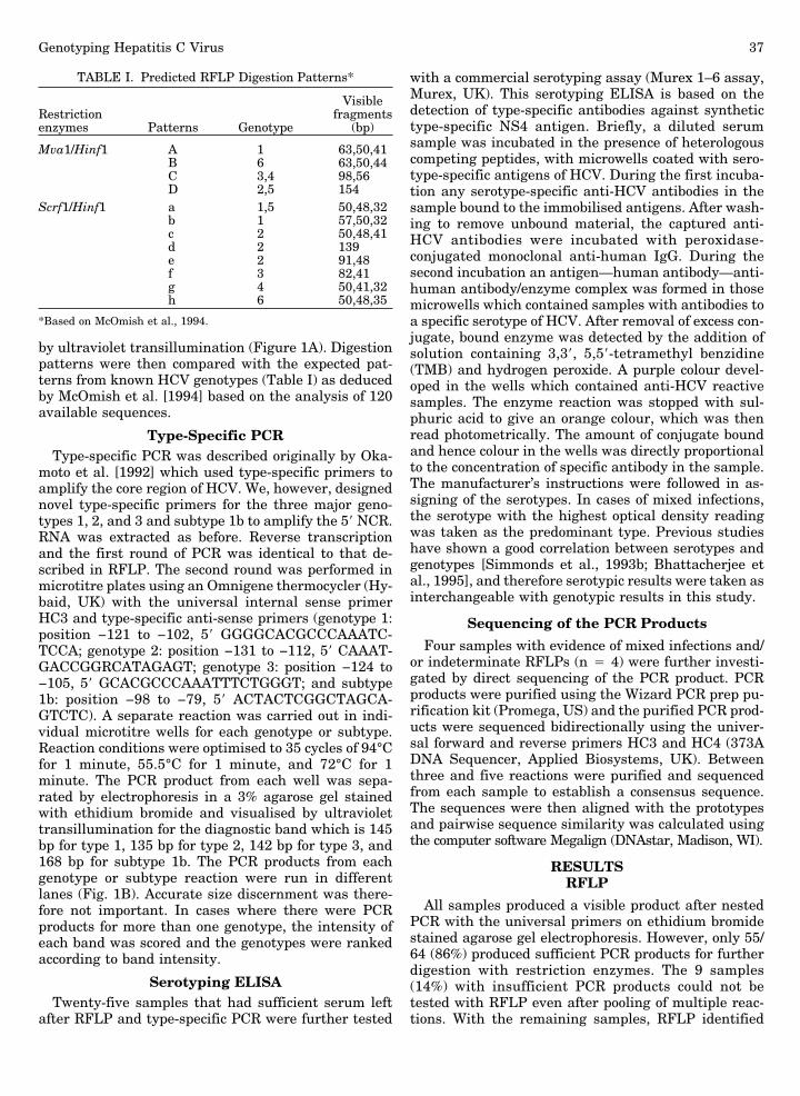
by ultraviolet transillumination (Figure 1A). Digestionpatterns were then compared with the expected pat-terns from known HCV genotypes (Table I) as deducedby McOmish et al. [1994] based on the analysis of 120available sequences.
Type-Specific PCRType-specific PCR was described originally by Oka-
moto et al. [1992] which used type-specific primers toamplify the core region of HCV. We, however, designednovel type-specific primers for the three major geno-types 1, 2, and 3 and subtype 1b to amplify the 58 NCR.RNA was extracted as before. Reverse transcriptionand the first round of PCR was identical to that de-scribed in RFLP. The second round was performed inmicrotitre plates using an Omnigene thermocycler (Hy-baid, UK) with the universal internal sense primerHC3 and type-specific anti-sense primers (genotype 1:position −121 to −102, 58 GGGGCACGCCCAAATC-TCCA; genotype 2: position −131 to −112, 58 CAAAT-GACCGGRCATAGAGT; genotype 3: position −124 to−105, 58 GCACGCCCAAATTTCTGGGT; and subtype1b: position −98 to −79, 58 ACTACTCGGCTAGCA-GTCTC). A separate reaction was carried out in indi-vidual microtitre wells for each genotype or subtype.Reaction conditions were optimised to 35 cycles of 94°Cfor 1 minute, 55.5°C for 1 minute, and 72°C for 1minute. The PCR product from each well was sepa-rated by electrophoresis in a 3% agarose gel stainedwith ethidium bromide and visualised by ultraviolettransillumination for the diagnostic band which is 145bp for type 1, 135 bp for type 2, 142 bp for type 3, and168 bp for subtype 1b. The PCR products from eachgenotype or subtype reaction were run in differentlanes (Fig. 1B). Accurate size discernment was there-fore not important. In cases where there were PCRproducts for more than one genotype, the intensity ofeach band was scored and the genotypes were rankedaccording to band intensity.
Serotyping ELISATwenty-five samples that had sufficient serum left
after RFLP and type-specific PCR were further tested
with a commercial serotyping assay (Murex 1–6 assay,Murex, UK). This serotyping ELISA is based on thedetection of type-specific antibodies against synthetictype-specific NS4 antigen. Briefly, a diluted serumsample was incubated in the presence of heterologouscompeting peptides, with microwells coated with sero-type-specific antigens of HCV. During the first incuba-tion any serotype-specific anti-HCV antibodies in thesample bound to the immobilised antigens. After wash-ing to remove unbound material, the captured anti-HCV antibodies were incubated with peroxidase-conjugated monoclonal anti-human IgG. During thesecond incubation an antigen—human antibody—anti-human antibody/enzyme complex was formed in thosemicrowells which contained samples with antibodies toa specific serotype of HCV. After removal of excess con-jugate, bound enzyme was detected by the addition ofsolution containing 3,38, 5,58-tetramethyl benzidine(TMB) and hydrogen peroxide. A purple colour devel-oped in the wells which contained anti-HCV reactivesamples. The enzyme reaction was stopped with sul-phuric acid to give an orange colour, which was thenread photometrically. The amount of conjugate boundand hence colour in the wells was directly proportionalto the concentration of specific antibody in the sample.The manufacturer’s instructions were followed in as-signing of the serotypes. In cases of mixed infections,the serotype with the highest optical density readingwas taken as the predominant type. Previous studieshave shown a good correlation between serotypes andgenotypes [Simmonds et al., 1993b; Bhattacherjee etal., 1995], and therefore serotypic results were taken asinterchangeable with genotypic results in this study.
Sequencing of the PCR Products
Four samples with evidence of mixed infections and/or indeterminate RFLPs (n 4 4) were further investi-gated by direct sequencing of the PCR product. PCRproducts were purified using the Wizard PCR prep pu-rification kit (Promega, US) and the purified PCR prod-ucts were sequenced bidirectionally using the univer-sal forward and reverse primers HC3 and HC4 (373ADNA Sequencer, Applied Biosystems, UK). Betweenthree and five reactions were purified and sequencedfrom each sample to establish a consensus sequence.The sequences were then aligned with the prototypesand pairwise sequence similarity was calculated usingthe computer software Megalign (DNAstar, Madison, WI).
RESULTSRFLP
All samples produced a visible product after nestedPCR with the universal primers on ethidium bromidestained agarose gel electrophoresis. However, only 55/64 (86%) produced sufficient PCR products for furtherdigestion with restriction enzymes. The 9 samples(14%) with insufficient PCR products could not betested with RFLP even after pooling of multiple reac-tions. With the remaining samples, RFLP identified
TABLE I. Predicted RFLP Digestion Patterns*
Restrictionenzymes Patterns Genotype
Visiblefragments
(bp)
Mva1/Hinf1 A 1 63,50,41B 6 63,50,44C 3,4 98,56D 2,5 154
Scrf1/Hinf1 a 1,5 50,48,32b 1 57,50,32c 2 50,48,41d 2 139e 2 91,48f 3 82,41g 4 50,41,32h 6 50,48,35
*Based on McOmish et al., 1994.
Genotyping Hepatitis C Virus 37
33/55 patients (60%) as genotype 1, 4/55 (7%) as geno-type 2, and 11/55 (20%) as genotype 3 (Figure 1A).Seven samples (13%) had restriction digestion patternsthat did not fall into any of the predicted patterns andtheir genotypes were therefore indeterminate. Overall,this approach was successful in assigning genotypes to48/64 (75%) samples. Improvement in PCR efficiencyshould improve the success rate as insufficient PCRproduct, not the RFLP method itself, was the problemin 9/64 (14%) cases.
Type-Specific PCR
Type-specific PCR was successful with all 64samples. Mixed infections were evident in 11 samples(17%) in which there were bands associated with morethan 1 genotype. If the predominant band only wasused in analysis, type-specific PCR identified 44/64(67%) as type 1, of which 8 had evidence of mixed in-fection; 4/64 (6%) were identified as type 2, of which 2had evidence of mixed infection; 16/64 (25%) were iden-tified as type 3, of which 1 had evidence of mixed in-fection. Of the 44 samples with genotype 1 infection, 27(61%) were also positive with the subtype 1b primer.None of the non–type 1 genotypes reacted with the type1b–specific primers.
RFLP vs. Type-Specific PCR
Of the 48 samples that had a typeable result by bothRFLP and type-specific PCR, 40/48 (83%) samples hadconcordant results: 28 were genotype 1, two were ge-notype 2, and 10 were genotype 3. The 9 samples withinsufficient PCR product for RFLP analysis were alltypeable with type-specific PCR: 6 were genotype 1 and3 were genotype 3. Of the 7 samples which gave anindeterminate pattern with RFLP, 2 were genotype 1,two were genotype 3, and 3 had triple mixed infectionwith genotypes 1, 2, and 3 (genotype 1 was predomi-nant in all). The results for the remaining 8 sampleswhich showed disagreement between the 2 methodswere all mixed infections as identified by type-specificPCR (Table II). However, if only the predominant bandfrom the mixed infections was considered, then the re-sults of the typeable samples were in complete agree-ment with type-specific PCR.
Serotyping ELISA
Serotyping ELISA was carried out on 25 sampleswhich had sufficient serum left after RFLP and type-specific PCR. Eleven of these 25 were samples that hadcomplete RFLP and type-specific PCR agreement. Se-rotyping ELISA also agreed in 7/11 (64%); 3 sampleswere untypable by ELISA; 1 sample which both RFLPand type-specific PCR identified as type 3, serotypingELISA identified as mixed infection of type 2 and type3 (type 3 predominant). Of the 3 samples indetermi-nate with RFLP but typable with type-specific PCR, 1was serotype 1 and 2 were serotype 3: serotypingELISA agreed with type-specific PCR on all. The re-maining 11 samples all had mixed infections as iden-tified by type-specific PCR: serotyping ELISA identi-fied 2 of these 11 as mixed infections (Table II). If onlythe predominant type was considered, the typeable re-sults of serotyping ELISA were in complete agreementwith both RFLP and type-specific PCR.
Risk Factors
Combining the results of the 3 methods and usingthe predominant genotypes only, patients were cat-egorised according to their risk factors. Of the 39 hae-mophiliacs, 30 (77%) were genotype 1, two (5%) weregenotype 2, and 7 (18%) were genotype 3. Of the 16intravenous drug users, 10 (63%) were genotype 1, one(6%) was genotype 2, and 5 (31%) were genotype 3. Thesingle post-transfusion patient had genotype 2 infec-tion. The patient whose only risk factor was being ahealth care worker had genotype 3 infection. Of the 2patients on haemodialysis, 1 had genotype 1 and theother had genotype 3 infection. In 5 patients where theroute of infection was not known, 3 were genotype 1and 2 were genotype 3. Comparing the 2 main groups ofpatients—haemophiliacs and intravenous drug users—there was no statistically significant difference in theirgenotype distribution (x2 4 1.27 and P 4 .529). As-suming that the mixed infections as identified by type-specific PCR were correct, then 6/39 (15%) haemophili-acs, 3/16 (18%) intravenous drug users, and 2/2 (100%)haemodialysis patients had mixed infection.
Sequencing
The PCR product from 4 samples with indeterminateRFLPs, of which 2 also had evidence of mixed infectionby the type-specific PCR method, were sequenced (Fig-ure 2). Cases 1, 2, and 3 showed significant sequencehomology with genotype 1 (pairwise sequence similar-ity between positions −184 and −92 4 89.2, or 97.8%)and the sequence predicted binding with the genotype1–specific primer. All these sequences had a restrictionmapping pattern deviated from that of the predicted(Table III). The sequence of cases 1 and 2 did not pre-dict binding with type 2– or type 3–specific primersalthough amplification products were available withthe type 2– and the type 3–specific primers, yieldingmixed infection of genotypes 1 and 2 for case 1 and
TABLE II. Samples Showing Evidence of Mixed Infectionby One or More Methods
RFLP Type-specific PCR ELISA
1 1 > 3 Untypeable1 1 > 2 11 1 > 3 1 > 6 > 21 1 > 2 11 1 > 2 Untypeable2 2 > 1 2 > 1 > 42 2 > 1 23 3 3 > 23 3 > 1 UntypeableIndeterminate 1 > 2 4 3 UntypeableIndeterminate 1 > 3 > 2 1Indeterminate 1 > 3 > 2 1
38 Khan et al.
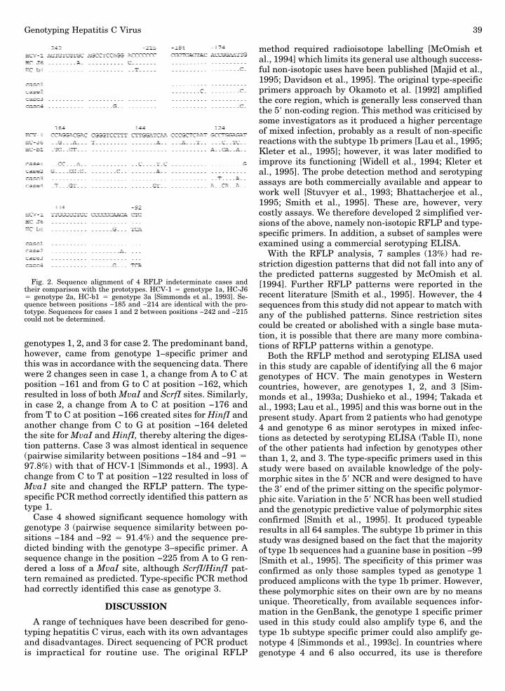
genotypes 1, 2, and 3 for case 2. The predominant band,however, came from genotype 1–specific primer andthis was in accordance with the sequencing data. Therewere 2 changes seen in case 1, a change from A to C atposition −161 and from G to C at position −162, whichresulted in loss of both MvaI and ScrfI sites. Similarly,in case 2, a change from A to C at position −176 andfrom T to C at position −166 created sites for HinfI andanother change from C to G at position −164 deletedthe site for MvaI and HinfI, thereby altering the diges-tion patterns. Case 3 was almost identical in sequence(pairwise similarity between positions −184 and −91 497.8%) with that of HCV-1 [Simmonds et al., 1993]. Achange from C to T at position −122 resulted in loss ofMva1 site and changed the RFLP pattern. The type-specific PCR method correctly identified this pattern astype 1.
Case 4 showed significant sequence homology withgenotype 3 (pairwise sequence similarity between po-sitions −184 and −92 4 91.4%) and the sequence pre-dicted binding with the genotype 3–specific primer. Asequence change in the position −225 from A to G ren-dered a loss of a MvaI site, although ScrfI/HinfI pat-tern remained as predicted. Type-specific PCR methodhad correctly identified this case as genotype 3.
DISCUSSION
A range of techniques have been described for geno-typing hepatitis C virus, each with its own advantagesand disadvantages. Direct sequencing of PCR productis impractical for routine use. The original RFLP
method required radioisotope labelling [McOmish etal., 1994] which limits its general use although success-ful non-isotopic uses have been published [Majid et al.,1995; Davidson et al., 1995]. The original type-specificprimers approach by Okamoto et al. [1992] amplifiedthe core region, which is generally less conserved thanthe 58 non-coding region. This method was criticised bysome investigators as it produced a higher percentageof mixed infection, probably as a result of non-specificreactions with the subtype 1b primers [Lau et al., 1995;Kleter et al., 1995]; however, it was later modified toimprove its functioning [Widell et al., 1994; Kleter etal., 1995]. The probe detection method and serotypingassays are both commercially available and appear towork well [Stuvyer et al., 1993; Bhattacherjee et al.,1995; Smith et al., 1995]. These are, however, verycostly assays. We therefore developed 2 simplified ver-sions of the above, namely non-isotopic RFLP and type-specific primers. In addition, a subset of samples wereexamined using a commercial serotyping ELISA.
With the RFLP analysis, 7 samples (13%) had re-striction digestion patterns that did not fall into any ofthe predicted patterns suggested by McOmish et al.[1994]. Further RFLP patterns were reported in therecent literature [Smith et al., 1995]. However, the 4sequences from this study did not appear to match withany of the published patterns. Since restriction sitescould be created or abolished with a single base muta-tion, it is possible that there are many more combina-tions of RFLP patterns within a genotype.
Both the RFLP method and serotyping ELISA usedin this study are capable of identifying all the 6 majorgenotypes of HCV. The main genotypes in Westerncountries, however, are genotypes 1, 2, and 3 [Sim-monds et al., 1993a; Dushieko et al., 1994; Takada etal., 1993; Lau et al., 1995] and this was borne out in thepresent study. Apart from 2 patients who had genotype4 and genotype 6 as minor serotypes in mixed infec-tions as detected by serotyping ELISA (Table II), noneof the other patients had infection by genotypes otherthan 1, 2, and 3. The type-specific primers used in thisstudy were based on available knowledge of the poly-morphic sites in the 58 NCR and were designed to havethe 38 end of the primer sitting on the specific polymor-phic site. Variation in the 58 NCR has been well studiedand the genotypic predictive value of polymorphic sitesconfirmed [Smith et al., 1995]. It produced typeableresults in all 64 samples. The subtype 1b primer in thisstudy was designed based on the fact that the majorityof type 1b sequences had a guanine base in position −99[Smith et al., 1995]. The specificity of this primer wasconfirmed as only those samples typed as genotype 1produced amplicons with the type 1b primer. However,these polymorphic sites on their own are by no meansunique. Theoretically, from available sequences infor-mation in the GenBank, the genotype 1 specific primerused in this study could also amplify type 6, and thetype 1b subtype specific primer could also amplify ge-notype 4 [Simmonds et al., 1993c]. In countries wheregenotype 4 and 6 also occurred, its use is therefore
Fig. 2. Sequence alignment of 4 RFLP indeterminate cases andtheir comparison with the prototypes. HCV-1 4 genotype 1a, HC-J64 genotype 2a, HC-b1 4 genotype 3a [Simmonds et al., 1993]. Se-quence between positions −185 and −214 are identical with the pro-totype. Sequences for cases 1 and 2 between positions −242 and −215could not be determined.
Genotyping Hepatitis C Virus 39

limited. However, sequence analysis suggests that it isalso possible to design specific primers for genotype 4and genotype 6 in the 58 NCR [Smith et al., 1995],though this would increase the complexity of the assay.However, the assay in its present format will provideinformation about the presence or absence of the badprognostic genotypes and may thus be of some use inclinical practice.
The most perplexing and controversial aspect is thephenomenon of mixed infection. The prevalence ofmixed infection depended very much on the methodused. Methods such as RFLP, which match genotypeswith expected patterns, will not easily detect mixedinfections. In a situation with true mixed infection, ei-ther only the predominant pattern prevails or an inde-terminate pattern emerges as the mixed digestion pat-tern appear together. A study on HCV genotypes inhaemophiliacs has shown that a combined RFLP andsequencing approach detected mixed infection in 7%,whereas a serotyping method identified 14% [Prestonet al., 1995]. The type-specific primer approach usingthe core region has been associated in particular withdetection of mixed infections [Lau et al., 1995]. Morethan 10% of patients with chronic liver disease hadmixed genotype infection in one study from Taiwan us-ing this typing method [Chen et al., 1994]. Here, use ofthe type-specific primer method for the 58 NCR showedthat 11/64 (17%) patients had mixed infection. The se-rotyping ELISA method identified 3/25 (12%) as mixedinfection, 2 of which were the same samples identifiedby type-specific PCR as mixed infections. Interestingly,with these 2 patients, while 1 showed reasonableagreement between the 2 methods (Table II), the otherhad genotype 3 identified by type-specific PCR as theminor type and type 6 and type 2 were identified byserotyping ELISA as the minor types. Discrepancy ispossible, however, as the serotyping assay detects bothcurrent and past infections, whereas the genotypingassay is for current infections only. The mixed infec-tions were seen in haemophiliacs, intravenous drug us-ers, and haemodialysis patients. These are the groupswhere multiple exposure is likely and mixed infectionsare possible.
Some studies have followed the courses of definitemixed infections, and although all combination ofevents could occur, at least in some patients there wasa tendency to select for a particular type, usually themore pathogenic subtypes [Villa et al., 1995; Widell etal., 1995]. This was compatible with our findings that apredominant type was always identifiable in our pa-
tients. Alternative explanations are possible. For ex-ample, the presence of a wide range of quasispecies insome samples may lead to minor species with varia-tions in the polymorphic sites and this could lead toamplification resulting in PCR products apparentlyspecific for another genotype. If this is true, then thefindings of these apparent mixed infections by type-specific PCR could be of some clinical interest as thepresence of quasispecies has been linked with a badprognosis [Moribe et al., 1995].
The sequence results from the 2 patients with evi-dence of mixed infection by the type-specific PCRmethod (cases 1 and 2) confirmed the validity of thepredominant genotypes as there was significant se-quence homology with the predominant genotype andit also predicted binding with the relevant primer.However, it did not resolve the problem of whetherthere were co-existing mixed infections with minorpopulation of sequences or mispriming as a result ofquasispecies. The real prevalence of mixed infectioncan be resolved only by cloning of the PCR productsand undertaking multiple sequencing on differentclones.
To conclude, the 2 simplified genotyping approachesappeared to produce valid and generally concordant re-sults with the type-specific PCR method producingmore typeable results than RFLP. With apparentmixed infections, while some cases probably had truemixed infection, some may be artefacts as a result ofquasispecies.
ACKNOWLEDGMENTS
Dr. R.U. Khan is supported by grants from the Com-mittee of Vice Chancellors and Principals of the Uni-versities of the United Kingdom (CVCP), the BestwayFoundation, and the Gunter Trust.
REFERENCES
Bhattacherjee V, Prescott LE, Pike I, Rodgers B, Bell H, El-ZayadiAR, Kew MC, Conradie J, Lin CK, Marsden H, Saeed AA, ParkerD, Yap P-L, Simmonds P (1995): Use of NS-4 peptides to identifytype-specific antibody to hepatitis C virus genotypes 1, 2, 3, 4, 5,and 6. Journal of General Virology 76:1737–1748.
Chan SW, McOmish F, Holmes EC, Dow B, Peutherer JF, Follet E,Yap PL, Simmonds P (1992): Analysis of a new hepatitis C virustype and its phylogenetic relationship to existing variants. Journalof General Virology 73:1131–1141.
Chen CH, Sheu JC, Wang JT, Huang GT, Yang PM, Lee HS, Lee CZ,Chen DS (1994): Genotypes of hepatitis C virus in chronic liverdisease in Taiwan. Journal of Medical Virology 44:234–236.
Choo QL, Kuo G, Weiner AJ, Overby LR, Bradley DW, Houghton M(1989): Isolation of cDNA clone derived from a blood-borne non-A,non-B viral hepatitis genome. Science 244:359–362.
TABLE III. Comparison of Results From the Different Methods in the 4 Cases Where Direct Sequencing Was Performed
Cases
Predicted RFLP patternsType specificPCR results
SerotypeELISA results
Sequence(% pairwise similarity
to prototype)MvaI/HinfI ScrfI/HinfI
Case 1 112, 58 58, 41, 39, 32 1 > 2 not tested Type 1 (92.5)Case 2 59, 50, 48 59, 50, 48 1 > 2 > 43 untypeable Type 1 (89.2)Case 3 92, 63 83, 48 1 1 Type 1 (97.8)Case 4 99, 74 83, 41 3 not tested Type 3 (91.4)
40 Khan et al.

Choo QL, Richman KH, Han JH, Berger K, Lee C, Dong C, GallegosC, Coit D, Medina-Selby A, Barr PJ, Weiner AJ, Bradley DW, KuoG, Houghton M (1991): Genetic organisation and diversity of hepa-titis C virus. Proceedings of the National Academy of Sciences ofthe United States of America 88:2451–2455.
Davidson F, Simmonds P, Ferguson JC, Jarvis LM, Dow BC, FolletAC, Seed CRG, Krusius T, Lin C, Medgyesi GA, Kiyokawa H, OlimG, Duraisamy G, Cuypers T, Saeed AA, Teo D, Conradie J, KewMC, Lin M, Nuchaprayoon, Ndimbie OK, Yap PL (1995): Survey ofminor genotypes and subtypes of hepatitis C virus using RFLP ofsequences amplified from the 58 non-coding region. Journal ofGeneral Virology 76:1197–1204.
Dixit V, Quan S, Martin P, Larson D, Brezina M, DiNello R, Sra K,Lau JYN, Chien D, Kolberg J, Tagger A, Davis G, Polito A, GitnickG (1995): Evaluation of a novel serotyping system for hepatitis Cvirus: Strong correlation with standard genotyping methodologies.Journal of Clinical Microbiology 33:2978–2983.
Dusheiko G, Schmilovitz-Weiss H, Brown D, McOmish F, Yap P-L,Sherlock S, McIntyre N, Simmonds P (1994): Hepatitis C virusgenotypes: An investigation of type-specific differences in geo-graphic origin and disease. Hepatology 19:13–18.
Kanai K, Kako M, Okamoto H (1992): HCV genotypes in chronic hepa-titis C and response to interferon. Lancet 339:1543.
Kleter GEM, Van-Doorn LJ, Stuyver L, Maertens G, Brouwer JT,Schalm SW, Heijtink RA, Quint WG (1995): Rapid genotyping ofhepatitis C virus RNA-isolates obtained from patients residing inwestern Europe. Journal of Medical Virology 47(1):35–42.
Lau JYN, Mizokami M, Kolberg JA, Davis GL, Prescott LE, Ohno T,Perrillo RP, Lindsay KL, Gish RG, Qian K-P, Kohara M, Sim-monds P, Urdea MS (1995): Application of six hepatitis C virusgenotyping systems to sera from chronic hepatitis C patients inthe United States. The Journal of Infectious Diseases 171:281–289.
Majid A, Holmes R, Desselberger U, Simmonds P, McKee TA (1995):Molecular epidemiology of hepatitis C virus infection among in-travenous drug users in rural communities. Journal of MedicalVirology 46:48–51.
McOmish F, Yap PL, Dow BC, Follet EAC, Seed C, Keller AJ, CobainTJ, Krusius T, Kolho E, Naukkarinen R, Lin C, Lai C, Leong S,Medgyesi GA, Hejjas M, Kiyokawa H, Fukada K, Cuypers T, SaeedAA, Al-Rasheed AM, Lin M, Simmonds P (1994): Geographicaldistribution of hepatitis C virus genotypes in blood donors: Aninternational collaborative survey. Journal of Clinical Microbiol-ogy 32:884–892.
Moribe T, Hayashi N, Kanazawa Y, Mita E, Fusamoto H, Negi M,Kaneshige T, Igimi H, Kamada T, Uchida K (1995): Hepatitis Cviral complexity detected by single-strand conformation polymor-phism and response to interferon therapy. Gastroenterology 108:789–795.
Nakao T, Enomoto N, Takada N, Takada A, Date T (1991): Typing ofhepatitis C virus genomes by restriction fragment length polymor-phisms. Journal of General Virology 72:2105–2112.
Nousbaum JB, Pol S, Nalpas B, Landais P, Berthelot P, Brechot C,The Collaborative Study Group (1995): Hepatitis C virus type 1b(II) infection in France and Italy. Annals of Internal Medicine122:161–168.
Okamoto H, Sugiyama Y, Okada S, Kurai K, Akahane Y, Sugai Y,Tanaka T, Sato K, Tsuda F, Miyakawa Y, Mayumi M (1992): Typ-ing hepatitis C virus by polymerase chain reaction with type-specific primers: Application to clinical surveys and tracing infec-tious sources. Journal of General Virology 73:673–679.
Pozatto G, Moretti M, Franzin F, Croce LS, Tiribelli C (1991): Severityof liver disease with different hepatitis C viral clones. Lancet 338:509.
Preston FE, Jarvis LM, Makris M, Philp L, Underwood JCE, LudlamCA, Simmonds P (1995): Heterogeneity of hepatitis C virus geno-types in haemophilia: relationship with chronic liver disease.Blood 85:1259–1262.
Sambrook J, Fritsch EF, Maniatis T (1989): Extraction, purification,and analysis of messenger RNA from eukaryotic cells. In ‘‘Molecu-lar Cloning: A Laboratory Manual,’’ 2nd ed. New York: ColdSpring Harbour Laboratory Press, pp 7.3–7.8.
Simmonds P, Holmes EC, Cha TA, Chan S-W, McOmish F, Irvine B,Beall E, Yap PL, Kolberg J, Urdea MS (1993a): Classification ofhepatitis C virus into six major genotypes and a series of subtypesby phylogenetic analysis of NS-5 region. Journal of General Virol-ogy 74:2391–2399.
Simmonds P, Rose KA, Graham S, Chan S-W, McOmish F, Dow BC,Follett EAC, Yap PL, Marsden H (1993b): Mapping of serotype-specific immunodominant epitopes in the NS-4 region of hepatitisC virus (HCV): Use of type-specific peptides to serologically differ-entiate infections with HCV types 1, 2, and 3. Journal of ClinicalMicrobiology 31:1493–1503.
Simmonds P, McOmish F, Yap PL, Chan S-W, Lin CK, Dusheiko G,Saeed AA, Holmes EC (1993c): Sequence variability in the 58 non-coding region of hepatitis C virus: Identification of the new virustype and restrictions on sequence diversity. Journal of GeneralVirology 74:661–668.
Simmonds P, Alberti A, Alter HJ, Bonino F, Bradley DW, Brechot C,Brouwer JT, Chan S-W, Chayama K, Chen DS, Choo QL, ColomboM, Cuypers HTM, Date T, Dusheiko GM, Esteban JI, Fay O, Had-ziyannis SJ, Han J, Hatzakis A, Holmes EC, Hotta K, HoughtonM, Irvine B, Kohara M, Kolberg JA, Kuo G, Lau JYN, Lelie PN,Maertens G, McOmish F, Miyamura T, Mizokami M, Nomoto A,Prince AM, Reesink HW, Rice C, Roggendorf M, Schalm SW, Shi-kata T, Shimotohno K, Stuyver L, Trepo C, Weiner A, Yap PL,Urdea MS (1994): A proposed system for the nomenclature ofhepatitis C viral genotypes. Hepatology 19:1321–1324.
Smith DB, Mellor J, Jarvis LM, Davidson F, Kolberg J, Urdea M, YapP-L, Simmonds P, The International HCV Collaborative StudyGroup (1995): Variation of the hepatitis C virus 58 non-codingregion: Implications for secondary structure, virus detection andtyping. Journal of General Virology 76:1749–1761.
Stuyver L, Rossau R, Wyseur A, Duhamel M, Vanderborght B, Heu-verswyn HV, Maertens G (1993): Typing of hepatitis C virus iso-lates and characterisation of new subtypes using a line probe as-say. Journal of General Virology 74:1093–1102.
Takada N, Takase S, Takada A, Date T (1993): Differences in hepa-titis C virus genotypes in different countries. Journal of Hepatol-ogy 17:277–283.
Van Doorn LJ, Kleter B, Stuyver L, Maertens G, Brouwer H, SchalmS, Heijtink R, Quint W, (1994): Analysis of hepatitis C virus geno-types by a line probe assay and correlation with antibody profiles.Journal of Hepatology 21:122–129.
Villa E, Buttafoco P, Merighi A, Grottola A, Ferretti I, Perrari A,Callea F, Trande P, Rebecchi AM, Manenti F (1995): Selection ofmore pathogenic hepatitis C virus genotype II during long-termfollow-up of interferon-treated patients. Journal of MolecularMedicine 73:249–254.
Widell A, Shev S, Mansson S, Zhang YY, Foberg U, Norkrans G,Fryden A, Weiland O, Kurkus J, Nordenfelt E (1994): Genotypingof hepatitis C virus isolates by a modified polymerase chain reac-tion assay using type specific primers: epidemiological applica-tions. Journal of Medical Virology 44(3):272–279.
Widell A, Mansson S, Persson NH, Thysell H, Hermodsson S, BlohmeI (1995): Hepatitis C superinfection in hepatitis C virus (HCV)-infected patients transplanted with an HCV-infected kidney.Transplantation 60:642–647.
Genotyping Hepatitis C Virus 41