evaluation of pseudocontinuous arterial spin labeling...
TRANSCRIPT
EVALUATION OF PSEUDOCONTINUOUS ARTERIAL SPIN
LABELING TECHNIQUE IN 1.5 T MRI: A PHANTOM STUDY
By
NORAIN LIYANA BT YUSOFF
Dissertation Submitted in
Partial Fulfillment of the
Requirements for the Degree of
Master of Science
(Medical Research)
UNIVERSITI SAINS MALAYSIA
2015

CERTIFICATE
This is to certify that the dissertation entitled ‘Evaluation of Pseudocontinuous Arterial
Spin Labeling Technique in 1.5 T MRI: A Phantom Study’ is the bona fide record of
research work done by Norain Liyana Yusoff, Matric Number: P-IPM0066/14 during the
period of September 2014 to June 2015 under the supervision. This dissertation submitted
in partial fulfillment of the requirement for the Degree of Master of Science (Medical
Research). Every research work and collection of data belongs to Advanced Medical and
Dental Institute, Universiti Sains Malaysia.
………………..
Main Supervisor,
Dr. Rafidah Zainon.

ACKNOWLEDGEMENT
I would like to express my special appreciation and thanks to my supervisor Dr.
Rafidah Zainon who has been a tremendous mentor for me. I would like to thank you for
encouraging my research project and for allowing me to grow as a research scientist. I
would also like to thank my project team members Awatif Mohd Rusli, Nurul Fara Natasha
and Nur Jihan for their continuous commitment in time and work. I would especially like to
thank all dedicated radiology team members at Clinical Trial Centre, Advanced Medical &
Dental Institute (AMDI) who have put in great effort in order to facilitate our project. I owe
special thanks to Ms Suzana and her colleagues that has helped us to overcome many
research barriers.
A special thanks to my family. Words cannot express how grateful I am to my
parents and siblings for the sacrifices. Their prayer for me was what sustained me this far
and I will always be thankful for their unconditional love. Their love, encouragement, and
invaluable supports in my study, both mentally and financially is what keeping me strong. I
would also like to thank all of my friends who supported me in writing, and pushed me to
strive towards my goal. To them, I would like to express my gratitude for their support,
encouragement and sacrifice.

Contents
INTRODUCTION .............................................................................................................. 1 1.1 Arterial Spin Labeling (ASL) .................................................................................. 1
1.2 Objectives ................................................................................................................ 5
LITERATURE REVIEW ................................................................................................... 6
2.1 Arterial Spin Labeling. ................................................................................................. 6 2.2 Cerebral Blood Flow. .................................................................................................... 7 2.3 Different types of ASL. ................................................................................................ 8 2.4 Advantages of ASL. ................................................................................................... 11
2.5 Limitations of ASL. ................................................................................................... 12 2.6 Susceptibility Artifact. ............................................................................................... 13
2.7 Clinical application of ASL. ....................................................................................... 14 2.7 Direction in future research. ....................................................................................... 16
METHODOLOGY ........................................................................................................... 18 3.1 Materials (The Phantom). ........................................................................................... 18
3.2 Procedure. ................................................................................................................... 21 3.3 Imaging Protocol and Hardware. ................................................................................ 25
3.4 Parameters . ................................................................................................................. 26 3.5 Data Analysis. ............................................................................................................. 28
RESULTS AND ANALYSIS ........................................................................................... 30 4.1 MR Compatibility. ...................................................................................................... 30
4.2 Analysis of the ASL SNR. .......................................................................................... 42
DISCUSSIONS ................................................................................................................. 45
CONCLUSION ................................................................................................................. 50 6.1 Conclusion. ........................................................................................................... 50 6.2 Limitation .............................................................................................................. 51
6.3 Future Studies ....................................................................................................... 52
REFERENCES ................................................................................................................. 53
LIST OF FIGURES
Number of figures Page
Figure 2.1 Control and labeled image acquisition 7
Figure 2.2 Schematic diagram of imaging and labeling regions for
cASL/pASL and pcASL. 10
Figure 2.3 Image acquisition after allowing some time pass after
labeling. 11
Figure 3.1 The 2D design of phantom. 20
Figure 3.2 Cross section of phantom. 20
Figure 3.3 Isometric view of the phantom. 21
Figure 3.4 Phantom that is used during scan. 21
Figure 3.5 The schematic diagram for setting up ASL phantom. 22
Figure 3.6 Phantom assembly inside the MRI room. 24
Figure 4.1-4.9i The images obtained from scanning using various parameters. 31
-41
.
LIST OF TABLES
Number of tables
Page
Table 3.1 Parameters that is used to get an optimal
images of the phantom. 27
Table 3.3 Parameters that are used together with
BRAVO sequence. 28
Table 4.1 The SNR value with different parameters using
adult phantom. 42
Table 4.2,4.3 & 4.4 The SNR value with different parameters using
paediatric phantom. 43-44
LIST OF ABBREVIATIONS
ASL Arterial Spin Labeling
CBF Cerebral Blood Flow
MRI Magnetic Resonance Imaging
ROI Region of Interest
ICA Internal Carotid Artery
PET Positron Emission Tomography
CT Computed Tomography
DSC-MRI Dynamic Susceptibility Contrast Magnetic Resonance Imaging
SNR Signal-to-noise ratio
pASL Pulsed Arterial Spin Labeling
cASL Continuous Arterial Spin Labeling
pcASL Pseudocontinuous Continuous Arterial Spin Labeling
RF Radiofrequency
VSASL Velocity Selective Arterial Spin Labeling
GRASE Gradient and Spin Echo (GRASE)
ACADEMIC CONTRIBUTION
PAPER IN PREPARATION :
1) Jihan M. Z., Fara A., Awatif M. R., Norain Y., and Zainon R., Design and Fabrication
of Arterial Spin Labeling Magnetic Resonance Imaging Flow Phantom for Evaluation of
Imaging Parameters, (2015). Paper was submitted to Journal of Instrumentation and to MRI
Journal.
2) Awatif M. R., Jihan M. Z., Fara A., Norain Y., and Zainon R., Design and Fabrication
of Flow Phantom for Pulsed Arterial Spin Labeling Technique in 3.0 Tesla Magnetic
Resonance Imaging, (2015).
3) Norain Y., Awatif. M. R., Jihan M. Z., Fara A., and Zainon R., Evaluation of Pseudo-
Continuous Arterial Spin Labeling Technique in 1.5 Tesla MRI: A Phantom Study, (2015).
This paper was submitted to Official Journal of The European Society for Magnetic
Resonance In Medicine and Biology (MAGMA).
ABSTRACT
The routine use of arterial spin labeling (ASL) in diagnosis various brain pathology
still facing challenges in image acquisition, post processing and analysis of MR images.
The application of ASL technique is still much limited in various clinical settings due to
relatively lower sensitivity and image coverage compared to other existing method. This
study is to review this advanced technique in Magnetic Resonance Imaging (MRI) with the
fabricated phantom using pseudocontinuous ASL (pcASL) with better SNR. pcASL is also
used in most hardware setup of MRI scanners. The main imaging parameters such as field
of view (FOV), slice thickness (ST) and matrix size were evaluated to obtain optimal image
acquisition. The T1, T2 weighted images and BRAVO sequences were also investigated in
this study. A fabricated flow phantom was scanned with 1.5 T MRI scanner (GE system).
Various labeling parameters were evaluated to obtain optimal image quality. The blood
mimicking solution was prepared with a combination of 40 % glycerol and 60 % distilled
water. The MR image quality was evaluated by measuring the the signal-to-noise ratio
(SNR) of each image. The pcASL showed that it provides optimal SNR by applying
optimal imaging parameters. The highest SNR value (94.9) was obtained in paediatric
carotid artery size and 75 % stenosis with imaging parameters: slice thickness 9 mm, FOV
320 mm x 320 mm and matrix size 256. This study also found that some susceptibility
artifacts can be found in the images. This is due to the turbulent flow inside narrow tubes or
leakage inside the phantom. Many SNR cannot be obtained due to artifacts. Thus,
familiarity with commonly encountered artifacts is important in determining the

interpretation of image. Improvement of the SNR in pcASL measurements is crucial so that
it could take a more central role in MRI studies.
ABSTRAK
Rutin penggunaan arteri pelabelan spin (ASL) dalam diagnosis pelbagai
patologi otak masih menghadapi banyak cabaran dalam pemerolehan imej, pemprosesan
dan analisis terutamanya dalam aliran darah serebral. Tujuan kajian ini adalah untuk
menyiasat teknik canggih baru ‘pseudo-continous ASL (pcASL)’ dengan fantom yang
direka menggunakan 1.5 Tesla MRI. Parameter utama seperti medan pengimejan (FOV),
ketebalan hirisan (ST) dan saiz matriks telah diuji untuk mendapatkan perolehan imej yang
optimum. Di samping itu, T1, T2 imej dan teknik BRAVO juga telah dinilai. Artifak imej
juga adalah penting untuk dalam menentukan tafsiran imej. Dalam kajian ini, phantom
tersebut telah diimbas dengan menggunakan 1.5 T MRI (sistem GE). Pelbagai parameter
pelabelam diuji satu per satu bagi membolehkan imej yang terbaik untuk ditangkap dan
dibandingkan. Kandungan darah ditiru dengan menggunakan gabungan gliserol dan air
masing-masing pada jumlah 40 % dan 60 %. Nisbah isyarat-kepada-hingar (SNR) bagi
setiap imej juga dikira. ASL telah menunjukkan bahawa ia juga boleh memberikan jumlah
munasabah SNR. Nilai tertinggi (94.9) ditangkap di phantom yang mempunyai saiz arteri
kanak-kanak dan 75 % stenosis dengan parameter: ketebalan hirisan 9 mm, FOV 320 mm x
320 mm, dan saiz matriks 256. Banyak SNR juga tidak dapat diperolehi kerana masalah
artifak. Antara penyebab artifak adalah disebabkan oleh tiub yang sempit dan kebocoran di
dalam phantom. Kajian ini memberi tumpuan kepada teknik pelabelan arteri berputar secara
berterusan (pcASL). Objektif utama kerja yang dibentangkan dalam disertasi ini telah
memberi tumpuan kepada mendapatkan parameter yang terbaik untuk digunakan dalam

teknik pcASL. Penambahbaikan nilai SNR untuk ASL adalah penting supaya ia boleh
mengambil peranan yang lebih penting dalam kajian Pengimejan Resonans Magnetik
(MRI).

1
CHAPTER I
INTRODUCTION
1.1 ARTERIAL SPIN LABELING (ASL)
Magnetic Resonance Imaging (MRI) is an important diagnostic tool that can assess
brain structure and its function (Smith, 2014). Arterial spin-labeling (ASL) technique can
quantitatively measure the cerebral blood flow (CBF) by using the arterial water as a tracer
and it is completely non-invasive method without any needs of injectable contrast which is
more suitable for patient with significant renal insufficiency. This ASL technique has
become increasingly popular in clinical settings to evaluate and diagnose various brain
disorders. It is safer without radioactive tracer and needs a shorter preparation and scanning
time compared to radionuclide-based imaging assessment methods (A.R. Deibler et al.,
2008). The non-invasive technique of ASL MRI will provide as an alternative to the needs
of injectable tracers without compromising the results (Tracy R. Melzer et al., 2011).
Perfusion is defined as the blood delivery from artery to capillary network of living
cells. In MRI, there are two main methods to measure the perfusion which are arterial spin-
labeling (ASL) and dynamic contrast-enhanced MRI (DCE-MRI). ASL uses water in the
living cells as an endogenous tracer, while DCE-MRI will need a paramagnetic tracer
injection to measure the signal changes (Sourbron, 2010). In ASL MRI, the hydrogen
protons from the arteries in neck region are inverted and after a while allowing the labeled

2
protons reaching brain tissue, images will be captured. This is called as labeled image.
Control images also will be captured to get perfusion weighted images when subtracting
them with labeled images (Jill B. De Vis et al., 2013).
Perfusion-sensitive images are obtained by inverting or saturating water protons in
the blood vessels that supply the region of interest (ROI), usually the neck. Then, the
intensity changes in the brain can be measured. The saturation or inversion step is known as
labeling or tagging. Spin labeling can be done by introducing a radiofrequency (RF) pulse
over the imaging slab. After labeling, a post-label delay takes place that will allow the
labeled protons to reach the slice of interest, leave the blood vessels and perfuse the tissue
(Anne C Zappe et al., 2008).
The general application of ASL MRI is not limited to brain but also other main
organs such as lungs and kidney. Cerebral blood flow (CBF) also important to be measured
in patients before and after pharmacological interventions (John A. Detre and Alsop,
1999). Any obstruction in the internal carotid artery (ICA) will lead to decrease in
perfusion pressure in the brain circulation (R.P.H. Bokkers et al., 2008). Any disturbance in
blood flow can result in oxygen level reduction which is very crucial in brain cells. Thus
the unique structures of various collateral blood vessels help to minimise the damage. As a
result of occlusion of the ICA, blood flows through the alternative routes including Circle
of Willis or leptomeningeal and ophthalmic collaterals. This will increase the transit times
of labeled blood to the brain cells (R.P.H. Bokkers et al., 2008).
The studies of ASL in multiple delay times have been performed by R.P.H. Bokkers
and colleagues in patients with symptomatic ICA occlusion. They were able to identify the
brain regions which were impaired and quantify the arterial blood inflow using ASL

3
(R.P.H. Bokkers et al., 2008). This can help in accuracy of diagnosis as the centre which is
affected more can be localised and the treatment can be more specified. The origin of the
emboli for example can be clearly located after reviewing the brain perfusion map. ASL
MR imaging has been able to give valuable information in term of hemodynamic function
as compared to normal MR protocols. The association between the vessels structure, tissue
perfusion and also brain function can be further asses (Peter Jan van Laar et al., 2008).
This study is to review this advanced technique in MR imaging with the fabricated
phantom. As we know, the application of ASL technique is still much limited in various
clinical settings. We hope to familiarise his technique in our center and gain as much
knowledge on using ASL technique. The ASL method will be used to scan images from a
phantom of blood vessel mimicking tube with different diameters and stenosis. The
obstruction of the brain blood vessels are the main factor in determining the outcome of
stroke patients. The carotid arteries when severely obstructed can lead to acute ischemic
stroke which resulted in damaged in brain cells due to lack of oxygen. The effect of brain
function is directly dependent on the area of blood vessels occlusion. Thus, fast and correct
diagnosis is very crucial in determining patient's survival and disability outcomes. ASL can
provide valuable information as it directly measures the CBF which is also the value of
oxygen exchange in tissues.
The effects of arterial tagging on distal images can be quantified in terms of tissue
perfusion because the regional changes in signal intensity are determined by blood flow and
T1 relaxation. The tracer for spin-tag imaging is magnetically labeled water, which will
decay during T1 of blood (approximately 1200 ms at 1.5 T) (A.R. Deibler et al,

4
2008). This allows for quantification of regional perfusion without the administration of
exogenous tracers or arterial blood sampling (Chalela et al, 2000). In addition, structural
images of brain can also be collected at the same time. It is also the best method to show
the area of decreased CBF compare to normal CBF in the grey matter region (Donahue et
al., 2102). As for cancer patients, ASL is very useful in the selecting and evaluating the
therapies. This is because brain tumour mainly very aggressive and by determining the
perfusion information, it will help the medical team to decide the best treatment for their
patients (Petersen et al. 2014).
5
1.2 OBJECTIVES
1. To evaluate the new advanced technique of pcASL with fabricated phantom using 1.5
Tesla MRI.
2. To obtain the best parameters in using pcASL technique in order to get optimal image.
3. To gather image and data by testing different parameters of MRI sequences including
PCASL, T1 and T2 weighted image and BRAVO.
4. To test the scanner compatibility with the pcASL protocol with flow phantom.
6
CHAPTER II
LITERATURE REVIEW
2.1 Arterial Spin Labeling.
Arterial spin labeling (ASL) technique is introduced since 10-15 years ago and has
gained popularity in clinical perfusion imaging. Many research studies are able to prove its
potential use in clinical world. It is soon will be a routine MR imaging sequence as it is
non-invasive that will make it much safer. Thus, the familiarities of users with ASL
technique will help in patient diagnosis and treatments in the future (Jeffrey M. Pollock et
al., 2009). Many studies data has shown that ASL gave stable noise characteristics over the
entire frequency spectrum, which makes it really suitable in studying low-frequency events
in brain function (Jiong Jiong Wang et al., 2003). The purpose of ASL technique is to
produce a flow-sensitized image (known as a labeled image) and a control image in which
the static tissue signals are identical. This can be achieved by inverting or saturating the
water protons in the blood supplying of captured area (Esben Thade Peterson et al., 2006).
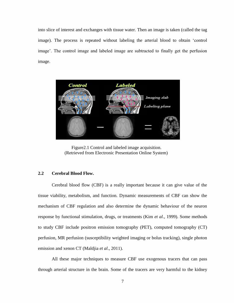
Figure 2.1 shows how the labeled and control image obtained during scanning. Arterial
blood water is magnetically labeled at the labeling plane. After some time, the tracer flows
7
into slice of interest and exchanges with tissue water. Then an image is taken (called the tag
image). The process is repeated without labeling the arterial blood to obtain ‘control
image’. The control image and labeled image are subtracted to finally get the perfusion
image.
Figure2.1 Control and labeled image acquisition.
(Retrieved from Electronic Presentation Online System)
2.2 Cerebral Blood Flow.
Cerebral blood flow (CBF) is a really important because it can give value of the
tissue viability, metabolism, and function. Dynamic measurements of CBF can show the
mechanism of CBF regulation and also determine the dynamic behaviour of the neuron
response by functional stimulation, drugs, or treatments (Kim et al., 1999). Some methods
to study CBF include positron emission tomography (PET), computed tomography (CT)
perfusion, MR perfusion (susceptibility weighted imaging or bolus tracking), single photon
emission and xenon CT (Maldjia et al., 2011).
All these major techniques to measure CBF use exogenous tracers that can pass
through arterial structure in the brain. Some of the tracers are very harmful to the kidney
8
and could not certainly be used in paediatric patients. ASL will provide the CBF value in
quantitative physiological units (mL blood/100 g tissue per minute) that suitable to use in
longitudinal or multicenter studies (Patrick W Hales et al., 2014). ASL technique can be
used in repeated times to track CBF changes in critical patients. ASL magnetically labeled
water molecules in the arterial blood vessels. This is totally different from perfusion
measurements using gadolonium bolus tracking in dynamic susceptibility contrast magnetic
resonance imaging (DSC-MRI), or PET (positron emission tomography). The usage of
exogenous contrast agents can be a very big problem in patients with renal disease and
paediatrics population (Clark et al., 2013).
2.3 Different types of ASL.
The ASL technique has two main subcategories called pulsed ASL (pASL) and
continuous (cASL). cASL has higher signal to noise ratio (SNR) than pASL, but less
available on commercial MRI scanners due to the requirement of long and continuous
radiofrequency (RF) transmission. The latest technique in ASL is known as
pseudocontinuous ASL (pcASL). It divides the long continuous radiofrequency (RF) pulse
in cASL into multiple separates short pulses but eventually will have same labeling effects
as cASL. As a result, better SNR is achieved compared to cASL and can be convenient to
be used in most hardware setup of MRI scanners (Sung-Hong Park et al., 2013).
In pASL, a wider labeling area which located proximal to the imaging plane
received single and short (2-5 milliseconds) RF pulses. pASL has several advantages with
higher tagging efficiency, less power deposition and improved transit time for the labeled
9
spins to travel from the labeling region to the imaging plane. But the main disadvantages in
pASL are low SNR and increased transit delay (Maldjia et al., 2011).
Some methods are also can be used to spatially select the labeling that will allow the
perfusion distribution of single arterial territories to be measured. Velocity selective arterial
spin labeling (vsASL) is being explored as a means of eliminating arterial transit time
dependence. The recent technique known as time-resolved ASL has been developed as a
noninvasive angiography method.
Figure 2.2 shows that in cASL and pcASL, the labeling plane is located at the base
of brain region. At this region, a continuous RF pulse is used to label the arterial blood
before it traveled up to cerebral arterial network higher up (imaging volume). While in the
pASL the labeling slab received short RF pulses in larger area which includes tissue and
arterial blood.

10
Figure 2.2 Schematic diagram of imaging and labeling regions for
CASL/PCASL and PASL. In CASL/PCASL, labeling occurs as
blood flow through a single labeling plane, while in PASL, a slab
of tissue, including arterial blood, is labeled.
(This image retrieved from article: Recommended Implementation of
Arterial Spin; Labeled Perfusion MRI for Clinical Applications: A
Consensus of the ISMRM).
The measurement of perfusion value using ASL is done by inverting the
longitudinal magnetisation of the arterial blood flow through the tissue, waiting for a given
'inflow time' (T1), then capturing an image (labeled image). Then, the same process is
repeated but without labeling the arterial blood flow (controlled image). Perfusion of the
region of interest is the result of subtraction between the 'labeled image' and 'controlled
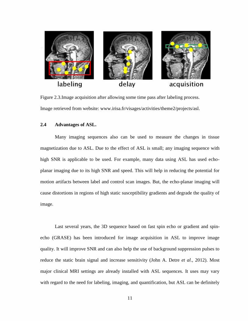
image' (Patrick W Hales et al., 2014). Figure 2.3 shows how the process of ASL imaging
started with labeling of arterial blood water at the base of brain region. Then after a short
delay of time, the arterial blood water will exchange and perfusion process happen before
finally acquisition of perfused tracer with brain tissues.
11
Figure 2.3.Image acquisition after allowing some time pass after labeling process.
Image retrieved from website: www.irisa.fr/visages/activities/theme2/projects/asl.
2.4 Advantages of ASL.
Many imaging sequences also can be used to measure the changes in tissue
magnetization due to ASL. Due to the effect of ASL is small; any imaging sequence with
high SNR is applicable to be used. For example, many data using ASL has used echo-
planar imaging due to its high SNR and speed. This will help in reducing the potential for
motion artifacts between label and control scan images. But, the echo-planar imaging will
cause distortions in regions of high static susceptibility gradients and degrade the quality of
image.
Last several years, the 3D sequence based on fast spin echo or gradient and spin-
echo (GRASE) has been introduced for image acquisition in ASL to improve image
quality. It will improve SNR and can also help the use of background suppression pulses to
reduce the static brain signal and increase sensitivity (John A. Detre et al., 2012). Most
major clinical MRI settings are already installed with ASL sequences. It uses may vary
with regard to the need for labeling, imaging, and quantification, but ASL can be definitely
12
use together with other clinical imaging protocols. Although the main consideration for use
is still in the brain imaging, it can later be extensively uses for other major organs. The
clinical example is in cerebrovascular diseases which primarily affect the brain perfusion.
Many earlier studies showed that ASL MRI was feasible in acute stroke. But, lacking in
robust methodology, low sensitivity for hypo-perfusion, and long signal requirement limits
its use. Until now, dynamic susceptibility contrast (DSC) perfusion MRI is still used
predominantly in diagnostic tool for acute stroke (John A. Detre et al., 2012).
2.5 Limitations of ASL.
Active research is still going on in ASL technique but some limitations has been
recognized. Firstly, in 1.5 T MR imaging unit, the theoretical perfusion-induced signal
change is just 1% for gray matter and 0.4% for white matter. This can cause ASL signal
disturbed by phenomena other than perfusion. Arterial signal artifacts, magnetisation
transfer artifacts, and the influence of noise in the input images are such examples. Long
radiofrequency (RF) pulses and multiple images acquisition also seems to be troublesome
in the commercial scanners (Jackson et al., 2005).
ASL method have low signal-to-noise ratio (SNR), difficult to plan the process, and
also doubt regarding cerebrovascular kinetics or blood equilibrium, directly affecting
perfusion estimates. As many studies actively done combining with technical advances,
ASL will become possible to use as the main diagnostic tool in clinical settings. Also, the
variability of continuous ASL (cASL), pulsed ASL (pASL), and pseudo-continuous ASL
(pcASL) sequences still need to be further understood (Sanna Gevers et al., 2011). The

13
product sequence should be readily available from a vendor to make it easier to use in any
clinical setting. The neurologic diseases which include steno-occlusive disease is rather
difficult because of increased in arterial transit times (ATTs). It will need long delays
between labeling and image capturing (>2 seconds). The signal of labeled blood decays
over time and lower the SNR. So, multiple images acquisition are needed and averaging
over multiple repetitions is not possible with all ASL sequences within reasonable
acquisition times. To solve this issue, three-dimensional (3D) gradient and spin echo
(GRASE) readout module is necessary. It can increase the SNR of a single measurement
and scanning will be done in multiple times (Steve Z Martin et al., 2014).
2.6 Susceptibility Artifact.
ASL also will have susceptibility artifact as on any echo-planar MR imaging
sequence. It is represented as signal intensity void on ASL CBF maps. This may be caused
by metallic hardware, blood products, calcification and air that surround the region of
interest. It also represents a significant limitation of ASL in the evaluation of diseases
because of magnetic distortion. Neurosurgical metal, such as orthodontic appliances and
other materials also significantly disturb interpretation of ASL CBF maps by producing
focally diminished signal intensity. Blood products can give local gradient susceptibility
artifact and thus are seen as low signal intensity on gradient sequences, including ASL.
This can be seen in hemorrhagic infarct type of stroke. Motion is also a common problem
in most clinical MR examinations. Small amounts of motion can produce a significant
source of error in clinical ASL. Motion artifact may produce increases or decreases in
signal intensity on a focal or global basis (A.R Deibler et al., 2008).
14
2.7 Clinical application of ASL.
ASL gives better spatial and temporal resolution compare to other current technique
(Asllani., 2012). Stroke is ranked in third place of mortality cause in western countries. It
also a major cause of disabilities that affect not only health but resulted in loss of financial
source in single bearing families. A quarter of stroke is caused by lacunar infarction due to
ischemia in the territory of the small perforating intracerebral arteries. The patients will
have gait disturbance, dementia and also parkinsonian syndrome (H S Markus et al., 2000).
These types of disabilities will surely affect patient's daily life. Immediate diagnosis and
treatment are the major factors for patient’s outcome. Post-ischemic hyper-perfusion which
commonly associated with cerebral infarction is both beneficial (i.e., prevent more infarct)
and harmful (i.e., more edema and hemorrhage, and neuronal damage due to reperfusion
injury). In addition, post-ischemic hyper-perfusion in the subacute stage (48 hours after
onset) is linked with tissue necrosis. Thus, it is very valuable for the physician to able to
measure the CBF accurately after stroke attacks (Yoji Tanaka et al., 2011).
Although ASL sequence used for scanning organs other than the brain is still very
new but several studies has been done to measure myocardial blood flow (MBF) in human
and animal models. Studies have been performed to demonstrate the effect of drugs to MBF
in vasodilation. It can be an important diagnostic tool for detecting ischemic heart disease
in early stage before a fatal heart attack occur (Hung Phi Do et al., 2014). ASL images also
can show the regional cerebral blood flow (rCBF) that makes it possible to do direct
physiological interpretation. This has lead to pharmacologic studies of various psychoactive

15
drugs such as Citalopram and amphetamine. Prior to ASL technique, pharmacologic studies
on brain often compared the brain activity on drug and brain activity off drug (Stephanie B.
Stewart et al., 2014). The non-invasive method of ASL is really valuable for repeated CBF
measurements in patient follow-up, pharmacologic studies as well as paediatric population.
ASL is very useful in studying slow changes of brain function.
As compared to BOLD fMRI, ASL is more appealing and well suited for
pharmacology MRI (phMRI) studies. This includes the direct pharmacological effects of
drugs which can take effect over hours or days including measuring drug effects indirectly
by their modulation of a stimulus or task response. ASL gives quantitative CBF
measurement both at rest and during task activation which is critical in separating drug
effects on resting brain with task-induced activation. This advantage of ASL technique
have been recognised by the scientific research community with several review articles
pointed out the value of ASL for assessing drug effects on baseline brain function. But, the
main limitation for ASL such as limited availability, low signal to noise ratio (SNR), and
image coverage still remains as the major obstacles ( Danny J J Wang et al., 2011).
MRI is always the choice of technique use study the developing physiology of the
brain. It is non-ionising and also has more spatial resolution make it the suitable method to
look into the transition and development of brain from childhood, through adolescence and
into adulthood. Reference range values can be developed using the hemodynamic
properties of the ASL signal in healthy children and young adult that can be used as
comparison to the abnormal cerebral perfusion pathologies. For example Sturge-Weber
Syndrome which caused abnormality in intracranial venous, a cutaneous capillary angioma,

16
and ocular deformation due to anomalous embryonic development. All of these conditions
will lead to perfusion defects that lead to epilepsy and contralateral hemiparesis (Patrick W
Hales et al., 2014). Follow ups of patients can be safely done with the ASL technique to
look into the level of damage and appropriate treatment plan.
The wide application of ASL technique will include various types of diseases such
as stroke, chronic vascular disease, dementia, and assessment of tumour blood flow. The
latest approach in pseudo-continuous ASL (pcASL) with high SNR is showing good result
to be used in clinical world. The pcASL will use advance magnetic field strength,
multichannel coils, background suppression and improved labeling schemes. Studies by
Wong manage to use this high SNR of the pcASL labeling scheme together with the
flexibility of tagging arteries within a single plane. Different from the use of pulsed ASL
(pASL), which require complex planning procedure to position a three-dimensional (3D)
slab over vessels of interest (Thomas W Okell et al., 2013). The higher SNR value in
pcASL is due to longer labeling period. However, the labeling efficiency will also decrease
as the higher blood velocity risks the underestimation of increase in CBF (Esther Ah
Warnert et al., 2014).
2.7 Direction in future research.
To enable more widespread use of ASL MRI in clinical practice, it is important to
develop widely available MRI sequence and software analysis platforms. Future research is
crucial in investigating its utilisation in various medical settings (Keith A Gillis et al.,
2014). It is still perplexing that ASL MRI unable to found its way into routine clinical

17
practice. This is due to multifactorial issues such as weak signals and ASL methodologies
are somewhat seen as more complex than other MRI routine methods. Furthermore, ASL
benefits also have been downgraded by more widely technologies that commonly available
such as dynamic susceptibility contrast perfusion MRI and BOLD fMRI. Clinicians are also
not familiar with being able to quantify CBF easily and thus rarely demand it. The
availability of truly robust ASL MRI implementations and growing research in this area
will help to more widespread use especially for the benefits of patient itself (John A.Detre
et al., 2012). ASL surely can be applied in healthy populations and also on specific patient
populations (Nederveen).

18
CHAPTER III
METHODOLOGY
3.1 Materials (The Phantom).
In this study, fabricated phantom is used. The body of the phantom used Perspex
because it is solid, stable, non-hazardous, easy to handle and most important it mimic the
real organ characteristics. The phantom is compact and also portable that can be well suited
for reassembly in multi-center research setting.
This phantom was designed of inside the vessel modules. It included the adoption of
two constituent geometries:
i. A set of straight tubes (adult: 0.8 cm and paediatric: 0.5 cm in diameter)
ii. A constant-diameter tube in the shape of a U-bend. It has stenosis of 75% and 50%
each).
The straight tubes provide standard geometry for testing accuracy and precision as well as
the effect of slice position and image resolution. The first straight tube with 0.8 cm in
diameter represents adult’s carotid artery while the other straight tube with 0.5 cm in
diameter mimics the pediatrics carotid artery. Basically the mimicking diameter of the
carotid arteries were chosen as it capable of reproducibly generating the requisite flow rates
and patterns.

19
The specifications of the phantom are as followed :
i. Dimension: 24 x 24 x 2.5 cm³.
ii. Radius U-bend: 2.5 cm.
iii. Diameter U-bend: 5 cm.
iv. Diameter U-bend tube: 0.8 cm.
v. Diameter 1st straight tube: 0.8 cm.
vi. Diameter 2nd straight tube: 0.5 cm.
vii. 50% stenose: 0.4 cm.
viii. 75% stenose: 0.2 cm.
ix. Width: 24 cm.
x. Length: 24 cm.
xi. Length straight tubes: 17 cm each.
Figure 3.1 shows 2D design of phantom that is used during the studies. It has a
Perspex base with 2 pairs of straight tubes and u-tubes. The straight tubes have different
diameters that represent adult and paediatric arteries. The u-tubes have different percentage
of stenosis at the distal ends. This phantom will be cut at the middle to fit the head coil of
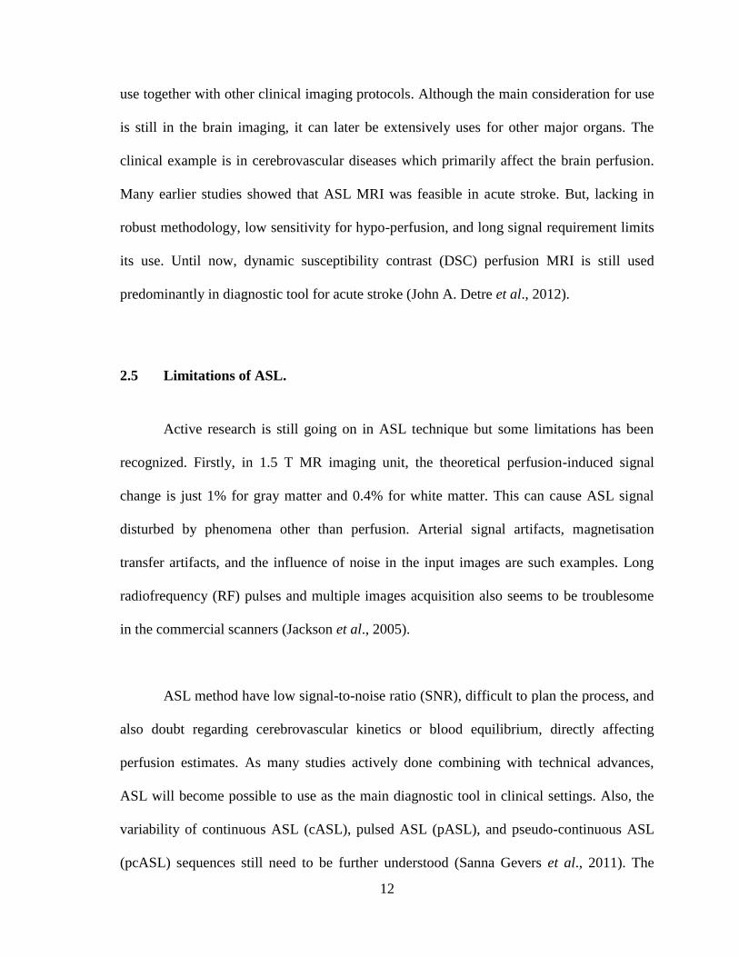
MRI scanner. Figure 3.2 and 3.3 show the cross section of the phantom in clearer view. It
shows that the phantom has different diameters of straight and u-shaped tubes and is cut at
the middle into two parts.

20
Figure 3.1: The 2D design of phantom (red line shows where it is
cut to fit head coil of MRI).
Figure 3.2: Cross section of phantom (red line shows where it
is cut to fit the MRI head coil of MRI).
21
Figure 3.3: Isometric view of the phantom (red line shows where it is
cut to fit head coil of MRI).
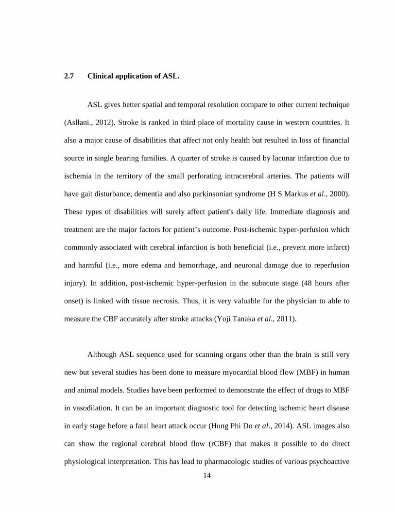
Figure 3.4: Phantom that is used during scan.
22
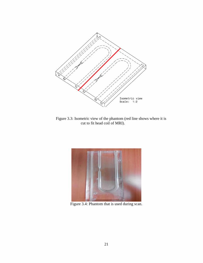
3.2 Procedure.
Assemble the phantom and assess performance
The blood is mimicked in the container by using combination of glycerol and
distilled water 40% and 60% respectively. Water pump is placed in the container to pump
the blood mimic into the phantom and circulate it during the experiment. The pump is
connected to the joint plastic pipes which are the combination of one T-shape pipe, and two
L-shape pipes which made two holes for blood mimic to flow into the phantom. These
holes are connected with plastic tubes which affiliated with phantom at one end of U-tube
and one end of straight tube. Other plastic tubes are connected from another one end of U-
tube and one end of straight tube for blood mimic flow out from phantom to blood mimic
container. This container is placed outside the room. The phantom is set up as figure below:
Figure 3.5: The schematic diagram for setting up ASL phantom.
23
The water pump is switched on and make sure that there is no bubble in the tubes
and phantom during circulation of blood mimic. The phantom and tubes are checked to
make sure there is no leakage. The velocity of blood mimic is measured to make sure it is
close to reference with 75 cm/s for adult and 97 cm/s for paediatric. The velocity is
measured by calculating how long the mixture travels in certain distance of tube. Colour
dye is used to clearly visualise the distance of blood mimic fluid travel from starting of
connector tubes to it ends. The lengths of the tubes are 25 cm each. So we will divide the
length of tubes (25 cm) with the time we get for the dye travel in the tubes. This method
will be repeated using different colour of dyes in order to get the mean value.

24
Figure 3.6 shows the real environment inside the MRI room when the phantom has been
connected to plastic tubes and placed inside the head coil of MRI scanner. The tubes are
then put through a hole inside the wall in between scanner room and the computers outside
to the water pump in water container.
Figure 3.6: Phantom assembly inside the MRI room.