evaluation of possible modes of action for acute effects
TRANSCRIPT
Inhalation Toxicology, 2009; 21(6): 537–551
R e s e a R c h a R t i c l e
Evaluation of possible modes of action for acute effects of methyl iodide in laboratory animals
Christopher R. Kirman1, Lisa M. Sweeney2, Michael L. Gargas2, and John H. Kinzell3
1The Sapphire Group, Inc., Beachwood, Ohio, USA, 2The Sapphire Group, Inc., Dayton, Ohio, USA, and 3Arysta LifeScience Corporation, Cary, NC, USA
Address for Correspondence: Christopher R. Kirman, The Sapphire Group, Inc., 2000 Auburn Drive, Suite 200, Beachwood, Ohio 44122, USA. E-mail: CRK@ thesapphiregroup.com
(Received 15 October 2008; accepted 05 November 2008)
Introduction
Recent studies have indicated that exposures to methyl iodide (MeI) produce a number of effects in laboratory ani-mals, including fetal toxicity, neurotoxicity, and degenera-tion of the nasal epithelium. Through its proposed use as an alternative to methyl bromide as a fumigant, there is potential for human exposures to MeI. These exposures, however, are expected to be acute in nature due to its intended use, which involves soil fumigation at the beginning of the growing sea-son, and its physical–chemical properties. For this reason, a risk assessment is required to characterize the potential for adverse health effects associated with acute exposures. An understanding of the mode(s) of action by which these effects are produced by MeI is important to guide criti-cal decisions used in the acute exposure risk assessment. These decisions include the selection of the appropriate
internal dose measure(s) calculated using physiologically based pharmacokinetic (PBPK) modeling, and evaluat-ing the relevance of the observations in animals to human health. Adapted from US Environmental Protection Agency guidelines (US EPA, 2005), the mode of action is defined here as a sequence of key events and processes, starting with exposure, proceeding through operational and ana-tomical changes, and resulting in toxicity. The term “mode of action” is contrasted with “mechanism of action,” which implies a more detailed understanding and description of events, often at the molecular level, than is meant by mode of action. Because risk assessments are often conducted with incomplete information, and because mode of action is used to guide critical decisions, it is important to provide risk managers with information regarding the level of confi-dence in the mode of action.
ISSN 0895-8378 print/ISSN 1091-7691 online © 2009 Informa UK LtdDOI: 10.1080/08958370802601510
abstractRecent studies have indicated that exposures to methyl iodide (MeI) produce a number of effects in laboratory animals, including fetal toxicity, neurotoxicity, and degeneration of the nasal epithelium. An understanding of the mode of action by which the effects of MeI are produced is useful in guiding critical decisions used in risk assessment. These decisions include the selection of the appropriate internal dose measure(s) calculated using physiologically based pharmacokinetic (PBPK) modeling, and evaluating the relevance of the observations in animals to human health. Modified Hill criteria were used to evaluate several possible mode(s) of action through which MeI produces toxicity in animals. For each endpoint, the key studies were summarized and several possible modes of action were compared to the modified Hill criteria. The available data best support the hypothesis that the fetal effects were likely associated with modulation of the thyroid hormones by iodide during development. This mode of action dictates the use of an internal dose measure in the risk assessment that is indicative of fetal iodide status, such as cumulative iodide concentration (area-under-the-curve or AUC) for iodide in fetal blood. The acute transient neurotoxicity observed in rats exposed to MeI is best supported by a mode of action involv-ing modification of ion currents by the parent chemical in nerve cells. In the case of assessing the potential acute neurotoxicity of MeI, the peak concentration of MeI in the brain would be the appropriate internal dose measure. Finally, the nasal lesions associated with exposure to high concentrations of MeI in rats are best supported by a mode of action that involves glutathione (GSH) depletion in the nasal epithelial tissue. The daily minimum GSH level in olfactory epithelium is the most appropriate internal dose measure for use in risk assessment for this endpoint. Confidence in these modes of action is considered low for the neurotoxic effects, medium for the nasal effects, and high for the fetal effects.
http://www.informapharmascience.com/iht
Inha
latio
n T
oxic
olog
y D
ownl
oade
d fr
om in
form
ahea
lthca
re.c
om b
y C
DC
Inf
orm
atio
n C
ente
r on
07/
06/1
2Fo
r pe
rson
al u
se o
nly.
538 Christopher R. Kirman et al.
Methods
Modified Hill criteria, as described in the US EPA’s Guidelines for carcinogen risk assessment (US EPA, 2005), were used to evaluate the possible mode(s) of action through which MeI produces three acute toxicity endpoints in animals: (1) developmental toxicity; (2) neurotoxicity; and (3) nasal lesions. Although the criteria were taken from guidelines intended for cancer risk assessment, they are considered to be equally applicable to evaluations made for the mode of action of non-cancer risk endpoints. These criteria, and questions associated with them, are listed below.
• Strength, consistency, and specificity of association. What is the level of statistical and biological significance for each event and for the adverse effect? Do independent studies and different experimental hypothesis-testing approaches produce the same associations? Does the agent produce effects other than those hypothesized? Is the key event associated with precursor lesions?
• Dose–response concordance. What are the correlations among doses producing events and the adverse effect? Are they observed in similar dose ranges?
• Temporal relationship. What is the ordering of events that underlie the toxicological process? Is this ordering consistent among independent studies? Is the mode of action consistent with any windows of susceptibility identified?
• Biological plausibility and coherence. Is the mode of action consistent with what is known about toxicology in general and for the case specifically? Are toxic effects and events consistent across structural analogues? Is the database on the agent internally consistent in sup-porting the purported mode of action?
Using these criteria, the possible modes of action (MOA) for MeI are discussed below with respect to acute developmen-tal effects, neurological effects, and nasal effects. For each endpoint, the key studies are summarized and the possible modes of action are compared to the modified Hill criteria. Upon identifying a mode of action that is best supported by the available data, the internal dose measure and other potential risk issues corresponding to the proposed mode of action are recommended. In the absence of sufficient infor-mation on mode of action, the use of the parent chemical in target tissue is considered as an appropriate default internal dose measure, by analogy to use of the external dose for the parent chemical in risk assessments without PBPK mod-eling. In light of this default assumption, the key questions regarding internal dosimetry as it relates to MOA are: (1) Is there sufficient information on the MOA to move away from the default internal dose measure? and (2) If so, what is the most appropriate internal dose measure?
Results
The proposed modes of action of the developmental, neu-rological, and nasal effects of MeI are summarized below.
Proposed mode of action for the developmental effects of MeI in rabbitsKey studiesRecent studies have shown that exposure of pregnant rabbits to MeI during late-stage pregnancy produces fetal losses. In a developmental toxicity study, groups of 24 pregnant New Zealand White rabbits were exposed to concentrations of 0, 2, 10, or 20 ppm MeI for 6 hours/day on days 6 through 28 of gestation (Nemec, 2002a). The number of fetuses, resorp-tions, implantations, and corpora lutea was determined on gestation day (GD) 29. An increased number of late resorp-tions (5.2–5.6% vs. 1.7–1.8% in controls) and post-implanta-tion losses, resulting in a reduced number of viable fetuses, were observed in animals exposed to 20 ppm. This study identified a NOAEL (no observed adverse effect level) and LOAEL (lowest observed adverse effect level) of 10 and 20 ppm for the developmental effects of MeI.
A follow-up study was conducted in rabbits to identify the “window of susceptibility” for the developmental effects of MeI (Nemec, 2003). In this study, groups of 24 pregnant New Zealand White rabbits were exposed to 20 ppm MeI for 6 hours/day on GD 6 through 28, 6 through 14, 15 through 22, 23 through 24, 25 through 26, or 27 through 28. An increased number of late resorptions were observed in ani-mals exposed on gestation days 6–28, 23–24, and 25–26. An increased number of post-implantation losses, decreased number of viable fetuses, and decreased mean fetal weight were observed in animals exposed on days 6–28 and 25–26 of gestation. No signs of developmental toxicity were observed in animals exposed on gestation days 6–14, 15–22, or 27–28. This study identified GD 23–26 as the window of susceptibil-ity for the developmental effects of MeI in rabbits.
Possible modes of actionFour possible modes of action for the developmental effects of MeI in rabbits were considered, as well as their implica-tions for the internal dose measure used in the risk assess-ment, and are summarized below.
• DNA methylation. The developmental effects of MeI could be attributed to its ability to methylate DNA. DNA adducts can cause miscoding or alter the expression of DNA, which may contribute to the developmental effects of MeI. This mode of action would dictate the use of the parent chemical in fetal blood as an appropriate internal dose measure for risk assessment.
• Glutathione (GSH) depletion. The developmental effects of MeI could also be attributed to its ability to deplete cellular GSH levels as a result of its metabolic conjuga-tion with GSH (Di Simplicio et al., 1984; Chamberlain et al., 1998a, 1998b, 1999). Once GSH is depleted below a critical level, cells and tissues are vulnerable to damage by oxidative stress. This could contribute to the devel-opmental toxicity observed with MeI. A review of the lit-erature has suggested that for extrahepatic tissues, tox-icity may be observed when GSH levels fall below 50% of their normal levels (Biaglow et al., 1986; Frederick et al., 1992; Plopper et al., 2001; Lee et al., 2005). Because
Inha
latio
n T
oxic
olog
y D
ownl
oade
d fr
om in
form
ahea
lthca
re.c
om b
y C
DC
Inf
orm
atio
n C
ente
r on
07/
06/1
2Fo
r pe
rson
al u
se o
nly.
Acute MEI modes of action 539
GSH depletion requires additional events (oxidative stress) before toxicity is observed, it is considered to be necessary but not sufficient for cytotoxicity. This mode of action would dictate the use of the daily minimum GSH concentrations in fetal blood or fetal tissues as an appropriate internal dose measure for risk assessment.
• Thyroid hormone modulation. Alternatively, the devel-opmental effects of MeI could be attributed to the release of excess iodide as a result of its metabolism. Excess iodide has been shown to alter thyroid hormone synthesis (Wolff–Chaikoff effect). Thyroid hormones are necessary for successful fetal development, and therefore altered thyroid function may contribute to the developmental toxicity of MeI. This mode of action would dictate the use of the cumulative iodide concen-tration (area-under-the-curve or AUC) for iodide in fetal blood (as a surrogate for AUC in fetal thyroid) as an appropriate internal dose measure for risk assessment.
• Reproductive hormone modulation. The developmental effects of MeI may be attributed to effects on reproductive hormones (e.g. estradiol) that are required for success-ful development of the fetus. In the absence of specific information to implicate one of the metabolites of MeI for this mode of action, a default internal dose measure (parent chemical in blood) would be assumed as an appropriate internal dose measure for risk assessment.
In addition, the possibility remains that the effects of MeI are related to a combination of these modes of action, which may operate at different concentrations and times, or to other cellular events not yet considered. The four possible modes of action described above are compared for causa-tion using the modified Hill criteria and discussed below. This information is also summarized in Table 1.
Strength/consistency/specificity of associationOne of the key pieces of information that can be used to discern between the possible modes of action is that rabbits appear to be more sensitive to the developmental effects of MeI than other species. With this observation in consid-eration, each possible mode of action is assessed below with respect to its strength, consistency, and specificity.
• DNA methylation. A single study was located in the pub-lished literature that demonstrated that MeI could pro-duce significant DNA methylation in rats following oral and inhalation exposures (Bolt & Gansewendt, 1993).
The extent of methylation is uncertain because a “major” (unspecified) part of radiolabel was due to incorporation via the single carbon pool rather than direct alkylation (Gansewendt et al., 1991). However, MeI is considered to be a very weak mutagen (Takahashi & Kawazoe, 1987). Although it is expected to occur to some extent in all spe-cies, no information was located to indicate that DNA methylation occurs in rabbits, or that rabbits are more sensitive to DNA methylation than other species.
• GSH depletion. A number of studies have shown that the conjugation of MeI with GSH can result in a deple-tion of cellular GSH in vivo in several species, includ-ing rabbits (Di Simplicio et al., 1984; Gandy et al., 1990; Chamberlain et al., 1998a; Sloter et al., 2007) and in ani-mal tissues in vitro (Chamberlain et al., 1998b, 1999). Although a causal link between GSH depletion and fetal resorptions has not been demonstrated, there are data that suggest that the rabbit conceptus is more sensitive than the rat conceptus to GSH depletion, as has been observed for thalidomide in vitro (Hansen et al., 1999).
• Thyroid hormone modulation. The levels of circulating thyroid hormones (tri-iodothyronine/thyroxine, T3/T4) are decreased in fetal and maternal blood of rab-bits following exposure to MeI (Sloter et al., 2007). In this study, circulating levels of thyroid stimulating hor-mone (TSH) were increased, and MeI produced a com-plete loss of colloid in the fetal thyroid. Another study reported that exposures of pregnant rabbits to either MeI by inhalation or to sodium iodide (NaI) by injection resulted in decreased circulating T3 and T4 in maternal blood (Sloter et al., 2007). Circulating levels of T4 in fetal blood were decreased (nondetected in a number of samples, particularly on GD 25), while the levels of T3 were variable (no change at some time points, an appar-ent increase at other time points) in rabbits exposed to MeI by inhalation or to NaI by injection (Sloter et al., 2007). Exposure to either MeI or NaI produced an increase in the ratio of T3:T4 in fetal rabbit blood, and both chemicals produced a decrease in fetal thyroid colloid. Decreases in circulating thyroid hormone levels were reported for rats exposed to 25–100 ppm MeI for 2 days (Himmelstein et al., 2005). Deiodinase inhibition was observed in rats exposed to MeI, which may also contribute to the effects on circulating thyroid hormone levels (Farwell, 2005). The effects of excess iodide on the thyroid have been well documented. Excess iodide inhibits thyroid hormone synthesis by
Table 1. Comparison of possible modes of action for the developmental effects of MeI in rabbits using modified Hill criteria.
Possible modes of action
Criteria DNA methylation GSH depletionThyroid hormone modulation
by iodideReproductive hormone
modulation
Strength, consistency, and specificity of association ? ? + +
Dose–response concordance + + + +
Temporal relationship – – + ?
Coherence and plausibility – – + ?
Note. +, data available to support mode of action; –, data available to refute mode of action; ?, data are equivocal.
Inha
latio
n T
oxic
olog
y D
ownl
oade
d fr
om in
form
ahea
lthca
re.c
om b
y C
DC
Inf
orm
atio
n C
ente
r on
07/
06/1
2Fo
r pe
rson
al u
se o
nly.
540 Christopher R. Kirman et al.
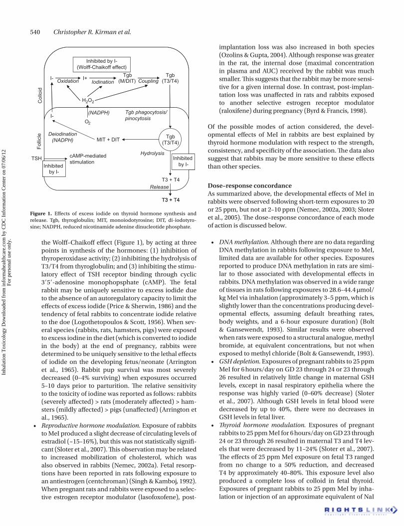
the Wolff–Chaikoff effect (Figure 1), by acting at three points in synthesis of the hormones: (1) inhibition of thyroperoxidase activity; (2) inhibiting the hydrolysis of T3/T4 from thyroglobulin; and (3) inhibiting the stimu-latory effect of TSH receptor binding through cyclic 3959-adenosine monophopsphate (cAMP). The fetal rabbit may be uniquely sensitive to excess iodide due to the absence of an autoregulatory capacity to limit the effects of excess iodide (Price & Sherwin, 1986) and the tendency of fetal rabbits to concentrate iodide relative to the doe (Logothetopoulos & Scott, 1956). When sev-eral species (rabbits, rats, hamsters, pigs) were exposed to excess iodine in the diet (which is converted to iodide in the body) at the end of pregnancy, rabbits were determined to be uniquely sensitive to the lethal effects of iodide on the developing fetus/neonate (Arrington et al., 1965). Rabbit pup survival was most severely decreased (0–4% surviving) when exposures occurred 5–10 days prior to parturition. The relative sensitivity to the toxicity of iodine was reported as follows: rabbits (severely affected) > rats (moderately affected) > ham-sters (mildly affected) > pigs (unaffected) (Arrington et al., 1965).
• Reproductive hormone modulation. Exposure of rabbits to MeI produced a slight decrease of circulating levels of estradiol (~15–16%), but this was not statistically signifi-cant (Sloter et al., 2007). This observation may be related to increased mobilization of cholesterol, which was also observed in rabbits (Nemec, 2002a). Fetal resorp-tions have been reported in rats following exposure to an antiestrogen (centchroman) (Singh & Kamboj, 1992). When pregnant rats and rabbits were exposed to a selec-tive estrogen receptor modulator (lasofoxofene), post-
implantation loss was also increased in both species (Ozolins & Gupta, 2004). Although response was greater in the rat, the internal dose (maximal concentration in plasma and AUC) received by the rabbit was much smaller. This suggests that the rabbit may be more sensi-tive for a given internal dose. In contrast, post-implan-tation loss was unaffected in rats and rabbits exposed to another selective estrogen receptor modulator (raloxifene) during pregnancy (Byrd & Francis, 1998).
Of the possible modes of action considered, the devel-opmental effects of MeI in rabbits are best explained by thyroid hormone modulation with respect to the strength, consistency, and specificity of the association. The data also suggest that rabbits may be more sensitive to these effects than other species.
Dose–response concordanceAs summarized above, the developmental effects of MeI in rabbits were observed following short-term exposures to 20 or 25 ppm, but not at 2–10 ppm (Nemec, 2002a, 2003; Sloter et al., 2005). The dose–response concordance of each mode of action is discussed below.
• DNA methylation. Although there are no data regarding DNA methylation in rabbits following exposure to MeI, limited data are available for other species. Exposures reported to produce DNA methylation in rats are simi-lar to those associated with developmental effects in rabbits. DNA methylation was observed in a wide range of tissues in rats following exposures to 28.6–44.4 µmol/kg MeI via inhalation (approximately 3–5 ppm, which is slightly lower than the concentrations producing devel-opmental effects, assuming default breathing rates, body weights, and a 6-hour exposure duration) (Bolt & Gansewendt, 1993). Similar results were observed when rats were exposed to a structural analogue, methyl bromide, at equivalent concentrations, but not when exposed to methyl chloride (Bolt & Gansewendt, 1993).
• GSH depletion. Exposures of pregnant rabbits to 25 ppm MeI for 6 hours/day on GD 23 through 24 or 23 through 26 resulted in relatively little change in maternal GSH levels, except in nasal respiratory epithelia where the response was highly varied (0–60% decrease) (Sloter et al., 2007). Although GSH levels in fetal blood were decreased by up to 40%, there were no decreases in GSH levels in fetal liver.
• Thyroid hormone modulation. Exposures of pregnant rabbits to 25 ppm MeI for 6 hours/day on GD 23 through 24 or 23 through 26 resulted in maternal T3 and T4 lev-els that were decreased by 11–24% (Sloter et al., 2007). The effects of 25 ppm MeI exposure on fetal T3 ranged from no change to a 50% reduction, and decreased T4 by approximately 40–80%. This exposure level also produced a complete loss of colloid in fetal thyroid. Exposures of pregnant rabbits to 25 ppm MeI by inha-lation or injection of an approximate equivalent of NaI
T3 + T4
I-
Hydrolysis
MIT + DIT
/
I+
Tgb(T3/T4)
T3 + T4
Release
O2
(NADPH)
-Inhibited by I-
T3 + T4
Col
loid
I-
Hydrolysis
Iodination
Fol
licle
MIT + DIT
Tgb phagocytosis/pinocytosis
CouplingOxidationI-Tgb
(M/DIT)Tgb
(T3/T4)
Tgb(T3/T4)
T3 + T4
Release
Deiodination(NADPH)
O2
(NADPH)
Inhibited by I-
Inhibited by I-(Wolff-Chaikoff effect)
Inhibited by I-
cAMP-mediatedstimulation
TSH
H2O2
Figure 1. Effects of excess iodide on thyroid hormone synthesis and release. Tgb, thyroglobulin; MIT, monoiodotyrosine; DIT, di-iodotyro-sine; NADPH, reduced nicotinamide adenine dinucleotide phosphate.
Inha
latio
n T
oxic
olog
y D
ownl
oade
d fr
om in
form
ahea
lthca
re.c
om b
y C
DC
Inf
orm
atio
n C
ente
r on
07/
06/1
2Fo
r pe
rson
al u
se o
nly.
Acute MEI modes of action 541
resulted in altered thyroid hormones in both maternal and fetal blood (Sloter et al., 2007).
• Reproductive hormone modulation. Changes in estra-diol concentrations were reported in rabbits following short-term exposure to 25 ppm MeI (Nemec, 2002a).
All of the possible modes of action are generally consistent with the dose range observed for developmental effects of MeI. Therefore, dose–response concordance cannot be used to discern between the modes of action considered.
Temporal relationshipAs summarized above, the developmental effects of MeI in rabbits were observed following a short-term exposure dur-ing a narrow and specific window of susceptibility (GD 23 through 26) (Nemec, 2002a, 2003). With this observation in consideration, the temporal relationship concordance of each potential mode of action is discussed below.
• DNA methylation. DNA methylation is observed shortly following exposure to MeI (Bolt & Gansewendt, 1993), and it is likely that this endpoint can occur during the defined window of susceptibility. However, DNA methylation would also be expected to occur through-out the gestation period with similar intensity. DNA methylation, therefore, does not explain the temporal relationship observed for the developmental effects of MeI in rabbits. As such, a mode of action involving DNA methylation in fetal tissues is unlikely to only cause the late-stage resorptions observed for MeI.
• GSH depletion. GSH depletion also occurs relatively quickly, with minimum GSH levels achieved in rab-bit or rat tissues within 6 hours of exposure to MeI (Himmelstein et al., 2005; Sloter et al., 2007). Therefore, GSH depletion is also expected to occur during the defined window of susceptibility. However, like DNA methylation, GSH depletion would also occur through-out the gestation period with similar intensity. Therefore, GSH depletion also does not explain the temporal rela-tionship observed for the developmental effects of MeI in rabbits. Although MeI is capable of depleting GSH in fetal tissues, any oxidative damage produced as a result of a compromised defense capability would produce fetal toxicity throughout the pregnancy, and is unlikely to be related to the late-stage resorptions observed for MeI.
• Thyroid hormone modulation. Circulating levels of thyroid hormones are reduced in maternal and fetal rabbits shortly following exposure to MeI (Sloter et al., 2007). In addition, the fetal thyroid showed a complete loss of colloid during the window of susceptibility in this study. The window of susceptibility identified for devel-opmental effects in rabbits corresponds well to the time (GD 22 through 26) at which the fetal thyroid begins to function on its own in preparation for birth.
• Reproductive hormone modulation. Estrogen levels are normally low at the beginning of pregnancy and
gradually increase to maximal levels during the late stages of pregnancy. Therefore, a reduction of estra-diol during the late stages of pregnancy is theoretically consistent with the temporal relationship observed for MeI. Data for other chemicals suggest that alterations in estrogen status during pregnancy can result in fetal resorptions. For example, exposure of rats to an anties-trogen (centchroman) on GD 5 of pregnancy produced dose-dependent resorptions (Singh & Kamboj, 1992).
Of the four possible modes of action considered, the devel-opmental effects of MeI in rabbits are best explained by thyroid hormone modulation with respect to temporal relationship.
Coherence and plausibilityThe coherence and plausibility of each mode of action is discussed below.
• DNA methylation. DNA methylation has been observed for MeI’s structural analogue, methyl bromide, but not for methyl chloride (Bolt & Gansewendt, 1993). This pattern corresponds with that observed for the nonen-zymatic cleavage of the carbon–halide bond, resulting in methylation, which occurs with MeI and methyl bro-mide, but not methyl chloride. However, methyl bro-mide exposure is not associated with fetal resorptions. Fetal toxicity or teratogenicity were also not observed in rats or rabbits exposed to methyl bromide at 30 and 10 mg/kg/day, respectively (Kaneda et al., 1998), sug-gesting that DNA methylation is not involved in the developmental effects of MeI.
• GSH depletion. Glutathione depletion has been observed with structural analogues, methyl bromide and methyl chloride (Landry et al., 1983; Kornbrust & Bus, 1984; Chellman et al., 1986; Davenport et al., 1992). Although no developmental effects were observed in animals exposed to methyl bromide (Kaneda et al., 1993, 1998), some developmental effects have been observed for methyl chloride. High concentrations of methyl chloride (3000 ppm) produced dominant lethal mutations in rats, whereas lower concentrations of 1000 ppm methyl chloride were without effect (Working et al., 1985). Methyl chloride was also teratogenic in mice exposed to 500 ppm, but not to 100 ppm (Wolkowski-Tyl et al., 1983). While GSH depletion may be an impor-tant event for some developmental effects, no reports of late-stage fetal resorptions were located for either methyl bromide or methyl chloride, suggesting that GSH depletion is not involved.
• Thyroid hormone modulation. Normal thyroid function is critical to a successful pregnancy outcome since thy-roid hormones play a critical role in the normal growth and maturation of several organ systems (Devaskar et al., 1986). Development of the fetal lung (Ballard et al., 1980), mechanisms involved in normal thermogen-esis (Steele & Wekstein, 1972), and glucose homeostasis
Inha
latio
n T
oxic
olog
y D
ownl
oade
d fr
om in
form
ahea
lthca
re.c
om b
y C
DC
Inf
orm
atio
n C
ente
r on
07/
06/1
2Fo
r pe
rson
al u
se o
nly.
542 Christopher R. Kirman et al.
(Klein et al., 1981) are especially influenced by thyroid hormonal status. Proper maturation of these homeo-static mechanisms is essential for independent extrau-terine survival since thyroid dysfunction as indicated by perturbation of maternal or fetal thyroid hormonal status has been associated with both fetal and neonatal morbidity and mortality (Pennington, 1990). Both MeI and NaI were shown to produce similar effects on cir-culating rabbit fetal thyroid hormones (decreased T4; varied impact on T3; increased T3:T4 ratios) (Sloter et al., 2007). Clear species differences have been reported for the effects of excess iodine (converted to iodide) dur-ing pregnancy (Arrington et al., 1965). Because thyroid hormone levels were not measured in this study, the possibility that the effects were related to toxic effects of iodide unrelated to effects on the thyroid (which would also require the use of iodide in fetal blood as an internal dose measure) cannot be excluded. The effects observed with excess dietary iodine (Arrington et al., 1965) and injected NaI (Sloter et al., 2007) are similar in nature to those observed for MeI (Nemec, 2002a, 2003). Furthermore, the species differences in sensitiv-ity to excess iodine (Arrington et al., 1965) correspond well to the species differences in the relative importance of nonshivering thermogenesis (NST) in neonates for maintaining body temperature after birth, for which the following has been reported: rabbits (rely exclusively on NST) > rats > hamsters > pigs (lack NST) (Slebodzinski, 1988). Since MeI exhibits substantial effects on the levels of thyroid hormones (increased T3:T4 ratio) in a man-ner that has been associated with acute lethality in adult rabbits (Ghosh & Bhattacharya, 1992), an association between the reported fetal resorptions in rabbits and the disruption of the thyroid hormones is well supported.
• Reproductive hormone modulation. Although circulating estradiol levels are reduced in pregnant rabbits (Sloter et al., 2007), this reduction is relatively small (~15%), particularly when contrasted with the dramatic changes in the fetal thyroid. The reduction in estradiol is may be secondary to changes in lipid metabolism (e.g. increased mobilization of cholesterol) (Matsui et al., 1982; Sloter et al., 2007), rather than a direct antiestrogenic effect of MeI. For these reasons, this mode of action appears less likely than the thyroid hormone effects to be responsible for the fetal resorptions in rabbits.
Based upon a consideration of the overall coherence in the possible modes of action, modulation of thyroid hormone levels is considered to be the most consistent with the avail-able information.
Conclusions for developmental effects of MeIA comparison of the possible modes of action to the modi-fied Hill criteria indicates that there is sufficient information available to support a role for thyroid hormone modulation in the developmental effects of MeI. This mode of action supports the use of the cumulative iodide concentration
(area-under-the-curve or AUC) for iodide in fetal blood or fetal thyroid as an appropriate internal dose measure used in risk assessment.
With respect to relevance to human health, data are available to suggest that there are important species dif-ferences in thyroid hormone modulation that should be considered in the risk assessment. Logothetopoulos and Scott (1956) demonstrated active transport of 131I across the placenta of the guinea pig, rabbit, and to a lesser extent, the rat. Depending on the gestation day, Logothetopoulos and Scott (1956) found that the fetal/maternal blood con-centration ratios were as high as 4–5-fold for guinea pigs and up to six-fold for rabbits. In this study the rats did not seem to concentrate iodide in their fetuses. In stud-ies using 131I in pregnant rabbits, Crone and Waago (1961) found that maternal serum and the maternal portion of the placenta had similar levels of iodide. Likewise, fetal serum and the fetal portion of the placenta had similar levels of iodide; however, the fetal portion of the placenta had 2–6 times higher concentrations of iodide than the maternal portion of the placenta (consistent with Logothetopoulos & Scott, 1956). Roti et al. (1983) also describe studies that demonstrate the concentrating of iodide in the fetuses of sheep (4–5-fold compared to maternal blood) and rabbits (5–9-fold compared to maternal blood), suggesting that the placenta of certain animals likely possesses an active mechanism for transporting iodide from mother to fetus (this is most likely what we now call the sodium/iodide symporter).
More recent studies with rabbits and rats have con-firmed the fetal/maternal iodide blood concentration ratios described in the preceding paragraph. The fetuses from pregnant rabbits dosed intravenously with NaI or exposed via inhalation to 20 ppm MeI had fetal plasma iodide levels that were about 2–3-fold higher than the maternal plasma levels (Morris et al., 2005; Sloter et al., 2007) during exposure or shortly after injection, and roughly 4–7-fold higher during the clearance phase of the plasma concentration curves. In addition, the fetuses from unexposed control pregnant rab-bits in the GD 23–26 timeframe had plasma iodide concen-trations that were about 8–10-fold higher than those of their respective mothers (Sloter et al., 2007). Pregnant rats, on the other hand, have blood iodides that are slightly higher than or equal to those measured in their fetuses at GD 20 when dosed intravenously with a tracer dose of radiolabeled iodide (see Clewell et al., 2003).
The rabbit has been shown to be more sensitive with regard to fetal viability than the rat, hamster, and swine when dosed with KI or NaI in feed at certain times during gestation (Arrington et al., 1965). A rough comparison shows that the rabbit is about 10 times more sensitive than the rat based on iodide concentration in the feed. The concentrat-ing of iodide in the rabbit fetus likely explains the increased sensitivity of rabbits to iodide toxicity compared to the rat. A two-generation study of rats exposed by inhalation to MeI reported increased postnatal loss following exposure to 50 ppm (Nemec, 2002b). Predicted fetal iodide levels for the
Inha
latio
n T
oxic
olog
y D
ownl
oade
d fr
om in
form
ahea
lthca
re.c
om b
y C
DC
Inf
orm
atio
n C
ente
r on
07/
06/1
2Fo
r pe
rson
al u
se o
nly.

Acute MEI modes of action 543
two-generation rat study with 50 ppm MeI (LOAEL dose, early postnatal loss as the endpoint) using a PBPK pregnant rat model were similar to those measured in rabbits exposed to 20 ppm MeI (Sweeney et al., unpublished results). This provides additional information to support the judgment that fetal plasma iodide concentration is a good dose metric to evaluate the developmental effects produced by exposure to MeI and is consistent with the rabbit as a more sensitive species to iodide toxicity in the fetus compared to the rat. The pregnant rat MeI model was developed by modifying the male rat MeI model (Sweeney et al., 2005) to account for physiological differences (e.g. blood flow to placenta, reduced fractional blood flow to other tissues) and inclusion of the validated pregnant rat iodide submodel per Clewell et al. (2003).
The key developmental risk assessment issue is whether the human fetal iodide concentrations following exposure to MeI (or iodide) are equal to, less than, or greater than those measured in their respective mothers. In other words, is a human fetus more like a rat fetus or a rabbit fetus with regard to fetal/maternal plasma iodide concentration ratio? Paired human maternal and fetal plasma iodide concen-trations in unexposed subjects were measured in a recent study by Rayburn et al. (2007) to provide the necessary data to reduce the uncertainty in the human fetal/maternal iodide ratio. The maternal and cord serum inorganic iodide levels reported by Rayburn et al. (2007) were consistent with previous reports of average serum inorganic iodide levels during human pregnancy (Liberman et al., 1998) and term cord blood inorganic iodide (Herve et al., 1997).
Rayburn et al. (2007) found that the cord plasma/maternal plasma iodide ratio averaged approximately 0.9 in preterm deliveries and 1.2 in term deliveries. Additional support for a fetal/maternal iodide ratio of about one comes from the work of Cottino et al. (1972). Total iodine was measured in serum of paired maternal and cord blood at birth, 0.25–48 hrs after intramuscular injection of iodomethylsparteine. The iodine in iodomethylsparteine is ionic “with an ‘iodine’ behaviour in the biological sense.” Aboul-Khair et al. (1966) demonstrated that maternal blood levels of organic compounds with incorporated radi-olabeled iodine are not detectable until more than 12 hours after administration of radiolabel. Although there is some uncertainty regarding what the assay for total iodine is measuring, it is reasonable to assume that for times up to 12 hours after administration, excess blood iodine in women dosed with iodomethylsparteine is attributable to free iodide. For women delivering <12 hrs after the injec-tion, total serum iodide levels were, on average, 14-fold higher at delivery than at discharge and cord blood levels averaged 10-fold higher than neonatal levels at discharge. For paired maternal and cord blood samples in Cottino et al. (1972), the ratios were reported as group average mater-nal/cord ratios, which we inverted to cord/maternal ratios for consistency with the rest of this document. For times of 0.25–1 hour after injection, the average cord/maternal blood iodine ratio was 0.6; 1.5–3 hrs after injection, the
average ratio was 0.8; and 4–9 hrs after injection, the aver-age ratio was 1.0. Because the excess iodine in the serum is anticipated to be iodide, these data demonstrate a ratio for paired mothers and neonates circa 1.
Unlike many animals (see description above for sheep and rabbits from Roti), humans do not seem to have the same ability to concentrate iodide in their fetuses via the placenta. Hence, this attribute likely explains why the human fetus is more prone to cretinism in geographic areas that produce maternal dietary iodine deficiency, as the human fetus is not capable of concentrating iodide.
Proposed mode of action for neurological effects of MeI in ratsKey studiesTransient neurological effects have been observed in rats following acute inhalation exposures to MeI. Groups of 24 Crl:CD(SD)IGS BR rats (12 males and 12 females in each) were exposed to concentrations of 0, 27, 93, or 401 ppm for 6 hours (Schaefer, 2002). Clinical signs of neurological effects included drooping eyelids, repetitive movement of the mouth, abnormal gait, uncoordinated air-righting reflex, and reduced rotorod performance in animals exposed to 97 or 401 ppm. In addition, body temperature was reduced 1 and 4°C in animals exposed to 97 and 401 ppm, respec-tively. These effects were acute in nature. Clinical signs of neurotoxicity were generally resolved by the following day, with the exception of one animal that subsequently died on day 5. In addition, all performance deficits were resolved when retested on days 7 and 14. No treatment-related neu-ropathological lesions were observed in animals exposed to the highest concentration. No effects were observed in ani-mals exposed to 27 ppm. This study identifies NOAEL and LOAEL values of 27 and 93 ppm for the acute neurological effects of MeI.
Case studies of accidental exposures to high concentra-tions of methyl iodide indicate that neurological effects, including histopathological changes, can occur in humans (Hermouet et al., 1996). However, the exposures associated with these effects are likely much higher than those reported for the transient effects in rats, and are likely related to a dif-ferent mode of action.
Possible modes of actionFour possible modes of action for the acute neurotoxic effects of MeI, and their implications for the internal dose measure used in the risk assessment, were considered and are summarized below.
• Modification of ion currents in neurons. A wide variety of solvents are known to produce anesthetic or seda-tive effects in animals and humans by altering nerve cell membrane properties (Snyder & Andrews, 1996). MeI may depress spontaneous and evoked activity of neurons in the brain, possibly through nonspecific actions with the lipid matrix of the nerve membrane or by a more specific action on the -amino butyric
Inha
latio
n T
oxic
olog
y D
ownl
oade
d fr
om in
form
ahea
lthca
re.c
om b
y C
DC
Inf
orm
atio
n C
ente
r on
07/
06/1
2Fo
r pe
rson
al u
se o
nly.

544 Christopher R. Kirman et al.
acid (GABAA) receptor chloride channel, a major
mediator of inhibitory synaptic transmission (Trevor & White, 2004). If this mode of action is correct, the peak concentration of MeI in the brain should serve as an appropriate internal dose measure for assessing the risks of neurological effects from MeI exposure. In addition, a recent in vitro study suggests that the formation of S-methyl-cysteine may contribute to the neurotoxicity of methyl halides, either by interact-ing with the membrane or with the GABA receptor (Pancetti et al., 2004). However, any contribution of S-methyl-cysteine to neurotoxicity would also indi-cate the use of peak MeI in the brain as an internal dose surrogate.
• GSH depletion. The acute neurological effects of MeI could be attributed to its ability to deplete cellular GSH levels as a result of its metabolic conjugation with GSH. Once depleted below critical levels of GSH, cells and tissues are vulnerable to damage by oxidative stress. This action may contribute to the neurotoxicity of MeI. As noted above, a review of the literature has suggested that for extrahepatic tissues, toxicity may be observed when GSH levels fall below 50% of their nor-mal levels. If correct, this mode of action would dictate the use of the daily minimum GSH concentration in the brain as an appropriate internal dose measure for risk assessment.
• Hydrogen sulfide formation. The acute neurological effects of MeI could also be attributed to its ability to form hydrogen sulfide, which requires at least two metabolic steps (GSH conjugation followed by oxida-tion). Hydrogen sulfide is an inhibitor of cytochrome c in mitochondria, and has been proposed as being possibly responsible for some of the neurotoxicity observed for MeI following exposure to high concen-trations (Hermouet et al., 1996). If correct, this mode of action would dictate the use of the amount of MeI conjugated with glutathione as an appropriate internal dose surrogate.
• Iodide release. The acute neurological effects of MeI could be associated with the release of iodide, which occurs following conjugation with glutathione. Iodide exposure has been shown to produce neurodevelop-mental effects, secondary to thyroid effects, in rats (Vorhees et al., 1984). If correct, this mode of action would dictate the use of iodide in blood or thyroid as an appropriate internal dose measure.
In addition, the possibility remains that the effects of MeI are related to a combination of these modes of action, which may operate at different concentrations and times, or to other cellular events not yet considered. The four possible modes of action described above are compared using modi-fied Hill criteria for causation as summarized in Table 2 and discussed below.
Strength/consistency/specificity of associationOne of the important pieces of information from the key study that can be used to discern between the possible modes of action is that the neurological effects are revers-ible. The three modes of action are discussed below with respect to the strength, consistency, and specificity of an association.
• Modification of ion currents in neurons. Although not assessed specifically for MeI, many organic vapors possess general anesthetic properties at high concen-trations that are reversible upon cessation of exposure (Richards et al., 1978; Pringle et al., 1981; Janoff & Miller, 1982; Franks & Lieb, 1987; Chiou et al., 1990; Curry et al., 1990; Janes et al., 1992; Messaoudi et al., 1992; Dickinson et al., 1993; Slater et al., 1993).
• GSH depletion. As noted above, a number of studies have shown that the conjugation of MeI with GSH can result in a depletion of cellular GSH. A role for GSH depletion in nerve cell toxicity, particularly within the mitochondria, is supported by observations made dur-ing in vitro studies (Bonnefoi et al., 1991; Bonnefoi, 1992; Chamberlain et al., 1999). Depletion of GSH levels would leave nerve cells or cell-fractions susceptible to oxidative damage. Although some oxidative damage may be repairable over time, it is expected that some deficit in nerve cell function would persist if injury were the result of oxidative damage.
• Hydrogen sulfide formation. Acute exposures to high hydrogen sulfide concentrations are associated with neurological effects in animals and humans. These effects typically are only associated with concentra-tions sufficiently large to result in loss of consciousness, coma, and death (Warenycia et al., 1989; Snyder et al., 1995; Dorman et al., 2002; Solnyshkova et al., 2004). Since these cases often also result in persistent loss of neurological performance in survivors, association of MeI’s neurotoxicity with hydrogen sulfide would be expected to demonstrate residual neurological deficits.
Table 2. Comparison of possible modes of action for the acute neurotoxic effects of MeI in rats using modified Hill criteria.
Possible modes of action
CriteriaCell membrane
disruption GSH depletion Hydrogen sulfide formation Iodide release
Strength, consistency, and specificity of association + ? ? ?
Dose–response concordance + + – –
Temporal relationship + – – –
Coherence and plausibility + – – –
Note. +, data available to support mode of action; –, data available to refute mode of action; ?, data are equivocal.
Inha
latio
n T
oxic
olog
y D
ownl
oade
d fr
om in
form
ahea
lthca
re.c
om b
y C
DC
Inf
orm
atio
n C
ente
r on
07/
06/1
2Fo
r pe
rson
al u
se o
nly.
Acute MEI modes of action 545
• Iodide release. No studies were located which reported neurotoxicity following acute exposures to iodide. However, potassium iodide exposure produced neu-rodevelopmental effects (delayed auditory startle, delayed olfactory orientation to the home-cage scent, decreased running-wheel activity) in rats following repeated exposures via the diet prior to, during, and post-pregnancy (Vorhees et al., 1984). These effects differ from the neurological effects observed for MeI in that they are delayed in onset, not reversible, and asso-ciated with repeated exposures, and therefore are not considered to be directly relevant.
The acute, transient neurological effects of MeI are best explained by the cell membrane disruption mode of action with respect to the strength, consistency, and specificity of the association.
Dose–response concordanceIn the key study neurological effects were observed follow-ing acute exposure to 97–401 ppm MeI (Schaefer, 2002). The dose–response concordance for each mode of action is dis-cussed below.
• Modification of ion currents in neurons. Although there are no specific data regarding the concentrations of MeI producing membrane disruption, the anesthetic or nar-cotic effects of lipophilic organic vapors are generally observed at high concentrations sufficient to disrupt membrane excitability. For a structurally related chemi-cal, acute exposures of rats to a similar range of concen-tration (188–250 ppm) of methyl bromide resulted in a reduced locomotor activity, which also was resolved within 24 hours of exposure without residual effects (Honma et al., 1985).
• Glutathione depletion. In rats exposed to 100 ppm MeI, the brain levels of GSH were reduced by 20–30%, and rats showed no signs of a neurological effect (Chamberlain et al., 1998). When rats were pretreated with phorone/buthionine sulfoximine (GSH depletors), marked signs of CNS neurotoxicity were observed. Because the pre-treatment did not result in substantially lower brain GSH levels, the authors attributed the neurotoxicity to an increased delivery of the parent chemical to the brain by decreasing a first-pass metabolic effect for MeI due to its conjugation with GSH in other tissues (Chamberlain et al., 1998).
• Hydrogen sulfide formation. Acute exposures of rats to 30 ppm or higher hydrogen sulfide produces an inhibition of cytochrome oxidase, which is the primary biochemi-cal biomarker associated with lethal exposures, in respi-ratory tissues, but cytochrome oxidase was increased in liver tissue (Dorman et al., 2002). In this study, the sulfide levels in the brain of exposed animals were unaffected. For this mode of action to be relevant, a significant frac-tion (>30%) of absorbed MeI would need to be converted to hydrogen sulfide in a relatively short period of time.
• Iodide release. Neurodevelopmental effects were observed in rats following repeated exposures to 0.025–0.1% potassium iodide in the diet (correspond-ing to approximately 12.5–50 mg/kg/day) (Vorhees et al., 1984). However, the cumulative dose of iodide to which rats were exposed to in this study is much larger than the acute dose of iodide received by rats from MeI in the key study.
Based upon a consideration of the dose–response concord-ance, the cell membrane disruption and glutathione deple-tion modes of action are best supported by the available data.
Temporal relationshipOne of the important pieces of information reported in the key study is that the neurological effects of MeI in rats were observed soon after the beginning of the exposure period, and resolved shortly after exposure ended. With this obser-vation in consideration, each of the possible modes of action is assessed below.
• Modification of ion currents in neurons. Although no specific information was located regarding the temporal relationship for MeI and ion current modi-fication, this effect for organic vapors in general is expected to be transient in nature, and becomes resolved once the tissue levels for the parent chemi-cal are reduced due to metabolism and excretion after exposure ends.
• Glutathione depletion. Although GSH depletion may occur quickly, any oxidative damage that accumu-lates during this time is expected to be only slowly resolved, if at all. Neurological damage that might occur would, therefore, be expected to persist for some time after exposure ceases. For this reason, GSH deple-tion does not provide concordance with the temporal relationship.
• Hydrogen sulfide formation. Although no specific information was located regarding the temporal rela-tionship for hydrogen sulfide formation by MeI, its for-mation requires several metabolic steps. Peak tissue levels for hydrogen sulfide formation may be delayed and only occur after exposure. If this is the case, this mode of action does not provide concordance with the observed temporal relationship for the neurological effects of MeI.
• Iodide release. Because the neurodevelopmental effects were observed in rats following subchronic exposure to potassium iodide rather than acute exposure, this mode of action does not provide concordance with the observed temporal relationship for the acute neurologi-cal effects of MeI.
Based upon a consideration of the temporal relationship, the cell membrane disruption mode of action is best supported by the available data.
Inha
latio
n T
oxic
olog
y D
ownl
oade
d fr
om in
form
ahea
lthca
re.c
om b
y C
DC
Inf
orm
atio
n C
ente
r on
07/
06/1
2Fo
r pe
rson
al u
se o
nly.

546 Christopher R. Kirman et al.
Coherence and plausibilityThe coherence and plausibility of the possible modes of action for MeI neurological effects are discussed below.
• Modification of ion currents in neurons. A mode of action involving nerve cell ion current modification by the parent chemical is consistent with observations made for a wide variety of organic vapors and solvents (Richards et al., 1978; Pringle et al., 1981; Janoff & Miller, 1982; Franks & Lieb, 1987; Chiou et al., 1990; Curry et al., 1990; Janes et al., 1992; Messaoudi et al., 1992; Dickinson et al., 1993; Slater et al., 1993), including those that are structurally similar to MeI (Honma et al., 1985).
• Glutathione depletion. Although MeI is clearly capable of depleting cellular glutathione in the brain and other tissues, the type of damage expected for this mode of action would be more persistent and severe than the transient effects observed in the key study. A potential role for GSH depletion cannot be ruled out as a mode of action for the more severe neurological effects asso-ciated with high concentrations or repeated exposures, but it is unlikely to play an important role in the tran-sient effects observed in the key study.
• Hydrogen sulfide formation. This mode of action would require a significant fraction of the absorbed dose of MeI to be converted to hydrogen sulfide. However, there is no evidence that this conversion would occur or would occur preferentially in the brain. In addition, the relative persistence and severity of the neurologi-cal effects associated with high-dose hydrogen sulfide exposure are inconsistent with the transient nature of the neurological effects of MeI observed in the key study. Therefore, this mode of action is not supported based upon a lack of coherence and plausibility.
• Iodide release. Because the only reports of neurotoxic-ity of iodide are secondary to effects on the thyroid fol-lowing repeated exposures during development, this mode of action is not supported by available informa-tion. Although the reduced body temperature in rats exposed to MeI might at first glance suggest a potential role for decreased thermogenesis, an important role of the thyroid, the fact that similar effects (central nervous system (CNS) depression, decreased body temperature) have been reported in rats acutely exposed to methyl bromide (Honma et al., 1985), which is not iodogenic, suggests that iodide release is not responsible for the acute neurotoxicity of MeI.
Based upon a consideration of the overall coherence, the cell membrane disruption mode of action is best supported by the available information.
Conclusions for the neurological effects of MeIBased on a comparison of the possible modes of action to the modified Hill criteria, there is limited information to sup-port a role for modification of ion currents in neurons in the acute neurotoxicity of MeI. This mode of action supports the
use of the peak concentration of methyl iodide in the brain as an appropriate internal dose measure used in risk assess-ment, which, although is based upon limited information, corresponds to the default internal dose measure for CNS effects. With respect to relevance to human health, no data were located to suggest that species differences exist for the proposed mode of action for the neurological effects, and therefore the relevance to human health is assumed.
Proposed mode of action for the nasal effects of MeI in ratsKey studiesGroups of 40 rats (20 males and 20 females) were exposed to concentrations of 0, 5, 21, and 70 ppm MeI for 6 hours/day, 5 days/week for up to 13 weeks (Kirkpatrick, 2002). Ten animals per sex per group were sacrificed after 4 weeks. No treatment-related effects were observed in animals exposed to 5 or 21 ppm. Degeneration of the olfactory epithelium and respiratory epithelial metaplasia were observed in animals of both sexes exposed to 70 ppm at both 4 and 13 weeks. This study identifies NOAEL and LOAEL values of 21 and 70 ppm for repeated exposures to MeI. This study is supported by similar observations made in rats exposed to 50 ppm, but not 20 ppm, in a two-generation reproductive toxicity study (Nemec, 2002b); in rats exposed to 60 ppm, but not 20 ppm, in a chronic toxicity study (1-year interim sacrifice) (Kirkpatrick, 2003); and in rats exposed to 100 ppm MeI in an acute study (Chamberlain et al., 1998a).
Possible modes of actionThree possible modes of action, as well as their implications to the internal dose measure used in the risk assessment, were considered and are summarized below.
• Glutathione depletion. The nasal effects of MeI could be attributed to its ability to deplete cellular GSH levels as a result of its conjugation with GSH. Once GSH becomes significantly depleted, cells and tissues are vulnerable to damage by oxidative stress, which may contribute to point-of-contact nasal lesions seen with MeI. As noted above, a review of the literature has suggested that for extrahepatic tissues, toxicity may be observed when GSH levels fall below 50% of their normal levels. If cor-rect, this mode of action would dictate the use of the daily minimum GSH concentration in olfactory epithe-lium as an appropriate internal dose measure for risk assessment.
• Methylation of macromolecules. The nasal effects of MeI could also be attributed to its ability to methylate macro-molecules. These macromolecular adducts could cause altered function of key proteins, which may contribute to the nasal lesions seen with MeI. If correct, this mode of action would dictate the use of the parent chemical in olfactory epithelium as an appropriate internal dose measure for risk assessment.
• Formaldehyde production. Under this mode of action, the nasal lesions of MeI would be attributed to the
Inha
latio
n T
oxic
olog
y D
ownl
oade
d fr
om in
form
ahea
lthca
re.c
om b
y C
DC
Inf
orm
atio
n C
ente
r on
07/
06/1
2Fo
r pe
rson
al u
se o
nly.
Acute MEI modes of action 547
metabolic formation of the reactive formaldehyde molecule. The effects of formaldehyde on nasal tissue have been attributed to its ability to form DNA–protein crosslinks (Conolly et al., 2000). If correct, this mode of action would dictate the use of formaldehyde in olfac-tory epithelium as an appropriate internal dose meas-ure for risk assessment.
In addition, the possibility remains that the effects of MeI are related to a combination of these modes of action, which may operate at different concentrations and times, or to other cellular events not yet considered. The three possible modes of action described above are compared using modi-fied Hill criteria for causation as summarized in Table 3 and discussed below.
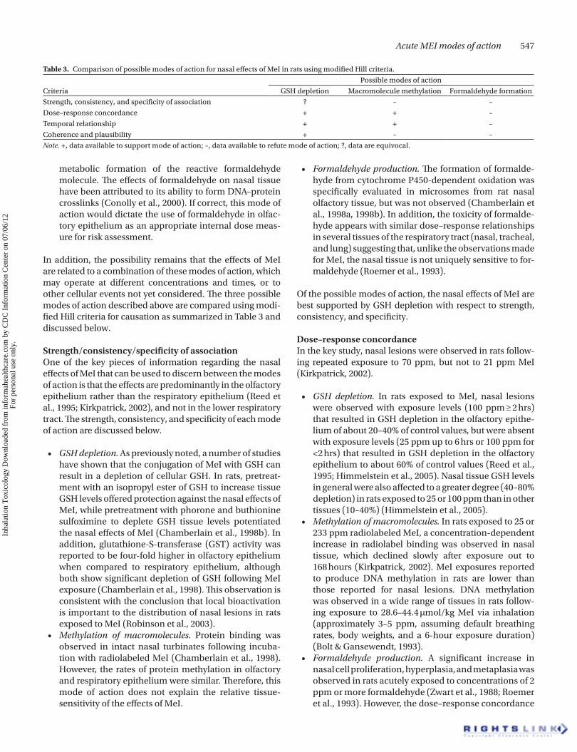
Strength/consistency/specificity of associationOne of the key pieces of information regarding the nasal effects of MeI that can be used to discern between the modes of action is that the effects are predominantly in the olfactory epithelium rather than the respiratory epithelium (Reed et al., 1995; Kirkpatrick, 2002), and not in the lower respiratory tract. The strength, consistency, and specificity of each mode of action are discussed below.
• GSH depletion. As previously noted, a number of studies have shown that the conjugation of MeI with GSH can result in a depletion of cellular GSH. In rats, pretreat-ment with an isopropyl ester of GSH to increase tissue GSH levels offered protection against the nasal effects of MeI, while pretreatment with phorone and buthionine sulfoximine to deplete GSH tissue levels potentiated the nasal effects of MeI (Chamberlain et al., 1998b). In addition, glutathione-S-transferase (GST) activity was reported to be four-fold higher in olfactory epithelium when compared to respiratory epithelium, although both show significant depletion of GSH following MeI exposure (Chamberlain et al., 1998). This observation is consistent with the conclusion that local bioactivation is important to the distribution of nasal lesions in rats exposed to MeI (Robinson et al., 2003).
• Methylation of macromolecules. Protein binding was observed in intact nasal turbinates following incuba-tion with radiolabeled MeI (Chamberlain et al., 1998). However, the rates of protein methylation in olfactory and respiratory epithelium were similar. Therefore, this mode of action does not explain the relative tissue-sensitivity of the effects of MeI.
• Formaldehyde production. The formation of formalde-hyde from cytochrome P450-dependent oxidation was specifically evaluated in microsomes from rat nasal olfactory tissue, but was not observed (Chamberlain et al., 1998a, 1998b). In addition, the toxicity of formalde-hyde appears with similar dose–response relationships in several tissues of the respiratory tract (nasal, tracheal, and lung) suggesting that, unlike the observations made for MeI, the nasal tissue is not uniquely sensitive to for-maldehyde (Roemer et al., 1993).
Of the possible modes of action, the nasal effects of MeI are best supported by GSH depletion with respect to strength, consistency, and specificity.
Dose–response concordanceIn the key study, nasal lesions were observed in rats follow-ing repeated exposure to 70 ppm, but not to 21 ppm MeI (Kirkpatrick, 2002).
• GSH depletion. In rats exposed to MeI, nasal lesions were observed with exposure levels (100 ppm ≥ 2 hrs) that resulted in GSH depletion in the olfactory epithe-lium of about 20–40% of control values, but were absent with exposure levels (25 ppm up to 6 hrs or 100 ppm for <2 hrs) that resulted in GSH depletion in the olfactory epithelium to about 60% of control values (Reed et al., 1995; Himmelstein et al., 2005). Nasal tissue GSH levels in general were also affected to a greater degree (40–80% depletion) in rats exposed to 25 or 100 ppm than in other tissues (10–40%) (Himmelstein et al., 2005).
• Methylation of macromolecules. In rats exposed to 25 or 233 ppm radiolabeled MeI, a concentration-dependent increase in radiolabel binding was observed in nasal tissue, which declined slowly after exposure out to 168 hours (Kirkpatrick, 2002). MeI exposures reported to produce DNA methylation in rats are lower than those reported for nasal lesions. DNA methylation was observed in a wide range of tissues in rats follow-ing exposure to 28.6–44.4 µmol/kg MeI via inhalation (approximately 3–5 ppm, assuming default breathing rates, body weights, and a 6-hour exposure duration) (Bolt & Gansewendt, 1993).
• Formaldehyde production. A significant increase in nasal cell proliferation, hyperplasia, and metaplasia was observed in rats acutely exposed to concentrations of 2 ppm or more formaldehyde (Zwart et al., 1988; Roemer et al., 1993). However, the dose–response concordance
Table 3. Comparison of possible modes of action for nasal effects of MeI in rats using modified Hill criteria.
Possible modes of action
Criteria GSH depletion Macromolecule methylation Formaldehyde formation
Strength, consistency, and specificity of association ? – –
Dose–response concordance + + –
Temporal relationship + + –
Coherence and plausibility + – –
Note. +, data available to support mode of action; –, data available to refute mode of action; ?, data are equivocal.
Inha
latio
n T
oxic
olog
y D
ownl
oade
d fr
om in
form
ahea
lthca
re.c
om b
y C
DC
Inf
orm
atio
n C
ente
r on
07/
06/1
2Fo
r pe
rson
al u
se o
nly.
548 Christopher R. Kirman et al.
of this mode of action is plausible only if a fraction of the inhaled MeI is rapidly converted to formaldehyde, which is not supported by the metabolic capacity of rat nasal tissue (Chamberlain et al., 1998a, 1998b).
Based upon a consideration of the dose–response con-cordance, either glutathione depletion or methylation of macromolecules is supported by the available data.
Temporal relationshipThe nasal lesions of MeI are observed following repeated exposures to MeI, including at both 4 and 13 weeks of expo-sure (Kirkpatrick, 2002), in a two-generation reproductive toxicity study (Nemec, 2002b), and following repeated expo-sures to MeI for 1 year (Kirkpatrick, 2003).
• GSH depletion. GSH depletion occurs relatively quickly, with the minimum GSH levels achieved in rat tissues at the end of the 6-hour exposure to MeI (Himmelstein et al., 2005; Sloter et al., 2007). Therefore, GSH depletion meets the requirements of a potential precursor lesion in that it is expected to occur well in advance of the for-mation of nasal lesions.
• Methylation of macromolecules. The methylation of macromolecules is also observed shortly following exposure to MeI (Bolt & Gansewendt, 1993). Therefore, it is likely that this endpoint would also occur prior to the development of nasal lesions.
• Formaldehyde production. Because formaldehyde was not observed with the metabolism of MeI in microsomes prepared from rat nasal tissue, a temporal relationship between formaldehyde formation and nasal lesions is not supported.
Based upon a consideration of the temporal relationship, either glutathione depletion or methylation of macromol-ecules is supported by the available data.
Coherence and plausibilityThe overall coherence and plausibility of each possible mode of action is discussed below.
• GSH depletion. As noted above, GSH depletion has been observed with structural analogues of MeI. The structural analogue methyl bromide, which also pro-duces GSH depletion, has been reported to produce nasal lesions in rodents (Eustis et al., 1988; National Toxicology Program, 1992).
• Methylation of macromolecules. As noted above, mac-romolecule methylation has been observed for the MeI structural analogue, methyl bromide, which produceds nasal lesions, but not for methyl chloride. However, exposure of rats to another methylating agent (dimethyl sulfate) produced greater methylation of macromol-ecules in respiratory epithelium compared to olfactory epithelium (Mathison et al., 1995). Therefore, this mode of action is not well supported.
• Formaldehyde production. Although formaldehyde is capable of producing nasal lesions in animals, the lack of detectable levels formed in microsomal preparations from rat nasal epithelium suggests that this mode of action is not applicable to MeI.
Based upon a consideration of the overall coherence, either GSH depletion or methylation of macromolecules is supported by the available information.
Conclusions for the nasal effects of MeIBased on a comparison of the three possible modes of action to modified Hill criteria, there is sufficient informa-tion available to support a role for GSH depletion in the nasal effects of MeI. This mode of action supports the use of the daily minimum GSH level in olfactory epithelium as an appropriate internal dose measure in risk assessment.
With respect to relevance to human health, limited data are available that indicate there may be important species differences that should be considered in the risk assessment. Background levels of GSH in nasal tissue appear lower in humans (Westerveld et al., 1997; Frederick et al., 2002) than in rats (Himmelstein et al., 2005), which would suggest that humans may be more sensitive than rats to the GSH-depleting effects of MeI. However, there are two additional factors that suggest that this may not necessarily be the case. First, GST activity in human nasal tissue is expected to be much lower than in rat nasal tissue, suggesting that the rate of GSH depletion would be slower in human tissue than in rodent tissue, and thereby allowing for GSH repletion pathways to maintain GSH levels. Like other halomethanes (methyl chloride, methyl bromide) (Thier et al., 1999; Muller et al., 2001), MeI conjugation with GSH is catalyzed by glutathione-S-transferase isozyme theta-1 (GST-T1) (Hallier et al., 1994; Chamberlain et al., 1998). The levels of GST-T1 in human nasal tissue have not been directly measured, but are likely to be low, and possibly negligible, based upon observations made for this isozyme in the respiratory tract (lung), and this represents an important species difference with respect to relative susceptibility to halomethane toxic-ity (Sherratt et al., 1997, 2002). Human GST-T1 is expected to be substantially less efficient at catalyzing MeI–GSH conju-gation based on findings with a GST mutant (Shokeer et al., 2005). Low/negligible levels of GST-T1 in human nasal tissue are consistent with reports for a lack of detectable conjuga-tion of cumene hydroperoxide (an indicator substrate for GST-T1 activity) in human nasal tissue (Aceto et al., 1989). Secondly, human nasal tissue possesses additional anti-oxidants which may be important for MeI. Specifically, uric acid has been identified as a major antioxidant in human nasal airway secretions. Although methylation of uric acid has not been measured in nasal tissue, this can be inferred from MeI’s ability to methylate DNA (uric acid is structur-ally similar to DNA bases). Furthermore, MeI is used in the formation of methylated derivatives of uric acid (Maruyama et al., 2000). Interestingly, methylated derivatives are them-selves excellent antioxidants (Nishida, 1991), suggesting that
Inha
latio
n T
oxic
olog
y D
ownl
oade
d fr
om in
form
ahea
lthca
re.c
om b
y C
DC
Inf
orm
atio
n C
ente
r on
07/
06/1
2Fo
r pe
rson
al u
se o
nly.
Acute MEI modes of action 549
conjugation of MeI to uric acid would not leave tissues sus-ceptible to oxidative damage.
Discussion and conclusions
Based on the results of MeI toxicity studies, three endpoints of concern were identified: late-stage fetal resorption (rab-bit), acute neurotoxicity (rat), and nasal lesions (rat). In reviewing potential modes of action for these endpoints, the available data suggested that the late-stage fetal resorp-tions were likely associated with modulation of the thyroid hormones by iodide during development (although con-tributions of general iodide toxicity on the fetus cannot be excluded). This mode of action is best evaluated using the cumulative iodide concentration (area-under-the-curve or AUC) for iodide in fetal blood as an appropriate internal dose measure. The acute transient neurotoxicity observed in rats exposed to MeI was felt to be due to a generalized modification of ion currents in nerve cells that is associated with high concentrations of many chemicals. In the case of assessing the potential acute neurotoxicity of MeI, the peak concentration of methyl iodide in the brain would be the appropriate internal dose measure. Finally, the nasal lesions associated with exposure to high concentrations of MeI in rats were supported as being associated with GSH depletion in the nasal epithelial tissue. The daily minimum GSH level in olfactory epithelium is the most appropriate internal dose measure for use in risk assessment for this endpoint.
Confidence in the proposed modes of action differs for each of the three endpoints considered, based upon the quality and quantity of information available to support it. Confidence in the proposed mode of action for the developmental effects of MeI is considered high because it is well supported by focused, mechanistic studies. Confidence in the proposed mode of action for the nasal effects of MeI is considered medium. Confidence in the proposed mode of action for the neuro-logical effects is considered low, because mechanistic studies for this endpoint are not available. Although confidence in this mode of action is low, the impact to the risk assessment is minimal since the endpoint is assumed to be relevant to human health, and the proposed internal dose measure is equivalent to the default dose measure (parent chemical in target tissue). The higher confidence levels attributed to the modes of action for developmental and nasal effects of MeI permit the use of internal dose measures in the risk assess-ment that depart from the default dose measure (iodide in fetal blood and minimum GSH, respectively). Additionally, although potential species differences are identified for the developmental and nasal effects of MeI, these endpoints are also considered to be relevant to human health.
Acknowledgments
Declaration of interest: The authors of this manuscript were hired as consultants or employees of Arysta LifeScience North America, LLC, the organization that funded this work, and is the registrant for MeI in the United States.
ReferencesAboul-Khair, S.A., Buchanan, T.J., Crooks, J., and Turnbull, A.C. 1966.
Structural and functional development of the human foetal thyroid. Clin. Sci. 31:415–424.
Aceto, A., Di Ilio, C., Angelucci, S., Longo, V., Gervasi, P.G., and Federici, G. 1989. Glutathione transferases in human nasal mucosa. Arch. Toxicol. 63:427–431.
Arrington, L.R., Taylor, R.N., Jr., Ammerman, C.B., and Shirley, R.L. 1965. Effects of excess dietary iodine upon rabbits, hamsters, rats and swine. J. Nutr. 87:394–398.
Ballard, P.L., Benson, B.J., Brehier, A., Carter, J.P., Kriz, B.M., and Jorgensen, E.C. 1980. Transplacental stimulation of lung development in the fetal rabbit by 3,5-dimethyl-3’-isopropyl-L-thyronine. J. Clin. Invest. 65:1407–1417.
Biaglow, J.E., Varnes, M.E., Roizen-Towle, L., Clark, E.P., Epp, E.R., Astor, M.B., and Hall, E.J. 1986. Biochemistry of reduction of nitro heterocycles. Biochem. Pharmacol. 35:77–90.
Bolt, H.M., and Gansewendt, B. 1993. Mechanisms of carcinogenicity of methyl halides. Crit. Rev. Toxicol. 23:237–253.
Bonnefoi, M.S., Davenport, C.J., and Morgan, K.T. 1991. Metabolism and toxicity of methyl iodide in primary dissociated neural cell cultures. Neurotoxicology 12:33–46.
Bonnefoi, M.S. 1992. Mitochondrial glutathione and methyl iodide-induced neurotoxicity in primary neural cell cultures. Neurotoxicology 13:401–412.
Byrd, R.A., and Francis, P.C. 1998. The selective estrogen receptor modulator, raloxifene: Segment II studies in rats and rabbits. Reprod. Toxicol. 12:261–270.
Chamberlain, M.P., Lock, E.A., and Reed, C.J. 1998a. Investigations of the pathways of toxicity of methyl iodide in the rat nasal cavity. Toxicology 129:169–181.
Chamberlain, M.P., Lock, E.A., Gaskell, B.A., and Reed, C.J. 1998b. The role of glutathione S-transferase and cytochrome P450-dependent metabolism in the olfactory toxicity of methyl iodide in the rat. Arch. Toxicol. 72:420–428.
Chamberlain, M.P., Sturgess, N.C., Lock, E.A., and Reed, C.J. 1999. Methyl iodide toxicity in rat cerebellar granule cells in vitro: The role of glutathione. Toxicology 139:27–37.
Chellman, G.J., White, R.D., Norton, R.M., and Bus, J.S. 1986. Inhibition of the acute toxicity of methyl chloride in male B6C3F1 mice by glutathione depletion. Toxicol. Appl. Pharmacol. 86:93–104.
Chiou, J.-S., Ma, S.-M., Kamaya, H., and Ueda, I. 1990. Anæsthesia cutoff phenomenon: Interfacial hydrogen bonding. Science 248:583–585.
Clewell, R.A., Merrill, E.A., Yu, K.O., Mahle, D.A., Sterner, T.R., Mattie, D.R., Robinson, P.J., Fisher, J.W., and Gearhart, J.M. 2003. Predicting fetal perchlorate dose and inhibition of iodide kinetics during gestation: a physiologically-based pharmacokinetic analysis of perchlorate and iodide kinetics in the rat. Toxicol. Sci. 73:235–255.
Conolly, R.B., Lilly, P.D., and Kimbell, J.S. 2000. Simulation modeling of the tissue disposition of formaldehyde to predict nasal DNA-protein cross-links in Fischer 344 rats, rhesus monkeys, and humans. Environ. Health Perspect. 108(suppl. 5):919–924.
Cottino, F., Zoppetti, G., Favero, A., Lombardi, M., Cenderelli, G., and Costa, A. 1972. Placental tissue and maternal and cord serum iodine following pharmacological loading. Folia Endocrinol. 25:278–285.
Crone, M., and Waago, G. 1961. Transfer of radioactive iodide between mother and foetus in the rabbit. Acta Physiol. Scand. 51:84–93.
Curry, S., Lieb, W.R. and Franks, N.P. 1990. Effects of general anæsthetics on the bacterial luciferase enzyme from Vibrio harveyi: An anæsthetic target site with differential sensitivity. Biochemistry 29:4641–4652.
Davenport, C.J., Ali, S.F., Miller, F.J., Lipe, G.W., Morgan, K.T., and Bonnefoi, M.S. 1992. Effect of methyl bromide on regional brain glutathione, glutathione-S-transferases, monoamines, and amino acids in F344 rats. Toxicol. Appl. Pharmacol. 112:120–127.
Devaskar, U.P., Devaskar, S.U., Sadiq, H.F. and Chechani, V. 1986. Ontogeny of plasma-free thyroxine and triiodothyronine concentrations during the perinatal period and maternofetal transfer of thyroid hormones in the rabbit. Dev. Pharmacol. Ther. 9:115–123.
Di Simplicio, P., Dolara, P., and Lodovici, M. 1984. Blood glutathione as a measure of exposure to toxic compounds. J. Appl. Toxicol. 4:227–229.
Dickinson, R., Franks, N.P. and Lieb, W.R. 1993. Thermodynamics of anæsthetic/protein interactions. Temperature studies on firefly luciferase. Biophys. J. 64:1264–1271.
Dorman, D.C., Moulin, F.J., McManus, B.E., Mahle, K.C., James, R.A., and Struve, M.F. 2002. Cytochrome oxidase inhibition induced by acute hydrogen sulfide inhalation: Correlation with tissue sulfide
Inha
latio
n T
oxic
olog
y D
ownl
oade
d fr
om in
form
ahea
lthca
re.c
om b
y C
DC
Inf
orm
atio
n C
ente
r on
07/
06/1
2Fo
r pe
rson
al u
se o
nly.
550 Christopher R. Kirman et al.
concentrations in the rat brain, liver, lung, and nasal epithelium. Toxicol. Sci. 65:18–25.
Eustis, S.L., Haber, S.B., Drew, R.T., and Yang, R.S. 1988. Toxicology and pathology of methyl bromide in F344 rats and B6C3F1 mice following repeated inhalation exposure. Fundam. Appl. Toxicol. 11:594–610.
Farwell, 2005. Manuscript in preparation.Franks, N.P., and Lieb, W.R. 1994. Molecular and cellular mechanisms of
general anæsthesia. Nature 367:607–614.Frederick, C.B., Potter, D.W., Chang-Mateu, M.I., and Andersen, M.E. 1992. A
physiologically based pharmacokinetic and pharmacodynamic model to describe the oral dosing of rats with ethyl acrylate and its implications for risk assessment. Toxicol. Appl. Pharmacol. 114:246–260.
Gandy, J., Millner, G.C., Bates, H.K., Casciano, D.A., and Harbison, R.D. 1990. Effects of selected chemicals on the glutathione status in the male reproductive system of rats. J. Toxicol. Environ. Health 29:45–57.
Gansewendt, B., Xu, D., Foest, U., Hallier, E., Bolt, H.M., and Peter, H. 1991. DNA binding of methyl iodide in male and female F344 rats. Carcinogenesis 12:463–7.
Ghosh, N., and Bhattacharya, S. 1992. Thyrotoxicity of the chlorides of cadmium and mercury in rabbit. Biomed. Environ. Sci. 5:236–240.
Hallier, E., Deutschmann, S., Reichel, C., Bolt, H.M., and Peter, H. 1990. A comparative investigation of the metabolism of methyl bromide and methyl iodide in human erythrocytes. Int. Arch. Occup. Environ. Health 62:221–225.
Hansen, J.M., Carney, E.W., and Harris, C. 1999. Differential alteration by thalidomide of the glutathione content of rat vs. rabbit conceptuses in vitro. Reprod. Toxicol. 13:547–554.
Hermouet, C., Garnier, R., Efthymiou, M., and Fournier, P. 1996. Methyl iodide poisoning: report of two cases. Am. J. Ind. Med. 30:759–764.
Himmelstein, M.W., et al. 2005. Iodomethane: In vivo 2-day inhalation mechanistic toxicity study in the rat. Unpublished report, DuPont Haskell Laboratory
Honma, T., Miyagawa, M., Sato, M., and Hasegawa, H. 1985. Neurotoxicity and metabolism of methyl bromide in rats. Toxicol. Appl. Pharmacol. 81:183–191.
Howdeshell, K.L. 2002. A model of the development of the brain as a construct of the thyroid system. Environ. Health Perspect. 110(suppl. 3):337–348.
Hudson, D.G., and Hull, D. 1975. Growth of adipose tissue in the fetal rabbit. Biol. Neonate 27:71–79.
Janes, N., Hsu, J.W.,Rubin, E., and Taraschi, T.F. 1992. Nature of alcohol and anæsthetic action on cooperative membrane equilibria. Biochemistry 31:9467–9472.
Janoff, A.S., and Miller, K.W. 1982. A critical assessment of the lipid theories of general anæsthetic action. In Biological membranes, ed. Chapman D., pp. 417–476. London:Academic Press.
Kaneda, M., Hatakenaka, N., Teramoto, S., and Maita, K. 1993. A two-generation reproduction study in rats with methyl bromide-fumigated diets. Food Chem. Toxicol. 31:533–542.
Kaneda, M., Hojo, H., Teramoto, S., and Maita, K. 1998. Oral teratogenicity studies of methyl bromide in rats and rabbits. Food Chem. Toxicol. 36:421–427.
Kirkpatrick, D.T. 2002. A 13-week inhalation toxicity study (with a 4-week interim necropsy) of iodomethane in albino rats, WIL-418015. Wildlife International Ltd., Maryland, January 28.
Kirkpatrick, D.T. 2003. A 24-month inhalation combined chronic toxicity/carcinogenicity study of iodomethane in rats, interim report (chronic, one-year toxicity assessment), WIL-418019. Wildlife International Ltd., Maryland, December 2.
Klein, A.H., Jenkins, J.J., Reviczky, A., and Fisher, D.A. 1981. Thyroid hormone-sensitive brown adipose tissue respiration in the newborn rabbit. Am. J. Physiol. 241:E449–E453.
Kornbrust, D.J., and Bus, J.S. 1984. Glutathione depletion by methyl chloride and association with lipid peroxidation in mice and rats. Toxicol. Appl. Pharmacol. 72:388–399.
Lampe, L., Kertesz, L., Vachter, J., and Dzvonyar, J. 1967. Maternal-foetal iodine metabolism in the rabbit. II. Distribution of 131I administered to the mother in placenta, and maternal and foetal blood. Acta Physiol. Acad. Sci. Hung. 31:303–308.
Landry, T.D., Gushow, T.S., Langvardt, P.W., Wall, J.M., and McKenna, M.J. 1983. Pharmacokinetics and metabolism of inhaled methyl chloride in the rat and dog. Toxicol. Appl. Pharmacol. 68:473–486.
Lee, M.S., Faller, T.H., Kreuzer, P.E., Kessler, W., Csanady, G.A., Putz, C., Rios-Blanco, M.N., Pottenger, L.H., Segerback, D., Osterman-Golkar, S., Swenberg, J.A., and Filser, J.G. 2005. Propylene oxide in blood and soluble nonprotein thiols in nasal mucosa and other tissues of male Fischer
344/N rats exposed to propylene oxide vapors–relevance of glutathione depletion for propylene oxide-induced rat nasal tumors. Toxicol. Sci. 83:177–189.
Liberman, C.S., Pino, S.C., Fang, S.L., Braverman, L.E., and Emerson, C.H. 1998. Circulating iodide concentrations during and after pregnancy. J. Clin. Endocrinol. Metab. 83:3545–3549.
Logothetopoulos, J., and Scott, R.F. 1956. Active iodide transport across the placenta of the guinea pig, rabbit and rat. J. Physiol. 132:365–371.
Maruyama, T., Kozai, S., and Sasaki, F. 2000. Method for the synthesis of uric acid derivatives. Nucleosides Nucleotides Nucleic Acids 19:1193–1203.
Mathison, B.H., Taylor, M.L., and Bogdanffy, M.S. 1995. Dimethyl sulfate uptake and methylation of DNA in rat respiratory tissues following acute inhalation. Fundam. Appl. Toxicol. 28:255–263.
Matsui, H., Wada, O., Manabe, S., Ushijima, Y., Komatsu, S., and Ono, T. 1982. Pathogenesis of hyperlipidemia induced in rabbits by methyl iodide. Toxicol. Appl. Pharmacol. 65:245–249.
McCarver, D.G., and Hines, R.N. 2002. The ontogeny of human drug-metabolizing enzymes: phase II conjugation enzymes and regulatory mechanisms. J. Pharmacol. Exp. Ther. 300:361–366.
Messaoudi, S., Lee, K.-H., Beaulieu, D., Baribeau, J., and Boucher, F. 1992. Equilibria between multiple spectral forms of bacteriorhodopsin effect of delipidation, anæsthetics and solvents on their pH dependance. Biochim. Biophys. Acta 1140:45–52.
Muller, M., Voss, M., Heise, C., Schulz, T., Bunger, J., and Hallier, E. 2001. High-performance liquid chromatography/fluorescence detection of S-methylglutathione formed by glutathione-S-transferase T1 in vitro. Arch. Toxicol. 74:760–767.
National Toxicology Program. 1992. NTP toxicology and carcinogenesis studies of methyl bromide (CAS 74–83-9) in B6C3F1 mice (inhalation studies). Natl. Toxicol. Program Tech. Rep. Ser. 385:1–212.
Nemec, M.D. 2002a. An inhalation prenatal developmental toxicity study of iodomethane in rabbits, WIL-418002. Wildlife International Ltd., Maryland, January 28.
Nemec, M.D. 2002b. An inhalation two-generation reproductive toxicity study of iodomethane in rats, WIL-418004. Wildlife International Ltd., Maryland, June 28.
Nemec, M.D. 2003. A phased-exposure prenatal developmental toxicity study of iodomethane in rabbits, WIL-418023. Wildlife International Ltd., Maryland, February 3.
Nishida, Y. 1991. Inhibition of lipid peroxidation by methylated analogues of uric acid. J. Pharm. Pharmacol. 43:885–887.
Nour, B.M., Boudreaux, J.P., and Rowe, M.I. 1984. An experimental model to study thermogenesis in the neonatal surgical patient. J. Pediatr. Surg. 19:764–770.
Ozolins, T.R., and Gupta, U. 2004. Embryo/fetal toxicity assessment of lasofoxifene, a selective estrogen receptor modulator (SERM), in rats and rabbits. Birth Defects Res. Part B Dev. Reprod. Toxicol. 71:161–170.
Pancetti, F., Oyarce, M., Aranda, M., Parodi, J., Aguayo, L.G., Morales, B., Westphal, G., Muller, M., Hallier, E., and Zeise, M.L. 2004. S-methylcysteine may be a causal factor in monohalomethane neurotoxicity. Neurotoxicology 25:817–823.
Pennington, J.A.T. 1990. A review of iodine toxicity reports. J. Am. Diet. Assoc. 90:1571–1580.
Peter, H., Laib, R.J., Ottenwalder, H., Topp, H., Rupprich, N., and Bolt, H.M. 1985. DNA-binding assay of methyl chloride. Arch. Toxicol. 57:84–87.
Plopper, C.G., Van Winkle, L.S., Fanucchi, M.V., Malburg, S.R., Nishio, S.J., Chang, A., and Buckpitt, A.R. 2001. Early events in naphthalene-induced acute Clara cell toxicity. II. Comparison of glutathione depletion and histopathology by airway location. Am. J. Respir. Cell. Mol. Biol. 24:272–281.
Price, D.J., and Sherwin, J.R. 1986. Autoregulation of iodide transport in the rabbit: Absence of autoregulation in fetal tissue and comparison of maternal and fetal thyroid iodination products. Endocrinology 119:2547–2552.
Pringle, M.J., Brown, K.B., and Miller, K.W. 1981. Can the lipid theories of anæsthesia account for the cutoff in anæsthetic potency in homologous series of alcohols. Mol. Pharmacol. 19:49–55.
Raijmakers, M.T., Steegers, E.A., and Peters, W.H. 2001. Glutathione S-transferases and thiol concentrations in embryonic and early fetal tissues. Hum. Reprod. 16:2445–2450.
Rayburn, W., Robinson, A., Braverman, L., et al. 2007. Iodide concentrations in matched maternal plasma, cord plasma and amniotic fluid from term and pre-term human pregnancies. Unpublished study prepared by The Sapphire Group, Technology Sciences Group Inc., and New Mexico St. University.
Reed, C.J., Gaskell, B.A., Banger, K.K., and Lock, E.A. 1995. Olfactory toxicity of methyl iodide in the rat. Arch. Toxicol. 70:51–56.
Inha
latio
n T
oxic
olog
y D
ownl
oade
d fr
om in
form
ahea
lthca
re.c
om b
y C
DC
Inf
orm
atio
n C
ente
r on
07/
06/1
2Fo
r pe
rson
al u
se o
nly.
Acute MEI modes of action 551
Richards, C.D., Martin, K., Gregory, S., Keightley, C.A., Hesketh, T.R., Smith, G.A., Warren, G.B., and Metcalfe, J.C. 1978. Degenerate perturbations of protein structure as the mechanism of anæsthetic action. Nature 276:775–779.
Robinson, D.A., Foster, J.R., Nash, J.A., and Reed, C.J. 2003. Three-dimensional mapping of the lesions induced by beta-beta’-iminodiproprionitrile, methyl iodide and methyl methacrylate in the rat nasal cavity. Toxicol. Pathol. 31:340–347.
Roemer, E., Anton, H.J., and Kindt, R. 1993. Cell proliferation in the respiratory tract of the rat after acute inhalation of formaldehyde or acrolein. J. Appl. Toxicol. 13:103–107.
Roti, E., Gnudi, A., and Braverman, L.E. 1983. The placental transport, synthesis, and metabolism of hormones and drugs which affect thyroid function. Endocr. Rev. 4:131–149.
Roti, E., and Vagenakis, A.G. 2004. Effect of excess iodine: Clinical aspects. In Werner & Ingbar’s The thyroid, a fundamental and clinical text, 9th ed., eds. Braverman and Utiger, , pp. 288–305. Lippincott Williams & Wilkins.
Sannolo, N., Mamone, G., Ferranti, P., Basile, A., and Malorni, A. 1999. Biomonitoring of human exposure to methyl bromide by isotope dilution mass spectrometry of peptide adducts. J. Mass Spectrom. 34:1028–1032.
Schaefer, G.J. 2002. An acute neurotoxicity study of iodomethane in rats, WIL-418008. WIL Research Laboratories, Ashland, OH, January 7.
Sherratt, P.J., Pulford, D.J., Harrison, D.J., Green, T., and Hayes, J.D. 1997. Evidence that human class Theta glutathione S-transferase T1-1 can catalyse the activation of dichloromethane, a liver and lung carcinogen in the mouse. Comparison of the tissue distribution of GST T1-1 with that of classes Alpha, Mu and Pi GST in human. Biochem. J. 326(Pt 3):837–846.
Sherratt, P.J., Williams, S., Foster, J., Kernohan, N., Green, T., and Hayes, J.D. 2002. Direct comparison of the nature of mouse and human GST T1-1 and the implications on dichloromethane carcinogenicity. Toxicol. Appl. Pharmacol. 179:89–97.
Shokeer, A., Larsson, A.K., and Mannervik, B. 2005. Residue 234 in glutathione transferase T1-1 plays a pivotal role in the catalytic activity and the selectivity against alternative substrates. Biochem. J. 388:387–392.
Singh, M.M., and Kamboj, V.P. 1992. Fetal resorption in rats treated with an antiestrogen in relation to luteal phase nidatory estrogen secretion. Acta Endocrinol. (Copenh.) 126:444–450.
Slater, S.J., Cox, K.J.A., Lombardi, J.V., Ho, C., Kelly, M.B., Rubin, E., and Stubbs, C.D. 1993. Inhibition of protein kinase C by alcohols and anæsthetics. Nature 364:82–84.
Slebodzinski, A.B. 1988. Hyperiodothyroninaemia of neonates, its significance for thermogenesis. Acta Physiol. Pol. 39:364–379.
Sloter, E.D., Nemec, M., Stump, D., Holson, J., Kirkpatrick, D., Gargas, M., and Kinzell, J. 2007. Methyl iodide induced fetal hypothyroidism implicated in late-stage fetal death in rabbits. Submitted.
Snyder, J.W., Safir, E.F., Summerville, G.P., and Middleberg, R.A. 1995. Occupational fatality and persistent neurological sequelae after mass exposure to hydrogen sulfide. Am. J. Emerg. Med. 13:199–203.
Snyder, R., and Andrews, L.S. 1996. Toxic effects of solvents and vapors. In Casarett and Doull’s Toxicology: The basic science of poisons, 5th ed., ed. Klassen, C.D. pp.New York: McGraw-Hill.
Solnyshkova, T.G., Shakhlamov, V.A., and Volodina, E.P. 2004. Cerebral cortex ultrastructure during exposure to hydrogen sulfide gas. Neurosci. Behav. Physiol. 34:343–345.
Steele, R.E., and Wekstein, D.R. 1972. Influence of thyroid hormone on homeothermic development of the rat. Am. J. Physiol. 222:1528–1533.
Sweeney, L.M., Kirman, C.R., and Gargas, M.L. 2005. Derivation of human toxicity reference values for methyl iodide using physiologically based pharmacokinetic (PBPK) modeling. Submitted.
Takahashi, K., and Kawazoe, Y. 1987. Potent induction of the adaptive response by a weak mutagen, methyl iodide, in Escherichia coli. Mutat. Res. 180:163–169.
Thier, R., Bruning, T., Roos, P.H., Rihs, H.P., Golka, K., Ko, Y., and Bolt, H.M. 2003. Markers of genetic susceptibility in human environmental hygiene and toxicology: The role of selected CYP, NAT and GST genes. Int. J. Hyg. Environ. Health 206:149–171.
Trevor, A.J., and White, P.F. 2004. General anesthetics. In Basic and clinical pharmacology, 9th ed., ed. Katzung B. G., pp. New York: Lange Medical Books, McGraw-Hill.
US EPA. 2005. Guidelines for carcinogen risk assessment, EPA/630/P-03/001F. Washington, DC: US Environmental Protection Agency.
Utiger, , pp. 1013–1028. Lippincott Williams & Wilkins.Vorhees, C.V., Butcher, R.E., and Brunner, R.L. 1984. Developmental
toxicity and psychotoxicity of potassium iodide in rats: a case for the inclusion of behaviour in toxicological assessment. Food Chem. Toxicol. 22:963–970.
Warenycia, M.W., Goodwin, L.R., Benishin, C.G., Reiffenstein, R.J., Francom, D.M., Taylor, J.D., and Dieken, F.P. 1989. Acute hydrogen sulfide poisoning. Demonstration of selective uptake of sulfide by the brainstem by measurement of brain sulfide levels. Biochem. Pharmacol. 38:973–981.
Westerveld, G.J., Dekker, I., Voss, H.P., Bast, A., and Scheeren, R.A. 1997. Antioxidant levels in the nasal mucosa of patients with chronic sinusitis and healthy controls. Arch. Otolaryngol. Head Neck Surg. 123:201–204.
Wolkowski-Tyl, R., Phelps, M., and Davis, J.K. 1983. Structural teratogenicity evaluation of methyl chloride in rats and mice after inhalation exposure. Teratology 27:181–195.
Working, P.K., Bus, J.S., and Hamm, T.E., Jr. 1985. Reproductive effects of inhaled methyl chloride in the male Fischer 344 rat. I. Mating performance and dominant lethal assay. Toxicol. Appl. Pharmacol. 77:133–143.
Zwart, A., Woutersen, R.A., Wilmer, J.W., Spit, B.J., and Feron, V.J. 1988. Cytotoxic and adaptive effects in rat nasal epithelium after 3-day and 13-week exposure to low concentrations of formaldehyde vapour. Toxicology 51:87–99.
Inha
latio
n T
oxic
olog
y D
ownl
oade
d fr
om in
form
ahea
lthca
re.c
om b
y C
DC
Inf
orm
atio
n C
ente
r on
07/
06/1
2Fo
r pe
rson
al u
se o
nly.