evaluation of diagnostic methods for the detection of ... aparecida gomes1, carlos graeff-teixeira2,...
TRANSCRIPT
RESEARCH ARTICLE
Evaluation of diagnostic methods for the
detection of intestinal schistosomiasis in
endemic areas with low parasite loads: Saline
gradient, Helmintex, Kato-Katz and rapid
urine test
Warllem Junio Oliveira1☯, Fernanda do Carmo Magalhães1☯, Andressa Mariana
Saldanha Elias1, Vanessa Normandio de Castro1, Vivian Favero2, Catieli
Gobetti Lindholz2, Aureo Almeida Oliveira3, Fernando Sergio Barbosa1, Frederico Gil1,
Maria Aparecida Gomes1, Carlos Graeff-Teixeira2, Martin Johannes Enk4, Paulo Marcos
Zech Coelho3, Mariangela Carneiro1, Deborah Aparecida Negrão-Corrêa1, Stefan
Michael Geiger1*
1 Departamento de Parasitologia, Instituto de Ciências Biologicas, Universidade Federal de Minas Gerais,
Belo Horizonte, Brasil, 2 Grupo de Parasitologia Medica, Pontifıcia Universidade Catolica de Rio Grande do
Sul, Porto Alegre, Brasil, 3 Laboratorio de Esquistossomose, Centro de Pesquisas Rene Rachou, Fundacão
Oswaldo Cruz, Belo Horizonte, Brasil, 4 Secretaria de Vigilancia em Saude, Instituto Evandro Chagas,
Ministerio da Saude, Belem, Brasil
☯ These authors contributed equally to this work.
Abstract
Background
In some tropical countries, such as Brazil, schistosomiasis control programs have led to a
significant reduction in the prevalence and parasite burden of endemic populations. In this
setting, the Kato-Katz technique, as the standard diagnostic method for the diagnosis of
Schistosoma mansoni infections, which involves the analysis of two slides from one fecal
sample, loses its sensitivity. As a result, a significant number of infected individuals are not
detected. The objective of this study was to perform extensive parasitological testing of up
to three fecal samples and include a rapid urine test (POC-CCA) in a moderate prevalence
area in Northern Minas Gerais, Brazil, and evaluate the performance of each test separately
and in combination.
Methods and findings
A total of 254 individuals were examined with variants of the standard Kato-Katz technique
(up to18 Kato-Katz slides prepared from three fecal samples), a modified Helmintex (30 g of
feces), the saline gradient (500 mg of feces), and the POC-CCA methods. We established a
reference standard taking into consideration all the positive results in any of the parasitologi-
cal exams. Evaluation of the parasite burden by two Kato-Katz slides confirmed that most of
the individuals harbored a light infection. When additional slides and different parasitological
PLOS Neglected Tropical Diseases | https://doi.org/10.1371/journal.pntd.0006232 February 22, 2018 1 / 22
a1111111111
a1111111111
a1111111111
a1111111111
a1111111111
OPENACCESS
Citation: Oliveira WJ, Magalhães FdC, Elias AMS,
de Castro VN, Favero V, Lindholz CG, et al. (2018)
Evaluation of diagnostic methods for the detection
of intestinal schistosomiasis in endemic areas with
low parasite loads: Saline gradient, Helmintex,
Kato-Katz and rapid urine test. PLoS Negl Trop Dis
12(2): e0006232. https://doi.org/10.1371/journal.
pntd.0006232
Editor: Lisette van Lieshout, Leiden University
Medical Center, NETHERLANDS
Received: August 1, 2017
Accepted: January 11, 2018
Published: February 22, 2018
Copyright: © 2018 Oliveira et al. This is an open
access article distributed under the terms of the
Creative Commons Attribution License, which
permits unrestricted use, distribution, and
reproduction in any medium, provided the original
author and source are credited.
Data Availability Statement: All relevant data are
within the paper and its Supporting Information
files.
Funding: The authors received financial support
from the National Brazilian Research Council
(CNPq) for research in neglected tropical diseases,
DECIT program 2012 #404405/2012-6. MC is
grateful to CNPq for research fellowships. MC and
DANC received financial support from Fundacão de
methods were included, the estimated prevalence rose 2.3 times, from 20.4% to 45.9%.
The best sensitivity was obtained with the Helmintex method (84%). All parasitological
methods readily detected a high or moderate intensity of infection; however, all lost their
high sensitivity in the case of low or very low intensity infections. The overall sensitivity of
POC-CCA (64.9%) was similar to the six Kato-Katz slides from three fecal samples. How-
ever, POC-CCA showed low concordance (κ = 0.34) when compared with the reference
standard.
Conclusions
The recommended Kato-Katz method largely underestimated the prevalence of S. mansoni
infection. Because the best performance was achieved with a modified Helmintex method,
this technique might serve as a more precise reference standard. An extended number of
Kato-Katz slides in combination with other parasitological methods or with POC-CCA was
able to detect more than 80% of egg-positive individuals; however, the rapid urine test
(POC-CCA) produced a considerable percentage of false positive results.
Author summary
Human infection with the flatworm Schistosoma mansoni continues to be a public health
problem in many tropical countries, including Brazil. The parasitological method recom-
mended by the World Health Organization for the detection of intestinal schistosomiasis,
the Kato-Katz method (KK), underestimates the prevalence of the infection in endemic
areas with reduced parasite burden. When extensive and supplementary parasitological
exams were performed, the prevalence of schistosomiasis in the examined population
increased 2.3 times. Additional KK slides and other parasitological methods, such as saline
gradient and Helmintex, allowed us to establish a strong reference standard that was used
to assess the parasitological tests and the rapid urine test for the detection of the circulat-
ing cathodic antigen of S. mansoni (POC-CCA). All tests readily detected the presence of
the flatworm in individuals with medium to high parasite loads. The Helmintex method
showed the best performance as it detected almost 84% of all infected individuals. A vari-
ant of the standard KK method, involving six fecal smears from three stool samples,
detected two-thirds of all infections, thus having a performance comparable to that found
with the POC-CCA. A combination of this variant KK method with the POC-CCA may
be a field-applicable alternative to improve the diagnosis of S. mansoni infections in indi-
viduals with low parasite loads in endemic areas.
Introduction
Recent estimates of helminth infections indicate the existence of more than one billion
infected individuals in underdeveloped areas of Africa, Asia, and in Central and South Amer-
ica [1]. Among the different trematode species infecting humans, schistosome species are the
parasites with the highest impact on public health, affecting more than 240 million individuals
and with 700–800 million people living at risk of infection [2–4]. In sub-Saharan Africa,
approximately 280,000 deaths per annum have been attributed to schistosome infections and
their clinical complications [5]. In Brazil, the only schistosome species transmitted among the
Diagnosis of schistosomiasis in individuals with low parasite load
PLOS Neglected Tropical Diseases | https://doi.org/10.1371/journal.pntd.0006232 February 22, 2018 2 / 22
Amparo à Pesquisa de Minas Gerais (FAPEMIG),
within the program to support researchers from
the State of Minas Gerais, Brazil (PPM program).
SMG received additional financial support from the
World Health Organization, TDR Program (Small
Grants Scheme, #A-869/2015). The funders had no
role in study design, data collection and analysis,
decision to publish, or preparation of the
manuscript.
Competing interests: The authors have declared
that no competing interests exist.

human population is Schistosoma mansoni and estimates vary between 1.5 and 6 million
infected individuals [1, 6, 7, 8, 9].
Since the implementation of the National Schistosomiasis Control Program (NSCP) in the
1970s and decades of consequent chemotherapeutic interventions, the Brazilian health author-
ities reported significant improvements in terms of transmission, prevalence, and parasite load
in the country’s endemic regions, especially in the states of Minas Gerais and Bahia [10]. In
this new epidemiological scenario, most of the infected individuals in endemic areas harbor
low parasite loads and are very unlikely to be detected with the commonly used parasitological
methods [11,12].
The Kato-Katz method (KK) [13] is recommended by the World Health Organization
(WHO) as the standard method for the detection of S. mansoni infection [14–16]. It is very
efficient in individuals with high to medium parasite loads, e.g. more than 100 eggs per gram
of feces, but shows reduced sensitivity in individuals with low parasite loads. As a consequence,
the real prevalence in an endemic setting may be significantly underestimated and that has led
to shortcomings in the control of schistosomiasis in these areas [17–20]. An important result
of the NSCP was a significant reduction in the number of severe clinical cases and deaths due
to S. mansoni infection [21, 22]. However, the failure to correctly identify all or most of the
individuals with low parasite burden by the standard parasitological approach (1 or 2 KK
slides) has contributed to the continuation of S. mansoni infection, with accompanying con-
tamination of the environment, especially the water bodies, and hence, allow reinfection in
endemic areas. Therefore, if new WHO guidelines about the elimination of schistosome infec-
tions in the world are sought to be achieved [16], new and more sensitive methods, apart from
the standard KK test, will have to be applied.
Due to the reduced performance of the KK method for the diagnosis of S. mansoni infection
in areas with low endemicity, new parasitological methods have been developed such as saline
gradient [23] and Helmintex [24]. Even immunological methods have been re-evaluated in
order to improve detection of S. mansoni infection in endemic populations [25–27]. As an
alternative to enhance the specificity of immunological methods for the diagnosis of schisto-
some infections, some assays focus on the detection of parasite-secreted antigens in serum or
urine samples of infected individuals [28]. Indeed, circulating cathodic antigens (CCA) of S.
mansoni are released into the circulation by juvenile and adult schistosomes and the levels of
these antigens correlate with the worm burden, thus indicating active infection [29–31]. Based
on these initial studies, a rapid antigen test, the Point-of-Care-CCA rapid test (POC-CCA) was
developed and is commercially available. It detects the circulating antigen in urine samples
and has a higher sensibility than the standard KK method when it was evaluated in schistoso-
miasis endemic areas in Africa [32–35]. However, most of these studies were restricted to
Africa and they only compared the POC-CCA reactivity in urine samples with parasitological
results obtained with the standard KK method and using this method as the reference standard
[36]. Since the KK method is not sensitive enough to identify individuals with low parasite
burden and serve as a ‘gold standard’, the real efficiency of the POC-CCA to detect S. mansoniinfection in endemic populations remains to be validated in relation to more sensitive parasi-
tological, molecular, and serological methods.
In the present study, we performed a combination of alternative parasitological methods to
detect more precisely intestinal schistosomiasis in an endemic area in Brazil. The thorough
parasitological investigation allowed us to implement a new reference standard to detect active
S. mansoni infection and to evaluate each of the parasitological methods for its performance
and accuracy. Moreover, we analyzed the potential of POC-CCA rapid urine test as an alterna-
tive for time-consuming parasitological exams in detecting individuals with low parasite
Diagnosis of schistosomiasis in individuals with low parasite load
PLOS Neglected Tropical Diseases | https://doi.org/10.1371/journal.pntd.0006232 February 22, 2018 3 / 22

burden commonly found in endemic areas subjected to long-term chemotherapeutic
interventions.
Materials and methods
Ethics statement
The present study was approved by the Ethics Committee of the Research Center Rene Rachou—
FIOCRUZ and all project details have been registered on the Brazilian Platform for Research with
Human Subjects (Plataforma Brasil) under the following number: CAAE#21824513.9.0000.5091.
Before any research activities, the local health authorities were contacted and agreed to collaborate
with the researchers from the different institutions. All enrolled participants were required to sign
an informed consent form. Parents or legal guardians signed the informed consent when minors
were involved.
When the parasitological results were positive, the relevant individuals were informed and
received free oral treatment at the local health clinic. Schistosomiasis: praziquantel (40 mg/kg
for adults and 60 mg/kg for children); intestinal helminths: albendazole (400 mg); protozoan
parasites: metronidazole (250 mg/2x/ 5 days).
Study area and population
The study was conducted in a rural area of the district of Brejo do Amparo, Municipality of
Januaria (S1 Fig Supplemental Information), located in the northern part of Minas Gerais
State, Brazil, approximately 600 km from the capital Belo Horizonte. The community is located
along the margins of the Tocantins brook and consists of roughly 270 individuals in total. In
local meetings and house-to-house visits, the project was explained to all interested inhabi-
tants, and stool exams were offered. A family-based socio-economic questionnaire was applied
to gather information on household construction, water supply, sanitation, and other socio-
economical aspects. Also, an individual questionnaire was used to record demographic and
occupational information and to indicate previous clinical conditions that might be relevant
for the research. Based on past interventions carried out by the local health authorities respon-
sible for schistosomiasis control, a prevalence of S. mansoni infection between 15–20% was
expected in this area. According to these authorities, no schistosomiasis control interventions
had been performed in the localities during the last two years before the beginning of the pres-
ent study.
Collection of biological samples and laboratory procedures
Participants were asked to provide a urine sample and three fecal samples, which were col-
lected on consecutive days. Fecal samples were brought to the field laboratory in Januaria to be
processed by the different parasitological methods. The flow diagram in Fig 1 shows the total
number of samples analyzed by each parasitological test and the results obtained with the
rapid urine test (POC-CCA). At least 50 grams of feces were collected with the first fecal sam-
ple using a 500 ml plastic container, which is sufficient for a complete fecal evacuation. The
fecal samples collected in the following days were small and, therefore, 80 ml plastic cups were
used.
Variants of the standard KK technique [13] were perfomed by preparing 14 slides with the
first fecal sample and two slides for the second, and third samples. Slides were examined under
the microscope (100x) for the presence of S. mansoni eggs and other intestinal helminths. The
exams were conducted by experienced microscopists at the Centro de Pesquisas Rene Rachou
and the Universidade Federal de Minas Gerais. At least 15% of all slides had their reading
Diagnosis of schistosomiasis in individuals with low parasite load
PLOS Neglected Tropical Diseases | https://doi.org/10.1371/journal.pntd.0006232 February 22, 2018 4 / 22

confirmed by a second microscopist, after random selection. The intensity of infection was cal-
culated by determining the mean number of S. mansoni eggs found in each slide and multiply-
ing the mean obtained by 24 to determine the number of eggs per gram of feces (EPG).
According to the World Health Organization [14], the intensity of S. mansoni infection can be
categorized as light (1–99 EPG), moderate (100–399 EPG), or heavy (�400 EPG). The sponta-
neous sedimentation method [37] was used to evaluate the presence of protozoan parasites in
fecal samples.
Next, a subsample was taken from the first fecal sample and processed following the saline
gradient technique and a modified Helmintex method. For the saline gradient method [23], a
suspension of 500 mg of feces was subjected to a slow flow of a 3% saline solution during one
hour. Subsequently, the supernatant was removed and the sediment was placed onto micro-
scope slides to search for S. mansoni eggs. The modified Helmintex method was performed as
described by Favero and colleagues [38]. Briefly, 30 grams of feces from the first fecal sample
were suspended in 70% ethanol, treated with detergent (Tween-20), subjected to repetitive fil-
tration and sedimentation steps, the addition of a solution with magnetic particles, and the
separation of S. mansoni eggs using a magnetic field. Finally, the free suspension was discarded
and the attached particles, which formed the final sediment, were mixed with 3% ninhydrin
solution and transferred onto microscope slides to search for S. mansoni eggs [38].
As mentioned above, each participant was also asked to provide a urine sample to perform
the rapid urine test (POC-CCA, Rapid Medical Diagnostics, Pretoria, South Africa) and detect
the circulating cathodic antigen of S. mansoni. To this end, first-morning urine samples were
Fig 1. Flowchart describing the workflow for the diagnosis of intestinal schistosomiasis in an endemic population
within the district of Brejo do Amparo, Januaria, Minas Gerais, Brazil. Fecal samples were examined with the Kato-
Katz technique with one fecal sample and two (SPL1 K1-K2), six (SPL1 K1-K6), 12 (SPL1 K1-K12), and 14 thick-
smears (SPL1 K1-K14), or with three fecal samples with two slides each (SPL1-3 K1-K2), saline gradient, Helmintex
and spontaneous sedimentation technique (HPJ). Further, individual urine samples were analyzed with the point-of-
care rapid urine test (POC-CCA) that detects the circulating cathodic antigen of Schistosoma mansoni. The numbers in
brackets indicate the number of individuals tested with each method.
https://doi.org/10.1371/journal.pntd.0006232.g001
Diagnosis of schistosomiasis in individuals with low parasite load
PLOS Neglected Tropical Diseases | https://doi.org/10.1371/journal.pntd.0006232 February 22, 2018 5 / 22

collected, transferred to the field laboratory in Januaria, aliquoted in 10–15 ml samples, and
stored at -20˚C until further testing. The test followed the manufacturers’ guidelines, and was
read 20 minutes after addition of the urine sample and buffer solution. Test results were scored
as negative if the circulating cathodic antigen band was absent. Positive results were scored as
trace (very light band), weak (+), medium (++) and strong (+++) depending on the intensity
of the circulating cathodic antigen band [28, 39]. Cases with trace results for the circulating
cathodic antigen of S. mansoni were considered as positive. The tests were scored indepen-
dently by two investigators. In case of conflicting results, a third investigator was consulted.
Statistical analyses and performance of the parasitological methods
Analyses were performed using Open Epi, version 3.03 and GraphPad Prism, version 5.0. In
order to evaluate the performance of the different diagnostic tests, a “Reference Standard” was
established, which included all positive results (visible eggs) from any of the parasitological
methods used (18 KK slides, saline gradient, and Helmintex). Normal distribution of the data
was verified by the Shapiro-Wilk test. For non-parametric data and categorical variables, the
Chi-square test was used. To compare the means for continuous variables, the Manny-Whit-
ney U-test and Kruskal-Wallis test were used, with a p-value� 0.05 considered significant.
The overall prevalence of S. mansoni infection in the endemic area was calculated by the num-
ber of egg-positive individuals found in any of the parasitological exams, as defined by the
“Reference Standard”, divided by the total number of participants. To compare the perfor-
mance and accuracy of each method, we calculated the sensitivity, specificity, positive (PPV)
and negative predictive values (NPV), and concordance (kappa index). To evaluate the degree
of concordance between the different methods, the kappa index (κ), which varies from 0 to
1.0, followed the following categorization: no agreement if κ<0.01; bad if 0.01�κ�0.20; weak
if 0.21�κ� 0.40; moderate if 0.41�κ�0.60; good if 0.61�κ�0.80, and excellent agreement if
κ>0.81 [40]. The relationship between the intensity of infection, as determined by the mean
EPG value of two slides from the first fecal sample and the semi-quantitative intensity of
POC-CCA results was examined by the Spearman’s rank correlation test.
Results
Characterization of the study population
As shown in Table 1, the parasitological study included 257 individuals, of which 122 were
male (47.5%) and 135 female (52.5%). Age of the participants ranged from 2–88 years, with a
mean age of 34.9 years (SD ±22.6) and a median age of 32 years (interquartile range 15–51
years). The number of individuals was equally distributed throughout the different age groups.
The study population was of low income and educational level: 90% of adult individuals
earned minimum Brazilian wages, and almost 80% had only elementary education or less. The
primary drinking water source is the local brook (60% of the residences) and the domestic sew-
age receives no treatment.
The initial fecal analyses performed with the saline gradient and the standard KK (two
slides) methods revealed that 85 individuals were positive for protozoan cysts and 81 individu-
als eliminated helminth eggs in the fecal samples (Table 2). The most prevalent helminthic
infections were intestinal schistosomiasis (20.4%) and hookworm (9.8%). The mean number
of S. mansoni eggs in infected individuals was 210 ± 645.8 EPG. Among these 48 infected indi-
viduals, most (66.7%; n = 32) had a low parasite load of less than 100 EPG, 25% (n = 12) had a
moderate infection, and 8.3% were heavily infected (Fig 2A). There was no statistically signifi-
cant association between the prevalence and the intensity of schistosomiasis with gender. Also,
Diagnosis of schistosomiasis in individuals with low parasite load
PLOS Neglected Tropical Diseases | https://doi.org/10.1371/journal.pntd.0006232 February 22, 2018 6 / 22
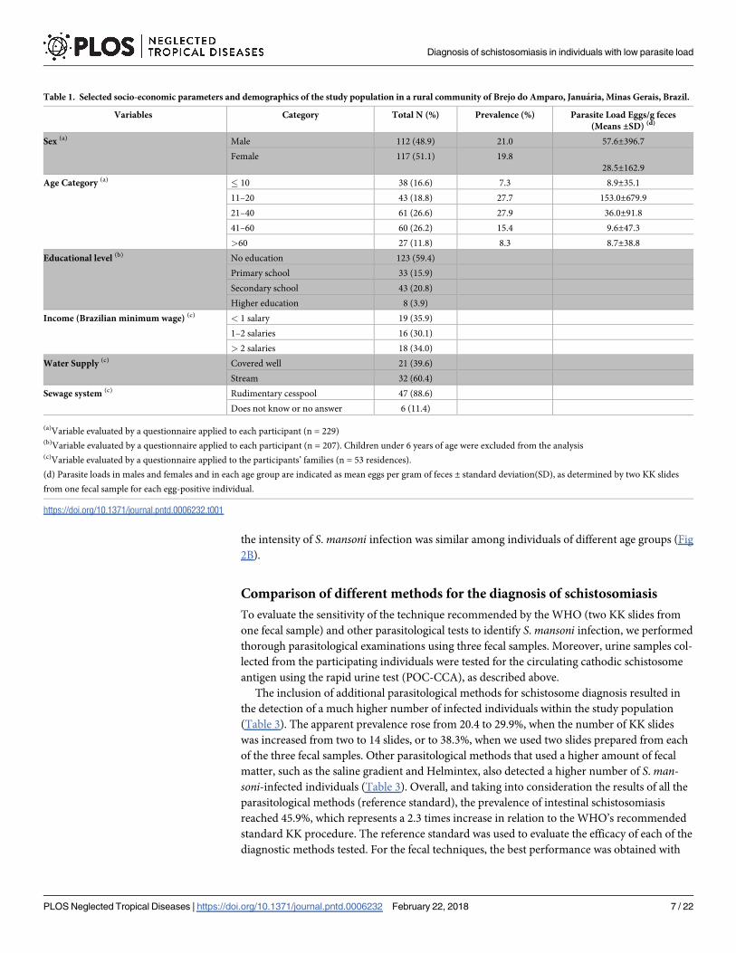
the intensity of S. mansoni infection was similar among individuals of different age groups (Fig
2B).
Comparison of different methods for the diagnosis of schistosomiasis
To evaluate the sensitivity of the technique recommended by the WHO (two KK slides from
one fecal sample) and other parasitological tests to identify S. mansoni infection, we performed
thorough parasitological examinations using three fecal samples. Moreover, urine samples col-
lected from the participating individuals were tested for the circulating cathodic schistosome
antigen using the rapid urine test (POC-CCA), as described above.
The inclusion of additional parasitological methods for schistosome diagnosis resulted in
the detection of a much higher number of infected individuals within the study population
(Table 3). The apparent prevalence rose from 20.4 to 29.9%, when the number of KK slides
was increased from two to 14 slides, or to 38.3%, when we used two slides prepared from each
of the three fecal samples. Other parasitological methods that used a higher amount of fecal
matter, such as the saline gradient and Helmintex, also detected a higher number of S. man-soni-infected individuals (Table 3). Overall, and taking into consideration the results of all the
parasitological methods (reference standard), the prevalence of intestinal schistosomiasis
reached 45.9%, which represents a 2.3 times increase in relation to the WHO’s recommended
standard KK procedure. The reference standard was used to evaluate the efficacy of each of the
diagnostic methods tested. For the fecal techniques, the best performance was obtained with
Table 1. Selected socio-economic parameters and demographics of the study population in a rural community of Brejo do Amparo, Januaria, Minas Gerais, Brazil.
Variables Category Total N (%) Prevalence (%) Parasite Load Eggs/g feces
(Means ±SD) (d)
Sex (a) Male 112 (48.9) 21.0 57.6±396.7
Female 117 (51.1) 19.8
28.5±162.9
Age Category (a) � 10 38 (16.6) 7.3 8.9±35.1
11–20 43 (18.8) 27.7 153.0±679.9
21–40 61 (26.6) 27.9 36.0±91.8
41–60 60 (26.2) 15.4 9.6±47.3
>60 27 (11.8) 8.3 8.7±38.8
Educational level (b) No education 123 (59.4)
Primary school 33 (15.9)
Secondary school 43 (20.8)
Higher education 8 (3.9)
Income (Brazilian minimum wage) (c) < 1 salary 19 (35.9)
1–2 salaries 16 (30.1)
> 2 salaries 18 (34.0)
Water Supply (c) Covered well 21 (39.6)
Stream 32 (60.4)
Sewage system (c) Rudimentary cesspool 47 (88.6)
Does not know or no answer 6 (11.4)
(a)Variable evaluated by a questionnaire applied to each participant (n = 229)(b)Variable evaluated by a questionnaire applied to each participant (n = 207). Children under 6 years of age were excluded from the analysis(c)Variable evaluated by a questionnaire applied to the participants’ families (n = 53 residences).
(d) Parasite loads in males and females and in each age group are indicated as mean eggs per gram of feces ± standard deviation(SD), as determined by two KK slides
from one fecal sample for each egg-positive individual.
https://doi.org/10.1371/journal.pntd.0006232.t001
Diagnosis of schistosomiasis in individuals with low parasite load
PLOS Neglected Tropical Diseases | https://doi.org/10.1371/journal.pntd.0006232 February 22, 2018 7 / 22

the modified Helmintex method, which identified schistosome eggs in feces of 88 individuals
(40.4% of prevalence). This parasitological method showed a high sensitivity (86.6%) and the
highest degree of concordance in relation to the reference standard (kappa = 0.84) (Table 3).
The analysis also demonstrated that the sensitivity of the KK method increased from 41.4%
with two slides from one fecal sample to up to 66.7% with six slides from three fecal samples.
In comparison, if only one fecal sample was processed, the sensitivity remained around 60%,
even when the number of examined slides was increased to 12 or 14 (Table 3 and Fig 3). The
improved performance of the KK method due to an increased number of examined slides (14
slides) or increased sampling effort (three fecal samples) is shown in Fig 3.
Fig 4 shows the prevalence of S. mansoni infection for the different age groups and as a
function of the parasitological methods, e.g. the standard KK method (2 slides from one fecal
sample) versus the reference standard (18 Kato-Katz slides, saline gradient, and Helmintex).
Using the reference standard, we found that children and young adults (11–20 years of age)
had the highest prevalence (55%) for S. mansoni infection. In contrast, the prevalence was
reduced to less than 50% in the other age groups, being further reduced in the elderly (older
than 60 years of age). Importantly, the prevalence found in each age group, considering the
combination of all parasitological exams (reference standard), was 1.7 to 4.7 times higher than
the prevalence obtained with the recommended two KK slides from one fecal sample (Fig 4).
Classification of infected individuals and performance of the different
parasitological methods according to the parasite load
The parasite load in S. mansoni infected individuals was determined by counting the eggs
found in two KK slides from one fecal sample and converting the counts in eggs per gram of
feces (EPG), according to standard procedures recommended by the WHO [14]. We assigned
an EPG value of 1 for the individuals who were not detected by two KK slides, but who were
Table 2. Prevalence of Schistosoma mansoni infection and other intestinal parasites in a rural community of the
Municipality of Januaria, Minas Gerais, Brazil.
Number of infected Prevalence (CI 95%)
Intestinal protozoa (n = 249) 85 34.1 (28.3–40.4)
Entamoeba coli 31 12.5 (8.6–17.2)
Endolimax nana 25 10.0 (6.6–14.5)
Blastocystis hominis 13 5.2 (2.8–8.8)
Entamoeba histolytica/díspar 9 3.6 (1.7–6.8)
Giardia lamblia 4 1.6 (0.4–4.1)
Iodamoeba butschlii 2 0.8 (0.1–2.9)
Entamoeba hartmani 1 0.4 (0.01–2.3)
Number of Infected Prevalence (CI 95%)
Helminths (n = 235) 81 34.5 (28.4–40.9)
Schistosoma mansoni� 48 20.4 (15.5–26.2)
Hookworm 23 9.8 (6.3–14.3)
Enterobius vermicularis 8 3.4 (1.5–6.6)
Strongyloides stercoralis 1 0.4 (0.01–2.3)
Trichuris trichiura 1 0.4 (0.01–2.3)
Coinfection (S. mansoni + Protozoa) 30 12.1 (8.6–16.7)
Coinfection (S. mansoni + other helminths) 11 4.7 (2.6–8.2)
� Positive for Schistosoma mansoni, as determined by two KK slides from one fecal sample.
https://doi.org/10.1371/journal.pntd.0006232.t002
Diagnosis of schistosomiasis in individuals with low parasite load
PLOS Neglected Tropical Diseases | https://doi.org/10.1371/journal.pntd.0006232 February 22, 2018 8 / 22

found positive when additional slides were analysed or when other fecal exams were used.
Thus, we classified 102 individuals with a light parasite load (EPG: 1–99), 12 individuals with a
moderate parasite load (EPG: 100–399), and four individuals with a heavy parasite load (EPG:
400 or more). Therefore, most of the infected individuals within the studied population had a
light parasitic infection.
Next, we analyzed the performance of the parasitological methods in relation to the parasite
load, with the individuals with a light infection being arbitrarily divided into three subgroups
(Table 4). All the diagnostic methods readily detected individuals with heavy to moderate
infections. On the other hand, the diagnostic methods decreased their sensitivity to detect
individuals with a low parasite load, especially in fecal samples with less than 12 EPG. In this
case, the best performance of the KK method (SPL1-3 K1-K2) reached a sensitivity of only
40%. In the group with a very low parasite load, the saline gradient and the rapid urine test had
sensitivities of 33.9 and 50.8%, respectively. The Helmintex method showed the highest sensi-
tivity for the group with very low parasite load (84.1%).
Fig 2. Classification of Schistosoma mansoni-infected individuals according to their parasitic load. A: Individual egg counts of one fecal
sample analyzed with two Kato-Katz slides and classification of egg-positive individuals according to their parasite load. Infection intensity was
determined by the number of eggs per gram of feces (EPG) and was classified as light (1–99 EPG, triangles), moderate (100–399 EPG, squares)
or heavy (�400 EPG, circles). Individual EPG values are plotted on a logarithmic scale and the horizontal bars indicate the mean EPG value in
each category. B: Boxplots showing the median, interquartile ranges, and 95% intervals of the parasite load (EPG) by the different age groups
and indicated on a logarithmic scale. Non-parametric Kruskal-Wallis test revealed no statistical significance between age groups (p> 0.05).
https://doi.org/10.1371/journal.pntd.0006232.g002
Table 3. Performance of different parasitological and immunochromatographic methods for the detection of intestinal schistosomiasis in comparison with the ref-
erence standard (18 Kato-Katz slides, saline gradient, and Helmintex).
Method Prevalence (%) Sensitivity % (CI 95%) Kappa Index (CI 95%)
SPL1 K1-K2 20.4 41.4 (32.8–50.5) 0.42 (0.31–0.52)
SLP1 K1-K6 29.0 56.4 (47.0–65.3) 0.56 (0.44–0.67)
SLP1 K1-K12 30.3 58.7 (49.3–67.5) 0.58 (0.46–0.70)
SLP1 K1-K14 29.9 58.2 (48.8–67.0) 0.58 (0.46–0.70)
SLP1-3 K1-K2 38.3 66.7 (57.3–74.9) 0.63 (0.50–0.76)
Saline Gradient 21.3 44.7 (35.0–54.7) 0.46 (0.34–0.58)
Helmintex 40.4 83.8 (75.6–89.6) 0.84 (0.71–0.97)
POC-CCA 47.4 64.9 (55.6–73.1) 0.34 (0.22–0.47)
Data shows the prevalence, sensitivity, and kappa index of concordance for the Kato-Katz technique obtained with the analysis of one fecal sample using two (SPL1
K1-K2), six (SPL1 K1-K6), 12 (SPL1 K1-K12), and 14 slides (SPL1 K1-K14), or obtained from two slides prepared from each of three fecal samples (SPL1-3 K1-K2), or
obtained with the saline gradient, Helmintex, or with POC-CCA methods.
https://doi.org/10.1371/journal.pntd.0006232.t003
Diagnosis of schistosomiasis in individuals with low parasite load
PLOS Neglected Tropical Diseases | https://doi.org/10.1371/journal.pntd.0006232 February 22, 2018 9 / 22

Performance of the POC-CCA as diagnostic method for intestinal
schistosomiasis
The POC-CCA identified 108 out of a total of 228 individuals as infected, which resulted in a
prevalence of 47.4% and a sensitivity of 64.9%, when compared with the reference standard
(18 Kato-Katz slides, saline gradient, and Helmintex). The sensitivity of the POC-CCA was
Fig 3. Prevalence of intestinal schistosomiasis and sensitivity of the Kato-Katz method, according to the number
of examined slides and stool samples. Prevalence of Schistosoma mansoni infection (white bars), as determined by the
analysis of one fecal sample with two (SPL1 K1-K2), six (SPL1 K1-K6), 12 (SPL1 K1-K12) or 14 slides (SPL1 K1-K14),
or obtained with the analysis of two slides prepared from each of three fecal samples (SPL1-3 K1-K2). The sensitivity of
the different numbers of Kato-Katz slides examined (black bars) was calculated in relation to the reference standard,
which included the combined results of 18 Kato-Katz slides, the saline gradient, and the Helmintex methods.
https://doi.org/10.1371/journal.pntd.0006232.g003
Fig 4. Prevalence profile of intestinal schistosomiasis in an endemic population divided by different age groups
according to the different parasitological methods. Black circles indicate the prevalence profile in the population
considering the sum of all parasitological methods used (reference standard: 18 Kato-Katz slides, saline gradient, and
Helmintex); grey squares indicate the prevalence profile considering the recommended two KK slides from one fecal
sample. Prevalence values (%) for each age group are indicated.
https://doi.org/10.1371/journal.pntd.0006232.g004
Diagnosis of schistosomiasis in individuals with low parasite load
PLOS Neglected Tropical Diseases | https://doi.org/10.1371/journal.pntd.0006232 February 22, 2018 10 / 22

superior to the saline gradient and comparable to the results obtained with six KK slides from
three fecal samples. However, the kappa index of the urine test was considerably lower than
that obtained with the other parasitological tests (Table 5).
The performance of the POC-CCA is illustrated in Fig 5. The visual scores ranged from
negative to trace, weak (+), moderate (++), and strongly positive (+++), with trace results con-
sidered a positive reaction for S. mansoni infection, as recommended by the manufacturer (Fig
5A). Comparing the POC-CCA result with the other parasitological analyses, we observed
that, of the 139 negative individuals in the parasitological tests, only 116 participants provided
urine samples for the POC-CCA test. Of these individuals, 81 (70%) were also found not reac-
tive (negative) in the urine test. However, 33 urine samples (28%) from the individuals found
negative by the other parasitological tests showed a trace reaction and another two samples
(1.7%) of parasitologically negative individuals had a weak positive result (+) (Fig 5B). Among
these 35 individuals, only four (11.4%) individuals had a hookworm infection and eight
(22.9%) individuals presented with protozoan cysts in their feces.
From 118 individuals found positive for S. mansoni eggs in any of the parasitological exams
(reference standard), 112 participants provided urine samples. Out of these 112 samples, 73
(65%) were tested positive for the circulating cathodic antigen of S. mansoni and were in
Table 4. Sensitivity of different diagnostic methods for the detection of intestinal schistosomiasis considering the parasite load, as defined by egg counts of two
Kato-Katz slides.
Classification by Parasite Load (EPG value)
Sensitivity (%) of each diagnostic method
Diagnostic Method Heavy
(EPG > 399)
% Sensitivity
Moderate
(EPG: 100–399)
% Sensitivity
Light
(EPG: 99–50)
% Sensitivity
(EPG: 49–12)
% Sensitivity
(EPG < 12)
% Sensitivity
SPL1 K1-K2 100 (04/04) 100 (12/12) 100 (04/04) 100 (28/28) 0 (0/70)
SPL1 K1-K6 100 (04/04) 100 (12/12) 100 (04/04) 100 (28/28) 22.6 (14/62)
SPL1 K1-K12 100 (04/04) 100 (11/11) 100 (04/04) 100 (28/28) 27.4 (17/62)
SPL1 K1-K14 100 (04/04) 100 (11/11) 100 (04/04) 100 (28/28) 27.0 (17/63)
SPL1-3 K1-K2 100 (04/04) 100 (12/12) 100 (04/04) 100 (28/28) 40.0 (24/60)
Saline Gradient 100 (04/04) 100 (06/06) 33.3 (01/03) 50.0 (11/22) 33.9 (20/59)
Helmintex 100 (03/03) 100 (11/11) 75.0 (03/04) 75.0 (18/24) 84.1 (53/63)
POC-CCA 100 (04/04) 100 (10/10) 100 (04/04) 77.8 (21/27) 50.8 (34/67)
Data show the sensitivity (%) of each diagnostic method and the number of individuals detected positive for intestinal schistosomiasis versus the total number of
examined individuals (in brackets), according to parasite load classification. Individuals with a light infection were arbitrarily divided into three subgroups with egg
counts of 99–50 eggs per gram of feces (EPG), 49–12 EPG, and less than 12 EPG.
https://doi.org/10.1371/journal.pntd.0006232.t004
Table 5. Performance of the rapid urine test for circulating cathodic antigen of Schistosoma mansoni (POC-CCA), as compared with the reference standard of a pos-
itive result in any of the used parasitological methods.
Method TP FP TN FN Prevalence %
(n positives/ n total)
Sensitivity %
(CI 95%)
Specificity %
(CI 95%)
PPV (%) NPV (%) Kappa
(CI 95%)
POC-CCA � 73 35 81 39 47.4
(n = 108 of 228)
64.9
(55.6–73.1)
69.2
(60.4–76.9)
66.7
(57.3–74.9)
67.5
(58.7–75.2)
0.34 (0.22–0.47)
POC-CCA�� 30 2 114 82 14.0
(n = 32 of 228)
26.8
(19.5–35.7)
98.3
(93.9–99.5)
93.8
(79.9–98.3)
58.2
(51.2–64.9)
0.25 (0.17–0.35)
The POC-CCA test was evaluated considering trace results as a positive result (�), as indicated by the manufacturer, or considering trace results as a negative result (��).
The data show the number of true positive (TP), false positive (FP), true negative (TN), and false negative (FN) individuals, as well as the prevalence (%), sensitivity,
specificity, and kappa index, together with the respective confidence intervals (CI), of concordance of each test.
https://doi.org/10.1371/journal.pntd.0006232.t005
Diagnosis of schistosomiasis in individuals with low parasite load
PLOS Neglected Tropical Diseases | https://doi.org/10.1371/journal.pntd.0006232 February 22, 2018 11 / 22

agreement with the results of the other parasitological exams. The results were classified as trace,
weak (+), medium (++), or strongly positive (+++) in 43 (38%), 17 (15%), 10 (9%), and three
(3%) of the examined urine samples, respectively (Fig 5B). In contrast, 39 urine samples (35%)
from egg-positive individuals were not reactive in the urine test and, therefore, were misclassified
as uninfected (false negatives). Interestingly, the mean EPG value from these missclassified indi-
viduals was considered as very low (mean EPG: 4.3; minimum: 1 EPG, maximum: 36 EPG).
Out of the 73 samples that were positive according to the reference standard and in the
POC-CCA, 59 (81%), 10 (14%), and four (5%) individuals were considered to have a light, moder-
ate or high parasite load, respectively. A significantly positive correlation was found between the
scores of the POC-CCA and intensity of infection, as determined by individual EPG values
(R = 0.537; p = 0.0001). The agreement between POC-CCA and the reference standard, as the sum
of all parasitological exams, showed a low concordance (κ = 0.34), which was even lower when
trace results in the urine test were considered as a negative result (κ = 0.25) (see also Table 5).
Combination of methods for an improved diagnosis of intestinal
schistosomiasis
Since the KK method is the recommended technique for the diagnosis of intestinal schistoso-
miasis [14], we compared the combination of more KK slides and fecal samples with the
Fig 5. Performance of the rapid urine test (POC-CCA) for the diagnosis of Schistosoma mansoni infection. A:
Photograph showing the different reactions possible with the POC-CCA: negative, trace, weak (+), medium (++) and
strong (+++). B: Distribution of the POC-CCA results in individuals from an endemic area classified as negative (n = 116)
or positive (n = 112) for S. mansoni infection by extensive parasitological testing (Reference standard: 18 Kato-Katz slides,
saline gradient, and Helmintex); total n = 228). Data indicate the percentages of POC-CCA reactivities in each group of
parasitologically negative or positive individuals. Red circles indicate discordant results in comparison with the reference
standard (false positive: 28 and 2%; or false negative: 35%).
https://doi.org/10.1371/journal.pntd.0006232.g005
Diagnosis of schistosomiasis in individuals with low parasite load
PLOS Neglected Tropical Diseases | https://doi.org/10.1371/journal.pntd.0006232 February 22, 2018 12 / 22
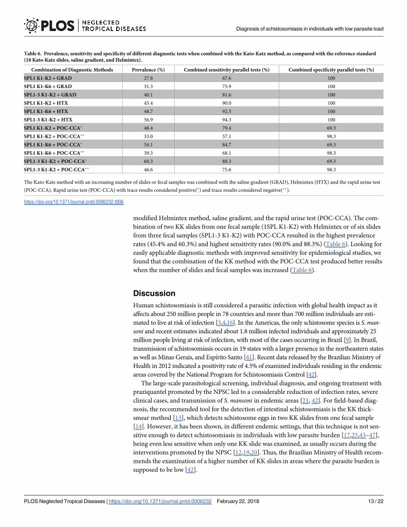
modified Helmintex method, saline gradient, and the rapid urine test (POC-CCA). The com-
bination of two KK slides from one fecal sample (1SPL K1-K2) with Helmintex or of six slides
from three fecal samples (SPL1-3 K1-K2) with POC-CCA resulted in the highest prevalence
rates (45.4% and 60.3%) and highest sensitivity rates (90.0% and 88.3%) (Table 6). Looking for
easily applicable diagnostic methods with improved sensitivity for epidemiological studies, we
found that the combination of the KK method with the POC-CCA test produced better results
when the number of slides and fecal samples was increased (Table 6).
Discussion
Human schistosomiasis is still considered a parasitic infection with global health impact as it
affects about 250 million people in 78 countries and more than 700 million individuals are esti-
mated to live at risk of infection [3,4,16]. In the Americas, the only schistosome species is S. man-soni and recent estimates indicated about 1.8 million infected individuals and approximately 25
million people living at risk of infection, with most of the cases occurring in Brazil [9]. In Brazil,
transmission of schistosomiasis occurs in 19 states with a larger presence in the northeastern states
as well as Minas Gerais, and Espırito Santo [41]. Recent data released by the Brazilian Ministry of
Health in 2012 indicated a positivity rate of 4.5% of examined individuals residing in the endemic
areas covered by the National Program for Schistosomiasis Control [42].
The large-scale parasitological screening, individual diagnosis, and ongoing treatment with
praziquantel promoted by the NPSC led to a considerable reduction of infection rates, severe
clinical cases, and transmission of S. mansoni in endemic areas [21, 42]. For field-based diag-
nosis, the recommended tool for the detection of intestinal schistosomiasis is the KK thick-
smear method [13], which detects schistosome eggs in two KK slides from one fecal sample
[14]. However, it has been shown, in different endemic settings, that this technique is not sen-
sitive enough to detect schistosomiasis in individuals with low parasite burden [17,25,43–47],
being even less sensitive when only one KK slide was examined, as usually occurs during the
interventions promoted by the NPSC [12,19,20]. Thus, the Brazilian Ministry of Health recom-
mends the examination of a higher number of KK slides in areas where the parasite burden is
supposed to be low [42].
Table 6. Prevalence, sensitivity and specificity of different diagnostic tests when combined with the Kato-Katz method, as compared with the reference standard
(18 Kato-Katz slides, saline gradient, and Helmintex).
Combination of Diagnostic Methods Prevalence (%) Combined sensitivity parallel tests (%) Combined specificity parallel tests (%)
SPL1 K1-K2 + GRAD 27.8 67.6 100
SPL1 K1-K6 + GRAD 31.3 75.9 100
SPL1-3 K1-K2 + GRAD 40.1 81.6 100
SPL1 K1-K2 + HTX 45.4 90.0 100
SPL1 K1-K6 + HTX 48.7 92.5 100
SPL1-3 K1-K2 + HTX 56.9 94.3 100
SPL1 K1-K2 + POC-CCA� 48.4 79.4 69.3
SPL1 K1-K2 + POC-CCA�� 33.0 57.1 98.3
SPL1 K1-K6 + POC-CCA� 54.1 84.7 69.3
SPL1 K1-K6 + POC-CCA�� 39.3 68.1 98.3
SPL1-3 K1-K2 + POC-CCA� 60.3 88.3 69.3
SPL1-3 K1-K2 + POC-CCA�� 46.6 75.6 98.3
The Kato-Katz method with an increasing number of slides or fecal samples was combined with the saline gradient (GRAD), Helmintex (HTX) and the rapid urine test
(POC-CCA). Rapid urine test (POC-CCA) with trace results considered positive(�) and trace results considered negative(��).
https://doi.org/10.1371/journal.pntd.0006232.t006
Diagnosis of schistosomiasis in individuals with low parasite load
PLOS Neglected Tropical Diseases | https://doi.org/10.1371/journal.pntd.0006232 February 22, 2018 13 / 22

In the present study, we tested the sensitivity of an increasing number of KK slides using up
to three fecal samples and compared its performance with other parasitological methods in an
area endemic for intestinal schistosomiasis. By performing thorough parasitological exams, we
aimed to get close to the ‘real’ picture of S. mansoni infections in this endemic region, which is
located within the area of action of the NSPC, but had not suffered any intervention in the two
years before the beginning of the present study. The diagnostic effort presented herein allowed
us to evaluate different parasitological methods in the light of a strong reference standard,
uniquely defined in this study.
Most of the population from the rural area studied herein had no adequate water supply
and sanitation. While waterborne protozoan infections were common, other intestinal hel-
minth infections were less frequent. S. mansoni infection was initially estimated to be 20.4%,
after examination of two KK slides from one fecal sample, which led the area to be classified as
with moderate risk of infection [48]. The classification of infected individuals according to
their parasite load [14] confirmed that two thirds of the initially diagnosed individuals har-
bored a light S. mansoni infection and less than 10% had a heavy infection. After performing
additional KK slides and other parasitological methods, the prevalence rose to 45.9%, which
represented a 2.3 times increase when compared with the initial exams and indicated nearly
half of the examined population as infected. As revealed by previous studies in areas of low
transmission of S. mansoni [12,19,20,25], the prevalence of infection for this parasite in the
area studied herein was largely underestimated when only the standard KK method was used.
The prevalence profile for intestinal schistosomiasis in different age groups revealed herein
matched that from other studies [4, 49,50]. However, if the standard method of two KK slides
was compared with our reference standard (18 KK slides + saline gradient + Helmintex), we
identified up to 4.7 times higher prevalences in the different age groups. This is in line with
results published previously [19, 20], showing that an increase in the number of examined KK
slides considerably augmented the number of egg-positive individuals. However, and this goes
beyond the already existing data on the evaluation and performance of multiple KK slides, we
showed that even using the superior version of the KK technique, which involves analyzing
two slides from three different fecal samples, we still missed more than one third of the
infected population.
Besides the KK technique, the other parasitological tests composing our reference standard
included a saline gradient using 500 mg of fecal matter from the first fecal sample [23] and per-
formed modified Helmintex method [38], which used up to 30 grams of feces. Using these
methods, a considerable number of additional egg-positive individuals were detected, with the
Helmintex method presenting the best performance and a sensitivity of over 80%. Initially, the
Helmintex method was described of being 30 times more sensitive than the standard KK
method [24], which is mainly due to the high amount of examined fecal matter, the successive
sieving and concentration processes and the separation and distinction of eggs by paramag-
netic beads and additional staining methods in the modified version [38]. A study investigat-
ing a low transmission area in the northeast of Brazil using the Helmintex method showed
similar results to ours published [51]. However, it has to be emphasized that, in the present set-
ting, none of the parasitological methods tested herein was able to detect eggs in every positive
sample. In this context it is interesting to note, that in seeding experiments with 30 grams of
feces, the recovery of schistosome eggs in fecal samples processed by the Helmintex method
was about 27%, only [38]. Further, it was not the aim of the study to evaluate and compare the
different methods in terms of applicability in field surveys, as well as operational, personnel,
and logistics and other factors that influence their implementation, as stated by others [19, 52–
54].
Diagnosis of schistosomiasis in individuals with low parasite load
PLOS Neglected Tropical Diseases | https://doi.org/10.1371/journal.pntd.0006232 February 22, 2018 14 / 22

An interesting alternative to the time consuming and labor intensive parasitological meth-
ods are rapid immunochromatographic tests for circulating antigens. Therefore, we included
the commercialized rapid urine test (POC-CCA) [28] in our study and evaluated it in compari-
son with our parasitological reference standard The POC-CCA has shown promising results
for the detection of intestinal schistosomiasis in various settings in Africa and Asia [32–34,
55,56]. When the S. mansoni egg-positive individuals tested herein were classified according to
their parasite load, the parasitological tests and the POC-CCA readily detected the individuals
with heavy or moderate infections. In contrast, all tests (parasitological and the POC-CCA)
showed reduced sensitivities when individuals with a low (99–12 EPG) or very low (less than
12 EPG) parasite load had to be detected. Especially in the case of very low parasite load, the
KK technique, at its best, only detected 40% of the infected individuals. In the case of the indi-
viduals with very low parasite load, the POC-CCA and the Helmintex methods showed the
best performance with sensitivities of more than 50 and 84%, respectively.
The rapid urine test (POC-CCA) has been successfully tested in different regions of Africa
and Asia [32,34, 56–58] and there are initiatives which favor this test for screening and map-
ping of intestinal schistosomiasis and improve transmission control and the elimination of
schistosomiasis [59,60]. However, the epidemiological situation of intestinal schistosomiasis in
most areas in Brazil is different from that found in many endemic settings in other tropical
countries. This is probably because the country has a national program for schistosomiasis
control since the 1970s with regular intervals of diagnosis and treatment rounds in endemic
populations. Data from the Brazilian Ministry of Health [42] and risk mapping of schistosomi-
asis in the country [61–63] indicated a considerable decrease in infection rates and high preva-
lence risk areas with ongoing interventions [42, 63]. However, these claims might be overly
optimistic since they are based on data from one KK slide from one fecal sample. In any case,
according to the government-published data, as a result of the NPSC interventions, the para-
site burden, significant morbidity, and mortality rates decreased during the last two decades
[42]. We evaluated the performance of POC-CCA and compared it with the reference standard
to detect S. mansoni infection in individuals of a community where NPSC’s interventions
including varying rounds of treatment had been promoted. The POC-CCA showed a sensitiv-
ity of approximately 65%, which is superior to that obtained with the saline gradient method,
comparable with the results obtained with KK variant using six KK slides from three fecal sam-
ples, and inferior to the sensitivity found with Helmintex, when the criteria of evaluation were
used as indicated by the manufacturer that is, if ‘trace’ was considered a positive result. A simi-
lar result for the sensitivity of POC-CCA and comparison with the performance of multiple
KK smears was obtained in an endemic area in Africa [64]. In our study, the main shortcom-
ing of the rapid urine test (POC-CCA) was a low concordance with the reference standard,
since we found 30 and 35% of false positive and false negative results, respectively. This low
concordance for the rapid urine test was not observed in other studies where parasitological
efforts for detection of schistosome eggs in feces were far less rigorous [35, 65]. Also, the dis-
crepancy might be partially explained by the discontinuous distribution of eggs in the fecal
matter, intermittent egg excretion, a small number of female worms, or by occult infections
with just one sex or aging worms. This is somewhat expected in elderly individuals since they
rarely visit contaminated water streams and are, therefore, less prone to reinfection
[32,43,46,66,67]. If ‘trace’ was not considered as positive, then the specificity increased to more
than 98%, but the sensitivity dropped to less than 27%, which we consider insufficient for a
screening method. In a recent study, the performance of POC-CCA was compared with that of
a KK test with two slides of one fecal sample, as recommended by WHO, and without further
extensive parasitological testing [65]. Even in that experimental setting, without a strong refer-
ence standard, the rapid urine test had a considerable percentage of false positive results, and
Diagnosis of schistosomiasis in individuals with low parasite load
PLOS Neglected Tropical Diseases | https://doi.org/10.1371/journal.pntd.0006232 February 22, 2018 15 / 22
that occurred even for individuals from an area considered as non-endemic for schistosomia-
sis. Additionally, 14% were classified as negative by the urine test, but these were proven to be
positive during parasitological exams [65]. Previous studies have reported cross-reactivity
between schistosomes and other intestinal helminths or other clinical conditions that can lead
to a false positive POC-CCA result [68–70]. However, we were not able to correlate any intesti-
nal protozoan or helminth infection with a ‘trace’ or positive POC-CCA result.
In order to improve the performance of POC-CCA test and elucidate the situation of indi-
viduals who were tested as ‘trace’, prior concentration of urine by lyophilization significantly
improved the concordance of the test in individuals with low parasite burden [70]. Also, recent
investigations on the specificity and sensitivity of methods for the detection of circulating
anodic antigen (CAA) from schistosomes seem to be even more promising [71–74].
To reach a maximum sensitivity and specificity and indicate alternatives for schistosomiasis
control programs, we tried to combine the standard KK method and different modifications of
this technique with the other parasitological methods or the POC-CCA. The best KK variant
tested herein (six slides prepared from three fecal samples) achieved a sensitivity of 82, 88, and
94% when combined with the saline gradient, POC-CCA or Helmintex methods, respectively.
Whether any of these scenarios is applicable to large-scale national control programs has to be
carefully evaluated, considering logistic and economic aspects [54]. In any case, maybe a first para-
sitological test has to be combined with a second more specific test for schistosomiasis in order to
join efforts against soil-transmitted helminthiasis and schistosomiasis [54,59,75]. Further, we
believe that in areas of low endemicity or low intensity infections, serology or molecular biology,
as proposed elsewhere [11,25,76–78], might be valuable alternatives to be included as additional
diagnostic procedures. We are currently investigating the performance of molecular biological
methods and serology in the parasitologically well-defined population studied herein.
In conclusion, we showed that in endemic areas of intestinal schistosomiasis with low-
intensity infections, the actual prevalence can be underestimated by up to 4.7 times when mea-
sured by the recommended standard procedure. The rigorous parasitological testing of three
fecal samples allowed us to evaluate parasitological and immunochromatographic methods for
diagnosis of infection with S. mansoni. The KK technique, even at its best was able to detect
only two-thirds of the infected individuals. The best sensitivity rate (over 80%) was achieved
with the Helmintex method. However, in its present form, Helmintex is not applicable for
large-scale screening due to the required sample size and the time-consuming sieving and sedi-
mentation processes [38], but might be an adequate reference standard or gold standard for
the evaluation of newly developed, field-based diagnostic tools. In addition, the performance
of the POC-CCA was in the range of the best KK variant (six slides from three fecal samples),
but a high number of individuals were not correctly diagnosed (false positive or false negative).
Furthermore, studies are underway, in order to re-evaluate the use of standard serological
methods and PCR-based detection of parasite DNA with our well-defined biological samples.
We believe that a combination of methods has to be implemented since the schistosomiasis
control programs in different regions of the world are moving from morbidity control towards
transmission control and elimination.
Supporting information
S1 Fig. Geographical localization of Minas Gerais State within Brazil (small red window)
and localization of the endemic area in the Municipality of Januaria, northern region of
Minas Gerais (zoom). Source: https://pt.wikipedia.org/wiki/Janu%C3%A1ria#/media/File:
MinasGerais_Municip_Januaria.svg.
(TIF)
Diagnosis of schistosomiasis in individuals with low parasite load
PLOS Neglected Tropical Diseases | https://doi.org/10.1371/journal.pntd.0006232 February 22, 2018 16 / 22

Acknowledgments
We would like to thank the people from the communities Pe da Serra, Tocantins, and Santana
for their collaboration and the warm reception during the field activities. We are also thankful
to the municipal government of Januaria for the logistic support during the field studies and to
Mr. Adailton Viana Bitencourt and the technicians from the Schistosomiasis Control Program
for their valuable help.
Author Contributions
Conceptualization: Maria Aparecida Gomes, Carlos Graeff-Teixeira, Martin Johannes Enk,
Paulo Marcos Zech Coelho, Mariangela Carneiro, Deborah Aparecida Negrão-Corrêa, Ste-
fan Michael Geiger.
Data curation: Warllem Junio Oliveira, Fernanda do Carmo Magalhães, Andressa Mariana
Saldanha Elias, Vanessa Normandio de Castro, Vivian Favero, Catieli Gobetti Lindholz,
Aureo Almeida Oliveira, Fernando Sergio Barbosa, Frederico Gil, Maria Aparecida Gomes,
Martin Johannes Enk, Mariangela Carneiro, Deborah Aparecida Negrão-Corrêa, Stefan
Michael Geiger.
Formal analysis: Warllem Junio Oliveira, Fernanda do Carmo Magalhães, Vanessa Norman-
dio de Castro, Vivian Favero, Catieli Gobetti Lindholz, Aureo Almeida Oliveira, Fernando
Sergio Barbosa, Frederico Gil, Maria Aparecida Gomes, Carlos Graeff-Teixeira, Martin
Johannes Enk, Mariangela Carneiro, Deborah Aparecida Negrão-Corrêa, Stefan Michael
Geiger.
Funding acquisition: Carlos Graeff-Teixeira, Martin Johannes Enk, Paulo Marcos Zech
Coelho, Deborah Aparecida Negrão-Corrêa, Stefan Michael Geiger.
Investigation: Warllem Junio Oliveira, Fernanda do Carmo Magalhães, Andressa Mariana
Saldanha Elias, Vanessa Normandio de Castro, Vivian Favero, Catieli Gobetti Lindholz,
Aureo Almeida Oliveira, Fernando Sergio Barbosa, Frederico Gil, Maria Aparecida Gomes,
Carlos Graeff-Teixeira, Martin Johannes Enk, Paulo Marcos Zech Coelho, Mariangela Car-
neiro, Deborah Aparecida Negrão-Corrêa, Stefan Michael Geiger.
Methodology: Warllem Junio Oliveira, Fernanda do Carmo Magalhães, Andressa Mariana
Saldanha Elias, Vanessa Normandio de Castro, Vivian Favero, Catieli Gobetti Lindholz,
Aureo Almeida Oliveira, Frederico Gil, Maria Aparecida Gomes, Carlos Graeff-Teixeira,
Martin Johannes Enk, Paulo Marcos Zech Coelho, Mariangela Carneiro, Deborah Apare-
cida Negrão-Corrêa, Stefan Michael Geiger.
Project administration: Carlos Graeff-Teixeira, Martin Johannes Enk, Paulo Marcos Zech
Coelho, Deborah Aparecida Negrão-Corrêa, Stefan Michael Geiger.
Resources: Carlos Graeff-Teixeira, Paulo Marcos Zech Coelho, Mariangela Carneiro, Deborah
Aparecida Negrão-Corrêa, Stefan Michael Geiger.
Supervision: Maria Aparecida Gomes, Carlos Graeff-Teixeira, Martin Johannes Enk, Paulo
Marcos Zech Coelho, Mariangela Carneiro, Deborah Aparecida Negrão-Corrêa, Stefan
Michael Geiger.
Validation: Warllem Junio Oliveira, Fernanda do Carmo Magalhães, Andressa Mariana Salda-
nha Elias, Vanessa Normandio de Castro, Vivian Favero, Catieli Gobetti Lindholz, Aureo
Almeida Oliveira, Fernando Sergio Barbosa, Frederico Gil, Carlos Graeff-Teixeira, Martin
Diagnosis of schistosomiasis in individuals with low parasite load
PLOS Neglected Tropical Diseases | https://doi.org/10.1371/journal.pntd.0006232 February 22, 2018 17 / 22

Johannes Enk, Mariangela Carneiro, Deborah Aparecida Negrão-Corrêa, Stefan Michael
Geiger.
Visualization: Paulo Marcos Zech Coelho, Stefan Michael Geiger.
Writing – original draft: Warllem Junio Oliveira, Maria Aparecida Gomes, Carlos Graeff-
Teixeira, Martin Johannes Enk, Paulo Marcos Zech Coelho, Deborah Aparecida Negrão-
Corrêa, Stefan Michael Geiger.
Writing – review & editing: Fernanda do Carmo Magalhães, Carlos Graeff-Teixeira, Martin
Johannes Enk, Paulo Marcos Zech Coelho, Mariangela Carneiro, Deborah Aparecida
Negrão-Corrêa, Stefan Michael Geiger.
References1. Hotez PJ, Bottazzi ME, Franco-Paredes C, Ault SK, Periago MR. The neglected tropical diseases of
Latin America and the Caribbean: A review of disease burden and distribution and a roadmap for control
and elimination. PLoS Negl Trop Dis. 2008; 2(9):e300. https://doi.org/10.1371/journal.pntd.0000300
PMID: 18820747
2. Gryseels B, Polman K, Clerinx J, Kestens L. Human Schistosomiasis. Lancet. 2006; 368 (9541):1106–
1118. https://doi.org/10.1016/S0140-6736(06)69440-3 PMID: 16997665
3. Steinmann P, Keiser J, Bos R, Tanner M, Utzinger J. Schistosomiasis and water resources develop-
ment: systematic review, meta-analysis, and estimates of people at risk. Lancet Infect Dis. 2006; 6
(7):411–425. https://doi.org/10.1016/S1473-3099(06)70521-7 PMID: 16790382
4. Colley DG, Bustinduy AL, Secor WE, King CH. Human schistosomiasis. Lancet. 2014; 383(9936):
2253–2264. https://doi.org/10.1016/S0140-6736(13)61949-2 PMID: 24698483
5. van der Werf MJ, de Vlas SJ, Brooker S, Looman CW, Nagelkerke NJ, Habbema JD, et al. Quantifica-
tion of clinical morbidity associated with schistosome infection in sub-Saharan Africa. Acta Trop. 2003;
86(2–3): 125–139. https://doi.org/10.1016/S0001-706X(03)00029-9 PMID: 12745133
6. Katz N, Peixoto SV. Critical analysis of the estimated number of Schistosomiasis mansoni carriers in
Brazil. Rev Soc Bras Med Trop. 2000; 33(3):303–8. https://doi.org/10.1590/S0037-
86822000000300009 PMID: 10967599
7. Passos ADC, Amaral RS. Esquistossomose mansonica: aspectos epidemiologicos e de controle. Epi-
demiological and control aspects of schistosomiasis in Brazilian endemic areas. Rev Soc Bras Med
Trop.1998; 31 (2 Suppl): S61–74.
8. Scholte RG, Gosoniu L, Malone JB, Chammartin F, Utzinger J, Vounatsou P. Predictive risk mapping of
schistosomiasis in Brazil using Bayesian geostatistical models. Acta Trop. 2014; 132: 57–63. https://
doi.org/10.1016/j.actatropica.2013.12.007 PMID: 24361640
9. Noya O, Katz N, Pontier JP, Theron A, Noya BA. Schistosomiasis in America. In: Franco-Paredes C,
Santos-Preciado JI, editors. Neglected tropical diseases: Latin America and the Caribbean. Viena:
Springer; 2015. pp. 11–43.
10. Rabello A, Pontes LA, Enk MJ, Montenegro SML, De Morais CNL. Diagnostico parasitologico, imunolo-
gico e molecular da esquistossomose mansoni. In: Carvalho OS, Coelho PMZ, editors. Schistosoma
mansoni & Esquistossomose: Uma visão multidisciplinar. Rio de Janeiro: Fiocruz;2008. pp. 895–926.
11. Grenfell R, Harn DA, Tundup S, Da’Dara A, Siqueira L, Coelho PMZ. New approaches with different
types of circulating cathodic antigen for the diagnosis of patients with low Schistosoma mansoni load.
PloS Negl Trop Dis. 2013; 7(2):e2054. https://doi.org/10.1371/journal.pntd.0002054 PMID: 23469295
12. Siqueira LMV, Gomes LI, Oliveira E, Oliveira ER, Oliveira AA, Enk MJ, et al. Evaluation of parasitologi-
cal and molecular techniques for the diagnosis and assessment of cure of schistosomiasis mansoni in a
low transmission area. Mem Inst Oswaldo Cruz. 2015; 110:209–214. https://doi.org/10.1590/0074-
02760140375 PMID: 25946244
13. Katz N, Chaves A, Pellegrino JA. A simple device for quantitative stool thick-smear technique in schisto-
somiasis mansoni. Rev Soc Bras Med Trop. 1972; 14 (6): 397–400.
14. World Health Organization (2002). Prevention and Control of Schistosomiasis and Soil-Transmitted
Helminthiasis. Technical Series Report 912. Available: http://whqlibdoc.who.int/trs/WHO_TRS_912.
pdf.
15. Doenhoff MJ, Chiodini PL, Hamilton JV. Specific and sensitive diagnosis of schistosome infection: can it
be done with antibodies? Trends Parasitol. 2004; 20:35–39. https://doi.org/10.1016/j.pt.2003.10.019
PMID: 14700588
Diagnosis of schistosomiasis in individuals with low parasite load
PLOS Neglected Tropical Diseases | https://doi.org/10.1371/journal.pntd.0006232 February 22, 2018 18 / 22

16. World Health Organization. Schistosomiasis: population requiring preventive chemotherapy and num-
ber of people treated in 2010. Wkly Epidemiol Rec. 2012; 87: 37–44. PMID: 22308580
17. de Vlas SJ, Gryseels B. Underestimation of Schistosoma mansoni prevalences. Parasitol Today. 1992;
8 (8):274–77. https://doi.org/10.1016/0169-4758(92)90144-Q PMID: 15463638
18. Gryseels B. Uncertainties in the epidemiology and control of schistosomiasis. Am J Trop Med
Hyg.1996; 55 (5 Suppl): S103–108.
19. Enk MJ, Lima ACL, Drummond SC, Schall VT, Coelho PMZ. The effect of the number of stool samples
on the observed prevalence and the infection intensity with Schistosoma mansoni among a population
in an area of low transmission. Acta Trop. 2008; 108 (2–3):222–228. https://doi.org/10.1016/j.
actatropica.2008.09.016 PMID: 18973744
20. Siqueira LM, Coelho PM, Oliveira AA, Massara CL, Carneiro NF, Lima AC, et al. Evaluation of two
coproscopic techniques for the diagnosis of schistosomiasis in a low-transmission area in the state of
Minas Gerais, Brazil. Mem Inst Oswaldo Cruz. 2011; 106(7):844–50. https://doi.org/10.1590/S0074-
02762011000700010 PMID: 22124557
21. Amaral RS, Tauil PL, Lima DD, Engels D. An analysis of the impact of the Schistosomiasis Control Pro-
gramme in Brazil. Mem. Inst. Oswaldo Cruz. 2006; 101(1 Suppl): S79–85.
22. Sarvel AK, Oliveira AA, Silva AR, Lima AC, Katz N. Evaluation of a 25-year-program for the control of
schistosomiasis mansoni in an endemic area in Brazil. PLoS Negl Trop Dis. 2011; 5(3):e990. https://
doi.org/10.1371/journal.pntd.0000990 PMID: 21423644
23. Coelho PMZ, Jurberg A, Oliveira AA, Katz N. Use of a saline gradient for the diagnosis of schistosomia-
sis. Mem Inst Oswaldo Cruz. 2009; 104 (5): 720–723. https://doi.org/10.1590/S0074-
02762009000500010 PMID: 19820832
24. Teixeira CF, Neuhauss E, Bem R, Romanzini J, Graeff-Teixeira C. Detection of Schistosoma mansoni
eggs in feces through their interaction with paramagnetic beads in a magnetic field. PLoS Negl Trop
Dis. 2007; 14; 1(2):e73. https://doi.org/10.1371/journal.pntd.0000073
25. Goncalves MML, Barreto MGM, Peralta RHS, Gargioni C, Goncalves T, Igreja RP, et al. Immunoassays
as an auxiliary tool for the serodiagnosis of Schistosoma mansoni infection in individuals with low inten-
sity of egg elimination. Acta Trop. 2006; 100 (1–2): 24–30. https://doi.org/10.1016/j.actatropica.2006.
09.004 PMID: 17069742
26. Jin YM, Lu K, Zhou WF, Fu ZQ, Liu JM, Shi YJ, et al. Comparison of recombinant proteins from Schisto-
soma japonicum for schistosomiasis diagnosis. Clin Vaccine Immunol. 2010; 17:476–480. https://doi.
org/10.1128/CVI.00418-09 PMID: 20053872
27. Espırito-Santo MC, Alvarado-Mora MV, Dias-Neto E, Botelho-Lima LS, Moreira JP, Amorim M, et al.
Evaluation of real-time PCR assay to detect Schistosoma mansoni infections in a low endemic setting.
BMC Infect Dis. 2014; 14:558. https://doi.org/10.1186/s12879-014-0558-4 PMID: 25338651
28. van Dam GJ, Wichers JH, Ferreira TMF, Ghati D, van Amerongen A, Deelder AM. Diagnosis of schisto-
somiasis by reagent strip test for detection of circulating cathodic antigen. J Clin Microbiol. 2004; 42
(12):5458–5461. https://doi.org/10.1128/JCM.42.12.5458-5461.2004 PMID: 15583265
29. De Clercq D, Sacko M, Vercruysse J, Bussche V, Landoure A, Diarra A, et al. Circulating anodic and
cathodic antigen in serum and urine of mixed Schistosoma haematobium and S. mansoni infections in
Office du Niger, Mali. Trop Med Int Health.1997; 2 (7): 680–685. https://doi.org/10.1046/j.1365-3156.
1997.d01-354.x PMID: 9270735
30. Disch J, Garcia MM, Krijger GW, Amorim MN, Katz N, Deelder AM, et al. Daily fluctuation of levels of cir-
culating cathodic antigen in urine of children with Schistosoma mansoni infection in Brazil. Trans R Soc
Trop Med Hyg.1997; 91 (2):222–225. https://doi.org/10.1016/S0035-9203(97)90233-9. PMID:
9196777
31. Polman K, Engels D, Fathers L, Deelder AM, Gryseels B. Day-to-day fluctuation of schistosome circu-
lating antigen levels in serum and urine of humans infected with Schistosoma mansoni in Burundi. Am J
Trop Med Hyg. 1998; 59(1):150–154. https://doi.org/10.4269/ajtmh.1998.59.150 PMID: 9684644
32. Stothard JR, Kabatereine NB, Tukahebwa EM, Kazibwe F, Rollinson D, Mathieson W, et al. Use of cir-
culating cathodic antigen (CCA) dipsticks for detection of intestinal and urinary schistosomiasis. Acta
Trop. 2006; 97(2): 219–228. https://doi.org/10.1016/j.actatropica.2005.11.004 PMID: 16386231
33. Standley CJ, Lwambo NJ, Lange CN, Kariuki HC, Adriko M, Stothard JR. Performance of circulating
cathodic antigen (CCA) urine-dipsticks for rapid detection of intestinal schistosomiasis in schoolchildren
from shoreline communities of Lake Victoria. Parasit Vectors. 2010; 3(1):7. https://doi.org/10.1186/
1756-3305-3-7 PMID: 20181101
34. Colley DG, Binder S, Campbell C, King CH, Tchuem Tchuente LA, N’Goran EK, et al. A five-country
evaluation of a point-of-care circulating cathodic antigen urine assay for the prevalence of Schistosoma
mansoni. Am J Trop Med Hyg. 2013; 88(3):426–432. https://doi.org/10.4269/ajtmh.12-0639 PMID:
23339198
Diagnosis of schistosomiasis in individuals with low parasite load
PLOS Neglected Tropical Diseases | https://doi.org/10.1371/journal.pntd.0006232 February 22, 2018 19 / 22

35. Coulibaly JT, N’Goran EK, Utzinger J, Doenhoff MJ, Dawson EM. A new rapid diagnostic test for detec-
tion of anti-Schistosoma mansoni and anti-Schistosoma haematobium antibodies. Parasit Vectors.
2013; 6:29. https://doi.org/10.1186/1756-3305-6-29 PMID: 23360734
36. Kittur N, Castleman JD, Campbell CH Jr, King CH, Colley DG. Comparison of Schistosoma mansoni
prevalence and intensity of infection, as determined by the circulating cathodic antigen urine assay or
by the Kato-Katz fecal assay: A systematic review. Am J Trop Med Hyg. 2016; 94(3):605–10. https://
doi.org/10.4269/ajtmh.15-0725 PMID: 26755565
37. Hoffman WA, Pons JA, Janer JL. The sedimentation-concentration method in schistosomiasis mansoni.
PR J Publ Hlth Trop Med. 1934; 9: 283–298.
38. Favero V, Candido RR, Verissimo CM, Jones MK, Pierre TG, Lindholz CG, et al. Optimization of the
Helmintex method for schistosomiasis diagnosis. Exp Parasitol. 2017; 177: 28–34. https://doi.org/10.
1016/j.exppara.2017.04.001 PMID: 28431921
39. Midzi N, Butterworth AE, Mduluza T, Munyati S, Deelder AM, van Dam GJ. Use of circulating cathodic
antigen strips for the diagnosis of urinary schistosomiasis. Trans R Soc Trop Med Hyg.2009; 103
(1):45–51. https://doi.org/10.1016/j.trstmh.2008.08.018 PMID: 18951599
40. Landis JR, Koch GG. The measurement of observer agreement for categorical data. Biometrics. 1977;
33(1): 159–174. https://doi.org/10.2307/2529310 PMID: 843571
41. Coura JR, Amaral RS. Epidemiological and control aspects of schistosomiasis in Brazilian endemic
areas. Mem Inst Oswaldo Cruz 2004; 99 (5 Suppl):S13–19. https://doi.org//S0074-
02762004000900003
42. BRASIL. Secretaria de Vigilancia em Saude (2014). Vigilancia da esquistossomose mansoni: diretrizes
tecnicas. 4. ed. Ministerio da Saude do Brasil, Brasılia, p.146, 2014. Available: http://bvsms.saude.gov.
br/bvs/publicacoes/vigilancia_esquistossome_mansoni_diretrizes_tecnicas.pdf.
43. Engels D, Sinzinkayo E, Gryseels B. Day-to-day egg count fluctuation in Schistosoma mansoni infection
and its operational implications. Am J Trop Med Hyg.1996; 54(4):319–24. PMID: 8615440
44. De Vlas SJ, Engels D, Rabello AL, Oostburg BF, van Lieshout L, Polderman AM, et al. Validation of a
chart to estimate true Schistosoma mansoni prevalences from simple egg counts. Parasitology. 1997;
114 (2): 113–121. https://doi.org/10.1017/S0031182096008207
45. Utzinger JN, Goran EK, Dri A, Lengeler C, Tanner M. Efficacy of praziquantel against Schistosoma
mansoni with particular consideration for intensity of infection. Trop Med Int Health. 2000; 5(2): 771–
778. https://doi.org/10.1046/j.1365-3156.2000.00646.x
46. Berhe N, Medhin G, Erko B, Smith T, Gedamu S, Bereded D, et al. Variations in helminth faecal egg
counts in Kato–Katz thick smears and their implications in assessing infection status with Schistosoma
mansoni. Acta Trop. 2004; 92 (3): 205–212. https://doi.org/10.1016/j.actatropica.2004.06.011 PMID:
15533288
47. Lin DD, Liu JX, Liu YM, Hu F, Zhang YY, Xu JM, et al. Routine Kato–Katz technique underestimates the
prevalence of Schistosoma japonicum: A case study in an endemic area of the People’s Republic of
China. Parasitol Int. 2008; 57 (3): 281–286. https://doi.org/10.1016/j.parint.2008.04.005 PMID:
18485807
48. World Health Organization. Schistosomiasis: population requiring preventive chemotherapy and num-
ber of people treated in 2010. Wkly Epidemiol Rec. 2012; 87(4): 37–44. PMID: 22308580
49. Coura-Filho P, Farah MW, Rezende FD, Lamartine SS, Carvalho OS, Katz N. Environmental and social
determinants in schistosomiasis mansoni in Ravena, Minas Gerais, Brazil. Cad Saude Publica. 1995;
11 (2): 254–265. PMID: 14528332
50. Fleming FM, Brooker S, Geiger SM, Caldas IR, Correa-Oliveira R, Hotez PJ, et al. Synergistic associa-
tions between hookworm and other helminth species in a rural community in Brazil. Trop Med Int Health.
2006: 11(1):56–64. https://doi.org/10.1111/j.1365-3156.2005.01541.x PMID: 16398756
51. Pinheiro MC, Carneiro TR, Hanemann AL, Oliveira SM, Bezerra FS. The combination of three faecal
parasitological methods to improve the diagnosis of schistosomiasis mansoni in a low endemic setting
in the state of Ceara, Brazil. Mem Inst Oswaldo Cruz. 2012; 107(7):873–876. https://doi.org/10.1590/
S0074-02762012000700006 PMID: 23147142
52. Teles HM, Ferreira CS, Carvalho ME, Zacharias F, Magalhães LA. The efficiency of the examination of
compressed fecal samples for Schistosoma mansoni eggs. Rev Soc Bras Med Trop. 2003; 36(4):503–
507. https://doi.org/10.1590/S0037-86822003000400012 PMID: 12937729
53. Speich B, Knoop S, Mohammed KA, Komis IS, Rinald L, Cringoli G, et al. Comparative cost assessment
of the Kato-Katz and FLOTAC techniques for soil-transmitted helminth diagnosis in epidemiological sur-
veys. Parasit Vectors. 2010; 3(71). https://doi.org/10.1186/1756-3305-3-71
54. Turner HC, Betis AA, Dunn JC, Whitton JM, Hollingsworth TD, Fleming FM, et al. Economic consider-
ations for moving beyond the Kato-Katz technique for diagnosing intestinal parasites as we move
Diagnosis of schistosomiasis in individuals with low parasite load
PLOS Neglected Tropical Diseases | https://doi.org/10.1371/journal.pntd.0006232 February 22, 2018 20 / 22

towards elimination. Trends Parasitol. 2017; 33(6): 435–443. https://doi.org/10.1016/j.pt.2017.01.007
PMID: 28187989
55. Sousa-Figueiredo JC, Betson M, Kabatereine NB, Stothard JR. The urine circulating cathodic antigen
(CCA) dipstick: a valid substitute for microscopy for mapping and point-of-care diagnosis of intestinal
schistosomiasis. PLoS Negl Trop Dis. 7 (1): e2008. https://doi.org/10.1371/journal.pntd.0002008
PMID: 23359826
56. Cai YC, Xu JF, Steinmann P, Chen SH, Chu YH, Tian LG, et al. Field comparison of circulating antibody
assays versus circulating antigen assays for the detection of schistosomiasis japonica in endemic areas
of China. Parasit Vectors. 2014. 7: 138. https://doi.org/10.1186/1756-3305-7-138 PMID: 24684924
57. Stothard JR, Webster BL, Weber T, Nyakaana S, Webster JP, Kazibwe F, et al. Molecular epidemiology
of Schistosoma mansoni in Uganda: DNA barcoding reveals substantive genetic diversity within Lake
Albert and Lake Victoria populations. Parasitology. 2009; 136:1–12. https://doi.org/10.1017/
S0031182008005167
58. Tchuem Tchuente LA, Fouodo CJ, Ngassam RI, Sumo L, Noumedem CD, Kenfack CM, et al. Evalua-
tion of circulating cathodic antigen (CCA) urine-tests for diagnosis of Schistosoma mansoni infection in
Cameroon. PloS Negl Trop Dis. 2012; 6(7):e1758. https://doi.org/10.1371/journal.pntd.0001758 PMID:
22860148
59. Hawkins KR, Cantera JL, Storey HL, Leader BT, Santos T. Diagnostic tests to support late-stage control
programs for schistosomiasis and soil-transmitted helminthiases. PLoS Negl Trop Dis. 2016; 10(12):
e0004985. https://doi.org/10.1371/journal.pntd.0004985 PMID: 28005900
60. Tchuem Tchuente LA, Rollinson D, Stothard JR, Molyneux D. Moving from control to elimination of
schistosomiasis in sub-Saharan Africa: time to change and adapt strategies. Infect Dis Poverty. 2017;
6 (42). https://doi.org/10.1186/s40249-017-0256-8
61. Guimarães RJ, Freitas CC, Dutra LV, Scholte RG, Martins-Bede FT, Fonseca FR, et al. A geoproces-
sing approach for studying and controlling schistosomiasis in the state of Minas Gerais, Brazil. Mem
Inst Oswaldo Cruz. 2010; 105(4):524–31. https://doi.org/10.1590/S0074-02762010000400030 PMID:
20721503
62. Guimarães RJ, Freitas CC, Dutra LV, Moura AC, Amaral RS, Drummond SC, et al. Schistosomiasis
risk estimation in Minas Gerais State, Brazil, using environmental data and GIS techniques. Acta Trop.
2008; 108(2–3):234–41. https://doi.org/10.1016/j.actatropica.2008.07.001 PMID: 18692017
63. Scholte RG, Gosoniu L, Malone JB, Chammartin F, Utzinger J, Vounatsou P.Predictive risk mapping of
schistosomiasis in Brazil using Bayesian geostatistical models. Acta Trop. 2014; 132: 57–63. https://
doi.org/10.1016/j.actatropica.2013.12.007 PMID: 24361640
64. Lamberton PH, Kabatereine NB, Oguttu DW, Fenwick A, Webster JP. Sensitivity and specificity of multi-
ple Kato-Katz thick smears and a circulating cathodic antigen test for Schistosoma mansoni diagnosis
pre- and post-repeated-praziquantel treatment. PLoS Negl Trop Dis. 2014; 8(9):e3139. https://doi.org/
10.1371/journal.pntd.0003139 PMID: 25211217
65. Silveira AMS, Costa EGD, Ray D, Suzuki BM, Hsieh MH, Fraga LA, et al. Evaluation of the CCA
immuno-chromatographic test to diagnose Schistosoma mansoni in Minas Gerais State, Brazil. PLoS
Negl Trop Dis. 2016; 10(1): e0004357. https://doi.org/10.1371/journal.pntd.0004357 PMID: 26752073
66. Barreto ML, Smith DH, Sleigh AC. Implications of faecal egg count variation when using the Kato-Katz
method to assess Schistosoma mansoni infections. Trans R Soc Trop Med Hyg. 1990; 84 (4): 545–555.
67. Rabelo ALT. Revisão crıtica dos metodos de diagnostico da esquistossomose mansoni. Rev Soc Bras
Med Trop. 1994; 27(4 Suppl): 177–180.
68. Van Dam GJ, Bergwerff AA, Thomas-Oates JE, Rotmano JP, Kamerling JP, Vliegenthard JF, et al. The
immunologically reactive O-linked polysaccharide chains derived from circulating cathodic antigen iso-
lated from the human blood fluke Schistosoma mansoni have Lewis x as repeating unit. Eur J Bio-
chem.1994; 225(1):467–482. https://doi.org/10.1111/j.1432-1033.1994.00467.x PMID: 7925469
69. van Dam GJ, Claas FH, Yazdanbakhsh M, Kruize YC, van Keulen AC, Ferreira ST, et al. Schistosoma
mansoni excretory circulating cathodic antigen shares Lewis-x epitopes with a human granulocyte sur-
face antigen and evokes host antibodies mediating complement-dependent lysis of granulocytes.
Blood. 1996; 88(11):4246–4251. PMID: 8943860
70. Coelho PM, Siqueira LM, Grenfell RF, Almeida NB, Katz N, Almeida A, et al. Improvement of POC-CCA
interpretation by using lyophilization of urine from patients with Schistosoma mansoni low worm burden:
towards an elimination of doubts about the concept of trace. PLoS Negl Trop Dis. 2016; 10(6):
e0004778. https://doi.org/10.1371/journal.pntd.0004778 PMID: 27326453
71. Corstjens PL, van Lieshout L, Zuiderwijk M, Kornelis D, Tanke HJ, Deelder AM, et al. Up-converting
phosphor technology-based lateral flow assay for detection of Schistosoma circulating anodic antigen
in serum. J Clin Microbiol. 2008; 46(1): 171–176. https://doi.org/10.1128/JCM.00877-07 PMID:
17942645
Diagnosis of schistosomiasis in individuals with low parasite load
PLOS Neglected Tropical Diseases | https://doi.org/10.1371/journal.pntd.0006232 February 22, 2018 21 / 22

72. van Dam GJ, de Dood CJ, Lewis M, Deelder AM, van Lieshout L, Tanke HJ, et al. A robust dry reagent
lateral flow assay for diagnosis of active schistosomiasis by detection of Schistosoma circulating anodic
antigen. Exp Parasitol. 2013; 135(2):274–282. https://doi.org/10.1016/j.exppara.2013.06.017 PMID:
23850995
73. Corstjens PL, De Dood CJ, Kornelis D, Wilson RA, Kauriuki TM, Nyakundi RK, et al. Tools for diagnosis,
monitoring and screening of Schistosoma infections utilizing lateral-flow based assays and upconvert-
ing phosphor labels. Parasitology. 2014; 141(14): 1841–1855. https://doi.org/10.1017/
S0031182014000626 PMID: 24932595
74. van Dam GJ, Xu J, Bergquist R, de Dood CJ, Utzinger J, Quin ZQ, et al. An ultra-sensitive assay target-
ing the circulating anodic antigen for the diagnosis of Schistosoma japonicum in a low-endemic area,
People’s Republic of China. Acta Trop. 2015; 141:190–197. https://doi.org/10.1016/j.actatropica.2014.
08.004 PMID: 25128703
75. Lo NC, Lai YS, Dimitrios-Alexios KV, Bogoch I, Coulibaly JT, Bendavid E, et al. Assessment of global
guidelines for preventive chemotherapy against schistosomiasis and soil-transmitted helminthiasis: a
cost-effectiveness modelling study. Lancet Infect Dis. 2016; 16(9): 1065–1075. https://doi.org/10.1016/
S1473-3099(16)30073-1 PMID: 27286968
76. Alarcon de Noya B, Ruiz R, Losada S, Colmenares C, Contreras R, Cesari IM, et al. Detection of schis-
tosomiasis cases in low-transmission areas based on coprologic and serologic criteria: The Venezuelan
experience. Acta Trop. 2007: 103 (1): 41–49. https://doi.org/10.1016/j.actatropica.2007.04.018 PMID:
17606217
77. Cavalcanti MG, Silva LF, Peralta RHS, Barreto MG, Peralta JM. Schistosomiasis in areas of low
endemicity: a new era in diagnosis. Trends Parasitol 2013; 29 (2): 75–82. https://doi.org/10.1016/j.pt.
2012.11.003 PMID: 23290589
78. Stothard JR, Stanton MC, Bustinduy AL, Sousa-Figueiredo JC, van Dam GJ, Betson M, et al. Diagnos-
tics for schistosomiasis in Africa and Arabia: A review of present options in control and future needs for
elimination. Parasitology. 2014; 141(14):1947–61. https://doi.org/10.1017/S0031182014001152
PMID: 25158604
Diagnosis of schistosomiasis in individuals with low parasite load
PLOS Neglected Tropical Diseases | https://doi.org/10.1371/journal.pntd.0006232 February 22, 2018 22 / 22