evaluation of dat positive aiha cases by elution study at
TRANSCRIPT

Corresponding author: Kavitha G Department of Transfusion Medicine, Madras Medical College, Chennai, India.
Copyright © 2021 Author(s) retain the copyright of this article. This article is published under the terms of the Creative Commons Attribution Liscense 4.0.
Evaluation of DAT positive AIHA cases by elution study at a tertiary care centre in South India
Kavitha G 1, *, Latha B 1 and Swathandran Hamsavardhini 2
1 Department of Transfusion Medicine, Madras Medical College, Chennai, India. 2 Department of Transfusion Medicine, the TN Dr. MGR Medical University, Chennai, India.
GSC Biological and Pharmaceutical Sciences, 2021, 15(03), 041–051
Publication history: Received on 26 April 2021; revised on 29 May 2021; accepted on 31 May 2021
Article DOI: https://doi.org/10.30574/gscbps.2021.15.3.0146
Abstract
The Direct Antiglobulin Test [DAT] is widely used in Immunohematological laboratory test because it is simple, quick and inexpensive test. It is performed when the presence of haemolysis and is the most important diagnostic tests for determining Immune Hemolytic Anemias. With this background this study was conducted, to evaluate the DAT positive cases Autoimmune Hemolytic Anemia [AIHA], along with antibody classes, severity and finally to perform elution studies to specify the antibody coated onto the red cells.
34 DAT positive AIHA cases with clinical and laboratory evidence of hemolysis were evaluated in this study. In these cases 25 warm Autoimmune Hemolytic Anemia [WAIHA]. 6 Cold Agglutinin Disease [CAD] and 3 Mixed AIHA. Our study suggests a significant association between the strength of DAT, the IgG class and subclass of Immunoglobulins either alone or in combination with other classes of immunoglobulins and/or complements. The specificity of auto and alloantibodies were identified by adsorption and elution techniques, which revealed exclusively anti-e in 6 cases of WAIHA and 5 cases with alloantibodies. The elution provides unbound RBCs for phenotyping and provide appropriate transfusion support for the needy patients.
In our study revealed a strong association between the severity of hemolysis and the strength of DAT, the IgG and subclass (IgG 1 & IgG 3) of IgG Antibodies either alone or in combination with other classes of Antibodies and/or complements. The autoantibody specificity had anti-e in 6 cases of WAIHA and 5 cases had clinically significant alloantibodies. The positive DAT with falling hematocrit, jaundice appear to be clinically helpful in identifying alloantibodies.
Keywords: AIHA; WAIHA; DAT; Monospecific DAT; Elution; Adsorption
1. Introduction
The direct antiglobulin test (DAT) is used to determine whether the red blood cells are coated in vivo with antibodies such as immunoglobulin, complement or both. The direct antiglobulin test is also referred as the Direct Coombs’ test since it is based on the test developed by Coombs, Mourant and Race [1].
Depending on the technique and the reagents used, a positive direct antiglobulin test has been reported in 1:1000 to 1:14,000 blood donors and 1% to 15% of hospital patients. The direct antiglobulin test is used most commonly to investigate possible haemolytic transfusion reactions, Haemolytic Disease of the Fetus and Newborn (HDFN), autoimmune haemolytic anaemia (primary or secondary), alloimmune haemolytic anaemia and drug induced immune

GSC Biological and Pharmaceutical Sciences, 2021, 15(03), 041–051
42
haemolysis [2]. These coated red cells are difficult to cross match, which is required for selection of an appropriate unit of blood for transfusion in this patients [3].
Clinical picture of WAIHA is highly variable. Most patients have symptoms correlated to anaemia, such as fatigue, palpitations and shortness of breath. Occasionally massive haemolysis manifested by haemoglobinuria, haemglobinemia and profound anaemia can be seen with secondary WAIHA [4].
Direct antiglobulin test (DAT) is used to determine whether the red cells have been coated in vivo with IgG or complement or both. Stronger the DAT, the antibody is expected to cause more haemolysis if the antigen positive donor unit is transfused [5]. The DAT can be initially performed with a polyspecific antihuman globulin (AHG) reagent that is capable of detecting both IgG and C3d. If the results are positive, tests with monospecific reagents (anti-IgG and anti-complement) need to be performed to appropriately characterize the immune process involved and determine the
diagnosis [6]. Then removal of antibodies from in vivo sensitized red cells by elution technique to identify them. Various elution procedures are used for dissociating antibodies from red cells. In studies Elution removes antibody molecules from the red cell membrane either by disrupting the antigen or changing conditions to favor dissociation of antibody from antigen conducted for the efficacy of various elution methods viz., Acid elution, Glycine-HCl/EDTA, heat elution and Chloroquine diphosphate, and Cold elution, the Acid elution method is suitable for eluting auto and allo Antibodies present on the RBCs [7].
This cross sectional study is conducted to find out serological characterization of red cell bound antibodies with regard to antibody class, subclass, DAT strength and their correlation with in vivo haemolysis and also the effect of acid elution (glycine acid /Glycine acid EDTA) in DAT positive AIHA patients. This study was done over a period of one year.
2. Material and methods
2.1. Inclusion criteria
Patients with clinical and laboratory evidence of hemolysis with positive DAT.
Patients who are willing to participate in the study.
2.2. Exclusion criteria
Patients with evidence of hemolysis, but DAT negative.
Patients who are not willing to participate in the study.
All samples were received for DAT in Ethylene Diamine Tetra Acetic acid (EDTA) tube/Vaccutainers. Blood grouping and Rh typing was done by tube technique. Polyspecific DAT was performed by CAT using LISS Coombs ID card “DAT IgG/C3d” which can detect IgG and C3d. [9].The blood samples of the patients’, who could potentially be included in the
study, were temporarily stored at room temperature 37℃ and samples were processed within 24 hours of collection.
Patients who had positive polyspecific DAT results were further evaluated by using monospecific LISS Coombs ID card “IgG, IgM, IgA, anti C3d and anti C3c” which could detect the presence of anti-IgG, anti-IgM, anti-IgA, anti-C3d and anti-C3c. If monospecific cards are positive, further IgG subtyping was performed using anti-human globulin IgG1 and IgG3 by CAT from BIORAD and this was done in two dilutions of 1:1 and 1:100.
Further, to find out the specificity of the antibody, elution studies were carried out using Glycine acid elution (diacidel) and Glycine acid /EDTA (immucor) elution procedures followed by Antibody screening and Identification. Auto adsorption and allo adsorption along with Antibody screening and Identification were carried out in AIHA samples having IAT more than or equal that of DAT to identify the presence of underlying alloantibodies.
AIHA is further classified into Primary and Secondary AIHA based on history, laboratory and radiological results. The degree of severity of AIHA was classified into moderate or severe based on following laboratory parameters
Haemoglobin < 9gm/dl
Unconjugated Bilirubin >2mg/dl
LDH > 500 IU/ml and
Reticulocytes >2%

GSC Biological and Pharmaceutical Sciences, 2021, 15(03), 041–051
43
The hemolysis is classified into severe if all the above said parameters were fulfilled and classified into moderate on the basis of whether two or three of the above said laboratory parameters mentioned above are abnormal [16,17,18].
2.3. Algorithm for DAT positive AIHA cases
3. Results
Out of 34 AIHA patients 25 patients were found to have warm AIHA, 6 had cold AIHA and 3 had mixed type of AIHA. In that 20 (59%) patients were diagnosed to have primary AIHA and the remaining 14 (41%) patients had secondary AIHA.
Causes of secondary AIHA are, Autoimmune Disorders, Lymphoproliferative disorders, Infection, Sickle cell anemia, Ca Cervix and Ovarian tumour.
3.1. Age Distribution of AIHA Patients
The age distribution of AIHA patients ranged from 3 years to 79 years with a median age of 38 years. When comparing the age distribution of between primary and secondary AIHA, median age was 40 years in primary (Range 3-79years) and 26 years in secondary AIHA (Range 5-70 years). Primary AIHA was most predominantly seen in ˃41 years of age (58%) and Secondary AIHA was more common among < 30 years of age (57%).
3.2. Gender distribution
In our study of 34 AIHA patients, 11 (33%) were males and 23(67%) were females with overall ratio of 1:2 (11:23).

GSC Biological and Pharmaceutical Sciences, 2021, 15(03), 041–051
44
X axis – Primary and Secondary AIHA; Y axis – number of Patients (Gender)
Figure 1 Gender distribution in primary and secondary AIHA
3.3. Bilirubin, Reticulocytes and LDH values in severe and moderate hemolysis
Values of Bilirubin, Reticulocytes and Lactate Dehydrogenase [LDH]. Values were assessed in AIHA patients with severe and moderate hemolysis by Mann Whitney test and they are statistically significant with a p value of ˂0.001.
3.4. Mean HGB value in primary and secondary AIHA patients
X axis – number of Patients; Y axis – Values of hemolytic parameters
Figure 2 clinical markers of hemolysis in AIHA
Mean hemoglobin was assessed in AIHA patients with severe and moderate hemolysis by unpaired T test. There is statistically significant difference in mean hemoglobin in patients with severe and moderate hemolysis. The mean hemoglobin of severe hemolysis is 5.6gm/dl and of moderate hemolysis is 8.2gm/dl. (P ˂0.0001).
GSC Biological and Pharmaceutical Sciences, 2021, 15(03), 041–051
45
3.5. Severity of AIHA
Hemolysis was classified into Severe, Moderate and no hemolysis. Out of 34 AIHA patients, 20 (60%) patients were belonged to severe hemolysis, 13(38%) patients had moderate hemolysis, 1(3%) had no hemolysis [16, 17].
3.6. Severity of hemolysis in primary and secondary AIHA patients
Among 20 AIHA patients with severe hemolysis, 12 had primary AIHA and 8 had secondary AIHA. The association of primary AIHA with severe Hemolysis found to be statistically significant with p ˂0.006. Moderate hemolysis was common among secondary AIHA patients.
3.7. Immunohematological Parameter
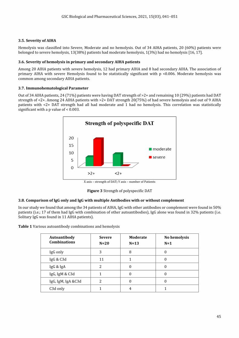
Out of 34 AIHA patients, 24 (71%) patients were having DAT strength of >2+ and remaining 10 (29%) patients had DAT strength of <2+. Among 24 AIHA patients with >2+ DAT strength 20(75%) of had severe hemolysis and out of 9 AIHA patients with <2+ DAT strength had all had moderate and 1 had no hemolysis. This correlation was statistically significant with a p value of < 0.003.
X axis – strength of DAT; Y axis – number of Patients
Figure 3 Strength of polyspecific DAT
3.8. Comparison of IgG only and IgG with multiple Antibodies with or without complement
In our study we found that among the 34 patients of AIHA, IgG with other antibodies or complement were found in 50% patients (i.e.; 17 of them had IgG with combination of other autoantibodies), IgG alone was found in 32% patients (i.e. Solitary IgG was found in 11 AIHA patients).
Table 1 Various autoantibody combinations and hemolysis
Autoantibody Combinations
Severe
N=20
Moderate
N=13
No hemolysis
N=1
IgG only 3 8 0
IgG & C3d 11 1 0
IgG & IgA 2 0 0
IgG, IgM & C3d 1 0 0
IgG, IgM, IgA &C3d 2 0 0
C3d only 1 4 1

GSC Biological and Pharmaceutical Sciences, 2021, 15(03), 041–051
46
3.9. Thermal Amplitude of auto antibodies
Out of 34 AIHA patients, 25 warm autoantibodies reacted at 37℃, among 6 Cold AIHA 3 had thermal amplitude of 4℃
to 22℃ and 3 had wide thermal amplitude of 4℃ to 37℃. In 3 Mixed AIHA cold autoantibodies had thermal amplitude
of 4℃ to 22℃ and warm autoantibodies reacted at 37℃.
3.10. Acid Elution in AIHA patients
Among the 34 DAT positive samples, Elution was done in all AIHA patients. Out of 34 elusions, 28(78%) samples were reactive with RBC reagent panel and 6 (23%) samples were nonreactive with the same. All the reactive eluates belonged to either warm or mixed AIHA patients and the nonreactive eluates belonged to patients with Cold AIHA.
X axis – results of elution; Y axis – number of patient samples
Figure 4 Acid Elution
3.11. Elution with Glycine Acid Ethylene Diamine Tetra Acetic acid [EDTA]
X axis – results of elution; Y axis – number of patient samples
Figure 5 Glycine acid EDTA
Among 34 patients with AIHA Glycine Acid EDTA was carried out in 29 patients who are diagnosed warm AIHA of AIHA and in patients with h/o previous transfusion more than 3 months and with a probable alloantibody. Test is not done in 3 Cold AIHA and 2 patients with a recent history of transfusion (< 7days). Glycine Acid EDTA rendered 19 samples of DAT completely negative and in the remaining 9 wAIHA the strength of DAT is reduced than its original strength.
GSC Biological and Pharmaceutical Sciences, 2021, 15(03), 041–051
47
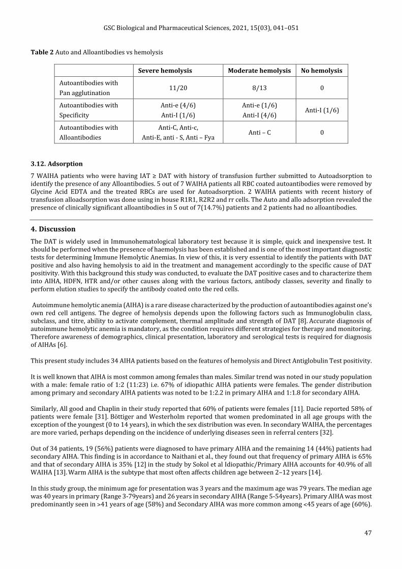
Table 2 Auto and Alloantibodies vs hemolysis
Severe hemolysis Moderate hemolysis No hemolysis
Autoantibodies with
Pan agglutination 11/20 8/13 0
Autoantibodies with
Specificity
Anti-e (4/6)
Anti-I (1/6)
Anti-e (1/6)
Anti-I (4/6) Anti-I (1/6)
Autoantibodies with
Alloantibodies
Anti-C, Anti-c,
Anti-E, anti - S, Anti – Fya Anti – C 0
3.12. Adsorption
7 WAIHA patients who were having IAT ≥ DAT with history of transfusion further submitted to Autoadsorption to identify the presence of any Alloantibodies. 5 out of 7 WAIHA patients all RBC coated autoantibodies were removed by Glycine Acid EDTA and the treated RBCs are used for Autoadsorption. 2 WAIHA patients with recent history of transfusion alloadsorption was done using in house R1R1, R2R2 and rr cells. The Auto and allo adsorption revealed the presence of clinically significant alloantibodies in 5 out of 7(14.7%) patients and 2 patients had no alloantibodies.
4. Discussion
The DAT is widely used in Immunohematological laboratory test because it is simple, quick and inexpensive test. It should be performed when the presence of haemolysis has been established and is one of the most important diagnostic tests for determining Immune Hemolytic Anemias. In view of this, it is very essential to identify the patients with DAT positive and also having hemolysis to aid in the treatment and management accordingly to the specific cause of DAT positivity. With this background this study was conducted, to evaluate the DAT positive cases and to characterize them into AIHA, HDFN, HTR and/or other causes along with the various factors, antibody classes, severity and finally to perform elution studies to specify the antibody coated onto the red cells.
Autoimmune hemolytic anemia (AIHA) is a rare disease characterized by the production of autoantibodies against one’s own red cell antigens. The degree of hemolysis depends upon the following factors such as Immunoglobulin class, subclass, and titre, ability to activate complement, thermal amplitude and strength of DAT [8]. Accurate diagnosis of autoimmune hemolytic anemia is mandatory, as the condition requires different strategies for therapy and monitoring. Therefore awareness of demographics, clinical presentation, laboratory and serological tests is required for diagnosis of AIHAs [6].
This present study includes 34 AIHA patients based on the features of hemolysis and Direct Antiglobulin Test positivity.
It is well known that AIHA is most common among females than males. Similar trend was noted in our study population with a male: female ratio of 1:2 (11:23) i.e. 67% of idiopathic AIHA patients were females. The gender distribution among primary and secondary AIHA patients was noted to be 1:2.2 in primary AIHA and 1:1.8 for secondary AIHA.
Similarly, All good and Chaplin in their study reported that 60% of patients were females [11]. Dacie reported 58% of patients were female [31]. Böttiger and Westerholm reported that women predominated in all age groups with the exception of the youngest (0 to 14 years), in which the sex distribution was even. In secondary WAIHA, the percentages are more varied, perhaps depending on the incidence of underlying diseases seen in referral centers [32].
Out of 34 patients, 19 (56%) patients were diagnosed to have primary AIHA and the remaining 14 (44%) patients had secondary AIHA. This finding is in accordance to Naithani et al., they found out that frequency of primary AIHA is 65% and that of secondary AIHA is 35% [12] in the study by Sokol et al Idiopathic/Primary AIHA accounts for 40.9% of all WAIHA [13]. Warm AIHA is the subtype that most often affects children age between 2–12 years [14].
In this study group, the minimum age for presentation was 3 years and the maximum age was 79 years. The median age was 40 years in primary (Range 3-79years) and 26 years in secondary AIHA (Range 5-54years). Primary AIHA was most predominantly seen in ˃41 years of age (58%) and Secondary AIHA was more common among ˂45 years of age (60%).

GSC Biological and Pharmaceutical Sciences, 2021, 15(03), 041–051
48
The most common cause for secondary AIHA was Autoimmune disorders (SLE and Rheumatoid Arthritis) and followed by sickle cell anemia and lymphoproliferative disorders. This finding is reflected in the younger age of presentation of secondary AIHA [15].
The degree of hemolysis in our study population was evaluated based on the following four parameters viz., Hemoglobin <9gm/dl, Bilirubin >2mg/dl, Lactate dehydrogenase 500IU/ml and Reticulocyte count >2% [16,17]. Based on these parameters the hemolysis is classified into moderate, severe hemolysis.
In our study 20(59%) patients had severe hemolysis, 13(41%) AIHA patients had moderate hemolysis. SS Das et al in their study classified hemolysis among AIHA patients into two groups only and they observed that 39.53% had severe hemolysis and 60.47% had moderate hemolysis [16].
4.1. Correlation of strength of Polyspecific DAT with severity of hemolysis
The strength of DAT positivity correlates with the number of Immunoglobulin molecules adherent to RBC. To assess the strength of DAT various techniques have been performed viz, tube technique, CAT, flow cytometry etc. In literature many studies have been done to assess the correlation of strength of Polyspecific DAT with severity of hemolysis. In our study, polyspecific DAT, Monospecific DAT and IgG subtype was done by CAT technique.
In the present study of 34 patients with AIHA 24 patients had the DAT strength of >2+ and remaining 10 patients had <2+ DAT strength. Correlating the strength of the DAT with the severity of hemolysis, we found that Among 24 AIHA patients with >2+ DAT strength, 18 (75%) of had severe hemolysis, but only 2 (10%) out of 10 AIHA patients with <2+ DAT strength had severe hemolysis. This correlation was statistically significant with a p value of < 0.003. Similarly Wheeler et al., in their study found out that the relationship between the presences or absence of hemolysis and the DAT strength was highly statistically significant [17]. Wikman in his study revealed that there was a significant correlation between strength of DAT and severe hemolysis [19].
4.2. Auto and Alloantibody in AIHA vs Severity of hemolysis
Alloantibodies were identified in 4/20 (25%) in relation to AIHA patients with severe hemolysis and 1/14 had alloantibody with moderate hemolysis and all of them had previous history of transfusion. 4 of 5 patients had single clinically significant alloantibody and 1 of 4 had multiple clinically significant alloantibodies. The study done by Wikman A et al had 28% of alloantibodies in AIHA patients all of them fell in moderate hemolysis group [19]. Similarly the study conducted by Branch DR and Petz LD 25-47% of sera from AIHA patients showed the presence of alloantibodies [20].
4.3. Autoantibodies in AIHA
In our study we found that among the 28 AIHA patients with IgG antibodies, IgG with other antibodies or complement were found in 50% patients (i.e.; 17of them had IgG with combination of other autoantibodies), IgG alone was found in 32% patients (i.e. Solitary IgG was found in 11 AIHA patients). Similarly in the study by Lai et al on AIHA patients he found that 55.6% of the patients had multiple autoantibodies and 44.4% had solitary IgG autoantibody [21].
In this conjuncture, our study found that 80% of the AIHA patients with severe hemolysis had combination of antibodies in comparison to only 15% of AIHA patients with severe hemolysis who had solitary IgG antibody. This association was statistically significant with a p value <0.002. This findings in accordance to the studies conducted by Das SS et al and Wheeler et al respectively [16, 17].
In our study C3d alone was identified in 6 (18%) of AIHA patients which raising the possibility of cold agglutination disease, and 3 (9%) of the AIHA patients had mixed AIHA having IgG, IgM& C3d. Similarly in the study conducted by Shulman et al 8.3% of the patients having mixed type AIHA [22].
The present study experienced that out of 34 AIHA patients 29 (85.2%) had autoantibodies and 5 (14.8%) patients had both auto and alloantibodies. Similarly, the study done by Wikman A et al had 28% of alloantibodies in AIHA patients all of them fell in moderate hemolysis group [19]. Similarly the study conducted by Branch DR and Petz LD 25-47% of sera from AIHA patients showed the presence of alloantibodies [20].
4.4. IgG subtypes
IgG1 was the most common subtype present in 11 (40%) patients, followed by combination of both IgG1 and IgG3 which was present in 8 (29%) patients and solitary IgG3 was found in 4 (14%) patients with AIHA. In our study 5 (17%) patients had neither IgG1 nor IgG3. In the study conducted by Das SS et al., 46.5% of AIHA patients, the subclass was

GSC Biological and Pharmaceutical Sciences, 2021, 15(03), 041–051
49
IgG1 or IgG3 or both, while in the remaining patients, no IgG1 or IgG3 was detected [16]. In the other study, IgG1 was the most frequent subclass (96%) coating the red cells [23].
The presence of both IgG1 and IgG3 has a significant effect on severity of hemolysis. A total of 8 patients were noted to have both IgG1 and IgG3 in which 7 (87%) had severe hemolysis in par with 1 (13%) moderate hemolysis. A similar impact was noted in patients with solitary IgG1 [95% CI= 25.0570 to 88.9932; P = 0.0042]. Among 11 AIHA patients with only IgG1 autoantibody 10 (90%) had moderate hemolysis and 1 (10%) 95% [CI= 39.2760 to 91.4404; P = 0.0002]. was having severe hemolysis. Both of these associations were statistically significant. Among 4 AIHA patients who had IgG3 subclass, 3(75%) had moderate hemolysis and 1 (25%) had severe hemolysis. In 5 AIHA patients who had neither IgG1 nor IgG3, 4 (80%) had moderate hemolysis and 1 (10%) had severe hemolysis.
Similarly in Das SS et al study 60% of the patients with combination of IgG1 with IgG3 and IgG1 alone were more likely to present with severe hemolysis as compared to 21.7% of the patients having IgG3 only and neither IgG1 nor IgG3 [16].
4.5. Thermal amplitude
Out of 34 AIHA patients, 25(74%) warm autoantibodies reacted at 37oC only, among 6 (18%) Cold AIHA in which 3 (9%) had thermal amplitude of 4oC to 22oC and 3 (9%) had wide thermal amplitude of 4oC to 37oC. The remaining 3 patients belonged to Mixed type AIHA. In a similar type of study conducted by Das SS 81.4% was WAIHA, 16.3% were mixed type and there was only one cold AIHA (2.32%) [16].
4.6. Elution (by Acid Elution and Glycine Acid EDTA)
Among the 34 DAT positive samples, Elution was done in all AIHA patients using commercially available kits, 28 elutes (25WAIHA & 3 Mixed AIHA) were reactive and 6 cold AIHA elutes were nonreactive. Among 28 reactive eluates 22 were showing panagglutination, in which 21 (73%) were WAIHA with, 1 (4%) was due to mixed AIHA and 6 (23%) AIHA samples showed the presence of autoantibody specificity as anti-e which was confirmed with serum studies (4 were due to WAIHA and 2 were mixed AIHA).Similar to this a study conducted by Marilyn Johnston FM, Mary Kay Belota, observed that 68% of patients, in which 37% yielded positive eluates and 63%, had nonreactive ones. Of the positive elution studies, 73% demonstrated only warm autoantibody on red blood cells. 2.5% of these had warm autoantibody in serum as well. 3% of these specimens were from previously but not recently transfused patients and had alloantibody/i.e. in serum. 33% eluates with positive results were from patients transfused 26-48 days before testing [24]. Similarly Dacie J in his study reported that Autoanti-e was the most common specificity; it has been pointed out that the reported relative incidence of different specific Rh autoantibodies corresponds well with the incidence of Rh antigens in the population (i.e., ‘e’ antigen is present on the RBCs of approximately 98% of the population) [10].
Among 34 patients with AIHA Glycine Acid EDTA was carried out in patients who are diagnosed case of AIHA and in patients with h/o previous transfusion more than 3 months. Glycine Acid EDTA rendered DAT completely negative in 18 of the cases and in remaining 11 cases the strength of DAT is reduced than its original strength. Similarly in the study conducted by Katharia R Glycine Acid EDTA 59 DAT positive samples showed decrease in reaction of which 22 samples were DAT negative [25].
7 WAIHA patients who were having IAT ≥ DAT further submitted to Autoadsorption to identify the presence of any Alloantibodies. 5 out of 7 WAIHA patients all RBC coated autoantibodies were removed by Glycine Acid EDTA and the treated RBCs are used for Autoadsorption 2 WAIHA patients with recent history of transfusion alloadsorption was done using in house R1R1, R2R2 and rr cells. The Auto and allo adsorption revealed the presence of clinically significant alloantibodies in 5 out of 7(14.7%) patients and 2 patients had no alloantibodies. Adsorptions with autologous RBCs, Laine and Beattie, James and colleagues, Issitt et al, and Morel and coworkers detected alloantibodies in 27%, 32% , 38%, and 40% of sera, respectively [26,27,28,29].
In 19 of 34 (55%) autoantibodies reacting with all test erythrocytes (panagglutination) were detected, Autoantibodies with specificity against Rh antigen (anti-e) were identified in 5 of 20 patients in the group with severe haemolysis, in 1 of 13 with moderate haemolysis and Autoantibodies with specificity against I antigen (anti-I) were identified in 1 of 20 patients in the group with severe haemolysis, in 5 of 13 with moderate haemolysis. Similarly Dacie J in his study reported that Autoanti-e was the most common specificity; it has been pointed out that the reported relative incidence of different specific Rh autoantibodies corresponds well with the incidence of Rh antigens in the population (i.e., ‘e’ antigen is present on the RBCs of approximately 98% of the population) [10]. Jenkins and coworkers found that sera containing cold autoagglutinins that had previously been called “nonspecific cold agglutinins” had anti-I specificity and it is more common to other cold autoantibodies [30].

GSC Biological and Pharmaceutical Sciences, 2021, 15(03), 041–051
50
5. Conclusion
A positive DAT with falling hematocrit, jaundice, elution and adsorption studies appear to be clinically helpful in investigating and identifying alloantibodies in autoimmune hemolytic anemia. In our study revealed a strong association between the severity of hemolysis and DAT strength, the IgG and subclass (IgG 1 & IgG 3) of IgG Antibodies either alone or in combination with other classes of Antibodies and/or complements. Adsorption and elution techniques are useful in AIHA cases with history of transfusion and while suspecting the presence of clinically significant alloantibodies. Prewarming techniques and Titration are effective in the diagnosis and management of Cold AIHA. The Glycine acid EDTA elution technique provides unbound RBCs for phenotyping it helps in transfusion of compatible blood. Use of DTT is useful in mixed AIHA. Further, this study reinstates a schematic approach in dealing with of DAT positive autoimmune haemolytic anemia cases by designing institutional based algorithm for efficient patient management.
Compliance with ethical standards
Acknowledgments
I am very grateful to Dr. P. Arumugam MD, Professor & HOD Department of Transfusion Medicine, and The TN Dr.MGR Medical University for his guidance and immense support in my study.
Disclosure of conflict of interest
There is no Conflict of interest.
Statement of ethical approval
Ethical approval was obtained from concerned institutions.
Statement of informed consent
Informed consent was obtained from all individual participants included in the study.
References
[1] Coombs RRA, Mourant AE, Race RR. A new test for the detection of weak and “incomplete” Rh agglutinins. Br J Exp Pathol. 1945; 26: 255.
[2] Petz D, Garratty G. Immune Hemolytic Anemias. 2nd ed. Philadelphia, Pennsylvania: Elsevier Inc. (USA). 2004; 1-28.
[3] Kaplan HS, Garratty G. Predictive value of direct antiglobulin test results Diagnostic Med. 1985; 8: 29-32.
[4] Petz LD. Blood transfusion in haemolytic anaemia. Immunohematology. 1999; 15: 15-23.
[5] South SF. Use of direct antiglobulin test in routine testing. In: Wallace M, Levitt J, eds. Current applications and interpretations of the direct antiglobulin test. Arlington, VA: American Association of blood banks. 1988: 25-45.
[6] Vanamala Alwar et al. Clinical Patterns and Hematological Spectrum in Autoimmune Haemolytic Anaemia. J Lab Physicians. 2010 Jan-Jun 2; 2: 17- 20.
[7] Judd WJ. Elution-Dissociation of Antibody from Red Blood Cells: Theoretical and Practical Considerations. Transfusion Medicine Reviews. 1999; 13: 297-310.
[8] Pirofsky B. Autoimmunization and the Autoimmune Hemolytic Anemias. Baltimore: Williams & Wilkins Company. 1969: 3-20.
[9] Parker V, Christopher A.Tormey. The Direct Antiglobulin Test. Indications, Interpretation, and Pitfalls. Arch Pathol Lab Med-Vol 141 2017.
[10] Dacie J. The haemolytic anaemias. The autoimmune haemolytic anaemias, vol. 3. New York: Churchill Livingstone; 1992.
[11] Allgood JW, Chaplin H. Jr: Idiopathic acquired autoimmune hemolytic anemia: A review of forty-seven cases treated from 1955 through 1965. Am J Med. 1967; 43: 254−273.

GSC Biological and Pharmaceutical Sciences, 2021, 15(03), 041–051
51
[12] Naithani R, Agarwal N, Mahapatra M et al,. Autoimmune hemolytic anemia in India: clinico-hematological spectrum of 79 cases. Hematology: 2006;11:73– 76
[13] Sokol RJ, Hewitt S, Stamps BK. Autoimmune haemolysis: an 18-year study of cases referred to a regional transfusion centre. Br Med J 1981;282:2023.
[14] Ware, RE, Nathan, DG, Orkin, SH, Ginsburg, P, Look, AT. “Autoimmune Hemolytic Anemia in Hematology of Infancy
and Childhood”. 2003.
[15] Rausen AR, LeVine R, Hsu TC, et al. Compatible transfusion therapy for paroxysmal cold hemoglobinuria. Pediatrics. 1975; 55: 275-278.
[16] Das SS, Nityanand S, Chaudhary R. Clinical and serological characterisation of autoimmune hemolytic anemia in a tertiary care hospital in North India. Ann Hematol. 2009 Aug; 88(8): 727-32.
[17] Christine A, Wheeler MD, Douglas P, Blackall MD, et al. Warm Reactive Autoantibodies Clinical and Serologic Correlations. Am J Clin Pathol. 2004; 122: 680-685.
[18] Nance SJ, Arndt PA, Garratty G. Correlation of IgG subclass with the severity of hemolytic disease of the newborn. Transfusion. 1990; 30: 381 –382.
[19] Agneta Wikman, Ulla Axdorph, Gunilla Gryfelt, et al. Characterization of red cell autoantibodies in consecutive DAT-positive patients with relation to in vivo haemolysis. Ann Hematol. 2005; 84: 150–158.
[20] Branch DR, Petz LD. Detecting alloantibodies in patients with autoantibodies. Transfusion. 1999; 39: 6–10
[21] Jeanne E. Hendrickson, Christopher A. Tormey. Red Blood Cell Antibodies in Hematology/Oncology Patients. Interpretation of Immunohematologic tests and clinical Significance of Detected Antibodies. Hematol oncol Clin N Am. 2016; 30: 635-651.
[22] Shulman IA, Branch DR, Nelson JM, et al. Autoimmune hemolytic anemia with both cold and warm autoantibodies. JAMA. 1985; 253: 1746.
[23] J. Fabijan´ska-Mitek et al. Gel Test Application for IgG Subclass Detection in Auto-Immune Haemolytic Anaemia. Vox Sang. 1997; 72: 233–237.
[24] Marilyn FM. Johnston, Mary Kay Belota. Determination of Need for Elution Studies for Positive Direct Antiglobulin Tests in Pretransfusion Testing. American Journal of Clinical Pathology. 1988 July; 90(1): 58–62.
[25] Rahul Katharia, Rajendra K Chaudhary. Removal of antibodies from red cells: Comparison of three elution methods. Asian J Transfus Sci. 2013 Jan-Jun; 7(1): 29–32.
[26] Laine ML, Beattie KM. Frequency of alloantibodies accompanying autoantibodies. Transfusion. 1985; 25: 545–546.
[27] James P, Rowe GP, Tozzo GG. Elucidation of alloantibodies in autoimmune haemolytic anaemia. Vox Sang. 1988; 54: 167–171.
[28] Issitt PD, Combs MR, Bumgarner DJ, Allen J, Kirkland A, Melroy-Carawan H. Studies of antibodies in the sera of patients who have made red cell autoantibodies. Transfusion. 1996; 36: 481–486.
[29] Morel PA, Bergen MO, Frank BA. A simple method for the detection of allo-antibody in the presence of warm autoantibody [abstract]. Transfusion. 1978; 18: 388.
[30] Jenkins WJ, Marsh WL, Noades J, et al. The I antigen and antibody. Vox Sang. 1960; 5: 97.
[31] Dacie JV, Worlledge SM: Auto-immune hemolytic anemias. In: Brown EB, Moore CV (eds): Progress in Hematology. New York: Grune & Stratton, 1969:82−120.
[32] Bottiger LE, Westerholm B: Acquired haemolytic anaemia. I: Incidence and aetiology. Acta Med Scand 1973;193:223−226.