evaluating the neuroprotective effects of fermented
TRANSCRIPT
EVALUATING THE NEUROPROTECTIVE EFFECTS OF
FERMENTED ROOIBOS HERBAL TEA IN WISTAR RATS
EXPOSED TO BISPHENOL-A DURING GESTATION AND
LACTATION
By
BUSHRA KHALIFA GAMOUDI
Student Number 3607196
A thesis submitted in fulfilment of the requirements for the degree of
Magister Scientiae (MSc)
Department of Medical Biosciences
Faculty of Natural Sciences
Supervisor
Prof. Okobi Ekpo
http://etd.uwc.ac.za/

ii
DECLARATION
I declare that the thesis titled: “Evaluating the neuroprotective effects of
fermented Rooibos Herbal Tea in Wistar rats exposed to Bisphenol-A during
gestation and lactation” is my work and is hereby submitted for the Master of
Science degree in Medical Biosciences at the University of the Western Cape,
South Africa. This work has not been submitted for any degree or examination in
any other university, and all sources used or quoted have been duly indicated and
acknowledged by complete references
Full name: Bushra Khalifa Gamoudi
Signed.......................................
http://etd.uwc.ac.za/

iii
DEDICATION
To my dearest family for their love, support, encouragement, and making my
dreams a reality.
The Prophet Muhammad (Peace Be upon Him) said, regarding knowledge:
“Acquire knowledge and impart it to the people...One who treads a path in search
of knowledge has his path to Paradise made easy by God” (Sunan Tirmidhi,
Hadith 107; Riyadh us-Saleheen, 245).
http://etd.uwc.ac.za/

iv
ACKNOWLEDGEMENTS
First of all, I would like to acknowledge my Allah for giving me the faith, strength,
good health, wisdom and perseverance, without you nothing is possible, and I
would not have completed my Masters!
This research would not have been completed without the generous help and
support received from different people. I want to express my sincere gratitude for
the valued assistance I received throughout my MSc programme from the
following people:
A special thanks to my husband who stood by me through the rough times
and my children whose presence always brings joy to my life.
My supervisor Prof. Okobi Ekpo for the knowledge gained,
encouragement, for his confidence in me, his patience, support and
motivation throughout the duration and completion of this research. I
acknowledge him for proofreading my thesis. THANK YOU!
I would also like to thank my parents and sisters for their love, care,
prayers, and support during my period of study.
Staff at the histology laboratory of the University of Stellenbosch, for their
help and the technical support.
To my Lab mates and colleagues at the Medical Bioscience department of
UWC, I say thank you for your kind and the friendly working relationship;
I enjoyed my times with you.
http://etd.uwc.ac.za/

v
Lastly, but by no means the least, big thanks to the Libyan government for
funding the project and my studies at the University of the Western Cape.
http://etd.uwc.ac.za/

vi
TABLE OF CONTENTS
DECLARATION ............................................................................................... ii
DEDICATION .................................................................................................. iii
ACKNOWLEDGEMENTS ............................................................................. iv
TABLE OF CONTENTS ....................................................................................... vi
LIST OF FIGURES ............................................................................................... xi
LIST OF TABLES ............................................................................................... xiii
LIST OF ABBREVIATIONS .............................................................................. xiv
ABSTRACT ......................................................................................................... xvi
CHAPTER 1 ........................................................................................................... 1
INTRODUCTION ............................................................................................. 1
1.1. RESEARCH HYPOTHESIS .................................................................... 5
1.2. RESEARCH AIM AND OBJECTIVES................................................... 5
1.2.1. AIM ........................................................................................................ 5
1.2.2. OBJECTIVES ........................................................................................ 5
CHAPTER 2 ........................................................................................................... 7
LITERATURE REVIEW ................................................................................. 7
2.1. THE CENTRAL NERVOUS SYSTEM (CNS) ....................................... 7
2.1.1. THE CEREBRUM ................................................................................. 8
2.1.1.1. CYTOARCHITECTURE OF THE CEREBRAL CORTEX ............. 9
2.1.1.2. FUNCTION OF THE CEREBRAL CORTEX ................................ 10
2.1.1.3. THE HIPPOCAMPUS ...................................................................... 11
http://etd.uwc.ac.za/

vii
2.1.2. THE CEREBELLUM .......................................................................... 13
2.1.2.1. CYTOARCHITECTURE OF THE CEREBELLAR CORTEX ...... 15
2.2. THE ENDOCRINE SYSTEM ................................................................ 16
2.2.1. ENDOCRINE DISRUPTOR COMPOUNDS (EDCs) ........................ 17
2.2.1.1. BISPHENOL A (BPA) ..................................................................... 21
2.2.1.1.1. PHYSICAL AND CHEMICAL PROPERTIES OF BPA ............. 22
2.2.1.2. RELEASE AND HUMAN EXPOSURE TO BPA .......................... 23
2.2.1.3. METABOLISM OF BPA ................................................................. 24
2.2.1.4. ACTIVITY OF BPA ......................................................................... 26
2.3. ROOIBOS (Aspalathus linearis) ............................................................ 28
2.3.1. CHEMICAL COMPOSITION OF ROOIBOS ................................... 29
2.3.1.1. POLYPHENOLS .............................................................................. 30
2.3.2. BIOLOGICAL PROPERTIES OF ROOIBOS TEA ........................... 32
2.3.4.1. NEUROPROTECTIVE EFFECT OF ROOIBOS ............................ 33
CHAPTER 3 ......................................................................................................... 36
MATERIALS AND METHODS .................................................................... 36
3.1. ETHICAL CONSIDERATION .............................................................. 36
3.2. MATERIALS.......................................................................................... 36
3.3. ANIMALS .............................................................................................. 37
3.4. TREATMENT PROTOCOL .................................................................. 38
3.5. PREPARATION OF REAGENTS ......................................................... 39
3.5.1. ROOIBOS HERBAL TEA PREPARATION ..................................... 39
3.5.2. BISPHENOL A (BPA) AND NORMAL SALINE ............................. 40
http://etd.uwc.ac.za/

viii
3.5.3. PREPARATION OF 4% FORMALDEHYDE SOLUTION .............. 40
3.5.4. PHOSPHATE BUFFERED SALINE (PBS) ....................................... 40
3.6. MOTOR FUNCTION TESTS (NEUROBEHAVIORAL TESTS) ........ 41
3.6.1. OPEN FIELD TEST (OFT) ................................................................. 41
3.6.2. NOVEL OBJECT RECOGNITION (NOR) TEST ............................. 43
3.7. ANIMAL SACRIFICE ........................................................................... 44
3.8. HISTOLOGICAL PREPARATION OF BRAIN SAMPLES ................ 44
3.8.1. AUTOMATED TISSUE PROCESSING ............................................ 44
3.8.2. EMBEDDING ..................................................................................... 45
3.8.3. SECTIONING ..................................................................................... 45
3.8.4. STAINING........................................................................................... 45
3.8.4.1. HAEMATOXYLIN AND EOSIN.................................................... 45
3.8.4.2. CRESYL VIOLET (CV) / NISSL STAINING ................................ 46
3.8.4.3. IMMUNOHISTOCHEMICAL (IHC) STAINING .......................... 47
3.9. NEUROCHEMICAL ASSAYS ............................................................. 47
3.9.1. HOMOGENIZATION OF TISSUES .................................................. 48
3.9.1.1. SUPEROXIDE DISMUTASE (SOD) .............................................. 48
3.9.1.2. CATALASE...................................................................................... 48
3.10. STATISTICAL ANALYSIS ................................................................ 49
CHAPTER 4 ......................................................................................................... 50
RESULTS ......................................................................................................... 50
4.1. INTRODUCTION .................................................................................. 50
4.2. EFFECT OF BPA AND FERMENTED ROOIBOS HERBAL TEA ON
BODY MASS ................................................................................................ 51
http://etd.uwc.ac.za/

ix
4.3. NEUROBEHAVIOURAL TESTS ......................................................... 52
4.3.1. THE OPEN FIELD TEST ................................................................... 52
4.3.1.1. THE FREQUENCY OF REARING EPISODES ............................. 52
4.3.1.2. TOTAL FREEZING TIME .............................................................. 53
4.3.1.3. TOTAL SELF-GROOMING TIME ................................................. 54
4.3.2. NOVEL OBJECT RECOGNITION (NOR) TEST ............................. 55
4.4. EFFECT OF BPA AND FERMENTED ROOIBOS HERBAL TEA ON
THE ACTIVITY OF ANTIOXIDANT ENZYMES ..................................... 57
4.4.1. SUPEROXIDE DISMUTASE ............................................................. 57
4.4.2. CATALASE......................................................................................... 59
4.5. HISTOLOGICAL STUDIES .................................................................. 60
4.5.1. LENGTH OF HIPPOCAMPAL CA1 REGION IN EXPERIMENTAL
RATS ............................................................................................................. 61
4.5.2. NUMBER OF PURKINJE CELLS IN PURKINJE CELL LAYER
(PCL) IN EXPERIMENTAL RATS ............................................................. 62
4.6. IMMUNOHISTOCHEMISTRY (IHC) .................................................. 64
4.6.1. GLIAL FIBRILLARY ACIDIC PROTEIN (GFAP) .......................... 64
CHAPTER 5 ......................................................................................................... 66
DISCUSSION ................................................................................................... 66
5.1. INTRODUCTION .................................................................................. 66
5.2. ROOIBOS TEA PREVENTED BPA-INDUCED GAIN IN BODY
MASS............................................................................................................. 67
5.3. ROOIBOS TEA MITIGATED BPA-INDUCED
NEUROBEHAVIOURAL DEFICITS .......................................................... 68
5.4. ROOIBOS TEA REGULATED THE ACTIVITY OF ANTIOXIDANT
ENZYMES..................................................................................................... 72
http://etd.uwc.ac.za/

x
5.5. ROOIBOS TEA ATTENUATED NEURODEGENERATION IN RATS
EXPOSED TO BPA ...................................................................................... 73
5.6. CONCLUSION .................................................................................................. 77
REFERENCES ...................................................................................................... 79
http://etd.uwc.ac.za/

xi
LIST OF FIGURES
Figure 2.1: The brain structure 7
Figure 2.2: The layers of the cerebral cortex 8
Figure 2.3: The lobes of the cerebral cortex 9
Figure 2.4: The structure of the Hippocampus 10
Figure 2.5: The layers of the hippocampus proper 11
Figure 2.6: The structure of the cerebellum A) Sagittal and B) Dorsal sections 12
Figure 2.7: The structure of the cerebellar cortex 13
Figure 2.8: Synthesis of Bisphenol A 19
Figure 2.9: Metabolism of BPA 22
Figure 2.10: Rooibos tea; (A) Unfermented (B) Fermented 25
Figure 2.11: Classification of polyphenols 27
Figure 2.12: Chemical structure of Aspalathin 28
Figure 3.1: Flow diagram showing the experimental design for the study 34
Figure 3.2: Open Field Test Apparatus 38
Figure 4.1: Average body mass of experimental rats on PND 42 47
Figure 4.2: Number of rearing episodes of experimental rats on PND 42 49
Figure 4.3: Total freezing time of experimental rats on PND 42 50
Figure 4.4: Total self-grooming time of experimental rats on PND 42 51
Figure 4.5: Recognition index of experimental rats on PND 42 52
Figure 4.6: Effects of different treatments on SOD enzyme activity in the
cerebral homogenate of experimental rats on PND 42 53
http://etd.uwc.ac.za/
xii
Figure 4.7: Effects of different treatments on SOD enzyme activity in the
cerebellar homogenate of experimental rats on PND 42 54
Figure 4.8: Effects of different treatments on CAT enzyme activity in the
cerebral homogenate of experimental rats on PND 42 55
Figure 4.9: Effects of different treatments on CAT enzyme activity in the
cerebellar homogenate of experimental rats on PND 42 55
Figure 4.10: Diagram of the hippocampus showing the landmarks used for
determining hippocampal length in the experimental rats. 57
Figure 4.11: CA1 hippocampal length in experimental rats on PND42 57
Figure 4.12: Number of Purkinje cells in experimental rats on PND42 58
Figure 4.13: Number of GFAP positive cells in experimental rats on PND42 60
http://etd.uwc.ac.za/
xiii
LIST OF TABLES
Table 2.1: Examples of EDCs and their endocrine disruptor effects 16
Table 2.2: Physical properties of BPA 19
Table 2.3: Quantification of the major flavonoids in aqueous rooibos extract 26
Table 3.1: Chemicals used in this study 32
Table 3.2: Equipment used in this study 33
Table 3.3: Tissue Processing Protocol 40
Table 3.4: IHC staining procedure 43
http://etd.uwc.ac.za/

xiv
LIST OF ABBREVIATIONS
•OH
Hydroxyl radical
ANOVA Analysis of variance
ATP Automated tissue processor
BPA Bisphenol A
CA Cornu ammonis
CAT Catalase
CNS Central nervous system
CV Cresyl Violet
DDT Dichlorodiphenyltrichloroethane
DG Dentate gyrus
EDCs Endocrine Disruptor compounds
FRHT Fermented rooibos herbal tea
GFAP Glial fibrillary acidic protein
H&E Haematoxylin and eosin
H2O2 Hydrogen peroxide
HCB Hexachlorobenzene
IHC Immunohistochemistry
NOR Novel Object Recognition
NS Normal saline
NTP National Toxicology Program
O−2 Superoxide
OFT Open Field Test
PAHs Polycyclic aromatic hydrocarbons
http://etd.uwc.ac.za/

xv
PBS Phosphate buffered saline
PCBs Polychlorinated biphenyls
PCDF Polychlorinated dibenzofurans
PCL Purkinje cell layer
PND Post-natal day
PNS Peripheral nervous system
RI Recognition index
ROS Reactive oxygen species
SOD Superoxide dismutase
UGT UDP-glucuronosyl transferase
http://etd.uwc.ac.za/

xvi
ABSTRACT
Exposure to endocrine-disrupting chemicals as bisphenol A (BPA) during
gestation and early postnatal life is known to disrupt normal developmental
processes and alter the body’s endocrine system leading to deleterious effects in
the developing central nervous system (CNS). BPA is an industrial synthetic
chemical commonly used in the production of a range of polymers and consumer
products, despite concerns about its safety. There is therefore the need to protect
the developing CNS from potential damage through the administration of
neuroprotective agents. Most medicinal plants are reported to possess significant
protective potential against tissue damage through different mechanisms that
prevent cell death, oxidative stress, inflammation, immunodeficiency, etc. In this
study, the protective effects of fermented rooibos (Aspalathus linearis) tea against
the deleterious effects of BPA were investigated. Rooibos is a herbal beverage
indigenous to South Africa with widely acclaimed health benefits often linked to
the bioactivity of its polyphenolic compounds, especially aspalathin. The anti-
allergic, cardiovascular, antioxidant and neuroprotective effects of this herb have
been previously reported hence, the present study aims to investigate if regular
consumption of rooibos tea during pregnancy and lactation could protect the
developing brain from the deleterious effects of BPA in a Wistar rat model. A
total of 40 three-month old adult female pregnant dams, with an average weight of
250g, were divided into four groups (n=10). Group 1 control rats received 9%
normal saline ad libitum; group 2 rats received 400µg/kg/day BPA only; group 3
rats received 20% fermented rooibos tea as well as 400µg/kg/day BPA, while
group 4 rats received ad libitum 20% fermented rooibos tea only. Offspring rats
were housed in the same cages as the dams and only separated after weaning on
postnatal day (PND) 21. Neurobehavioural assessment using the open field test
was done on postnatal day (PND) 42 after which the final body masses were taken
before the rats were decapitated under deep anaesthesia, and the desired CNS
parts carefully dissected out and processed for histological, biochemical and
immunohistochemical studies. The results obtained showed that there was
http://etd.uwc.ac.za/

xvii
significant impairment of neurobehavioural activity, decreased cerebral and
cerebellar antioxidant enzyme activity, reduced hippocampal CA1 length,
significant loss of cerebellar Purkinje cells and significant astrocyte activation
demonstrated by increased glial fibrillary acidic protein (GFAP) activity in
experimental rats exposed to BPA only. However, co-administration of rooibos
tea significantly attenuated the BPA-induced distortions. Taken together, these
findings suggest that rooibos could be a potent neuroprotective agent against
BPA-induced structural, functional and biochemical alterations in the developing
CNS.
Keywords: Endocrine disrupting chemicals, Bisphenol A, rooibos, antioxidants,
neurodegeneration, neuroprotection, neurobehavioural activity.
http://etd.uwc.ac.za/

1
CHAPTER 1
INTRODUCTION
Many chemical substances have been identified as Endocrine Disruptor
Compounds (EDCs) and humans become exposed to them either at the workplace,
through food or environment sources (air, water and soil). EDCs have been widely
reported to directly or indirectly affect the normal functioning of the endocrine,
nervous and reproductive systems (Colborn et al., 1993; Clotfelter, et al., 2004;
Kajta and Wójtowicz, 2013). Bisphenol A (BPA) is one of the most recognised
EDCs and is a widespread estrogenic chemical (Krishnan et al., 1993, Eladak et
al., 2015) and more than 8 billion pounds (4 billion kilograms) of BPA are
produced for the manufacturing of polycarbonate plastic food and beverage
containers per year (Rubin, 2011). BPA is also used in the resin lining of metal
cans, dental sealants, and is employed as an additive in a wide array of other
products (Vandenberg et al., 2007).
BPA has been reported to be capable of crossing the placental barrier during
pregnancy and is present in different human fluids including foetal and maternal
serum, as well as in full-term amniotic fluid (Ikezuki et al., 2002, Yamada et al.,
2002). In one study, BPA was identified in developing organs of mice including
the CNS (Mita et al., 2012). Other studies have also reported the effects of BPA
on the developing and adult brain (MacLusky et al., 2005, Zsarnovszky et al.,
2005, Rubin and Soto, 2009, Negri-Cesi, 2015, Gupta et al., 2018). The
hippocampus and cerebellum are some of the CNS structures which have been
http://etd.uwc.ac.za/

2
identified as susceptible targets of BPA-induced distortions. Hippocampal
apoptosis and neurodegeneration (Tiwari, et al., 2015; Xu et al., 2010a) as well as
alteration of early cerebellar morphology (Mathisen, et al., 2013) have been
reported in developing animals. Both these structures were the focus of the
present study.
The cerebellum is a component of the CNS situated at the posterior aspect of the
brainstem, inferior to the occipital and temporal lobes of the cerebral cortex. It is
mainly involved in maintaining balance, posture and the coordination of voluntary
movements through a stabilising control system which receives early warning
signals from each motor impulse (Fonnum and Lock, 2000). It is also involved in
motor, learning and cognitive functions (albeit via poorly understood
mechanisms). Although motor commands are mostly initiated in other CNS
regions, the cerebellum modifies them according to rate, range, force and
sequence of contractions by allowing more adaptive and accurate movements
(Fonnum and Lock, 2000).
The cerebellum belongs to a distributed sensorimotor coordination network that
includes the cerebral cortex, basal motor nuclei, thalamus and the reticular
formation (Doya, 1999). Therefore, any dysfunction of the cerebellum could
disrupt the entire sensorimotor coordination network and manifest as locomotor
dysfunctions. The cerebellum accounts for approximately 10% of the brain’s
volume and contains over 50% of the total number of neurons in the brain
(Herculano-Houzel, 2009). The remarkable diversity in its neuronal population
together with its unique post-natal development makes it an appropriate neural
http://etd.uwc.ac.za/
3
structure for studying the effects of different substances on cellular dynamics
during pre- and post-natal development. Whereas, granule and Purkinje cells are
the most important neurons in the cerebellum, Purkinje cells are very important
because they are the largest neurons in the brain and the sole output of the
cerebellum, thus implying that any distortions in their structure and function could
affect overall cerebellar function (Fonnum and Lock, 2000).
Neurodevelopmental deficits potentially related to cerebellar injury may be arise
from the effects of environmental substances leading to such impaired motor
functions as hypotonia, fine motor incoordination, ataxia and impaired motor
sequencing (Powls et al., 1995, Goyen et al., 1998). Cerebellar injury has also
been implicated in cognitive, social and behavioural dysfunction among older
patients (Berquin et al., 1998, Levisohn et al., 2000) and may contribute to the
long-term cognitive, language, and behavioural dysfunction seen among 25-50%
formerly pre-term infants (Limperopoulos et al., 2007, Messerschmidt et al.,
2008). Only a few studies have focused on the vulnerability of the cerebellum to
toxic agents (Bist and Bhatt, 2009, Sanders et al., 2009), mainly focused on the
effects of lead, polychlorinated biphenyl and mercury on the nervous system (Yun
and Hoyer, 2000, Mervis et al., 2002, Verstraeten et al., 2008).
The functions of the hippocampus in memory acquisition and storage as well as
spatial learning have been adequately described (Sutherland and Rudy, 1989).
Hippocampal CA1 neurons are the most sensitive and susceptible to selective
degeneration in response to such EDCs as BPA, ischemia and other toxic
substances (Kirino, 1982, Pulsinelli et al., 1982, Elsworth et al., 2013, Kimura et
http://etd.uwc.ac.za/

4
al., 2016). In addition, axons of CA1 have been considered the main output of the
hippocampus (Duvernoy, 2013) which informs the choice of this region for
investigation in the current study.
The use of herbal products for the treatment of diseases is as old as humanity, and
certain plants have been reported to contain active compounds which could
protect the brain against such EDCs as BPA. Herbal products are considered to be
natural, green, pure, and with minimal side effects (Prashant et al., 2014), often
representing a source of new compounds with potent antioxidant activity to
address oxidative stress (Auddy et al., 2003). Rooibos (Aspalathus linearis) a
herbal beverage indigenous to South Africa, is one such plant with high
antioxidant activity (McKay and Blumberg, 2007, Joubert et al., 2008). Rooibos
naturally grows in the Western Cape Fynbos region of Clanwilliam and is
naturally caffeine-free, with a low tannin content (Erickson, 2003). The highly
acclaimed antioxidant properties of rooibos could be due to its unique polyphenol
content, aspalathin, known to be capable of delaying, inhibiting, or preventing
oxidation through scavenging of free radicals to diminish oxidative stress (McKay
and Blumberg, 2007, Joubert et al., 2008).
It has been reported that long-term consumption of rooibos could protect rat
brains from age-related changes, possibly due to its ability to prevent the
accumulation of lipid peroxides in the brain (Inanami et al., 1995). Also, rooibos
herbal tea has been shown to decrease steroidal hormone levels, indicating the
possibility that it could protect against the effects of BPA (Schloms et al., 2012).
This study was therefore designed to investigate the potential neuroprotective
http://etd.uwc.ac.za/

5
activity of rooibos herbal tea through the amelioration of the effects of the
endocrine disruptor BPA in an in vivo model.
1.1. RESEARCH HYPOTHESIS
This study hypothesizes that regular consumption of fermented rooibos herbal tea
could protect the developing brain against the deleterious effects of BPA. It is
believed that the polyphenolic compounds in rooibos tea are responsible for its
biological and pharmacological activities.
1.2. RESEARCH AIM AND OBJECTIVES
1.2.1. AIM
This study aims to evaluate the potential neuroprotective effects of fermented
rooibos herbal tea on the development of some CNS regions in rats exposed to
BPA during pregnancy and lactation.
1.2.2. OBJECTIVES
The following objectives were set for this study:
To determine the effects of maternal exposure to rooibos herbal tea and BPA
on the body masses of offspring rats.
To determine the effects of maternal exposure to rooibos and BPA on
neurobehavioural activity in offspring rats.
To determine the effects of maternal exposure to rooibos and BPA on
antioxidant enzymatic activity in cerebri and cerebella of offspring rats.
To determine the effects of maternal exposure to rooibos and BPA on
hippocampal and cerebellar morphology in offspring rats.
http://etd.uwc.ac.za/

6
To determine the effects of rooibos and BPA exposure on the activation of
astrocytes in offspring rats.
http://etd.uwc.ac.za/

7
CHAPTER 2
LITERATURE REVIEW
2.1. THE CENTRAL NERVOUS SYSTEM (CNS)
The nervous system consists of two major parts or subdivisions, the central
nervous system (CNS) and the peripheral nervous system (PNS) (Walsh and
Marshall, 1957). The CNS is the control centre for the entire nervous system and
is comprised of the brain and spinal cord. The brain is further subdivided into
three major sections, cerebrum, brainstem and cerebellum (Figure 2.1). The
cerebrum is the largest part of the human brain and is responsible for the
processing of speech, learning, emotions, muscular contractions as well as the
interpretation of sensory data related to hearing, vision and touch (Abhang et al.,
2016). The brainstem consists of the midbrain, pons and medulla oblongata, and
connects the cerebrum and cerebellum to the spinal cord. The cerebellum lies
posterior to the cerebrum, has important functions in motor control and is
responsible for the maintenance of balance and equilibrium (Morton and Bastian,
2004).
http://etd.uwc.ac.za/

8
Source: https://www.ncbi.nlm.nih.gov/pubmedhealth/PMHT0024735/
Figure 2.2: The brain structure
2.1.1. THE CEREBRUM
The cerebrum is the largest subdivision of the brain, located superiorly and
anteriorly in relation to the brainstem. It consists of a pair of cerebral hemispheres
(right and left), separated by the midline longitudinal cerebral fissure (the falx
cerebri) (Patestas and Gartner 2016). The cerebral cortex forms the outer covering
of the cerebral hemispheres and consists of a central core of white matter and a
thin outer covering of grey matter. It is estimated to comprise approximately 85%
volume of the adult human brain (Stephan et al., 1981, Rilling and Insel, 1999),
and each hemisphere is further subdivided into four lobes: the frontal, parietal,
temporal, and occipital lobes. The cerebral cortex is few millimetres thick and is
http://etd.uwc.ac.za/

9
highly convoluted to permit a large surface area of the brain to fit inside skulls
(Hilgetag and Barbas, 2009).
2.1.1.1. CYTOARCHITECTURE OF THE CEREBRAL CORTEX
The cerebral cortex is organized into six layers, and each of these layers has
different roles.
Source: https://medicine.academic.ru/135149/layers_of_cerebral_cortex
Figure 2.2: The layers of the cerebral cortex
The layers of the cerebral cortex, from superficial to deep, are:
Molecular layer (Plexiform) contains scattered horizontal cells and the apical
dendrites of pyramidal neurons of the cerebral cortex
The external granular layer contains small neuronal cell bodies.
http://etd.uwc.ac.za/

10
The external pyramidal layer is composed of small and medium pyramidal
cells.
The internal granular layer is made up of densely packed small round
pyramidal cells.
Internal pyramidal layer (Ganglionic) contains larger pyramidal cells and
scattered non-pyramidal cells.
Fusiform or Multiform layer comprises of variably shaped cells.
2.1.1.2. FUNCTION OF THE CEREBRAL CORTEX
Different regions of the cerebral cortex are associated with certain functions. For
instance, the frontal lobe is associated with conscious thought, memory,
personality and control of voluntary muscle movements (Chayer and Freedman,
2001).
Source:https://www.wpclipart.com/medical/anatomy/brain/brain_3/four_lobes_of_the_cerebral_c
ortex.png.html
Figure 2.3: The lobes of the cerebral cortex
http://etd.uwc.ac.za/

11
The parietal lobe gives individuals perspective and to help them understand space,
touch, and volume; the occipital lobe processes visual information while temporal
lobe is involved in auditory reception, helps to understand speech and is involved
in retrieving visual as well as verbal memories (Aversi-Ferreira et al., 2010,
Kiernan, 2012). The process of speech involves two different areas of the
cerebrum, namely, the Broca’s area which is responsible for the coordination of
the muscles for speaking and translation of thought into speech (Fadiga et al.,
2009), and the Wernicke’s area which is involved in the ability to read,
understand, and speak written word (Ardila et al., 2016).
2.1.1.3. THE HIPPOCAMPUS
The hippocampus is a small neuronal curved structure located on the medial
aspect of the temporal lobe of each cerebral cortex and forms an important part of
the limbic system. The hippocampus plays an important role in long-term memory
formation and spatial navigation. The hippocampus consists of two interlocking
U-shaped grey matter structures, the hippocampus proper (cornu ammonis, CA;
Figure 2.4) which is differentiated into four regions, CA1-4, and the dentate gyrus
separated from each other by hippocampal sulcus (Lorente de Nó, 1934;
Duvernoy, 2005).
http://etd.uwc.ac.za/

12
Source: https://www.slideshare.net/amandahessborzacchini/hippocampus-13053807
Figure 2.4: The structure of the Hippocampus
The CA is divided into three primary layers, each defined by a particular feature
of the large pyramidal cells or their afferents (Figure 2.5). The polymorph layer is
found between the alveus and the pyramidal layer, and contains cell bodies of the
basket cells and the basal dendrites of the pyramidal cells, as well as afferents
from the septum; the cell bodies of the pyramids dominate the pyramidal layer
(stratum pyramidale), and the molecular layer contains pyramidal dendrites
(Bayer, 1985).
Source: https://www.slideshare.net/drpraveenktripathi/limbic-system-brain
Figure 2.5: The layers of the hippocampus proper
http://etd.uwc.ac.za/

13
2.1.2. THE CEREBELLUM
The cerebellum is located conspicuously in the hindbrain with pons and medulla
oblongata. Although it is only 10% of total brain mass, the cerebellum has about
75% as much surface area as either of the much larger cerebral hemispheres,
containing approximately 70-101 billion neurons in the brain (Snider, 1958,
Lange, 1975, Andersen et al., 1992). The cerebellum has a relatively longer
developmental period and is one of the first structures in the brain to differentiate
and one of the last to mature (Liu et al., 2011). It is derived from the rhombic lips
and thickenings along the margins of the embryonic hindbrain. By the second
trimester of pregnancy, the fissures of the cerebellar cortex have appeared (Liu et
al., 2011).
The cerebellum consists of two paired lateral extensions known as hemispheres
united by the midline part called the vermis (Figure 2.6). Typically, the
cerebellum is divided into the anterior, posterior and flocculonodular lobes as
defined by the major fissures.
http://etd.uwc.ac.za/

14
Source: http://what-when-how.com/neuroscience/the-cerebellum-motor-systems-part-1/
Figure 2.6: The structure of the cerebellum A) Sagittal and B) Dorsal sections
The cerebellum consists of an outer layer of grey matter called the cortex and an
inner core of white matter known as medulla, containing four paired deep nuclei
namely, dentate nucleus, globose nucleus, emboliform nucleus, and fastigial
nucleus (Snell, 2010). All cerebellar input goes to the cortex, and all output comes
from the deep nuclei. The cerebellum is connected to the brainstem by three pairs
of stalks called cerebellar peduncles. Whereas the inferior peduncles connect the
cerebellum to the medulla oblongata, the middle peduncles connect the
cerebellum to the pons and the superior peduncles connect the cerebellum to the
midbrain (Snell, 2010).
http://etd.uwc.ac.za/

15
2.1.2.1. CYTOARCHITECTURE OF THE CEREBELLAR CORTEX
The cerebellar cortex is made up of three layers: the molecular layer, the granule
cell layer and the Purkinje cell layer in the middle (Figure 2.7). The input to the
cerebellum comes via the mossy and climbing fibres. The molecular layer consists
of two main types of neurons: stellate cells and basket cells (Apps and Garwicz,
2005). The Purkinje layer is formed by a single row of large Purkinje cells and
their axons which provide the only efferent pathway to the deep cerebellar nuclei,
thus constituting the sole output of all motor coordination in the cerebellar cortex
(Apps and Garwicz, 2005). The granular layer is densely populated by small
granule cells with dark-staining nuclei and scanty cytoplasm (Apps and Garwicz,
2005).
Source: http://vanat.cvm.umn.edu/neurHistAtls/pages/cns9.html
Figure 2.7: The structure of the cerebellar cortex.
Early studies considered the cerebellum a motor structure because cerebellar
damage mostly leads to impairments in motor function. Although the cerebellum
does not initiate movement, it contributes to coordination, precision, and accurate
http://etd.uwc.ac.za/

16
timing of movements (Penhune et al., 1998, Salman, 2002). The cerebellum
receives input from the sensory system of the spinal cord and other parts of the
brain, and integrate these inputs to fine-tune motor activity (Fine et al., 2002).
Damage to the cerebellum may occur as a result of stroke, head injury, cancer,
cerebral palsy, viral infection, or neurodegenerative diseases and may
subsequently cause individuals to experience tremors, difficulties in maintaining
balance, lack of muscle tone, speech difficulties, lack of control over eye
movement, difficulty in standing upright, and inability to perform accurate
movements (Fine et al., 2002). In addition, cerebellar neurons may become
damaged resulting in the loss of muscle control or coordination of movement in a
condition known as ataxia.
Some studies have shown that ataxia may also be triggered by the exposure to
toxins such as alcohol, drugs, or heavy metals (Manto, 2012, Barsottini et al.,
2014).
2.2. THE ENDOCRINE SYSTEM
The physiological activity of the endocrine system is closely linked to that of the
nervous system, with both systems coordinating the activity of the other (Brück,
1983). However, the differentiating aspect of the endocrine system is that its
effect is deployed through hormones secreted by glands located throughout the
body, conveyed in the bloodstream to particular areas in the body to regulate an
array of activities such as digestion, growth, metabolism and blood pressure
(Hiller-Sturmhöfel and Bartke, 1998).
http://etd.uwc.ac.za/

17
The hypothalamus is the brain structure that connects the nervous and the
endocrine systems (Cleghorn, 1955). It contains a small group of nuclei found at
the base of the forebrain and is responsible for the control of behaviour and such
rudimentary needs as sleep, hunger, thirst and sex as well as emotional and stress
responses (Zha and Xu, 2015). The hypothalamus also regulates the secretion of
hormones from the pituitary glands and other endocrine glands in the body
(Charlton, 2008).
2.2.1. ENDOCRINE DISRUPTOR COMPOUNDS (EDCs)
The United States Environmental Protection Agency (EPA) defines EDCs as “an
exogenous agent that interferes with synthesis, secretion, transport, metabolism,
binding action, or elimination of natural blood-borne hormones that are present
in the body and are responsible for homeostasis, reproduction, and developmental
process” (Diamanti-Kandarakis et al., 2009). Reports show that EDCs alter and
modify natural endocrine function, thus emerging as a major public health
challenge (Diamanti-Kandarakis et al., 2009, Schug et al., 2011, Zoeller et al.,
2014). These deleterious effects are primarily due to their potentially disruptive
activity on physiological processes, particularly through direct interaction with
steroid hormone receptors which are expressed abundantly in the hypothalamus
and other brain areas involved in the regulation of neuroendocrine functions
(Thomas and Doughty, 2004, Tokumoto et al., 2007).
Humans can take up EDCs or its precursors through consumption of contaminated
food, drinking water, inhalation and direct dermal contact with contaminated
products (Caballero-Gallardo et al., 2016). Studies have linked EDCs to adverse
http://etd.uwc.ac.za/

18
biological effects in animals, giving rise to concerns that its exposure might cause
serious medical effects (Colborn et al., 1993). Examples of some common EDCs
and their effects are summarized in Table 2.1.
http://etd.uwc.ac.za/

19
Table 2.3: Examples of EDCs and their endocrine disruptor effects
Name Uses Effects References
Endrin Insecticide and
pesticide
Competitively binds to androgen receptors; Toxic to fish and the
CNS
(Coble et al., 1967, Bhattacharya
and Mukherjee, 1975, Lemaire et
al., 2004)
Polychlorinated dibenzofurans (PCDF)
Insulators and
lubricants
Toxic to infants and is associated with memory and attention
deficits, decreased verbal abilities, and adverse behavioural as
well as emotional effects in early childhood.
(Henretig, 2009, Kodavanti et al.,
2017)
Heptachlor Insecticide and
pesticide
Toxic to the liver and the central nervous system as well as
reproductive, hematopoietic, immune, and renal systems.
(Fendick et al., 1990)
Hexachlorobenzene (HCB) Pesticide In animals, it induces neurological symptoms such as paralysis,
tremors and convulsions. In humans, it causes damage to the
liver, thyroid, nervous system, bones, kidneys, blood, and
immune and endocrine systems
(Addae et al., 2013)
Mirex Insecticide Human carcinogen and high toxicity to fish and other aquatic
animals
(Sanders et al., 1981, Spinelli et
al., 2007)
Dichlorodiphenyltrichloroethane
(DDT)
Pesticide Carcinogenic, early pregnancy loss, toxic to the reproductive
and nervous systems.
(Longnecker et al., 1997, Salleh et
al., 2015)
Aldrin Pesticide Carcinogenic (de Jong et al., 1997, Hooker et al.,
2014)
Chlordane Insecticide Carcinogenic, toxic to the reproductive systems (Fry, 1995, Persson and
Magnusson, 2015, Xiong, 2017)
http://etd.uwc.ac.za/
20
The adverse effect of EDCs can be seen during critical developmental stages such
as intrauterine, perinatal and in puberty, during which neuroendocrine systems are
modulated by steroid and other hormones (Frye et al., 2012). EDCs cross the
blood-placental and blood-brain barriers to interfere with development and
function, thus affecting pregnant mothers and children, both categories of which
have been considered the most susceptible to the harmful effects of exposure to
EDCs (Perera and Herbstman, 2011, Schug et al., 2015). Various reports indicate
that foetuses and offspring animals are exposed to EDCs through the placenta and
breast milk respectively, with such resultant effects as learning difficulties,
infertility and increased vulnerability to cancer not becoming evident until much
later in life (Poongothai et al., 2007, Li et al., 2013b). The major adverse health
effects arising from exposure to EDCs in humans include a myriad of
reproductive problems such as reduced count, motility and quality of sperms,
decreased female fertility, longer time to conception, higher miscarriage rates
(Singleton and Khan, 2003, Crain et al., 2008, Campion et al., 2012), increased
occurrence of testicular, prostate and breast cancer (Manibusan and Touart, 2017,
Balabanič and Klemenčič, 2018) and premature puberty in females (Zama et al.,
2016).
Some nervous system disorders may have their origins in endocrine disruptions,
particularly if the hippocampus and hypothalamus are targeted resulting in such
conditions as schizophrenia, bipolar disorders and cognitive dysfunctions (Brown
Jr, 2008; Meeker, 2012). A typical EDC that has been implicated in organ
function disruption and several other nervous system pathologies is 2, 2-bis (4-
hydroxyphenyl) propane, commonly known as BPA (Brown Jr, 2008).
http://etd.uwc.ac.za/

21
2.2.1.1. BISPHENOL A (BPA)
BPA is an industrial synthetic chemical commonly used in the production of a
range of polymers such as polycarbonate plastics and epoxy resins, as well as in
the production of several indoor applications and consumer products such as
thermal printer paper, in the lining of cans presently used for food and beverages.
It is also used for electronic equipment, water pipes, dental sealants, sports
equipment, medical devices, tableware, among others (Geens et al., 2011, Ribeiro
et al., 2017)
BPA is reported to be one of the highest chemicals produced worldwide, and there
has been an exponential increase in its use during the last 30 years, thus making
its potential for food and environment contamination higher (vom Saal and Myers,
2008, Gao et al., 2015). Human exposure to BPA is recognized as a widespread
occurrence, especially in developed countries, with reports indicating that
approximately 100 tonnes of BPA are released into the atmosphere each year, and
analyses of human samples showed the presence of BPA in most tested subjects
(Geens et al., 2012). The consumption of BPA-contaminated food is anticipated to
contribute to the overall BPA environmental exposure by more than 90% for all
age groups and its presence in the air, waste and drinking water, as well as in dust
particles has been reported (Vandenberg et al., 2007, Rudel et al., 2011, vom Saal
and Welshons, 2014).
Structurally, BPA is a diphenyl compound composed of two hydroxyl groups in
the ‘‘para’’ position connected by a methyl bridge, with two methyl functional
groups attached to the bridge making it structurally similar to hormones such as
http://etd.uwc.ac.za/

22
synthetic oestrogen, diethylstilbestrol (Kang et al., 2006). BPA is synthesized by
the condensation of acetone with two equivalents of phenol, and the reaction is
catalyzed by strong acids like hydrochloric acid (Figure 2.8) or a sulfonated
polystyrene resin (Uglea and Negulescu, 1991).
Figure 2.8: Synthesis of Bisphenol A
Prenatal BPA exposure is associated with neural, behavioural and bipolar
disorders in neonates, infants and children (Brown Jr, 2008). Reports indicate that
BPA induces hyperinsulinemia relating to type 2 diabetes mellitus and obesity at
very low doses and also triggers a high risk of miscarriage in women (Sugiura-
Ogasawara et al., 2005, Alonso-Magdalena et al., 2010).
2.2.1.1.1. PHYSICAL AND CHEMICAL PROPERTIES OF BPA
The physical and chemical properties of BPA are listed below.
Table 2.4: Physical properties of BPA.
Properties
Boiling point 220 oC (4 mmHg)
Melting point 158-159 oC
Molecular formula (CH3)2C(C6H4OH)2
Molar Mass 228.29 g·mol−1
Water solubility 21.5 oC
Vapour Pressure 25 oC
Density 1.20 g/cm³
BPA Acetone
Phenol Phenol
http://etd.uwc.ac.za/

23
2.2.1.2. RELEASE AND HUMAN EXPOSURE TO BPA
BPA has received considerable attention in recent years due to widespread
sources for human exposure through such routes as the environment (soil, air, and
aquatic) and contaminated foods. As a result of the high quantities of BPA
produced and an increase in the number of products based on epoxy resins and
polycarbonate plastics, there is widespread environmental contamination and
well-documented human exposure to BPA (Le et al., 2008). Although BPA is a
colourless solid soluble in organic solvents and poorly soluble in water, studies
have shown that BPA leaches from baby bottles, tin cans, reusable plastic water
bottles, and polycarbonate plastic containers into water, beverages, drink solutions
and food liquors, mostly due to the exposure of these BPA-containing plastics to
high temperatures (Grumetto et al., 2008, Le et al., 2008, De Coensel et al., 2009).
For instance, boiling to sterilize infant feeding bottles or addition of very hot
water or beverages to drinking bottles was reported to increase the rate of BPA
leaching by up to 55-fold (Le et al., 2008). Also, the discolouration, scratching of
polycarbonate, abnormal increase in pH and prolonged storage of water, drinks or
food in BPA-containing products increases the chances of leaching (Munguia-
Lopez et al., 2005, Biedermann-Brem et al., 2008).
In most industrialized countries, BPA detection in human amniotic fluid, serum,
breast milk and urine using various evaluation techniques has been reported
(Vandenberg et al., 2007) leading to the observation that BPA concentration in
human serum ranges from 0.2–1.6 ng/mL or 0.88–7.0 nM (Sajiki et al., 1999,
Takeuchi and Tsutsumi, 2002). In the United States, BPA was found to be present
in 95% of urine samples of 394 adults with a median concentration of 1.28 μg/L
http://etd.uwc.ac.za/

24
(Calafat et al., 2004) and according to the U.S. Environmental Protection Agency,
a reference dose of BPA is 50 mg kg/day was set while the European Union set a
no-observed-adverse-effect level (NOAEL) of 5 mg kg/day (Moriyama et al.,
2002, Gao et al., 2015).
2.2.1.3. METABOLISM OF BPA
Following oral intake of BPA in humans and rodents, reports show that BPA is
absorbed from the gastrointestinal tract and is glucuronidated by UDP-
glucuronosyltransferase (UGT, Figure 2.9) as a first-pass metabolism in the liver
to its main metabolite, BPA-glucuronide (Pottenger et al., 2000, Pritchett et al.,
2002, Völkel et al., 2002). The glucuronidation of BPA is a detoxification reaction
by UGT to detoxify BPA, its metabolite BPA-glucuronide is quickly excreted in
the urine due to its half-life of less than 6 hours. Bisphenol A-sulphate on the
other hand is reported to be a minor urinary BPA metabolite in humans (Ye et al.,
2005, 2006).
Following the oral intake of BPA in humans, the first-pass metabolism in the liver
is reported to be a super-effective process that results in a tremendously reduced
systemic availability (Pottenger et al., 2000, Upmeier et al., 2000). Also, BPA-
glucuronide and BPA-sulphate do not hinder hormonal control of reproduction,
thus indicating that these reactions are strictly detoxification pathways (Snyder et
al., 2000, Shimizu et al., 2002, Willhite et al., 2008). In rats, BPA is also
predominantly glucuronidated, with sulphation representing a minor pathway
(Pottenger et al., 2000), however the BPA-glucuronide formed is excreted from
the liver via the bile into the gastrointestinal tract, cleaved back to BPA and
http://etd.uwc.ac.za/
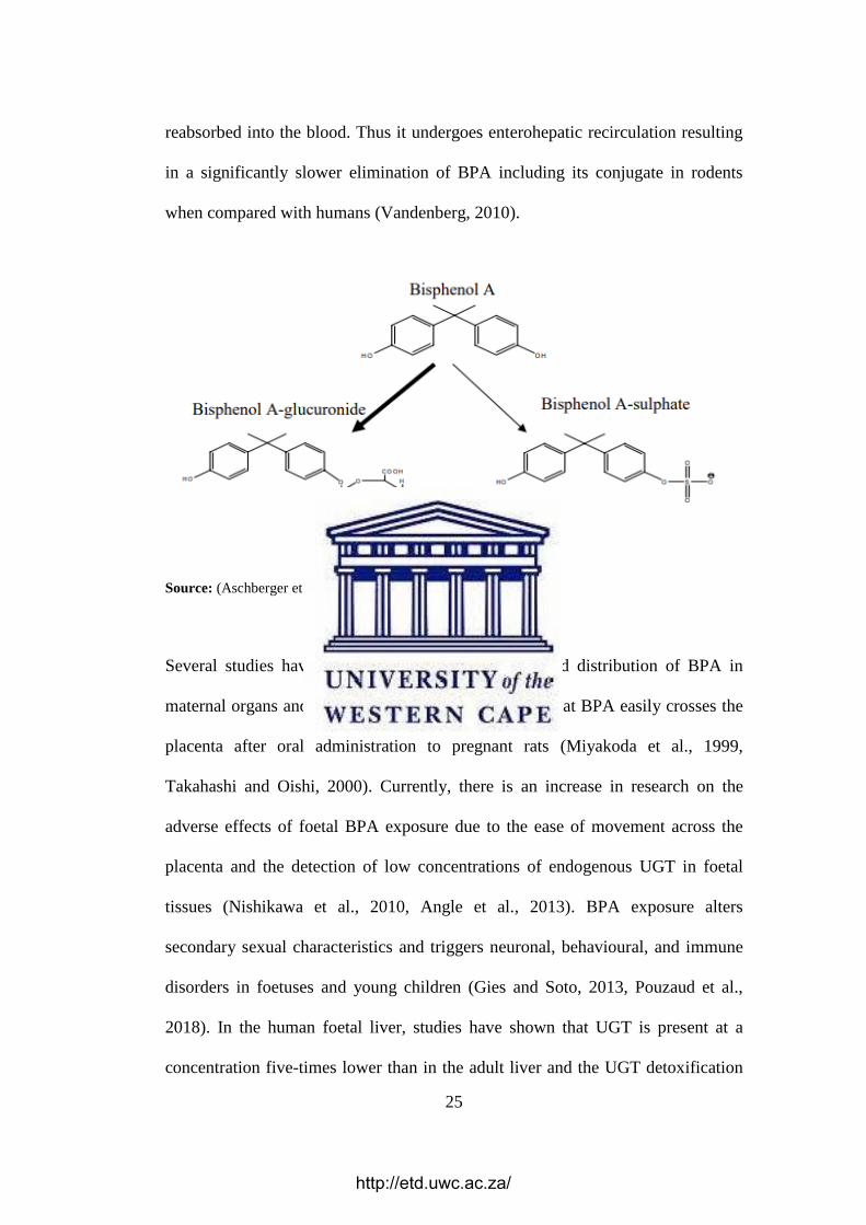
25
reabsorbed into the blood. Thus it undergoes enterohepatic recirculation resulting
in a significantly slower elimination of BPA including its conjugate in rodents
when compared with humans (Vandenberg, 2010).
Source: (Aschberger et al., 2010)
Figure 2.9: Metabolism of BPA
Several studies have suggested that the absorption and distribution of BPA in
maternal organs and foetuses are extremely rapid and that BPA easily crosses the
placenta after oral administration to pregnant rats (Miyakoda et al., 1999,
Takahashi and Oishi, 2000). Currently, there is an increase in research on the
adverse effects of foetal BPA exposure due to the ease of movement across the
placenta and the detection of low concentrations of endogenous UGT in foetal
tissues (Nishikawa et al., 2010, Angle et al., 2013). BPA exposure alters
secondary sexual characteristics and triggers neuronal, behavioural, and immune
disorders in foetuses and young children (Gies and Soto, 2013, Pouzaud et al.,
2018). In the human foetal liver, studies have shown that UGT is present at a
concentration five-times lower than in the adult liver and the UGT detoxification
http://etd.uwc.ac.za/

26
activities are not detected in foetal rat liver, thus suggesting that foetuses may be
more susceptible to the deleterious effects of BPA as most of it could be
accumulating in the tissues with potential consequencies (Matsumoto et al., 2002,
Jalal et al., 2017).
2.2.1.4. ACTIVITY OF BPA
BPA possesses estrogenic properties and acts as an agonist for oestrogen receptors
(Matsushima et al., 2010, Shanle and Xu, 2010b). For instance, the E-SCREEN,
often regarded as the most sensitive assay for oestrogenicity based on its ability to
differentiate between partial and full agonists, has confirmed the estrogenic
properties of BPA (Matsushima et al., 2010). Prepubescent CD-1 mice treated
with BPA demonstrated estrogenic responses such as increased expression of the
oestrogen-inducible protein lactoferrin, improved uterine wet weight and luminal
epithelial height (Markey et al., 2001). In another study, ovariectomized rats
exposed to BPA treatment experienced triggered proliferation of the uterine and
vaginal epithelial cells and induced estrogenic responses in the mammary and
pituitary glands (Colerangle and Roy, 1997, Steinmetz et al., 1998).
There is evidence in literature which indicates that exposure to BPA during early
development may increase breast cancer risk (Murray et al., 2007). This assertion
is supported by studies showing that BPA activates genes involved in the growth
of mammary glands and promotes breast cancers in rats after exposure to known
carcinogens at a dose that would not cause cancer in BPA-untreated mice
(Betancourt et al., 2010, Wadia et al., 2013). In treated male rats, a reduction in
sperm count, testosterone and luteinizing hormone (LH) as well as in the weights
http://etd.uwc.ac.za/

27
of the testes, epididymis, seminal vesicle and prostate gland were reported
(Sakaue et al., 2001, Akingbemi et al., 2004, Nakamura et al., 2010).
A report by the National Institutes of Health in Norway indicates that BPA has
adverse effects on foetal and infant brain development and behaviour. In
agreement with the previous report, the U.S. National Toxicology Program (NTP)
expressed concerns on the effects of BPA on the prostate gland, brain and
behaviour in foetuses, infants, and children (Abdallah, 2016). In rodents and non-
human primates, it has been reported that BPA affects the brain even at relatively
low exposure levels (Leranth et al., 2008, Nakagami et al., 2009). In-vivo studies
demonstrate that perinatal or neonatal BPA exposure modifies brain sexual
differentiation (Patisaul et al., 2006, Rubin et al., 2006), and initiates aggression,
anxiety, cognitive deficits, and learning-memory impairment (Miyagawa et al.,
2007, Tian et al., 2010, Xu et al., 2010b). BPA also induces changes in the
differentiation of ectodermal tissues, including neural tissues in cynomolgus
monkeys (Yamamoto et al., 2007), and triggers apoptosis in the central neurons of
tadpoles leading to head, vertebral and abdominal developmental deficits (Oka et
al., 2003).
Whereas BPA exposure in male rats during lactation is linked to hyperactivity and
the loss of dopaminergic neurons (Ishido et al., 2007), exposure during gestation
and lactation led to significant changes in the Glutamate/Aspartate (G/A) ratio in
the hippocampus of the cerebrum in Sprague-Dawley rats, thus suggesting
neuronal and glial developmental alterations (Kunz et al., 2011).
http://etd.uwc.ac.za/

28
2.3. ROOIBOS (Aspalathus linearis)
Rooibos tea is prepared from a leguminous shrub, Aspalathus linearis which is
native to the Cederberg Mountains of the Western Cape region of South Africa
(Joubert et al., 2004) and is commonly used in the fermented (red; Figure 2.10) or
unfermented (green) form (Joubert et al., 2004). Fermentation is the enzymatic
oxidation of polyphenols and other components in the leaves of the plant, thus
converting the green grassy-smelling and malty tasting tea to the sweet-smelling
and fruity red tea (Krafczyk and Glomb, 2008).
(A) (B)
Source: http://www.carmientea.co.za/tea-variants/rooibos/ & http://amavida.com/learn/
Figure 2.10: Rooibos tea; (A) Unfermented (B) Fermented
Rooibos tea has become a popular and reputable herbal beverage due to its
characteristic taste, absence of alkaloids and low tannin content (Cheney and
Scholtz, 1963, Jaganyi and Wheeler, 2003). In 1968, Annetjie Theron discovered
that an infusion of rooibos alleviated prolonged restlessness, vomiting and
stomach cramps when administered to her colicky baby, thus leading to its
classification as a healthy beverage. This led to a broader consumer base, and
http://etd.uwc.ac.za/

29
several babies have since been reared with rooibos either added to their milk or
given as a weak brew (Joubert et al., 2008).
Other pharmacological reports suggest that rooibos relieves ingestion, heartburn
and nausea, improves appetite, decreases nervous tension, enhances sound sleep
and has anti-allergic effects (Morton, 1983, Van Wyk et al., 1997). Rooibos
formulations have also been reported to be capable of relieving such
dermatological complications as eczema, rashes and acne, leading to the
production of toiletries and cosmetic products, now sold through many
supermarkets and beauty shops (Joubert et al., 2008).
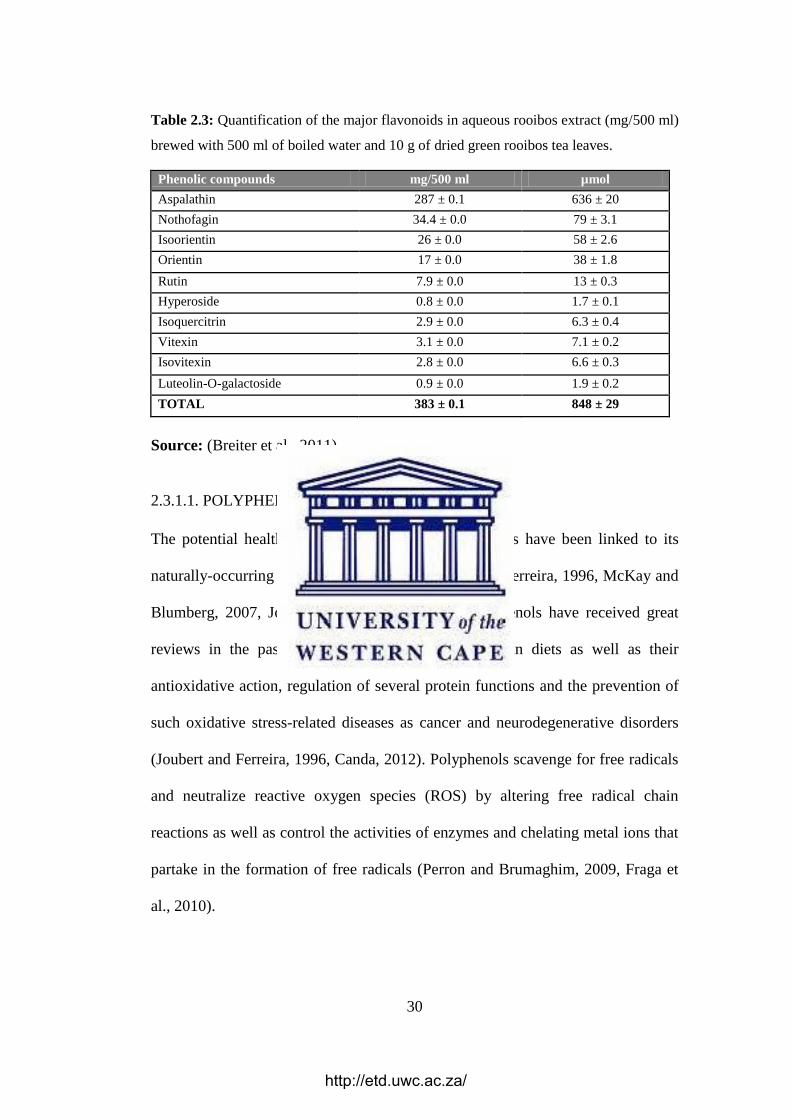
2.3.1. CHEMICAL COMPOSITION OF ROOIBOS
The leaf tannin content of fermented rooibos is between 3.2% to 4.4% (Reynecke
et al., 1949, Blommaert and Steenkamp, 1978) and traces of the alkaloid,
sparteine, flavonoids (aspalathin and aspalalinin) have been detected in this herbal
tea (Rabe et al., 1994, Shimamura et al., 2006). Other compounds detected in
rooibos include nothofagin, flavones (orientin, iso-orientin, vitexin, isovitexin,
chrysoeriol, luteolin and luteolin-7-O-glucoside), flavanones (dihydro-orientin,
dihydro-iso-orientin and hemiphlorin), flavonols (quercetin and its glycosides,
quercetin-3-robinobioside, hyperoside, isoquercitrin and rutin), phenolic acids,
lignans, esculetin, orientin, iso-orientin, and trace quantities of the aglycones,
luteolin, quercetin and chrysoeriol (Joubert et al., 2008).
http://etd.uwc.ac.za/
30
Table 2.3: Quantification of the major flavonoids in aqueous rooibos extract (mg/500 ml)
brewed with 500 ml of boiled water and 10 g of dried green rooibos tea leaves.
Phenolic compounds mg/500 ml µmol
Aspalathin 287 ± 0.1 636 ± 20
Nothofagin 34.4 ± 0.0 79 ± 3.1
Isoorientin 26 ± 0.0 58 ± 2.6
Orientin 17 ± 0.0 38 ± 1.8
Rutin 7.9 ± 0.0 13 ± 0.3
Hyperoside 0.8 ± 0.0 1.7 ± 0.1
Isoquercitrin 2.9 ± 0.0 6.3 ± 0.4
Vitexin 3.1 ± 0.0 7.1 ± 0.2
Isovitexin 2.8 ± 0.0 6.6 ± 0.3
Luteolin-O-galactoside 0.9 ± 0.0 1.9 ± 0.2
TOTAL 383 ± 0.1 848 ± 29
Source: (Breiter et al., 2011)
2.3.1.1. POLYPHENOLS
The potential health benefits and bioactivity of rooibos have been linked to its
naturally-occurring polyphenolic content (Joubert and Ferreira, 1996, McKay and
Blumberg, 2007, Joubert and de Beer, 2011). Polyphenols have received great
reviews in the past decade due to their abundance in diets as well as their
antioxidative action, regulation of several protein functions and the prevention of
such oxidative stress-related diseases as cancer and neurodegenerative disorders
(Joubert and Ferreira, 1996, Canda, 2012). Polyphenols scavenge for free radicals
and neutralize reactive oxygen species (ROS) by altering free radical chain
reactions as well as control the activities of enzymes and chelating metal ions that
partake in the formation of free radicals (Perron and Brumaghim, 2009, Fraga et
al., 2010).
http://etd.uwc.ac.za/

31
Polyphenols are subdivided into flavonoids and phenolic acids (Figure 2.11), and
may also be further classified as monomeric molecules (containing one unit) or
polymeric molecules (containing more than one unit) (Erickson, 2003).
Source: Modified from (Mentor, 2014)
Figure 2.11: Classification of polyphenols
Regardless of its considerable reduction during fermentation, aspalathin (Figure
2.12) is the main flavonoid of unfermented rooibos and is one of the major
components of the water extract of fermented rooibos (Joubert, 1996, Bramati et
al., 2002).
Figure 2.12: Chemical structure of Aspalathin
http://etd.uwc.ac.za/

32
2.3.2. BIOLOGICAL PROPERTIES OF ROOIBOS TEA
As a result of its high polyphenolic contents, rooibos contains antioxidants that
scavenge free radicals and thus prevent cellular oxidative damage (Joubert et al.,
2008). The antidiabetic potential of the rooibos tea extract was confirmed using a
glucose-lowering test of its flavonoids in streptozotocin-treated rats (Muller et al.,
2012). In this regard, aspalathin has been reported to have a beneficial effect on
glucose homoeostasis through the stimulation of glucose uptake in muscle tissues
and increased insulin secretion from pancreatic β-cell, thus preventing type II
diabetes in mice (Kawano et al., 2009).
The aqueous extract of rooibos tea extract also exhibited bronchodilatory,
antispasmodic and blood pressure lowering activities in rabbits, guinea-pigs and
rats through the activation of KATP with the selective bronchodilatory effect, thus
validating its therapeutic potential in the treatment of congestive respiratory
diseases (Khan and Gilani, 2006). In different studies, treatment of rats with
rooibos tea improved sperm concentration, motility and viability (Opuwari and
Monsees, 2014) while consumption of the fermented tea considerably improved
the lipid profiles and redox status in adults predisposed to the development of
cardiovascular diseases (Marnewick et al., 2011).
The natural antioxidant and hepatoprotective activity of rooibos tea extract were
demonstrated via its inhibition of increased concentrations of triacylglycerols,
cholesterol, malondialdehyde, aminotransferases, bilirubin and alkaline
phosphatase in rats treated with carbon tetrachloride (Bosek and Nakano, 2003).
Fermented rooibos tea was reported to prevent obesity through the inhibition of
http://etd.uwc.ac.za/

33
adipogenesis and adipocyte metabolism in 3T3-L1 adipocytes, thus highlighting
its potential in preventing obesity (Sanderson et al., 2014). In a different study,
rooibos increased the activity of cytosolic glutathione S-transferase alpha,
microsomal UGT and glutathione, in addition to the modulation of phase II drug
metabolizing enzymes and oxidative status in the liver of treated rats, thus
suggesting that it is important in protecting against the adverse effects related to
mutagenesis and oxidative damage (Marnewick et al., 2003). Similar
antimutagenic properties of fermented and unfermented rooibos tea in preventing
the transformation of a mutagenic compound into a mutagen was investigated
with the Salmonella typhimurium mutagenicity assay, and rooibos showed potent
antimutagenic activity against 2-acetylaminofluorene and aflatoxin B1-induced
mutagenesis (Van der Merwe et al., 2006).
2.3.4.1. NEUROPROTECTIVE EFFECTS OF ROOIBOS
The brain is one of the most sensitive targets of oxidative stress (Garbarino et al.,
2015). It accounts for approximately 2% of total body mass but consumes 15–
20% of the energy produced in the human body, and this increase in mass-specific
metabolic rate is ascribed to the high amount of omega-three polyunsaturated fatty
acids in brain tissue which are vulnerable to peroxidation (Hulbert et al., 2007). In
the brain tissue, there are high amounts of redox-active iron and copper, thereby
increasing its susceptibility to oxidative damage. Also, the brain is mostly unable
to replace impaired cells since it is composed generally of terminally
differentiated glia and neurons (Garbarino et al., 2015).
http://etd.uwc.ac.za/

34
When there is excessive production of ROS, endogenous defence mechanisms
against ROS may not be sufficient to suppress ROS-associated oxidative damage,
consequently, exogenous dietary antioxidants are needed to supplement and
protect neurons against oxidative damage (Liu et al., 2018). Several natural
beverages such as rooibos are reported to be good sources of antioxidants due to
its polyphenolic content and have been subsequently utilized against a variety of
oxidative stress-induced neurodegenerative disorders such as Alzheimer’s and
Parkinson’s disease (Hong et al., 2014). In one study, rooibos tea showed efficient
protection against immobilization-induced oxidative stress in rats through the
reduction of stress-related metabolites (5-HIAA and FFA), prevention of lipid
peroxidation, restoration of stress-induced protein degradation, regulation of
glutathione metabolism and modulation of such antioxidant enzymes as
superoxide dismutase and catalase (Hong et al., 2014). The administration of
rooibos was observed to suppress the age-related accumulation of lipid peroxides
in several regions of rat brains (Inanami et al., 1995). Also, prolonged
consumption of fermented rooibos tea was found to offer neuroprotection against
ischemic brain injury through the attenuation of brain oedema and neuronal
apoptosis as well as improving neurobehavioural outcomes in rats given rooibos
tea compared to the untreated rats (Akinrinmade et al., 2017).
Although there is evidence in literature on the protective activity of rooibos
against some oxidative stress-inducing agents, its neuroprotective activity on the
development and function of the developing CNS is not clearly known. In the
present study, the neuroprotective effects of rooibos on the developing Wistar rat
hippocampus and cerebellum exposed to BPA during gestation and lactation was
http://etd.uwc.ac.za/

35
investigated. Results from this study are expected to provide the first research
evidence on the benefits of rooibos tea as a remedy for ameliorating potential
BPA toxicity in the developing CNS using Wistar rats exposed to BPA.
http://etd.uwc.ac.za/

36
CHAPTER 3
MATERIALS AND METHODS
3.1. ETHICAL CONSIDERATION
Ethical clearance for this study was provided by the Faculty of Natural Science
Research Ethics Committee of the University of the Western Cape, Cape Town,
South Africa. Ethical and project registration numbers were assigned to the
research project before commencement (Ethics Registration No: 2013-0410).
3.2. MATERIALS
The materials and equipment used in this study are shown in Table 3.1 and 3.2.
Table 3.4: Chemicals used in this study
Chemicals Supplier
Bisphenol A (BPA) Sigma-Aldrich (USA)
Ethanol Sigma-Aldrich (USA)
Sodium Chloride (NaCl) Merck ( SA)
Formaldehyde Sigma-Aldrich (USA)
Sodium hydroxide (NaOH) Merck ( SA)
Di-sodium hydrogen phosphate (anhydrous) (Na2HPO4) Merck (Germany)
Sodium di-hydrogen orthophosphate (Anhydrous)
(NaH2PO4)
Associated chemical enterprises
(SA)
Potassium chloride (KCL) Merck ( SA)
Sodium pentobarbitone Norpham Medical (SA)
DPX Kimix (SA)
Paraffin wax Merck (Germany)
Haematoxylin and eosin (H&E) stain Kimix (SA)
Cresyl Violet (CV) stain Kimix (SA)
Perchloric acid (PCA) Sigma-Aldrich (USA)
http://etd.uwc.ac.za/

37
Table 3.5: Equipment used in this study
Product Supplier
Automatic tissue processor Duplex processor, Shandon Elliott (UK)
Autostainer machine Leica AutoStainer XL, (Germany)
Weighing Balance Adam, Keynes (USA) and Radwag Wagi (Poland)
Centrifuge Eppendorf centrifuge (Germany)
Embedding system Leica, EG1160 (Germany)
Liquid chromatographic system Waters Acquity (USA)
Microtome Leica, RM 2125 RT (Germany)
Water bath Leica SMM (Germany)
3.3. ANIMALS
Forty (40) adult female Wistar rats approximately three months old, with an
average weight of 250g were used in this study. Animals were procured from the
University of Stellenbosch animal facility, Cape Town, South Africa and
maintained under standard laboratory conditions at the University of the Western
Cape animal house. Daily body masses of rats were measured using a weighing
balance, and progressive weight changes relative to the initial weights were noted.
Mating was implemented by keeping one male rat with one female rat (1:1) in a
cage for seven days (Figure 3.1).
During mating, pregnancy and post-partum periods, animals were allowed free
access to standard rat chow and tap water. Successful mating was confirmed by
the presence of a vaginal plug or spermatozoa in the vaginal smear the following
morning (Ramírez-López et al., 2016). Pregnant dams were randomly assigned to
four treatment groups of ten dams each (n = 10; total = 40).
http://etd.uwc.ac.za/

38
Figure 3.3: Flow diagram showing the experimental design for the study (10 rats per
group; G1=control, G2= BPA only, G3= rooibos + BPA and G4= rooibos tea only
3.4. TREATMENT PROTOCOL
Throughout pregnancy, dams were randomly assigned to four different groups of
ten rats each (n=10) and housed in separate cages as summarized below:
G1 received 9 % normal saline ad libitum (NS)
G2 received 400µg/ kg/day BPA (BPA)
G3 received 20 % fermented rooibos tea and 400µg/ kg/day BPA (RB)
G4 received 20 % fermented rooibos tea ad libitum (RT)
After parturition, the pups remained with mothers in the respective cages
maintained at temperature 21–24ºC under a 12 hours’ light and 12 hours’ darkness
cycle. The treatment was stopped after delivery on PND 21 (weaning day) and the
pups were kept in separate cages and were allowed food and tap water ad libitum.
Female Rat
G1: NS G2: BPA G3: RB G4: RT
Male Rat
http://etd.uwc.ac.za/

39
The day of birth was considered as PND 0 and pups were sacrificed at the end of
the experiment on postnatal day (PND) 42. It is important to note that the
treatment was only applied to the pregnant dams in the groups and not the
offspring. The offspring rats were thus exposed to treatment only during
pregnancy (via the placenta) and lactation. A total of 12 pups per group were
sacrificed on each of the assigned days.
3.5. PREPARATION OF REAGENTS
3.5.1. ROOIBOS HERBAL TEA PREPARATION
The fermented rooibos herbal tea used in this study was a gift from Rooibos Ltd
(Clanwilliam, South Africa) to the research laboratory of Prof. Thomas Moonses
at the Department of Medical Biosciences, University of the Western Cape, South
Africa. A concentration of 0.02g/ml of fermented rooibos herbal tea reported to be
the routine amount used by rooibos consumers for tea-making purposes
(Marnewick et al., 2003, Pantsi et al., 2011) was used throughout this study.
Briefly, 1000ml freshly boiled tap water was added to 20g of fermented rooibos
leaves and stems for 5 minutes followed by filtration with cheesecloth and
Whatman’s filter papers (no 4 and 1 respectively). The aqueous extract was then
allowed to cool off to room temperature before administration to rats ad libitum.
Fresh tea was prepared daily while intake of rooibos tea and water was measured
throughout the experimentation period by subtracting the volume of the remaining
fluid from the initial volume. No major fluid leakage from water bottles was
observed, and the average consumption rate per cage was 40ml/day.
http://etd.uwc.ac.za/

40
3.5.2. BISPHENOL A (BPA) AND NORMAL SALINE
BPA was dissolved in 1% absolute ethanol, diluted in water, and administered
orally as the vehicle at a concentration of 400µg/ kg/day. This concentration was
selected based on the findings from a previous study (Ahmed and Eid, 2015). An
oral route of BPA administration to the pregnant and lactating female was chosen
throughout this study to mimic the most likely route of exposure of the compound
in humans and wildlife. Glass water bottles were used in this study to ensure that
related compounds did not leach from plastic water bottles to possibly confound
the results of this study. The preparation of 0.9% normal saline was done by
adding 9g of Sodium Chloride (NaCl) to a litre (1000ml) of distilled water.
3.5.3. PREPARATION OF 4% FORMALDEHYDE SOLUTION
The formaldehyde solution was needed to preserve the specimens for histological
and immunohistochemical studies. Briefly, a litre of 4% formaldehyde was
prepared by adding 40 g of paraformaldehyde powder into 700 ml of distilled
water at 60 °C. The paraformaldehyde solution was stirred on a burner, and two
drops of 2 M sodium hydroxide (NaOH) solution were added until the
paraformaldehyde solution was clear. The volume of solution was made up to 1 L
with distilled water. The solution was prepared in small amounts as needed to
ensure that only fresh solutions were used each time.
3.5.4. PHOSPHATE BUFFERED SALINE (PBS)
PBS was prepared by adding 1.44 g anhydrous di-sodium hydrogen phosphate
(Na2HPO4), 0.24 g anhydrous sodium di-hydrogen orthophosphate (NaH2PO4), 8
g of sodium chloride (NaCl) and 0.2 g of potassium chloride (KCL). The
http://etd.uwc.ac.za/

41
chemicals were dissolved in 800 ml until complete dissolution. The pH of the
solution was confirmed to be 7.4 by a pH meter and was thereafter made up to
1000 L.
3.6. MOTOR FUNCTION TESTS (NEUROBEHAVIORAL TESTS)
Neurobehavioral studies are commonly used to evaluate behavioural changes
induced by exposure to treatments of varying types, concentrations and dosages
(Seale et al., 2012). Common neurobehavioral parameters include locomotor
activity, cognitive function (e.g. memory deficit, etc.), gait, depression, anxiety,
equilibrium, etc. In this study, only the open field test (OFT) and the novel object
recognition (NOR) tests were done due to some constraints.
3.6.1. OPEN FIELD TEST (OFT)
The OFT is a commonly used neurobehavioral assessment tool that provides
simultaneous measurement of locomotion and anxiety in laboratory animals
(Kendigelen et al., 2012). On PND 42, the OFT was performed in the OFT
apparatus (Figure 3.2), a square plexiglass box (72×72 cm with 36cm walls) with
a video camera (Samsung HMX-F90, South Korea) positioned above the
apparatus to record each trial for subsequent analysis.
The open-field arena was divided into 16 equal squares, via a 4 × 4 grid, for ease
of data analysis. Animals were tested singly, each transported from the housing
room to the testing room and allowed to acclimatize in the OFT apparatus. Pre-
testing was done for two days to prepare the animals. On the third day, each rat
was placed in the centre zone of the OFT arena and observed for 5 minutes. This
was repeated twice after which the rat was returned into its home cage and the
http://etd.uwc.ac.za/

42
OFT box cleaned with 70% ethanol before testing the next rat. Video recording of
all tests was done using the overhead camera, and recordings were analysed using
the Smart video tracking software version 3.0, from Panlab Harvard Apparatus
(Massachusetts, USA) to measure the locomotor activity of each rat by extracting
the total distance travelled in the OFT arena. As a measure of anxiety, the total
distance travelled in the 12 squares near the walls was compared with the distance
travelled in the four squares at the centre of the arena. These assays were
performed my the researcher as well as two blind” observers, and the results were
averaged.
Source: http://www.neuralconnections.net/2011/10/stress-receptors-and-responses.html
Figure 3.4: Open Field Test Apparatus.
The activities evaluated include the frequency of rearing episodes (the number of
times the rat stood on its hind limbs with its forelimbs against the wall of the
http://etd.uwc.ac.za/

43
testing cage), total freezing time and total self-grooming activity (the duration that
a rat spends licking or scratching itself while remaining stationary).
3.6.2. NOVEL OBJECT RECOGNITION (NOR) TEST
The NOR test is widely used to evaluate cognition, particularly recognition
memory, in rodent models of CNS disorders. This test measures the ability of rats
to recognize a novel object in an otherwise familiar environment, thus reflecting
the use of learning and recognition memory (Ennaceur and Delacour, 1988,
Ennaceur, 2010). The apparatus for the NOR test included a square plastic/glass
box (40x40x25 cm), with a digital camera (Samsung HMX-F90, South Korea)
mounted directly above it. The object test arena was divided into four equal
squares via a 2 × 2 grid, and rats were tested singly.
The NOR test was conducted with two different kinds of objects which were
different in shape, colour, and texture. The task procedure consisted of three
phases: habituation, familiarization, and test phase. During habituation, the rats
were allowed to explore an empty arena. Twenty-four hours after habituation
(familiarization), the rats were exposed to the familiar arena with two identical
objects placed at an equal distance. The next day (test phase), the rats were
allowed to explore the arena in the presence of the familiar object (old object) and
a new object to test recognition memory. Recognition memory was evaluated
using a recognition index (RI) for each animal calculated using the formula: RI =
TN/ (TN + TF) where TN and TF represent the time an animal spends with the
novel object (TN) versus a familiar (old) one (TF) respectively. The testing box
was cleaned with 70% ethanol before testing the next rat.
http://etd.uwc.ac.za/

44
3.7. ANIMAL SACRIFICE
On PND 42, the final body masses of the animals were taken before injection of
150 mg/bw (i.p) sodium pentobarbital and subsequent decapitation under deep
anaesthesia. The neural specimens of interest were carefully dissected out and
either fixed in 4% paraformaldehyde solution (for histology and
immunohistochemical studies) or stored in cold phosphate buffered saline (PBS)
(mainly for biochemical studies).
3.8. HISTOLOGICAL PREPARATION OF BRAIN SAMPLES
3.8.1. AUTOMATED TISSUE PROCESSING
After two days in 4% paraformaldehyde solution, brain specimens were put in
appropriately labelled cassettes before processing in a Leica-2125 automated
tissue processor, ATP (Leica, Germany) for 7 hours in preparation for sectioning,
staining and microscopic analysis. The programme selected for the specimens
consisted of 4 hours of dehydration (through different grades of ethanol to remove
water), 1 hour 30 minutes of xylene clearing (to remove ethanol) and 1 hour 30
minutes of infiltration with molten paraffin wax (to displace xylene, Table 3.3).
Table 3.6: Tissue Processing Protocol
Step Solution Time (min) Temperature (ºC)
1 10% Formalin 30 Room Temperature
2 70% Ethanol 30 Room Temperature
3 96% Ethanol 30 Room Temperature
4 96% Ethanol 30 Room Temperature
5 99.9% Ethanol 30 Room Temperature
6 99.9% Ethanol 30 Room Temperature
7 99.9% Ethanol 30 Room Temperature
8 Xylene 30 Room Temperature
9 Xylene 30 Room Temperature
10 Paraffin 60 60
11 Paraffin 60 60
12 Paraffin 60 60
http://etd.uwc.ac.za/

45
3.8.2. EMBEDDING
The cassettes of CNS specimens were removed from the last change of paraffin
wax in the ATP (Leica, Germany), and samples were manually removed from
cassettes. After that, samples were carefully placed in steel moulds containing
liquid paraffin wax in the desired orientation, and more liquid wax was added
until the samples were completely covered by paraffin wax. The moulds were
then transferred to the refrigerating plate of an embedding machine to allow the
wax to solidify. Tissue blocks were later removed from the moulds and
refrigerated until sectioning.
3.8.3. SECTIONING
Tissue blocks were removed from refrigeration and allowed for two hours before
trimming to appropriate dimensions followed by sectioning at 5 microns using a
Leica TP-1020 microtome (Leica, Germany). Each section was placed in warm
water to allow for spreading before mounting on a labelled glass slide.
3.8.4. STAINING
Standard haematoxylin and eosin (H&E), cresyl violet as well as
immunohistochemical (GFAP) staining were done for morphometric studies.
3.8.4.1. HAEMATOXYLIN AND EOSIN
H & E staining was done with an autostainer machine (Leica AutoStainer XL) at
the Histology Laboratory of the University of Stellenbosch, South Africa using a
standardized protocol. Briefly, the glass slides were placed on a staining rack,
deparaffinized in two changes of xylene for 5 minutes, rehydrated by immersion
http://etd.uwc.ac.za/

46
in two changes of 99% ethanol for 2 minutes followed by 96% and 70% ethanol
for two minutes each. Thereafter the slides were rinsed in running tap water for at
least two minutes before staining in haematoxylin for 8 minutes. The slides were
then placed in running water at room temperature for 5 minutes, differentiated in
acid alcohol for 2-3 seconds before being rinsed in running water. Slides were
counterstained in eosin for 4 minutes, and thereafter rinsed in running tap water
for at least one minute followed by dehydration in three different baths of alcohol
(70%, 96%, and 99%) for 30 seconds, one minute and 30 seconds respectively and
finally cleared in xylene for one minute. Coverslips were then mounted on the
slides using DPX mounting medium and left to dry for 48hours (Cardiff et al.,
2014).
3.8.4.2. CRESYL VIOLET (CV) / NISSL STAINING
The CV staining technique is commonly used to study cellular cytoarchitecture in
neural tissues because it presents clearly delineated neuronal perikaryons in
comparison to the modest rendition of shape and size of cell bodies offered by
staining with haematoxylin and eosin (Akinrinmade et al., 2017). Briefly, sections
were deparaffinized in xylene twice for 10 minutes each and then hydrated in
100% ethanol twice for 5 minutes each, then in 95% ethanol for 3 minutes and
again in 70% ethanol for 3 minutes. Hydrated sections were then rinsed in tap
water and distilled water, stained in 0.1% Cresyl violet for 5 minutes and
thereafter rinsed quickly in distilled water. Sections were further differentiated in
95% ethanol for 2 minutes and then dehydrated in 100% ethanol for 10 minutes,
cleared in xylene for 10 minutes before coverslip mounting with DPX and drying
for 48 hours.
http://etd.uwc.ac.za/

47
3.8.4.3. IMMUNOHISTOCHEMICAL (IHC) STAINING
All immunohistochemical (IHC) staining procedures were done using the Leica
Bond Autostainer and the Bond Polymer at the Histology Laboratory of the
University of Stellenbosch, South Africa using a standardized protocol. The tissue
sections were then manually rehydrated in ethanol grades, cleared in xylene,
mounted with DPX solution and then left to dry for 48 hours. (Table 3.4).
Table 3.4: IHC staining procedure
Step Type Incubation
Time
Temperature Dispense Type
1 Peroxide Block 5 min Ambient selected vol.
2-4 Bond Wash Solution 0 min each Ambient selected vol.
5 Primary Antibody 15 min Ambient selected vol.
6-8 Bond Wash Solution 0 min each Ambient selected vol.
9 Post Primary 8 min Ambient selected vol.
10-12 Bond Wash Solution 2 min each Ambient selected vol.
13 Polymer 8 min Ambient selected vol.
14-15 Bond Wash Solution 2 min each Ambient selected vol.
16-17 Deionized Water 0 min each Ambient selected vol.
18 Mixed DAB Refine 10 min Ambient selected vol.
19-21 Deionized Water 0 min each Ambient selected vol.
22 Haematoxylin 5min Ambient selected vol.
23-25 Deionized Water 0 min Ambient selected vol.
Rehydration and clearing
Step Solution Duration
1 70% alcohol 5 dips
2-3 96% alcohol 5 dips each
4-5 99% alcohol 5 dips each
6-7 Xylene Dip for 1 min each
Mounting for observation was done using DPX and slide cover slips.
3.9. NEUROCHEMICAL ASSAYS
Neurochemical assays are often done to determine the quantities of biological
molecules and compounds of interest, ranging from neurotransmitters,
neuromodulators, reactive oxygen species, neurosteroids, ions, neurohormones,
neuro enzymes, immunogens, ingested drugs, neurotoxins etc. The neurochemical
http://etd.uwc.ac.za/

48
assays done in this study include superoxide dismutase (SOD) and catalase (CAT)
mainly to evaluate cerebral and cerebellar antioxidant enzyme activity.
3.9.1. HOMOGENIZATION OF TISSUES
Brain tissues were homogenized in 10 times (w/v) 0.1M PBS (pH 7.4) in a Teflon
glass homogenizer (IKA Laboratories, Germany) for two periods of 10 seconds
each. The homogenate obtained was then centrifuged at 15,000 rpm in a
microcentrifuge at 4°C for 10 minutes and the supernatant collected and
transferred into newly marked Eppendorf tubes for relevant neurochemical assays.
3.9.1.1. SUPEROXIDE DISMUTASE (SOD)
SOD activity assays measure the amount of enzyme that inhibited the oxidation of
pyrogallol, expressed as units per milligram of protein. In this study, the
supernatants were collected on ice and the SOD colorimetric activity kit (Life
Technologies, USA) was used to measure SOD activity according to the
manufacturer’s instructions. Briefly, 10μL of standards and samples, 50μL of 1X
substrate and 25µL of 1X xanthine oxidase were added to each well of a 96 well
half area plate and then incubated for 20 minutes at room temperature.
Absorbance was read using a Polarstar Omega plate reader (BMG Labtech, USA)
at 450nm and a curve-fitting software was used to generate the standard curve
using a four parameter algorithm.
3.9.1.2. CATALASE
The catalase colorimetric activity kit (Life Technologies, USA) was used to
measure the activity of catalase in the samples. Catalase is a highly conserved
http://etd.uwc.ac.za/
49
enzyme expressed in all mammalian tissues and catalyzes the hydrolysis of
hydrogen peroxide (H2O2) into H2O and O2, to prevent the potential harmful
effects of excessive levels of H2O2 (Nagata et al., 1999, Agati et al., 2012). The
supernatants were collected on ice and experiments were performed according to
manufacturer’s instructions. Briefly, 25μL of standards, 25μL of the sample and
25μL of hydrogen peroxide reagent were added into each well and incubated for
30 minutes at room temperature. Thereafter, 25 µL of the substrate and 25μL of
1X HRP solution were added to each well and further incubated for 15 minutes at
room temperature. Absorbance was read using a Polarstar Omega plate reader at
560nm, and a curve-fitting software was used to generate the standard curve using
a four parameter algorithm.
3.10. STATISTICAL ANALYSIS
The GraphPad Prism Software V7 was used for all statistical analyses
(www.graphpad.com/scientific-software/prism/) and data obtained was expressed
as mean ± standard error of mean (SEM). A one-way analysis of variance
(ANOVA) followed by Tukey's multiple comparison post-hoc test was performed
to determine statistical significance (p<0.05).
http://etd.uwc.ac.za/

50
CHAPTER 4
RESULTS
4.1. INTRODUCTION
In this study, the potential neuroprotective effects of fermented rooibos herbal tea
on the development of the cerebrum and cerebellum was evaluated in rats exposed
to BPA during pregnancy and lactation. Two groups of pregnant Wistar rat dams
(RB and RT) were administered fermented rooibos herbal tea ad libitum during
pregnancy and lactation while the third group of pregnant Wistar rat dams (BPA)
were administered with BPA only. The fourth group of dams (NS) was
administered normal saline ad libitum and offspring from this group served as
controls.
Daily consumption of rooibos tea and water was measured throughout the
experimentation period by deducting the volume of the remaining fluid from the
original volume; the average intake per cage was 40ml/day throughout the study
period. No major fluid leakage from the water bottles was detected. Rats were
allowed ad libitum access to tap water, normal saline and rooibos tea depending
on the treatment groups. Offspring rats from each group (n = 12) were subjected
to neurobehavioural assessment on PND 42, weighed and sacrificed for further
neurochemical, histological and immunohistochemical evaluation.
The control group (NS) results were used as non-pathological reference for all
other groups and a one-way ANOVA multiple comparisons test and Turkey’s
http://etd.uwc.ac.za/

51
multiple comparisons post-hoc test (GraphPad Prism version 7.0) were used to
compare differences between respective treatment groups for all parameters.
Findings from this study will provide information on the ability of natural
products to protect the developing brain against the deleterious effects of EDCs.
4.2. EFFECTS OF BPA AND FERMENTED ROOIBOS HERBAL TEA ON
BODY MASS
Previous studies have indicated that perinatal BPA exposure leads to body mass
increases in offspring rats (Rubin et al., 2001, Miyawaki et al., 2007). The effects
of rooibos tea and BPA treatments on changes in body mass in offspring Wistar
rats was investigated as part of the present study. Findings show that by PND 42,
BPA treatment led to a significant increase (p<0.0001, Figure 4.1) in body masses
when compared to the controls. The consumption of rooibos tea appeared to have
prevented the BPA-induced body mass increment as corroborated by a significant
reduction (p<0.0001) in the body masses of offspring rats in the RB group when
compared to the BPA group. There was no significant difference (p>0.05) in the
body masses of offspring rats in the RT group when compared to the control
group.
http://etd.uwc.ac.za/

52
NS
BP
A
RB
RT
0
2 0
4 0
6 0
8 0
1 0 0
1 2 0
Av
era
ge
bo
dy
we
igh
t (g
)
NS: Normal saline, BPA: Bisphenol A, RB: Rooibos tea + Bisphenol A, RT: Rooibos tea.
Bars represent the mean + SEM (n=12). P<0.0001 vs NS; фP<0.0001 vs BPA
Figure 4.2: Average body mass of experimental rats on PND 42
4.3. NEUROBEHAVIOURAL TESTS
4.3.1. THE OPEN FIELD TEST
The open field test is one of the most commonly used measures of animal
neurobehavioral deficits. In the present study, video recordings were analysed to
evaluate the frequency of rearing episodes, total freezing time and total self-
grooming time on PND 42. Three different observers captured the data and results
were averaged to obtain the final values which were analysed statistically for
comparisons between the respective groups.
4.3.1.1. THE FREQUENCY OF REARING EPISODES
Rearing episodes indicate the frequency at which the animal stands on hind limbs
or leans against the walls of the open field apparatus with the forelimb paws
(Davies et al., 2012). It is considered one of the measures of anxiety in rodents
http://etd.uwc.ac.za/

53
and has been reported to reveal exploratory behaviour triggered by new stimuli
(Van Abeelen, 1977, Alves et al., 2012). Findings show that BPA treatment led to
a significant reduction (p=0.0005, Figure 4.2) in rearing episodes when compared
to the control, however, in the RB group of rats, there was a significant increase
(p<0.0001) in rearing episodes when compared to BPA group of rats. Also, there
was no significant difference (p>0.05) between the RT group of rats and the NS
control group.
NS
BP
A
RB
RT
0
1 0
2 0
3 0
4 0
Re
ari
ng
ep
iso
de
s
NS: Normal saline, BPA: Bisphenol A, RB: Rooibos tea + Bisphenol A, RT: Rooibos tea.
Bars represent the mean + SEM (n=6). P<0.001 vs NS; фP<0.0001 vs BPA
Figure 4.2: Number of rearing episodes of experimental rats on PND 42
4.3.1.2. TOTAL FREEZING TIME
Freezing (complete absence of body movements) is commonly used as an
indicator of anxiety and high-stress conditions (Walsh and Cummins, 1976, Díaz-
Morán et al., 2014). Total freezing time (seconds) was determined by scoring with
a stopwatch and reviewing video recordings of each rat during the test period.
Findings show that BPA treatment led to a significant increase (p<0.0001, Figure
http://etd.uwc.ac.za/
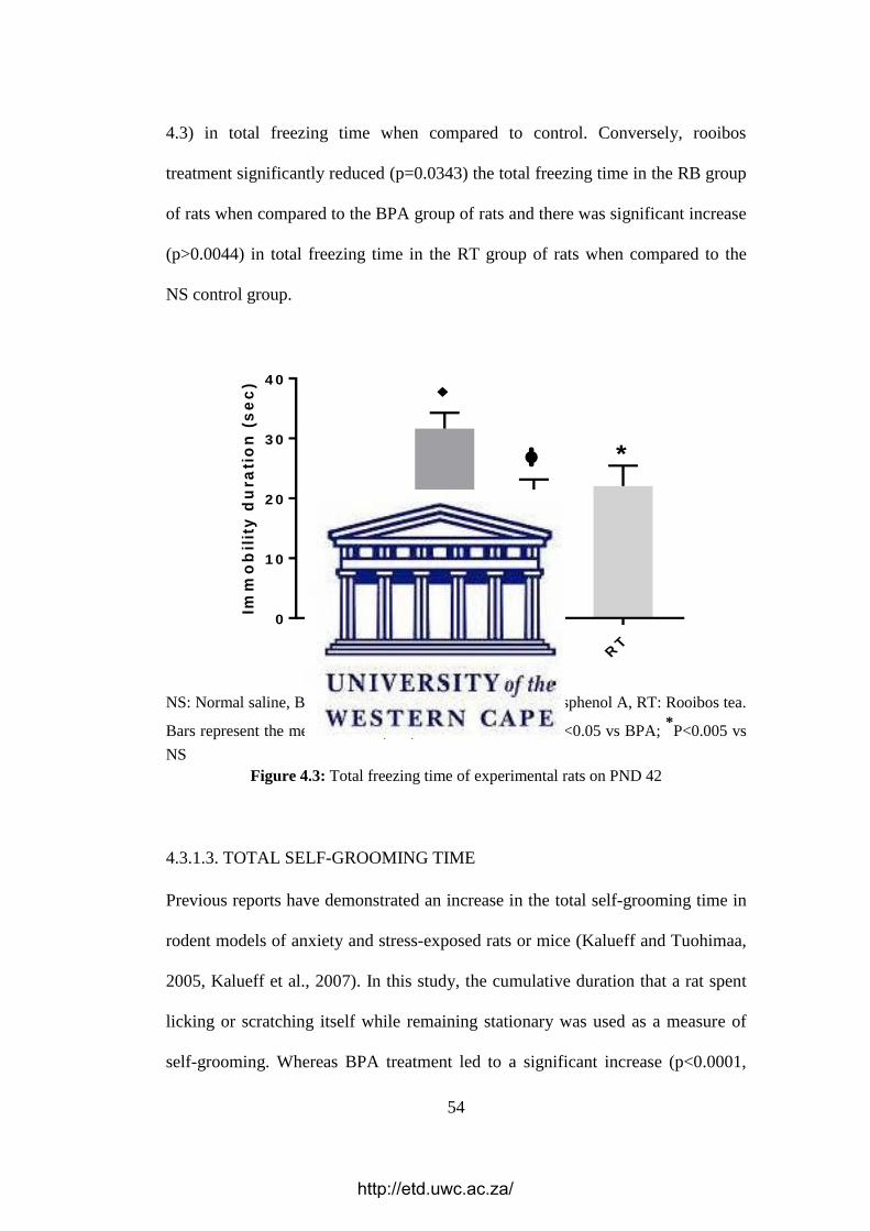
54
4.3) in total freezing time when compared to control. Conversely, rooibos
treatment significantly reduced (p=0.0343) the total freezing time in the RB group
of rats when compared to the BPA group of rats and there was significant increase
(p>0.0044) in total freezing time in the RT group of rats when compared to the
NS control group.
NS
BP
A
RB
RT
0
1 0
2 0
3 0
4 0
Imm
ob
ilit
y d
ura
tio
n (
se
c)
*
NS: Normal saline, BPA: Bisphenol A, RB: Rooibos tea + Bisphenol A, RT: Rooibos tea.
Bars represent the mean + SEM (n=6). P<0.001 vs NS; фP<0.05 vs BPA; *P<0.005 vs
NS
Figure 4.3: Total freezing time of experimental rats on PND 42
4.3.1.3. TOTAL SELF-GROOMING TIME
Previous reports have demonstrated an increase in the total self-grooming time in
rodent models of anxiety and stress-exposed rats or mice (Kalueff and Tuohimaa,
2005, Kalueff et al., 2007). In this study, the cumulative duration that a rat spent
licking or scratching itself while remaining stationary was used as a measure of
self-grooming. Whereas BPA treatment led to a significant increase (p<0.0001,
http://etd.uwc.ac.za/

55
Figure 4.4) in total self-grooming time when compared to the control, rooibos
treatment significantly reduced (p<0.0001) the total self-grooming time as seen in
the RB group when compared to the BPA group. Also, there was no significant
difference (p>0.05) in total freezing time in the RT group of rats when compared
to the NS control group.
NS
BP
A
RB
RT
0
1 0
2 0
3 0
4 0
5 0
Gro
om
ing
tim
e (
se
c)
NS: Normal saline, BPA: Bisphenol A, RB: Rooibos tea + Bisphenol A, RT: Rooibos tea.
Bars represent the mean + SEM (n=6). P<0.001 vs NS; фP<0.05 vs BPA.
Figure 4.4: Total self-grooming time of experimental rats on PND 42
4.3.2. NOVEL OBJECT RECOGNITION (NOR) TEST
The NOR test was used in this study to determine the ability of a rat to recognize
and spend longer time exploring a novel object introduced into its environment. In
this study, rats were exposed to both novel (N) and familiar (old, F) objects for 3
minutes, and the Recognition Index (RI) was calculated from the formula below:
http://etd.uwc.ac.za/

56
𝑅𝐼 = 𝑇𝑁
(𝑇𝑁 + 𝑇𝐹)… … … … … . 𝐹𝑜𝑟𝑚𝑢𝑙𝑎 1,
Where RI = Recognition Index, TN = Time spent with a novel object and TF = time spent
with a familiar object
Findings show that BPA treatment led to a reduction (p=0.6289, Figure 4.5) in RI
when compared to the control, however, in the RB group of rats, there was a
significant increase (p<0.0269) in RI when compared to the BPA group of rats.
Also, there was no significant difference (p>0.05) between the RT group of rats
and the NS control group.
NS
BP
A
RB
RT
0
5
1 0
1 5
2 0
2 5
Re
co
gn
itio
n I
nd
ex
NS: Normal saline, BPA: Bisphenol A, RB: Rooibos tea + Bisphenol A, RT: Rooibos tea.
Bars represent the mean + SEM (n=6). фP<0.05 vs BPA.
Figure 4.5: Recognition index of experimental rats on PND 42
http://etd.uwc.ac.za/

57
4.4. EFFECT OF BPA AND FERMENTED ROOIBOS HERBAL TEA ON THE
ACTIVITY OF ANTIOXIDANT ENZYMES
4.4.1. SUPEROXIDE DISMUTASE
Reactive oxygen species (ROS) are generally regarded as cytotoxic agents that
induce oxidative damage by attacking cell membranes and DNA (Ozaydın et al.,
2018). The production of ROS may be triggered by inflammation, radiation,
drugs, aging and such chemical substances as BPA. However, antioxidant
enzymes such as SOD and CAT are known to inhibit the damage caused by ROS.
An increase in ROS generation or a reduction in cellular antioxidant levels results
in a condition known as oxidative stress (Hassan et al., 2012, Abdel-Wahab,
2014). BPA has been reported to cause damage in many organs such as the liver,
kidney and brain by lowering antioxidant activity, thus increasing oxidative stress
(Song et al., 2014, Moghaddam et al., 2015).
Superoxide dismutases (SOD) are enzymes that counteract oxidative damage by
catalyzing the dismutation of superoxide radical (O2-) into either molecular
oxygen O2 or H2O2 which are less toxic molecules (Hollman and Katan, 1999).
Findings from this study show that BPA treatment resulted in a significant
decrease (p<0.005, Figure 4.6-4.7) in cerebral and cerebellar SOD activity;
however, in the RB group of rats, there was a significant increase (p<0.05) in
SOD activity when compared to BPA group of rats.
There was no significant difference (p<0.05) in SOD activity in the RT group of
rats when compared to the NS control group.
http://etd.uwc.ac.za/

58
T re a tm e n t
SO
D a
cti
vit
y (
U/m
g p
ro
tein
)
NS
BP
AR
BR
T
0 .0 0
0 .0 5
0 .1 0
0 .1 5
0 .2 0
0 .2 5
NS: Normal saline, BPA: Bisphenol A, RB: Rooibos tea + Bisphenol A, RT: Rooibos tea.
Bars represent the mean + SEM (n=3). P<0.005 vs NS; фP<0.005 vs BPA.
Figure 4.6: Effects of different treatments on SOD enzyme activity in the cerebral
homogenate of experimental rats on PND 42
T re a tm e n t
SO
D a
cti
vit
y (
U/m
g p
ro
tein
)
NS
BP
AR
BR
T
0 .0 0
0 .0 5
0 .1 0
0 .1 5
0 .2 0
NS: Normal saline, BPA: Bisphenol A, RB: Rooibos tea + Bisphenol A, RT: Rooibos tea.
Bars represent the mean + SEM (n=3). P<0.05 vs NS; фP<0.05 vs BPA.
Figure 4.7: Effects of different treatments on SOD enzyme activity in the cerebellar
homogenate of experimental rats on PND 42
http://etd.uwc.ac.za/

59
4.4.2. CATALASE
CAT is involved in the detoxification of H2O2 through the conversion of two
molecules of H2O2 to two molecules of H2O and one molecule of O2 in tissues
(Nagata et al., 1999, Agati et al., 2012). The rate of H2O2 disintegration is directly
proportional to catalase concentration hence, higher CAT concentrations indicate
highly potent antioxidant effects and vice versa. Following treatment of rats with
BPA, there was a significant reduction in the cerebral and cerebellar catalase
activity (p<0.05, Figure 4.8-4.9). In the RB group, there was a significant increase
(p<0.05) in CAT activity when compared to BPA group. Also, there was no
significant difference (p<0.05) in CAT activity in the RT group of rats when
compared to the NS control group.
T re a tm e n t
CA
T a
cti
vit
y (
U/m
l)
NS
BP
AR
BR
T
0
2
4
6
8
NS: Normal saline, BPA: Bisphenol A, RB: Rooibos tea + Bisphenol A, RT: Rooibos tea.
Bars represent the mean + SEM (n=3). P<0.05 vs NS; фP<0.05 vs BPA.
Figure 4.8: Effects of different treatments on CAT enzyme activity in the cerebral
homogenate of experimental rats on PND 42
http://etd.uwc.ac.za/

60
T re a tm e n t
CA
T a
cti
vit
y (
U/m
l)
NS
BP
AR
BR
T
0
2
4
6
8
NS: Normal saline, BPA: Bisphenol A, RB: Rooibos tea + Bisphenol A, RT: Rooibos tea.
Bars represent the mean + SEM (n=3). P<0.05 vs NS; фP<0.05 vs BPA.
Figure 4.9: Effects of different treatments on CAT enzyme activity in the cerebellar
homogenate of experimental rats on PND 42
4.5. HISTOLOGICAL STUDIES
In this study, coronal sections of the cerebrum were obtained and stained to study
the potential neuroprotective effects of fermented rooibos herbal tea on the
development of CNS structures in rats exposed to BPA during pregnancy and
lactation. Coronal sections were cut and stained with H&E and Cresyl violet (CV)
for cell counting and histological analysis of the shapes and sizes of cell bodies in
the (CA1) region of the hippocampus while GFAP immunostaining was used for
analysis of damage responses in astrocytes. Similar analysis was done for the
Purkinje cells and the glial cells in the cerebellum.
http://etd.uwc.ac.za/

61
4.5.1. LENGTH OF HIPPOCAMPAL CA1 REGION IN EXPERIMENTAL
RATS
The H & E staining revealed very clear outlines of the pyramidal cell layer of the
hippocampal region of interest - the CA1 region. The length of this region was
determined by measurement along the dotted red landmark using an appropriate
Image-j tool as shown in Figure 4.10. In order to ensure that the sampling size
was sufficient for this analysis, specimens from six rats were assigned to each
group and three (3) separate staggered serial sections were used per animal, giving
a total of 18 sections per group. The singular CA1 region of each slide was
measured and average values were computed and plotted for each group using the
Graph Pad Prism software version 6.
CA - cornua ammonis (subregions - CA1, CA2 and CA3); DG – Dentate gyrus.
Figure 4.10: Diagram of the hippocampus showing the landmarks used for determining
the hippocampal length in experimental rats.
Findings show that on PND 42, there was a significant reduction (p<0.0001,
Figure 4.11) in the hippocampal CA1 length of rats treated with BPA when
http://etd.uwc.ac.za/
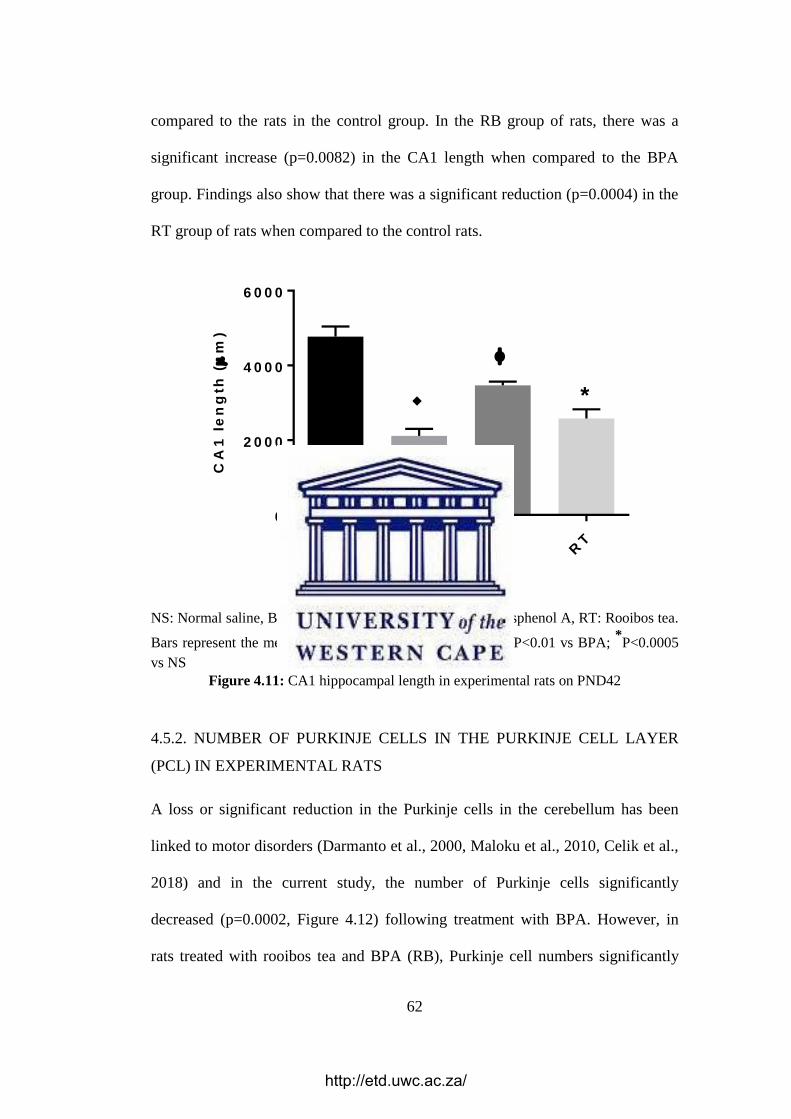
62
compared to the rats in the control group. In the RB group of rats, there was a
significant increase (p=0.0082) in the CA1 length when compared to the BPA
group. Findings also show that there was a significant reduction (p=0.0004) in the
RT group of rats when compared to the control rats.
T re a tm e n t
CA
1 l
en
gth
(
m)
NS
BP
AR
BR
T
0
2 0 0 0
4 0 0 0
6 0 0 0
*
NS: Normal saline, BPA: Bisphenol A, RB: Rooibos tea + Bisphenol A, RT: Rooibos tea.
Bars represent the mean + SEM (n=3). P<0.0001 vs NS; фP<0.01 vs BPA; *P<0.0005
vs NS
Figure 4.11: CA1 hippocampal length in experimental rats on PND42
4.5.2. NUMBER OF PURKINJE CELLS IN THE PURKINJE CELL LAYER
(PCL) IN EXPERIMENTAL RATS
A loss or significant reduction in the Purkinje cells in the cerebellum has been
linked to motor disorders (Darmanto et al., 2000, Maloku et al., 2010, Celik et al.,
2018) and in the current study, the number of Purkinje cells significantly
decreased (p=0.0002, Figure 4.12) following treatment with BPA. However, in
rats treated with rooibos tea and BPA (RB), Purkinje cell numbers significantly
http://etd.uwc.ac.za/

63
increased (p<0.0001) when compared to rats treated with BPA only. In the RT
group, there was no significant difference (p=0.9849) in the number of Purkinje
cells when compared to rats in the NS control group.
T re a tm e n t
Nu
mb
er o
f P
urk
inje
ce
lls
NS
BP
AR
BR
T
0
2 0
4 0
6 0
8 0
NS: Normal saline, BPA: Bisphenol A, RB: Rooibos tea + Bisphenol A, RT: Rooibos tea.
Bars represent the mean + SEM (n=4). P<0.0005 vs NS; фP<0.0001 vs BPA.
Figure 4.12: Number of Purkinje cells in experimental rats on PND42
http://etd.uwc.ac.za/

64
4.6. IMMUNOHISTOCHEMISTRY (IHC)
4.6.1. GLIAL FIBRILLARY ACIDIC PROTEIN (GFAP)
GFAP is an essential protein of intermediate glial filaments in astrocytes used for
the assessment of damage responses in astrocytes (Cikriklar et al., 2016). An
increase in the expression of GFAP is a key feature in astrocytic and CNS damage
(Hausmann et al., 2000, Yu et al., 2004). A total of three rats were assigned per
group, and tissue sections were processed for immunostaining to investigate
GFAP. The immunoreactive astrocytes for GFAP were determined in six counting
boxes (440 × 330 µm) per slide along the CA1 region, which surrounded the
polymorphic and molecular layers adjoining the pyramidal layer. The average
values were computed, plotted and compared between the different treatment
groups using the Graph Pad Prism software version 6.
Results showed that there were significantly more (p<0.0001, Figure 4.13) GFAP-
immunoreactive astrocytes in the BPA group of rats when compared to control
while the rooibos (RB) group of rats had significantly fewer (p<0.0001) GFAP-
immunoreactive astrocytes when compared to the BPA only -treated group. Also,
there was no significant difference (p>0.05) between the RT group of rats and the
rats in the NS control group.
http://etd.uwc.ac.za/

65
T re a tm e n t
No
. o
f G
FA
P p
os
itiv
e c
ell
s
NS
BP
AR
BR
T
0
5 0
1 0 0
1 5 0
2 0 0
2 5 0
NS: Normal saline, BPA: Bisphenol A, RB: Rooibos tea + Bisphenol A, RT: Rooibos tea.
Bars represent the mean + SEM (n=3). P<0.0001 vs NS; фP<0.0001 vs BPA.
Figure 4.13: Number of GFAP positive cells in experimental rats on PND42
http://etd.uwc.ac.za/

66
CHAPTER 5
DISCUSSION
5.1. GENERAL INTRODUCTION
Endocrine-disrupting chemicals (EDCs) are a wide range of compounds that
disrupt normal growth and development by halting or modifying the action of
hormones upon ingestion or absorption into the body (Schug et al., 2015).
Examples of EDCs include a broad group of natural and synthetic chemical
substances, such as polycyclic aromatic hydrocarbons (PAHs), polychlorinated
biphenyls (PCBs), pesticides, herbicides, fungicides, BPA and
dichlorodiphenyltrichloroethane (DDT). EDCs are abundant in our environs, diet,
and consumer products, which makes exposure to these toxins very frequent and
regular (Waring and Harris, 2005). Exposure to EDCs during initial
developmental stages is known to interrupt normal developmental processes,
thereby altering the body’s endocrine system to initiate deleterious effects in
various systems in the human body (Schug et al., 2015). For instance, EDCs
initiates neurotoxic effects that disturb the synthesis, transport, and release of such
neurotransmitters as glutamate, dopamine, serotonin, norepinephrine, which play
important roles in regulating behaviour, cognition, learning, and memory
(Dickerson and Gore, 2007, Rasier et al., 2007).
This study was designed to investigate if continuous consumption of fermented
rooibos herbal tea (FRHT) could offer diverse protection against potential damage
http://etd.uwc.ac.za/

67
to the developing CNS of offspring Wistar rats exposed to BPA, following ad
libitum maternal consumption during and after pregnancy as observed on PND 42.
The offspring were considered to have ‘formed’ and ‘developed’ in the presence
of ad libitum rooibos tea consumption including postnatal consumption through
breastfeeding. After the evaluation of neurobehavioural activity, rats were
sacrificed, and cerebral as well as cerebellar samples were examined for changes
in antioxidant enzyme activities and histomorphology. The results obtained from
this study are discussed in the following sections.
5.2. ROOIBOS TEA PREVENTED BPA-INDUCED GAIN IN BODY MASS
In this study, findings revealed an increase in the body mass of experimental rats
treated with BPA only. This is in line with previous reports demonstrating that
perinatal BPA exposure increases body mass when compared to controls (Rubin
et al., 2001, Miyawaki et al., 2007, Ezz et al., 2015). There are confirmed reports
which link BPA to weight gain and obesity (Newbold et al., 2009, Rubin and
Soto, 2009, Shankar et al., 2012). For instance, Li and colleagues reported that
high BPA exposure might promote childhood obesity and might advance global
obesity epidemic in the human population (Li et al., 2013a). In the same regard,
Harley and co-workers revealed that elevated urinary BPA concentrations in
children aged nine were linked to higher odds of obesity, increased waist
circumference, BMI z-score and percentage body fat (Harley et al., 2013). An
appraisal of available literature reveals other reports of body mass gain in
offspring of rodent mothers exposed to BPA during gestation and/or lactation, or
during the early postnatal period (Howdeshell et al., 1999). Also, postnatal
exposure of BPA in male rat pups to investigate the effects of BPA on anxiety
http://etd.uwc.ac.za/

68
revealed that exposed males substantially weighed more than the controls
(Patisaul and Bateman, 2008). Consequently, the increase in body mass gain in
this study highlights the ability of BPA to promote the progression of obesity
which could in turn further aggravate several metabolic, cardiovascular and
neurological disorders associated with BPA exposure.
Several mechanisms have been proposed to explain the association between BPA
exposure and body mass gain including the ability of BPA to enhance
differentiation of adipocytes and promote lipid accumulation in target cells
(Masuno et al., 2002, Wada et al., 2007), as well as increased gene expression of
adipogenic transcription factors (Phrakonkham et al., 2008). Others include the
ability of BPA to inhibit the release of adiponectin from human adipose tissue
explants (Hugo et al., 2008), dysregulate neuronal appetite controlling circuits
through its action on the hypothalamus (Wade and Schneider, 1992, Simerly,
2008), and inhibit the signalling of PPARgamma, which is essential for lipid
homeostasis and differentiation of brown and white adipocytes (Grun and
Blumberg, 2006, Grün and Blumberg, 2007). Findings from this study confirm
that although perinatal BPA exposure is a major challenge with respect to weight
gain and possibly childhood obesity, these effects could be attenuated by rooibos
through its protective activity against BPA.
5.3. ROOIBOS TEA MITIGATED BPA-INDUCED NEUROBEHAVIOURAL
DEFICITS
In this study, the open field test (OFT) was employed to investigate certain
neurobehavioral tests such as the frequency of rearing episodes, total freezing
http://etd.uwc.ac.za/
69
time, total self-grooming time and the novel object recognition (NOR) test. The
OFT, initially developed in 1934, is a routine test that evaluates emotionality,
exploratory behaviour and general activity in rodents (Hall, 1934). It is one of the
most commonly used measures of behaviour in animal psychology because it
offers a prompt and easy evaluation of precise behaviours that require little or no
training to the test subject and the person administering the test (Walsh and
Cummins, 1976, Prut and Belzung, 2003). Also, its popularity stems from the
psychological and physiological conceptions underlying the tests, which are
largely uncomplicated and easy to understand (Seibenhener and Wooten, 2015).
Although the primary outcome of interest is “movement”, it is sometimes
influenced by other factors such as motor output, exploratory drive, freezing, fear-
related behaviour and sickness (Gould et al., 2009). The OFT is also employed to
assess the toxic effects of compounds in rodents and thus evaluates different
facets of behaviour other than movement/locomotion (Gould et al., 2009).
The frequency of rearing episodes is regarded as a measure of exploratory activity
in the OFT and indicates the number of times rats stood on both hind paws in a
vertically upright position (Erdoğan et al., 2004, Ennaceur, 2014). Reports show
that a reduction in rearing episodes is indicative of increased anxiety (Costall et
al., 1989) and is further demonstrated under experimental conditions, such as
stress (Masood et al., 2003, Fan et al., 2011), drug treatment (van Lier et al.,
2004) and brain stimulation (Racine, 1972, Hannesson et al., 2001). In this study,
the reduction in the frequency of rearing episodes in the group of rats exposed to
BPA only could be indicative of increased anxiety and stress as demonstrated in
previous studies. However, following exposure to rooibos, it was observed that
http://etd.uwc.ac.za/

70
experimental rats exhibited an increase in rearing episodes, thus indicating that
rooibos was protective against BPA-induced reduction in the frequency of rearing,
possibly indicative of induced anxiety or stress.
Freezing activity, an important indicator of anxiety in rodents (Díaz-Morán et al.,
2014), is defined as the duration of the OFT within which the rodent was
stationary. It is conspicuous in conditions where stress levels are noticeably high
and occurs in response to apparent danger, environmental displacement, sudden
changes and fright stimuli (Walsh and Cummins, 1976, Brandao et al., 2008). In
line with earlier studies showing increased freezing activity in stress conditions
(Ranjbar et al., 2017) and in rats with impaired brain function (Saikhedkar et al.,
2014), total freezing time was significantly elevated in this study in the group of
rats exposed to BPA only. Conversely, we observed a significant decline in total
freezing time in the BPA-and-rooibos treated rats, thus confirming the protective
activity of rooibos against BPA in experimental rats.
Total self-grooming time is defined as the duration of time the experimental rats
expended in licking or scratching itself while motionless. It is a very important
behavioural activity in rodents and is involved in biologically essential processes,
such as thermoregulation, social communication and de-arousal (Spruijt et al.,
1988, Spruijt et al., 1992, Leonard et al., 2005). Previous reports indicate that self-
grooming is enhanced by exposure to stress (Jolles et al., 1979, Katz and Roth,
1979) and in rodent models of anxiety such as stress-induction in rats or mice
after exposure to anxiogenic drugs (Kalueff and Tuohimaa, 2005, Kalueff et al.,
2007).
http://etd.uwc.ac.za/

71
In this study, the increase in self-grooming observed in the BPA-only group could
be indicative of increased anxiety and stress. However, following exposure to
rooibos in the BPA-and-rooibos group, it was observed that the rats exhibited a
significant reduction in self-grooming, thus demonstrating that rooibos tea
consumption could have offered protection against BPA-induced anxiety-like
behaviour demonstrated as increase in self-grooming in this study.
The integrity of the medial temporal lobe is essential for memory processing and
this can be evaluated through the recognition index (RI), which is a measure of
the capability of a person or an animal to determine if a previously encountered
item is familiar or not (Squire et al., 2007). Tests that evaluate RI are becoming
gradually valuable tools for research designed to explore the neural basis of
memory (Winters et al., 2004). One such tests is the novel object recognition task
(NOR), studied for the first time by Ennaceur and Delacour to evaluate short-term
memory in rodents (Ennaceur and Delacour, 1988) which highlights the capacity
of the rodent to recall information for a short amount of time (Mathiasen and
DiCamillo, 2010). The NOR can be designed to evaluate working memory,
attention, anxiety, and preference for novelty in rodents (Silvers et al., 2007,
Goulart et al., 2010). It has also been utilized to investigate the effects of several
pharmacological treatments and brain impairment in rodents (Goulart et al.,
2010).
Findings from the present study showed that RI was significantly lowered in
BPA-only treated rats when compared to the NS controls. This is in line with
previous reports which show reduced RI in stress conditions (Nagata et al., 2009,
http://etd.uwc.ac.za/

72
Eagle et al., 2013). Conversely, when compared to rats exposed to BPA only, a
significant increase in RI was observed in the RB group thus suggesting a
neuroprotective role by rooibos tea against possible BPA-induced short-term
memory loss.
5.4. ROOIBOS TEA REGULATED THE ACTIVITY OF ANTIOXIDANT
ENZYMES
Reports show that exposure to BPA during pregnancy can result in a variety of
foetal malformations and growth retardations as well as early embryonic losses
(Bajaj et al., 1993, Friedler, 1996, Khattak et al., 1999), possibly via the
production of excessive ROS known to be toxic to cells (Nicol et al., 2000, Wells
et al., 2005). BPA-induced ROS production has been investigated in many cell
types and at different concentrations (Huc et al., 2012, Babu et al., 2013, Gassman
et al., 2015). For instance, nanomolar BPA concentration ranges were found to
result in short and transient elevations of ROS (Koong and Watson, 2015, Pfeifer
et al., 2015); nevertheless prolonged and increased levels of oxidative stress have
been reported (Huc et al., 2012, Porreca et al., 2016). BPA exposure in
micromolar concentration ranges have been reported to induce higher levels of
oxidative stress (Huc et al., 2012, Babu et al., 2013, Gassman et al., 2015, Leem et
al., 2017), and cytotoxicity via ROS-triggered DNA damage (Ooe et al., 2005,
Huc et al., 2012, Leem et al., 2017). Different in vivo studies have shown an
association between high urinary BPA levels and elevated concentrations of such
oxidative stress biomarkers as lipid peroxidation and malondialdehyde (Watkins
et al., 2015, Zhang et al., 2016), thus supporting the pro-oxidant activity of BPA.
http://etd.uwc.ac.za/

73
Cellular antioxidants mitigate the deleterious effects arising from interactions
between lipid, protein, DNA molecules and ROS (Hassan et al., 2012).
Irrespective of the existence of a potent antioxidant system, an imbalance in the
generation of ROS due to natural or synthetic chemicals may result in some
disorders. The concentration of free radical scavengers and the levels of such
antioxidant enzymes as SOD and CAT regulate the antioxidant capacity of cells;
thus numerous endpoints have been investigated following BPA exposure to
evaluate its effect on antioxidant capacity (Gassman, 2017).
The enzyme SOD is known to defend tissues from oxidative damage by
catalyzing the conversion of O2− to H2O2 (Fridovich, 1997), while catalase is
responsible for the detoxification of the H2O2 generated by SOD through the
conversion of two molecules of H2O2 to form two molecules of H2O and one
molecule of O2 (Nagata et al., 1999, Agati et al., 2012), thus providing protection
against ROS. Findings from the current study showed that whereas BPA induced
marked oxidative damage by inhibiting the activities of the antioxidant enzymes
SOD and CAT, the intake of rooibos tea could attenuate these effects of BPA.
These findings are in agreement with other studies that report a significant
decrease in SOD and CAT levels following BPA treatment (Eid et al., 2015,
Moghaddam et al., 2015, Ozaydın et al., 2018).
5.5. ROOIBOS TEA ATTENUATED NEURODEGENERATION IN RATS
EXPOSED TO BPA
The development of the brain involves a complex interplay between dendritic
growth and the production of neuronal connections to form a neural network
(McAllister et al., 1997, Wong and Ghosh, 2002). Different brain regions are
http://etd.uwc.ac.za/

74
tasked with different functions, and histological damage to these brain regions
hinders their function (Mcallister et al., 2004, Kimura et al., 2016). The
hippocampus is subdivided into two main parts which have been studied
comprehensively for spatial navigation, learning and memory functions; these are
the cornu ammonis (subfields: CA1, CA2, CA3, CA4) and the dentate gyrus
(Bannerman et al., 2014, Moser et al., 2014). Alterations in hippocampal
morphology are linked to changes in cognitive function (Van Praag et al., 2000)
and the hippocampal CA1 (H-CA1) neurons in rodents are reported to be essential
for the processing of hippocampus-dependent memory (Bartsch et al., 2011).
These neurons are also the most sensitive and most susceptible neurons to
selective degeneration in response to such EDCs as BPA, ischemia and other toxic
substances (Kirino, 1982, Pulsinelli et al., 1982, Elsworth et al., 2013, Kimura et
al., 2016).
Previous studies have shown that there was a reduction in the dendritic spines of
neurons in the H-CA1 region following exposure of mice to BPA (Eilam-Stock et
al., 2012, Kimura et al., 2016, Liu et al., 2016). The results obtained from the
present study are in agreement with the above findings as there was a significant
reduction in the average length of the H-CA1 region in rats exposed to BPA-only
when compared to the NS control group. However, a significant increase in the
average H-CA1 length was observed in rats treated with BPA-and-rooibos when
compared to BPA-only rats. These morphological findings indicate a protective
effect of rooibos in the CA1 region of the hippocampus in rats, thus attenuating
BPA-induced impairment of cognition in the rats.
http://etd.uwc.ac.za/

75
In certain human inherited disease conditions, such as ataxia telangiectasia,
paraneoplastic cerebellar degeneration and lysosomal storage diseases, a
significant loss of Purkinje cells has been reported (Gilman et al., 1981, Anderson
et al., 1987) as a noticeable component of the histopathological alterations
detected in cerebellar injury. Some of the clinical indicators of cerebellar
dysfunction frequently manifested in these conditions include ataxia and tremor
(Riedel et al., 1990). Purkinje cell loss is a significant observation in many
diseases but the exact cause is unknown.
In some in vivo studies, the significant reduction in Purkinje cell population is due
to the neurotoxic effect of EDCs on Purkinje cells (Kimura-Kuroda et al., 2007,
Ibhazehiebo et al., 2011) and the reduction in Purkinje cells observed in this study
is similar to findings from these previous studies, possibly indicating that the
deleterious effects of BPA on the cerebellum (Ahmed and Eid, 2015). The number
of Purkinje cells however significantly increased in rats treated with BPA-and-
rooibos tea when compared to the BPA-only rats, thus suggesting a
neuroprotective role for rooibos tea.
Apart from neuronal damage caused by most EDCs, it is plausible to also
investigate possible damage to the support cells of the CNS. Astrocytes (also
called astroglia) are the most abundant support cells in the CNS (Zuchero and
Barres, 2015) and contain GFAP which is an important protein of intermediate
glial filaments in astrocytes commonly used to investigate astrocytic damage
response (Cikriklar et al., 2016). In this study, there was an increase in the
expression of GFAP-positive astrocytes in BPA-exposed rats when compared to
http://etd.uwc.ac.za/

76
controls. Increased GFAP expression is prominent in conditions of CNS injury
and astrocyte damage (Hausmann et al., 2000, Yu et al., 2004, Cai et al., 2006)
previous studies have reported on the various mechanisms of astrocyte activation
possibly associated with BPA exposure. For instance, increased generation of
hydroxyl radicals has been reported in the adult mouse brain following BPA
exposure (Kabuto et al., 2003) and elevated concentrations of lipid peroxidation in
postnatal mouse brains was reported following BPA exposure during pregnancy
and lactation (Kabuto et al., 2004). Consequently, BPA-triggered oxidative stress
and peroxidation could be said to be responsible for the astroglial activation
reported in this study. However, rats exposed to rooibos tea had significantly
reduced GFAP levels when compared to BPA-only treated rats. Taken together,
findings from this study seem to indicate that rooibos tea is protective against the
deleterious effects of BPA in the developing brain.
http://etd.uwc.ac.za/

77
5.6. CONCLUSION
This study provides the first research evidence of the neuroprotective potential of
fermented rooibos herbal tea against BPA exposure on brain development in
Wistar rats. The protective activity observed is possibly due to the natural and
bioactive constituents of this herb, reported to be responsible for the regulation of
cellular antioxidants which inhibit BPA-induced oxidative damage. These
findings suggest that rooibos tea offers protection against BPA toxicity, and may
provide the basis for clinical trials and policy decisions aimed at regulating the
use of products containing such EDCs as BPA.
Limitations of this study include investigation of few immunohistochemical
markers in the brains of experimental rats, few neurobehavioural tests to confirm
BPA-induced impairment of cognitive function, and the evaluation of only few
oxidative stress markers to fully elucidate the impact of oxidative stress on BPA
exposure. These limitations are partly due to financial constraints and the need to
pitch the volume of research data at the MSc level.
Future recommendations
Isolation of the bioactive compounds in rooibos and investigation of their
neuroprotective activity against BPA.
Investigation of additional immunohistochemical, apoptotic, inflammatory and
oxidative stress biomarkers to corroborate findings from this study.
http://etd.uwc.ac.za/

78
Assessment of additional OFT activities and neurobehavioural tests (such as
T-maze test and water maze test) to offer more information on BPA-induced
impairment of locomotion, learning and memory.
http://etd.uwc.ac.za/

79
REFERENCES
Abdallah, M. a. M. 2016. Endocrine Disruptors as Pollutants in Marine Ecosystem: A
Case Study in Egypt. The Open Biotechnology Journal, 10.
Abdel-Wahab, W. M. 2014. Thymoquinone attenuates toxicity and oxidative stress
induced by bisphenol A in liver of male rats. Pak J Biol Sci, 17, 1152-1160.
Abhang, P. A., Gawali, B. W. & Mehrotra, S. C. 2016. Introduction to EEG-and Speech-
Based Emotion Recognition, Academic Press.
Addae, C., Cheng, H. & Martinez-Ceballos, E. 2013. Effect of the environmental
pollutant hexachlorobenzene (HCB) on the neuronal differentiation of mouse
embryonic stem cells. International journal of environmental research and public
health, 10, 5244-5256.
Agati, G., Azzarello, E., Pollastri, S. & Tattini, M. 2012. Flavonoids as antioxidants in
plants: location and functional significance. Plant Science, 196, 67-76.
Ahmed, M. a. a. S. & Eid, R. A. 2015. Effect of Bisphenol-A on the Post-Natal
Development and Structure of Rat Cerebellum. Int. J. Curr. Microbiol. App. Sci,
4, 14-35.
Akingbemi, B. T., Sottas, C. M., Koulova, A. I., Klinefelter, G. R. & Hardy, M. P. 2004.
Inhibition of testicular steroidogenesis by the xenoestrogen bisphenol A is
associated with reduced pituitary luteinizing hormone secretion and decreased
steroidogenic enzyme gene expression in rat Leydig cells. Endocrinology, 145,
592-603.
Akinrinmade, O., Omoruyi, S., Dietrich, D. & Ekpo, O. 2017. Long‑term consumption of
fermented rooibos herbal tea offers neuroprotection against ischemic brain injury
in rats. Acta Neurobiol Exp, 77, 94-105.
Alonso-Magdalena, P., Ropero, A. B., Soriano, S., Quesada, I. & Nadal, A. 2010.
Bisphenol-A: a new diabetogenic factor. Hormones (Athens), 9, 118-26.
Alves, R., De Carvalho, J. G. B. & Venditti, M. a. C. 2012. High-and low-rearing rats
differ in the brain excitability controlled by the allosteric benzodiazepine site in
the GABAA receptor. Journal of Behavioral and Brain Science, 2, 315.
Andersen, B. B., Korbo, L. & Pakkenberg, B. 1992. A quantitative study of the human
cerebellum with unbiased stereological techniques. Journal of Comparative
Neurology, 326, 549-560.
Anderson, N., Cunningham, J. & Posner, J. 1987. Autoimmune pathogenesis of
paraneoplastic neurological syndromes. Critical reviews in neurobiology, 3, 245-
299.
Angle, B. M., Do, R. P., Ponzi, D., Stahlhut, R. W., Drury, B. E., Nagel, S. C., Welshons,
W. V., Besch-Williford, C. L., Palanza, P. & Parmigiani, S. 2013. Metabolic
http://etd.uwc.ac.za/

80
disruption in male mice due to fetal exposure to low but not high doses of
bisphenol A (BPA): evidence for effects on body weight, food intake, adipocytes,
leptin, adiponectin, insulin and glucose regulation. Reproductive toxicology, 42,
256-268.
Apps, R. & Garwicz, M. 2005. Anatomical and physiological foundations of cerebellar
information processing. Nature Reviews Neuroscience, 6, 297.
Ardila, A., Bernal, B. & Rosselli, M. 2016. The role of Wernicke's area in language
comprehension. Psychology & Neuroscience, 9, 340.
Aschberger, K., Castello, P., Hoekstra, E., Karakitsios, S., Munn, S., Pakalin, S. &
Sarigiannis, D. 2010. Bisphenol A and baby bottles: challenges and perspectives.
Luxembourg: Publications Office of the European Union, 5-50.
Auddy, B., Ferreira, M., Blasina, F., Lafon, L., Arredondo, F., Dajas, F., Tripathi, P.,
Seal, T. & Mukherjee, B. 2003. Screening of antioxidant activity of three Indian
medicinal plants, traditionally used for the management of neurodegenerative
diseases. Journal of Ethnopharmacology, 84, 131-138.
Aversi-Ferreira, T. A., Araújo, M. F. P. D., Lopes, D. B. & Nishijo, H. 2010. History,
citoarchitecture and neurophysiology of human and non human primates' parietal
lobe: A review. Dementia & Neuropsychologia, 4, 173-180.
Babu, S., Uppu, S., Claville, M. O. & Uppu, R. M. 2013. Prooxidant actions of bisphenol
A (BPA) phenoxyl radicals: implications to BPA-related oxidative stress and
toxicity. Toxicology mechanisms and methods, 23, 273-280.
Bajaj, J., Misra, A., Rajalakshmi, M. & Madan, R. 1993. Environmental release of
chemicals and reproductive ecology. Environmental health perspectives, 101,
125-130.
Balabanič, D. & Klemenčič, A. K. 2018. Endocrine-disrupting chemicals and male
reproductive health: a review. Slovenian Medical Journal, 87.
Bannerman, D. M., Sprengel, R., Sanderson, D. J., Mchugh, S. B., Rawlins, J. N. P.,
Monyer, H. & Seeburg, P. H. 2014. Hippocampal synaptic plasticity, spatial
memory and anxiety. Nature reviews neuroscience, 15, 181.
Barsottini, O. G. P., Albuquerque, M. V. C. D., Braga Neto, P. & Pedroso, J. L. 2014.
Adult onset sporadic ataxias: a diagnostic challenge. Arquivos de neuro-
psiquiatria, 72, 232-240.
Bartsch, T., Döhring, J., Rohr, A., Jansen, O. & Deuschl, G. 2011. CA1 neurons in the
human hippocampus are critical for autobiographical memory, mental time travel,
and autonoetic consciousness. Proceedings of the National Academy of Sciences,
201110266.
Bayer, S. A. 1985. Hippocampal region. The rat nervous system, 1, 335-352.
http://etd.uwc.ac.za/

81
Berquin, P., Giedd, J., Jacobsen, L., Hamburger, S., Krain, A., Rapoport, J. &
Castellanos, F. 1998. Cerebellum in attention-deficit hyperactivity disorder A
morphometric MRI study. Neurology, 50, 1087-1093.
Betancourt, A. M., Eltoum, I. A., Desmond, R. A., Russo, J. & Lamartiniere, C. A. 2010.
In utero exposure to bisphenol A shifts the window of susceptibility for
mammary carcinogenesis in the rat. Environmental health perspectives, 118,
1614.
Bhattacharya, S. & Mukherjee, S. 1975. Toxic effects of endrin on hepatopancreas of the
teleost fish, Clarias batrachus (Linn.). Indian journal of experimental biology, 13,
185-186.
Biedermann-Brem, S., Grob, K. & Fjeldal, P. 2008. Release of bisphenol A from
polycarbonate baby bottles: mechanisms of formation and investigation of worst
case scenarios. European Food Research and Technology, 227, 1053-1060.
Bist, R. & Bhatt, D. K. 2009. The evaluation of effect of alpha-lipoic acid and vitamin E
on the lipid peroxidation, gamma-amino butyric acid and serotonin level in the
brain of mice (Mus musculus) acutely intoxicated with lindane. Journal of the
neurological sciences, 276, 99-102.
Blommaert, K. & Steenkamp, J. 1978. Tannien-en moontlike kafeieninhoud van
rooibostee, Aspalathus (subgen. nortieria) linearis (Burm. Fil.) R. Dahlgr.
Agroplantae.
Bosek, P. & Nakano, M. 2003. Hepatoprotective effect of rooibos tea (Aspalathus
linearis) on CCl 4-induced liver damage in rats. Physiological Research, 52, 461-
466.
Bramati, L., Minoggio, M., Gardana, C., Simonetti, P., Mauri, P. & Pietta, P. 2002.
Quantitative characterization of flavonoid compounds in rooibos tea (Aspalathus
linearis) by LC− UV/DAD. Journal of Agricultural and Food Chemistry, 50,
5513-5519.
Brandao, M. L., Zanoveli, J. M., Ruiz-Martinez, R. C., Oliveira, L. C. & Landeira-
Fernandez, J. 2008. Different patterns of freezing behavior organized in the
periaqueductal gray of rats: association with different types of anxiety.
Behavioural brain research, 188, 1-13.
Breiter, T., Laue, C., Kressel, G., Gröll, S., Engelhardt, U. H. & Hahn, A. 2011.
Bioavailability and antioxidant potential of rooibos flavonoids in humans
following the consumption of different rooibos formulations. Food chemistry,
128, 338-347.
Brown Jr, J. S. 2008. Effects of bisphenol-A and other endocrine disruptors compared
with abnormalities of schizophrenia: an endocrine-disruption theory of
schizophrenia. Schizophrenia bulletin, 35, 256-278.
Brück, K. 1983. Functions of the endocrine system. Human physiology. Springer.
http://etd.uwc.ac.za/

82
Caballero-Gallardo, K., Olivero-Verbel, J. & L Freeman, J. 2016. Toxicogenomics to
evaluate endocrine disrupting effects of environmental chemicals using the
zebrafish model. Current genomics, 17, 515-527.
Cai, Z., Lin, S., Fan, L.-W., Pang, Y. & Rhodes, P. 2006. Minocycline alleviates
hypoxic–ischemic injury to developing oligodendrocytes in the neonatal rat brain.
Neuroscience, 137, 425-435.
Calafat, A. M., Kuklenyik, Z., Reidy, J. A., Caudill, S. P., Ekong, J. & Needham, L. L.
2004. Urinary concentrations of bisphenol A and 4-nonylphenol in a human
reference population. Environmental health perspectives, 113, 391-395.
Campion, S., Catlin, N., Heger, N., Mcdonnell, E. V., Pacheco, S. E., Saffarini, C.,
Sandrof, M. A. & Boekelheide, K. 2012. Male reprotoxicity and endocrine
disruption. Molecular, Clinical and Environmental Toxicology. Springer.
Canda, B. D. 2012. Modulation of oxidative stress by rooibos (aspalathus linearis) herbal
tea, chinese green (camellia sinensis) tea and commercial tea supplements using
a rodent model. Cape Peninsula University of Technology.
Cardiff, R. D., Miller, C. H. & Munn, R. J. 2014. Manual hematoxylin and eosin staining
of mouse tissue sections. Cold Spring Harbor protocols, 2014, pdb. prot073411.
Celik, I., Seker, M. & Salbacak, A. 2018. Histological and histomorphometric studies on
the cerebellar cortex and silver stained nucleolus organizer regions of Purkinje
neurons in chronic morphine-treated rats. Veterinarski arhiv, 88, 75-88.
Charlton, H. 2008. Hypothalamic control of anterior pituitary function: a history. Journal
of neuroendocrinology, 20, 641-646.
Chayer, C. & Freedman, M. 2001. Frontal lobe functions. Current neurology and
neuroscience reports, 1, 547-552.
Cheney, R. H. & Scholtz, E. 1963. Rooibos tea, a South African contribution to world
beverages. Economic Botany, 17, 186-194.
Cikriklar, H. I., Uysal, O., Ekici, M. A., Ozbek, Z., Cosan, D. T., Yucel, M., Yurumez, Y.
& Baydemir, C. 2016. Effectiveness of GFAP in Determining Neuronal Damage
in Rats with Induced Head Trauma. Turkish neurosurgery, 26.
Cleghorn, R. 1955. The hypothalamic-endocrine system. Psychosomatic medicine, 17,
367-376.
Clotfelter, E. D., Bell, A. M., & Levering, K. R. (2004). The role of animal behaviour in
the study of endocrine-disrupting chemicals. Animal behaviour, 68(4), 665-676.
Coble, Y., Hildebrandt, P., Davis, J., Raasch, F. & Curley, A. 1967. Acute endrin
poisoning. JAMA, 202, 489-493.
http://etd.uwc.ac.za/

83
Colborn, T., Vom Saal, F. S. & Soto, A. M. 1993. Developmental effects of endocrine-
disrupting chemicals in wildlife and humans. Environmental health perspectives,
101, 378.
Colerangle, J. B. & Roy, D. 1997. Profound effects of the weak environmental estrogen-
like chemical bisphenol A on the growth of the mammary gland of Noble rats.
The Journal of steroid biochemistry and molecular biology, 60, 153-160.
Costall, B., Jones, B., Kelly, M., Naylor, R. J. & Tomkins, D. 1989. Exploration of mice
in a black and white test box: validation as a model of anxiety. Pharmacology
Biochemistry and Behavior, 32, 777-785.
Crain, D. A., Janssen, S. J., Edwards, T. M., Heindel, J., Ho, S.-M., Hunt, P., Iguchi, T.,
Juul, A., Mclachlan, J. A. & Schwartz, J. 2008. Female reproductive disorders:
the roles of endocrine-disrupting compounds and developmental timing. Fertility
and sterility, 90, 911-940.
Darmanto, W., Inouye, M., Takagishi, Y., Ogawa, M., Mikoshiba, K. & Murata, Y. 2000.
Derangement of Purkinje cells in the rat cerebellum following prenatal exposure
to X-irradiation: decreased Reelin level is a possible cause. Journal of
Neuropathology & Experimental Neurology, 59, 251-262.
Davies, K. G., Ekpenyong, C., Nwangwa, J. & Antai, A. B. 2012. Locomotor and
Exploratory Behaviour in Mice with Treated Oral Artemether Suspension.
Research in Neuroscience, 1, 17-24.
De Coensel, N., David, F. & Sandra, P. 2009. Study on the migration of bisphenol‐A
from baby bottles by stir bar sorptive extraction‐thermal desorption‐capillary GC‐
MS. Journal of separation science, 32, 3829-3836.
De Jong, G., Swaen, G. & Slangen, J. 1997. Mortality of workers exposed to dieldrin and
aldrin: a retrospective cohort study. Occupational and environmental medicine,
54, 702-707.
Diamanti-Kandarakis, E., Bourguignon, J.-P., Giudice, L. C., Hauser, R., Prins, G. S.,
Soto, A. M., Zoeller, R. T. & Gore, A. C. 2009. Endocrine-disrupting chemicals:
an Endocrine Society scientific statement. Endocrine reviews, 30, 293-342.
Díaz-Morán, S., Estanislau, C., Cañete, T., Blázquez, G., Ráez, A., Tobeña, A. &
Fernández-Teruel, A. 2014. Relationships of open-field behaviour with anxiety in
the elevated zero-maze test: focus on freezing and grooming. World Journal of
Neuroscience, 4, 1.
Dickerson, S. M. & Gore, A. C. 2007. Estrogenic environmental endocrine-disrupting
chemical effects on reproductive neuroendocrine function and dysfunction across
the life cycle. Reviews in Endocrine and Metabolic Disorders, 8, 143-159.
Doya, K. 1999. What are the computations of the cerebellum, the basal ganglia and the
cerebral cortex? Neural networks, 12, 961-974.
http://etd.uwc.ac.za/

84
Duvernoy, H.M., 2005. The human hippocampus: functional anatomy, vascularization
and serial sections with MRI. Springer Science & Business Media.
Duvernoy, H.M., 2013. The human hippocampus: an atlas of applied anatomy. JF
Bergmann-Verlag.
Eagle, A. L., Fitzpatrick, C. J. & Perrine, S. A. 2013. Single prolonged stress impairs
social and object novelty recognition in rats. Behavioural brain research, 256,
591-597.
Eid, J. I., Eissa, S. M. & El-Ghor, A. A. 2015. Bisphenol A induces oxidative stress and
DNA damage in hepatic tissue of female rat offspring. The Journal of Basic &
Applied Zoology, 71, 10-19.
Eilam-Stock, T., Serrano, P., Frankfurt, M. & Luine, V. 2012. Bisphenol-A impairs
memory and reduces dendritic spine density in adult male rats. Behavioral
neuroscience, 126, 175.
Eladak, S., Grisin, T., Moison, D., Guerquin, M.-J., N'tumba-Byn, T., Pozzi-Gaudin, S.,
Benachi, A., Livera, G., Rouiller-Fabre, V. & Habert, R. 2015. A new chapter in
the bisphenol A story: bisphenol S and bisphenol F are not safe alternatives to
this compound. Fertility and sterility, 103, 11-21.
Elsworth, J. D., Jentsch, J. D., Vandevoort, C. A., Roth, R. H., Redmond Jr, D. E. &
Leranth, C. 2013. Prenatal exposure to bisphenol A impacts midbrain dopamine
neurons and hippocampal spine synapses in non-human primates.
Neurotoxicology, 35, 113-120.
Ennaceur, A. 2010. One-trial object recognition in rats and mice: methodological and
theoretical issues. Behavioural brain research, 215, 244-254.
Ennaceur, A. 2014. Tests of unconditioned anxiety—pitfalls and disappointments.
Physiology & behavior, 135, 55-71.
Ennaceur, A. & Delacour, J. 1988. A new one-trial test for neurobiological studies of
memory in rats. 1: Behavioral data. Behavioural brain research, 31, 47-59.
Erdoğan, F., Gölgeli, A., Arman, F. & Ersoy, A. Ö. 2004. The effects of
pentylenetetrazole-induced status epilepticus on behavior, emotional memory,
and learning in rats. Epilepsy & Behavior, 5, 388-393.
Erickson, L. 2003. Rooibos tea: research into antioxidant and antimutagenic properties.
HerbalGram, 59, 34-45.
Ezz, H. S. A., Khadrawy, Y. A. & Mourad, I. M. 2015. The effect of bisphenol A on
some oxidative stress parameters and acetylcholinesterase activity in the heart of
male albino rats. Cytotechnology, 67, 145-155.
Fadiga, L., Craighero, L. & D’ausilio, A. 2009. Broca's area in language, action, and
music. Annals of the New York Academy of Sciences, 1169, 448-458.
http://etd.uwc.ac.za/
85
Fan, S. J., Jiang, H., Yang, L.-J., Liu, X., Song, J. & Pan, F. 2011. Effects of adrenergic
agents on stress-induced brain microstructural and immunochemical changes in
adult male Wistar rats. Annals of Anatomy-Anatomischer Anzeiger, 193, 418-424.
Fendick, E., Mather-Mihaich, E., Houck, K., Clair, M. S., Faust, J., Rockwell, C. &
Owens, M. 1990. Ecological toxicology and human health effects of heptachlor.
Reviews of environmental contamination and toxicology. Springer.
Fine, E. J., Ionita, C. C. & Lohr, L. 2002. The history of the development of the cerebellar
examination. Seminars in neurology, 22, 375-384.
Fonnum, F. & Lock, E. 2000. Cerebellum as a target for toxic substances. Toxicology
letters, 112, 9-16.
Fraga, C. G., Galleano, M., Verstraeten, S. V. & Oteiza, P. I. 2010. Basic biochemical
mechanisms behind the health benefits of polyphenols. Molecular aspects of
medicine, 31, 435-445.
Fridovich, I. 1997. Superoxide anion radical (O· 2), superoxide dismutases, and related
matters. Journal of Biological Chemistry, 272, 18515-18517.
Friedler, G. 1996. Paternal exposures: impact on reproductive and developmental
outcome. An overview. Pharmacology Biochemistry and Behavior, 55, 691-700.
Fry, D. M. 1995. Reproductive effects in birds exposed to pesticides and industrial
chemicals. Environmental Health Perspectives, 103, 165.
Frye, C., Bo, E., Calamandrei, G., Calza, L., Dessì‐Fulgheri, F., Fernández, M., Fusani,
L., Kah, O., Kajta, M. & Le Page, Y. 2012. Endocrine disrupters: a review of
some sources, effects, and mechanisms of actions on behaviour and
neuroendocrine systems. Journal of neuroendocrinology, 24, 144-159.
Gao, H., Yang, B.-J., Li, N., Feng, L.-M., Shi, X.-Y., Zhao, W.-H. & Liu, S.-J. 2015.
Bisphenol A and hormone-associated cancers: current progress and perspectives.
Medicine, 94.
Garbarino, V. R., Orr, M. E., Rodriguez, K. A. & Buffenstein, R. 2015. Mechanisms of
oxidative stress resistance in the brain: lessons learned from hypoxia tolerant
extremophilic vertebrates. Archives of biochemistry and biophysics, 576, 8-16.
Gassman, N. R. 2017. Induction of oxidative stress by bisphenol A and its pleiotropic
effects. Environmental and molecular mutagenesis, 58, 60-71.
Gassman, N. R., Coskun, E., Stefanick, D. F., Horton, J. K., Jaruga, P., Dizdaroglu, M. &
Wilson, S. H. 2015. Bisphenol a promotes cell survival following oxidative DNA
damage in mouse fibroblasts. PloS one, 10, e0118819.
Geens, T., Aerts, D., Berthot, C., Bourguignon, J.-P., Goeyens, L., Lecomte, P.,
Maghuin-Rogister, G., Pironnet, A.-M., Pussemier, L. & Scippo, M.-L. 2012. A
review of dietary and non-dietary exposure to bisphenol-A. Food and chemical
toxicology, 50, 3725-3740.
http://etd.uwc.ac.za/

86
Geens, T., Goeyens, L. & Covaci, A. 2011. Are potential sources for human exposure to
bisphenol-A overlooked? International journal of hygiene and environmental
health, 214, 339-347.
Gies, A. & Soto, A. M. 2013. 10 Bisphenol A: contested science, divergent safety
evaluations. Late lessons from early warnings: science, precaution, innovation,
20.
Gilman, S., Bloedel, J. & Lechtenberg, R. 1981. Disorders of the Cerebellum:
Contemporary Neurology Series. Philadelphia, Davis.
Goulart, B., De Lima, M., De Farias, C., Reolon, G., Almeida, V., Quevedo, J.,
Kapczinski, F., Schröder, N. & Roesler, R. 2010. Ketamine impairs recognition
memory consolidation and prevents learning-induced increase in hippocampal
brain-derived neurotrophic factor levels. Neuroscience, 167, 969-973.
Gould, T. D., Dao, D. T. & Kovacsics, C. E. 2009. The open field test. Mood and anxiety
related phenotypes in mice. Springer.
Goyen, T. A., Lui, K. & Woods, R. 1998. Visual‐motor, visual‐perceptual, and fine motor
outcomes in very‐low‐birthweight children at 5 years. Developmental Medicine
& Child Neurology, 40, 76-81.
Grumetto, L., Montesano, D., Seccia, S., Albrizio, S. & Barbato, F. 2008. Determination
of bisphenol A and bisphenol B residues in canned peeled tomatoes by reversed-
phase liquid chromatography. Journal of agricultural and food chemistry, 56,
10633-10637.
Grün, F. & Blumberg, B. 2007. Perturbed nuclear receptor signaling by environmental
obesogens as emerging factors in the obesity crisis. Reviews in Endocrine and
Metabolic Disorders, 8, 161-171.
GruN, F. & Blumberg, B. 2006. Environmental obesogens: organotins and endocrine
disruption via nuclear receptor signaling. Endocrinology, 147, s50-s55.
Gupta, S., Guha, P., Majumder, S., Pal, P., Sen, K., Chowdhury, P., Chakraborty, A.,
Panigrahi, A. K. & Mukherjee, D. 2018. Effects of bisphenol A (BPA) on brain-
specific expression of cyp19a1b gene in swim-up fry of Labeo rohita.
Comparative Biochemistry and Physiology Part C: Toxicology & Pharmacology,
209, 63-71.
Hall, C. S. 1934. Emotional behavior in the rat. I. Defecation and urination as measures of
individual differences in emotionality. Journal of Comparative psychology, 18,
385.
Hannesson, D. K., Howland, J., Pollock, M., Mohapel, P., Wallace, A. E. & Corcoran, M.
E. 2001. Dorsal hippocampal kindling produces a selective and enduring
disruption of hippocampally mediated behavior. Journal of Neuroscience, 21,
4443-4450.
http://etd.uwc.ac.za/

87
Harley, K. G., Schall, R. A., Chevrier, J., Tyler, K., Aguirre, H., Bradman, A., Holland,
N. T., Lustig, R. H., Calafat, A. M. & Eskenazi, B. 2013. Prenatal and postnatal
bisphenol A exposure and body mass index in childhood in the CHAMACOS
cohort. Environmental health perspectives, 121, 514.
Hassan, Z. K., Elobeid, M. A., Virk, P., Omer, S. A., Elamin, M., Daghestani, M. H. &
Alolayan, E. M. 2012. Bisphenol A induces hepatotoxicity through oxidative
stress in rat model. Oxidative Medicine and Cellular Longevity, 2012.
Hausmann, R., Riess, R., Fieguth, A. & Betz, P. 2000. Immunohistochemical
investigations on the course of astroglial GFAP expression following human
brain injury. International journal of legal medicine, 113, 70-75.
Henretig, F. M. 2009. Toxins. Developmental-Behavioral Pediatrics (Fourth Edition).
Elsevier.
Herculano-Houzel, S. 2009. The human brain in numbers: a linearly scaled-up primate
brain. Frontiers in human neuroscience, 3, 31.
Hilgetag, C. C. & Barbas, H. 2009. Sculpting the brain. Scientific American, 300, 66-71.
Hiller-Sturmhöfel, S. & Bartke, A. 1998. The Endocrine System. Alcohol Health Res.
World, 22, 153-164.
Hollman, P. C. H. & Katan, M. B. 1999. Dietary flavonoids: intake, health effects and
bioavailability. Food and Chemical Toxicology, 37, 937-942.
Hong, I.-S., Lee, H.-Y. & Kim, H.-P. 2014. Anti-oxidative effects of Rooibos tea
(Aspalathus linearis) on immobilization-induced oxidative stress in rat brain.
PloS one, 9, e87061.
Hooker, E. P., Fulcher, K. G. & Gibb, H. J. 2014. Aldrin and Dieldrin: A Reevaluation of
the Cancer and Noncancer Dose‐Response Assessments. Risk Analysis, 34, 865-
878.
Howdeshell, K. L., Hotchkiss, A. K., Thayer, K. A., Vandenbergh, J. G. & Vom Saal, F.
S. 1999. Environmental toxins: exposure to bisphenol A advances puberty.
Nature, 401, 763.
Huc, L., Lemarié, A., Guéraud, F. & Héliès-Toussaint, C. 2012. Low concentrations of
bisphenol A induce lipid accumulation mediated by the production of reactive
oxygen species in the mitochondria of HepG2 cells. Toxicology in vitro, 26, 709-
717.
Hugo, E. R., Brandebourg, T. D., Woo, J. G., Loftus, J., Alexander, J. W. & Ben-
Jonathan, N. 2008. Bisphenol A at environmentally relevant doses inhibits
adiponectin release from human adipose tissue explants and adipocytes.
Environmental health perspectives, 116, 1642.
http://etd.uwc.ac.za/

88
Hulbert, A., Pamplona, R., Buffenstein, R. & Buttemer, W. 2007. Life and death:
metabolic rate, membrane composition, and life span of animals. Physiological
reviews, 87, 1175-1213.
Ibhazehiebo, K., Iwasaki, T., Shimokawa, N. & Koibuchi, N. 2011. 1, 2, 5, 6, 9, 10-
αHexabromocyclododecane (HBCD) impairs thyroid hormone-induced dendrite
arborization of Purkinje cells and suppresses thyroid hormone receptor-mediated
transcription. The Cerebellum, 10, 22-31.
Ikezuki, Y., Tsutsumi, O., Takai, Y., Kamei, Y. & Taketani, Y. 2002. Determination of
bisphenol A concentrations in human biological fluids reveals significant early
prenatal exposure. Human reproduction, 17, 2839-2841.
Inanami, O., Asanuma, T., Inukai, N., Jin, T., Shimokawa, S., Kasai, N., Nakano, M.,
Sato, F. & Kuwabara, M. 1995. The suppression of age-related accumulation of
lipid peroxides in rat brain by administration of Rooibos tea (Aspalathus linearis).
Neuroscience letters, 196, 85-88.
Ishido, M., Yonemoto, J. & Morita, M. 2007. Mesencephalic neurodegeneration in the
orally administered bisphenol A-caused hyperactive rats. Toxicology letters, 173,
66-72.
Jaganyi, D. & Wheeler, P. J. 2003. Rooibos tea: equilibrium and extraction kinetics of
aspalathin. Food Chemistry, 83, 121-126.
Jalal, N., Surendranath, A. R., Pathak, J. L., Yu, S. & Chung, C. Y. 2017. Bisphenol A
(BPA) the mighty and the mutagenic. Toxicology reports.
Jolles, J., Rompa-Barendregt, J. & Gispen, W. 1979. Novelty and grooming behavior in
the rat. Behavioral and neural biology, 25, 563-572.
Joubert, E. 1996. HPLC quantification of the dihydrochalcones, aspalathin and
nothofagin in rooibos tea (Aspalathus linearis) as affected by processing. Food
Chemistry, 55, 403-411.
Joubert, E. & De Beer, D. 2011. Rooibos (Aspalathus linearis) beyond the farm gate:
From herbal tea to potential phytopharmaceutical. South African Journal of
Botany, 77, 869-886.
Joubert, E. & Ferreira, D. 1996. Antioxidants of Rooibos tea-a possible explanation for its
health promoting properties? SA Journal of Food Science and Nutrition, 8, 79-84.
Joubert, E., Gelderblom, W., Louw, A. & De Beer, D. 2008. South African herbal teas:
Aspalathus linearis, Cyclopia spp. and Athrixia phylicoides—a review. Journal
of ethnopharmacology, 119, 376-412.
Joubert, E., Winterton, P., Britz, T. J. & Ferreira, D. 2004. Superoxide anion and α, α-
diphenyl-β-picrylhydrazyl radical scavenging capacity of rooibos (Aspalathus
linearis) aqueous extracts, crude phenolic fractions, tannin and flavonoids. Food
Research International, 37, 133-138.
http://etd.uwc.ac.za/

89
Kabuto, H., Amakawa, M. & Shishibori, T. 2004. Exposure to bisphenol A during
embryonic/fetal life and infancy increases oxidative injury and causes
underdevelopment of the brain and testis in mice. Life sciences, 74, 2931-2940.
Kabuto, H., Hasuike, S., Minagawa, N. & Shishibori, T. 2003. Effects of bisphenol A on
the metabolisms of active oxygen species in mouse tissues. Environmental
research, 93, 31-35.
Kajta, M. & Wójtowicz, A. K. 2013. Impact of endocrine-disrupting chemicals on neural
development and the onset of neurological disorders. Pharmacological Reports,
65, 1632-1639.
Kalueff, A. V., Aldridge, J. W., Laporte, J. L., Murphy, D. L. & Tuohimaa, P. 2007.
Analyzing grooming microstructure in neurobehavioral experiments. Nature
protocols, 2, 2538.
Kalueff, A. V. & Tuohimaa, P. 2005. Mouse grooming microstructure is a reliable
anxiety marker bidirectionally sensitive to GABAergic drugs. European journal
of pharmacology, 508, 147-153.
Kang, J., Kondo, F. & Katayama, Y. 2006. Human exposure to bisphenol A Toxicology
226 (2–3): 79–89. Find this article online.
Katz, R. J. & Roth, K. A. 1979. Stress induced grooming in the rat--an endorphin
mediated syndrome. Neuroscience Letters, 13, 209-212.
Kawano, A., Nakamura, H., Hata, S.-I., Minakawa, M., Miura, Y. & Yagasaki, K. 2009.
Hypoglycemic effect of aspalathin, a rooibos tea component from Aspalathus
linearis, in type 2 diabetic model db/db mice. Phytomedicine, 16, 437-443.
Kendigelen, P., Sun, D. & Cengiz, P. 2012. Neurobehavioral assessments of global
cerebral ischemia. Animal Models of Acute Neurological Injuries II. Springer.
Khan, A.-U. & Gilani, A. H. 2006. Selective bronchodilatory effect of Rooibos tea
(Aspalathus linearis) and its flavonoid, chrysoeriol. European journal of
nutrition, 45, 463.
Khattak, S., Guiti, K., Mcmartin, K., Barrera, M., Kennedy, D. & Koren, G. 1999.
Pregnancy outcome following gestational exposure to organic solvents: a
prospective controlled study. Jama, 281, 1106-1109.
Kiernan, J. 2012. Anatomy of the temporal lobe. Epilepsy research and treatment, 2012.
Kimura-Kuroda, J., Nagata, I. & Kuroda, Y. 2007. Disrupting effects of hydroxy-
polychlorinated biphenyl (PCB) congeners on neuronal development of
cerebellar Purkinje cells: a possible causal factor for developmental brain
disorders? Chemosphere, 67, S412-S420.
Kimura, E., Matsuyoshi, C., Miyazaki, W., Benner, S., Hosokawa, M., Yokoyama, K.,
Kakeyama, M. & Tohyama, C. 2016. Prenatal exposure to bisphenol A impacts
http://etd.uwc.ac.za/

90
neuronal morphology in the hippocampal CA1 region in developing and aged
mice. Archives of toxicology, 90, 691-700.
Kirino, T. 1982. Delayed neuronal death in the gerbil hippocampus following ischemia.
Brain research, 239, 57-69.
Kodavanti, P. R. S., Valdez, J., Yang, J.-H., Curras-Collazo, M. & Loganathan, B. G.
2017. Polychlorinated Biphenyls, Polybrominated Biphenyls, Polychlorinated
Dibenzo-p-dioxins, and Polychlorinated Dibenzofurans. Reproductive and
Developmental Toxicology (Second Edition). Elsevier.
Koong, L. Y. & Watson, C. S. 2015. Rapid, nongenomic signaling effects of several
xenoestrogens involved in early-vs. late-stage prostate cancer cell proliferation.
Endocrine Disruptors, 3, e995003.
Krafczyk, N. & Glomb, M. A. 2008. Characterization of phenolic compounds in rooibos
tea. Journal of Agricultural and Food Chemistry, 56, 3368-3376.
Krishnan, A. V., Stathis, P., Permuth, S. F., Tokes, L. & Feldman, D. 1993. Bisphenol-A:
an estrogenic substance is released from polycarbonate flasks during autoclaving.
Endocrinology, 132, 2279-2286.
Kunz, N., Camm, E. J., Somm, E., Lodygensky, G., Darbre, S., Aubert, M. L., Hüppi, P.
S., Sizonenko, S. V. & Gruetter, R. 2011. Developmental and metabolic brain
alterations in rats exposed to bisphenol A during gestation and lactation.
International Journal of Developmental Neuroscience, 29, 37-43.
Lange, W. 1975. Cell number and cell density in the cerebellar cortex of man and some
other mammals. Cell and tissue research, 157, 115-124.
Le, H. H., Carlson, E. M., Chua, J. P. & Belcher, S. M. 2008. Bisphenol A is released
from polycarbonate drinking bottles and mimics the neurotoxic actions of
estrogen in developing cerebellar neurons. Toxicology letters, 176, 149-156.
Leem, Y. H., Oh, S., Kang, H. J., Kim, J. H., Yoon, J. & Chang, J. S. 2017. BPA‐toxicity
via superoxide anion overload and a deficit in β‐catenin signaling in human bone
mesenchymal stem cells. Environmental toxicology, 32, 344-352.
Lemaire, G., Terouanne, B., Mauvais, P., Michel, S. & Rahmani, R. 2004. Effect of
organochlorine pesticides on human androgen receptor activation in vitro.
Toxicology and applied pharmacology, 196, 235-246.
Leonard, S. T., Alizadeh-Naderi, R., Stokes, K. & Ferkin, M. H. 2005. The role of
prolactin and testosterone in mediating seasonal differences in the self-grooming
behavior of male meadow voles, Microtus pennsylvanicus. Physiology &
behavior, 85, 461-468.
Leranth, C., Hajszan, T., Szigeti-Buck, K., Bober, J. & Maclusky, N. J. 2008. Bisphenol
A prevents the synaptogenic response to estradiol in hippocampus and prefrontal
cortex of ovariectomized nonhuman primates. Proceedings of the National
Academy of Sciences.
http://etd.uwc.ac.za/

91
Levisohn, L., Cronin-Golomb, A. & Schmahmann, J. D. 2000. Neuropsychological
consequences of cerebellar tumour resection in children: cerebellar cognitive
affective syndrome in a paediatric population. Brain, 123, 1041-1050.
Li, D.-K., Miao, M., Zhou, Z., Wu, C., Shi, H., Liu, X., Wang, S. & Yuan, W. 2013a.
Urine bisphenol-A level in relation to obesity and overweight in school-age
children. PloS one, 8, e65399.
Li, L.-X., Chen, L., Meng, X.-Z., Chen, B.-H., Chen, S.-Q., Zhao, Y., Zhao, L.-F., Liang,
Y. & Zhang, Y.-H. 2013b. Exposure levels of environmental endocrine disruptors
in mother-newborn pairs in China and their placental transfer characteristics.
PloS one, 8, e62526.
Limperopoulos, C., Bassan, H., Gauvreau, K., Robertson, R. L., Sullivan, N. R., Benson,
C. B., Avery, L., Stewart, J., Soul, J. S. & Ringer, S. A. 2007. Does cerebellar
injury in premature infants contribute to the high prevalence of long-term
cognitive, learning, and behavioral disability in survivors? Pediatrics, 120, 584-
593.
Liu, F., Zhang, Z., Lin, X., Teng, G., Meng, H., Yu, T., Fang, F., Zang, F., Li, Z. & Liu,
S. 2011. Development of the human fetal cerebellum in the second trimester: a
post mortem magnetic resonance imaging evaluation. Journal of anatomy, 219,
582-588.
Liu, Z.-H., Ding, J.-J., Yang, Q.-Q., Song, H.-Z., Chen, X.-T., Xu, Y., Xiao, G.-R. &
Wang, H.-L. 2016. Early developmental bisphenol-A exposure sex-independently
impairs spatial memory by remodeling hippocampal dendritic architecture and
synaptic transmission in rats. Scientific reports, 6, 32492.
Liu, Z., Ren, Z., Zhang, J., Chuang, C.-C., Kandaswamy, E., Zhou, T. & Zuo, L. 2018.
Role of ROS and Nutritional Antioxidants in Human Diseases. Frontiers in
physiology, 9.
Longnecker, M. P., Rogan, W. J. & Lucier, G. 1997. The human health effects of DDT
(dichlorodiphenyltrichloroethane) and PCBS (polychlorinated biphenyls) and an
overview of organochlorines in public health. Annual review of public health, 18,
211-244.
Maclusky, N. J., Hajszan, T. & Leranth, C. 2005. The environmental estrogen bisphenol
A inhibits estradiol-induced hippocampal synaptogenesis. Environmental health
perspectives, 113, 675.
Maloku, E., Covelo, I. R., Hanbauer, I., Guidotti, A., Kadriu, B., Hu, Q., Davis, J. M. &
Costa, E. 2010. Lower number of cerebellar Purkinje neurons in psychosis is
associated with reduced reelin expression. Proceedings of the National Academy
of Sciences, 107, 4407-4411.
Manibusan, M. & Touart, L. 2017. A comprehensive review of regulatory test methods
for endocrine adverse health effects. Critical reviews in toxicology, 47, 440-488.
http://etd.uwc.ac.za/

92
Manto, M. 2012. Toxic agents causing cerebellar ataxias. Handbook of clinical
neurology. Elsevier.
Markey, C. M., Michaelson, C. L., Veson, E. C., Sonnenschein, C. & Soto, A. M. 2001.
The mouse uterotrophic assay: a reevaluation of its validity in assessing the
estrogenicity of bisphenol A. Environmental health perspectives, 109, 55.
Marnewick, J. L., Joubert, E., Swart, P., Van Der Westhuizen, F. & Gelderblom, W. C.
2003. Modulation of hepatic drug metabolizing enzymes and oxidative status by
rooibos (Aspalathus linearis) and honeybush (Cyclopia intermedia), green and
black (Camellia sinensis) teas in rats. Journal of agricultural and food chemistry,
51, 8113-8119.
Marnewick, J. L., Rautenbach, F., Venter, I., Neethling, H., Blackhurst, D. M.,
Wolmarans, P. & Macharia, M. 2011. Effects of rooibos (Aspalathus linearis) on
oxidative stress and biochemical parameters in adults at risk for cardiovascular
disease. Journal of ethnopharmacology, 133, 46-52.
Masood, A., Banerjee, B., Vijayan, V. & Ray, A. 2003. Modulation of stress-induced
neurobehavioral changes by nitric oxide in rats. European journal of
pharmacology, 458, 135-139.
Masuno, H., Kidani, T., Sekiya, K., Sakayama, K., Shiosaka, T., Yamamoto, H. &
Honda, K. 2002. Bisphenol A in combination with insulin can accelerate the
conversion of 3T3-L1 fibroblasts to adipocytes. Journal of lipid research, 43,
676-684.
Mathiasen, J. R. & Dicamillo, A. 2010. Novel object recognition in the rat: a facile assay
for cognitive function. Current protocols in pharmacology, 49 (1), 5-59.
Mathisen, G.H., Yazdani, M., Rakkestad, K.E., Aden, P.K., Bodin, J., Samuelsen, M.,
Nygaard, U.C., Goverud, I.L., Gaarder, M., Løberg, E.M. and Bølling, A.K.,
2013. Prenatal exposure to bisphenol A interferes with the development of
cerebellar granule neurons in mice and chicken. International Journal of
Developmental Neuroscience, 31(8), 762-769.
Matsumoto, J., Yokota, H. & Yuasa, A. 2002. Developmental increases in rat hepatic
microsomal UDP-glucuronosyltransferase activities toward xenoestrogens and
decreases during pregnancy. Environmental health perspectives, 110, 193.
Matsushima, A., Liu, X., Okada, H., Shimohigashi, M. & Shimohigashi, Y. 2010.
Bisphenol AF is a full agonist for the estrogen receptor ERα but a highly specific
antagonist for ERβ. Environmental health perspectives, 118, 1267.
Mcallister, A. K., Katz, L. C. & Lo, D. C. 1997. Opposing roles for endogenous BDNF
and NT-3 in regulating cortical dendritic growth. Neuron, 18, 767-778.
Mcallister, T. W., Flashman, L. A., Sparling, M. B. & Saykin, A. J. 2004. Working
memory deficits after traumatic brain injury: catecholaminergic mechanisms and
prospects for treatment—a review. Brain Injury, 18, 331-350.
http://etd.uwc.ac.za/
93
Mckay, D. L. & Blumberg, J. B. 2007. A review of the bioactivity of South African
herbal teas: rooibos (Aspalathus linearis) and honeybush (Cyclopia intermedia).
Phytotherapy Research: An International Journal Devoted to Pharmacological
and Toxicological Evaluation of Natural Product Derivatives, 21, 1-16.
Meeker, J. D. 2012. Exposure to environmental endocrine disruptors and child
development. Archives of pediatrics & adolescent medicine, 166, 952-958.
Mentor, S. 2014. In vitro modulatory effects of fermented rooibos extract (Aspalathus
linearis) against ethanol-induced effects on the mouse blood-brain barrier. Msc,
University of the Western Cape.
Mervis, R., Bachstetter, A., Harry, G., Tilson, H. & Kodavanti, P. 2002. Long-lasting
neurostructural consequences in the rat hippocampus by developmental exposure
to a mixture of polychlorinated biphenyls. The Toxicologist, 133.
Messerschmidt, A., Fuiko, R., Prayer, D., Brugger, P. C., Boltshauser, E., Zoder, G.,
Sterniste, W., Weber, M. & Birnbacher, R. 2008. Disrupted cerebellar
development in preterm infants is associated with impaired neurodevelopmental
outcome. European journal of pediatrics, 167, 1141-1147.
Mita, L., Baldi, A., Diano, N., Viggiano, E., Portaccio, M., Nicolucci, C., Grumiro, L.,
Menale, C., Mita, D.G., Spugnini, E.P. and Viceconte, R., 2012. Differential
accumulation of BPA in some tissues of offspring of Balb-C mice exposed to
different BPA doses. Environmental toxicology and pharmacology, 33(1), pp.9-
15.
Miyagawa, K., Narita, M., Narita, M., Akama, H. & Suzuki, T. 2007. Memory
impairment associated with a dysfunction of the hippocampal cholinergic system
induced by prenatal and neonatal exposures to bisphenol-A. Neuroscience letters,
418, 236-241.
Miyakoda, H., Tabata, M., Onodera, S. & Tkeda, K. 1999. Passage of bisphenol A into
the fetus of the pregnant rat. Journal of health science, 45, 318-323.
Miyawaki, J., Sakayama, K., Kato, H., Yamamoto, H. & Masuno, H. 2007. Perinatal and
postnatal exposure to bisphenol a increases adipose tissue mass and serum
cholesterol level in mice. Journal of atherosclerosis and thrombosis, 14, 245-
252.
Moghaddam, H. S., Samarghandian, S. & Farkhondeh, T. 2015. Effect of bisphenol A on
blood glucose, lipid profile and oxidative stress indices in adult male mice.
Toxicology mechanisms and methods, 25, 507-513.
Moriyama, K., Tagami, T., Akamizu, T., Usui, T., Saijo, M., Kanamoto, N., Hataya, Y.,
Shimatsu, A., Kuzuya, H. & Nakao, K. 2002. Thyroid hormone action is
disrupted by bisphenol A as an antagonist. The Journal of Clinical Endocrinology
& Metabolism, 87, 5185-5190.
Morton, J. F. 1983. Rooibos tea, Aspalathus linearis, a caffeineless, low-tannin beverage.
Economic Botany, 37, 164-173.
http://etd.uwc.ac.za/

94
Morton, S. M. & Bastian, A. J. 2004. Cerebellar control of balance and locomotion. The
Neuroscientist, 10, 247-259.
Moser, E. I., Roudi, Y., Witter, M. P., Kentros, C., Bonhoeffer, T. & Moser, M.-B. 2014.
Grid cells and cortical representation. Nature Reviews Neuroscience, 15, 466.
Muller, C., Joubert, E., De Beer, D., Sanderson, M., Malherbe, C., Fey, S. & Louw, J.
2012. Acute assessment of an aspalathin-enriched green rooibos (Aspalathus
linearis) extract with hypoglycemic potential. Phytomedicine, 20, 32-39.
Munguia-Lopez, E., Gerardo-Lugo, S., Peralta, E., Bolumen, S. & Soto-Valdez, H. 2005.
Migration of bisphenol A (BPA) from can coatings into a fatty-food simulant and
tuna fish. Food additives and contaminants, 22, 892-898.
Murray, T. J., Maffini, M. V., Ucci, A. A., Sonnenschein, C. & Soto, A. M. 2007.
Induction of mammary gland ductal hyperplasias and carcinoma in situ following
fetal bisphenol A exposure. Reproductive toxicology, 23, 383-390.
Nagata, H., Takekoshi, S., Takagi, T., Honma, T. & Watanabe, K. 1999. Antioxidative
action of flavonoids, quercetin and catechin, mediated by the activation of
glutathione peroxidase. Tokai Journal of Experimental and Clinical Medicine,
24, 1-11.
Nagata, K., Nakashima-Kamimura, N., Mikami, T., Ohsawa, I. & Ohta, S. 2009.
Consumption of molecular hydrogen prevents the stress-induced impairments in
hippocampus-dependent learning tasks during chronic physical restraint in mice.
Neuropsychopharmacology, 34, 501.
Nakagami, A., Negishi, T., Kawasaki, K., Imai, N., Nishida, Y., Ihara, T., Kuroda, Y.,
Yoshikawa, Y. & Koyama, T. 2009. Alterations in male infant behaviors towards
its mother by prenatal exposure to bisphenol A in cynomolgus monkeys (Macaca
fascicularis) during early suckling period. Psychoneuroendocrinology, 34, 1189-
1197.
Nakamura, D., Yanagiba, Y., Duan, Z., Ito, Y., Okamura, A., Asaeda, N., Tagawa, Y., Li,
C., Taya, K. & Zhang, S.-Y. 2010. Bisphenol A may cause testosterone reduction
by adversely affecting both testis and pituitary systems similar to estradiol.
Toxicology letters, 194, 16-25.
Negri-Cesi, P. 2015. Bisphenol A interaction with brain development and functions.
Dose-Response, 13, 1559325815590394.
Newbold, R. R., Padilla-Banks, E. & Jefferson, W. N. 2009. Environmental estrogens and
obesity. Molecular and cellular endocrinology, 304, 84-89.
Nicol, C. J., Zielenski, J., Tsui, L.-C. & Wells, P. G. 2000. An embryoprotective role for
glucose-6-phosphate dehydrogenase in developmental oxidative stress and
chemical teratogenesis. The FASEB Journal, 14, 111-127.
http://etd.uwc.ac.za/

95
Nishikawa, M., Iwano, H., Yanagisawa, R., Koike, N., Inoue, H. & Yokota, H. 2010.
Placental transfer of conjugated bisphenol A and subsequent reactivation in the
rat fetus. Environmental health perspectives, 118, 1196.
Oka, T., Adati, N., Shinkai, T., Sakuma, K., Nishimura, T. & Kurose, K. 2003. Bisphenol
A induces apoptosis in central neural cells during early development of Xenopus
laevis. Biochemical and biophysical research communications, 312, 877-882.
Ooe, H., Taira, T., Iguchi-Ariga, S. M. & Ariga, H. 2005. Induction of reactive oxygen
species by bisphenol A and abrogation of bisphenol A-induced cell injury by DJ-
1. Toxicological Sciences, 88, 114-126.
Opuwari, C. & Monsees, T. 2014. In vivo effects of A spalathus linearis (rooibos) on
male rat reproductive functions. Andrologia, 46, 867-877.
Ozaydın, T., Oznurlu, Y., Sur, E., Celik, I., Uluısık, D. & Dayan, M. 2018. Effects of
bisphenol A on antioxidant system and lipid profile in rats. Biotechnic &
Histochemistry, 1-8.
Pantsi, W., Marnewick, J., Esterhuyse, A., Rautenbach, F. & Van Rooyen, J. 2011.
Rooibos (Aspalathus linearis) offers cardiac protection against
ischaemia/reperfusion in the isolated perfused rat heart. Phytomedicine, 18, 1220-
1228.
Patisaul, H. B. & Bateman, H. L. 2008. Neonatal exposure to endocrine active
compounds or an ERβ agonist increases adult anxiety and aggression in
gonadally intact male rats. Hormones and behavior, 53, 580-588.
Patisaul, H. B., Fortino, A. E. & Polston, E. K. 2006. Neonatal genistein or bisphenol-A
exposure alters sexual differentiation of the AVPV. Neurotoxicology and
teratology, 28, 111-118.
Penhune, V. B., Zatorre, R. J. & Evans, A. C. 1998. Cerebellar contributions to motor
timing: a PET study of auditory and visual rhythm reproduction. Journal of
cognitive neuroscience, 10, 752-765.
Perera, F. & Herbstman, J. 2011. Prenatal environmental exposures, epigenetics, and
disease. Reproductive toxicology, 31, 363-373.
Perron, N. R. & Brumaghim, J. L. 2009. A review of the antioxidant mechanisms of
polyphenol compounds related to iron binding. Cell biochemistry and biophysics,
53, 75-100.
Persson, S. & Magnusson, U. 2015. Environmental pollutants and alterations in the
reproductive system in wild male mink (Neovison vison) from Sweden.
Chemosphere, 120, 237-245.
Pfeifer, D., Chung, Y. M. & Hu, M. C. 2015. Effects of low-dose bisphenol A on DNA
damage and proliferation of breast cells: the role of c-Myc. Environmental health
perspectives, 123, 1271.
http://etd.uwc.ac.za/

96
Phrakonkham, P., Viengchareun, S., Belloir, C., Lombes, M., Artur, Y. & Canivenc-
Lavier, M.-C. 2008. Dietary xenoestrogens differentially impair 3T3-L1
preadipocyte differentiation and persistently affect leptin synthesis. The Journal
of steroid biochemistry and molecular biology, 110, 95-103.
Poongothai, S., Ravikrishnan, R. & Murthy, P. 2007. Endocrine disruption and
perspective human health implications: a review. The Internet Journal of
Toxicology, 4.
Porreca, I., Severino, L. U., D’angelo, F., Cuomo, D., Ceccarelli, M., Altucci, L.,
Amendola, E., Nebbioso, A., Mallardo, M. & De Felice, M. 2016. “Stockpile” of
slight transcriptomic changes determines the indirect genotoxicity of low-dose
BPA in thyroid cells. PloS one, 11, e0151618.
Pottenger, L. H., Domoradzki, J. Y., Markham, D. A., Hansen, S. C., Cagen, S. Z. &
Waechter Jr, J. M. 2000. The relative bioavailability and metabolism of bisphenol
A in rats is dependent upon the route of administration. Toxicological Sciences,
54, 3-18.
Pouzaud, F., Thierry-Mieg, M., Burga, K. & Vérines-Jouin, L. 2018. Concerns related to
ED-mediated effects of Bisphenol A and their regulatory consideration.
Molecular and Cellular Endocrinology, 475, 92-106.
Powls, A., Botting, N., Cooke, R. W. & Marlow, N. 1995. Motor impairment in children
12 to 13 years old with a birthweight of less than 1250 g. Archives of Disease in
Childhood-Fetal and Neonatal Edition, 73, F62-F66.
Prashant, T., Dheeraj, A. & Shubhangi, D. 2014. Impact of Plants having Potential Action
on CNS: An Overview. Asian Journal of Pharmacy and Technology, 4, 100-105.
Pritchett, J., Kuester, R. & Sipes, I. 2002. Metabolism of bisphenol a in primary cultured
hepatocytes from mice, rats, and humans. Drug Metabolism and Disposition, 30,
1180-1185.
Prut, L. & Belzung, C. 2003. The open field as a paradigm to measure the effects of drugs
on anxiety-like behaviors: a review. European journal of pharmacology, 463, 3-
33.
Pulsinelli, W. A., Brierley, J. B. & Plum, F. 1982. Temporal profile of neuronal damage
in a model of transient forebrain ischemia. Annals of Neurology: Official Journal
of the American Neurological Association and the Child Neurology Society, 11,
491-498.
Rabe, C., Steenkamp, J. A., Joubert, E., Burger, J. F. & Ferreira, D. 1994. Phenolic
metabolites from rooibos tea (Aspalathus linearis). Phytochemistry, 35, 1559-
1565.
Racine, R. J. 1972. Modification of seizure activity by electrical stimulation: II. Motor
seizure. Electroencephalography and clinical neurophysiology, 32, 281-294.
http://etd.uwc.ac.za/
97
Ramírez-López, M. T., Arco, R., Decara, J., Vázquez, M., Blanco, R. N., Alén, F.,
Suárez, J., De Heras, R. G. & De Fonseca, F. R. 2016. Exposure to a highly
caloric palatable diet during the perinatal period affects the expression of the
endogenous cannabinoid system in the brain, liver and adipose tissue of adult rat
offspring. PloS one, 11, e0165432.
Ranjbar, H., Radahmadi, M., Reisi, P. & Alaei, H. 2017. Effects of electrical lesion of
basolateral amygdala nucleus on rat anxiety‐like behaviour under acute, sub‐
chronic, and chronic stresses. Clinical and Experimental Pharmacology and
Physiology, 44, 470-479.
Rasier, G., Parent, A.-S., Gérard, A., Denooz, R., Lebrethon, M.-C., Charlier, C. &
Bourguignon, J.-P. 2007. Mechanisms of interaction of endocrine-disrupting
chemicals with glutamate-evoked secretion of gonadotropin-releasing hormone.
Toxicological Sciences, 102, 33-41.
Reynecke, J., Coetzee, W. & Bester, J. 1949. Rooibos tea. A preliminary report on the
composition. Farming in South Africa, 24, 397-398.
Ribeiro, E., Ladeira, C. & Viegas, S. 2017. Occupational exposure to Bisphenol A (BPA):
a reality that still needs to be unveiled. Toxics, 5, 22.
Riedel, C. J., Muraszko, K. M. & Youle, R. J. 1990. Diphtheria toxin mutant selectively
kills cerebellar Purkinje neurons. Proceedings of the National Academy of
Sciences, 87, 5051-5055.
Rilling, J. K. & Insel, T. R. 1999. The primate neocortex in comparative perspective
using magnetic resonance imaging. Journal of human evolution, 37, 191-223.
Rubin, B. S. 2011. Bisphenol A: an endocrine disruptor with widespread exposure and
multiple effects. The Journal of steroid biochemistry and molecular biology, 127,
27-34.
Rubin, B. S., Lenkowski, J. R., Schaeberle, C. M., Vandenberg, L. N., Ronsheim, P. M.
& Soto, A. M. 2006. Evidence of altered brain sexual differentiation in mice
exposed perinatally to low, environmentally relevant levels of bisphenol A.
Endocrinology, 147, 3681-3691.
Rubin, B. S., Murray, M. K., Damassa, D. A., King, J. C. & Soto, A. M. 2001. Perinatal
exposure to low doses of bisphenol A affects body weight, patterns of estrous
cyclicity, and plasma LH levels. Environmental health perspectives, 109, 675.
Rubin, B. S. & Soto, A. M. 2009. Bisphenol A: perinatal exposure and body weight.
Molecular and cellular endocrinology, 304, 55-62.
Rudel, R. A., Gray, J. M., Engel, C. L., Rawsthorne, T. W., Dodson, R. E., Ackerman, J.
M., Rizzo, J., Nudelman, J. L. & Brody, J. G. 2011. Food packaging and
bisphenol A and bis (2-ethyhexyl) phthalate exposure: findings from a dietary
intervention. Environmental health perspectives, 119, 914.
http://etd.uwc.ac.za/

98
Saikhedkar, N., Bhatnagar, M., Jain, A., Sukhwal, P., Sharma, C. & Jaiswal, N. 2014.
Effects of mobile phone radiation (900 MHz radiofrequency) on structure and
functions of rat brain. Neurological research, 36, 1072-1079.
Sajiki, J., Takahashi, K. & Yonekubo, J. 1999. Sensitive method for the determination of
bisphenol-A in serum using two systems of high-performance liquid
chromatography. Journal of Chromatography B: Biomedical Sciences and
Applications, 736, 255-261.
Sakaue, M., Ohsako, S., Ishimura, R., Kurosawa, S., Kurohmaru, M., Hayashi, Y., Aoki,
Y., Yonemoto, J. & Tohyama, C. 2001. Bisphenol-A affects spermatogenesis in
the adult rat even at a low dose. Journal of occupational health, 43, 185-190.
Salleh, N., Giribabu, N., Feng, A. O. M. & Myint, K. 2015. Bisphenol A,
Dichlorodiphenyltrichloroethane (DDT) and Vinclozolin Affect ex-vivo Uterine
Contraction in Rats via Uterotonin (Prostaglandin F2α, Acetylcholine and
Oxytocin) Related Pathways. International journal of medical sciences, 12, 914.
Salman, M. S. 2002. Topical review: the cerebellum: it's about time! but timing is not
everything-new insights into the role of the cerebellum in timing motor and
cognitive tasks. Journal of Child Neurology, 17, 1-9.
Sanders, H. O., Huckins, J., Johnson, B. T. & Skaar, D. 1981. Biological effects of
kepone and mirex in freshwater invertebrates. Archives of environmental
contamination and toxicology, 10, 531-539.
Sanders, T., Liu, Y., Buchner, V. & Tchounwou, P. B. 2009. Neurotoxic effects and
biomarkers of lead exposure: a review. Reviews on environmental health, 24, 15-
46.
Schloms, L., Storbeck, K.-H., Swart, P., Gelderblom, W. C. & Swart, A. C. 2012. The
influence of Aspalathus linearis (Rooibos) and dihydrochalcones on adrenal
steroidogenesis: Quantification of steroid intermediates and end products in
H295R cells. The Journal of steroid biochemistry and molecular biology, 128,
128-138.
Schug, T. T., Blawas, A. M., Gray, K., Heindel, J. J. & Lawler, C. P. 2015. Elucidating
the links between endocrine disruptors and neurodevelopment. Endocrinology,
156, 1941-1951.
Schug, T. T., Janesick, A., Blumberg, B. & Heindel, J. J. 2011. Endocrine disrupting
chemicals and disease susceptibility. The Journal of steroid biochemistry and
molecular biology, 127, 204-215.
Seale, S. M., Feng, Q., Agarwal, A. K. & El-Alfy, A. T. 2012. Neurobehavioral and
transcriptional effects of acrylamide in juvenile rats. Pharmacology Biochemistry
and Behavior, 101, 77-84.
Seibenhener, M. L. & Wooten, M. C. 2015. Use of the open field maze to measure
locomotor and anxiety-like behavior in mice. Journal of visualized experiments:
JoVE.
http://etd.uwc.ac.za/

99
Shankar, A., Teppala, S. & Sabanayagam, C. 2012. Urinary bisphenol a levels and
measures of obesity: results from the national health and nutrition examination
survey 2003–2008. ISRN endocrinology, 2012.
Shanle, E. K. & Xu, W. 2010. Endocrine disrupting chemicals targeting estrogen receptor
signaling: identification and mechanisms of action. Chemical research in
toxicology, 24, 6-19.
Shimamura, N., Miyase, T., Umehara, K., Warashina, T. & Fujii, S. 2006. Phytoestrogens
from Aspalathus linearis. Biological and Pharmaceutical Bulletin, 29, 1271-
1274.
Shimizu, M., Ohta, K., Matsumoto, Y., Fukuoka, M., Ohno, Y. & Ozawa, S. 2002.
Sulfation of bisphenol A abolished its estrogenicity based on proliferation and
gene expression in human breast cancer MCF-7 cells. Toxicology in vitro, 16,
549-556.
Silvers, J. M., Harrod, S. B., Mactutus, C. F. & Booze, R. M. 2007. Automation of the
novel object recognition task for use in adolescent rats. Journal of neuroscience
methods, 166, 99-103.
Simerly, R. B. 2008. Hypothalamic substrates of metabolic imprinting. Physiology &
behavior, 94, 79-89.
Singleton, D. W. & Khan, S. A. 2003. Xenoestrogen exposure and mechanisms of
endocrine disruption. Front Biosci, 8, s110-s118.
Snell, R. S. 2010. Clinical neuroanatomy, Lippincott Williams & Wilkins.
Snider, R. S. 1958. The cerebellum. Scientific American, 199, 84-91.
Snyder, R. W., Maness, S. C., Gaido, K. W., Welsch, F., Sumner, S. C. & Fennell, T. R.
2000. Metabolism and disposition of bisphenol A in female rats. Toxicology and
applied pharmacology, 168, 225-234.
Song, S., Zhang, L., Zhang, H., Wei, W. & Jia, L. 2014. Perinatal BPA exposure induces
hyperglycemia, oxidative stress and decreased adiponectin production in later life
of male rat offspring. International journal of environmental research and public
health, 11, 3728-3742.
Spinelli, J. J., Ng, C. H., Weber, J. P., Connors, J. M., Gascoyne, R. D., Lai, A. S.,
Brooks‐Wilson, A. R., Le, N. D., Berry, B. R. & Gallagher, R. P. 2007.
Organochlorines and risk of non‐Hodgkin lymphoma. International journal of
cancer, 121, 2767-2775.
Spruijt, B. M., Welbergen, P., Brakkee, J. & Gispen, W. H. 1988. An ethological analysis
of excessive grooming in young and aged rats. Annals of the New York Academy
of Sciences, 525, 89-100.
Spruijt, B. V., Van Hooff, J. & Gispen, W. 1992. Ethology and neurobiology of grooming
behavior. Physiological reviews, 72, 825-852.
http://etd.uwc.ac.za/

100
Squire, L. R., Wixted, J. T. & Clark, R. E. 2007. Recognition memory and the medial
temporal lobe: a new perspective. Nature Reviews Neuroscience, 8, 872.
Steinmetz, R., Mitchner, N. A., Grant, A., Allen, D. L., Bigsby, R. M. & Ben-Jonathan,
N. 1998. The xenoestrogen bisphenol A induces growth, differentiation, and c-fos
gene expression in the female reproductive tract. Endocrinology, 139, 2741-2747.
Stephan, H., Frahm, H. & Baron, G. 1981. New and revised data on volumes of brain
structures in insectivores and primates. Folia primatologica, 35, 1-29.
Sugiura-Ogasawara, M., Ozaki, Y., Sonta, S.-I., Makino, T. & Suzumori, K. 2005.
Exposure to bisphenol A is associated with recurrent miscarriage. Human
reproduction, 20, 2325-2329.
Sutherland, R.J. and Rudy, J.W., 1989. Configural association theory: The role of the
hippocampal formation in learning, memory, and amnesia. Psychobiology, 17(2),
129-144.
Takahashi, O. & Oishi, S. 2000. Disposition of orally administered 2, 2-Bis (4-
hydroxyphenyl) propane (Bisphenol A) in pregnant rats and the placental transfer
to fetuses. Environmental Health Perspectives, 108, 931.
Takeuchi, T. & Tsutsumi, O. 2002. Serum bisphenol A concentrations showed gender
differences, possibly linked to androgen levels. Biochemical and biophysical
research communications, 291, 76-78.
Thomas, P. & Doughty, K. 2004. Disruption of rapid, nongenomic steroid actions by
environmental chemicals: interference with progestin stimulation of sperm
motility in Atlantic croaker. Environmental science & technology, 38, 6328-6332.
Tian, Y. H., Baek, J. H., Lee, S. Y. & Jang, C. G. 2010. Prenatal and postnatal exposure
to bisphenol a induces anxiolytic behaviors and cognitive deficits in mice.
Synapse, 64, 432-439.
Tiwari, S. K., Agarwal, S., Seth, B., Yadav, A., Ray, R. S., Mishra, V. N., & Chaturvedi,
R. K. (2015). Inhibitory effects of bisphenol-A on neural stem cells proliferation
and differentiation in the rat brain are dependent on Wnt/β-catenin pathway.
Molecular neurobiology, 52(3), 1735-1757.
Tokumoto, T., Tokumoto, M. & Thomas, P. 2007. Interactions of diethylstilbestrol (DES)
and DES analogs with membrane progestin receptor-α and the correlation with
their nongenomic progestin activities. Endocrinology, 148, 3459-3467.
Uglea, C. V. & Negulescu, I. I. 1991. Synthesis and characterization of oligomers, CRC
Press.
Upmeier, A., Degen, G. H., Diel, P., Michna, H. & Bolt, H. M. 2000. Toxicokinetics of
bisphenol A in female DA/Han rats after a single iv and oral administration.
Archives of toxicology, 74, 431-436.
http://etd.uwc.ac.za/
101
Van Abeelen, J. 1977. Rearing responses and locomotor activity in mice: Single-locus
control. Behavioral biology, 19, 401-404.
Van Der Merwe, J., Joubert, E., Richards, E., Manley, M., Snijman, P., Marnewick, J. &
Gelderblom, W. 2006. A comparative study on the antimutagenic properties of
aqueous extracts of Aspalathus linearis (rooibos), different Cyclopia
spp.(honeybush) and Camellia sinensis teas. Mutation Research/Genetic
Toxicology and Environmental Mutagenesis, 611, 42-53.
Van Lier, H., Drinkenburg, W. H., Van Eeten, Y. J. & Coenen, A. M. 2004. Effects of
diazepam and zolpidem on EEG beta frequencies are behavior-specific in rats.
Neuropharmacology, 47, 163-174.
Van Praag, H., Kempermann, G. & Gage, F. H. 2000. Neural consequences of
enviromental enrichment. Nature Reviews Neuroscience, 1, 191.
Van Wyk, B.-E., Oudtshoorn, B. V. & Gericke, N. 1997. Medicinal Plants of South
Africa, Briza.
Vandenberg, L.N., Chahoud, I., Padmanabhan, V., Paumgartten, F.J. and Schoenfelder,
G., 2010. Biomonitoring studies should be used by regulatory agencies to assess
human exposure levels and safety of bisphenol A. Environmental health
perspectives, 118(8): 1051.
Vandenberg, L. N., Hauser, R., Marcus, M., Olea, N. & Welshons, W. V. 2007. Human
exposure to bisphenol A (BPA). Reproductive toxicology, 24, 139-177.
Verstraeten, S. V., Aimo, L. & Oteiza, P. I. 2008. Aluminium and lead: molecular
mechanisms of brain toxicity. Archives of toxicology, 82, 789-802.
Völkel, W., Colnot, T., Csanády, G. A., Filser, J. G. & Dekant, W. 2002. Metabolism and
kinetics of bisphenol A in humans at low doses following oral administration.
Chemical research in toxicology, 15, 1281-1287.
Vom Saal, F. S. & Myers, J. P. 2008. Bisphenol A and risk of metabolic disorders. Jama,
300, 1353-1355.
Vom Saal, F. S. & Welshons, W. V. 2014. Evidence that bisphenol A (BPA) can be
accurately measured without contamination in human serum and urine, and that
BPA causes numerous hazards from multiple routes of exposure. Molecular and
cellular endocrinology, 398, 101-113.
Wada, K., Sakamoto, H., Nishikawa, K., Sakuma, S., Nakajima, A., Fujimoto, Y. &
Kamisaki, Y. 2007. Life style-related diseases of the digestive system: endocrine
disruptors stimulate lipid accumulation in target cells related to metabolic
syndrome. Journal of pharmacological sciences, 105, 133-137.
Wade, G. N. & Schneider, J. E. 1992. Metabolic fuels and reproduction in female
mammals. Neuroscience & Biobehavioral Reviews, 16, 235-272.
http://etd.uwc.ac.za/

102
Wadia, P. R., Cabaton, N. J., Borrero, M. D., Rubin, B. S., Sonnenschein, C., Shioda, T.
& Soto, A. M. 2013. Low-dose BPA exposure alters the mesenchymal and
epithelial transcriptomes of the mouse fetal mammary gland. PloS one, 8,
e63902.
Walsh, E. G. & Marshall, J. 1957. Physiology of the nervous system, Longmans, Green.
Walsh, R. N. & Cummins, R. A. 1976. The open-field test: a critical review.
Psychological bulletin, 83, 482.
Waring, R. & Harris, R. 2005. Endocrine disrupters: a human risk? Molecular and
cellular endocrinology, 244, 2-9.
Watkins, D. J., Ferguson, K. K., Del Toro, L. V. A., Alshawabkeh, A. N., Cordero, J. F.
& Meeker, J. D. 2015. Associations between urinary phenol and paraben
concentrations and markers of oxidative stress and inflammation among pregnant
women in Puerto Rico. International journal of hygiene and environmental
health, 218, 212-219.
Wells, P. G., Bhuller, Y., Chen, C. S., Jeng, W., Kasapinovic, S., Kennedy, J. C., Kim, P.
M., Laposa, R. R., Mccallum, G. P. & Nicol, C. J. 2005. Molecular and
biochemical mechanisms in teratogenesis involving reactive oxygen species.
Toxicology and applied pharmacology, 207, 354-366.
Willhite, C. C., Ball, G. L. & Mclellan, C. J. 2008. Derivation of a bisphenol A oral
reference dose (RfD) and drinking-water equivalent concentration. Journal of
Toxicology and Environmental Health, Part B, 11, 69-146.
Winters, B. D., Forwood, S. E., Cowell, R. A., Saksida, L. M. & Bussey, T. J. 2004.
Double dissociation between the effects of peri-postrhinal cortex and
hippocampal lesions on tests of object recognition and spatial memory:
heterogeneity of function within the temporal lobe. Journal of Neuroscience, 24,
5901-5908.
Wong, R. O. & Ghosh, A. 2002. Activity-dependent regulation of dendritic growth and
patterning. Nature reviews neuroscience, 3, 803.
Xiong, J. 2017. Toxicity of chlordane at early developmental stage of zebrafish. bioRxiv,
119248.
Xu, X.H., Wang, Y.M., Zhang, J., Luo, Q.Q., Ye, Y.P. and Ruan, Q., 2010. Perinatal
exposure to bisphenol‐A changes N‐methyl‐D‐aspartate receptor expression in
the hippocampus of male rat offspring. Environmental toxicology and chemistry,
29(1), 176-181.
Xu, X.-H., Zhang, J., Wang, Y.-M., Ye, Y.-P. & Luo, Q.-Q. 2010. Perinatal exposure to
bisphenol-A impairs learning-memory by concomitant down-regulation of N-
methyl-D-aspartate receptors of hippocampus in male offspring mice. Hormones
and behavior, 58, 326-333.
http://etd.uwc.ac.za/

103
Yamada, H., Furuta, I., Kato, E. H., Kataoka, S., Usuki, Y., Kobashi, G., Sata, F., Kishi,
R. & Fujimoto, S. 2002. Maternal serum and amniotic fluid bisphenol A
concentrations in the early second trimester. Reproductive toxicology, 16, 735-
739.
Yamamoto, S., Onoe, H., Tsukada, H. & Watanabe, Y. 2007. Effects of increased
endogenous serotonin on the in vivo binding of [11C] DASB to serotonin
transporters in conscious monkey brain. Synapse, 61, 724-731.
Ye, X., Kuklenyik, Z., Needham, L. L. & Calafat, A. M. 2005. Quantification of urinary
conjugates of bisphenol A, 2, 5-dichlorophenol, and 2-hydroxy-4-
methoxybenzophenone in humans by online solid phase extraction–high
performance liquid chromatography–tandem mass spectrometry. Analytical and
bioanalytical chemistry, 383, 638-644.
Ye, X., Kuklenyik, Z., Needham, L. L. & Calafat, A. M. 2006. Measuring environmental
phenols and chlorinated organic chemicals in breast milk using automated on-line
column-switching–high performance liquid chromatography–isotope dilution
tandem mass spectrometry. Journal of Chromatography B, 831, 110-115.
Yu, H. M., Yuan, T. M., Gu, W. Z. & Li, J. P. 2004. Expression of glial fibrillary acidic
protein in developing rat brain after intrauterine infection. Neuropathology, 24,
136-143.
Yun, S. & Hoyer, S. 2000. Effects of low-level lead on glycolytic enzymes and pyruvate
dehydrogenase of rat brain in vitro: relevance to sporadic Alzheimer's disease?
Journal of neural transmission, 107, 355-368.
Zama, A. M., Bhurke, A. & Uzumcu, M. 2016. Effects of Endocrine-disrupting
Chemicals on Female Reproductive Health. The Open Biotechnology Journal, 10.
Zha, X. & Xu, X. 2015. Dissecting the hypothalamic pathways that underlie innate
behaviors. Neuroscience bulletin, 31, 629-648.
Zhang, T., Xue, J., Gao, C.-Z., Qiu, R.-L., Li, Y.-X., Li, X., Huang, M.-Z. & Kannan, K.
2016. Urinary concentrations of bisphenols and their association with biomarkers
of oxidative stress in people living near e-waste recycling facilities in China.
Environmental science & technology, 50, 4045-4053.
Zoeller, R. T., Bergman, Å., Becher, G., Bjerregaard, P., Bornman, R., Brandt, I., Iguchi,
T., Jobling, S., Kidd, K. A. & Kortenkamp, A. 2014. A path forward in the debate
over health impacts of endocrine disrupting chemicals. Environmental Health,
13, 118.
Zsarnovszky, A., Le, H. H., Wang, H.-S. & Belcher, S. M. 2005. Ontogeny of rapid
estrogen-mediated extracellular signal-regulated kinase signaling in the rat
cerebellar cortex: potent nongenomic agonist and endocrine disrupting activity of
the xenoestrogen bisphenol A. Endocrinology, 146, 5388-5396.
Zuchero, J. B., & Barres, B. A. 2015. Glia in mammalian development and disease.
Development, 142(22), 3805-3809.
http://etd.uwc.ac.za/

104
Electronic
http://vanat.cvm.umn.edu/neurHistAtls/pages/cns9.html accessed 28th September 2018
http://what-when-how.com/neuroscience/the-cerebellum-motor-systems-part-1/ accessed 16th September 2018
https://medicine.academic.ru/135149/layers_of_cerebral_cortex accessed 15th
August 2018
https://www.ncbi.nlm.nih.gov/pubmedhealth/PMHT0024735/ accessed 8th August
2018
https://www.slideshare.net/amandahessborzacchini/hippocampus-13053807
accessed 30th September 2018
https://www.slideshare.net/drpraveenktripathi/limbic-system-brain accessed 7th
August 2018
https://www.wpclipart.com/medical/anatomy/brain/brain_3/four_lobes_of_the_ce
rebral_cortex.png.html accessed 12th September 2018
http://etd.uwc.ac.za/