european workshop on particulate systems - · pdf fileeuropean workshop on particulate systems...
TRANSCRIPT

European Workshop on Particulate Systems
Berlin 2008
One Decade of EWPS
Berlin 2008
One Decade of EWPS
Program
30th and 31st May 2008
Henry-Ford-Building
Garystraße 35 14195 Berlin
Organizers: Prof. Dr. Rainer H. Müller, Dr. Cornelia M. Keck, Freie Universität Berlin

European Workshop on Particulate Systems
Berlin, May 30-31, 2008
Index of Content
Sponsors ………………………………………………………………………...... 3
Lecture Plan ……………………………………………………………………… 4
Poster Session (List of Posters) ………………………………………………….. 10
Conference location and closest train station ……..……………………………... 12
Hotel Ravenna and closest train station for getting to the conference ………..…. 12
Public transport system of Berlin (map) …………………………………………. 13
Venue of the conference dinner and how to get there …………………………… 14
List of participants ……………………………………………………………….. 69
Abstracts:
University of Geneva, Switzerland ………………………………………………. 15
University of Paris, France ………………………………………………………. 20
University of Copenhagen, Denmark ……………………………………………. 26
University of Porto, Portugal …………………………………………………….. 31
University of Lisbon, Portugal …………………………………………………... 34
University of Berlin, Germany …………………………………………………... 36
University of Munich, Germany …………………………………………………. 56
University of Utrecht, The Netherlands ………………………………………….. 60
University of Ljubljana, Slovenia ........................................................................... 66
Design and compilation of the abstract book: Jana Pardeike
Freie Universität Berlin
2

Academic Sponsors:
European Association of Pharma Biotechnology
Industrial Sponsors: Platinum Sponsors:
PharmaSol GmbHPharmaSol GmbH
Gold Sponsors:
3

European Workshop on Particulate Systems
Berlin, May 30-31, 2008
Lecture Plan
Friday:
13.00 - 13.50 Opening & Celebration Ceremony
13.50 – 14.10 Short Break (for supply with soft drinks)
Session 1 Chair: Elias Fattal (Paris) 14.10 – 14.30 Robert Gurny (Geneve) Paclitaxel loaded anti-Her2 immuno-
nanoparticles - Biodistribution studies in healthy mice (page 18)
14.30 – 14.50 Florence Delie (Geneve) Effect of size and surface hydrophilicity on biodegradable particle interaction with intestinal cells (page 19)
14.50 – 15.10 Claudia Di Tommaso
(Geneve) Hexyl-substituted polylactides as novel hydrophobic blocks in polymeric micellar drug delivery systems (page 17)
15.10 – 15.30 Magali Zeisser (Geneve) Hypericin-loaded nanoparticles to improve photodetection of ovarian metastases (page 16)
15.30 – 16.10 Coffee Break & Poster Viewing (presenters must be present at their poster)
4

European Workshop on Particulate Systems
Berlin, May 30-31, 2008
Session 2 Chair: Florence Delie (Geneve) 16.10 – 16.30 Eva H. Møller
(Copenhagen) Liposomal formulations of ghrelin (page 27)
16.30 – 16.50 Camilla Foged (Copenhagen)
In vivo and in vitro investigations of the potential of CAF01 liposomes as a mucosal vaccine adjuvant (page 28)
16.50 – 17.10 Dongmei Cun (Copenhagen)
Preparation and characterization of siRNA loaded PLGA nanoparticles (page 29)
17.10 – 17.30 Johannes Geiger (Munich) Targeting of the β2-adrenoceptor increases nonviral gene delivery to pulmonary epithelial cells in vitro and lungs in vivo (page 57)
17.30 – 17.50 Alparand H. Florindo (Lisbon)
Surface modified polymeric nanoparticles as vaccine carriers for mucosalimmunisation against streptococcus equi (page 35)
19.30 Conference Dinner (opening of buffet at 20.00)
5

European Workshop on Particulate Systems
Berlin, May 30-31, 2008
Saturday:
Session 1 Chair: Sven Frøkjaer (Copenhagen) 09.30 – 9.50 Julijana Kristl
(Ljubljana) Careful selection of stabilizers used for solid lipid nanoparticles preparation (page 67)
09.50 – 10.10 Medha Joshi (Mumbai/Berlin)
Lipid Nanocarrier based potent anti-malarial formulations (page 39)
10.10 – 10.30 Eliana B. Souto (Porto) Assessment and evaluation of oral bioavailability of anticancer drugs by lipid-based nanoparticles coated with polysaccharides (page 32)
10.30 – 10.50 Susana Martins (Porto) Camptothecin-loaded SLN based on Trimyristin (Dynasan 114) for brain delivery (page 33)
10.50 – 11.10 Louise Bastholm Jensen (Copenhagen)
Solid Lipid Nanoparticles as drug delivery system for corticosteroids: Influence of lipid and drug substance on the release profile in vitro (page 30)
11.10 – 11.30 Coffee Break
6

European Workshop on Particulate Systems
Berlin, May 30-31, 2008
Session 2 Chair: Eliana B. Souto (Porto) 11.30 – 11.50 Peggy Schlupp (Berlin) Solid lipid nanoparticles for improved skin
uptake (page 40)
11.50 – 12.10 Aiman Hommoss (Berlin) Nanostructured Lipid Carriers (NLC) in cosmetic dermal formulations (page 43)
12.10 – 12.30 Jana Pardeike (Berlin) PX-13/18 – New phospholipase A2-inhibitors for dermal application in nanoparticles (page 45)
12.30 – 12.50 Rainer H. Müller (Berlin) Second generation of drug nanocrystals: special features of smartCrystals (page 46)
12.50 – 13.10 Cornelia M. Keck (Berlin) Laser diffractometry of Submicron particles: Optical parameters and additional techniques - a pitfall? (page 52)
13.10 – 14.00 Lunch break (lunch provided)
7

European Workshop on Particulate Systems
Berlin, May 30-31, 2008
Session 3
Chair: Gert Storm (Utrecht)
14.00 – 14.20 Claudio Surace (Paris) Cationic liposomes containing a Dope-Hyaluronic conjugate for gene delivery (page 22)
14.20 – 14.40 Laure Lajavardi (Paris) Therapeutic effect of intravitreal injection of vasoactive intestinal peptide-loaded liposomes on experimental autoimmune uveoretinitis (page 21)
14.40 – 15.00 Caroline Roques (Paris) Influence of formulation parameters on organization of amphiphilic polymers/DNA systems and on their in vivo efficiency (page 23)
15.00 – 15.20 Raquel Díaz-López (Paris) Phospholipid–decorated microcapsules used as ultrasound contrast agent (page 25)
15.20 – 15.40 Hania Khoury (Paris) Lipidic prodrugs: a promising strategy for nucleoside analogues (page 24)
15.40 – 16.00 Coffee Break
8

European Workshop on Particulate Systems
Berlin, May 30-31, 2008
Session 4 Chair: Julijana Kristl (Ljubljana) 16.00 – 16.20 Marcel H.A.M. Fens
(Utrecht) Liposomal encapsulation of ZD6126 shows enhanced antitumor efficacy in murine B16.F10 melanoma (page 61)
16.20 – 16.40 Niels Hagenaars (Utrecht) Particulate structure, vaccine composition and route of administration determine the immunogenicity of commonly used influenza vaccines (page 65)
16.40 – 17.00 Joost H. van den Berg (Utrecht)
Dermal vaccination by DNA tattooing; characteristics and optimization in ex vivo human skin. (page 62)
17.00 – 17.20 Ethlinn V.B. van Gaal (Utrecht)
A new method based on flow cytometry for rapid determination of size of gene delivery nanoparticles in biological fluids (page 63)
17.20 – 17.40 Sophie R. Van Tomme (Utrecht)
Microsphere-based self-assembling dextran hydrogels for pharmaceutical applications (page 64)
17.40 – 17.50 Award Ceremony End of meeting
9

European Workshop on Particulate Systems
Berlin, May 30-31, 2008
Poster Session 1. Senta Uezguen (Munich)
Characterization of pDMAEMA-graft-PEG copolymers as non-viral gene transfer agents (page 58)
2. Senta Uezguen (Munich) Transgene expression of transfected supercoiled plasmid DNA concatemers in mammalian cells (page 59)
3. Sarah Küchler (Berlin) Comparison of Solid Lipid Nanoparticles and dendritic Core-Mutishell Nanoparticles as drug delivery systems for topical application (page 44)
4. Aiman Hommoss (Berlin) BMBM-loaded nanostructured lipid carriers (NLC): a carrier system for more efficient and save sunscreens (page 41)
5. Medha Joshi (Berlin) Hydrophobic nanostructured lipid carriers as novel modification for dermal application (page 42)
6. Marc Muchow (Berlin/Nancy)
Oral absorption enhancement by nanocarrier technology (page 50)
7. Julijana Kristl (Ljubljana)
Formulation of new poorly water soluble active compounds in nanoparticles to improve their inhibitory effect in the cancer cells(page 68)
8. Cornelia M. Keck (Berlin)
Size analysis of nanocrystals using dynamic light scattering (page 53)
9. Szymon Kobierski (Berlin) Production of Hesperidin dermal nanocrystals by novel smartCrystal combination technology (page 47)
10

European Workshop on Particulate Systems
Berlin, May 30-31, 2008
10. Rachmat Mauludin
(Berlin) Production of lyophilised coenzyme Q10 nanocrystals (page 48)
11. Hai-Long Yuan (Bejing/Berlin)
Preparation and long-term stability of ascorbyl palmitate nanosuspension by high pressure homogenization (page 49)
12. Cornelia M. Keck (Berlin) Parenteral Lipofundin Nanoemulsions: 20 years long-term stability (page 51)
13. Cornelia M. Keck (Berlin) Ultrasonic resonator technology: novel non-invasive method for assessing the physical stability of suspensions (page 54)
14. Susanne Schulze (Heidelberg/Berlin)
Ultrasonic resonator technology -Characterization of drug delivery systems by ultrasonic velocimetry (page 55)
11

European Workshop on Particulate Systems
Berlin, May 30-31, 2008
Conference location and closest underground (U) station Conference location: Henry-Ford-Building Garystraße 35 14195 Berlin-Dahlem Closest underground (U) Station: U-Bahn Thielplatz For detailed information check the following link: http://www.berlinonline.de/citymap/map.asp?sid=3b3fb44b8b475377444e8eea31769af7&id=3705&num=35
Ravenna Hotel and closest train stations for going to the conference
Ravenna Hotel
Grunewaldstraße 8-9 12165 Berlin
Bus lines X83, 83 a few moments from the Hotel in Grunewaldstraße
For detailed information check the following link: http://www.berlinonline.de/citymap/map.asp?sid=3b3fb44b8b475377444e8eea31769af7&id=4291&num=
12

European Workshop on Particulate Systems
Berlin, May 30-31, 2008
Public transport system of Berlin
13

European Workshop on Particulate Systems
Berlin, May 30-31, 2008
Venue of the conference dinner is the Academy of Sciences (Akademie der Wissenschaften)
at the Gendarmenmarkt, Markgrafenstraße 38
(entrance from Jägerstaße 22-23) For detailed information check the following link: http://www.berlinonline.de/citymap/map.asp?sid=3b3fb44b8b475377444e8eea31769af7&id=8163&num= How to get there from the Hotel Ravenna?
Take the S1 (direction Oranienburg) from S+U Rathaus Steglitz to S+U Potsdamer Platz. Change here to the U2 (direction Pankow) and take this train to U Stadtmitte (leave station in driving direction of train). The travel time will be approximately 40 min.
How to get there from the conference venue?
Take the U3 (direction Nollendorfplatz) from U Thielplatz to U Wittenbergplatz. Change here to the U2 (direction Pankow) and take this train to U Stadtmitte (leave station in driving direction of train). The travel time will be approximately 45 min.
14
European Workshop on Particulate Systems
Berlin, May 30-31, 2008
University of Geneva, Switzerland HYPERICIN-LOADED NANOPARTICLES TO IMPROVE PHOTODETECTION OF
OVARIAN METASTASES M. Zeisser-Labouèbe, N. Lange, R. Gurny and F. Delie
HEXYL-SUBSTITUTED POLYLACTIDES AS NOVEL HYDROPHOBIC BLOCS IN POLYMERIC MICELLAR DRUG DELIVERY SYSTEMS
Karine Mondon, Claudia Di Tommaso, Robert Gurny, Michael Möller
PACLITAXEL LOADED ANTI-HER2 IMMUNONANOPARTICLES - BIODISTRIBUTION STUDIES IN HEALTHY MICE
A. Cirstoiu-Hapca, F. Delie, L. Bossy, F. Buchegger, R. Gurny
EFFECT OF SIZE AND SURFACE HYDROPHILICITY ON BIODEGRADABLE PARTICLE INTERACTION WITH INTESTINAL CELLS
M. Gaumet, R. Gurny, F. Delie
15

European Workshop on Particulate Systems
Berlin, May 30-31, 2008
HYPERICIN-LOADED NANOPARTICLES TO IMPROVE PHOTODETECTION
OF OVARIAN METASTASES
M. Zeisser-Labouèbe, N. Lange, R. Gurny and F. Delie
Department of Pharmaceutics and Biopharmaceutics, School of Pharmaceutical Sciences, University of Geneva, University of Lausanne, Geneva, Switzerland
INTRODUCTION Ovarian cancer ranks fifth amongst the most fatal forms of woman cancer in Europe and the United States 1,2. When ovarian cancer is suspected, a first laparotomy is performed for histological analysis of all suspicious sites, staging and tumour debulking. After surgery, depending on the stage of the disease, various combination schemes of taxane- and platinum-based chemotherapy are used to lead to the complete clinical remission. The high recurrence rate and thus lethality of ovarian cancer is mainly due to the dissemination of the ovarian carcinoma as micrometastase in the peritoneal cavity 3. One strategy to enhance the efficacy of microtumour resection is to improve the detection of the micrometastasis, hardly visible in the abdominal cavity. Fluorescence photodetection (PD) using photosensitizers (PS) appears as a potent technique to optimise the lesions resection with a low normal tissue damaging rate4. Hypericin (Hy), a natural compound extracted from Hypericum perforatum is a potentially interesting drug for PD in oncology. Hy has shown promising results for the detection of bladder cancer in the chick chorioallantoic membrane model 5 and in humans 6. As Hy is a hydrophobic drug, systemic intravenous administration is problematic and restricts its medical applications. As already used for other PS 7, polymeric nanoparticles (NPs) have been suggested to overcome the delivery issues of Hy. Our objectives were first to produce and characterize Hy-loaded NPs in terms of size and drug loading. The feasibility of fluorescence photodetection using the encapsulated Hy was then evaluated in vivo and compared to the free Hy on Fischer 344 rats bearing ovarian cancer. METHODS AND RESULTS Hy was loaded in biodegradable polymeric NPs of polylactic acid (PLA) using the nanoprecipitation method 8. NPs of about 270 nm in diameter were obtained with a polydispersity index of 0.1. A good entrapment efficiency of Hy (74 %) was observed with PLA leading to a drug loading of 3.7 %. The NuTu-19 ovarian cancer cell line (kindly provided by Dr A. Major, HUG, Geneva Hospital) was injected in the peritoneal cavity of female Fischer rats. Tumour development was allowed for 5 weeks. Hy, either solubilised in polyethylenglycol, ethanol and water or as a suspension of NPs in NaCl 0.9%, was administered to the tumour-bearing rats at 2 mg/kg in the tail vein. To determine the accumulation of Hy in tumour tissues, the abdominal cavity of sacrificed rats was opened 3, 6 and 24h after Hy administration, to reach the tumour and fluorescence images were taken using a fluorescence endoscopy D-light system® (Karl Storz). The contrast of fluorescence seemed to be higher when NPs were used as compared to the solution. The fluorescence intensity increased with the circulation time of NPs. These visual observations were confirmed by analysis of tissue content after Hy extraction from tumour and surrounding muscle. For free drug, tumour concentration was maximal after 3h and decreased leading to an equal concentration in tumour and muscle. In contrast, with NPs, the maximal tumour concentration was reached at 24h with a tumour to muscle ratio of 3.5. More selective accumulation in ovarian tumour was achieved with NPs than free drug. References 1 Ferlay et al., Ann Oncol 2007; 18: 581. 5 Saw et al., J Photochem Photobiol B 2006; 86: 207. 2 Jemal et al., CA Cancer J Clin 2007; 57: 43. 6 D'Hallewin et al., BJU Int 2002; 89: 760. 3 Naora et al., Nat Rev Cancer 2005; 5: 355. 7 Zeisser-Labouèbe et al., In NtLS 2006; 6: 40. 4 Stringer et al., Photodiagn Photodyn Ther 2004; 1: 9. 8 Zeisser-Labouebe et al., Int J Pharm 2006; 326:174.
16
European Workshop on Particulate Systems Berlin, May 30-31, 2008
HEXYL-SUBSTITUTED POLYLACTIDES AS NOVEL HYDROPHOBIC BLOCS
IN POLYMERIC MICELLAR DRUG DELIVERY SYSTEMS
Karine Mondon, Claudia Di Tommaso, Robert Gurny, Michael Möller
Department of Pharmaceutics and Biopharmaceutics, School of Pharmaceutical Sciences University of Geneva, University of Lausanne, 1211 Geneva 4, Switzerland.
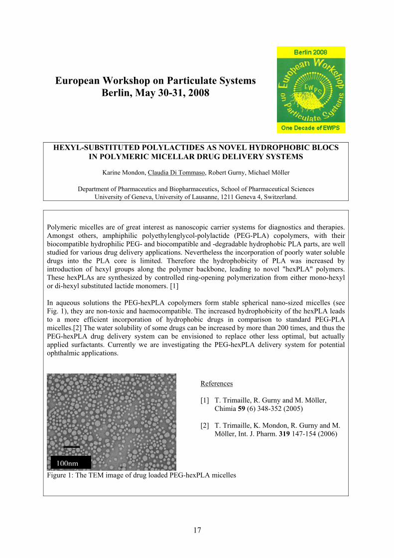
Polymeric micelles are of great interest as nanoscopic carrier systems for diagnostics and therapies. Amongst others, amphiphilic polyethylenglycol-polylactide (PEG-PLA) copolymers, with their biocompatible hydrophilic PEG- and biocompatible and -degradable hydrophobic PLA parts, are well studied for various drug delivery applications. Nevertheless the incorporation of poorly water soluble drugs into the PLA core is limited. Therefore the hydrophobicity of PLA was increased by introduction of hexyl groups along the polymer backbone, leading to novel "hexPLA" polymers. These hexPLAs are synthesized by controlled ring-opening polymerization from either mono-hexyl or di-hexyl substituted lactide monomers. [1] In aqueous solutions the PEG-hexPLA copolymers form stable spherical nano-sized micelles (see Fig. 1), they are non-toxic and haemocompatible. The increased hydrophobicity of the hexPLA leads to a more efficient incorporation of hydrophobic drugs in comparison to standard PEG-PLA micelles.[2] The water solubility of some drugs can be increased by more than 200 times, and thus the PEG-hexPLA drug delivery system can be envisioned to replace other less optimal, but actually applied surfactants. Currently we are investigating the PEG-hexPLA delivery system for potential ophthalmic applications.
Figure 1: The TEM image of drug loaded PEG-hexPLA micelles
References [1] T. Trimaille, R. Gurny and M. Möller,
Chimia 59 (6) 348-352 (2005) [2] T. Trimaille, K. Mondon, R. Gurny and M.
Möller, Int. J. Pharm. 319 147-154 (2006)
100nm
17

European Workshop on Particulate Systems
Berlin, May 30-31, 2008
PACLITAXEL LOADED ANTI-HER2 IMMUNONANOPARTICLES -
BIODISTRIBUTION STUDIES IN HEALTHY MICE
A. Cirstoiu-Hapcaa, F. Deliea L. Bossyb, F. Bucheggerc , R. Gurnya
a University of Geneva, University of Lausanne, Geneva , Switzerland, bTRB Chemedica International
SA, Geneva, Switzerland, c Division of Nuclear Medicine, University Hospital of Geneva, Switzerland
Introduction Paclitaxel (Tx) is one of the most efficient agents against a wide spectrum of cancers, such as ovarian, breast, lung, head and neck cancer. However, it suffers from a poor solubility in water and a low therapeutic index associated with serious side effects. Entrapping the drug into polymeric nanoparticles (NPs) provides the possibility to deliver it as a suspension, and also, due to enhanced permeability and retention effect (EPR), to promote its selective distribution to tumor site. To further enhance selective drug distribution to target cells overexpressing specific antigens or receptors, the surface of NPs can be decorated with specific ligands such as monoclonal antibodies (mAb) (1). In the present study, immunonanoparticles were obtained by coating paclitaxel-loaded nanoparticles (NPs-Tx) surface with mAb. Anti-Her2 mAb, Herceptin®, (HER) were used as targeting moieties for ovarian cancer cells overexpressing Her2 specific antigens(2). Biodistribution of three paclitaxel formulations: immunonanoparticles (NPs-Tx-HER), NPs-Tx and free Tx was studied in healthy mice, and the influence of the intraperitoneal (IP) versus intravenous (IV) administration route on distribution drug was investigated. Results The number of anti-Her2 mAb molecules bound per nanoparticle was about 290 and the encapsulation efficiency of Tx was approximately 70%. Biodistribution studies showed that after IV or IP injections, paclitaxel encapsulated either in NP-Tx-HER or NP-Tx was preferentially distributed in spleen, liver and lungs, whereas, free Tx had an equivalent distribution in all tested organs. The same tissue distribution of Tx after IV and IP administration was observed, and the Tx was still detected in organs 24h after injection of all Tx formulations. Conclusion In this study, paclitaxel-loaded anti-Her2 immunonanoparticles were successfully prepared to achieve specific tumour targeting. A preferential distribution of encapsulated Tx in reticulo-endothelial system (RES) organs was observed, compared to the non-specific tissue distribution of free Tx. The same tissue distribution of Tx after IV and IP administration observed in healthy mice could represent a main advantage considering the development of peritoneal micrometastasis in ovarian cancer. Indeed, IP administration could offer the possibility to associate local and systemic distribution of drug (3). In vivo studies are in progress to demonstrate the efficiency of paclitaxel-loaded immunonanoparticles in a xenograft animal model.
References (1) Sahoo S.K. et al., Int. J. Cancer, (2004) 112: 335-340, (2) Cirstoiu-Hapca A. et al., Int. J. Pharm., (2007) 331:190-196, (3) Tsai M. at al., Pharm. Research, (2007) Vol.24, No.9
18

European Workshop on Particulate Systems
Berlin, May 30-31, 2008
EFFECT OF SIZE AND SURFACE HYDROPHILICITY ON BIODEGRADABLE
PARTICLE INTERACTION WITH INTESTINAL CELLS.
M. Gaumet, R. Gurny, F. Delie
Department of Pharmaceutics and Biopharmaceutics, School of Pharmaceutical Sciences University of Geneva, University of Lausanne, Geneva , Switzerland
Polymeric nanoparticles held great promises as drug delivery systems, especially for new approaches in cancer therapy. Using polystyrene particles, size and surface properties were described as leading parameters influencing the interaction of particulates with cells (1). However, the physicochemical properties involved in cellular uptake are still not clearly defined, partly due to the fact that these parameters were studied with non biodegradable material and a large majority of the studies using biodegradable particles showed a lack of characterization of the final particles (2). To better understand the critical parameters involved in the interaction of biodegradable particles with intestinal cells, nano- and microparticles of well defined sizes were prepared by emulsion-evaporation method from poly(lactide-co-glycolide) (PLGA). Surface hydrophilicity was modulated by coating with chitosan. After encapsulation of a fluorescent dye, the interaction of these particles was studied on Caco-2 cells as a model for intestinal cells. After particle preparation, selective centrifugation enabled the isolation of five classes of particles: 0.1, 0.3, 0.6, 1, and 2 μm; with narrow size distribution and presenting the same surface morphology, charge, residual surfactant and hydrophilicity (3). A quantitative method based on fluorescence spectroscopy was developed to estimate the number of particles interacting with a single cell. The interaction was clearly dependant on particle size and concentration. Particles in the range of 100 nm presented a higher interaction when compared to larger particles. Cellular localisation of particles by confocal microscopy showed the intracellular location of small particles, whereas particles > 300 nm were associated with the cell apical membranes. Small PLGA nanoparticles were observed in the nuclei of the Caco-2 cells, unlike to polystyrene particles, also tested as a reference. Chitosan-coated nanoparticles obtained by adsorption showed an increase in hydrophilicity
determined by the Rose Bengal method and a slight increase in charge. A higher cellular uptake was observed for the small hydrophilic particles whereas no variations were seen with larger particles (>300 nm).
References (1) S. M. Moyes, et al. Int. J Pharm 337:133-141, 2007. (2) M. Gaumet, et al. Eur.J Pharm Biopharm., 2008 in press, (3) M. Gaumet, et al. Int.J Pharm 342 :222-230, 2007.
19

European Workshop on Particulate Systems
Berlin, May 30-31, 2008
University of Paris, France
THERAPEUTIC EFFECT OF INTRAVITREAL INJECTION OF VASOACTIVE INTESTINAL PEPTIDE-LOADED LIPOSOMES ON EXPERIMENTAL
AUTOIMMUNE UVEORETINITIS L. Lajavardi, A. Bochot, S. Camelo, Y. de Kozak, E. Fattal
CATIONIC LIPOSOMES CONTAINING A DOPE-HYALURONIC CONJUGATE FOR GENE DELIVERY
Claudio Surace, Silvia Arpicco, Véronique Marsaud, Céline Bouclier, Michel Renoir, Luigi Cattel, Elias Fattal1
INFLUENCE OF FORMULATION PARAMETERS ON ORGANIZATION OF AMPHIPHILIC POLYMERS/DNA SYSTEMS
AND ON THEIR IN VIVO EFFICIENCY Caroline Roques, Kawthar Bouchemal, Yves Fromes; Elias Fattal
LIPIDIC PRODRUGS: A PROMISING STRATEGY
FOR NUCLEOSIDE ANALOGUES H. Khoury, M. Lalanne, L. H. Reddy, A. Deroussent, A. Paci, Sinda Lepêtre-Mouelhi,
K. Andrieux, G. Vassal, Didier Desmaële, P. Couvreur
PHOSPHOLIPID – DECORATED MICROCAPSULES USED AS ULTRASOUND CONTRAST AGENT
R. Díaz-López, N. Tsapis, V. Nicolas, D. Libong, C. Bilem, M. Chehimi, P. Chaminade, E. Fattal
20

European Workshop on Particulate Systems
Berlin, May 30-31, 2008
THERAPEUTIC EFFECT OF INTRAVITREAL INJECTION OF VASOACTIVE
INTESTINAL PEPTIDE-LOADED LIPOSOMES ON EXPERIMENTAL AUTOIMMUNE UVEORETINITIS
L. Lajavardi1, A. Bochot1, S. Camelo2, Y. de Kozak2, E. Fattal1
1UMR CNRS 8612. Faculté de Pharmacie. 5, av JB Clément, 92296 Châtenay-Malabry. 2INSERM U598. 15,
rue de l’école de médecine, 75270 Paris cedex 06. The development of new approaches is needed for the treatment of human posterior uveitis, a potentially blinding disease. It has been previously shown in Lewis rats that a single intravitreal injection of Vasoactive Intestinal Peptide (VIP) was efficient at reducing ocular inflammation during endotoxin-induced uveitis only when formulated in liposomes. This formulation enhanced its immunosuppressive effect and controlled its delivery to all tissues affected by or involved in ocular inflammation (vitreous, ciliary body, conjunctiva, retina and sclera) 1. Moreover, after intravitreal injection in normal rats, VIP-loaded rhodamine-conjugated liposomes (VIP-Rh-Lip) reached cervical lymph nodes via conjunctival lymphatics and to a lesser extend the spleen via the conventional outflow pathway. In those tissues, VIP-Rh-Lip were internalized by resident macrophages 2. In the present study we tested the effect of a single intravitreal injection of VIP-Rh-Lip on experimental autoimmune uveoretinitis (EAU) induced by retinal soluble autoantigen (S-Ag) immunization in Lewis rats. Intravitreal injection of VIP-Rh-Lip simultaneously or 6 days after S-Ag stimulation, but not saline, saline-VIP or empty Rh-liposomes (E-Rh-Lip), reduced EAU clinical and pathological signs. All together, the data suggest that liposomes preserved VIP integrity. Twenty days after intravitreal injection, VIP-Rh-Lip and free VIP were detected in EAU rats in intraocular macrophages and in cells present in the spleen, inguinal and cervical lymph nodes. Locally, intravitreal injection of VIP-Rh-Lip reduced intraocular concentration of IL-2, IL-4, IL-6, IFN-gamma, IL-17, CCL2, CCL3 and CCL5 and expression of NOS-2 mRNA. At the systemic level, VIP-Rh-Lip treatment reduced lymphocyte proliferation, decreased IL-2 and increased IL-10 secretion by inguinal lymph node cells in response to S-Ag restimulation in vitro. Moreover, VIP-Rh-Lip treatment diminished delayed type hypersensitivity response to S-Ag and reduced serum concentration of IL-12 and IFN-gamma in vivo. Thus, a single intravitreal injection of VIP-Rh-Lip performed during the afferent stage of the immune response but not during the efferent stage, protected against retinal tissue damage through a reduced Th1-Th17 type of response with upregulation of IL-10 and change of macrophage phenotype.
1. Lajavardi L, Bochot A, Camelo S, Goldenberg B, Naud MC, Behar-Cohen F, Fattal E, de Kozak Y. Downregulation of endotoxin-induced uveitis by intravitreal injection of vasoactive intestinal peptide encapsulated in liposomes. Invest Ophthalmol Vis Sci. 2007; 48:3230-8.
2. Camelo S, Lajavardi L, Bochot A, Goldenberg B, Naud MC, Fattal E, Behar-Cohen F, de Kozak Y. Ocular and systemic bio-distribution of rhodamine-conjugated liposomes loaded with VIP injected into the vitreous of Lewis rats. Mol Vis. 2007; 13:2263-74.
3.
21
European Workshop on Particulate Systems
Berlin, May 30-31, 2008
CATIONIC LIPOSOMES CONTAINING A DOPE-HYALURONIC CONJUGATE
FOR GENE DELIVERY
Claudio Surace1,2, Silvia Arpicco2, Véronique Marsaud1, Céline Bouclier1, Michel Renoir1, Luigi Cattel2, Elias Fattal1
1UMR CNRS 8612, Paris-Sud University, Châtenay Malabry, France
2Gruppo di Chimica Farmaceutica Applicata, Dipartimento Scienza e Tecnologia del Farmaco, University of Turin, Italy
Purpose: Cationic liposomes [2-(2,3didodecyloxypropyl)hydroxyethyl ammonium bromide(DE) and dioleoylphosphatidylethanolamine(DOPE) 1:1 w/w] containing a new hyaluronic acid(HA)-DOPE conjugate were prepared in order to target the hyaluronan receptor CD44 in cancer cell lines. Methods: Liposomes were prepared by adding increasing amount of HA-DOPE conjugate to the DE-DOPE mixture. Complexes were successively formed by adding plasmid DNA(pCMV-luciferase). Size and zeta potential were measured Cytotoxicity and transfection efficiency of the complexes were determined in vitro in CD44+ (MDA-MB231) and CD44- (MCF-7) cell lines. MCF-7 and MDA cells were then treated with increasing amounts of the anti CD44+ primary antibody Hermes-1 before adding the lipoplexes to assess the role of HA-CD44 binding in the transfection. Results: All preparations display a size around 300nm and a strong positive zeta potential value which, as increasing the amount of HA-DOPE conjugate, decreased. Formulations containing the HA-DOPE conjugate were found to have a better efficiency than plain cationic liposomes in both cell lines. Furthermore, on the contrary of liposomes composed of commercial lipids (i.e. Lipofectin) which were cytotoxic, the lipoplexes containing HA-DOPE showed a better cell viability. As expected, transfection of MCF-7cells (CD44-), although lower than MDA-MB231 cells (CD44+), was not inhibited by the presence of Hermes-1 antibody. On the other hand, a decrease of the transfection efficiency was found in MDA-MB231 cells by increasing the amount of the antibody. Conclusions: Cationic liposomes containing HA-DOPE conjugate were shown to be a good system to deliver intracellullarly DNA both in MDA-MB231 and MCF-7 cells: HA-CD44 binding is supposed to be involved in the uptake pathway in CD44+ cell line (MDA-MB231). ). Work is in progress to evaluate the in-vivo behavior of the lipoplexes
22

European Workshop on Particulate Systems
Berlin, May 30-31, 2008
INFLUENCE OF FORMULATION PARAMETERS ON ORGANIZATION
OF AMPHIPHILIC POLYMERS/DNA SYSTEMS AND ON THEIR IN VIVO EFFICIENCY
Caroline Roques1,2, Kawthar Bouchemal1, Yves Fromes2 and Elias Fattal1
1UMR CNRS 8612, Université Paris 11, 92290 Châtenay-Malabry, France. 2Institut de Myologie - Inserm U582 - Université Pierre et Marie Curie, 75013 Paris, France.
As DNA is a hydrophilic, negatively charge macromolecule, it does not freely cross the membranes. To achieve safe and efficient gene delivery, formulation of nucleic acids is thus a major concern. Synthetic formulations based on polymers were first achieved with molecules exhibiting a high density of positive charges, as Polyethyleneimine (PEI). Interest has also focused on polymers displaying few or no charges. Among those, amphiphilic Tetronic 304 and Pluronic L64 seem of particular interest to transfer DNA in vivo. The mechanism by which these polymers are promoting gene transfer in vivo is yet not fully understood. Nevertheless, several studies have highlighted the influence of temperature and medium on the supramolecular organization of such amphiphilic polymers. In this context, our work has focused on determining the influence of several formulation parameters on the interaction and organization of polymer/DNA systems. We have studied the correlation between these modifications and the toxicity and efficiency of the systems in vivo.
For each polymer/DNA formulation, the morphology of the vectors was assessed by cryo- and conventional Transmission Electron Microscopy, their size by Dynamic Light Scattering and their electrophoretic mobility by Laser Doppler Velocimetry. PEI/DNA complexes are displaying a relatively small size and a strongly positive zeta potential. After in vivo administration, PEI/DNA complexes exhibited a high toxicity towards skeletal muscle. Amphiphilic polymers associated to DNA are leading to more complex systems displaying weaker interactions. Isothermal Titration Calorimetry (ITC) measurements carried out on the amphiphilic polymers have demonstrated that concentration of Tetronic 304 used in our studies are above the critical micelle concentration (CMC), whatever the temperature. On the contrary, with Pluronic L64, the CMC was reached only at 37°C in Tyrode’s salts solution. When adding Tetronic 304 to DNA, no differences were recorded when increasing the temperature, while interactions between DNA and Pluronic L64 are linked to the presence of micelles, and are thus depending on temperature and medium used. In vivo, no lesions were detected with amphiphilic polymers based formulations. Moreover, these formulations allowed significant improvement of gene transfer to the skeletal muscle with reference to naked DNA, even at low DNA doses. Afterwards, in vivo administration of the formulations was performed at 4, 20 and 37°C. The results are in good agreement with the ITC outcomes: no significant differences were observed for Tetronic/DNA systems, while Pluronic L64/DNA formulations exhibited maximum efficiency at 37°C in Tyrode.
Our studies have emphasized the interest of amphiphilic polymers displaying few or no charges to transfer DNA in the skeletal muscle. The supramolecular organization of Pluronic L64 based formulations, as well as the interactions between the polymer and the DNA, is strongly dependent on the temperature and the medium used. Moreover, these modifications have a direct impact on the in vivo efficiency of such vectors.
23

European Workshop on Particulate Systems
Berlin, May 30-31, 2008
LIPIDIC PRODRUGS: A PROMISING STRATEGY
FOR NUCLEOSIDE ANALOGUES
H. Khourya,b, M. Lalannea, L. H. Reddya, A. Deroussentb, A. Pacib, Sinda Lepêtre-Mouelhic, K. Andrieuxa, G. Vassala, Didier Desmaëlec, P. Couvreura
a UMR CNRS 8612, Faculty of Pharmacy, Paris-Sud University, Châtenay Malabry, France.
b UPRES EA 3535, Institut Gustave Roussy, Paris-Sud University, Villejuif, France. c Université Paris XI, Faculté de Pharmacie, UMR CNRS 8076, IFR 141, 92296 Châtenay-Malabry Cedex,
France Lipid and lipophilic excipients can have significant and beneficial effects on the absorption and exposure of co-administered prodrugs. Nucleoside analogues exhibit an anticancer and/or antiviral activity, by incorporation of their active metabolite with the DNA. However, they have various therapeutic limitations restricting their use, such as short plasma half-life or rapid metabolism. In order to improve their biodisponibility and their therapeutic index, we have developed two nucleoside analogues by coupling lipids; diglyceride to didanosine and squalene to gemcitabine. Hence, two glycerolipidic prodrugs of didanosine, a phosphorylated and a non-phosphorylated one, were synthesized. The followed strategy consists in obtaining molecules able to mimic triglycerides, and therefore able to follow their metabolism pathway, which consist in a phosphorylation before DNA incorporation. Glycerolipidic prodrug is an interesting concept to enhance lymphatic absorption and distribution of poor absorbed polar drugs. Such molecules could intend to oral delivery, replacing perfusion, a less convenient administration pathway. Gemcitabine, on the other hand was covalently bound to squalene, an acyclic isoprenoid chain, leading to gemcitabine-squalene. This isoprenoid moiety aims to protect gemcitabine from plasmatic deamination, improving thus its bioavailability through a better absorption and reducing its elimination. The newly synthesised prodrugs, glycerolipidic didanosine and gemcitabine-squalene were formulated in lipid-based formulation. Glycerolipidic didanosine was encapsulated into liposomes [Lalanne et al. 2007a, Lalanne et al. 2007b] and gemcitabine-squalene gave self nano-assemblies nanoparticles [Couvreur et al. 2006]. Both glycerolipidic didanosine prodrugs were incorporated into liposomes with an optimal lipid/prodrug ratio 1:5 and 1:10 (mol:mol) for phosphorylated and a non-phosphorylated prodrugs, respectively. Squalenoylation rendered gemcitabine amphiphilic and able to form nanoassemblies spontaneously in water. In order to validate this biomimetic strategy, an in vitro study of the prodrugs metabolism is proposed as well as pharmacokinetic profiles and biodistribution. Moreover, the antiviral and anticancer activities were verified both in vitro and in vivo. Altogether, our promising results demonstrate that lipid-based formulation of nucleoside analogues is of great interest in therapeutics. Indeed, these new prodrugs seemed to be more advantageous than the nucleoside analogues alone in solution: they display more potent efficiency, both in vitro and in vivo, in experimental cancers treatment and show more convenient pharmacokinetic profiles.
24

European Workshop on Particulate Systems
Berlin, May 30-31, 2008
PHOSPHOLIPID – DECORATED MICROCAPSULES USED AS
ULTRASOUND CONTRAST AGENT
R. Díaz-Lópeza,b, N. Tsapisa,b, V. Nicolasc, D. Libongd, C. Bileme, M. Chehimie, P. Chaminaded, and E. Fattala,b
a Univ Paris-Sud, CNRS UMR 8612, 5 rue J. B. Clément, 92296 Châtenay- Malabry, France. b CNRS, UMR 8612, 5 rue J. B. Clément, 92296 Châtenay- Malabry, France.
c Univ Paris-Sud, IFR141-ITFM, Plateau technique-Imagerie cellulaire, Châtenay- Malabry, France. d Univ Paris-Sud, Groupe Chimie Analytique, EA 4041, IFR 141, 92296 Châtenay- Malabry, France
e University of Paris VII, ITODYS, CNRS UPRESA 7086, 1 rue Guy de la Brosse, Paris 75005, France Ultrasonic imaging is a widely available, non-invasive and cost-effective diagnostic modality, but the weak difference of echogenicity between different tissues often hampers a clear diagnostic. To better visualize specific tissues, ultrasound contrast agents (UCAs) are frequently used. The most recent ones consist of gaseous perfluorocarbon bubbles encapsulated within polymer shells. These allow obtaining passage through the pulmonary capillary bed, and a prolonged plasmatic half-life. However, due to their hydrophobic surface, they are quickly eliminated by the monocyte phagocytic system. In addition, UCAs also lack specificity and active targeting would be needed to image specific tissues. Therefore, surface modification of polymeric UCAs has been considered to enable particles to cross leaky tumor vasculature and favor active targeting. Several strategies have been evaluated but we choose to explore a new one described by Fahmy et al. (2005). We present here an easy method to modify the surface properties of polymeric microcapsules of perfluorooctyl bromide used as ultrasound contrast agents. These capsules were obtained by a modified solvent emulsification-evaporation process. Phospholipids have been incorporated in the organic phase before emulsification. Several phospholipids were reviewed: fluorescent, pegylated and biotinylated phospholipids. The influence of phospholipid quantity on microcapsule size and morphology was evaluated by laser diffraction, fluorescent and confocal microscopy and scanning electron microscopy. The proportion of pegylated phospholipids associated to the microcapsules was quantified by high performance liquid chromatography (HPLC) with a Charged Aerosol Detection (CAD) and the surface modifications of the microcapsules were assessed by electron spectroscopy for chemical analysis (ESCA). Finally, the accessibility of biotin groups at the surface of microcapsules and their specific association with streptavidin was assessed by incubating microcapsules with fluorescent streptavidin. Experiments show that only a fraction of the phospholipids is associated to the microcapsules, the rest being dissolved with the surfactant in the aqueous phase. Microscopy shows that phospholipids are present within the shell and that the core/shell structure is preserved up to 0.5 mg fluorescent lipids, up to 0.26 mg pegylated lipids and up to 0.25 mg biotinylated lipids (for 100mg PLGA). ESCA shows the presence of Nitrogen at the surface of phospholipid decorated pegylated capsules. HPLC allows quantifying lipids associated to capsules: 10% of pegylated lipids introduced in the organic phase are associated with the capsules. Finally biotinylated microcapsules incubated with neutravidin tend to aggregate, which confirms the presence of biotin at the surface.
25

European Workshop on Particulate Systems
Berlin, May 30-31, 2008
University of Copenhagen, Denmark
LIPOSOMAL FORMULATION OF GHRELIN Eva H. Møller, Line H. Nielsen, Birgitte Holst, Pia S. Nielsen, and Jesper Østergaard
IN VIVO AND IN VOTRO INVESTIGATIONS OF THE POTENTIAL OF CAF01 LIPOSOMS AS A MUCOSAL VACCINE ADJUVANT
Dennis Christensen, Ida Rosenkrands, Else Marie Agger, Camilla Foged, Hanne Mørck Nielsen
PREPARATION AND CHARACTERIZATION OF siRNA LOADED PLGA NANOPARTICLES
Dongmei Cun, Camilla Foged, Mingshi Yang, Linda Boye Jensen, and Sven Frokjaer, Hanne Mørck Nielsen
SOLID LIPID NANOPARTICLES AS DRUG DELIVERY SYSTEM FOR CORTICOSTEROIDS: INFLUENCE OF LIPID AND DRUG SUBSTANCE ON THE
RELEASE PROFILE IN VITRO Louise Bastholm Jensen, Emily Magnusson, Linda Gunnarsson, Charlotte Vermehren,
Hanne Mørck Nielsen, Karsten Petersson
CAMPTOTHECIN-LOADED SLN BASED ON TRIMYRISTIN (DYNASAN 114) FOR BRAIN DELIVERY
Susana Martins, Domingos C. Ferreira, Eliana B. Souto, Martin Brandl
26

European Workshop on Particulate Systems
Berlin, May 30-31, 2008
LIPOSOMAL FORMULATIONS OF GHRELIN
Eva H. Møller1, Line H. Nielsen1, Birgitte Holst2, Pia S. Nielsen2, and Jesper Østergaard1
1Dept. of Pharmaceutics and Analytical Chemistry, Faculty of Pharmaceutical Sciences, Univ. of Copenhagen
2Dept. of Neuroscience and Pharmacology, Faculty of Health Sciences, University of Copenhagen Ghrelin is an appetite-stimulating peptide hormone produced by cells in the fundus of the stomach. Human ghrelin is a pharmacologically interesting peptide, albeit it has a very short half-life after administration. The peptide is acylated with an octanoyl group at its Ser-3 side chain. A formulation of ghrelin for prolonged release will be helpful in resolving some of the pharmacodynamic/pharmacokinetic aspects of ghrelin. In order to prolong the half-life of ghrelin, liposomal suspensions of DPPC liposomes, PC:chol liposomes, and negatively charged DPPC:DPPS-liposomes were prepared and ghrelin was added. The DPPC and PC:chol liposomes were chosen to elucidate whether ghrelin, having an n-octanoyl side chain, could associate to neutral liposomes by this lipophilic arm. In the negatively charged liposomes, electrostatic interactions could mediate binding and also cause an agglomeration of the liposomes to larger aggregates, hence slowing down the release of ghrelin from liposomes. Unilamellar liposomes (approx. 100 nm, lipid conc. approx. 2 mM) were prepared by the hydrated film method. After addition of human ghrelin to the liposomes, the suspensions were characterized and analyzed with regard to chemical and physical stability. The secondary structure of ghrelin in buffer as well as in the three liposomal formulations was studied. The release into the blood stream in vivo following s.c. administration of ghrelin in buffer and liposomal formulations was examined in male Sprague-Dawley rats (approx. 300 g). The ghrelin concentration was 0.125 mg/ml and the dose administered was 200 mg/kg. Blood samples were drawn from tail veins at regular time intervals and evaluated by a RIA assay. The results showed that the PC:chol-ghrelin formulation had a longer persistence as compared to the plain ghrelin solution and the other liposomal formulations. Capillary electrophoresis studies on the interaction between liposomes and ghrelin revealed an affinity between ghrelin and the negatively charged (DPPC:DPPS) liposomes, whereas only very small affinities could be discerned in the other liposomal formulations of ghrelin. Hence, the differences observed in vivo between the formulations were apparently not caused by affinity between ghrelin and the liposome, but could rather be caused by the different physical states of the liposomes.
27
European Workshop on Particulate Systems
Berlin, May 30-31, 2008
IN VIVO AND IN VITRO INVESTIGATIONS OF THE POTENTIAL OF
CAF01 LIPOSOMESAS A MUCOSAL VACCINE ADJUVANT
Dennis Christensen1, 2, Ida Rosenkrands2, Else Marie Agger2, Camilla Foged1, Hanne Mørck Nielsen1
1) Statens Serum Institut, Dept. Infectious Disease Immunology, Adjuvant research, Artillerivej 5,
DK-2300 Copenhagen S. 2) University of Copenhagen, Faculty of Pharm. Sci., Dept. Pharm. and Analyt. Chem., Universitetsparken 2,
DK-2100 Copenhagen Ø Nasal administration of vaccines has many advantages compared to parenteral vaccine delivery. A needle-free mucosal vaccine would be easily applicable, target the vaccine to the entry point of many pathogens, and reduce the risk of infection with other pathogens, compared to invasive methods. CAF01 is a novel vaccine adjuvant system with a remarkable immunostimulatory activity, characterized by induction of both TH1 and TH2 responses. CAF01 is based on liposomes consisting of dimethyldioctadecylammonium bromide and trehalose 6,6’-dibehenate, The purpose of this study was to investigate the potential of CAF01 liposomes as a drug delivery system for nasal vaccines. In addition, the toxic effect of CAF01 on epithelial cells was investigated. The mucus producing epithelial CALU-3 cell culture model was chosen as model system to elucidate the deposition, absorption, and retention of the antigen associated with the liposomes after application onto epithelial cells. The cell toxicity of CAF01 was investigated by analyzing the integrity and the metabolic activity of the CALU-3 cells. Finally, the adjuvant was tested nasally in a mouse model using the tuberculosis vaccine candidate Ag85B-ESAT-6 as a model antigen. The results suggest that CAF01 enhances the transport of antigen through the mucus layer of the CALU-3 cells, increasing the concentration of antigen at the cell surface, but not increasing the transport across the epithelial cells Neither the integrity or the metabolic activity was altered after exposing the CALU-3 cells to CAF01 indicating that the liposomes are not toxic. Finally, in vivo studies showed enhanced antigen-specific interferon (IFN)-γ release from blood lymphocytes from mice nasally vaccinated with Ag85B-ESAT-6 formulated with CAF01, as compared to mice vaccinated with the antigen alone or naive mice. The in-vitro data combined with the preliminary in-vivo study indicate that CAF01 has a potential as a mucosal vaccine delivery system, however, additional in-vivo investigations need to be done, and the vaccine formulation has to be further optimized to target the mucosal tissues in e.g. nose, lungs or vagina.
28

European Workshop on Particulate Systems
Berlin, May 30-31, 2008
PREPARATION AND CHARACTERIZATION OF SIRNA LOADED
PLGA NANOPARTICLES
Dongmei Cun1, Camilla Foged1, Mingshi Yang2, Linda Boye Jensen1, and Sven Frokjaer1, Hanne Mørck Nielsen1
1: Faculty of Pharmaceutical Sciences, Department of Pharmaceutics and Analytical
Chemistry, University of Copenhagen, Denmark, 2: Novo Nordisk A/S, Denmark
RNA interference (RNAi) mediated by small interfering RNA (siRNA) has appeared to be potent and highly specific for gene silencing compared to other antisence strategies. An obstacle to develop siRNA as a drug, however, is its rapid degradation and poor cellular uptake into the cytoplasm where it can enter the RNAi pathway.
In order to enhance gene therapy, specialized design features are required for delivery vector. A number of researchers have attempted to provide biodegradable nanoparticles vectors formulated with poly (D, L-lactide-co-glycolide) (PLGA) for delivery of DNA. But the efficiency of PLGA NPS as a carrier of siRNA delivery systems has not yet been extensively evaluated.
The aim of this study to prepare siRNA loaded PLGA nanoparticles with narrow size distribution and high encapsulation efficiency by a multiple emulsion [(W1/O) W2] solvent evaporation method. The nanoparticles were characterized by dynamic light scattering (DLS) and transmission electron microscopy (TEM) in terms of particle size, zeta-potential and morphology. An assay was established to determine the encapsulation efficiency of siRNA. The effects of some formulation and processing parameters, such as the inherent properties of the poly (D,L-lactic-co-glycolic acid) polymer, the concentration of polymer, volume ratio of inner water phase to oil phase and the power of sonication, on the size and encapsulation efficiency was investigated.
It was shown that nanoparticles have a size around 200nm with narrow distribution (PDI<0.1) could be produced and the encapsulation efficiency was increased gradually with the optimizing of parameters.
Keywords: siRNA delivery, PLGA, Nanoparticles, Gene therapy, Biodegradable.
29
European Workshop on Particulate Systems
Berlin, May 30-31, 2008
SOLID LIPID NANOPARTICLES AS DRUG DELIVERY SYSTEM FOR
CORTICOSTEROIDS: INFLUENCE OF LIPID AND DRUG SUBSTANCE ON THE RELEASE PROFILE IN VITRO
Louise Bastholm Jensen1+2, Emily Magnusson1, Linda Gunnarsson1, Charlotte Vermehren1, Hanne Mørck
Nielsen2, Karsten Petersson1
1 LEO Pharma A/S, Industriparken 55, DK-2750 Ballerup, Denmark 2 Faculty of Pharmaceutical Sciences, University of Copenhagen, Universitetsparken 2, DK-2100 Copenhagen,
Denmark Email: [email protected]
Solid lipid nanoparticles (SLN) is a novel drug delivery system in the submicron size range (50-1000 nm) composed of well tolerated lipids with a melting point above body temperature. Due to the solid state of the lipid, efficient drug encapsulation and controlled drug release can be achieved. In addition SLN has been shown to possess occlusive properties which may be advantageous in the treatment of skin diseases characterized by an increased TEWL such as atopic dermatitis. The ability of the SLN to control the release of the drug substance and thereby limit systemic absorption also makes it a suitable delivery system for topical administration where only a local effect is needed. The drug substance incorporation in the SLN particle and the distribution between the lipid and the water phase is likely to affect the release profile as well as the SLN system stability. The purpose of the present work was therefore to investigate the influence of lipid composition on the drug substance solubility in the lipid phase and the release profile of SLN in vitro. Furthermore, the influence of the physicochemical properties of the drug substance on the solubility in the lipid phase and on the release profile for a selected SLN lipid composition was investigated. The lipids used in the study were different regarding content of monoglycerides and chain length of the fatty acid moiety. The drug substances applied were corticosteroid derivatives varying in lipophilicity. Solubility of the drug substances in the melted lipids was estimated by hot stage microscopy. The hot high pressure homogenization method was used to prepare the SLN. Release studies were performed in diffusion cells with a cellulose membrane, withdrawing samples at 1, 2, 3, 4, 5, 6 and 24 h. The results showed that the solubility of the drug substances in the melted lipids was related to monoglyceride content and drug substance lipophilicity. The majority of the release profiles followed zero order. The total amount released in most cases could be correlated to the amount of monoglycerides in the lipid and drug substance lipohilicity. This may be related to differences in drug substance incorporation and affinity to the lipid particles or to differences in distribution between lipid and water phase.
30

European Workshop on Particulate Systems
Berlin, May 30-31, 2008
University of Porto, Portugal
ASSESSMENT AND EVALUATION OF ORAL BIOAVAILABILITY OF ANTICANCER DRUGS BY LIPID-BASED NANOPARTICLES
COATED WITH POLYSACCHARIDES Eliana B. Souto
CAMPTOTHECIN-LOADED SLN BASED ON TRIMYRISTIN (DYNASAN 114) FOR BRAIN DELIVERY
Susana Martins, Domingos C. Ferreira, Eliana B. Souto, Martin Brandl
31

European Workshop on Particulate Systems
Berlin, May 30-31, 2008
ASSESSMENT AND EVALUATION OF ORAL BIOAVAILABILITY OF
ANTICANCER DRUGS BY LIPID-BASED NANOPARTICLES COATED WITH POLYSACCHARIDES
Eliana B. Souto*
Faculty of Health Sciences, University of Fernando Pessoa, Rua Carlos da Maia, 296, 4200-150 Porto, Portugal
(*corresponding author: [email protected]) Despite decades of research, the developments in cancer chemotherapy are relatively slow, due to the lack of appropriate mechanisms to deliver anticancer drugs selectively to tumour tissues. To overcome the anatomical and physiological barriers, as well as to minimize the exposure of normal tissues to anticancer drugs, attempts have been made to develop novel drug delivery systems in the field of nanotechnology. Anticancer drugs such as paclitaxel [1,2], docetaxel [3], tamoxifen [4,5], and doxorubicin [6], have been encapsulated into biodegradable polysaccharide nanoparticles to minimize adverse side effects and drug toxicity. Suitable nanoparticulate systems for oral drug delivery would be those composed of a hybrid system based on lipids and polysaccharides. Lipophilic anticancers are encapsulated within lipid matrices [1], which are then coated with polysaccharides, e.g. chitosan. Chitosan is a natural polysaccharide that shows mucoadhesive properties [7,8]. Furthermore, it has been reported that chitosan nanoparticles reveal significant in vivo anti-tumour activity [9]. Oral delivery of anticancer drugs can cause severe toxicity limiting therefore their therapeutic potential. On the other hand, a suitable dose needs to be administered to maintain their therapeutic concentration in tumours. The risk of a subtherapeutic dosage is an important issue because tumours can develop drug resistance as a result of biochemical changes, drug export mechanisms, as well as limitations on the mechanisms of cellular drug uptake. Currently, several approaches are in progress to develop formulations for oral anticancer drugs, based on pharmaceutical (e.g. lecithin stabilized nanocarriers such as solid lipid nanoparticles, nanoemulsions and liposomes), chemical (e.g. prodrugs), and biological (e.g. gastrointestinal absorption) strategies. It is highly feasible for nanoparticles of biodegradable polymers to be applied to promote oral chemotherapy. This review focuses the novel approaches based on lipid/polysaccharide nanoparticles to deliver anticancer drugs through oral route, including the physicochemical analysis to assess the mechanisms of cellular drug uptake. The cellular uptake of nanoparticles can be evaluated in vitro by using Caco-2 cells, a human colon adenocarcinoma cell line. The effects of the particle size and particle surface coating with polysaccharides on the cellular uptake of the nanoparticles can be quantified by spectrofluorometric measurements. Images of confocal laser scanning microscopy (SEM), cryo-SEM and transmission electron microscopy (TEM) can be useful to visualize the internalization of nanoparticles by the Caco-2 cells. To assess the structure of nanoparticles, micro-FTIR and elastic neutron scattering experiments can be performed. References:
[1]. Arica Yegin, B., J.P. Benoit, and A. Lamprecht, Drug Dev Ind Pharm, 2006. 32(9): p. 1089-94. [2]. Park, J.S., et al., J Control Release, 2006. 115(1): p. 37-45. [3]. ten Tije, A.J., et al.,. Clin Pharmacokinet, 2003. 42(7): p. 665-85. [4]. Memisoglu-Bilensoy, E. and A.A. Hincal, Int J Pharm, 2006. 311(1-2): p. 203-8. [5]. Memisoglu-Bilensoy, E., et al., J Control Release, 2005. 104(3): p. 489-96. [6]. Hyung, W., et al., Biotechnol Bioeng, 2008. 99(2): p. 442-54. [7]. Chayed, S. and F.M. Winnik, Eur J Pharm Biopharm, 2007. 65(3): p. 363-70. [8]. Prego, C., et al., J Control Release, 2005. 101(1-3): p. 151-62. [9]. Qi, L. and Z. Xu, Bioorg Med Chem Lett, 2006. 16(16): p. 4243-5.
32

European Workshop on Particulate Systems
Berlin, May 30-31, 2008
CAMPTOTHECIN-LOADED SLN BASED ON TRIMYRISTIN (DYNASAN 114)
FOR BRAIN DELIVERY
Susana Martins1,2,4, Domingos C. Ferreira1, Eliana B. Souto3, Martin Brandl4
1 Faculty Pharmacy, University of Porto, Portugal 2 Faculty of Pharmaceutical Sciences, University of Copenhagen, Denmark
3 Faculty of Health Sciences, University of Fernando Pessoa, Portugal 4 Institute of Pharmacy, University of Tromso, Norway, E-mail: [email protected]
Introduction: Solid lipid nanoparticles (SLN) are submicron particles in the size range 50-1000 nm, composed of physiological and biodegradable lipids that remain in a solid state at room and body temperature. SLN combine the advantages of systems such as liposomes, polymeric nanoparticles and emulsions, but avoid or minimize the drawbacks associated with these traditional colloidal systems. The blood-brain-barrier represents the major obstacle to the delivery of drugs to the brain. Due to the high incidence of brain diseases, the development of a suitable carrier is a demand for improving drug delivery to treat brain diseases. Kreuter et al. discovered that polysorbate 80 loaded polybutylcyanoacrylate (PBCA) nanoparticles were able to deliver dalargin to the brain. Polisorbate 20 and polysorbate 80 are promising surface-modifiers to be used to deliver drugs to the brain since they can inhibit the efflux function of P-glycoprotein. Camptothecin (CPT) is a well-established topoisomerase I inhibitor against a broad spectrum of cancers. However, poor aqueous solubility, instability of the lactone ring, and toxic effects to normal tissues have limited CPT clinical development. This drug has previously been formulated in lipid nanoparticles and liposomes. The aim of this work was the production of SLN with size below 200 nm, low polidispersity index (PI) and with high CPT association efficiency intended for brain delivery. Materials and experimental methods: Unloaded and CPT-loaded SLN composed of trimyristin (Dynasan 114) and stabilized by either polysorbate 20, 60 or 80 were produced by hot high pressure homogenization (3 cycles at 500 MPa and 90ºC) using an APV Gaulin MicronLab 40. The particle size of SLN was analyzed by both, photon correlation spectroscopy (PSSS Nicomp 370) and optical single particle counting (PSS Nicomp). SLN stability was determined by following the particle size and PI of SLN stored at room temperature during one month. CPT association efficiency was determined after ultracentrifugation (2 runs at 100,000g, 20 min) of SLN diluted in 50 mM PBS buffer, pH 10.5. The supernatant obtained of the second ultracentrifugation was analysed by HPLC and CPT concentration in SLN thus detected indirectly. Results and discussion: Since unloaded- and CPT-loaded SLN have similar sizes (below 200 nm) and PI values (below 0.12), it can be concluded that encapsulation of this drug did not significantly change the physical characteristics of the produced lipid particles. Particle counting of both, unloaded- and CPT-loaded SLN showed similar results in the “50% particles smaller than [μm]” limit (~0.62 µm). For the “99% particles smaller than [μm]” limit some small differences could be detected between the unloaded- (~1.45 µm) and CPT-loaded (~1.88 µm) SLN. The incorporation of drug slightly shifted the 99% size limit to higher values which may perhaps be attributed to the presence of CPT crystals or some aggregation of the SLN provoked by CPT molecules. Further studies are being run to clarify the effect of trimyristin (Dynasan 114) on the size of SLN. Particles larger than 3 μm were not found in the SLN dispersions by optical particle sizer analysis. This is very important since the SLN produced are meant for iv administration. With regard to the effect of different surfactants used to stabilize SLN, the CPT association efficiency values were very similar, i.e. 81% for polysorbate 20, 82% for polysorbate 60 and 80% for polysorbate 80. Conclusion: CPT-loaded SLN with a mean diameter below 200 nm, of low PI, with no particles larger than 3 μm and with a relatively high association efficiency have been successfully produced.
33

European Workshop on Particulate Systems
Berlin, May 30-31, 2008
University of Lisbon, Portugal SURFACE MODIFIED POLYMERIC NANOPARTICLES AS VACCINE CARRIERS
FOR MUCOSALIMMUNISATION AGAINST SREPTOCOCCUS EQUI António J. Almeida, L.M.D. Gonçalves, S. Pandit, H.O. Alpar, H. Florindo
34
European Workshop on Particulate Systems
Berlin, May 30-31, 2008
SURFACE MODIFIED POLYMERIC NANOPARTICLES AS VACCINE
CARRIERS FOR MUCOSALIMMUNISATION AGAINST SREPTOCOCCUS EQUI
António J. Almeida, L.M.D. Gonçalves, S. Pandit, H.O. Alparand H. Florindo
1 iMed.UL, Faculty of Pharmacy, University of Lisbon, Lisbon Portugal 2Centre for Drug Delivery Research, School of Pharmacy, University of London, London, UK
Strangles is a highly contagious bacterial infection of Equidae family caused by Streptococcus equi subspecies equi, a Lancefield group C streptococcus [1]. Although S. equi is sensitive to some antibiotics, most of the treatments are ineffective and prevention is the key. New vaccines against strangles have been developed but limited efficacy has been confirmed [2]. Biodegradable polymeric particulate systems have enormous potential as antigen carriers for the induction of systemic and local immunity. Therefore, the aims of the present study were to produce surface modified polycaprolactone (PCL) and polylactic acid (PLA) nanoparticles, with cationic (glycochitosan) or anionic (sodium alginate or polyvinyl alcohol) surface charge, and absorption enhancers (spermine and oleic acid), as S. equi carriers for the induction of both systemic and local protective immunity in a mice model. The PCL and PLA nanoparticles were prepared by the double emulsion (w/o/w) solvent evaporation method described elsewhere [3]. Morphology, particle size and surface charge were duly controlled by laser scattering and scanning electron microscopy. Protein integrity throughout encapsulation was assessed by SDS-PAGE. In vivo studies involved female BALB/c mice (n = 4/group), which were intramuscularly (i.m.) or intranasally (i.n.) immunized on day 1 and boosted on day 29, with 50μl of solution containing S. equi antigen equivalent to 10μg of SeM protein, either free or encapsulated in nanoparticles. The levels of anti-S. equi specific IgG, IgG1, IgG2a and IgA were determined by indirect ELISA. Cellular immunity was assessed through interleukin concentrations (IL-2, IL-4, IL 6 and IFN-γ) using ELISA. The encapsulation efficiency varied from 30.4% to 95.4 % and SDS-PAGE showed that antigen integrity was maintained. Vaccination by i.m. route induced a strong increase of serum IgG levels. Negatively charged formulations induced significantly higher IgG and IgG2a levels. Differences were observed in serum antibody levels of animals vaccinated with bacterial extract encapsulated in PLA and PCL particles, when compared to the free form of the antigen. Overall, the IgG subclass titres obtained in all groups treated with particulate vaccine suggest the generation of Th1/Th2 mixed response. Spermine-containing formulations induced similar IL2 levels regardless of the polymer used to prepare nanoparticles. PLA nanoparticles induced higher IFN-γ levels when compared to PCL nanoparticles. However, interleukin levels induced by i.n. immunisation were lower than those obtained after i.m. vaccination. Moreover, the efficacy of vaccines to prevent strangles seems to be dependent on the induction of a mucosal immune response. Nasal mucosal antibody response (SIgA) was statistically higher in animals immunized by the i.n. route. Finally, PCL and PLA nanoparticles are potential vaccine carriers with strong immunoadjuvant properties for the effective delivery of S. equi antigens, thus confirming our previous findings [4].
References
1 Flock M, Jacobsson K, Frykberg L, Hirst TR, Franklin A, Guss B, Flock JI. (2004) Infect. Immun. 72: 32283236. 2 Hamlen HJ, Timoney J, Bell RJ. (1994) JAVMA 204: 768-775. 3 Conway BR, Alpar HO. (1996) Eur J Pharm Biopharm 42: 42-48. 4 Azevedo AF, Galhardas J, Cunha A, Cruz P, Gonçalves LMD, Almeida AJ. (2006) Eur J Pharm Biopharm. 64: 131-137. Acknowledgements This work was supported by Fundação para a Ciência e Tecnologia, Portugal, and FEDER (SFRH/BD/14300/2003; POCI/BIO/59147/2004; PPCDT/BIO/59147/2004).
35

European Workshop on Particulate Systems
Berlin, May 30-31, 2008
University of Berlin, Germany
LIPID NANOCARRIER BASED POTENT ANTI-MALARIAL FORMULATIONS Medha Joshi, Shobhona Sharma,Vandana Patravale
SOLID LIPID NANOPARTICLES FOR IMPROVED SKIN UPTAKE Peggy Schlupp, Monika Schäfer-Korting
BMBM-LOADED NANOSTRUCTURED LIPID CARRIERS (NLC): A CARRIER SYSTEM FOR MORE EFFICIENT AND SAVE SUNSCREENS
Aiman Hommoss, Mohammad Al-Samman, Rainer H. Müller
NANOSTRUCTURED LIPID CARRIERS (NLC) IN COSMETIC DERMAL FORMULATIONS
Aiman Hommoss, Kay Schwabe, Christian Rimpler, Rainer H. Müller
HYDROPHOBIC NANOSTRUCTURED LIPID CARRIERS AS NOVEL MODIFICATION FOR DERMAL APPLICATION
Medha Joshi, Dirk Franke, Rainer H. Müller
COMPARISON OF SOLID LIPID NANOPARTICLES AND DENDRITIC CORE-MULTISHELL NANOPARTICLES
AS DRUG DELIVERY SYSTEM FOR TOPICAL APPLICATION S. Küchler, M. R. Radowski, R. Haag, M. Schäfer-Korting
PX-13/18 – NEW PHOSPHOLIPASE A2 INHIBITORS FOR DERMAL APPLICTATION IN NANOPARTICLES
Jana Pardeike, Rainer H. Müller
36

European Workshop on Particulate Systems
Berlin, May 30-31, 2008
SECOND GENERATION OF DRUG NANOCRYSTALS: SPECIAL FEATURES OF SMARTCRYSTALS
Rainer H. Müller, Cornelia M. Keck, Rachmat Mauludin, Szymon Kobierski
PRODUCTION OF HESPERIDIN DERMAL NANOCRYSTALS BY SMARTCRYSTAL COMBINATION TECHNOLOGY
Szymon Kobierski, Rachmat Mauludin, Cornelia M. Keck, Rainer H. Müller
PRODUCTION OF LYOPHILISED COENZYME Q10 NANOCRYSTALS Rachmat Mauludin, Cornelia M. Keck
PREPARATION AND LONG-TERM STABILITY OF ASCORBYL PALMITATE NANOSUSPENSION BY SMARTCRYSTAL TECHNOLOGY
Hailong Yuan, Syzmon Kobierski, Rainer H. Müller
ORAL ABSORPTION ENHANCEMENT BY NANOCARRIER TECHNOLOGY Marc Muchow, Anne Sapin, Szymon Kobierski, Philippe Maincent, Rainer H. Müller
PARENTERAL LIPOFUNDIN NANO EMULSIONS: 20 YEARS LONG-TERM STABILITY
Cornelia M. Keck, Friedrich Koppe, Hannes Tiede, Corinna Schmidt, Rainer H. Müller
LASER DIFFRACTOMETRY OF SUBMICRON PARTICLES:
OPTICAL PARAMETERS AND ADDITIONAL TECHNIQUES - A PITFALL? Cornelia M. Keck
SIZE ANALYSIS OF NANOCRYSTALS USING DYNAMIC LIGHT SCATTERING Cornelia M. Keck
37

European Workshop on Particulate Systems
Berlin, May 30-31, 2008
ULTRASONIC RESONATOR TECHNOLOGY: NOVEL NON-INVASIVE METHOD
FOR ASSESSING THE PHYSICAL STABILITY OF SUSPENSIONS Cornelia M. Keck, Susanne Schulze, Sabine Koch, Philipp Elmer, Daniel Gau
ULTRASONIC RESONATOR TECHNOLOGY – CHARACTERIZATION OF DRUG DELIVERY SYSTEMS BY ULTRASONIC
VELOCIMETRY Susanne Schulze, Daniel Gau
38

European Workshop on Particulate Systems
Berlin, May 30-31, 2008
LIPID NANOCARRIER BASED POTENT ANTI-MALARIAL FORMULATIONS
Medha Joshi1&2†, Shobhona Sharma2, Vandana Patravale1
1University Institute of Chemical Technology (UICT), University of Mumbai, Department of Pharmaceutical Sciences and Technology, N P Marg, Matunga, Mumbai-400019.
2 Molecular Parasitology Lab, Department of biological Sciences, Tata Institute of Fundamental Research (TIFR), Colaba, Mumbai-400005.
† Current Affiliation : DAAD Post Doctoral Research Fellow, Free University of Berlin, Germany. Malaria is a serious public health problem affecting 200 million to 300 million people and accounts for 1 million to 2 million deaths per year, over 1 million of which are children. Numerous efforts have been made towards effective vaccines development against malaria as it may elicit a protective immune response in individuals of diverse genetic makeup and could complement other strategies for prevention and control of the disease. Although these studies have benefited us in terms of knowledge of the nature of the protective host immunological mechanisms and their respective target antigens but there is no effective malaria vaccine as yet on the horizon, and only chemotherapy remains major practical method of managing malaria. Existing treatments for malaria include a limited number of clinically effective drugs; and the emergence of drug-resistant parasite strains indicates an urgent need for discovering new and effective Antimalarial therapeutics as well as effective utilization of existing through the concept of novel drug delivery systems. Artemether is a semisynthetic antimalarial drug derived from natural product artemisinin, which is particularly effective against either the chloroquine resistant or the mefloquine resistant strains of Plasmodium falciparum. It is practically insoluble in water. The oral formulations of this drug are rapidly but incompletely absorbed, and their bioavailability is low. It has Tmax of 4-9hrs and bioavailability ~ 43%. Artemether shows increased absorption when administered with fatty meal with Cmax and AUC doubled. Hence artemether NanOsorb, a microemulsion preconcentrate adsorbed on an inert carrier was prepared and filled in hard gelatin capsules for oral delivery. Parenterally Artemether is currently administered intramuscularly as an oily injection, which causes severe pain during injection. Moreover, recent studies have indicated that Artemether is slowly and erratically absorbed from oily solutions. Hence, the need of hour is to have an intravenous aqueous system. In view of this, Nanostructured Lipid Carriers (NLC) of Artemether, which would present Artemether to body in a solubilized form with enhanced efficacy were fabricated. The lipid nanocarriers were fabricated using the microemulsion template technique and evaluated for particle size, encapsulation efficiency, in vitro release behavior, stability, Sub acute toxicity and in vivo pharmacodynamic activity in murine model. The results indicate tremendous potential of lipid nanocarriers for the effective treatment of malaria.
39

European Workshop on Particulate Systems
Berlin, May 30-31, 2008
SOLID LIPID NANOPARTICLES FOR IMPROVED SKIN UPTAKE
Peggy Schlupp, Monika Schäfer-Korting
Department of Pharmacology and Toxicology, Institute of Pharmacy,
Freie Universität Berlin, 14195 Berlin, Germany Skin is tremendously efficient in protecting the organism against the uptake of xenobiotics such as topically applied drugs. The stratum corneum, formed by corneocytes imbedded in a lipid matrix mainly composed of ceramides, cholesterol and free fatty acids (C24-C26), represents the primary barrier1. Surmounting this skin barrier is crucial for a successful treatment of skin diseases. The development of drug delivery systems like solid lipid nanoparticles2 (SLN) approaches to overcome this problem3. Although different publications have reported the potential of SLN as a drug delivery system for topical application, the mechanism of improved skin penetration of the loaded active ingredient has not been investigated in full. Thus we compared the drug release and the skin penetration of the nonhalogenated glucocorticoid diester prednicarbate (PC, logP 3.8) from lipid nanoparticles, lipid microparticles and a cream. Static Franz-cell set up and infinite dose approach were used. PC release was derived from the permeation through a polyamide membrane. Skin penetration was followed in excised human skin. Loading to lipid nanoparticles enhance PC release more than 10-fold and even 15-fold when loaded to microparticles as compared to the cream. In contrast, PC penetration into human skin increased 5-fold when loaded to SLN, yet only 2-fold following the microparticles. Furthermore, we studied the interaction of SLN with skin by scanning electron microscopy. The first results suggest a mixing of the lipid composing particles and the skin surface lipids, leading to dissolution of the particles on the surface over time which explains enhanced drug penetration into the skin when loaded to SLN. 1J.A. Bouwstra, P.L. Honeywell-Nguyen, G.S. Gooris, M. Ponec, Structure of the skin barrier and its modulation by vesicular formulations, Prog. Lipid Res. 42 (2003) 1–36 2W. Mehnert, K. Mäder, Solid lipid nanoparticles: production, characterization and applications, Adv. Drug Deliv. Rev. 47 (2001) 165–196. 3M. Schäfer-Korting, W. Mehnert, H.C. Korting, Lipid nanoparticles for improved topical application of drugs for skin diseases. Adv Drug
40
European Workshop on Particulate Systems
Berlin, May 30-31, 2008
BMBM-LOADED NANOSTRUCTURED LIPID CARRIERS (NLC): A CARRIER
SYSTEM FOR MORE EFFICIENT AND SAVE SUNSCREENS
Aiman Hommoss , Mohammad Al-Samman, Rainer H. Müller
Department of Pharmaceutical Technology, Biopharmaceutics and NutriCosmetics, The Free University of Berlin, Kelchstr. 31, 12169 Berlin, Germany
The principle of photoprotection in organic sunscreens is the absorption of UV radiation. Organic sunscreen products can induce photo-contact allergic reactions. The systemic absorption of sunscreens after topical application can also cause side effects. Moreover, the demand for water-resistant sunscreens encourages the production of more lipophilic sunscreens, which could further increase their dermal absorption. Avobenzone (BMBM) is an organic UV blocker used in sunscreen products to absorb the full spectrum of the UV-A rays. Enclosing the BMBM in NLC might increase the blocking efficacy (= synergistic effect), hence less sunscreen is required and consequently its dermal permeation is decreased. BMBM-loaded NLC and BMBM nanoemulsion were prepared by high pressure homogenization using an LAB 40 (APV Deutschland GmbH, Germany). The particle size analysis was performed by laser diffractometry, Coulter®LS 230 (Beckman-Coulter, Germany). The BMBM-loaded NLC dispersion and the BMBM nanoemulsion were diluted and the spectral absorption between 500 and 200 nm has been measured using a spectrophotometer, UV-1700 PharmaSpec (Shimadzu, Japan). Furthermore, the BMBM-loaded NLC were admixed to a cream base and the UV blocking activity of this cream and a conventional BMBM cream was compared by using a β-carotene solution. The solution was exposed to artificial sun light containing UV radiation and the β-carotene concentration was measured after a 1 and 2 hrs of exposure. The LD99% of the NLC and the nanoemulsion was below 1 µm. The UV absorption of the diluted BMBM-loaded NLC was around 2 times higher than the UV absorption of the BMBM nanoemulsion. The UV blocking activity of the BMBM-loaded NLC cream was 4 times higher than the UV blocking activity of the conventional BMBM cream after 120 min. It can be concluded that incorporating BMBM in NLC boosts the UV blocking activity of the BMBM and hence makes it possible to reduce the amount of BMBM in the finished product while maintaining the desired UV blocking activity. This will decrease the potential side effects caused by the BMBM dermal permeation.
41

European Workshop on Particulate Systems
Berlin, May 30-31, 2008
HYDROPHOBIC NANOSTRUCTURED LIPID CARRIERS AS NOVEL
MODIFICATION FOR DERMAL APPLICATION
Medha Joshi, Dirk Franke, Rainer H. Müller Department of Pharmaceutical Technology, Biopharmaceutics and NutriCosmetics,
The Free University of Berlin, 12169 Berlin, Germany Nanocarriers such as solid lipid nanoparticles (SLN) and nanostructured lipid carriers (NLC) are commonly used in pharmaceuticals for improved drug delivery to the skin. However, all the research work performed till date was aimed at producing SLN and NLC suspensions for skin delivery using general classical surfactants such as Tween 80 and Plantacare which are hydrophilic. After application of such lipid nanoparticles suspension onto the skin, they can relatively easily be removed from the skin because of their hydrophilicity. In case water is present, particularly in world regions with high temperatures, and in combination with high humidity, people tend to sweat, leading to easy rinsing off of lipid nanoparticles applied to the skin. This has a direct reductive effect on the action time of drugs incorporated into these particles, and is also of great importance for cosmetic products. Therefore the aim of the present investigation was to produce hydrophobic NLC not using conventional surfactants but using polymeric surfactants such as polyacrylates which should render the nanoparticles an antirinsing effect and hence enhanced action. The hydrophobicity of a particle suspension is a supercomposition of the hydrophobicity of the excipients used, i. e. the lipids forming the particle matrix and the surfactant stabilising the particles in aqueous suspension. A screening was performed to identify lipids which are most hydrophobic and also stabilisers showing a relatively high hydrophobicity based on contact angle measurements using a Goniometer. With the identified most hydrophobic excipients NLC suspensions were prepared by high pressure homogenisation. The lipid was melted, and dispersed by high speed stirring in the hot stabiliser solution and then passed through a high pressure homogeniser (pressure: 800 bar, 2 homogenisation cycles). The particles were characterised with regard to their physico-chemical parameters, such as particle size by photon correlation spectroscopy (PCS) and laser diffraction (LD), particle charge by laser doppler anemometry (LDA).
Fig. 1 contact angle of different formulations The hydrophobicity in terms of contact angle measurement of the films of these formulations generated using optimized production conditions and optimized amounts of stabilizers were determined using Kruss goniometer. Contact angle below 300 was considered as relatively hydrophilic, whereas the one around 900 was considered as hydrophobic formulation. NLC with classical surfactants such as plantacare lead to films with contact angles around 10-300. However, contact angles around 900 were found with both the conventional Tego care 450 and with formulations containing 1% carbopol. Based on this, 2 formulations are available to create novel cosmetic dermal formulations with prolonged retainment and at the same time different skin felling- very important for the asian cosmetic market.
010
2030
405060
7080
90100
Plantacare 2000 0.2% tego care 0.4% tego care 0.6% tego care 1.8% Tego care 1% carbopol
Con
tact
ang
le
42

European Workshop on Particulate Systems
Berlin, May 30-31, 2008
NANOSTRUCTURED LIPID CARRIERS (NLC)
IN COSMETIC DERMAL FORMULATIONS
Aiman Hommoss 1, Kay Schwabe 2, Christian Rimpler 2, Rainer H. Müller 1
1 Department of Pharmaceutical Technology, Biopharmaceutics and NutriCosmetics, The Free University of Berlin, Kelchstr. 31, 12169 Berlin, Germany
2 Dr. Rimpler GmbH, Neue Wiesen 10, D-30900 Wedemark Germany In the last 10 years the cosmetic market has grown up larger, and the efficiency of the cosmetic products became more important for the consumer than before. Also many new regulations concerning the cosmetic products manufacturing and its ingredients were applied. On the other hand some new technologies could find its way to the cosmetic industry in a relatively short time. One of these technologies is the Nanostructured lipid carriers (NLC), which is the second generation of the solid lipid nanoparticles (SLN). The lipid nanoparticles were originally developed to deliver drugs in pharmaceutical therapy, and to improve the delivery of drugs. But there is no principal difference between delivering of lipophilc pharmaceutical molecules and lipophilc cosmetic molecules. The NLC have been under investigation for dermal application since few years. This carrier system has many advantages that can be beneficial for cosmetic products. The so-called “SOPI” concept, which stands for stabilization, occlusion, protection and integrity, has been introduced last year for the cosmetic products based on the NLC technology. Dr. Rimpler GmbH was one of the first cosmetic companies that have developed a cosmetic product based on the NLC technology by the cooperation with PharmaSol GmbH, the company that owns the NLC technology patent. The first product line was the “Dr. Rimpler Nanocare”, which has been introduced to the market at the cosmetic fair “Beauty” in Munich in October 2005. In these products the NLC were loaded with Coenzyme Q 10 (Q10). The NLC in these products protected the Coenzyme Q 10 against the oxidative stress and enhanced the penetration of the Q10 into the skin. In 2006 a new product line “Surmer” was launched by Isabelle Lancray, one of the oldest French cosmetic trademarks. In these products Kukuinut oil was incorporated in the NLC to protect the omega-3 and omega-6 fatty acids in the Kukuinut oil. The special property of the Kukuinut oil NLC is that they reduce the transepidermal water loss (TEWL) and hence increase the skin hydration. Also the Kukuinut oil NLC help in repairing the damaged hydrolipid film on the skin surface. Since the end of 2005 around 25 cosmetic products based on the NLC technology were introduced to the market worldwide, and at the end of 2008 around 30 products are expected. References: Rimpler M. et al, Lipophiler Wirkstofftransport durch NLC-Technologie, Eurocosmetics magazine Vol. 13, 28-31, 2005. Müller, R.H., J. Pardeike, R. Petersen, and A. Hommoss, Nano lipid carriers (NLC): The new cosmetic carrier generation. SÖFW Journal, 2007. 5.
43
European Workshop on Particulate Systems
Berlin, May 30-31, 2008
COMPARISON OF SOLID LIPID NANOPARTICLES AND DENDRITIC CORE-
MUTISHELL NANOPARTICLES AS DRUG DELIVERY SYSTEMS FOR TOPICAL APPLICATION
S. Küchler1, M. R. Radowski2, R. Haag2, M. Schäfer-Korting1
1 Department of Pharmacology and Toxicology, Freie Universität Berlin, 14195 Berlin 2 Department of Chemistry and Biochemistry, Freie Universität Berlin, 14195 Berlin
During the last decades various nanoparticulate systems were developed and tested in view of facilitating topical treatment of skin diseases aiming an improvement of drug penetration and drug tolerability. Concerning production, characterization and topical use solid lipid nanoparticles (SLN) were investigated in detail [1; 2]. Besides lipid nanoparticles also polymeric particles gained interest as drug carrier for dermal application [3]. Dendritic core-multishell (CMS) nanotransporters are new typed nanosized polymeric carriers. These particles are build up from hyperbranched polymeric cores composed of polyglycerol surrounded by a double-layered shell consisting of a C18-alkyl chain and of monomethoxy poly(ethylene glycol). Encapsulation and transport of lipophilic as well as hydrophilic agents is possible [4; 5]. To test the potential as carrier system for topical use we compared the effect of those CMS nanocarriers and SLN on skin penetration using the model dye nile red. Conventional base cream served as control. SLN increased the penetration of nile red 4-fold in the stratum corneum and 8-fold in the viable epidermis compared to the cream. Furthermore, CMS nanotransporters were clearly superior to SLN, dye penetration was enhanced 8-fold in the stratum corneum and 14-fold in the viable epidermis. For a topical application in the therapy of skin diseases dermal safety is an essential precondition. To test the dermal safety of SLN and CMS nanocarriers the EPISKIN® skin irritation test was performed. Results predict no irritant potential according EU classification R38. As the nanocarrier systems can accidentally or intended come into contact with the eyes the eye irritation potential of both nanosystems was tested, too, using the HET-CAM test. The evaluation was carried out concerning the endpoints haemorrhage, coagulation and vessel lysis. SLN as well as CMS showed no eye irritating potential. In conclusion SLN and dendritic core-multishell carriers are promising systems to increase effectiveness and tolerability of the local treatment of skin diseases. [1] M. Schäfer-Korting, W. Mehnert, H.C. Korting, Lipid nanoparticles for improved topical application of drugs for skin diseases. Adv Drug Delivery Rev 59 (2007) 427-443. [2] R.H. Müller, M. Radtke, S.A. Wissing, Solid lipid nanoparticles (SLN) and nanostructured lipid carriers (NLC) in cosmetic and dermatological preparations. Adv Drug Delivery Rev 54 (2002) S131-155. [3] R. Alvarez-Roman, A. Naik, Y.N. Kalia, R.H. Guy, H. Fessi, Skin penetration and distribution of polymeric nanoparticles. J Control Release 99 (2004) 53-62. [4] M.R. Radowski, A. Shukla, H. von Berlepsch, C. Bottcher, G. Pickaert, H. Rehage, R. Haag, Supramolecular aggregates of dendritic multishell architectures as universal nanocarriers. Angew Chemie (Int Ed) 46 (2007) 1265-1269. [5] R. Haag, Supramolecular drug-delivery systems based on polymeric core-shell architectures. Angew Chemie (Int Ed) 43 (2004) 278-282.
44

European Workshop on Particulate Systems
Berlin, May 30-31, 2008
PX-13/18 – NEW PHOSPHOLIPASE A2 INHIBITORS
FOR DERMAL APPLICTATION IN NANOPARTICLES
Jana Pardeike, Rainer H. Müller
Department of Pharmaceutical Technology, Biopharmaceutics and NutriCosmetics, The Free University of Berlin, 12169 Berlin, Germany
PX-13 and PX-18 are poorly water soluble phospholipase A2–IIA inhibitors. Phospholipase A2 (PLA2) catalyzes the lysis of phospholipids resulting in free fatty acids, most importantly arachidonic acid, and lysophospholipids. sPLA2 plays a role in normal skin functions as well as in inflammatory skin diseases. An up regulation of sPLA2-IIA can be observed in keratinocytes of hyperproliferative psoriatic epidermis as well as in cells of psoriatic dermis [1]. Therefore using sPLA2-IIA inhibitors in therapy of psoriatic skin can be a potentially successful approach. To overcome the formulation problem arising by the poor water solubility of these compounds, nanosuspensions were prepared. In the following study the EPISKIN test and the hen’s egg-chorioallantoic membrane test (HET-CAM test) were used to test the skin irritation potential and the eye tolerability of the new compounds PX-13 and PX-18 formulated as nanosuspensions to assess there safety for dermal application. The particle size of the 5% PX-18 and 5% PX-13 nanosuspensions were measured directly after production. A skin irritation potential of a test material according to EU classification (R38 or no label) is predicted if the mean relative tissue viability of three individual tissues exposed to the test substance is reduced below 50% of the mean viability of the negative control [2]. 5% PX-18 nanosuspension and 5% PX-13 nanosuspension showed tissue viability close to 100%. Both nanoparticulate formulations can be classified as non irritant to the skin. A substance can be classified as none or only slightly irritant to the eye if the sum of all sores (haemorrhage, lysis and coagulation) from 6 eggs in the HET-CAM test is smaller than 12. Classification as a moderately irritant to the eye is given if the sum of all scores is between 12 and 16. Severe in vivo effects are expected if the sum of all scores is equal or greater than 16 [3]. The sum of the scores given for lysis, haemorrhage and coagulation on 6 eggs tested with the same sample was well below 12. Neither the 5% PX-13 nanosuspension nor the 5% PX-18 nanosuspension showed an irritation potential. None of the tested sample can be classified as irritant to the eye. The new sPLA2 inhibitors PX-18 and PX-13 formulated as nanosuspensions with an active concentration of 5% (w/w) can be classified as non irritant to the skin and non irritant to the eye. Furthermore the nanosuspensions can be easily incorporated into a gel, which makes the PLA2 inhibitors available for dermal application. 1. Haas, U., et al., Characterization and differentiation-dependent regulation of secreted phospholipases
A in human keratinocytes and in healthy and psoriatic human skin. J Invest Dermatol, 2005. 124(1): p. 204-11.
2. Liebsch, M., D. Traube, and H. Kandarova, In Vitro Skin Irritation Test: Human Skin Model. 2004, BfR. p. 1-31.
3. Steiling, W., The Hen's Egg Test on the Chorioallantoic Membrane (HET-CAM) INCITTOX n°96. 1994, ECVAM DB-ALM. p. 18.
45

European Workshop on Particulate Systems
Berlin, May 30-31, 2008
SECOND GENERATION OF DRUG NANOCRYSTALS:
SPECIAL FEATURES OF SMARTCRYSTALS
Rainer H. Müller, Cornelia M. Keck, Rachmat Mauludin, Szymon Kobierski
Department of Pharmaceutical Technology, Biopharmaceutics and NutriCosmetics, The Free University of Berlin, 12169 Berlin, Germany
Drug nanocrystals are a formulation to increase the oral bioavailability of poorly soluble drugs of the BCS class II. Nanocrystals in aqueous suspensions (nanosuspensions) can also be intravenously injected. The nanocrystal technology was developed at the beginning of the 1990s, whereas only the top down technologies are of industrial relevance (pearl/ball milling, high pressure homogenization). All products on the market or in clinical phases are based on these technologies of the first generation. Limitations of these technologies are for example the smallest size which can be reached (200/100 nm) and relatively long production times. In the last years the smartCrystal technology was developed which is considered as the second generation of drug nanocrystals. It is not one process, but a family of processes being in most cases a combination of a pre-treatment step followed by high pressure homogenization. In addition, the processes themselves can be modified by variation of the dispersion medium or additives to the dispersion medium. The smartCrystal technology is a toolbox of combination technologies and modification technologies, table 1 gives an overview. It can be summarized that production of nanocrystals is faster, depending on the drug a higher physical stability can be reached (important for good stability in the gut against electrolytes and bioavailability enhancement) and that nanocrystals below 100 nm can be generated, being of importance for i.v.-injected nanosuspension to minimize liver uptake and mimicking an injected solution.
Table 1: Overview of a smartCrystal combination process
Process code Pretreatment Main treatment
Nanopure No pre-treatment modification dipersion medium * HPH
H 42 spray-drying HPH
H 69 Parallel flow precipitation HPH
H 69 Lyophilisation HPH
CT ball milling HPH
* e.g. non-aqueous (oils), water mixtures
46

European Workshop on Particulate Systems
Berlin, May 30-31, 2008
PRODUCTION OF HESPERIDIN DERMAL NANOCRYSTALS
BY NOVEL SMARTCRYSTAL COMBINATION TECHNOLOGY
Szymon Kobierski, Rachmat Mauludin, Cornelia M. Keck, Rainer H. Müller
Department of Pharmaceutical Technology, Biopharmaceutics and NutriCosmetics, The Free University of Berlin, 12169 Berlin, Germany
The drug nanocrystals of the first generation are produced either by bottom-up technologies (various precipitation techniques) or alternatively by top-down technologies (ball milling, high pressure homogenization) the latter technologies being the most industrially feasible ones as proven by the products on the pharmaceutical market. The smartCrystal technology as second, advanced generation is a family of various processes to yield tailor-made nanocrystals for the respective application. The smartCrystal combination process of ball milling followed by high pressure homogenization was used to produce hesperidin nanocrystals, an antioxidative compound for dermal cosmetic application. In a principle study the optimum parameters were identified for the ball milling process. Parameters such as suspension concentration and pump capacity were investigated. The particle size was monitored as a function of passages through the ball mill (PML 2, Bühler, Switzerland). Particle size was analysed using photon correlation spectroscopy (PCS, Zetasizer ZS, Malvern Instruments, UK) and laser diffractometry (LS 230, Beckman-Coulter, Germany). At optimum pump capacity 5 passages through the pearl mill were sufficient to yield the smallest nanocrystal size achievable under the applied milling conditions. The PCS diameter was 550 nm, the polydispersity index 0.149 (PCS data). In the second step of the combination process the nanosuspension was passed through a high pressure homogenizer (Avestin C50, Avestin, Canada). This led to a further size reduction with a final diameter of 210 nm. Currently the hesperidin nanocrystals are being tested in various dermal cosmetic formulations. The smart Crystal combination process has the advantage of a faster production. It is not economic – as performed by the first generation high pressure homogenization process – to reduce the size of a micrometer powder by homogenization. The pre-treatment of milling in a ball mill is ideal because subsequently the product is further processed in the wet condition by high pressure homogenization. In addition, the more homogenous product of this combination technology possesses in most cases a higher stability than a product just pearl-milled.
47
European Workshop on Particulate Systems
Berlin, May 30-31, 2008
PRODUCTION OF LYOPHILISED COENZYME Q10 NANOCRYSTALS
Rachmat Mauludin, Cornelia M. Keck
Department of Pharmaceutical Technology, Biopharmaceutics and NutriCosmetics,
The Free University of Berlin, 12169 Berlin, Germany Nanocrystals are a formulation approach to enhance the bioavailability of poorly soluble drugs after oral administration; the first patents were filed at the beginning of the 1990s. Normally, other industries, such as the cosmetic industry watch very carefully pharmaceutical developments and exploit them for cosmetic products. The classical example are the liposomes which appeared first on the cosmetic market (1986) before they were introduced as pharmaceutical products around 1990. However, during the first 15 years of nanocrystal development, this carrier system was completely neglected by other industries. Recently the nanocrystals were employed for improved delivery of poorly soluble active rutin into cosmetic products by Juvena in 2007. However, they have also huge potential in nutritional food supplements and general as food additive. Coenzyme Q10 is a compound which - based on the biopharmaceutical classification system - belongs to the compounds of class II/IV, that means it has a poor bioavailability (only 1 µg/ml plasma level after taking a single 100 mg dose of Coenzyme Q10). At the same time it is a relatively costly compound. Therefore it makes sense to increase the bioavailability by production of coenzyme Q10 nanocrystals. Coenzyme Q10 nanocrystals were produced by high pressure homogenization applying the classical process of the first nanocrystal generation. The obtained particle size was about 220 nm. For production of tablets and capsules a dry nanocrystal product is required. To achieve this, the nanocrystals were lyophilised. It could be shown that the lyophilised product was very well re-dispersible. It is a prerequisite that nanocrystals are released as fine nanosuspension in the gastrointestinal tract because aggregated nanocrystals show reduced dissolution velocity and subsequently reduced bioavailability. The saturation solubility of Q10 nanocrystals was compared to Q10 microcrystals, showing clearly the superiority of the nano formulation. Based on the good re-dispersability and the higher saturation solubility, the lyophilised Q10 nanocrystal product can be used for production of tablets as nutritional supplement. It is estimated that the oral dose can be reduced by a factor of 5 – 10 but maintaining the same Q10 blood concentrations.
48

European Workshop on Particulate Systems
Berlin, May 30-31, 2008
PREPARATION AND LONG TERM STABILITY OF ASCORBYL PALMITATE
NANOSUSPENSION BY HIGH PRESSURE HOMOGENIZATION
Hailong Yuana,b, Syzmon Kobierskib, Rainer H. Müllerb
a302 Hospital of PLA&PLA Institute of Chinese Materia Medica, Xisihuanzhong Road 100, 100039 Beijing,China
bDepartment of Pharmaceutical Technology, Biopharmaceutics and NutriCosmetics, The Free University of Berlin, 12169 Berlin, Germany
Ascorbyl palmitate (AP) is an antioxidant used in both cosmetics and food industry. It is a poorly soluble, chemically labile drug with a high degradation rate in aqueous media. The aim of this study was to show the feasibility of AP stabilization by smartCrystal technology and to find optimal production parameters for a stable AP nanosuspension. 1% and 2% concentration of Tween 80 , Poloxamer 188, PVA, and PlantCare 2000 were chosen as emulsifying agents to stabilize the developed AP nanosuspensions. After 3 months of storage at three different temperatures (4 °C, 25 °C and 40 °C), the photon correlation spectroscopy (PCS) and laser diffraction (LD) analysis of AP nanosuspensions revealed that the mean particle size of those stabilized with 1% Tween 80 did not significantly change compared to those stabilized with other surfactants at 25°C and 4 °C. Even 3 month after production no discoloration and sedimentation was recognizable when the nanosuspension was stored at 25°C and 4 °C. As a result it can be stated that the production of nanosuspensions by high pressure homogenization is suitable for AP.
49

European Workshop on Particulate Systems
Berlin, May 30-31, 2008
ORAL ABSORPTION ENHANCEMENT BY NANOCARRIER TECHNOLOGY
Marc Muchow1,2, Anne Sapin2, Szymon Kobierski1, Philippe Maincent2, Rainer H. Müller1
1Freie Universität Berlin, Department of Pharmaceutical Technology, Biopharmaceutics & NutriCosmetics,
Kelchstraße 31, 12169 Berlin, Germany, [email protected] 2Université Henri Poincaré, Faculté de Pharmacie, 5 rue Lebrun, 54000 Nancy, France
Purpose: Testosterone (T) and its ester testosterone undecanoate (TU) were used as model drugs for the assessment of oral bioavailability enhancement by means of different nanostructured carriers: Nanosuspensions of testosterone and testosterone undecanoate and nanostructured lipid carriers (NLC) of testostereone undecanoate. The two drugs have a very low oral bioavailability due to a high first pass effect in the liver. However the bioavailability of the undecanoate ester dissolved in castor oil and lauroglycol FCC (Andriol Testocaps®) is distinctively higher due to an increase of lymphatic absorption. The aim of this work is to develop suitable nanostructured testosterone and testosterone undecanoate formulations in order to enhance the oral bioavailability of the drugs. The oral bioavailability of these formulations was assessed in vivo using a rat model. Materials and methods: Nanosuspensions and nanostructured lipid carriers (NLC) were prepared by high pressure homogenization (HPH) using a Micron LAB 40 (APV Homogenizers, Unna, Germany) and an Avestin Emulsiflex B3 (Avestin, Ottawa, Canada). Particle size was analysed by photon correlation spectroscopy (Zetasizer 3000HS, Malvern, UK) and by laser diffractometry (LS 230, Beckman-Coulter, Germany), crystalline status by a Mettler-Toledo DSC (Giessen,Germany). The drugs were quantified by HPLC with UV detector (Shimadzu, Kyoto, Japan). For the in vivo test, Wistar rats (Charles River Laboratories, Wilmington, MA, USA) were used. The serum testosterone level was determined using an Enzyme Immuno Assay Kit (EIA) (Cayman Chemical Company, Ann Arbor, Michigan, USA) Results and Conclusion: Nanosuspensions of both testosterone and testosterone undecanoate were produced with the sizes 474nm (crystalline) for testosterone undecanoate and 864nm for testosterone. Nanostructured lipid carriers (NLC) were produced with different sizes (from 170 to 600nm) and drug content. Bioavailability studies were conducted in a rat model after oral administration. The results were compared with results of a test with the commercial formulation (Andriol Testocaps) to assess relative bioavailability and with an intravenous TU emulsion previously described in literature to assess the absolute bioavailability. The NLC formulations exhibited a higher oral bioavailability than the commercial formulation whereas the results of the nanosuspensions were not better than the commercial product. The higher bioavailability of the lipid nanoparticle formulation is probably due to the presence of lipids, which enhance the lymphatic transport. This work was supported by: Galenos Fellowship in the Framework of the EU Project "Towards a European PhD in Advanced Drug Delivery, Marie Curie Contract MEST-CT-2004-404992
50

European Workshop on Particulate Systems
Berlin, May 30-31, 2008
PARENTERAL LIPOFUNDIN NANOEMULSIONS:
20 YEARS LONG-TERM STABILITY
Cornelia M. Keck, Friedrich Koppe, Hannes Tiede, Corinna Schmidt, Rainer H. Müller
Department of Pharmaceutical Technology, Biopharmaceutics and NutriCosmetics, The Free University of Berlin, 12169 Berlin, Germany
Oil-in-water (o/w) emulsions are thermodynamically not stable, they are metastable systems. The metastable state is achieved by localization of surfactants or other stabilizers (e. g. polymers) in the o/w interface. A typical emulsion stability ranges from months to several years. In this study lecithin-stabilized emulsions for parenteral nutrition, Lipofundin S and Lipofundin MCT, were investigated regarding their physical stability over a period of about 20 years. Storage was performed at room temperature. The batch numbers of the Lipofundin emulsions were 551281A (MCT 10 %), 604582A (MCT 20 %), 604481B (S 10 %) and 605382B(S 20 %). Shelf life of parenteral emulsions is typically 2 years, the expiry dates of these emulsions were 30 June 1987, and 31 December 1987 for the other three. Particle size analysis was performed by a photon correlation spectroscopy (PCS) and by laser diffractometry (LD). Zeta potential measurements were performed by laser Doppler anemometry (LDA). An additional difficulty in this study was that the lifetime of the emulsions was longer than the lifetime of the analytical instruments used. Analysis was performed with 3 different PCS/LDA systems of Malvern Instruments (UK); laser diffractometers used were Mastersizer 2000 und Coulter LS 230. Table 1 shows a selection of characteristic size parameters, clearly indicating the excellent long-term stability of the emulsions. The zeta potentials increased over 20 years storage slightly by 5 to 10 mV, being for all emulsions in the range of about -60 mV, that means the emulsions show a “very good” electrostatic stabilization according to the criteria by Riddick. Table 1: PCS mean diameter and LD diameter 90/95 % as function of time.
day 0 year 1 year 8 year 20
PCS [nm]
LD90% [µm]
PCS [nm]
LD95% [µm]
PCS [nm]
LD95% [µm]
Lipofundin S 10% 346 1.25 366 0.806 345 0.586 Lipofundin S 20% 327 1.28 325 0.753 367 0.550 Lipofundin MCT 10% 273 1.16 282 0.690 249 0.445
332 1.22 331 0.715 354 0.508 Lipofundin MCT 20% (*Please note: D95 % diameters after 8 and 20 years are smaller compared to D90 % (1year) due to a change in the measuring range of the instruments (40nm-2000µm vs. 400nm-200µm), LD combination technology and routine for size distribution calculations) Conclusion The data demonstrate nicely that metastable dispersions, even in the highly dispersed state of a nano-dispersion/emulsion can be stable for quite a long time.
51

European Workshop on Particulate Systems
Berlin, May 30-31, 2008
Laser diffractometry of Submicron particles:
Optical parameters and additional techniques - a pitfall?
Cornelia M. Keck
Department of Pharmaceutical Technology, Biopharmaceutics and NutriCosmetics, The Free University of Berlin, 12169 Berlin, Germany
The interest in nanoparticles is increasing worldwide. With that there is a strong need for sensitive and accurate sizing method. Laser diffractometry (LD) is one of the most often used techniques today, because it is giving fast and highly reproducible measurements and possesses a very broad measuring range (e.g. 20nm – 2000µm). With that it is possible to measure nanoparticles, microparticles, macroparticles and mixtures of those, making this technique an universal sizing approach. The broad measuring of modern instruments is only achieved by the application of additional techniques, which are applied to extend the measuring range down to a few nanometers being - very importantly - different to pure LD measurements. It is also important to realise that for all particles being smaller than approx. 25µm optical parameters need to be applied for analysis. Unfortunately, for many solid pharmaceutical compounds, no optical parameters are known. Therefore very often the size analysis is performed by using guessed optical parameters or just the Fraunhofer approximation, where no optical parameters are applied. By investigating the influence on the size result obtained by varying the optical properties and by analysing the sample also without additional techniques it was found, that the size result obtained is strongly influenced by these parameters. By changing the optical parameters the size and also the size distribution can change tremendously (e.g. mean particle size changes from 200nm to 1500nm!). The application of additional techniques for the extension of the submicron measuring range can lead to the overestimation of small particles and the neglect of larger particles in the system. This is of special importance, because in pharmaceutics the presence of larger particles within a small sized system can indicate not only physical instability; it may also cause severe side effects if such a product is administered intravenously. The findings clearly show that LD analysis can totally fail. Therefore particle size analysis using LD is only useful if the correct optical parameters are applied and if the existence or absence of large particles besides a small sized main population is assessed by also analysing the sample without the additional technique. Nevertheless it is also important to note, that the publication of LD data is only meaningful if the parameters of analysis are published as well.
52

European Workshop on Particulate Systems
Berlin, May 30-31, 2008
Size analysis of nanocrystals using dynamic light scattering
Cornelia M. Keck
Department of Pharmaceutical Technology, Biopharmaceutics and NutriCosmetics,
The Free University of Berlin, 12169 Berlin, Germany Nanocrystals are an universal formulation principle for poorly soluble drugs, because they possess a higher saturation solubility (cs) and a faster dissolution velocity (dc/dt), when compared to normally sized drug powders. An important pre-requisite for accurate and reproducible particle size analysis is the stability of the sample during the measurement. For the size analysis with dynamic light scattering the sample needs to be diluted to avaiod multi scattering effects of the particle. In case of nanocrystals this might be problematic, because of their special properties (increased cs and dc/dt), hence the nanocrystals might dissolve or partly dissolve during the measurement. To access the influence on the particle size obtained due to dissolution of the sample a rutin nanosuspension was analysed in different volumes of measuring medium (water). Depending on the amount of medium more or less dissolution should occur over the time of the measurement. Measurements were also obtained with rutin powder saturated medium (ps) and with medium which was saturated with the nanosuspension (ns) itself. All measurements were performed in 1ml medium and 2ml medium with a constant amount of added sample (5µl) using the Zetasizer Nano (Malvern, UK). As expected the higher volume of measuring medium, the more pronounced is the dissolution of the sample, which is visible as a decrease in particle size and in the polydispersity index. The effect is less if ps medium is used and can be avoided if ns-medium is used (Table 1). Table 1: particle size of a rutin nanosuspension measured in three different media (water, powder-saturated, nanosuspension saturated) and in 1ml and 2ml with each medium. Depending on the medium and the volume used nanoparticles dissolve during the measurement, leading to false results.
measuring medium z-average in nm polydispersity index in 1ml in 2ml in 1ml in 2ml
water 1019 947 0.190 0.146 saturated with powder 1138 1090 0.207 0.157
saturated with nanosuspension 1167 1165 0.150 0.150
Because the extent of dissolution varies also with time, accurate and reproducible measurements can only be obtained if the measuring medium is fully saturated with the nanosuspension. Otherwise partial dissolution can occur, which will give false results.
53
European Workshop on Particulate Systems
Berlin, May 30-31, 2008
ULTRASONIC RESONATOR TECHNOLOGY: NOVEL NON-INVASIVE METHOD FOR ASSESSING THE PHYSICAL STABILITY OF SUSPENSIONS
Cornelia M. Keck1, Susanne Schulze2, Sabine Koch1, Philipp Elmer1, Daniel Gau2
1. Department of Pharmaceutical Technology, Biopharmaceutics & NutriCosmetics, Free University, Berlin, Germany 2. TF Instruments GmbH, Heidelberg, Germany
Introduction: Nanocrystals increase the bioavailability of BCS II drugs, because diminution of particles down to the nano-range increases the dissolution velocity and the saturation solubility of actives. However these special properties can only be achieved if the nanosuspension is physically stable, also after oral administration. Possible aggregation due to the electrolytes within the gastrointestinal tract (GIT) would lead to a loss in dissolution velocity and saturation solubility. The physical stability after oral administration can be simulated by simulating the harsh conditions of the GIT in vitro. For that the formulations are suspended into simulated gastro intestinal fluid (GSF) or simulated intestinal fluid (SIF, after USP). The changes (e.g. aggregation of the particles) which occur over time can be accessed via different techniques. Normally they are assessed by particle size measurements (e.g. laser diffractometry and light microscopy techniques) requiring dilution of the samples. The aim of this study was to investigate the physical stability of different rutin nanosuspensions using not only conventional sizing techniques (laser diffractometry, light microscopy) but also a novel analytical method, the Ultrasonic Resonator Technology (URT), being non-invasive, i.e. requiring no dilution of the sample for the measurement. This is advantageous, because dilution is crucial for all particles which start dissolving, especially nanocrystals. Materials and Methods: The nanosuspensions investigated contained the antioxidative flavonoid rutin (quercetin-3 glycoside, 12% (w/w)) and were stabilized with Tween 80 (1% w/w). The suspensions investigated differed in respect to their production method. Suspension A was produced via pearl milling alone. Suspension B, C, D and E were produced via the new combination approach of pearl milling and high pressure homogenization (smartCrystal®
process). They only differed in the number of homogenization cycles and the applied pressure, respectively. Results: No aggregation was observed using laser diffractometry. The observed increase in the particle size was either neglectable or very little (e.g. the increase in the d <99% was only 0.9µm after 120min). In contrast, light microscopy clearly showed aggregation over time. However quantification or a differentiation between the five different suspensions investigated was not possible via this method. In conclusion, using the conventional methods to investigate the physical stability of the different nanosuspensions was not sufficient. Thus the measurements were repeated using URT. URT determines the changes of the ultrasonic velocity and ultrasound absorption in the sample over the time compared to a standard, where no changes occur over the time. With that it was possible to determine not only the extent of aggregation of the different suspensions, but also the aggregation kinetics of each suspension. It was found that the extent of the aggregation and time until the maximum of aggregation is reached is different for each of the suspensions investigated. The aggregation was stronger in SGF than in the SIF for all the suspensions. The suspension produced via pearl milling alone showed a stronger aggregation than the suspensions which were produced via the new smartCrystal combination method. The extent of aggregation and also the aggregation kinetics were almost similar for Suspension B-E. However there was a slight increase in stability with an increasing number of homogenization cycles. Conclusion: The results prove that URT was superior to laser diffractometry and that the new smartCrystal® combination method leads to more physically stable suspensions.
54
European Workshop on Particulate Systems
Berlin, May 30-31, 2008
ULTRASONIC RESONATOR TECHNOLOGY – CHARACTERIZATION OF DRUG DELIVERY SYSTEMS BY ULTRASONIC
VELOCIMETRY
Susanne Schulze, Daniel Gau
TF Instruments GmbH, Heidelberg, Germany In cooperation with Freie Universität Berlin
The most common approaches to investigate the physical properties of drug delivery systems from nano to millimeter range are measurements of particle size, distribution and form. Several techniques allow for precise measurements of these properties in solid and liquid particles including microscopy, dynamic light scattering, zetapotential. Ultrasonic Resonator Technology (URT) is a novel analytical method based on ultrasound, which has recently become a powerful tool for the analysis of all kinds of (nano-) particles. The distinct advantage over light based methods being the fact that sound waves travel through almost any medium, irrespective of its turbidity. Therefore concentrated solutions and dispersions are accessible to URT, and dilution or other forms of sample preparation are not necessary. URT determines the speed of sound (i.e. ultrasonic velocity) and attenuation (i.e. ultrasonic absorption) and thereby complements existing methods when subtle structural changes needs to be quantified or thermodynamic and mechanical properties are crucial for particle characterization. This includes binding of molecules (e.g. chemical compunds, proteins, nucleic acids) to the particle surface, grafting of molecules to the particle surface, incorportaion of molecules (e.g. chemical compunds, proteins, nucleic acids) into the particle core, and phase transtion at the surface or in the particle core (e.g. over time and temperature). Various model systems shall be discussed to demonstrate the applicability of URT for the characterization of drug delivery systems. The results indicate that high resolution measurement of ultrasonic parameters provides additional and complementary information for the better understanding of (nano-) particles, and their stability. Although URT has only recently been introduced the method is emerging to be a fully-fledged analytical tool for day-to-day use in the pharmaceutical and chemical laboratory with potential applications in the field of process control.
55

European Workshop on Particulate Systems
Berlin, May 30-31, 2008
Unisvertiy of Munich, Germany
TARGETING OF THE Β2-ADRENOCEPTOR INCREASES NONVIRAL GENE DELIVERY TO PULMONARY EPITHELIAL CELLS IN VITRO
AND LUNGS IN VIVO Markus Elfinger, Senta Üzgün, Nathalie Sieverling, Manish Aneja, Christof Maucksch,
Johannes Geiger, Carsten Rudolph
CHARACTERIZATION OF PDMAEMA-GRAFT-PEG COPOLYMERS AS NON-VIRAL GENE TRANSFER AGENTS
Senta Uezguen, Oezguer Akdemir, Christof Maucksch, Jean-François Lutz, Carsten Rudolph TRANSGENE EXPRESSION OF TRANSFECTED SUPERCOILED PLASMID DNA
CONCATEMERS IN MAMMALIAN CELLS Christof Maucksch, Senta Uezguen, Martin Schleef, Manish Kumar Aneja, Markus Elfinger,
Carsten Rudolph
56

European Workshop on Particulate Systems
Berlin, May 30-31, 2008
TARGETING OF THE Β2-ADRENOCEPTOR INCREASES NONVIRAL GENE
DELIVERY TO PULMONARY EPITHELIAL CELLS IN VITRO AND LUNGS IN VIVO
Markus Elfinger1, 2, Senta Üzgün1, 2, Nathalie Sieverling3, Manish Aneja1, 2,
Christof Maucksch1, 2, Johannes Geiger1, 2, Carsten Rudolph1, 2
1Department of Pediatrics, Ludwig-Maximilians University, 80337 Munich, Germany
2Department of Pharmaceutical Technology, Biopharmaceutics & NutriCosmetics, Free University of Berlin, 12169 Berlin, Germany
3Fraunhofer Institute for Applied Polymer Research, 14476 Golm, Germany There is a variety of diseases such as cystic fibrosis, α1– antitrypsin deficiency or surfactant protein B (SP-B) deficiency, where conventional clinical treatments are not sufficient but which could be accessible for a gene therapeutic approach. Furthermore, the lung offers the advantage of its relatively noninvasive accessibility for drug application using well-developed delivery technologies, like aerosol inhalation. Nevertheless, the resulting gene expression in lung tissue is limited due to phagocytosis of gene vector complexes by alveolar macrophages, which results in their rapid clearance from the lungs. To circumvent this problem, internalization kinetics of the used polymer/DNA nanoparticles by lung cells needs to be improved. This could be achieved by the development of ligand–associated gene vectors, which may result in receptor-mediated uptake by the lung epithelial cells more rapidly than their clearance by the mononuclear phagocytic system. A high number of β2-adrenoreceptors was detected on alveolar but not bronchial epithelial cells by FACS measurements and fluorescence microscopy. Therefore, in this study, the β2-adrenoreceptor agonist clenbuterol coupled to branched polyethylenimine 25kDa (PEI) was evaluated as a targeting ligand for receptor-mediated gene delivery to lung cells in vitro and in vivo. Transfection studies of PEI-g-clenbuterol on alveolar-like epithelial A549 cells showed a 14-fold higher gene expression compared to uncoupled PEI and could be inhibited by an excess of free clenbuterol. No significant increase in transfection efficiency compared to PEI/pDNA complexes was observed with bronchial epithelial BEAS-2B cells. In vivo experiments with the best working in vitro polymer ratio showed increased gene expression in lungs of mice, depending on the application route. No increase of gene expression was measured after instillation, but we obtained a 1.6-fold increase with aerosol delivery and a significant 2.5-fold increase with intravenous injection of PEI-g-clenbuterol compared to unmodified PEI. Therefore, we suggest that targeting of the β2-adrenoceptor represents an effective method for receptor-mediated gene delivery to the lung in vitro and in vivo.
57

European Workshop on Particulate Systems
Berlin, May 30-31, 2008
CHARACTERIZATION OF PDMAEMA-GRAFT-PEG COPOLYMERS AS
NON-VIRAL GENE TRANSFER AGENTS
Senta Uezguen1, 2, Oezguer Akdemir3, Christof Maucksch1, 2, Jean-François Lutz3, Carsten Rudolph1, 2
1 Department of Pediatrics, Ludwig-Maximilians University, 80337 Munich, Germany
2 Department of Pharmaceutical Technology, Biopharmaceutics and NutriCosmetics, The Free University of Berlin, 12169 Berlin, Germany
3 Fraunhofer Institut for Applied Polymer Research, 14476 Golm, Germany Cationic polymers such as polyethylenimine (PEI) or poly(N,N-dimethylaminoethyl methacrylate) (pDMAEMA) are well known non viral gene transfer agents (GTAs), which efficiently transfect cells in vitro. These polymers condense plasmid DNA (pDNA) into nanoparticles due to their high positive charge density. However, in vivo application is limited due to i) relatively high cytotoxicity, ii) colloidal instability in serum as well as high salt concentrations, and iii) rapid clearance from the bloodstream after systemic application. It has been previously shown that PEGylation is an efficient method to encounter these drawbacks. A series of tertiary amine methacrylate grafted poly(ethylene glycol) copolymers (polyDMAEMA-graft-PEG) with increasing PEG/ DMAEMA molar ratios and varying PEG molecular weight were synthesized by radical polymerisation and investigated for their potential to serve as novel GTAs. Correlations between the molecular structure, especially the influence of the PEG block, and properties of polymer and polyplexes such as i) biophysical characteristics, ii) transfection efficiencies in vitro and in vivo and iii) cytotoxicity were assessed in detail. We demonstrated that varying the amount of PEGgraftment in the copolymer strongly affected particle characteristics and biological activity of the polyplexes. Charge and particle dimension can be controlled by altering the degree of PEG grafting and molecular weight in the polymer backbone, while only a low degree of PEGylation is recommended to achieve good colloidal stability and reduce cytotoxicity, which are preconditions for successful in vivo application. However, condensation efficiencies were compromised. Transfection studies showed that PEGgaftment strongly affected transfection levels in vitro and in vivo. Although high PEG/pDMAEMA ratios resulted in low transfection rates, the transfection efficiency of low PEG/pDMAEMA ratios was only ~10-fold less than that of branched PEI 25 kDa at N/P=10. Our data suggest that this was not due to reduced cellular uptake of the complexes but rather caused by a decrease in the buffering capacity which is known to be indispensable for the endolysosomal escape of the particles according to the “proton sponge” hypothesis. Co-delivery of an endosome-disruptive peptide (INF7) increased gene expression resulting in transfection rates comparable to PEI, which may have great potential for in vivo gene delivery.
58

European Workshop on Particulate Systems Berlin, May 30-31, 2008
TRANSGENE EXPRESSION OF TRANSFECTED SUPERCOILED PLASMID DNA
CONCATEMERS IN MAMMALIAN CELLS
Christof Maucksch *,†, Senta Uezguen *,†, Martin Schleef ‡, Manish Kumar Aneja *, Markus Elfinger *,†, Carsten Rudolph *,†
*Department of Pediatrics, Ludwig-Maximilians-University, 80337 Munich, Germany
†Department of Pharmacy, Free University of Berlin, 14166 Berlin, Germany ‡PlasmidFactory GmbH & Co KG, 33607 Bielefeld, Germany
Supercoiled topology of transfected plasmid DNA (pDNA) is critical for transgene expression in mammalian cells. Here, we analysed transgene expression of transfected supercoiled pDNA concatemers. Jurkat T cells were transfected with a supercoiled 4.7 kb monomeric and in parallel a 9.4 kb dimeric pEGFP plasmid concatemer using electroporation. The number and mean fluorescent intensity (MFI) of EGFP expressing cells and the relative amounts of TOTO-1 fluorescently labeled pDNA associated with the cell, located in the cytoplasm, and the nucleus were analysed by flow cytometry. From these measurements, we could infer the relative gene copy (Egene) and plasmid expression efficiency (Eplasmid) by determining the ratio of the EGFP MFI of the transfected cells to TOTO-1 MFI per nucleus on the single cell level. Egene and Eplasmid were significantly 1.6-and 3.4-fold higher for EGFP-dimer than for EGFP-monomer, although the absolute transfection rates and amount of pDNA detected in the nucleus were significantly lower for EGFP-dimer than for EGFP-monomer. Together with hydrodynamic plasmid diameter measurements, these observations suggest that the concatemer arrangement increases relative gene expression efficiency, whereas plasmid size is important for cell and nucleus entry after electroporation. We propose to use preferably small supercoiled plasmid concatemers as the ideal plasmid vectors for nonviral gene therapy.
59

European Workshop on Particulate Systems
Berlin, May 30-31, 2008
University of Utrecht, The Netherlands
LIPOSOMAL ENCAPSULATION OF ZD6126 SHOWS ENHANCED ANTITUMOR EFFICACY IN MURINE B16.F10 MELANOMA
Marcel H.A.M. Fens, Kathryn J. Hill, Joêlle Issa, Susan E. Ashton, Russell Westwood, David C. Blakey, Gert Storm, Anderson J. Ryan, Raymond M. Schiffelers
DERMAL VACCINATION BY DNA TATTOOING; CHARACTERISTICS AND OPTIMIZATION IN EX VIVO HUMAN SKIN
Joost H. van den Berg, Bastiaan Nuijen, Jos H. Beijnen, Andrew Vincent, Harm van Tinteren, Wim E. Hennink, Gert Storm, Ton N. Schumacher , John B.A.G. Haanen
A NEW METHOD BASED ON FLOW CYTOMETRY FOR RAPID DETERMINATION OF SIZE OF GENE DELIVERY NANOPARTICLES
IN BIOLOGICAL FLUIDSE.V.B. van Gaal, R. Hulikunta, G. Spierenburg, W.E. Hennink, D.J.A. Crommelin,
E. Mastrobattista
MICROSPHERE-BASED SELF-ASSEMBLING DEXTRAN HYDROGELS FOR PHARMACEUTICAL APPLICATIONS
Sophie R. Van Tomme, Mies J. van Steenbergen, Stefaan C. De Smedt, Cornelus F. van Nostrum, Wim E. Hennink
PARTICULATE STRUCTURE, VACCINE COMPOSITION AND ROUTE OF ADMINISTRATION DETERMINE THE IMMUNOGENICITY OF COMMONLY
USED INFLUENZA VACCINES Niels Hagenaars, Enrico Mastrobattista, Harrie Glansbeek, Jacco Heldens,
Han van den Bosch, Herman Vromans, Wim Jiskoot
60

European Workshop on Particulate Systems
Berlin, May 30-31, 2008
LIPOSOMAL ENCAPSULATION OF ZD6126 SHOWS ENHANCED ANTITUMOR
EFFICACY IN MURINE B16.F10 MELANOMA
Marcel H.A.M. Fens,1 Kathryn J. Hill,2 Joêlle Issa,1 Susan E. Ashton,2 Russell Westwood,2 David C. Blakey,2 Gert Storm,1 Anderson J. Ryan,2 and Raymond M. Schiffelers1
1Department of Pharmaceutics, Utrecht Institute for Pharmaceutical Sciences (UIPS), Utrecht University,
Utrecht, The Netherlands. 2AstraZeneca, Mereside, Alderley Park, Macclesfield, Cheshire, United Kingdom Vascular disrupting agents (VDAs) are able to specifically change tumor endothelial cell morphology resulting in vessel occlusion and massive tumor cell necrosis. However, typically, a viable rim of cells can resume tumor growth. Moreover, VDAs have been limited by cardiovascular adverse effects in clinical trials. To improve the therapeutic window of VDAs we investigated liposomal targeting of ZD6126 as a model VDA. ZD6126 is a phosphate-prodrug of the tubulin-binding vascular disrupting agent ZD6126 phenol. ZD6126 was encapsulated into long circulating PEG-liposomes for passive targeting and PEG-liposomes conjugated with peptide ligands containing the RGD-motif for active targeting to αv-integrins on tumor endothelial cells. ZD6126 could be stably encapsulated, and liposomes displayed minimal leakage in vitro. In vivo, upon intravenous injection, free ZD6126 was rapidly converted into ZD6126 phenol which was cleared from the circulation within minutes. In contrast, ZD6126 encapsulated into either RGD-targeted or PEG-liposomes showed prolonged blood circulation times and ZD6126 phenol exposure was also prolonged. Both liposomal formulations displayed tumor accumulation plus hepatosplenic uptake by local macrophages. The altered pharmacokinetics and tissue distribution profiles of both liposomal ZD6126-formulations resulted, both in single dose and multiple dose regimes, in improved therapeutic efficacy in established murine B16.F10 melanomas compared with free ZD6126. The passively and actively targeted liposomes showed equal antitumor efficacy, indicating that delivery of ZD6126 to the tumor tissue may suffice to disrupt tumor blood vessels without the need for specific targeting to the tumor endothelium.
61

European Workshop on Particulate Systems
Berlin, May 30-31, 2008
DERMAL VACCINATION BY DNA TATTOOING; CHARACTERISTICS AND
OPTIMIZATION IN EX VIVO HUMAN SKIN.
Joost H. van den Berga,b,c, Bastiaan Nuijena, Jos H. Beijnena,d, Andrew Vincente, Harm van Tinterene, Wim E. Henninkb, Gert Stormb, Ton N. Schumacherc ,
John B.A.G. Haanenc.
Departments of Pharmacy & Pharmacologya, Immunologyc & Biostatistics e, The Netherlands Cancer Institute/Slotervaart Hospital, Louwesweg 6, 1066 EC, Amsterdam, The Netherlands
Departments of Pharmaceuticsb & Biomedical Analysisd, Utrecht Institute of Pharmaceutical Sciences, Sorbonnelaan 16, 3584 CA Utrecht, The Netherlands
The intradermal administration of DNA vaccines by tattooing is a promising delivery technique for genetic immunization. As DNA tattooing was developed in the furred skin of mice and non-human primates we set up an ex vivo human skin model to test the technique for clinical application. The characteristics of antigen expression in human skin were studied upon the tattooing with different reporter plasmids. Luciferase showed strong expression in intact skin, that peaked between 3-18 hours after tattooing and retained for 2-3 days. Tattooing of GFP encoding plasmid in human skin showed that the vast majority of GFP+ cells appeared to be epidermal keratinocytes, with a small fraction (1.29±0.53%) of GFP+ epidermal Langerhans cells. Because the majority of transfected cells is keratinocyte, cross-presentation is probably an important process in antigen presentation upon DNA tattoo immunization. The transfection efficiency of the technique was roughly estimated to be 2 out of 10,000,000 copies of plasmid, which is rather low. Additionally, the different settings of the tattoo procedure were optimized and validated by measuring luciferase expression in intact skin upon DNA tattooing. Therefore, 428 skin areas were tattooed divided over 10 different pieces of donor skin, using a full randomization of the tattoo variations. The data were analyzed using linear mixed effects modelling. The model showed that an increase in DNA concentration, needle depth or tattoo time all significantly increased antigen expression (all p’s <0.001). The DNA concentration was the most important tattoo variation influencing the level of antigen expression. In this study, only naked DNA vaccines were tested, but the developed model can also be useful to test non-viral DNA carriers or other delivery techniques for dermal administration of DNA vaccines.
62

European Workshop on Particulate Systems
Berlin, May 30-31, 2008
A NEW METHOD BASED ON FLOW CYTOMETRY FOR RAPID
DETERMINATION OF SIZE OF GENE DELIVERY NANOPARTICLES IN BIOLOGICAL FLUIDS
E.V.B. van Gaal1, R. Hulikunta, G. Spierenburg2, W.E. Hennink1, D.J.A. Crommelin1,
E. Mastrobattista1
1 Department of Pharmaceutics, Utrecht Institute for Pharmaceutical Sciences (UIPS), Faculty of Science,
Utrecht University, Sorbonnelaan 16, 3584 CA Utrecht, The Netherlands 2 Department of Immunology, UMC-Utrecht, Postbus 85.090, 3508 AB Utrecht, The Netherlands
Background The development of nanomedicines is accompanied by an increasing demand for sizing techniques in the nanoscale range. When designing DNA-containing particles for nonviral gene delivery, size is of utmost importance for cellular processing, with uptake routes being associated with size on the one end and with gene expression efficiency on the other end. Characterization of nonviral gene delivery vectors therefore requires a method that allows acquiring detailed size distribution data under biological conditions. Moreover, to study the direct biological effect of particle size on uptake route and efficiency a method is needed that enables preparative size fractionation in the submicron range. Here we describe the development of such a method based on flow cytometry. Methods SSC intensities of fluorescein-labeled carboxylated polystyrene beads of sizes 100-200-500-1000-2000nm were measured with flow cytometry and a calibration curve relating SSC to size was fitted. Simultaneous detection of fluorescence and SSC allowed us to set a threshold on fluorescence thereby providing the possibility to measure fluorescently labeled particles of interest in the presence of unlabeled particulate matter present in biological fluids. DNA-containing nanoparticles were prepared by mixing fluorescein-labeled plasmid DNA (pLacZ labeled with LabelIT Fluorescein Nucleic Acid Labeling Kits, Mirus) with 22kDa linear pEI (Exgen) in hepes buffered glucose (HBG) at a volume ratio of 1:4 and nitrogen to phosphate ratio 6:1 and incubation for 30 minutes at room temperature. Nanoparticles were diluted in HBG, hepes buffered saline (HBS) or cell culture medium containing 0 or 10% fetal calf serum (FCS) and size was measured with dynamic light scattering (DLS) and flow cytometry. Results o A method for particle size analysis in the nanoscale range (down to 100 nm) that combines fast
measurement of complete populations with information at the level of single particles was successfully developed.
o Flow cytometry was shown to have better resolving power than DLS, when measuring polydisperse populations.
o Using fluorescence as inclusion criterium enabled measurement of gene delivery particles in biological fluids containing particulate matter.
o Fractionation of submicron particles was proven in principle. Conclusions Flow cytometry can be used as a fast and powerful tool to study particle size (distribution) in the nanoscale range. Generation of number distributions and the possibility to study particle size in the presence of particular matter often present in biological fluids are major advantages of this method over conventional sizing methods. Preparative fractionation of submicron particles was proven in principle, but imposes new technical requirements on flow cytometry equipment.
63

European Workshop on Particulate Systems
Berlin, May 30-31, 2008
MICROSPHERE-BASED SELF-ASSEMBLING DEXTRAN HYDROGELS
FOR PHARMACEUTICAL APPLICATIONS
Sophie R. Van Tommea, Mies J. van Steenbergena, Stefaan C. De Smedtb, Cornelus F. van Nostruma, Wim E. Henninka
a Department of Pharmaceutics, Utrecht Institute for Pharmaceutical Sciences (UIPS), University Utrecht,
P.O. Box 80082, 3508 TB Utrecht, The Netherlands b Laboratory of General Biochemistry and Physical Pharmacy, Department of Pharmaceutics, Ghent University,
Harelbekestraat 72, 9000 Ghent, Belgium The aim of this research was to design injectable, self-gelling, dextran-based hydrogels suitable for protein delivery and tissue engineering applications. Charged microspheres were prepared by copolymerization of either methacrylic acid (MAA) or N,N-dimethylamino ethyl methacrylate (DMAEMA) with hydroxyethyl methacrylate-derivatized dextran (dex-HEMA) emulsified in an aqueous PEG solution. Macroscopic hydrogels were obtained by mixing negatively (dex-HEMA-MAA) and positively (dex-HEMA-DMAEMA) charged microspheres dispersed in buffer solutions of pH 7. Rheological analysis confirmed that mainly elastic networks were formed instantly upon mixing of equal volumes of oppositely charged microspheres. The hydrogel strength could be tailored by varying the water content of the gel. Importantly for applications, e.g. as injectable matrix for proteins, it was demonstrated that the hydrogels start to flow above a certain applied shear stress whereas once the stress is removed, the network rebuilds, making it particularly attractive as injectable matrix. In vitro release studies were carried out with model proteins (lysozyme, BSA and IgG), loaded in the hydrogels by simply mixing the microspheres with protein solution, avoiding the use of potentially damaging factors (organic solvents, extreme pH, temperature, etc). At pH 7, a diffusion-controlled release of the entrapped proteins was observed. Fifty % of the initial amount of lysozyme, BSA and IgG was released in respectively 4, 6 and 13 days, which is in accordance to the difference in hydrodynamic radii of the proteins. Importantly, lysozyme was quantitatively and with full preservation of its enzymatic activity released in about 25 days. The degradation behavior of the macrogels was investigated by means of swelling experiments. By varying the crosslink density of the microspheres and the % solid of the macroscopic gels, the degradation time of the gels could be tailored from 65 to 140 days. Furthermore, decreasing the ratio +/- microspheres from 100/0 to 0/100 resulted in increasing degradation times of 30 to 125 days. Incorporation of rhodamine-B-dextran (Mw 70000 g/mol) in both the anionic and cationic microspheres resulted in a degradation-controlled release with a tailorable release profile depending on the positively/negatively charged microspheres ratio. The possibility to tailor the network properties, release behavior and degradation profile of these in situ gelling systems makes them promising candidates for delivery of pharmaceutically active proteins and for tissue engineering applications.
64
European Workshop on Particulate Systems
Berlin, May 30-31, 2008
PARTICULATE STRUCTURE, VACCINE COMPOSITION AND ROUTE OF
ADMINISTRATION DETERMINE THE IMMUNOGENICITY OF COMMONLY USED INFLUENZA VACCINES
Niels Hagenaarsa, Enrico Mastrobattistaa, Harrie Glansbeekb, Jacco Heldensb,
Han van den Boschb, Herman Vromansa,c, Wim Jiskootd
a Department of Pharmaceutics, Utrecht Institute of Pharmaceutical Sciences, University Utrecht, Sorbonnelaan 16, 3584 CA Utrecht, The Netherlands
b Nobilon International BV, Exportstraat 39B, 5830 AH Boxmeer, The Netherlands c Department of Pharmaceutics, N.V. Organon, P.O. Box 20, 5340 BH Oss, The Netherlands
d Division of Drug Delivery Technology, Leiden/Amsterdam Center for Drug Research, Leiden University, PO box 9502, 2300 RA Leiden, The Netherlands
Purpose: Recent advances in vaccine delivery research have created possibilities to develop more effective vaccines. Among the alternatives for parenteral influenza vaccines, the nasal route is especially attractive. The nasal mucosa is easily accessible and allows non-invasive immunization. Additionally it also allows local mucosal immune responses on top of systemic ones, which is thought to be important for the first line of defence against influenza infection. The aim of this experiment was to study the influence of antigen composition, particulate structure and route of administration on the immunogenicity and protection of inactivated influenza vaccine types administered. Methods: Whole inactivated virus (WIV), split, subunit and virosome vaccines were tested. The virus used for vaccination was a cell culture grown, mouse-adapted influenza A/PR/8/34 at a dose of 5 μg hemagglutinin (HA). Mice were vaccinated via the intramuscular (i.m.) or intranasal (i.n.) route twice at 30 and 60 days before aerosol challenge containing homologous egg-grown influenza virus. Serum samples were taken before each vaccination and before the challenge. Nasal washes were performed on 3 mice 4 weeks after boost vaccination. Hemagglutination inhibition (HI) titers, total IgG, IgG1 and IgG2a-ELISA titres were determined in serum, as well as sIgA ELISA titres in nasal washes. Results: After i.m. vaccination, all vaccines induced protection against challenge. However, WIV induced highest HI titers and a mixed IgG1/IgG2a serum antibody response, and protected at lower dosages. Split, subunit and virosome formulation induced lower HI titers and only IgG1 titers. No nasal sIgA could be detected in any of the vaccinated groups. After i.n. vaccination, only mice immunized i.n. with WIV or Split were protected. Only mice i.n. vaccinated with WIV displayed nasal sIgA titres and showed mostly serum IgG2a immune responses, while i.n. split induced no nasal sIgA and mainly serum IgG1 responses. Subunit and virosome vaccines were not protective after i.n. immunization and induced no IgG1, IgG2a or sIgA titres. Conclusions: These data indicate that multi-component vaccines (WIV and split) are more immunogenic than vaccines containing only HA (subunit and virosomes). Additionally, the presence of particulate structure affects the type of immune response to a multi-component vaccine. Serum IgG2a titers, indicator for a Th1 immune response could only be detected in the WIV group while split mainly induced IgG1, indicative for a Th2 immune response. Higher serum immune responses were induced via the i.m. route of administration with all vaccine formulations, but mucosal sIgA responses were only induced after i.n. administration. Additionally, serum HI titres do not correlate with protection after intranasal vaccination with inactivated vaccine formulations, suggesting that other immune system components contribute to protection.
65

European Workshop on Particulate Systems
Berlin, May 30-31, 2008
University of Ljubljana, Slovenia
CAREFUL SELECTION OF STABILIZERS USED FOR SOLID LIPID NANOPARTICLES PREPARATION
Julijana Kristl, Karmen Teskač, Miha Milek, Irena Raščan-Mlinarič
FORMULATION OF NEW POORLY WATER SOLUBLE ACTIVE COMPOUNDS IN NANOPARTICLES TO IMPROVE THEIR INHIBITORY EFFECT
IN THE CANCER CELLS Petra Kocbek, Karmen Teskač, Stanislav Gobec, Julijana Kristl
66
European Workshop on Particulate Systems
Berlin, May 30-31, 2008
CAREFUL SELECTION OF STABILIZERS USED FOR
SOLID LIPID NANOPARTICLES PREPARATION
Julijana Kristl, Karmen Teskač, Miha Milek, Irena Raščan-Mlinarič
University of Ljubljana, Faculty of Pharmacy, Aškerčeva 7, 1000 Ljubljana, Slovenia Solid lipid nanoparticles (SLN) have been are noted for their advantageous drug delivery properties such as biocompatibility, controlled release and passive drug targeting. However, the cytotoxicity of SLN and their ingredients, especially over a longer time period usage, has not been investigated in detail. This paper examines the critical issues regarding the use of a surface active stabilizer Tyloxapol for the preparation of SLN and other nanoparticle carriers and its effects on cellular functions and viability. SLN composed of behenate, phospholipids and a stabilizer, Tyloxapol (Tyl) or Lutrol, were prepared by the lipid melt method, labelled with a fluorescent dye and incubated with Jurkat or HEK 293 cells. The nanoparticles were rapidly internalized and distributed around cell nuclei. Incubation of cells with SLN-Tyl resulted in a dose- and time-dependent cytostatic effect, and also caused moderate cytotoxicity. Furthermore, both the Tyloxapol solution and SLN-Tyl dispersion caused the appearance of a detached cell phenotype, as well as decreased cell proliferation and alterations in cellular morphology. Correlation with cell cycle analysis showed that, while the unfavourable effects of SLN-Tyl and Tyloxapol solution are similar initially, longer incubation results in cell recovery in the case of SLN-Tyl, whereas Tyloxapol solution causes cell death. These findings confirm that a careful selection of stabilizer is a crucial step in designing SLN with minimal cytotoxic properties.
67

European Workshop on Particulate Systems
Berlin, May 30-31, 2008
FORMULATION OF NEW POORLY WATER SOLUBLE ACTIVE COMPOUNDS
IN NANOPARTICLES TO IMPROVE THEIR INHIBITORY EFFECT IN THE CANCER CELLS.
Petra Kocbek, Karmen Teskač, Stanislav Gobec, Julijana Kristl
University of Ljubljana, Faculty of Pharmacy, Aškerčeva 7, 1000 Ljubljana, Slovenia Increased estradiol (E2) biological activity stimulates cell proliferation, leading to development and progression of breast cancer. Inhibitors of type 1 17β-hydroxysteroid dehydrogenase (17β-HSD) enzyme inhibit E2 biosynthesis and therefore have potential anticancer activity. Two new poorly water soluble inhibitors, various trans-cinnamic acid esters, were included in our research and their inhibitory activity was confirmed in an in vitro assay on human recombinant type 1 17β-HSD enzyme. We have formulated poly(ε-caprolactone) (PCL) nanoparticles loaded with inhibitors and investigated their effect using T-47D cell line. The effects of free active substance as suspensions of the inhibitors were also tested, but did not exert any biological effect. Besides that, one of inhibitors had low stability in aqueous medium. Evaluation of inhibitors in nanoparticles on T-47D cells, which express the target enzyme, showed reduced cell proliferation and changes in cell morphology most probably due to biological effect of incorporated inhibitors. Poorly soluble inhibitors usually do not reach the site of action in concentration high enough to exert inhibitory effect. However, these results show that loading in nanoparticles increase solubility, prevents drug degradation and biological activity as well.
68
European Workshop on Particulate Systems
Berlin, May 30-31, 2008
List of participants
Name First Name University Almeida Antonio University of Lisbon Al Shaal Loaye FU Berlin Bastholm-Jensen Louise University of Copenhagen van den Berg Joost Utrecht University Bochot Amélie Université Paris-Sud Brinkmann Ansgar FU Berlin Christensen Dennis University of Copenhagen Crieland Bart Utrecht University Cun Dongmei University of Copenhagen Delie Florence Université de Geneve Dick Sonja B. Braun Melsungen AG Diaz-Lopez Raquel Université Paris-Sud Fattal Elias Université Paris-Sud Fens Marcel Utrecht University Florence Alexander (Sandy) University of London Florindo Alparand H. University of Lisbon Foged Camilla University of Copenhagen Frøkjaer Sven University of Copenhagen van Gaal Ethlinn Utrecht University Geiger Johannes Ludwig-Maximilians-Universität Munich Gurny Robert Université de Geneve Hagenaars Niels Utrecht University Hanisch Jörg FU Berlin Heuck Gesine Université de Geneve Hommoss Aiman FU Berlin Horn Møller Eva University of Copenhagen Jørgensen Lene University of Copenhagen Joshi Medha FU Berlin Kasimova Marina University of Copenhagen Keck Cornelia M. FU Berlin Khoury Hania Université Paris-Sud Knudsen Nina University of Copenhagen Kobierski Szymon FU Berlin Küchler Sarah FU Berlin Lammers Twan Utrecht University
69

European Workshop on Particulate Systems
Berlin, May 30-31, 2008
Name First Name University Lajavardi Laure Université Paris-Sud Martins Susana University of Copenhagen Mauludin Rachmat FU Berlin Moeller Michael Université de Geneve Mørck Nielsen Hanne University of Copenhagen Muchow Marc FU Berlin Müller Rainer FU Berlin Pardeike Jana FU Berlin Petersen Charlotte University of Copenhagen Roques Caroline Université Paris-Sud Rudolph Carsten Ludwig-Maximilians-Universität Munich Schlupp Peggy FU Berlin Schulze Susanne TF Instruments GmbH, Heidelberg Schwabe Kay Dr. Rimpler GmbH Souto Eliana University of Porto Speiser Peter University of Zurich (em.) Surace Claudio Université Paris-Sud Talleli Marina Utrecht University Tsapis Nicola Université Paris-Sud di Tommaso Claudia Université de Geneve van Tomme Sophie Utrecht University Üzgün Senta Ludwig-Maximilians-Universität Munich Varkouhi Amir Utrecht University Yuan Hai-Long FU Berlin Zeisser Magali Université de Geneve
70