european society of medical imaging...
TRANSCRIPT

European Society of Medical Imaging Informatics
2014 Newsletter

Dear Members of EuSoMII,
It is my great pleasure to address you in my capacity as President of the European Society of Radiology (ESR) and to warmly welcome your society as one of ESR’s 15 European Subspecialty and Allied Sci-ences Members.
The ESR fully acknowledges the impor-tance of integrating information and com-munication technology into medical imag-ing and that your society represents an important interface to do so along with IHE-Europe, for which as you may be aware the ESR has taken over the man-agement services as of autumn 2013.
It is essential to join forces and collabo-rate on interoperability and ehealth issues and the ESR has a dedicated subcommit-tee dealing with ehealth and informatics to tackle these challenges.
Big data has become a buzzword and the imaging community needs to develop adequate storage, codification, stan-dardisation and interoperability in regard to imaging data in strong collaboration with the colleagues from medical imaging informatics.
The establishment of imaging biobanks is essential, which is why the ESR has set up a dedicated Working Group on Imaging Biobanks. Imaging biobanks have the po-tential to make a significant contribution to the future development of personalised medicine and the role of imaging and would facilitate European research activi-ties.
The availability of open, high-quality and large scale imaging biobanks and proc-essing facilities in terms of data, services and resources will significantly simplify access to knowledge, improve interopera-bility, standardisation and data manage-ment and will ensure a harmonised ap-proach to quality assurance of data.
The ESR has also gone digital in educa-tional matters and has set up a new eLearning Platform by providing facili-tated access to electronic learning mate-rial and fully acknowledges the important role of IT in the field of education and training.
I wish EuSoMII a lot of success in its en-deavours and look forward to strengthen-ing our collaboration.
With best regards,
Guy Frija ESR President
ESR President’s Note
European Society of Medical Imaging Informatics
EuSoMII Newsletter
In this issue:
From EuroPACS to EuSoMII
EuSoMII 2014 Meeting Announcement
Tablet’s in Radiology
ESR eHealth and Computer
Application Committee Plans and Projects
Society of Imaging Infor-
matics in Medicine (SIIM)
The Fifth Iranian Imaging Informatics Conference in
Shiraz
Imaging Informatics Blog
Inside this issue:
President’s Report 4
Secretary’s Report 6
Editor’s Note 6
EuSoMII Academies 20
Imaging Biobanks 7
Personalized Medicine
10
Corporate Members 22
February 2014
Guy Frija, ESR President

EuSoMII President’s Note
Dear Member, 2013 has been characterized by great changes in the history of the Society. The main has been the change of name, with the transition of EuroPACS to EuSoMII (European Society of Medical Imaging Informatics), and in parallel the definition of the new mission statement, both officially approved during the General Assembly which was held at the CARS 2013 meeting in Heidelberg (Germany). Below is the new mission statement : The vision of the Society is the integration of information and communication technology with diagnostic and therapeutic medical imaging . • The mission is to foster the transition from research to clinical application and education in the Following fields: • Intelligent infrastructures and processes for image and knowledge management in medical diagnosis and therapy • Clinical application of medical computer images • Seamless information sharing for healthcare delivery and for clinical research purposes • Standards and quality assurance methods and tools . Such big changes allows EuSoMII to widen its scientific horizons and place itself in the european landscape as a multidisciplinary environment of reference for medical doctors, computer scientists, engineers, etc, in the field of medical imaging informatics. But more happen in 2013, since the Society has been definitively welcomed in the european radiological community and affiliated by the European Society of Radiology as Institutional member. My gratitude on behalf of the EuSoMII board to the Executive Council of ESR for making this real. In consequence of this definitive affiliation, members of the EuSoMII board are officially serving the Society in the following ESR bodies: Research Committee, Education Committee, Subspecialty and Allied Science, and Professional organization Committee. The strong relationship with ESR is testified through the work done by many members of the EuSoMII board in other ESR bodies: MIR, EIBIR, WG on Imaging Biobanks, eHealth and Informatics Subcommitte, and eLearning . European Congress of Radiology and Computer Applications Subcommittee You are welcome to our ECR booth at the EuSoMII Booth (Entrance level - Society Booths) and the EuSoMII hospitality suite (room C 141 second floor, Foyer C). I look forward to meet members and non-members to exchange opinions and get suggestions how to improve the Society. I would like to highlight a special ECR/EuSoMII/
Computer Applications scientific event that will be held in the Conference, the Multimedia Classroom, coordinated by myself and Daniele Regge. The multimedia classroom (located in the 2nd floor) is a special auditorium with a network of workstations from several leading vendors, that represent the latest progress in computer applications at the congress. In this dedicated classroom, participants get a chance to solve cases on workstations, kindly supplied by several different companies. The aim is to provide direct (doctor-to-doctor) training on the workstation for the interpretation of cases in CT colonography, trauma MDCT, cardiac CT and oncologic imaging. Onsite registration is necessary.
My special thanks to the Computer Applications subcommittee for having proposed an excellent program in the field of medical imaging informatics at ECR 2014. The subcommitte is chaired by Josep Fernandez Bayo and members are F.H. Barneveld Binkhuysen/NL,U.W. Engelmann/DE; B. Gibaud /FR,D. Regge/IT,P. Sögner/AT,Z. Tarján /HU).
Looking at the final ECR program I would like to emphasize 2 hot topic in imaging informatics that will be discussed in the refresher course on Mobile IT in Radiology and the Special Focus session on Structure reporting.
Upcoming Annual meeting The annual meeting will be held under the local organization of our board member Wojciech Glinkowski in Warsaw the 24-27th of September in the Nałęcz Institute of Biocybernetics and Biomedical Engineering PAS. Your are welcome to submit abstract in all fields of your research and clinical application of medical imaging informatics. The following topics will be highlighted in the program: Quantitative Imaging, Image processing and visualization, Imaging Biobanks, Computer Aided Diagnosis, PACS-CAD Integration, Computer Aided Surgery and Interventions, Personalized medicine, Electronic Patient Record, Telemedicine/Teleradiology, PACS Architecture, Workflow and Ontologies, PACS Ontologies, Structured reporting, Image Sharing, Open
EuSoMII Newsletter—2014 Page 4
Emanuele Neri, EuSoMII President
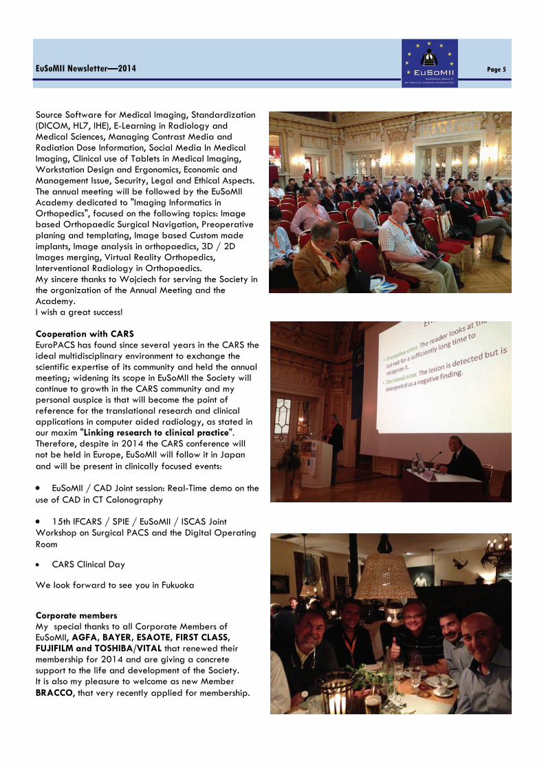
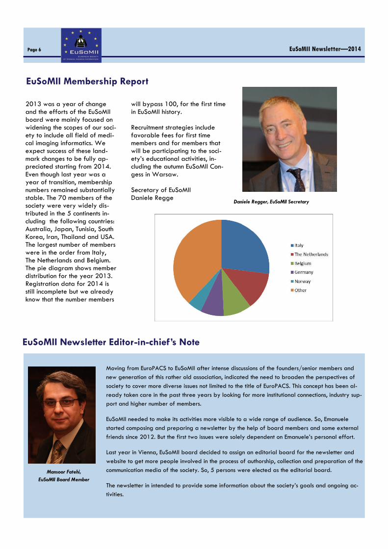
Source Software for Medical Imaging, Standardization (DICOM, HL7, IHE), E-Learning in Radiology and Medical Sciences, Managing Contrast Media and Radiation Dose Information, Social Media In Medical Imaging, Clinical use of Tablets in Medical Imaging, Workstation Design and Ergonomics, Economic and Management Issue, Security, Legal and Ethical Aspects. The annual meeting will be followed by the EuSoMII Academy dedicated to "Imaging Informatics in Orthopedics", focused on the following topics: Image based Orthopaedic Surgical Navigation, Preoperative planing and templating, Image based Custom made implants, Image analysis in orthopaedics, 3D / 2D Images merging, Virtual Reality Orthopedics, Interventional Radiology in Orthopaedics. My sincere thanks to Wojciech for serving the Society in the organization of the Annual Meeting and the Academy. I wish a great success! Cooperation with CARS EuroPACS has found since several years in the CARS the ideal multidisciplinary environment to exchange the scientific expertise of its community and held the annual meeting; widening its scope in EuSoMII the Society will continue to growth in the CARS community and my personal auspice is that will become the point of reference for the translational research and clinical applications in computer aided radiology, as stated in our maxim "Linking research to clinical practice". Therefore, despite in 2014 the CARS conference will not be held in Europe, EuSoMII will follow it in Japan and will be present in clinically focused events:
EuSoMII / CAD Joint session: Real-Time demo on the use of CAD in CT Colonography
15th IFCARS / SPIE / EuSoMII / ISCAS Joint Workshop on Surgical PACS and the Digital Operating Room
CARS Clinical Day
We look forward to see you in Fukuoka
Corporate members My special thanks to all Corporate Members of EuSoMII, AGFA, BAYER, ESAOTE, FIRST CLASS, FUJIFILM and TOSHIBA/VITAL that renewed their membership for 2014 and are giving a concrete support to the life and development of the Society. It is also my pleasure to welcome as new Member BRACCO, that very recently applied for membership.
EuSoMII Newsletter—2014 Page 5
EuSoMII Membership Report
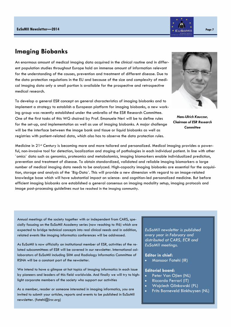
2013 was a year of change and the efforts of the EuSoMII board were mainly focused on widening the scopes of our soci-ety to include all field of medi-cal imaging informatics. We expect success of these land-mark changes to be fully ap-preciated starting from 2014. Even though last year was a year of transition, membership numbers remained substantially stable. The 70 members of the society were very widely dis-tributed in the 5 continents in-cluding the following countries: Australia, Japan, Tunisia, South Korea, Iran, Thailand and USA. The largest number of members were in the order from Italy, The Netherlands and Belgium. The pie diagram shows member distribution for the year 2013. Registration data for 2014 is still incomplete but we already know that the number members
will bypass 100, for the first time in EuSoMII history. Recruitment strategies include favorable fees for first time members and for members that will be participating to the soci-ety’s educational activities, in-cluding the autumn EuSoMII Con-gess in Warsaw. Secretary of EuSoMII Daniele Regge
EuSoMII Newsletter—2014 Page 6
Daniele Regger, EuSoMII Secretary
EuSoMII Newsletter Editor-in-chief’s Note
Mansoor Fatehi,
EuSoMII Board Member
Moving from EuroPACS to EuSoMII after intense discussions of the founders/senior members and
new generation of this rather old association, indicated the need to broaden the perspectives of
society to cover more diverse issues not limited to the title of EuroPACS. This concept has been al-
ready taken care in the past three years by looking for more institutional connections, industry sup-port and higher number of members.
EuSoMII needed to make its activities more visible to a wide range of audience. So, Emanuele
started composing and preparing a newsletter by the help of board members and some external
friends since 2012. But the first two issues were solely dependent on Emanuele’s personal effort.
Last year in Vienna, EuSoMII board decided to assign an editorial board for the newsletter and
website to get more people involved in the process of authorship, collection and preparation of the
communication media of the society. So, 5 persons were elected as the editorial board.
The newsletter in intended to provide some information about the society’s goals and ongoing ac-
tivities.
EuSoMII newsletter is published every year in February and distributed at CARS, ECR and EuSoMII meetings. Editor in chief: Mansoor Fatehi (IR)
Editorial board: Peter Van Ojien (NL) Riccardo Ferrari (IT) Wojciech Glinkowski (PL) Frits Barneveld Binkhuysen (NL)
Imaging Biobanks
An enormous amount of medical imaging data acquired in the clinical routine and in differ-
ent population studies throughout Europe hold an immense amount of information relevant
for the understanding of the causes, prevention and treatment of different disease. Due to
the data protection regulations in the EU and because of the size and complexity of medi-
cal imaging data only a small portion is available for the prospective and retrospective
medical research.
To develop a general ESR concept on general characteristics of imaging biobanks and to
implement a strategy to establish a European platform for imaging biobanks, a new work-
ing group was recently established under the umbrella of the ESR Research Committee.
One of the first tasks of this WG chaired by Prof. Emanuele Neri will be to define rules
for the set-up, and implementation as well as use of imaging biobanks. A major challenge
will be the interface between the image bank and tissue or liquid biobanks as well as
registries with patient-related data, which also has to observe the data protection rules.
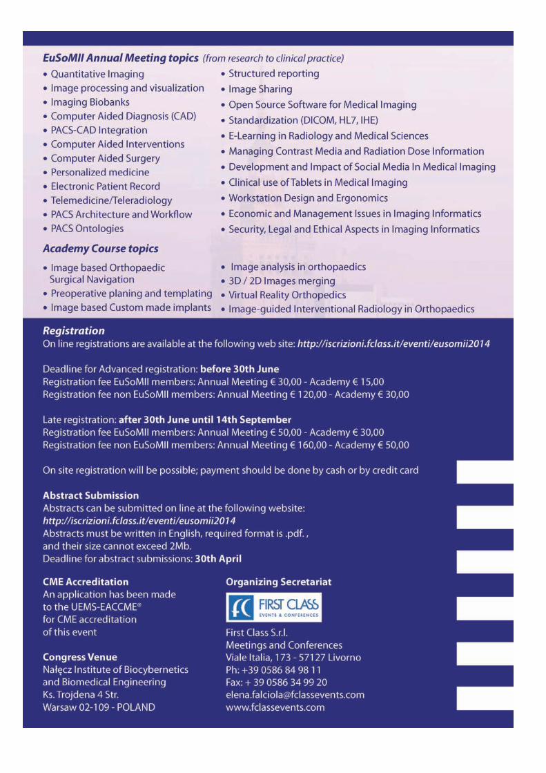
Medicine in 21st Century is becoming more and more tailored and personalized. Medical imaging provides a power-ful, non-invasive tool for detection, localization and staging of pathologies in each individual patient. In line with other `omics` data such as genomics, proteomics and metabolomics, imaging biomarkers enable individualized prediction, prevention and treatment of disease. To obtain standardized, validated and reliable imaging biomarkers a large number of medical imaging data needs to be analyzed. High-capacity imaging biobanks are essential for the acquisi-tion, storage and analysis of the ´Big-Data´. This will provide a new dimension with regard to an image-related knowledge base which will have substantial impact on science- and cognition-led personalized medicine. But before efficient imaging biobanks are established a general consensus on imaging modality setup, imaging protocols and image post-processing guidelines must be reached in the imaging community.
Page 7 EuSoMII Newsletter—2014
Hans-Ulrich Kauczor, Chairman of ESR Research
Committee
Annual meetings of the society together with or independent from CARS, spe-
cially focusing on the EuSoMII Academy series (now reaching to #6) which are
expected to bridge technical concepts into real clinical needs and in addition,
related events like imaging informatics conferences will be addressed.
As EuSoMII is now officially an institutional member of ESR, activities of the re-
lated subcommittees of ESR will be covered in our newsletter. International col-
laborators of EuSoMII including SIIM and Radiology Informatics Committee of
RSNA will be a constant part of the newsletter.
We intend to have a glimpse at hot topics of imaging informatics in each issue
by pioneers and leaders of this field worldwide. And finally we will try to high-
light corporate members of the society who support our activities
As a member, reader or someone interested in imaging informatics, you are
invited to submit your articles, reports and events to be published in EuSoMII
newsletter. ([email protected])


ECR 2014 - RC 1605: Improving workflow efficiency and quality
It will be my distinct pleasure to chair this Refresher Course on PACS, that will see the participation of friends such as Peter Mildenberger “Improving the quality and efficiency of computerized order entry through decision support”, Emanuele Neri “Improving quality and efficiency of reporting through structure and templates”, and Eliseo Vaño “Improving quality and efficiency of dose management trough exchange between modalities and reg-istries”.
My hospital in one of the oldest in Italy. It was founded in 1257, and now is the hub of a regional-wide integrated RIS/PACS system provided as a commercial service by Esaote and Fujifilm. Presently, our central archive stores 140 terabyte, corresponding to 3,800,000 exams.
Our group has been active in PACS research and implementation since two decades: my first paper on this topic published in European Radiology dates back to 1994 and was entitled: “Transmission of radiological images using broadband communications”.
Being a teaching hospital we developed a PACS add-on software aimed at allowing the seamless production of teaching files directly from the reporting environment. This is now a commercial product sold worldwide by Fujifilm.
More recently, we have been very active in testing software tools for the automatic tracking of radiation dose and contrast medium administration. These tools allow to easily discover bad practices and have the potential to greatly improve efficiency and quality.
In fact, we believe that being medical care an information-intensive activity, its overall quality is impaired if imaging-related data are not managed comprehensively and efficiently.
It is now apparent that just “storing” images and reports is not enough. This is why there are so many new develop-ments in the area of CPOE with integrated clinical decision support, critical results reporting software, and dose man-agement software able to automatically report and log radiation dose information and contrast media informa-tion.
I cordially invite you to attend this session at ECR 2014,
that will tke place on Monday, March 10, 08:30–10:
EuSoMII Newsletter—2014 Page 10
Davide Caramella,
Honorary Member of EuSoMII Board
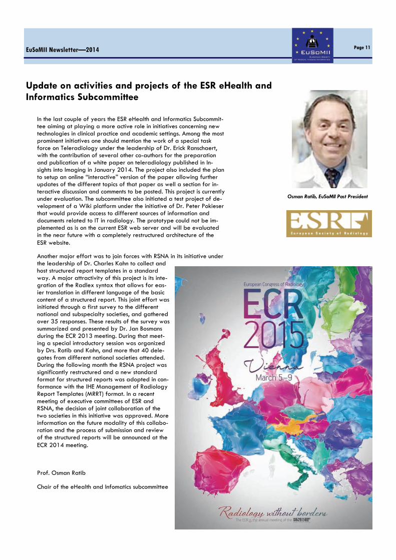
In the last couple of years the ESR eHealth and Informatics Subcommit-tee aiming at playing a more active role in initiatives concerning new technologies in clinical practice and academic settings. Among the most prominent initiatives one should mention the work of a special task force on Teleradiology under the leadership of Dr. Erick Ranschaert, with the contribution of several other co-authors for the preparation and publication of a white paper on teleradiology published in In-sights into Imaging in January 2014. The project also included the plan to setup an online “interactive” version of the paper allowing further updates of the different topics of that paper as well a section for in-teractive discussion and comments to be posted. This project is currently under evaluation. The subcommittee also initiated a test project of de-velopment of a Wiki platform under the initiative of Dr. Peter Pokieser that would provide access to different sources of information and documents related to IT in radiology. The prototype could not be im-plemented as is on the current ESR web server and will be evaluated in the near future with a completely restructured architecture of the ESR website.
Another major effort was to join forces with RSNA in its initiative under the leadership of Dr. Charles Kahn to collect and host structured report templates in a standard way. A major attractivity of this project is its inte-gration of the Radlex syntax that allows for eas-ier translation in different language of the basic content of a structured report. This joint effort was initiated through a first survey to the different national and subspecialty societies, and gathered over 35 responses. These results of the survey was summarized and presented by Dr. Jan Bosmans during the ECR 2013 meeting. During that meet-ing a special introductory session was organized by Drs. Ratib and Kahn, and more that 40 dele-gates from different national societies attended. During the following month the RSNA project was significantly restructured and a new standard format for structured reports was adopted in con-formance with the IHE Management of Radiology Report Templates (MRRT) format. In a recent meeting of executive committees of ESR and RSNA, the decision of joint collaboration of the two societies in this initiative was approved. More information on the future modality of this collabo-ration and the process of submission and review of the structured reports will be announced at the ECR 2014 meeting.
Prof. Osman Ratib
Chair of the eHealth and Infomatics subcommittee
EuSoMII Newsletter—2014 Page 11
Update on activities and projects of the ESR eHealth and Informatics Subcommittee
Osman Ratib, EuSoMII Past President

CARS 2014, June 25-28, 2014, Fukuoka Convention Center, Japan The CARS congress is the annual event for a distinguished international community of scientists, engineers and physi-cians to present and discuss the key innovations that shape modern medicine on a worldwide basis. Founded in 1985, CARS has played a leading role in medical and imaging informatics for more than 25 years by focusing on research and development on novel algo-rithms and systems and their applications in radiology and surgery. Its growth and impact is due to CARS’s close collaboration with the ISCAS and EuroPACS (now EuSoMII) societies, and CAR, CAD and CMI organizations. Following the long term successful cooperation, in many parts of the world, in 2014 these prestigious scientific communities will jointly hold their annual meetings as part of the 28th CARS Congress in Fukuoka, Japan. The CARS Congress Organizing Committee invites you to come to Fukuoka in June 25 – 28, 2014, for an extraordi-nary event in which scientific/medical presentations as well as stimulating discussions will foster new visions on the future of medicine.
Page 13 EuSoMII Newsletter—2014
At the CARS Congress you will have the opportunity to meet scholars and practising experts in the fields of radiology, surgery, engineering, informatics and healthcare management who have an interest in topics, such as
image- and model-guided interventions
advanced medical imaging
image processing and visualization
intelligent operating room of the future
decision and action support in surgical management
computer aided diagnosis
medical simulation and evaluation, and e-Learning
surgical navigation and robotics
model-guided and personalised medicine New PACS applications, including IT-infrastructures adapted for
the operating room, related results from the DICOM and IHE working groups, but in particular, new methods and IT-tools for modelling the patient and medical processes are increasingly shaping the scope of CARS. Clinical specialties represented at CARS include:
Computer Aided Neuro-, ENT-, Orthopaedic and Spinal Sur-gery, Cardiovascular- and Thoracic Surgery, Gastroenterological-, Gynaecological- and Urological Surgery
Imaging and Interventional Radiology
Computed Maxillofacial Imaging
Computer Assisted Radiation Therapy
Image Guided Navigation Surgery
Minimally Invasive and Robotic Surgery Additional events will take place during CARS 2014 in Fukuoka,
such as tutorials and hands-on training for biomedical engineers, a full IFCARS/ISCAS clinical day on management of liver tumour as well as the IPCAI and ACCAS annual conferences. Social events will accompany the conference, such as the opening ceremony and a boat cruise in Hakata’s bay. Scholarships are available for helping students and junior researchers to attend the conference.
Please note that the deadline for paper and abstract submis-sions for CARS 2014 in Fukuoka is January 10, 2014.
Recent successful CARS congresses have taken place in Berlin, Paris, Tokyo, San Francisco, London, Chicago, Osaka, Barcelona, Geneva, Pisa, and Heidelberg.
We would be delighted to welcome you at CARS 2014, which will take place for the first time in Fukuoka, a renowned Japanese city for culture and industry as a centre of Kyushu Island with many geographic attractions.
Makoto Hashizume, MD, PhD, CARS 2014 President Heinz U. Lemke, PhD, CARS Organizer

Tablet Computers
Since their introduction, tablet PCs have evolved extensively. They have become very popular, filling the gap between
laptop computers and smart mobile phones. Tablets are de-
signed for general purpose uses. They are not designed for
primary radiologic diagnosis; however, some of their display
characteristics and parameters make them attractive for
viewing images, and they are great devices for consulting and reviewing radiologic images.
In February 2011, the US Food and Drug Administration ap-proved the first mobile application for limited diagnostic
viewing, but its use is restricted to situations in which no diag-
nostic reading room facility with workstations is available.
Even in these cases, certain precautions are necessary—one
should take steps to ensure optimal conditions, for instance,
cleaning off the fingerprints that normally cover the screen and going to the dimmest part of the room. Most importantly,
the display should be DICOM-calibrated [1] like the displays
used in primary diagnostic workstations in the reading room.
Tablets lack the hardware for automatic DICOM calibration
and do not provide access to the video subsystem, so DICOM
GSDF calibration is not easy. However, nowadays tools are
available to calibrate the tablet display to make it DICOM
conformant. If we use one of these tools, then using
tablets for diagnostic
reading is a better option
than using a conventional
laptop without calibra-
tion.
Tablets can make access
easier, improving image
distribution and making
reports available to re-ferring clinicians; they
may be especially useful
for patient rounds, communicating with patients about
findings, and non-diagnostic consultations. However,
the images they display should look the same as those
available in the reading room, and their quality should
be similar, too. Clinicians need to know how to create the appropriate viewing conditions with these devices.
They need to develop good viewing habits like setting
the auto-brightness to “OFF” to ensure they always use
the maximum allowed brightness and going to a dim-
mest part of the room to avoid direct reflections on the
screen from windows or lighting lamps.
Focusing on displays, we can see how far tablets are
from matching the display characteristics of a diagnos-
tic workstation display in a reading room. The differ-
ence in some of those characteristics and parameters is
not great. For instance, tablets generally have more spatial resolution than diagnostic displays because
they are able to show more pixels per inch (pixel den-
sity). In this sense, they are even better than diagnostic
monitors. But one should take in consideration the
working distance: on workstations the eye is around
EuSoMII Newsletter—2014 Page 14
Josep Fernandez Bayo, EuSoMII
Treasurer
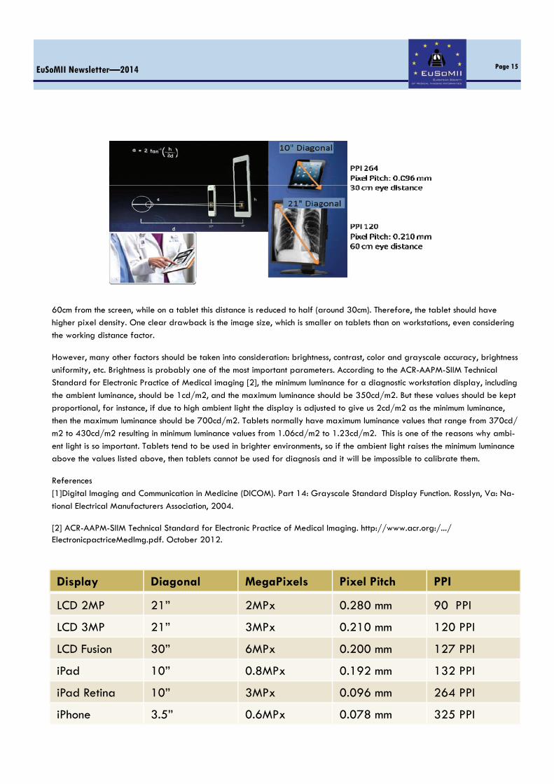
60cm from the screen, while on a tablet this distance is reduced to half (around 30cm). Therefore, the tablet should have higher pixel density. One clear drawback is the image size, which is smaller on tablets than on workstations, even considering
the working distance factor.
However, many other factors should be taken into consideration: brightness, contrast, color and grayscale accuracy, brightness
uniformity, etc. Brightness is probably one of the most important parameters. According to the ACR-AAPM-SIIM Technical Standard for Electronic Practice of Medical imaging [2], the minimum luminance for a diagnostic workstation display, including
the ambient luminance, should be 1cd/m2, and the maximum luminance should be 350cd/m2. But these values should be kept
proportional, for instance, if due to high ambient light the display is adjusted to give us 2cd/m2 as the minimum luminance,
then the maximum luminance should be 700cd/m2. Tablets normally have maximum luminance values that range from 370cd/
m2 to 430cd/m2 resulting in minimum luminance values from 1.06cd/m2 to 1.23cd/m2. This is one of the reasons why ambi-
ent light is so important. Tablets tend to be used in brighter environments, so if the ambient light raises the minimum luminance above the values listed above, then tablets cannot be used for diagnosis and it will be impossible to calibrate them.
References
[1]Digital Imaging and Communication in Medicine (DICOM). Part 14: Grayscale Standard Display Function. Rosslyn, Va: Na-
tional Electrical Manufacturers Association, 2004.
[2] ACR-AAPM-SIIM Technical Standard for Electronic Practice of Medical Imaging. http://www.acr.org:/.../ElectronicpactriceMedImg.pdf. October 2012.
EuSoMII Newsletter—2014 Page 15
Display Diagonal MegaPixels Pixel Pitch PPI
LCD 2MP 21” 2MPx 0.280 mm 90 PPI
LCD 3MP 21” 3MPx 0.210 mm 120 PPI
LCD Fusion 30” 6MPx 0.200 mm 127 PPI
iPad 10” 0.8MPx 0.192 mm 132 PPI
iPad Retina 10” 3MPx 0.096 mm 264 PPI
iPhone 3.5” 0.6MPx 0.078 mm 325 PPI
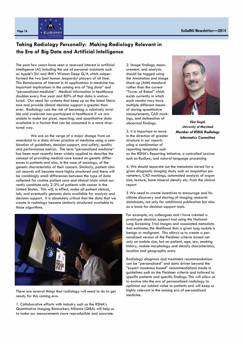
Taking Radiology Personally: Making Radiology Relevant in the Era of Big Data and Artificial Intelligence
The past few years have seen a renewed interest in artificial intelligence (AI) including the use of personal assistants such as Apple’s Siri and IBM’s Watson Deep Q/A which outper-formed the two best human Jeopardy! players of all time. This Renaissance of interest in AI applications in medicine has important implications in the coming era of “big data” and “personalized medicine”. Medical information in healthcare doubles every five year and 80% of that data is unstruc-tured. Our need for systems that keep up on the latest litera-ture and provide clinical decision support is greater than ever. Radiology runs the risk of becoming a relatively invisi-ble and irrelevant non-participant in healthcare if we are unable to make our pixel, reporting, and quantitative data available in a format that can be consumed in a more struc-tured way.
We are on the verge of a major change from an anecdotal to a data driven practice of medicine using a com-bination of guidelines, decision support, and safety, quality and performance metrics. The term “personalized medicine” has been most recently been widely applied to describe the concept of providing medical care based on genetic differ-ences in patients and also, in the case of oncology, of the genetic characteristics of their cancers. Similarly, patient clini-cal records will become more highly structured and there will be vanishingly small differences between the type of data collected for routine patient care and clinical trials which cur-rently constitute only 2-3% of patients with cancer in the United States. This will, in effect, make all patient clinical, lab, and eventually genomic data available for analysis and decision support. It is absolutely critical that the data that we create in radiology become similarly structured available to those algorithms.
There are several things that radiology will need to do to get ready for this coming era:
1. Collaborative efforts with industry such as the RSNA’s Quantitative Imaging Biomarkers Alliance (QIBA) will help us to make our measurements more reproducible and accurate.
2. Image findings, meas-urement, and analysis should be tagged using the Annotation and Image Mark-up (AIM) standard rather than the current “Tower of Babel” which exists currently in which each vendor may have multiple different means of storing quantitative measurements, CAD mark-ings, and delineation of abnormal findings.
3. It is important to move in the direction of greater structure in our reports using a combination of reporting templates such as the RSNA’s Reporting Initiative, a controlled Lexicon such as RadLex, and natural language processing
4. We should separate out the metadata stored for a given diagnostic imaging study such as acquisition pa-rameters, CAD markings, automated analysis of organ size, texture, bone mineral density etc. from the clinical report
5 We need to create incentives to encourage and fa-cilitate discovery and sharing of imaging research databases, not only for additional publication but also as a basis for decision support tools.
For example, my colleagues and I have created a prototype decision support tool using the National Lung Screening Trial images and associated metadata that estimates the likelihood that a given lung nodule is benign or malignant. This allows us to create a per-sonalized version of the Fleishner criteria based not only on nodule size, but on patient, age, sex, smoking history, nodule morphology and density characteristics, location and geographic area.
Radiology diagnosis and treatment recommendations can be “personalized” and data driven beyond the “expert consensus based” recommendations made in guidelines such as the Fleishner criteria and tailored to specific patients and specific findings. This will allow us to evolve into the era of personalized radiology to optimize our added value to patients and will keep us highly relevant in the coming era of personalized medicine.
EuSoMII Newsletter—2014 Page 16
Eliot Siegel,
University of Maryland
Member of RSNA Radiology Informatics Committee
The Society for Imaging Informatics in Medicine (SIIM) is a unique professional organization that includes among its members radiologists, other clinical specialists who use medical im-ages, clinical IT and imaging informatics professionals, physicists, imaging researchers, and imaging and IT industry professionals. SIIM has 47 European members, 2% of total mem-bership. The SIIM 2014 Annual Meeting will be held 15-17 May, 2014, in Long Beach, California. It includes learning tracks on enterprise imaging, new archiving strategies, data mining, quality and safety, and career development for imaging informatics professionals. Among the most exciting opportunities this year are a hackathon based on HL7 FHIR, DI-COMweb and RESTful objects, and a vibrant Open Source Plug Fest. SIIM also runs a one-day Imaging Informatics Professional Bootcamp, the most widely used exam resource for the ABII CIIP exam, and an excellent course to update imaging informatics skills. The meet-ing also has hands-on workshops in areas from Nagios monitoring to hacking Mirth, and focus sessions on radiation optimization, mHealth, analytics, interoperability, quantitative imaging, and reporting.
With the explosion in medical images across all specialties and the widespread shift to enterprise EMRs and VNAs, SIIM is responding with a renewed effort to explore the issues of managing all medical images, not just radiology. At the same time, SIIM continues to support basic research in radiology informatics. An important result of this research is work under the leadership of Brad Erickson, MD, PhD to produce the SIIM Workflow Initiative in Medicine (SWIM). SWIM provides a common definition of workflow steps inside medical imaging departments, defines key performance indicators using these workflow steps, and defines data elements used to capture information about the KPIs and workflow steps. This is a major resource for imaging departments that implement workflow engines to improve productivity, quality and safety. SWIM workflow terms are integrated into RadLex, and IHE has expressed interest in it. SIIM expects these definitions will be included in IHE pro-files in the next few years.
SIIM is also participating with ACR and RSNA in a major new initiative known as Imaging 3.0. Among other goals, Imaging 3.0 will help radiologists become ‘IT savvy’ by defining IT tools and the knowledge needed to use them to improve radiologists’ productivity and quality, and describ-ing how to use analytics to demonstrate radiologists’ value throughout the healthcare system. While this is primarily a USA-centric response to the change from volume-based reimbursement to value-based payment, the resulting IT tools should be universally applicable.
For more information and to view SIIM’s education offer-ings, please visit SIIM online at siim.org.
J. Raymond "Raym" Geis, MD Chair, Society for Imaging Informatics in Medicine
EuSoMII Newsletter—2014 Page 17
Society for Imaging Informatics in Medicine (SIIM)
J. Raymond "Raym" Geis, MD
Chair, Society for Imaging
Informatics in Medicine Member of RSNA Radiology
Informatics Committee

Blogging on Medical Imaging Informatics
In the past decades, developments in radiology have been rapidly introduced. Most of those development would not have been possible without informatics. Therefore, Medical Imaging Informatics is an area of growing interest. Medical Imaging Informatics is not just about Picture Archiving and Communication Systems (PACS) and advanced visualization but is involved in the full medical imaging chain on the topics of acquisition, reconstruction, archiving, distribution, visualiza-tion, and reporting. Imaging informatics developments on the one hand relies on the high-end computing developments and expensive, dedi-cated, hard- and software but on the other hand also on the developments in consumer electronics. One example is the gaming industry which demands on the graphic capabilities of gaming hardware led to the development of low-price, high quality, video cards that could be utilized also to benefit the advanced visualization application in radiology. To keep track of these developments and to collect and regis-ter anything related to Medical Imaging Informatics a blog was started in September 2009 with the following purpose: This blog provides information on conferences and novelties in the area of Medical Imaging Informatics (MII). MII has a broad scope ranging from the Radiology Information System and Picture Archiving and Communication System (PACS) to Ad-vanced Visualization and Computer Aided Diagnosis (CAD). To find new opportunities in healthcare we need to look at infor-matics solutions in other areas to apply them into the medical field to achieve higher level healthcare at lower costs. Currently 285 posts have been included and a total of close to 27.500 pageviews are registered (status February 17, 2014). If you are interested please access the blog at http://medicalimaginginformatics.blogspot.com or through the link at the EuSoMII webpage. Leave your comments at a post on the blog or send us your contributions at: [email protected]
EuSoMII Newsletter—2014 Page 18
P.M.A. van Ooijen, MSc, PhD, CPHIT Scientific Researcher,
Dept. of Radiology, UMCG

The Iranian Imaging Informatics Conferences have always been scientifically supported by EuSoMII (former EuroPACS). The first conference hosted Bernie Huang as honorary president and Davide Caramella as guest speaker. The second one had honorary presidency of Heinz Lemke and Peter Mildenberger also attended as guest speaker. For the third conference Emanuele Neri was the honorary president together with kind contribution of Daniele Regger. In the fourth conference the Peter Mildenberger was the honorary president and Utku Senol from Turkey took part as guest speaker.
This time in Shiraz, we hosted Josep Fernandez Bayo and Ustun Aydingoz from Turkey. Also Emenuele gave the opening lecture from Pisa by kind support of ESR office. So, Iranian Imaging Informatics Conferences should and has been considered as local EuroPACS/EuSoMII meetings.
In the fifth conference, the major themes were selected as: RIS, Teleradiology, Image Processing and Tablets. The meeting was very successful in attracting audience although mostly from local province but also from all over the country. The number of par-ticipants was above 400, among which 92 persons applied for EuSoMII membership. Alireza Shakibafard has been instrumental in this success by competent organization of the local facilities and people to actualize this event.
41 papers were submitted to the conference and as usual the award of the winner of the best paper was travel support to par-ticipate in CARS meeting. The title of the best paper was:
Evaluation of the Delicacy of Dynamic – SPECT with common gamma camera systems
The award was granted under the name of late Paolo Inchingolo who was supposed to be one of the guest speakers of the first IIC but could not manage to attend due to serious illness ended in his unexpected passing away.
Medical Imaging Informatics Research Center (MIIRC) that was established nearly a year ago in Tehran is a non-profit organization affiliated to Iranian Society of Radiology trying to play the role of a bridge between clinical and technical professionals and students for teaching and re-search purposes. At the same time, the center pro-vides consultancy services to hospitals and institu-tions looking for a digital transformation. IICs are currently events under the leadership of MIIRC.
Mansoor Fatehi, MD, CIIP Director, Medical Imaging Informatics Research Center, Tehran
EuSoMII Newsletter—2014 Page 19
Dr Ustun Aydingoz Professor of Radiology, Hacettepe University School of Medi-cine, Ankara, Turkey
"I was honored and delighted to take part in the 5th Iranian Imaging Informatics Conference. I was able to share with my Iranian colleagues some of my experi-ence regarding HIS-RIS-PACS integration, RIS workflow issues, and workplace ergonomics in radiology. One of the pleasant surprises that I had during the conference in beautiful Shiraz was the high number of radiologists and radiologists-in-training who at-tended the meeting. Honestly, we were not able to bring together such a satisfactory number of medical doctors to our national medical informatics meetings in Turkey. I was also impressed with the eagerness of the Iranian industry participants in terms of developing new hardware and software. Although it may be that HIS and RIS development and implementation has a long way to go in Iran, enthusiasm and talent of Iranian technical people and medical doctors promises a successful future in these fields. I believe we have reasons to believe that collaboration between Turkish and Ira-nian radiology and medical informatics communities will bear fruits for both nations, which have a time-honored tradition of good relations."
The Fifth Iranian Imaging Informatics Conference – Shiraz/IRAN

The 4th EuSoMII Academy Course, Imaging Informatics in Oncology
When Emanuele Neri called to ask me if I was willing to or-ganize the 2013 Academy course in Torino I was initially a bit reluctant. Academy courses are clinically oriented but lean heavily on informatics and the two worlds are not always so easy to reconcile. It is commonplace to think that informatics is for computer scientists and not for doctors. However, imaging doctors heavily rely on informatics for image processing, re-porting (e.g. structured reporting) and recently also for man-aging contrast agents and radiation dose. Finally it was de-cided, we would explore the role of imaging informatics in oncology. Cancer has important social and economic implications. In industrialized countries cancer is the second cause of death and an invalidating disease. Costs of therapy are increasing rapidly due to the extended life expectancy of patients and to the widespread use of the new target therapies. In 2030 the number of people living with cancer will be 3 times more than in 2013. The wide spectrum of therapeutic options re-quires accurate pre and post-treatment assessment, and im-aging is in the frontline. Tools to assess response to treatment are entering more and more into clinical practice and imag-ing doctors must know how to deal with them. Not only, but accurate monitoring requires standardization of imaging pro-tocols: same technique, same contrast media and same injec-tion protocol. Accurate monitoring means that radiologists must learn how to measure tumor changes in a reliable man-ner in one or more dimensions, by means of anatomical and functional imaging. The course was held on September 20-21. The faculty was very experienced and lectures state-of-the-art; large part of the second day was dedicated to the discussion of clinical cases using specialized software. Over 70 delegates from all over Europe attended Turin for the 4th Academy Course. Next stop Warsaw, good luck Wojciech! Daniele Regge Organizer of the 4th Academy Course in Torino, Italy
EuSoMII Newsletter—2014 Page 20
Day 1: September 20 Moderators: Emanuele Neri, Wojciech Glinkowski Session I: Fundamentals of Imaging Informatics applied to Oncol-ogy (14.00 - 15.40) Patient Protocol Standardization - Mathias Prokop Basic principles of image processing - Marleen de Bruijne Measuring cancer: from 2D to 4D - Irene Bargellini CAD: From benchmark to bedside - Daniele Regge Session II: Imaging Response to Therapy (16.00 - 18.00) Target therapies: what imaging specialists must know - Mas-simo Aglietta RECIST and beyond - Francesco Sardanelli Advanced imaging techniques to monitor treatment response - DW and perfusion imaging - Vicky Goh - PET/CT and PET/MRI - Osman Ratib - Dual energy CT - Heinz-Peter Schlemmer
Day 2: September 21 Moderators: Daniele Regge, Davide Caramella Session I: Practical Session: assessing response to treatment Case review 1: Metastatic Disease Case review 2: Locally Advanced Cancer Session II: Practical Session: Computer Aided Diagnosis Case review 3: Lung CAD Case review 4: Colon CAD Case review 5: Prostate CAD Final Lecture: Advanced representation and sharing of image processing data (16.30 - 17.00) –
Faculty Massimo Aglietta Irene Bargellini Hans-Christoph Becker Delia Campanella Davide Caramella Marleen de Bruijne Lorenzo Faggioni Nicola Flor Wojciech Glinkowski
Vicky Goh Anna Rita Larici Emanuele Neri Mathias Prokop Osman Ratib Daniele Regge Marco Rengo Filippo Russo Francesco Sardanelli Heinz-Peter Schlemmer
Agenda

The 5th EuSoMII Academy on “Tablets in Radiology”– Shiraz / IRAN
The fifth EuSoMII academy was planned to be held in Iran after the successful organization of the second academy on CAD. The subject of the 5th academy: “Tablets in Radiology” turned out to be very much attractive to the local audience leading to above 100 participants. In this program, we tried to organize the content in three separate groups: general information about tablets, ra-diologic applications, and hands-on experience with real time reading by the aid of tablets. So, the hands-on session was based on a local server to help participants browse cases and follow the instructions of the mobile viewer. The academy was honored by kind participation of Josep Fernandez Bayo, EuSoMII treasurer who gave an in-depth overview of technical aspects of tablets in radiology. But majority of the lectures were delivered by local faculty. The academy was part of the 5th Iranian Imaging Informatics Conference. Mansoor Fatehi, MD, CIIP Organizer of 5th EuSoMII Academy in Shiraz
EuSoMII Newsletter—2014 Page 21
Agneda Session I: Introduction to Tablets The 5th EuSoMII Academy: Learning Objective and Course Outline: Mansoor Fatehi, MD, CIIP Tablet-computers: a technical overview: Josep Fernandez Bayo Principle of iOS: Hossein Khalili, MD Introduction to Android Tablets: Hossein Khalili, MD Software Installation: Mohammad Farjadian Session II: Tablets & Radiology Osirix Viewer: Mojtaba Ebrahimi, MD Tablet-based Radiology Apps: Mojtaba Ebrahimi, MD Session III: Tablets & PACS Tablets for Clinical Radiology & Non-Radiology Practice: Mansoor Fatehi, MD, CIIP INFINITT Mobile PACS Viewer: A Live Demo Hands-on Exercises Using INFINITT PACS Viewers for Both iPAD & Android Tablets

Agfa HealthCare is a global leader in the fast growing market of integrated IT and imaging systems, offer-ing healthcare facilities a seamless flow of information and a 360º view of patient care. The company has a unique, holistic approach, enabling it to provide in-depth clinical know-how and fully integrated hospital-wide solutions. These specialized solutions integrate IT and imaging systems for Radiology, Cardiology, Mammography and Orthopedics. Agfa Healthcare's enterprise-wide IT platform integrates all administra-tive and clinical data within a healthcare facility and is designed to match the unique needs of specific healthcare professionals.
Agfa HealthCare in Radiology: Agfa has over 100 years experience in radiology imaging and one out of two hospitals worldwide uses Agfa HealthCare customized applications today. The wide range of Agfa's solutions combined with speech technology and integrated IT applications allows radiologists to keep a clear, accessible, and structured overview of all relevant patient data and images. Agfa HealthCare in Cardiology: Agfa HealthCare offers cardiovascular specialist a single point of access to patient-centric clinical information and radiology results. Structured reporting supports your quality diag-nostic decision process, enabling rapid turnaround and improved communication. Point-of-care data cap-ture and multi-modality integration optimizes efficient workflows. Agfa HealthCare in healthcare IT: Agfa HealthCare's enterprise-wide IT platform, ORBIS, integrates all administrative and clinical data within a healthcare facility and its network and is designed to match the unique needs of specific healthcare professionals. ORBIS is the result of 10 years' of healthcare IT experi-ence and is used by over 400,000 healthcare facility employees daily. The ORBIS platform and its unique workflow and information management capabilities contain over 70 modules that can be used to build and customize the system at the point of care, according to each healthcare facility's changing needs.
EuSoMII Corporate Members Page 22

EuSoMII Corporate Members Page 23

EuSoMII Corporate Members Page 24

FUJIFILM constantly sets new standards for diagnostic imaging technology.
EuSoMII Corporate Members Page 25

First Class ideates and organizes conferences, meetings and educational courses, with a special expertise in the field of medical sciences. Full respect of the rules and regulations is considered a must in each and every event. A leading conference organizer both in Italy and abroad, First Class boasts prestigious collaborations with major Scientific Societies, Institutions and Universities. A dynamic team specialized in the organization of educational courses, workshops and international master classes for specialist physicians (expertise in the field of Radiology), hosted within the most prestigious University and Hos‐pital Centers in the world. A unique meeting opportunity for both participants and the Faculty, which allows for an in‐depth and authoritative educational experience. Master classes can further be optimized with a series of customized and interactive activi‐ties.
First Class is an accredited CME Provider approved by the Italian Ministry of Health and the European UEMS‐EACCME. The accreditation allows First Class to organize residential and online continuing education courses both in Italy and Europe. The strategic support and organizational competence of the group are an added value to the scientific content of each course and are a key contributor to the professional experience of each participant. In order to understand the real expectations and needs and to be successful in achieving important results in view of ever changing goals, each client is followed with great care and accuracy. This is the only approach possible to organize impeccably successful events, to emphasize talent and generate value. To choose First Class means opting for a complete and carefully planned service.
Head Office
Viale Italia, 173 – 57127 Livorno – Italy Ph. +39 0586 849811 – Fax. +39 0586 349920
Registered Office
Via Malasoma, 14/16 – 56126 Pisa – Italy UK Office
U.K. Office: 37 Middle Stoke, Limpley Stoke, Bath BA2 7GF 01225 723 243
[email protected] - www.fclassevents.com
EuSoMII Corporate Members Page 26

SYNAPSE Suite FUJIFILM constantly sets new standards for diagnostic imaging technology.
SYNAPSE continues this tradition, with thousands of proven implementations worldwide benefiting from SYNAPSE’s im-provements in efficiency and workflow.
SYNAPSE is a suite of software products developed by FUJIFILM for integrated workflow and digital imaging in the medical field.
SYNAPSE Suite is the global solution of FUJIFILM able to provide the best tool for managing clinical data and diagnostic images in a safe and complete way, in all areas of medical imaging, whether for radiology or cardiology.
SYNAPSE PACS is the System for storage and distribution of radiological diagnostic images entirely based on web tech-nology.
SYNAPSE 3D is the software for advanced post-processing of medical imaging; state of the art system, allows for a pow-erful and full support for reading, interpretation and reporting, regardless of the type of modalities and vendors.
SYNAPSE Mobility is the application that allows access to Synapse PACS from any device platform based on PC/Mac via a multi-browser support, and from devices such as iPad/iPhone or based on Android.
SYNAPSE Cardiovascular is the innovative software solution, vendor and hardware-neutral, which allows a complete and effective integration of all specialties in cardiovascular care.
SYNAPSE Workflow (*) is the system for the integrated and safe management of patient data and workflow, not only in radiology, but in virtually every field concerned with medical imaging.
SYNAPSE Theca (*) is the software solution for the storage and preservation of documents, in full compliance with Italian law.
EuSoMII Corporate Members Page 27

EuSoMII Corporate Members Page 28


Head Office Corner
The EuSoMII has been managed by Arnold Stipsits since 2005, back when it was still
called "EuroPACS". In 2009, he received
support from Andreas Sonnleitner and ever
since, they have been in charge of the daily
business of the EuSoMII. In their capacity as
"head office", they keep the membership database up to date, collect the annual fees,
organize meetings and telephone
conferences for the board and collect,
archive and disseminate relevant information.
They keep up with the daily tasks of running
a society and are in constant contact with the
president and the secretary. The EuSoMII office is located in the heart of Vienna and if
you would like to drop by, feel free to do so
any time during office hours.
E u S o M I I
EuSoMll office Mr. Arnold Stipsits, EuSoMll Head Office
Neutorgasse 9/6, 1010 Vienna - AUSTRIA phone: 0043-1-533 40 64 - 10 - fax: 0043-1-535 70 37 -
European Society of Medical Imaging Informatics EuSoMII The vision of the Society is the integration of information and communication technology with diagnostic and thera-peutic medical imaging. •The mission is to foster the transition from research to clini-cal application and education in the following fields: •Intelligent infrastructures and processes for image and knowledge management in medical diagnosis and therapy •Clinical computer application of medical images •Seamless information sharing for healthcare delivery and for clinical research purposes •Standards and quality assurance methods and tools.
President Emanuele Neri (IT) Past President Osman Ratib (CH) Secretary Daniele Regge (IT) Treasurer Josep Fernandez Bayo (ES) Board members Aslak Aslaksen (Norway) Frits Barneveld Binkhuysen (NL) Erwin Bellon (BE) Mansoor Fatehi (IR) Bernard Gibaud (FR) Wojciech Glinkowski (PL) Hiroshi Kondoh (JP) Wiro Niessen (NL) Peter Sögner (AT) Honorary Board Members Albert R. Bakker (NL) Davide Caramella (IT) H.K. Huang (US) Heinz U. Lemke (DE) Hiromu Nishitani (JP) Osman Ratib (CH)
Auditors Davide Caramella (IT) Arnold Stipsits (AT)

EuSoMll office Mr. Arnold Stipsits, EuSoMll Head Office
Neutorgasse 9/6, 1010 Vienna - AUSTRIA phone: 0043-1-533 40 64 - 10 - fax: 0043-1-535 70 37 - [email protected]