european journal of medicinal...
TRANSCRIPT

lable at ScienceDirect
European Journal of Medicinal Chemistry 111 (2016) 126e137
Contents lists avai
European Journal of Medicinal Chemistry
journal homepage: http: / /www.elsevier .com/locate/ejmech
Research paper
Synthesis and biological evaluation of D-ring fused 1,2,3-thiadiazoledehydroepiandrosterone derivatives as antitumor agents
Hai-Wei Cui a, 1, Shihong Peng b, 1, Xiang-Zhong Gu c, Huang Chen b, Yuan He b, Wei Gao a,Fang Lv b, Jin-Hua Wang b, Yan Wang a, Jia Xie b, Ming-Yao Liu b, Zhengfang Yi b, **,Wen-Wei Qiu a, *
a Department of Chemistry, School of Chemistry and Molecular Engineering, East China Normal University, 500 Dongchuan Road, Shanghai 200241, Chinab Shanghai Key Laboratory of Regulatory Biology, Institute of Biomedical Sciences and School of Life Sciences, East China Normal University, Shanghai200241, Chinac Department of Research and Development, Jiangsu Jiaerke Pharmaceuticals Group Co., Ltd., Zhenglu Town, Changzhou 213111, China
a r t i c l e i n f o
Article history:Received 4 December 2015Received in revised form29 January 2016Accepted 30 January 2016Available online 1 February 2016
Keywords:AntitumorDehydroepiandrosterone1,2,3-ThiadiazoleApoptosisMigration
Abbreviations: IC50, half maximal inhibitory conctivity relationship; SI, selectivity index; SRB, sulforhosine kinases; HE, hematoxylin and eosin; HAF,adriamycin; DHEA, dehydroepiandrosterone.* Corresponding author.** Corresponding author.
E-mail addresses: [email protected] (Z. Yi)(W.-W. Qiu).
1 These authors contributed equally to this work.
http://dx.doi.org/10.1016/j.ejmech.2016.01.0580223-5234/© 2016 Elsevier Masson SAS. All rights re
a b s t r a c t
A series of D-ring fused 1,2,3-thiadiazole DHEA derivatives were synthesized and investigated for theiractivity against the growth of various tumor cell lines using the sulforhodamine B (SRB) assay. It isamazing that for these compounds, T47D cell line was much more sensitive than other tumor cell lines.The most potent saturated N-heterocyclic derivatives showed similar antitumor effect with the positivecontrol compound ADM (adriamycin) on T47D cells, that was 44e60 folds more potent than the leadcompound DHEA. Most compounds with potent antitumor activity displayed low toxicity on normalhuman fibroblasts (HAF). Especially compound 25 (CH33) showed an IC50 of 0.058 mM on T47D cells andits selectivity index (SI) between HAF and T47D was 364, which was 214 folds better than ADM (SI ¼ 1.7).The apoptosis, colony formation and transwell migration assays of 25 were performed on T47D cell line.The primary mechanism study showed that 25 caused a dose-dependent induction of apoptosis, andinduced phosphorylation of EphA2 and EphB3 in T47D cells. The in vivo antitumor effect of 25 was alsoobserved in T47D tumor-bearing mice without obvious toxicity.
© 2016 Elsevier Masson SAS. All rights reserved.
1. Introduction
Steroids are widely distributed in natural world and display avariety of biological activities, especially antitumor activity [1,2].Heterocycles are considered as privileged scaffolds in drug dis-covery and widely used in medicinal chemistry [3,4]. The intro-duction of A- and D-ring fused heterocycles into the steroidderivatives often result in amelioration of biological properties[5,6], for example, enhancing the antitumor effect [7e10].
Dehydroepiandrosterone (DHEA) also known as androstenolone
entration; SAR, structure-ac-damine; RTKs, receptor tyro-human fibroblasts; ADM,
served.
or prasterone, is the most abundant circulating steroid hormone inhumans [11]. DHEA and its sulfate ester also have a variety of po-tential biological effects, such as anti-aging [12], anti-inflammatory[13], immunomodulatory [14], antiviral [15,16], antidepressant [17]and anticancer effects [18,19]. As heterocycles were fused with theD-ring of DHEA, the antitumor activity was greatly improved. Forinstance (Fig. 1), compound A, that was obtained by introducing p-methoxyphenylpyrazoline fragment displayed more cytotoxic ac-tivity than cisplatin on malignant human cell lines [20]; a series ofD-ring fused thiazole, thiazole imines, imidazo thiazoles and pyri-dine were synthesized by Liu et al. showed good cytotoxicityagainst three tumor cell lines (EC109, EC9706 and MGC 803),especially compound B possessed the most potent antitumor ac-tivity [21,22]; compound C was synthesized by fusing pyrazole-scaffold with D-ring exhibited the best antitumor activity withIC50 values of 20e1.4 nM against four cell lines, and 1.03 mM againsta tamoxifen resistant breast cancer cell line, and a preliminarystructure-activity relationship (SAR) showed that with a linker in 3-position was favorable to increase the bioactivity [23].

Fig. 1. Chemical structures of D-ring fused heterocycles DHEA antitumor agents.
H.-W. Cui et al. / European Journal of Medicinal Chemistry 111 (2016) 126e137 127
Thiadiazoles occur widely in nature in four isomeric forms, thatis 1,2,3-thiadiazole, 1,2,5-thiadiazole, 1,2,4-thiadiazole and 1,3,4-thiadiazole. Many drugs containing thiadiazole nucleus are avail-able in the market for instance acetazolamide, methazolamide,cefazoline, sulfamethazole, etc. In recent decades, thiadiazole andtheir derivatives have attracted much attentation because of theirwide range of biological activities [24]. Especially the 1,3,4-thiadiazole derivatives have attracted widespread attention dueto their diverse biological activity such as antimicrobial, antifungal,antiviral, antitubercular, anti-inflammatory, analgesic, antiepi-leptic, and antitumor activity, and been described in recent reviews[25e27]. However, there are only a few researches performed onthe 1,2,3-thiadiazole derivatives and their biological activity[28e32]. In our laboratory, researches are focused on exploring the1,2,3-thiadiazole DHEA derivatives and their antitumor activity.
In order to obtain novel potent antitumor agents, a series ofcompounds with D-ring fused 1,2,3-thiadiazole of DHEA weresynthesized. Their inhibitory activity against the growth of T47Dcells and other 8 different tumor cell lines was evaluated using aSRB assay. DHEA and its derivative were screened by flow cytom-etry to determine their apoptotic behaviors, and their inhibitoryeffects on tumor cell migration and colony formation ability werealso tested. The primary anticancermechanismwas studied and thein vivo antitumor evaluation in T47D tumor-bearing mice was alsocarried out.
2. Chemistry
A series of D-ring fused 1,2,3-thiadiazole DHEA derivatives weresynthesized according to the pathways described in Schemes 1e2.
Compound 1 was prepared by protection of the 3-hydroxylgroup of DHEA with acetyl group under Ac2O and DMAP inCH2Cl2. The intermediate 2 was afforded by reaction of 1 withaminourea hydrochloride and sodium acetate in EtOH. Compound4 was obtained by condensation of 2 under thionyl chloride, thenhydrolysis with K2CO3 in MeOH. Reduction of the double bond at B-ring of 1 under H2 and palladium in carbon in autoclave gavecompound 5. Compound 8 (CH21) was prepared in a mannersimilar to that of compound 4.
Compounds 9e13 were obtained by reaction of compound 8with corresponding acyl chlorides under DMAP in CH2Cl2.
Scheme 1. Synthesis of derivatives 4 and 8. Reagents and conditions: (a) Ac2O, DMAP, CH2Clfor 6; (c) SOCl2, CH2Cl2, rt, 5 h, 82% for 3 and 78% for 7; (d) K2CO3, MeOH, rt, 4 h, 93% for
Compounds 20e25 were obtained by condensation of 8 with cor-responding N-Boc-amino-carboxylic acids under 1-ethyl-3-(3-dimethylaminopropyl) carbodiimide (EDCI), 1-hydroxybenzotriazole (HOBt) and DMAP in CH2Cl2, then depro-tection of the Boc group under boron trifluoride etherate in Et2O.
3. Result and discussion
3.1. Antiproliferative activity
The first round synthetic DHEA derivatives 4 and 8 were eval-uated for their in vitro antitumor activity in SRB assay against hu-man breast cancer cell lines (T47D, MDA-MB-231 and MCF-7),human prostate cancer cell lines (DU-145 and LNCaP), human coloncarcinoma cell lines (HCT-116 and HT-29), human promyelocyticleukemia cell line (HL-60) and human T lymphocyte cell line(Jurkat). It is amazing that for these tested compounds (DHEA, EPI,4 and 8), T47D was the most sensitive tumor cell line. However forother various tumor cell lines, these compounds just showed weakor even no inhibition activity (Table 1). All these tested compoundsdisplayed potent antiproliferative activity against T47D cells.Especially compound 8, which was about 2 and 4 folds higher ac-tivity than EPI and DHEA respectively. Thus, we considered the D-ring fused 1,2,3-thiadiazole as an effective modification and thesubsequent evaluation should be focused on the most sensitiveT47D cell line.
The second round synthetic analogues were obtained bymodification of the C3-hydroxyl group of compound 8 andscreened using the SRB assay on T47D cell line (Table 2). The resultsshowed that the alkyl esters (7, 9, 10, 20 and 21) and aromatic N-heterocyclic esters (11,12 and 13) in position C-3 were not favorablesubstituted groups for improving antitumor activity, which resul-ted in a lower inhibition of cell growth compared with 8. De-rivatives (22, 23, 24 and 25) bearing a saturated N-heterocycle inposition C-3, possessed more potent antiproliferative activity than8. It is interesting that for the five-membered saturated N-hetero-cycle, the unnatural D-proline derivative 25 (IC50 ¼ 0.058 mM)possessed much higher antiproliferative activity than its natural L-proline derivative 24 (IC50 ¼ 0.24 mM). The most potent saturatedN-heterocyclic derivatives 22, 23 and 25 showed similar antitumoreffect with the positive control compound ADM (adriamycin) on
2, rt, 5 h, 98%; (b) aminourea hydrochloride, CH3COONa, EtOH, rt, 6 h, 92% for 2 and 95%4 and 95% for 8; (e) H2 (4.0 Mp), 5% Pd/C, MeOH, 40 �C, 16 h, 87%.

Scheme 2. Synthesis of derivatives 9e13 and 20e25. Reagents and conditions: (a) acyl chlorides, DMAP, CH2Cl2, rt, 4 h, 83e90% for 9e13; (b) N-Boc-amino-carboxylic acids, EDCI,HOBt, DMAP, CH2Cl2, rt, 8 h, 86e98% for 14e19; (c) boron trifluoride etherate, Et2O, rt, 2 h, 72e82% for 20e25.
Table 1IC50 values of DHEA and its derivatives 4 and 8 against the growth of T47D cells and other various tumor cell lines.
Compound IC50 (mM)a
T47D MDA-MB-231 MCF-7 DU145 LNCaP HCT116 HT29 HL-60 Jurkat
DHEA 2.55 >50 >50 >50 >50 >50 >50 >50 46.5EPIb 1.23 >50 >50 >50 >50 >50 >50 >50 28.84 (CH12) 1.36 >50 24.1 >50 >50 >50 >50 >50 >508 (CH21) 0.67 20.1 >50 35.6 48.1 >50 >50 >50 22.7
From SRB assay after 96 h of treatment.a IC50 data are an average of at least 3 independent experiments, variation ± 10%.b Epiandrosterone (EPI) is a reduzate of DHEA.
H.-W. Cui et al. / European Journal of Medicinal Chemistry 111 (2016) 126e137128
T47D cells, that was 44e60 folds more potent than DHEA(IC50 ¼ 2.55 mM).
In order to further investigate the selectivity between T47Dtumor cell line and other tumor cells, we tested the most potentcompounds 22 and 25 and its L-proline isomer 24 against thegrowth of these tumor cell lines (Table 3). The results revealed thatselectivity was in accordance with the former (Table 1), T47D wasthe most sensitive tumor cells in the all tested tumor cell lines.Compound 25 (CH33) possessed the best selectivity, which was 43-to 793-fold for T47D over other tumor cells.
3.2. Selectivity
One of themajor hindrances for druggability of compoundswitheffective antitumor activity is their toxicity to normal cells. Thus, itis important to measure cytotoxicity on normal cells in anticancerdrug discovery.
Compound 8 and its derivatives were chosen for selectivity teston a normal human fibroblast (HAF) cell line using the SRB assay.The selectivity indexes (SI) were calculated by IC50 values in HAFcells divided by IC50 values in T47D cell line. The results (Table 2)revealed that most of the tested compounds were less toxic on HAFin comparison with the tumor cells. To our delight, the SI of com-pounds 22, 24 and 25 were satisfactory. Especially compound 25possessed the highest selectivity (SI ¼ 364), which was 214 foldsbetter than the positive control compound ADM (SI¼ 1.7), althoughthe two compounds possessed almost equivalent antiproliferativeactivity against T47D cells.
3.3. Colony formation
Colony formation assay is the gold standard for measuring theeffect of cytotoxic agents on tumor cells in vitro [33]. It is closer tophysiology and growth of tumor in vivo to mimic individual tumorcell development into macroscopic cell clones [34]. So the colonyformation assay on T47D cell line was performed to confirm theantiproliferative activity of compound 25. The colony formationability of T47D cells was significantly inhibited by 25 (Fig. 2,IC50 ¼ 0.35 mM) in a dose-dependent manner, which was muchmore effective than DHEA.
3.4. Migration
Migration is an important step during the metastasis of cancer.The antimetastatic activity of compound 25 was measured byin vitro transwell migration assay. The cell migration ability of T47Dcells was significantly inhibited by 25 (Fig. 3, IC50 ¼ 0.42 mM) in adose-dependent manner, which was much more effective thanDHEA.
3.5. Apoptosis and cell cycle
Previous results of cellular cytotoxicity and colony formationassay indicated that compound 25 inhibited cancer cell prolifera-tion. Many factors cause cell proliferation inhibition, including cellcycle arrest and induction of apoptosis [35e37]. So we evaluatedwhether the antiproliferative activity of the compound was
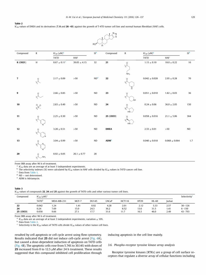
Table 2IC50 values of DHEA and its derivatives (7, 8 and 26e43) against the growth of T-47D tumor cell line and normal human fibroblast (HAF) cells.
RO
SNN
H
Compound R IC50 (mM)a SIb Compound R IC50 (mM)a SIb
T47D HAF T47D HAF
8 (CH21) H 0.67 ± 0.11c 39.05 ± 4.15 32 21 O
NH2
1.15 ± 0.10 18.0 ± 0.22 16
7 O 2.17 ± 0.09 >50 NDd 22 O
NH
0.042 ± 0.028 2.95 ± 0.28 70
9 O 2.66 ± 0.85 >50 ND 23 O
NH
0.051 ± 0.010 1.82 ± 0.03 36
10S
O O 2.83 ± 0.49 >50 ND 24 O
HN
0.24 ± 0.06 36.0 ± 2.05 150
11 O
N
2.25 ± 0.30 >50 ND 25 (CH33) O
HN
0.058 ± 0.016 21.1 ± 5.06 364
12 O
N
3.28 ± 0.51 >50 ND DHEA 2.55 ± 0.01 >50 ND
13 O
N
N3.04 ± 0.99 >50 ND ADMe 0.040 ± 0.018 0.068 ± 0.064 1.7
20 ONH
0.92 ± 0.05 26.1 ± 0.77 28
From SRB assay after 96 h of treatment.a IC50 data are an average of at least 3 independent experiments.b The selectivity indexes (SI) were calculated by IC50 values in HAF cells divided by IC50 values in T47D cancer cell line.c Data from Table 1.d ND ¼ not determined.e ADM is Adriamycin.
Table 3IC50 values of compounds 22, 24 and 25 against the growth of T47D cells and other various tumor cell lines.
Compound IC50 (mM)a Selectivityc
T47Db MDA-MB-231 MCF-7 DU145 LNCaP HCT116 HT29 HL-60 Jurkat
22 0.042 1.24 1.41 0.92 4.28 2.01 2.12 5.33 2.57 30e12624 0.24 7.69 15.0 24.3 36.2 8.52 13.6 31.5 1.41 6e15025 (CH33) 0.058 9.66 27.1 17.7 31.6 11.7 16.5 46.0 2.49 43e793
From SRB assay after 96 h of treatment.a IC50 data are an average of at least 3 independent experiments, variation ± 10%.b Data from Table 2.c Selectivity is the IC50 values of T47D cells divide IC50 values of other tumor cell lines.
H.-W. Cui et al. / European Journal of Medicinal Chemistry 111 (2016) 126e137 129
resulted by cell apoptosis or cell cycle arrest using flow cytometry.Results indicated that 25 did not induce cell-cycle arrest (Fig. 4A),but caused a dose-dependent induction of apoptosis on T47D cells(Fig. 4B). The apoptotic cells rose from 5.74% to 30.14% with doses of25 increased from 0 to 12.5 mM after 24 h treatment. These resultssuggested that this compound inhibited cell proliferation through
inducing apoptosis in the cell line mainly.
3.6. Phospho-receptor tyrosine kinase array analysis
Receptor tyrosine kinases (RTKs) are a group of cell surface re-ceptors that regulate a diverse array of cellular functions including
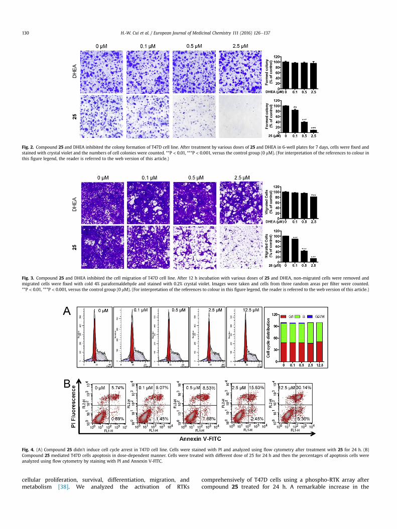
Fig. 2. Compound 25 and DHEA inhibited the colony formation of T47D cell line. After treatment by various doses of 25 and DHEA in 6-well plates for 7 days, cells were fixed andstained with crystal violet and the numbers of cell colonies were counted. **P < 0.01, ***P < 0.001, versus the control group (0 mM). (For interpretation of the references to colour inthis figure legend, the reader is referred to the web version of this article.)
Fig. 3. Compound 25 and DHEA inhibited the cell migration of T47D cell line. After 12 h incubation with various doses of 25 and DHEA, non-migrated cells were removed andmigrated cells were fixed with cold 4% paraformaldehyde and stained with 0.2% crystal violet. Images were taken and cells from three random areas per filter were counted.**P < 0.01, ***P < 0.001, versus the control group (0 mM). (For interpretation of the references to colour in this figure legend, the reader is referred to the web version of this article.)
Fig. 4. (A) Compound 25 didn't induce cell cycle arrest in T47D cell line. Cells were stained with PI and analyzed using flow cytometry after treatment with 25 for 24 h. (B)Compound 25 mediated T47D cells apoptosis in dose-dependent manner. Cells were treated with different dose of 25 for 24 h and then the percentages of apoptosis cells wereanalyzed using flow cytometry by staining with PI and Annexin V-FITC.
H.-W. Cui et al. / European Journal of Medicinal Chemistry 111 (2016) 126e137130
cellular proliferation, survival, differentiation, migration, andmetabolism [38]. We analyzed the activation of RTKs
comprehensively of T47D cells using a phospho-RTK array aftercompound 25 treated for 24 h. A remarkable increase in the

H.-W. Cui et al. / European Journal of Medicinal Chemistry 111 (2016) 126e137 131
phosphorylation of EphA2 and EphB3 was detected (Fig. 5). Ac-cording to previous studies, there is accumulating evidence tosuggest that EPH expression and function in the absence of kinaseactivity is tumor promoting, whereas EPH forward signaling(phosphorylated Eph) is tumor suppressive [39e42]. So, thephosphorylation of EphA2 and EphB3 may play pivotal role in theproliferation suppression, migration inhibition and apoptosis-inducing of 25 in T47D cells and further in-depth mechanismresearch is also required.
3.7. In vivo antitumor activity
In order to determine the efficacy of compound 25 in vivo, T47Dsubcutaneous xenograft growth model and orthotopic xenograftgrowth and metastasis model were evaluated. Results of subcu-taneous xenograft growth model suggested that the tumor growthof T47D was significantly inhibited by 25 (20 mg/kg/day) comparedwith the control group (Fig. 6A, left), and there was almost noobvious effect on body weight in the compound treated mice(Fig. 6A, right). The tumor volume and body weight of 25 treatedmice in orthotopic xenograft model displayed the same results withsubcutaneous xenograft model (Fig. 6B), moreover, the compounddisplayed remarkable antimetastatic ability, the number ofmetastasized T47D nodules in the lungs of the compound (20 mg/kg/day) treated mice reduced to about 40% compared with thecontrol group (Fig. 6C). HE staining results of liver and kidneyshowed that 25 presented no obvious influence to the anatomicalmorphologies of mice (Fig. 6D).
4. Conclusion
In this study we synthesized a novel class of D-ring fused 1,2,3-thiadiazole DHEA derivatives and screened them for anti-proliferative activity using the SRB assay on various cancer celllines. It is amazing that for these compounds, T47D cell line wasmuchmore sensitive than other tumor cell lines. Compound 25wasan unnatural D-proline modified derivative, which possessed themost potent antiproliferative activity on T47D cells and the bestselectivity between the tumor cell line and HAF cells. The colonyformation and metastatic ability of T47D cells was significantlyinhibited by 25 in a dose-dependent manner, which was muchmore effective than lead compound DHEA. The primarymechanismstudy showed that 25 did not induce cell-cycle arrest, but caused adose-dependent induction of apoptosis, and induced phosphory-lation of EphA2 and EphB3 in T47D cells. In vivo antitumor activityevaluation displayed that the tumor volume was significantlyinhibited and there was no obvious change in body weight of 25treated mice in subcutaneous xenograft model and orthotopic
Fig. 5. Compound 25 increased the phosphorylation of EphA2 and EphB3 in T47D cells.After treated with 2.5 mM 25 for 24 h, T47D cells were lysed and analyzed for theactivation of RTKs comprehensively using a phospho-RTK array.
xenograft model. Moreover, the compound also displayedremarkable antimetastatic ability, the number of metastasizedT47D nodules in the lungs reduced to about 40% compared with thecontrol group, and the HE staining results showed that the com-pound presented no obvious influence to the anatomical mor-phologies of mice.
In conclusion, we report D-ring fused 1,2,3-thiadiazole DHEAderivatives as a series of new chemical entities for the first time.Especially 25, which exhibited potent antitumor and antimetastaticactivity in vitro and in vivo, and could be used as a promising leadfor the development of a new class of antitumor and antimetastaticagents.
5. Experimental section
5.1. General
All reagents and chemicals were purchased from commercialsuppliers and used without further purification unless otherwisestated. When needed, the reactions were carried out in oven-driedglassware under a positive pressure of dry N2. Column chroma-tography was performed on silica gel (QinDao, 200e300 mesh)using the indicated eluents. Thin-layer chromatography was car-ried out on silica gel plates (QinDao) with a layer thickness of0.25 mm. Melting points were determined using the MEL-TEMP 3.0apparatus and uncorrected. 1H (400 MHz) and 13C (100 MHz) NMRspectra were recorded on Bruker AM-400 spectrometer with CDCl3or DMSO-d6 as solvent and tetramethylsilane (TMS) as the internalstandard. All chemical shift values were reported in units ofd (ppm). The following abbreviations were used to indicate thepeak multiplicity: s ¼ singlet; d ¼ doublet; t ¼ triplet;m ¼ multiplet; br ¼ broad. High-resolution mass data were ob-tained on a BrukermicroOTOF-Q II spectrometer.
5.2. 3b-Acetoxy-5-androstene-17-one (1)
To a solution of DHEA (5 g, 17.2 mmol) in dry CH2Cl2 (40 mL),Ac2O (15.6 g, 103.3 mmol) and DMAP (315 mg, 2.58 mmol) wereadded at room temperature. The reaction mixture was stirred for5 h under nitrogen atmosphere at room temperature and concen-trated. The residuewas poured into H2O (30mL) and extractedwithAcOEt (20 mL � 3). The organic layer was washed with brine, driedwith anhydrous Na2SO4 and concentrated to give compound 1(5.61 g, 98%) as a white solid without purified. 1H NMR (400 MHz,CDCl3) d 5.40 (d, J ¼ 4.7 Hz, 1H), 4.63e4.56 (m, 1H), 2.45 (dd,J ¼ 19.2, 8.9 Hz, 1H), 2.33 (dd, J ¼ 13.1, 2.7 Hz, 2H), 2.15e2.05 (m,2H), 2.03 (s, 3H), 1.96 (dd, J ¼ 12.1, 5.8 Hz, 1H), 1.86 (s, 3H), 1.65 (d,J ¼ 8.3 Hz, 3H), 1.33e1.24 (m, 2H), 1.18e1.12 (m, 1H), 1.05 (s, 3H),0.88 (s, 3H). 13C NMR (100 MHz, CDCl3) d 220.88, 170.41, 139.87,121.84, 73.65, 51.64, 50.10, 47.46, 38.04, 36.90, 36.68, 35.79, 31.342,37.37, 30.73, 27.66, 21.85, 21.38, 20.28, 19.31, 13.51.
5.3. 3b-Acetoxy-17-(2-carbamoylhydrazono)-5-androstene (2)
To a solution of compound 1 (400 mg, 1.2 mmol) in absoluteEtOH (20 mL), aminourea hydrochloride (675 mg, 6 mmol) andanhydrous CH3COONa (492 mg, 6 mmol) were added at roomtemperature. The reaction mixture was stirred for 6 h at roomtemperature and concentrated. The residue was poured into H2O(30 mL) and extracted with AcOEt (20 mL � 3). The organic layerwas washed with brine, dried with anhydrous Na2SO4 andconcentrated. The residue was purified by silica gel column chro-matography (CH2Cl2/MeOH, 20/1, v/v) to give compound 2 (431mg,92%) as a white solid. 1H NMR (400 MHz, CDCl3) d 7.82 (s, 1H), 5.43(d, J ¼ 5.1 Hz, 1H),4.73e4.63 (m, 1H), 2.38 (dd, J ¼ 13.1, 5.2 Hz, 2H),

Fig. 6. (A) T47D cells subcutaneous xenograft mice were intraperitoneally treated with compound 25 (20 mg/kg/day) or DMSO (served as control) for 19 days. Tumor volume andmice body weight were measured every other day. N ¼ 8; ns, no significant; ***P < 0.001. (B) T47D cells orthotopic xenograft mice intraperitoneally treated with 25 (20 mg/kg/day)or DMSO (served as control) for 16 days. Tumor volume and mice body weight were measured every three days. N ¼ 5; ns, no significant; *P < 0.05. (C) Lungs were fixed,photographed, and amount of metastasis nodules were manually counted using a dissecting microscope. *P < 0.05. (D) Liver and kidney hematoxylin and eosin (HE) staining ofcontrol and 25 treatment groups.
H.-W. Cui et al. / European Journal of Medicinal Chemistry 111 (2016) 126e137132
2.27e2.14 (m, 1H), 2.02 (s, 3H), 0.86 (s, 3H), 0.84 (s, 3H).
5.4. 3b-Acetoxy-androsta-5,16-dieno[17,16-d][1,2,3]thiadiazole (3)
To a solution of compound 2 (435 mg, 1.12 mmol) in dry CH2Cl2(10 mL), thionyl chloride (2 mL) was added at 0 �C. The reactionmixture was stirred for 5 h at room temperature and then pouredinto H2O (30 mL) and extracted with CH2Cl2 (10 mL � 3). Theorganic layer was washed with brine, dried with anhydrous Na2SO4and concentrated. The residue was purified by silica gel columnchromatography (CH2Cl2/MeOH, 40/1, v/v) to give compound 3
(335 mg, 82%) as a white solid. IR (ATR) cm�1: 2930, 2840, 1719,728. 1H NMR (400 MHz, CDCl3) d 5.42 (d, J ¼ 5.1 Hz, 1H), 4.68e4.56(m,1H), 3.05 (dd, J¼ 15.5, 6.3 Hz,1H), 2.63 (dd, J¼ 15.5,11.9 Hz,1H),2.48 (dd, J¼ 10.2, 2.4 Hz,1H), 2.42e2.29 (m, 2H), 2.28e2.19 (m,1H),2.15e2.06 (m, 1H), 2.04 (s, 3H), 1.13 (s, 3H), 1.12 (s, 3H). 13C NMR(100 MHz, CDCl3) d 179.86, 170.48, 153.88, 140.30, 121.49, 73.65,62.91, 50.41, 41.54, 38.09, 36.89, 36.82, 34.24, 30.96, 30.62, 27.67,27.45, 21.41, 20.34, 19.29, 17.71.

H.-W. Cui et al. / European Journal of Medicinal Chemistry 111 (2016) 126e137 133
5.5. 3b-Hydroxy-androsta-5,16-dieno[17,16-d][1,2,3]thiadiazole (4)
To a solution of 3 (230 mg, 0.62 mmol) in MeOH (10 mL), K2CO3(428 mg, 3.1 mmol) was added at room temperature. The reactionmixture was stirred for 4 h at room temperature and concentrated.The residue was poured into H2O (30 mL) and acidified to pH 5e6with hydrochloric acid (1 M), then extracted with AcOEt(20 mL � 3). The organic layer was washed with brine, dried withanhydrous Na2SO4 and concentrated. The residue was purified bysilica gel column chromatography (CH2Cl2/MeOH, 30/1, v/v) to givecompound 4 (190 mg, 93%) as a white solid; mp: 198e200 �C. IR(ATR) cm�1: 3420, 2920, 2853, 720. 1H NMR (400 MHz, CDCl3)d 5.42e5.35 (m, 1H), 3.61e3.49 (m, 1H), 3.07e3.01 (m, 1H),2.67e2.59 (m,1H), 2.50e2.47 (m,1H), 2.36e2.19 (m, 3H), 2.12e2.06(m,1H), 1.13 (s, 3H), 1.10 (s, 3H). 13C NMR (100MHz, CDCl3) d 180.06,154.06, 141.57, 120.61, 77.48, 77.16, 76.84, 71.66, 63.15, 50.63, 42.31,41.68, 37.20, 36.93, 34.39, 31.66, 31.10, 30.80, 27.57, 20.51, 19.48,17.82. HRMS (ESI): calcd for C19H27N2OS [M þ H]þ; 331.1839; found331.1830.
5.6. 3b-Acetoxy-5a-androstan-17-one (5)
To a solution of 1 (200 mg, 0.6 mmol) in MeOH (15 mL) in anautoclave, Pd/C (100 mg, 5%) and the H2 were added and main-tained at 4.0 Mp. The reaction mixture was stirred at 40 �C for 16 h,then fitered with diatomite, and the filter cake was washed withCH2Cl2 (15 mL � 3). The filtrate was concentrated and the residuewas purified by silica gel column chromatography (petroleumether/AcOEt, 5/1, v/v) to give compound 5 (172 mg, 87%) as a whitesolid. 1H NMR (400 MHz, CDCl3) d 4.68 (s, 1H), 2.42 (dd, J ¼ 19.2,8.9 Hz, 1H), 2.11e2.03 (m, 1H), 2.01 (d, J ¼ 2.3 Hz, 3H), 0.84 (d,J ¼ 2.7 Hz, 6H). 13C NMR (100 MHz, CDCl3) d 221.14, 170.60, 73.45,54.27, 51.31, 47.73, 44.61, 36.67, 35.81, 35.61, 34.99, 33.91, 31.49,30.77, 28.24, 27.37, 21.74, 21.42, 20.43, 13.79, 12.18.
5.7. 3b-Acetoxy-17-(2-carbamoylhydrazono)-5a-androstan (6)
By a similar procedure described for compound 2, compound 6was obtained as awhite solid; yield: 95%. 1H NMR (400MHz, CDCl3)d 7.82 (s, 1H), 4.73e4.63 (m, 1H), 2.38 (dd, J ¼ 13.1, 5.2 Hz, 2H),2.27e2.14 (m, 1H), 2.02 (s, 3H), 0.86 (s, 3H), 0.84 (s, 3H).
5.8. 3b-Acetoxy-5a-androst-16-eno[17,16-d][1,2,3]thiadiazole (7)
By a similar procedure described for compound 3, compound 7was obtained as awhite solid; yield: 78%; mp: 184e186 �C. IR (ATR)cm�1: 2917, 2853, 1716, 1245, 728. 1H NMR (400 MHz, CDCl3)d 4.75e4.65 (m,1H), 3.02 (dd, J¼ 15.5, 6.3 Hz,1H), 2.59 (dd, J¼ 15.5,11.9 Hz, 1H), 2.43 (dd, J ¼ 12.0, 3.1 Hz, 1H), 2.25e2.18 (m, 1H), 2.02(s, 3H), 1.09 (s, 3H), 0.91 (s, 3H). 13C NMR (100MHz, CDCl3) d 180.09,170.76, 153.99, 73.54, 62.96, 54.83, 44.86, 41.91, 36.60, 35.92, 34.41,34.33, 34.04, 31.32, 28.31, 27.50, 27.50, 21.54, 20.81, 18.06, 12.33.HRMS (ESI): calcd for C21H30N2NaO2S [M þ Na]þ; 397.1920; found397.1922.
5.9. 3b-Hydroxy-5a-androst-16-eno[17,16-d][1,2,3]thiadiazole (8)
By a similar procedure described for compound 4, compound 8was obtained as awhite solid; yield: 95%; mp: 208e210 �C. IR (ATR)cm�1: 3410, 2923, 2847, 1720, 735. 1H NMR (400 MHz, CDCl3)d 3.66e3.56 (m,1H), 3.02 (dd, J¼ 15.5, 6.3 Hz,1H), 2.59 (dd, J¼ 15.5,11.9 Hz, 1H), 2.48e2.38 (m, 1H), 2.25e2.17 (m, 1H), 1.10 (s, 3H), 0.90(s, 3H)$13C NMR (100 MHz, CDCl3) d 180.15, 154.06, 71.17, 63.06,54.96, 45.06, 41.92, 38.15, 36.84, 35.93, 34.44, 34.36, 31.53, 31.41,28.44, 27.51, 20.86, 18.07, 12.43. HRMS (ESI): calcd for
C19H28N2NaOS [M þ Na]þ; 355.1815; found 355.1818.
5.10. General procedure for the preparation of compounds 9e13
To a solution of compound 8 (150 mg, 0.45 mmol) in dry CH2Cl2(20 mL), acyl chloride (9.0 mmol) and DMAP (55 mg, 0.45 mmol)were added at room temperature. The reaction mixture was stirredfor 4 h at room temperature under nitrogen atmosphere, thenpoured into H2O (30 mL) and extracted with CH2Cl2 (20 mL � 3).The organic layer was washed with brine, dried with anhydrousNa2SO4 and concentrated. The residue was purified by silica gelcolumn chromatography (petroleum ether/AcOEt, 3/1, v/v) to givethe desired product.
5.10.1. 3b-Propionyloxy-5a-androst-16-eno[17,16-d][1,2,3]thiadiazole (9)
White solid; yield: 88%; mp: 178e180 �C. IR (ATR) cm�1: 2920,2847, 1727, 1196. 1H NMR (400 MHz, CDCl3) d 4.75e4.67 (m, 1H),3.02 (dd, J ¼ 15.5, 6.3 Hz, 1H), 2.59 (dd, J ¼ 15.5, 12.0 Hz, 1H), 2.43(dd, J ¼ 11.9, 3.2 Hz, 1H), 2.29 (d, J ¼ 7.6 Hz, 2H), 2.24e2.17 (m, 1H),1.13 (t, J ¼ 7.6 Hz, 3H), 1.09 (s, 3H), 0.92 (s, 3H)$13C NMR (100 MHz,CDCl3) d 180.11, 174.18, 154.01, 73.31, 62.97, 54.84, 44.87, 41.91,36.62, 35.94, 34.42, 34.34, 34.08, 31.33, 28.33, 28.06, 27.53, 27.50,20.82, 18.07, 12.35, 9.31. HRMS (ESI): calcd for C22H32N2NaO2S [M þNa]þ; 411.2077; found 411.2109.
5.10.2. 3b-Methylsulfonyloxy-5a-androst-16-eno[17,16-d][1,2,3]thiadiazole (10)
White solid; yield: 90%; mp: 166e167 �C. IR (ATR) cm�1: 2919,2855, 1728, 728. 1H NMR (400 MHz, CDCl3) d 4.69e4.58 (m, 1H),3.04 (d, J ¼ 6.3 Hz, 1H), 3.00 (s, 3H), 2.60 (dd, J ¼ 15.5, 11.9 Hz, 1H),2.44 (dd, J ¼ 11.4, 3.2 Hz, 1H), 2.21 (d, J ¼ 29.5 Hz, 1H), 2.02 (d,J¼ 14.0 Hz, 1H), 1.09 (s, 3H), 0.92 (s, 3H)$13C NMR (100MHz, CDCl3)d 179.98, 153.95, 81.78, 62.85, 54.65, 44.98, 41.87, 38.94, 36.62,35.68, 35.19, 34.34, 34.24, 31.21, 28.69, 28.18, 27.47, 20.81, 18.05,12.28. HRMS (ESI): calcd for C20H30N2NaO3S2 [M þ Na]þ; 433.1590;found 433.1612.
5.10.3. 3b-Nicotinoyloxy-5a-androst-16-eno[17,16-d][1,2,3]thiadiazole (11)
White solid; yield: 83%; mp: 223e225 �C. IR (ATR) cm�1: 2923,2850, 1724, 1280, 730. 1H NMR (400 MHz, CDCl3) d 9.24 (s, 1H), 8.79(d, J ¼ 4.3 Hz, 1H), 8.41 (d, J ¼ 7.7 Hz, 1H), 7.54e7.45 (m, 1H),5.06e4.95 (m, 1H), 3.03 (dd, J ¼ 15.5, 6.4 Hz, 1H), 2.61 (dd, J ¼ 15.5,11.9 Hz, 1H), 2.46 (dd, J ¼ 11.7, 3.1 Hz, 1H), 2.28e2.20 (m, 1H), 2.00(d, J ¼ 12.5 Hz, 1H), 1.11 (s, 3H), 0.98 (s, 3H). 13C NMR (100 MHz,CDCl3) d 180.07, 164.88, 153.98, 153.35, 150.99, 137.11, 126.73,123.32, 74.81, 62.94, 54.82, 44.90, 41.91, 36.60, 35.97, 34.41, 34.33,34.07, 31.32, 28.32, 27.56, 27.50, 20.84, 18.07, 12.41. HRMS (ESI):calcd for C25H32N3O2S [M þ H]þ; 438.2210; found 438.2232.
5.10.4. 3b-Isonicotinoyloxy-5a-androst-16-eno[17,16-d][1,2,3]thiadiazole (12)
White solid; yield: 89%; mp: 235e237 �C. IR (ATR) cm�1: 2914,2851,1721,1280, 727. 1H NMR (400MHz, CDCl3) d 8.78 (d, J¼ 5.2 Hz,2H), 7.90 (d, J ¼ 5.5 Hz, 2H), 5.04e4.94 (m, 1H), 3.03 (dd, J ¼ 15.5,6.3 Hz, 1H), 2.61 (dd, J ¼ 15.5, 11.9 Hz, 1H), 2.45 (dd, J ¼ 11.7, 3.2 Hz,1H), 2.27e2.20 (m, 1H), 2.04e1.98 (m, 1H), 1.11 (s, 3H), 0.97 (s,3H)$13C NMR (100 MHz, CDCl3) d 180.07, 164.71, 153.98, 150.62,150.62,138.13, 122.97, 122.97, 75.25, 62.95, 54.82, 44.90, 41.92,36.59, 35.98, 34.41, 34.35, 34.00, 31.32, 28.33, 27.51, 27.50, 20.86,18.08, 12.41. HRMS (ESI): calcd for C25H32N3O2S [M þ H]þ;438.2210; found 438.2250.

H.-W. Cui et al. / European Journal of Medicinal Chemistry 111 (2016) 126e137134
5.10.5. 3b-[(Pyrazine-2-carbonyl)oxy]-5a-androst-16-eno [17,16-d][1,2,3]thiadiazole (13)
White solid; yield: 89%; mp: 222e224 �C. IR (ATR) cm�1: 2916,2853, 1712, 733. 1H NMR (400 MHz, CDCl3) d 9.30 (s, 1H), 8.74 (d,J ¼ 5.6 Hz, 2H), 5.15e5.04 (m, 1H), 3.03 (dd, J ¼ 15.5, 6.3 Hz, 1H),2.60 (dd, J ¼ 15.5, 12.0 Hz, 1H), 2.45 (dd, J ¼ 11.8, 3.2 Hz, 1H),2.28e2.20 (m, 1H), 2.06e2.01 (m, 1H), 1.10 (s, 3H), 0.97 (s, 3H)$13CNMR (100 MHz, CDCl3) d 180.04, 163.57, 153.95, 147.58, 146.38,144.50, 143.97, 75.83, 62.92, 54.78, 44.94, 41.89, 36.61, 35.97, 34.39,34.31, 33.93, 31.29, 28.28, 27.49, 27.43, 20.82, 18.05,12.40. HRMS(ESI): calcd for C24H30N4NaO2S [M þ Na]þ; 461.1982; found461.1986.
5.11. General procedure for the preparation of compounds 14e19
To a solution of compound 8 (250 mg, 0.75 mmol) in dry CH2Cl2(20 mL), N-BOC-amino-carboxylic acid (3.0 mmol), HOBT (405 mg,3.0 mmol), EDCI (575mg, 3.0 mmol) and DMAP (366mg, 3.0 mmol)were added at room temperature. The reaction mixture was stirredfor 8 h at room temperature under nitrogen atmosphere, thenpoured into water (30 mL) and extracted with CH2Cl2 (20 mL � 3).The organic layer was washed with brine, dried with anhydrousNa2SO4 and concentrated. The residue was purified by silica gelcolumn chromatography (petroleum ether/AcOEt, 3/1, v/v) to givethe desired product.
5.11.1. 3b-{2-[(tert-butoxycarbonyl)amino]acetoxy}-5a-androst-16-eno[17,16-d][1,2,3] thiadiazole (14)
White solid; yield: 93%. 1H NMR (400 MHz, CDCl3) d 5.01 (s, 1H),4.83e4.72 (m,1H), 3.89 (d, J¼ 4.7 Hz, 2H), 3.04 (dd, J¼ 15.5, 6.3 Hz,1H), 2.62 (dd, J ¼ 15.4, 12.0 Hz, 1H), 2.46 (dd, J ¼ 11.6, 3.4 Hz, 1H),2.27e2.16 (m, 1H), 1.48 (s, 9H), 1.12 (s, 3H), 0.94 (s, 3H). 13C NMR(100 MHz, CDCl3) d 179.97, 169.87, 155.71, 153.91, 79.91, 74.64,62.82, 54.66, 44.70, 42.67, 41.79, 36.41, 35.78, 34.27, 34.19, 33.82,31.18, 28.33, 28.16, 27.40, 27.31, 20.70, 17.97, 12.22.
5.11.2. 3b-{[4-((tert-butoxycarbonyl)amino)butanoyl]oxy}-5a-androst-16-eno[17,16-d][1,2,3] thiadiazole (15)
White solid; yield: 96%. 1H NMR (400 MHz, CDCl3) d 4.76e4.67(m, 1H), 4.65 (d, J ¼ 11.9 Hz, 1H), 3.15 (d, J ¼ 6.3 Hz, 2H), 3.01 (dd,J¼ 15.5, 6.3 Hz,1H), 2.59 (dd, J¼ 15.5,12.0 Hz,1H), 2.43 (dd, J¼ 11.8,3.3 Hz, 1H), 2.31 (t, J ¼ 7.3 Hz, 2H), 2.26e2.16 (m, 1H), 1.43 (s, 9H),1.09 (s, 3H), 0.91 (s, 3H). 13C NMR (100MHz, CDCl3) d 179.98,172.82,155.94, 153.92, 79.17, 73.51, 62.83, 54.67, 44.73, 41.78, 39.96, 36.46,35.80, 34.27, 34.19, 33.92, 32.00, 31.20, 28.41, 28.19, 27.39, 25.33,20.69, 17.97, 14.14, 12.23.
5.11.3. 3b-{[1-(tert-butoxycarbonyl)piperidine-3-carbonyl]oxy}-5a-androst-16-eno [17,16-d][1,2,3]thiadiazole (16)
White solid; yield: 98%. 1H NMR (400 MHz, CDCl3) d 4.78e4.65(m, 1H), 3.99 (d, J ¼ 13.4 Hz, 2H), 3.01 (dd, J ¼ 15.5, 6.3 Hz, 1H),2.92e2.77 (m, 2H), 2.59 (dd, J ¼ 15.4, 12.0 Hz, 1H), 2.49e2.35 (m,2H), 2.26e2.16 (m, 1H), 1.45 (s, 9H), 1.09 (s, 3H), 0.91 (s, 3H). 13CNMR (100MHz, CDCl3) d 180.00, 173.01, 154.71, 153.91, 79.66, 73.46,62.84, 54.67, 44.71, 41.79, 41.59,41.57, 36.44, 35.81, 34.28, 34.20,33.90, 33.87, 31.21, 28.43, 28.19, 27.37, 27.35, 27.33, 24.33, 20.70,17.97, 12.24.
5.11.4. 3b-{[1-(tert-butoxycarbonyl)piperidine-4-carbonyl]oxy}-5a-androst-16-eno [17,16-d][1,2,3]thiadiazole (17)
White solid; yield: 96%. 1H NMR (400 MHz, CDCl3) d 4.78e4.64(m,1H), 4.12 (d, J¼ 7.1 Hz,1H), 3.94e3.83 (m,1H), 3.01 (dd, J¼ 15.5,6.3 Hz, 1H), 2.86e2.76 (m, 1H), 2.59 (dd, J ¼ 15.4, 12.0 Hz, 1H),2.46e2.34 (m, 2H), 2.27e2.15 (m, 1H), 2.02 (d, J ¼ 15.8 Hz, 1H), 1.46(s, 9H), 1.09 (s, 3H), 0.91 (s, 3H). 13C NMR (100MHz, CDCl3) d 179.98,
174.15, 154.72, 153.91, 79.55, 73.42, 62.83, 54.67, 44.71, 41.79, 41.29,36.45, 35.81, 34.27, 34.20, 33.91, 31.20, 28.44, 28.19, 28.01, 27.98,27.40, 27.37, 20.71, 17.97, 12.25.
5.11.5. 3b-[N-tert-butoxycarbonyl-(L-prolyl)oxy]-5a-androst-16-eno[17,16-d][1,2,3]thiadiazole (18)
White solid; yield: 98%. 1H NMR (400 MHz, CDCl3) d 4.80e4.68(m, 1H), 4.31e4.16 (m, 1H), 3.59e3.34 (m, 2H), 3.02 (dd, J ¼ 15.5,6.3 Hz, 1H), 2.59 (dd, J ¼ 15.1, 12.2 Hz, 1H), 2.44 (d, J ¼ 9.1 Hz, 1H),2.28e2.11 (m, 2H), 1.46 (s, 3H), 1.42 (s, 6H), 1.09 (s, 3H), 0.91 (s, 3H).13C NMR (125MHz, DMSO-d6) d 179.81,172.60,155.17,153.34, 79.13,73.75, 62.50, 59.11, 54.25, 46.61, 44.44, 41.79, 36.25, 35.80, 34.49,34.07, 31.07, 30.82, 29.88, 28.52, 28.38, 27.66, 24.28, 23.50, 20.77,18.32, 12.30.
5.11.6. 3b-[N-tert-butoxycarbonyl-(D-prolyl)oxy]-5a-androst-16-eno[17,16-d][1,2,3]thiadiazole (19)
White solid; yield: 86%. 1H NMR (400 MHz, CDCl3) d 4.80e4.67(m, 1H), 4.33e4.14 (m, 1H), 3.61e3.35 (m, 2H), 3.02 (dd, J ¼ 15.5,6.3 Hz, 1H), 2.65e2.53 (m, 1H), 2.43 (d, J ¼ 11.4 Hz, 1H), 2.30e2.11(m, 2H), 1.46 (s, 3H), 1.42 (s, 6H), 1.09 (s, 3H), 0.91 (s, 3H). 13C NMR(125 MHz, CDCl3) d 179.96, 172.71, 154.36, 153.88, 79.77, 73.85,62.82, 59.30, 54.68, 46.33, 44.72, 41.79, 36.43, 35.81, 34.28, 34.21,34.01, 31.19, 31.00, 28.45, 28.39, 28.21, 27.39, 23.53, 20.71, 17.96,12.25.
5.12. General procedure for the preparation of compounds 20e25
One of compounds 14e19 (0.4 mmol) was dissolved in anhy-drous diethyl ether (20 mL) and boron trifluoride etherate (5 mL)was added. The reaction mixture was stirred for 2 h at room tem-perature and then quenched with saturated aqueous NaHCO3(20 mL). The mixture was extracted with AcOEt (20 mL � 3). Thecombined organic extract was washed with brine, dried overanhydrous Na2SO4 and concentrated. The residue was purified bysilica gel chromatography (CH2Cl2/MeOH, 10/1, v/v) to give thedesired product.
5.12.1. 3b-(2-Aminoacetoxy)-5a-androst-16-eno[17,16-d][1,2,3]thiadiazole (20)
White solid; yield: 77%; mp: 163e165 �C. IR (ATR) cm�1: 3378,2918, 2853, 1718, 1280, 728. 1H NMR (400 MHz, CDCl3) d 4.82e4.71(m,1H), 3.45 (s, 2H), 3.01 (dd, J¼ 15.5, 6.3 Hz,1H), 2.59 (dd, J¼ 15.5,11.9 Hz, 1H), 2.43 (dd, J ¼ 11.7, 3.2 Hz, 1H), 1.09 (s, 3H), 0.91 (s, 3H).13C NMR (100 MHz, CDCl3) d 180.04, 173.69, 153.97, 74.19, 62.92,54.79, 44.82, 44.19, 41.87, 36.54, 35.88, 34.37, 34.29, 34.01, 31.28,28.27, 27.47, 27.47, 20.79, 18.04, 12.30. HRMS (ESI): calcd forC21H32N3O2S [M þ H]þ; 390.2210; found 390.2211.
5.12.2. 3b-[(4-Aminobutanoyl)oxy]-5a-androst-16-eno[17,16-d][1,2,3]thiadiazole (21)
White solid; yield: 72%; mp: 170e172 �C. IR (ATR) cm�1: 3342,2918, 2855, 1719, 1685, 728. 1H NMR (400 MHz, CDCl3) d 4.77e4.65(m, 1H), 3.02 (dd, J ¼ 15.5, 6.3 Hz, 1H), 2.73 (t, J ¼ 7.0 Hz, 1H), 2.59(dd, J ¼ 15.5, 11.9 Hz, 1H), 2.43 (dd, J ¼ 12.0, 3.0 Hz, 1H), 2.39e2.26(m, 2H), 1.09 (s, 3H), 0.91 (s, 3H). 13C NMR (100 MHz, CDCl3)d 180.06, 173.01, 154.01, 73.60, 71.07, 62.90, 54.75, 44.81, 41.86,41.10, 38.13, 36.54, 35.89, 34.35, 34.26, 34.01, 32.09, 31.28, 28.27,27.48, 20.78, 18.06, 12.32. HRMS (ESI): calcd for C23H36N3O2S [M þH]þ; 418.2523; found 418.2528.
5.12.3. 3b-[(Piperidine-3-carbonyl)oxy]-5a-androst-16-eno[17,16-d][1,2,3]thiadiazole (22)
White solid; yield: 75%; mp: 179e181 �C. IR (ATR) cm�1: 3527,2923, 2853, 1722, 728. 1H NMR (400 MHz, CDCl3) d 7.24 (s, 1H), 4.75

H.-W. Cui et al. / European Journal of Medicinal Chemistry 111 (2016) 126e137 135
(s, 1H), 3.63e3.10 (m, 4H), 3.01 (dd, J¼ 15.5, 6.3 Hz,1H), 2.90 (s, 1H),2.59 (dd, J ¼ 15.4, 12.0 Hz, 1H), 2.43 (dd, J ¼ 11.9, 2.9 Hz, 1H), 1.09 (s,3H), 0.92 (s, 3H). 13C NMR (100MHz, CDCl3) d 180.02,171.99,154.06,74.95, 62.96, 54.81, 45.42, 44.99, 44.82, 41.90, 38.08, 36.50, 35.90,34.41, 34.29, 33.78, 31.31, 28.27, 27.51, 27.30, 25.30, 20.95, 20.84,18.07, 12.34. HRMS (ESI): calcd for C25H38N3O2S [M þ H]þ;444.2679; found 444.2679.
5.12.4. 3b-[(Piperidine-4-carbonyl)oxy]-5a-androst-16-eno[17,16-d][1,2,3]thiadiazole (23)
White solid; yield: 82%; mp: 175e177 �C. IR (ATR) cm�1: 3340,2917, 2852, 1717, 728. 1H NMR (400 MHz, CDCl3) d 4.79e4.61 (m,1H), 3.14e3.05 (m, 2H), 3.02 (dd, J¼ 15.5, 6.4 Hz,1H), 2.68e2.60 (m,2H), 2.48e2.33 (m, 2H), 2.25e2.18 (m, 1H), 1.09 (s, 3H), 0.91 (s, 3H).13C NMR (100 MHz, CDCl3) d 180.07, 174.73, 153.98, 73.26, 62.93,54.79, 45.87, 44.83, 41.88, 41.81, 36.56, 35.91, 34.38, 34.30, 34.02,31.30, 29.32, 29.29, 28.29, 27.47, 20.79, 18.04, 12.33. HRMS (ESI):calcd for C25H38N3O2S [M þ H]þ; 444.2679; found 444.2686.
5.12.5. 3b-[(L-prolyl)oxy]-5a-androst-16-eno[17,16-d][1,2,3]thiadiazole (24)
White solid; yield: 74%; mp: 82e84 �C. IR (ATR) cm�1: 3391,2917, 2853, 1719, 728. 1H NMR (400 MHz, CDCl3) d 4.83e4.66 (m,1H), 3.79e3.65 (m,1H), 3.13e3.04 (m,1H), 3.01 (dd, J¼ 15.5, 6.3 Hz,1H), 2.95e2.84 (m, 1H), 2.58 (dd, J ¼ 15.3, 12.1 Hz, 1H), 2.45e2.40(m, 1H), 2.25e2.09 (m, 2H), 1.08 (s, 3H), 0.91 (s, 3H). 13C NMR(100MHz, CDCl3) d 180.10, 175.05, 153.99, 74.12, 62.96, 59.99, 54.82,47.15, 44.85, 41.92, 36.58, 35.94, 34.41, 34.34, 33.99, 31.33, 30.54,28.31, 27.51, 27.47, 25.58, 20.83, 18.07, 12.36. HRMS (ESI): calcd forC24H36N3O2S [M þ H]þ; 430.2523; found 430.2546.
5.12.6. 3b-[(D-prolyl)oxy]-5a-androst-16-eno[17,16-d][1,2,3]thiadiazole (25)
White solid; yield: 81%; mp: 148e150 �C. IR (ATR) cm�1: 3389,2931, 2853, 1719, 728. 1H NMR (400 MHz, CDCl3) d 4.80e4.68 (m,1H), 3.71e3.68 (m, 1H), 3.10e3.06 (m, 1H), 3.04e2.99 (m, 1H),2.91e2.85 (m, 1H), 2.59 (dd, J ¼ 15.5, 11.9 Hz, 1H), 2.43 (dd, J ¼ 11.8,3.2 Hz, 1H), 2.25e2.18 (m, 1H), 2.16e2.07 (m, 1H), 1.09 (s, 3H), 0.91(s, 3H). 13C NMR (100 MHz, CDCl3) d 180.07, 175.17, 153.97, 74.00,62.94, 60.08, 54.80, 47.21, 44.85, 41.89, 36.55, 35.91, 34.39, 34.32,33.98, 31.30, 30.53, 28.29, 27.49, 27.45, 25.61, 20.82, 18.06, 12.35.HRMS (ESI): calcd for C24H36N3O2S [M þ H]þ; 430.2523; found430.2543.
5.13. Biological assay
5.13.1. Cell lines and culture conditionsTumor cell lines used in this study were obtained from the
American Type Culture Collection (ATCC). LNCaP, DU145, HCT116,HT29, T47D, MCF7, HL60 and Jurkat cell lines were cultured in RPMI1640 medium, MDA-MB-231 cell line was cultured in MEM me-dium, and HAF cell line was cultured in DMEM medium with anadditional final concentration of 2 mM L-glutamine. Medium wassupplemented with 10% FBS, and all tumor cells were incubated at37 �C and 5% CO2 incubator.
5.13.2. Cell viability assayAdherent cell viability was determined by SRB assay which was
described previously [43]. In brief, cells (including LNCaP, DU-145,HCT-116, HT-29, T47D, MDA-MB-231, MCF7 and HAF) wereseeded into 96-well plates. After 24 h, the cells were treated withvarious doses of the compounds for 96 h. Then fixed and washedafter dyeing with SRB. After dried, 10 mM Tris-based solution wasadded and absorbance was measured at 515 nm. Suspension cellviability was determined by MTS assay which was described
previously [44]. In brief, cells (including HL60 and Jurkat) wereseeded into 96-well plates. After 24 h, the cells were treated withvarious doses of the compounds. MTS was added and incubatedafter 96 h treatment and absorbance was measured at 490 nm. TheIC50 was calculated using GraphPad software.
5.13.3. In vitro transwell migration assayTranswell migration assay was performed as previously re-
ported [45]. The inhibition of tumor cell migration was assessed bythe Boyden chamber (Corning Falcon) migration assay with 8.0 mmpore. Briefly, T47D cells were collected, centrifuged, and resus-pended with serum-free medium. The top chambers were seededwith 5 � 104 cells in serum-free 1640 medium containing differentdose of compounds. The bottom chambers were filled with com-plete medium supplemented with different dose of compounds.After 12 h incubation, non-migrated cells were removed, andmigrated cells were fixed and stained. Images were taken and cellsfrom three random areas per filter were counted.
5.13.4. Cell cycle analysisCell cycle analysis was conducted by PI staining as described
previously [43]. T47D cells were plated in 6 cm dishes and weretreated with different dose of compound for 24 h. After ethanolfixation, cells were washed in PBS once and suspended in PBS withRNAase and propidium iodide (PI) in dark for 30 min. Then cellswere analyzed by flow cytometry (FACS Calibur, BD Biosciences).
5.13.5. Cell apotosis analysisApoptotic cells were monitored with Annexin V-FITC Apoptosis
Detection Kit I (BD Biosciences) as described previously [44]. T47Dcells were plated in 6 cm dishes and were treated with differentdose of compound for 24 h. Cells were washed with cold PBS,harvested and re-suspended in 1 � binding buffer, and incubatedwith Annexin V fluorescein isothiocyanate and propidium iodidefor 15 min in dark at room temperature. Then 1 � binding bufferwas added and analyzed immediately with flow cytometry (FACSCalibur, BD Biosciences).
5.13.6. Colony formation assayT47D cells were trypsinized and seeded 2000 per well in 6-well
dishes. Cells were allowed to attach overnight and then exposed todifferent dose of compound for a week. After fixed for 20 min, cellswere stained. The number of cell colonies was calculated andanalyzed as the ratio of the number of treated samples to untreatedsamples. Triplicate wells were set up for each concentration.
5.13.7. Subcutaneous and orthotopic xenograft animal model ofT47D
These assays were performed as described previously [46,47]. Insubcutaneous xenograft animal model, 2 � 106 T47D cells wereimplanted subcutaneously on 8-week-old female nude mice. Onday 3, mice were divided into two groups (n ¼ 8) randomly.Compound (20 mg/kg) or DMSO was injected intraperitoneallyevery day. The tumor length, width and body weight were evalu-ated every other day and xenograft tumor growth rate wasmeasured as the following equation,volume ¼ length � width2 � 0.52. Mice were continually observeduntil they were sacrificed at the twenty-first day. The liver andkidney were fixed and prepared for H&E staining. In orthotopicxenograft animal model, 1 � 105 T47D cells were injected subcu-taneously into the 4th abdominal mammary fat pad on 8-week-oldfemale nude mice. On day 3, mice were divided into two groups(n¼ 5) randomly. Compound (20mg/kg/day) or DMSOwas injectedintraperitoneally every day. The tumor length, width and bodyweight were evaluated every three days and xenograft tumor

H.-W. Cui et al. / European Journal of Medicinal Chemistry 111 (2016) 126e137136
growth rate was measured as the following equation,volume ¼ length � width2 � 0.52. Mice were continually observeduntil they were sacrificed at the eighteenth day. Lung metastaseswere manually counted using a dissecting microscope.
5.13.8. Phospho-receptor tyrosine kinase array analysisA Human Phospho-RTK Array Kit (R&D Systems) was used to
measure the relative level of tyrosine phosphorylation of 49different receptor tyrosine kinases (RTKs). T47D cells were plated in10 cm dishes and treated with 2.5 mM compound or DMSO for 24 h.Cells were lysed with Lysis Buffer 17 and diluted to 1000 mg/mLwith Array Buffer 1. The arrays were blocked with Array Buffer 1 for1 h at room temperature on a rocking platform shaker and thenincubated with 1 mL lysate overnight at 4 �C on a rocking platformshaker. The arrays were washed, incubated with Anti-Phospho-Tyrosine-HRP Detection Antibody, treated with Chemi ReagentMix, and exposed to film.
Acknowledgment
This work was supported by Shanghai Science and TechnologyCouncil (Grant 12ZR1408500), Major Research Plan of the NationalNatural Science Foundation of China (Grant 91413112), the OpenProject Program of Jiangsu Key Laboratory of Drug Screening(JKLDS2015KF-04) and National Natural Science Foundation ofChina (81272463, 81472788).
Appendix A. Supplementary data
Supplementary data related to this article can be found at http://dx.doi.org/10.1016/j.ejmech.2016.01.058.
References
[1] G.O. Ifere, E. Barr, A. Equan, K. Gordon, U.P. Singh, J. Chaudhary, J.U. Igietseme,G.A. Ananaba, Differential effects of cholesterol and phytosterols on cell pro-liferation, apoptosis and expression of a prostate specific gene in prostatecancer cell lines, Cancer Detect. Prev. 32 (2009) 319e328.
[2] R. Bansal, P.C. Acharya, Man-made cytotoxic steroids: exemplary agents forcancer therapy, Chem. Rev. 114 (2014) 6986e7005.
[3] Y. Song, P. Zhan, Q. Zhang, X. Liu, Privileged scaffolds or promiscuous binders:a glance of pyrrolo[2,1-f][1,2,4]triazines and related bridgehead nitrogenheterocycles in medicinal chemistry, Curr. Pharm. Des. 19 (2013) 1528e1548.
[4] M. Boiani, M. Gonz�alez, Imidazole and benzimidazole derivatives as chemo-therapeutic agents, Mini Rev. Med. Chem. 5 (2005) 409e424.
[5] S. Gogoi, K. Shekarrao, A. Duarah, T.C. Bora, S. Gogoi, R.C. Boruah, A microwavepromoted solvent-free approach to steroidal quinolines and their in vitroevaluation for antimicrobial activities, Steroids 77 (2012) 1438e1445.
[6] H.Y. Hana, W.K. Khalil, A.I. Elmakawy, G.A. Elmegeed, Androgenic profile andgenotoxicity evaluation of testosterone propionate and novel synthesizedheterocyclic steroids, J. Steroid Biochem. Mol. Biol. 110 (2008) 284e294.
[7] M. Elfar, G.A. Elmegeed, E.A. Eskander, H.M. Rady, M.A. Tantawy, Novelmodified steroid derivatives of androstane as chemotherapeutic anti-canceragents, Eur. J. Med. Chem. 44 (2009) 3936e3964.
[8] H. Bai, S. Xia, Z. Li, J. Wang, Q. Wang, Synthesis of [1,2,4]-triazolo-annulated 3-aza-A-homocholestanesea novel class of pentacyclic compounds, Steroids 77(2012) 521e527.
[9] L.H. Huang, Y.F. Zheng, Y.Z. Lu, C.J. Song, Y.G. Wang, B. Yu, H.M. Liu, Synthesisand biological evaluation of novel steroidal[17,16-d][1,2,4]triazolo[1,5-a]py-rimidines, Steroids 77 (2012) 710e715.
[10] M.M. Abdelhalim, E.M. Kamel, S.T. Rabie, N.R. Mohamed, Synthesis and bio-logical evaluation of some nitrogen containing steroidal heterocycles, Steroids76 (2011) 78e84.
[11] Q. Mo, S.F. Lu, N.G. Simon, Dehydroepiandrosterone and its metabolites: dif-ferential effects on androgen receptor trafficking and transcriptional activity,J. Steroid Biochem. Mol. Biol. 99 (2006) 50e58.
[12] H. Nawata, T. Yanase, K. Goto, T. Okabe, K. Ashida, Mechanism of action ofanti-aging DHEA-S and the replacement of DHEA-S, Mech. Ageing Dev. 123(2002) 1101e1106.
[13] K. Rutkowski, P. Sowa, J. Rutkowska-Talipska, A. Kuryliszyn-Moskal,R. Rutkowski, Dehydroepiandrosterone (DHEA): hypes and hopes, Drugs 74(2014) 1195e1207.
[14] M.W. Knoferl, M.K. Angele, R.A. Catania, M.D. Diodato, K.I. Bland, I.H. Chaudry,Immunomodulatory effects of dehydroepiandrosterone in proestrus female
mice after trauma-hemorrhage, J. Appl. Physiol. 95 (2003) 529e535.[15] N.I. Torres, V. Castilla, A.C. Bruttomesso, J. Eiras, L.R. Galagovsky,
M.B. Wachsman, In vitro antiviral activity of dehydroepiandrosterone, 17synthetic analogs and ERK modulators against herpes simplex virus type 1,Antivir. Res. 95 (2012) 37e48.
[16] E.G. Acosta, A.C. Bruttomesso, J.A. Bisceglia, M.B. Wachsman, L.R. Galagovsky,V. Castilla, Dehydroepiandrosterone, epiandrosterone and synthetic de-rivatives inhibit Junin virus replication in vitro, Virus Res. 135 (2008)203e212.
[17] S. do Vale, L. Selinger, J.M. Martins, M. Bicho, I. do Carmo, C. Escera, Dehy-droepiandrosterone (DHEA) and dehydroepiandrosterone-sulfate (DHEAS)and emotional processing e a behavioral and electrophysiological approach,Horm. Behav. 73 (2015) 94e103.
[18] F. Labrie, V. Luu-The, C. Labrie, A. B�elanger, J. Simard, S.X. Lin, G. Pelletier,Endocrine and intracrine sources of androgens in women: inhibition of breastcancer and other roles of androgens and their precursor dehydroepiandros-terone, Endocr. Rev. 24 (2003) 152e182.
[19] S. Yoshida, A. Honda, Y. Matsuzaki, S. Fukushima, N. Tanaka, A. Takagiwa,Y. Fujimoto, H. Miyazaki, G. Salen, Anti-proliferative action of endogenousdehydroepiandrosterone metabolites on human cancer cell lines, Steroids 68(2003) 73e83.
[20] E. Frank, Z. Mucsi, I. Zupk�o, B. R�ethy, G. Falkay, G. Schneider, J. W€olfling,Efficient approach to androstene-fused arylpyrazolines as potent anti-proliferative agents. Experimental and theoretical studies of substituent ef-fects on BF (3)-catalyzed intramolecular [3 þ 2] cycloadditions of olefinicphenylhydrazones, J. Am. Chem. Soc. 131 (2009) 3894e3904.
[21] B.L. Zhang, E. Zhang, L.P. Pang, L.X. Song, Y.F. Li, B. Yu, H.M. Liu, Design andsynthesis of novel D-ring fused steroidal heterocycles, Steroids 78 (2013)1200e1208.
[22] B.L. Zhang, L.X. Song, Y.F. Li, Y.L. Li, Y.Z. Guo, E. Zhang, H.M. Liu, Synthesis andbiological evaluation of dehydroepiandrosterone-fused thiazole, imidazo[2,1-b]thiazole, pyridine steroidal analogues, Steroids 80 (2014) 92e101.
[23] Y. Huang, M. Liu, L. Meng, P. Feng, Y. Guo, M. Ying, X. Zhu, Y. Chen, Synthesisand antitumor evaluation of novel hybrids of phenylsulfonylfuroxan andepiandrosterone/dehydroepiandrosterone derivatives, Steroids 101 (2015)7e14.
[24] Y. Li, J. Geng, Y. Liu, S. Yu, G. Zhao, Thiadiazole-a promising structure in me-dicinal chemistry, ChemMedChem. 8 (2013) 27e41.
[25] A.K. Jain, S. Sharma, A. Vaidya, V. Ravichandran, R.K. Agrawal, 1,3,4-thiadiazoleand its derivatives: a review on recent progress in biological activities, Chem.Biol. Drug. Des. 81 (2013) 557e576.
[26] Y. Hu, C.Y. Li, X.M. Wang, Y.H. Yang, H.L. Zhu, 1,3,4-Thiadiazole: synthesis,reactions, and applications in medicinal, agricultural, and materials chemistry,Chem. Rev. 114 (2014) 5572e5610.
[27] J. Matysiak, Biological and pharmacological activities of 1,3,4-thiadiazolebased compounds, Mini Rev. Med. Chem. 15 (2015) 762e775.
[28] N.M. Mhaidat, M. Al-Smadi, F. Al-Momani, K.H. Alzoubi, I. Mansi, Q. Al-Balas,Synthesis, antimicrobial and in vitro antitumor activities of a series of 1,2,3-thiadiazole and 1,2,3-selenadiazole derivatives, Drug. Des. Devel. Ther. 9(2015) 3645e3652.
[29] K. Paulrasu, A. Duraikannu, M. Palrasu, A. Shanmugasundaram,M. Kuppusamy, B. Thirunavukkarasu, Synthesis of 4-methyl-N0-(3-alkyl-2r,6c-diarylpiperidin-4-ylidene)-1,2,3- thiadiazole-5-carbohydrazides with antiox-idant, antitumor and antimicrobial activities, Org. Biomol. Chem. 12 (2014)5911e5921.
[30] F. Hayat, A. Salahuddin, J. Zargan, A. Azam, Synthesis, characterization, anti-amoebic activity and cytotoxicity of novel 2-(quinolin-8-yloxy) acetohy-drazones and their cyclized products (1,2,3-thiadiazole and 1,2,3-selenadiazole derivatives), Eur. J. Med. Chem. 45 (2010) 6127e6134.
[31] M. Wu, Q. Sun, C. Yang, D. Chen, J. Ding, Y. Chen, L. Lin, Y. Xie, Synthesis andactivity of combretastatin A-4 analogues: 1,2,3-thiadiazoles as potent anti-tumor agents, Bioorg. Med. Chem. Lett. 17 (2007) 869e873.
[32] H.B. He, L.X. Gao, Q.F. Deng, W.P. Ma, C.L. Tang, W.W. Qiu, J. Tang, J.Y. Li, J. Li,F. Yang, Synthesis and biological evaluation of 4,4-dimethyl lithocholic acidderivatives as novel inhibitors of protein tyrosine phosphatase 1B, Bioorg.Med. Chem. Lett. 22 (2012) 7237e7242.
[33] D. Katz, E. Ito, K.S. Lau, J.D. Mocanu, C. Bastianutto, A.D. Schimmer, F.F. Liu,Increased efficiency for performing colony formation assays in 96-well plates:novel applications to combination therapies and high-throughput screening,Biotechniques 44 (2008) ixexiv.
[34] P.G. Wylie, W.P. Bowen, Determination of cell colony formation in a high-content screening assay, Clin. Lab. Med. 27 (2007) 193e199.
[35] N. Nordin, N.A. Majid, N.M. Hashim, M.A. Rahman, Z. Hassan, H.M. Ali, Lir-iodenine, an aporphine alkaloid from Enicosanthellum pulchrum, inhibitsproliferation of human ovarian cancer cells through induction of apoptosis viathe mitochondrial signaling pathway and blocking cell cycle progression,Drug. Des. Devel. Ther. 9 (2015) 1437e1448.
[36] M.D. Shi, C.K. Shiao, Y.C. Lee, Y.W. Shih, Apigenin, a dietary flavonoid, inhibitsproliferation of human bladder cancer T-24 cells via blocking cell cycle pro-gression and inducing apoptosis, Cancer Cell Int. 15 (2015) 1e12.
[37] Y. Li, N. He, C. Zhai, Peperotetraphin inhibits the proliferation of humanprostate cancer cells via induction of cell cycle arrest and apoptosis, Med.Oncol. 32 (2015) 1e6.
[38] A. Gschwind, O.M. Fischer, A. Ullrich, The discovery of receptor tyrosine ki-nases: targets for cancer therapy, Nat. Rev. Cancer 4 (2004) 361e370.

H.-W. Cui et al. / European Journal of Medicinal Chemistry 111 (2016) 126e137 137
[39] S.R. Kumar, J.S. Scehnet, E.J. Ley, J. Singh, V. Krasnoperov, R. Liu,P.K. Manchanda, R.D. Ladner, D. Hawes, F.A. Weaver, R.W. Beart, G. Singh,C. Nguyen, M. Kahn, P.S. Gill, Preferential induction of EphB4 over EphB2 andits implication in colorectal cancer progression, Cancer Res. 69 (2009)3736e3745.
[40] N.K. Noren, G. Foos, C.A. Hauser, E.B. Pasquale, The EphB4 receptor suppressesbreast cancer cell tumorigenicity through an AbleCrk pathway, Nat. Cell Biol.8 (2006) 815e825.
[41] D.P. Zelinski, N.D. Zantek, J.C. Stewart, A.R. Irizarry, M.K. Kinch, EphA2 over-expression causes tumorigenesis of mammary epithelial cells, Cancer Res. 61(2001) 2301e2306.
[42] H. Miao, D.Q. Li, A. Mukherjee, H. Guo, A. Petty, J. Cutter, J.P. Basilion, J. Sedor,J. Wu, D. Danielpour, A.E. Sloan, M.L. Cohen, B. Wang, EphA2 mediates ligand-dependent inhibition and ligand-independent promotion of cell migrationand invasion via a reciprocal regulatory loop with Akt, Cancer Cell 16 (2009)9e20.
[43] M. Qin, S. Peng, N. Liu, M. Hu, Y. He, G. Li, H. Chen, Y. He, A. Chen, X. Wang,
M. Liu, Y. Chen, Z. Yi, LG308, a novel synthetic compound with anti-microtubule activity in prostate cancer cells, exerts effective antitumor ac-tivity, J. Pharmacol. Exp. Ther. 355 (2015) 473e483.
[44] M. Hu, S. Peng, Y. He, M. Qin, X. Cong, Y. Xing, M. Liu, Z. Yi, Lycorine is a novelinhibitor of the growth and metastasis of hormone-refractory prostate cancer,Oncotarget 6 (2015) 15348e15361.
[45] C. Gao, F.J. Dai, H.W. Cui, S.H. Peng, Y. He, X. Wang, Z.F. Yi, W.W. Qiu, Synthesisof novel heterocyclic ring-fused 18b-glycyrrhetinic acid derivatives withantitumor and antimetastatic activity, Chem. Biol. Drug Des. 84 (2014)223e233.
[46] T. Zhang, J. Li, Y. Dong, D. Zhai, L. Lai, F. Dai, H. Deng, Y. Chen, M. Liu, Z. Yi,Cucurbitacin E inhibits breast tumor metastasis by suppressing cell migrationand invasion, Breast Cancer Res. Treat. 135 (2012) 445e458.
[47] S.U. Dalm, J.W. Martens, A.M. Sieuwerts, C.H. van Deurzen, S.J. Koelewijn, E. deBlois, T. Maina, B.A. Nock, L. Brunel, J.A. Fehrentz, J. Martinez, M. de Jong,M. Melis, In vitro and in vivo application of radiolabeled gastrin-releasingpeptide receptor ligands in breast cancer, J. Nucl. Med. 56 (2015) 752e757.