establishment and validation of hippocampal ltp …...establishment and validation of hippocampal...
TRANSCRIPT

Establishment and validation of hippocampal LTP
for characterization of memory enhancing drugs as
potential treatment of Alzheimer`s disease
Doctoral thesis for a doctoral degree at the
Julius-Maximilians-Universität Würzburg
Submitted by
Katja Kroker
from Nürnberg
Würzburg 2011
JULIUS-MAXIMILIANS-UNIVERSITÄT WÜRZBURG

Submitted on: ……………………………………….
Members of the Promotionskomitee:
Chairperson: Prof. Dr. Thomas Dandekar
Supervisor (First): PD. Dr. Martin Lenter
Supervisor (Second): Prof. Dr. Wolfgang Rössler
Date of Public Defense: …………………………….
Date of Receipt of Certificates: ……………………..

Affidavit
(Eidesstattliche Erklärung)
According §4 Abs. 3 Zff. 3, 5 und 8 of the
“Promotionsordung der Julius-Maximilians-Universität Würzburg”
I hereby declare that my thesis entitled “Establishment and validation of
hippocampal LTP for characterization of memory enhancing drugs as
potential treatment of Alzheimer`s disease” is the result of my own work. I
did not receive any help or support from commercial consultants. All sources
and /or materials applied are listed and specified in the thesis.
Furthermore, I verify that this thesis has not yet been submitted as part of
another examination process neither in identical nor in similar form.
Biberach, ……………………………………
The thesis is based on the following manuscripts:
I. Kroker KS, Rosenbrock H, Rast G (2011) A multi-slice recording system for
stable late phase hippocampal long-term potentiation experiments. Journal of
Neuroscience Methods 194:394–401.
This manuscript is presented as chapter 2 in the thesis.
II. Kroker KS, Rast G, Rosenbrock H (2011) Differential effect of the mGlu5
receptor positive allosteric modulator ADX-47273 on early and late
hippocampal LTP. Neuropharmacology 61(4):707-714.
This manuscript is presented as chapter 3 in the thesis.
III. Kroker KS, Rast G, Rosenbrock H. Differential effects of subtype-specific
nicotinic acetylcholinergic receptor agonists on early and late hippocampal
LTP. European Journal of Pharmacology: under minor revision.
This manuscript is presented as chapter 4 in the thesis.
IV. Kroker KS, Rast G, Giovannini R, Marti A, Dorner-Ciossek C, Rosenbrock H
(2011) Inhibition of acetylcholinesterase and phosphodiesterase-9A has
differential effects on hippocampal early and late LTP. Neuropharmacology:
submitted.
This manuscript is presented as chapter 5 in the thesis.
V. Kroker KS, Moreth J, Rast G, Kussmaul L, Rosenbrock H (2011) Effects of
cognitive enhancing drugs on amyloid-beta oligomer induced impairment of
hippocampal LTP. In preparation.
Parts of this manuscript are presented as chapter 6 in the thesis.

“Dissertation unter Einschluss mehrerer Manuskripte”
Erklärung zu Eigenanteilen an Publikationen und Zweitpuplikationsrechten
Puplikation (Vollständiges Zitat):
Kroker KS, Rosenbrock H, Rast G (2011) A multi-slice recording system for stable late phase hippocampal long-
term potentiation experiments. Journal of Neuroscience Methods 194:394–401.
Beteiligt an Autoren-Initialen, Verantwortlichkeit abnehmend von links nach rechts
Planung der Untersuchungen KSK GR HR
Datenerhebung KSK GR
Daten-Analyse und Interpretation KSK GR
Schreiben des Manuskripts KSK GR HR
ggf. Erläuterung:
Puplikation (Vollständiges Zitat):
Kroker KS, Rast G, Rosenbrock H (2011) Differential effect of the mGlu5 receptor positive allosteric modulator
ADX-47273 on early and late hippocampal LTP. Neuropharmacology 61(4):707-714
Beteiligt an Autoren-Initialen, Verantwortlichkeit abnehmend von links nach rechts
Planung der Untersuchungen KSK
Datenerhebung KSK
Daten-Analyse und Interpretation KSK HR GR
Schreiben des Manuskripts KSK HR GR
ggf. Erläuterung:
Puplikation (Vollständiges Zitat):
Kroker KS, Rast G, Rosenbrock H (2011) Differential effects of subtype-specific nicotinic acetylcholinergic receptor
agonists on early and late hippocampal LTP. European Journal of Pharmacology: under minor revision.
Beteiligt an Autoren-Initialen, Verantwortlichkeit abnehmend von links nach rechts
Planung der Untersuchungen KSK
Datenerhebung KSK
Daten-Analyse und Interpretation KSK HR GR
Schreiben des Manuskripts KSK HR GR
ggf. Erläuterung:

Puplikation (Vollständiges Zitat):
Kroker KS, Rast G, Giovannini R, Marti A, Dorner-Ciossek C, Rosenbrock H (2011) Inhibition of
acetylcholinesterase and phosphodiesterase-9A has differential effects on hippocampal early and late LTP.
Neuropharmacology: submitted.
Beteiligt an Autoren-Initialen, Verantwortlichkeit abnehmend von links nach rechts
Planung der Untersuchungen KSK
Datenerhebung KSK
Daten-Analyse und Interpretation KSK HR GR AM / CDC
Schreiben des Manuskripts KSK HR GR
ggf. Erläuterung: RG: Synthese von BAY 73-6691
Puplikation (Vollständiges Zitat):
Kroker KS, Moreth J, Rast G, Kussmaul L, Rosenbrock H (2011) Effects of cognitive enhancing drugs on amyloid-
beta oligomer induced impairment of hippocampal LTP. In preparation.
Beteiligt an Autoren-Initialen, Verantwortlichkeit abnehmend von links nach rechts
Planung der Untersuchungen KSK JM
Datenerhebung KSK
Daten-Analyse und Interpretation KSK HR GR
Schreiben des Manuskripts KSK HR GR
ggf. Erläuterung: JM und LK: Bereitstellung / Charakterisierung der ADDLs
Für alle in dieser „kumulativen Dissertation“ verwendeten Manuskripte liegen die
notwendigen Genehmigungen der Verlage und Co-Autoren für die Zweitpublikation vor.
Mit meiner Unterschrift bestätige ich die Kenntnisnahme und das Einverständnis meiner
direkten Betreuer.
Katja Kroker,
Kandidat(in), Datum, Unterschrift
PRE-INDEX
Summary ................................................................................................................................. I
Zusammenfassung ............................................................................................................... III
TABLE OF CONTENTS
1. General introduction ..................................................................................................... 1
1.1. Alzheimer`s disease .................................................................................................. 2
1.2. Hippocampal learning and memory ....................................................................... 17
1.3.Thesis outline ........................................................................................................... 28
2. A multi-slice recording system for stable hippocampal LTP experiments ............ 30
2.1. Introduction ............................................................................................................ 32
2.2. Materials and methods ............................................................................................ 34
2.3. Results .................................................................................................................... 42
2.4. Discussion ............................................................................................................... 47
3. Validation of early and late LTP and the effect of the mGlu5 receptor positive
allosteric modulator ADX-47273 ............................................................................... 49
3.1. Introduction ............................................................................................................ 51
3.2. Materials and methods ............................................................................................ 53
3.3. Results .................................................................................................................... 56
3.4. Discussion ............................................................................................................... 62
4. Differential effects of subtype-specific nAChR agonists on early and late LTP ... 67
4.1. Introduction ............................................................................................................ 69
4.2. Materials and methods ............................................................................................ 71
4.3. Results .................................................................................................................... 74
4.4. Discussion ............................................................................................................... 79
5. Inhibition of AChE and PDE9A has differential effects on early and late LTP ... 83
5.1. Introduction ............................................................................................................ 85
5.2. Materials and methods ............................................................................................ 87
5.3. Results .................................................................................................................... 92
5.4. Discussion ............................................................................................................. 101
6. Effects of cognitive enhancing drugs on amyloid-beta oligomer induced
impairment of LTP .................................................................................................... 105
6.1. Introduction .......................................................................................................... 107
6.2. Materials and methods .......................................................................................... 109
6.3. Results .................................................................................................................. 112
6.4. Discussion ............................................................................................................. 121
7. General discussion and outlook ............................................................................... 126
8. References .................................................................................................................. 132
9. Acknowledgements .................................................................................................... 179
Summary I
Summary
Alzheimer‟s disease (AD) is a progressive neurodegenerative disease of the brain. Today
AD is the most common form of dementia in elderly people, accounting for around 50 –
60% of all cases of mental deterioration among persons over 65 years of age. It is
clinically characterized by a progressive loss of memory and later on a decline in higher
cognitive functions. The pathological hallmarks of AD, consistently demonstrated in brain
tissue of patients, are extracellular amyloid- (A plaques, intracellular neurofibrillary
tangles of tau protein and a profound loss of mainly cholinergic and glutamatergic
synapses and ultimatively neurons. Estimates foresee that more than 80 million individuals
will be affected by the disease by 2040 due to population aging worldwide underlining the
high medical need for this disease. In general, there exist two different potential
approaches to treat AD patients: curative approaches, e.g. drugs inhibiting β-/ -secretase
and enhancing α-secretase as well as Aβ antibodies, and palliative approaches, e.g.
memory enhancing drugs. In order to find suitable drugs for the treatment of AD,
experimental model systems are utilized to explore potential drug candidates. Such an
experimental system is hippocampal long-term potentiation (LTP), which is widely
accepted as an in vitro model of cellular processes fundamentally involved in memory
formation.
The present thesis focuses on the establishment and validation of LTP in rat hippocampal
slices to characterize memory enhancing drugs as a potential treatment of AD. First, a
multi-slice recording system was set up enabling stable measurements of LTP for up to
seven hours from several slices simultaneously (chapter 2). Then, distinct protocols to
induce early and late CA1 LTP, resembling short-term and long-term memory, were
established. They were validated by addressing the hallmarks accepted for these forms of
LTP: protein-synthesis independence and NMDA receptor dependence without
contribution of L-VDCCs for early LTP, as opposed to protein-synthesis and NMDA / L-
VDCCs dependence for late LTP (chapter 3).
As in AD patients a loss of mainly cholinergic and glutamatergic synapses is obvious,
these validated forms of LTP were used to study drugs potentially being able to enhance
cholinergic and/or glutamatergic neuronal functions. The effects of two drugs exclusively
interfering with cholinergic function on LTP were tested: the α4β2 nicotinic
acetylcholinergic receptor agonist TC-1827 (chapter 4) and the acetylcholine esterase
inhibitor donepezil (chapter 5). Both drugs were found to increase early LTP, but to not
Summary II
affect late LTP. Furthermore, two drugs exclusively interfering with glutamatergic function
were analyzed: the metabotropic glutamate 5 receptor postive allosteric modulator ADX-
47273 (chapter 3) and the phosphodiesterase (PDE) 9A inhibitor BAY 73-6691 (chapter
5). ADX-47273 increased late LTP, but had no effect on early LTP, whereas BAY 73-6691
showed enhancing effects on both early and late LTP and even transformed early into late
LTP. The same effects like for the PDE9A inhibitor were observed for the α7 nicotinic
acetylcholinergic receptor partial agonist SSR180711 (chapter 4), which interferes with
both, cholinergic and glutamatergic function. Thus, drugs facilitating glutamatergic
function or both glutamatergic and cholinergic function seem to be more efficacious in
enhancing LTP than drugs facilitating solely cholinergic function.
To evaluate whether this finding also proves true for experimental circumstances
mimicking decreased cognitive function together with pathophysiology in AD patients, the
ability of the drugs to ameliorate LTP impaired by soluble Aβ oligomer was analyzed
(chapter 6). Soluble Aβ oligomers, also referred to as amyloid-β derived diffusible ligands
(ADDLs), are thought to a putative cause of AD. Here, they were demonstrated to impair
early and late LTP to different extents by exclusively targeting NMDA receptors and/or
their signaling. These results further contribute to the hypothesis that soluble Aβ oligomers
cause synaptic dysfunction which might lead to cognitive decline seen in AD patients.
Regarding drug effects, donepezil and TC-1827 slightly restored ADDLs induced
impairment of early LTP, but had no effect on late LTP impaired by ADDLs. In contrast,
both, SSR180711 and BAY 73-6691 completely rescued early as well as late LTP impaired
by ADDLs. ADX-47273 had no restoring effect on ADDLs induced early LTP
impairment, but partially restored late LTP impaired by ADDLs. Thus, the earlier finding
of the present thesis was confirmed: drugs facilitating glutamatergic function not only
seem to be more efficacious in enhancing LTP than drugs facilitating solely cholinergic
function, but are also superior in ameliorating soluble Aβ oligomer induced LTP deficits.
Therefore, from a preclinical perspective and based on the results of the present thesis,
drugs interfering with glutamatergic function seem to have a high therapeutic potential as
alternative treatment concerning cognitive deficits. Probably, they represent more
efficacious approaches for the symptomatic treatment of AD than current treatments solely
facilitating cholinergic function.
Zusammenfassung III
Zusammenfassung
Die Alzheimer‟sche Erkrankung ist eine neurodegenerative Erkrankung des Gehirns. Heute
ist die Alzheimer‟sche Erkrankung die am häufigsten auftretende Form von Demenz bei
älteren Menschen und verantwortlich für 50 – 60% aller mentalen Beeinträchtigungen bei
über 65-jährigen. Eine zunehmende Verschlechterung des Gedächtnisses und in späteren
Stadien auch ein Rückgang höherer kognitiver Funktionen stellen den typischen
Krankheitsverlauf dar. Charakteristisch für das Gehirngewebe von Alzheimer-Patienten
sind extrazelluläre β-amyloide (Aβ) Plaques und aus intrazellulärem Tau-Protein
bestehende neurofibrilläre Bündel sowie ein Verlust hauptsächlich von cholinergen und
glutamatergen Neuronen. Aufgrund der alternden Bevölkerung wird für das Jahr 2040 die
Zahl der Betroffenen weltweit auf mehr als 80 Millionen geschätzt. Somit wird es in der
Zukunft einen noch größeren Bedarf an Medikamenten zur Behandlung der
Alzheimer‟schen Erkrankung geben. Prinzipiell lassen sich zwei verschiedene
therapeutische Ansätze unterscheiden: Den Krankheitsverlauf beeinflussende Ansätze, z.B.
β-/ -Sekretase inhibierende und α-Sekretase aktivierende Substanzen sowie Aβ-
Antikörper, und symptomatische Ansätze, z.B. gedächtnissteigernde Substanzen. Um
geeignete Medikamente für die Behandlung der Alzheimer‟schen Erkrankung zu finden,
werden experimentelle Modellsysteme zur Erforschung von Substanzkandidaten
verwendet. Ein solches experimentelles System ist die hippocampale Langzeitpotenzierung
(LTP), welche ein anerkanntes in vitro Modell für die Erforschung der zugrundeliegenden
zellulären Prozesse der Gedächtnisbildung ist.
Die vorliegende Arbeit beschäftigt sich mit der Etablierung und Validierung von LTP in
hippocampalen Hirnschnitten der Ratte um gedächtnissteigernde Substanzen zur
potentiellen Behandlung der Alzheimer‟schen Erkrankung zu charakterisieren. Dazu wurde
zunächst ein Messsystem zur parallelen Charakterisierung mehrerer Schnitte aufgebaut,
das Messungen bis zu sieben Stunden erlaubt (Kapitel 2). Dann wurden unterschiedliche
Protokolle etabliert um Früh- und Spätphasen-LTP zu generieren. Dabei würde
Frühphasen-LTP konzeptionell eher mit dem Kurzzeitgedächtnis einhergehen, während
Spätphasen-LTP dem Langzeitgedächtnis gleichkommen würde. Die Protokolle wurden so
validiert, dass sie den literaturbasierten Definitionen für Früh- und Spätphasen-LTP
entsprechen: Proteinsynthese-Unabhängigkeit und Abhängigkeit von NMDA Rezeptoren
für Frühphasen-LTP, gegenüber Proteinsynthese-Abhängigkeit sowie additive
Abhängigkeit von NMDA Rezeptoren und L-VDCCs für Spätphasen-LTP (Kapitel 3).
Zusammenfassung IV
Da in Alzheimer-Patienten hauptsächlich ein Defizit cholinerger und glutamaterger
Neurone vorliegt, wurden die validierten LTP Formen benutzt, um solche Substanzen zu
analysieren, die potentiell cholinerge und/oder glutamaterge neuronale Funktion erhöhen.
Die Effekte zweier ausschließlich cholinerge Funktion erhöhender Substanzen wurden
analysiert: Der α4β2 nicotinische Acetylcholin-Rezeptor Agonist TC-1827 (Kapitel 4) und
der Acetylcholinesterase-Inhibitor Donepezil (Kapitel 5). Beide Substanzen erhöhten
Frühphasen-LTP, aber hatten keinen Effekt auf Spätphasen-LTP. Desweiteren wurden
zwei Substanzen getestet, die ausschließlich mit glutamaterger Funktion interferieren: Der
metabotrope Glutamatrezeptor 5 positiv allosterische Modulator ADX-47273 (Kapitel 3)
und der Phosphodiesterase (PDE) 9A-Inhibitor BAY 73-6691 (Kapitel 5). ADX-47273
erhöhte Spätphasen-LTP, aber hatte keinen Effekt auf Frühphasen-LTP, wohingegen BAY
73-6691 eine erhöhende Wirkung auf beide LTP Formen aufwies und sogar Früh- in
Spätphasen-LTP umwandelte. Die gleichen Effekte, wie bei dem PDE9A-Inhibitor,
konnten auch mit dem partiellen α7 nicotinische Acetylcholin-Rezeptor Agonisten
SSR180711 (Kapitel 4) demonstriert werden. SSR180711 wirkt sowohl auf cholinerge, als
auch auf glutamaterge neuronale Funktion. Somit scheinen Substanzen, die glutamaterge
Funktionen unterstützen, wirksamer im Bezug auf die Erhöhung von LTP zu sein als
Substanzen, die ausschließlich cholinerge Funktionen fördern.
Um herauszufinden, ob diese Erkenntnisse auch auf experimentelle Umstände übertragbar
sind, welche die beeinträchtigten kognitiven Funktionen in Zusammenhang mit
pathologischen Veränderungen in Alzheimer-Patienten imitieren, wurde die Fähigkeit der
Substanzen überprüft, durch lösliche Aβ Oligomere verschlechtertes LTP zu verbessern
(Kapitel 6). Lösliche Aβ Oligomere, auch als amyloid-β derived diffusible ligands
(ADDLs) bezeichnet, werden zurzeit als eine mutmaßliche Ursache der Alzheimer‟schen
Erkrankung angesehen. In der vorliegenden Arbeit wurde gezeigt, dass ADDLs Früh- und
Spätphasen-LTP in verschiedenem Ausmaß vermindern, indem sie ausschließlich auf
NMDA Rezeptoren und/oder ihre Signalkaskaden einwirken. Diese Ergebnisse leisten
einen weiteren Beitrag zu der Bestätigung der Hypothese, dass lösliche Aβ Oligomere eine
synaptische Dysfunktion verursachen, die zum Leistungsrückgang der kognitiven
Fähigkeiten in Alzheimer-Patienten beiträgt. Donepezil und TC-1827 konnten die durch
ADDLs induzierten Defizite bei Frühphasen-LTP geringfügig wiederherstellen, aber sie
hatten keinen Einfluss auf das durch ADDLs verschlechterte Spätphasen-LTP. Im
Gegensatz dazu, konnten sowohl SSR180711 als auch BAY 73-6691 ein durch ADDLs
verschlechtertes Früh- und Spätphasen-LTP komplett wiederherstellen. ADX-47273 hatte
Zusammenfassung V
keinen positiven Effekt auf Frühphasen-LTP, welches durch ADDLs verschlechtert
worden war, konnte aber ein durch ADDLs verschlechtertes Spätphasen-LTP teilweise
wiederherstellen. Somit wurde der vorherige Befund der Arbeit bestätigt: Substanzen,
welche die glutamaterge Funktion verbessern, scheinen nicht nur wirksamer im Bezug auf
LTP-Erhöhung zu sein als Substanzen die ausschließlich cholinerge Funktion erhöhen,
sondern sie sind auch in der Lage, durch lösliche Aβ Oligomere verursachte Defizite bei
LTP zu verbessern.
Aus einem präklinischen Blickwinkel und basierend auf den Ergebnissen der vorliegenden
Arbeit weisen demnach Substanzen, die glutamaterge Funktionen verbessern, ein hohes
therapeutisches Potential als alternative Ansätze bezüglich kognitiver Defizite auf.
Möglicherweise könnten sie sogar wirksamere Ansätze für die symptomatische
Behandlung der Alzheimer‟schen Erkrankung darstellen, als derzeitige Behandlungen, die
ausschließlich cholinerge Funktion verbessern.

Chapter 1 1
CHAPTER 1
General introduction

Chapter 1 2
1.1. Alzheimer’s disease
Alzheimer‟s disease (AD) was first described in 1907 by the German psychiatrist and
neuropathologist Alois Alzheimer (Alzheimer, 1907 reviewed by Vishal et al., 2011). It is
a progressive, irreversible neurodegenerative disease of the brain (Sonkusare et al., 2005)
and today the most common form of dementia in elderly people, accounting for around 50–
60% of all cases of mental deterioration among persons over 65 years of age (Blennow et
al., 2006; Francis et al., 1999; Kar et al., 2004). It is clinically characterized by a loss of
memory, beginning early in the disease process, and a decline in higher cognitive functions
(Arehart-Treichel, 2011; Boyle et al., 2006). As the disease progresses, other cognitive
dysfunctions appear - e.g. disorientation, confusion and problems with reasoning – along
with behavioral/emotional disturbances - e.g. agitation, anxiety, delusion, depression and
insomnia – impairing functions in activities of daily living (Kar et al., 2004; Schifilliti et
al., 2010; Waldemar et al., 2007). The time between onset of clinical symptoms and death
of AD patients is around 8.5 years, but the time course of the disease is variable (Ashford
and Schmitt, 2001; Francis et al., 1999). The number of dementia cases worldwide in 2010
was estimated to be about 35.6 million. Over the next decades, a fourfold increase in the
prevalence of AD is expected due to population aging worldwide. Estimates foresee that
worldwide more than 80 million individuals will be affected by the disease by 2040 –
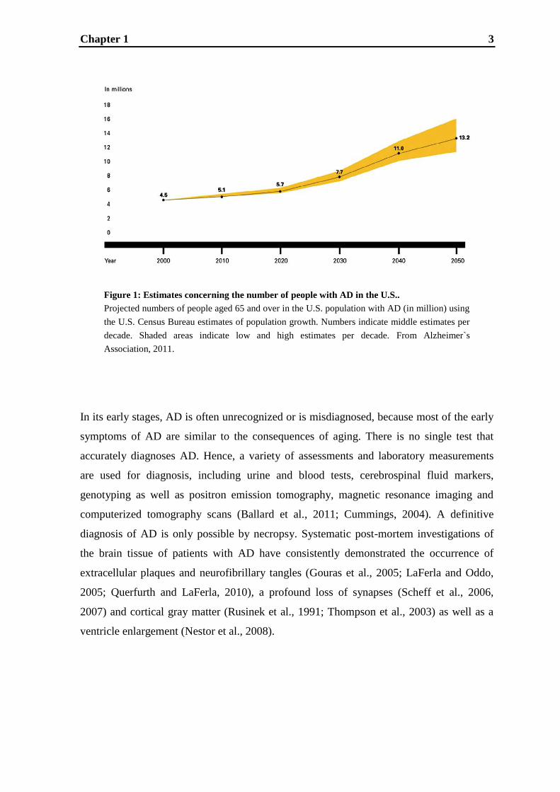
alone in the U.S. about 11 million (Fig.1; Alzheimer's Association, 2011; Brookmeyer et
al., 1998, 2007; Forlenza et al., 2010; Scatena et al., 2007). Both genetic and
environmental factors are considered to contribute to the development of AD. The minority
of cases has an obvious genetic origin with linkage studies indicating predominantly a
missense mutation in the amyloid precursor protein or Presenilin-1 or -2 gene (Bertram and
Tanzi, 2005; Cai et al., 1993; Citron et al., 1992, 1994; Goate et al., 1991; Haass and De
Strooper, 1999; Holmes, 2002; Levy-Lahad et al., 1995; Selkoe, 2001; Selkoe et al., 2002;
Sherrington et al., 1995), but also other candidate genes were identified. One example is
the ε4 allele of the apolipoprotein E gene, which is associated with a significant increase of
up to 70% for the risk of developing AD during aging (Corder et a., 1993; Holmes, 2002;
Mahley, 1988; Masters and Beyreuther, 1998; Strittmatter et al., 1993).
Chapter 1 3
Figure 1: Estimates concerning the number of people with AD in the U.S..
Projected numbers of people aged 65 and over in the U.S. population with AD (in million) using
the U.S. Census Bureau estimates of population growth. Numbers indicate middle estimates per
decade. Shaded areas indicate low and high estimates per decade. From Alzheimer`s
Association, 2011.
In its early stages, AD is often unrecognized or is misdiagnosed, because most of the early
symptoms of AD are similar to the consequences of aging. There is no single test that
accurately diagnoses AD. Hence, a variety of assessments and laboratory measurements
are used for diagnosis, including urine and blood tests, cerebrospinal fluid markers,
genotyping as well as positron emission tomography, magnetic resonance imaging and
computerized tomography scans (Ballard et al., 2011; Cummings, 2004). A definitive
diagnosis of AD is only possible by necropsy. Systematic post-mortem investigations of
the brain tissue of patients with AD have consistently demonstrated the occurrence of
extracellular plaques and neurofibrillary tangles (Gouras et al., 2005; LaFerla and Oddo,
2005; Querfurth and LaFerla, 2010), a profound loss of synapses (Scheff et al., 2006,
2007) and cortical gray matter (Rusinek et al., 1991; Thompson et al., 2003) as well as a
ventricle enlargement (Nestor et al., 2008).
Chapter 1 4
1.1.1. Amyloid hypothesis
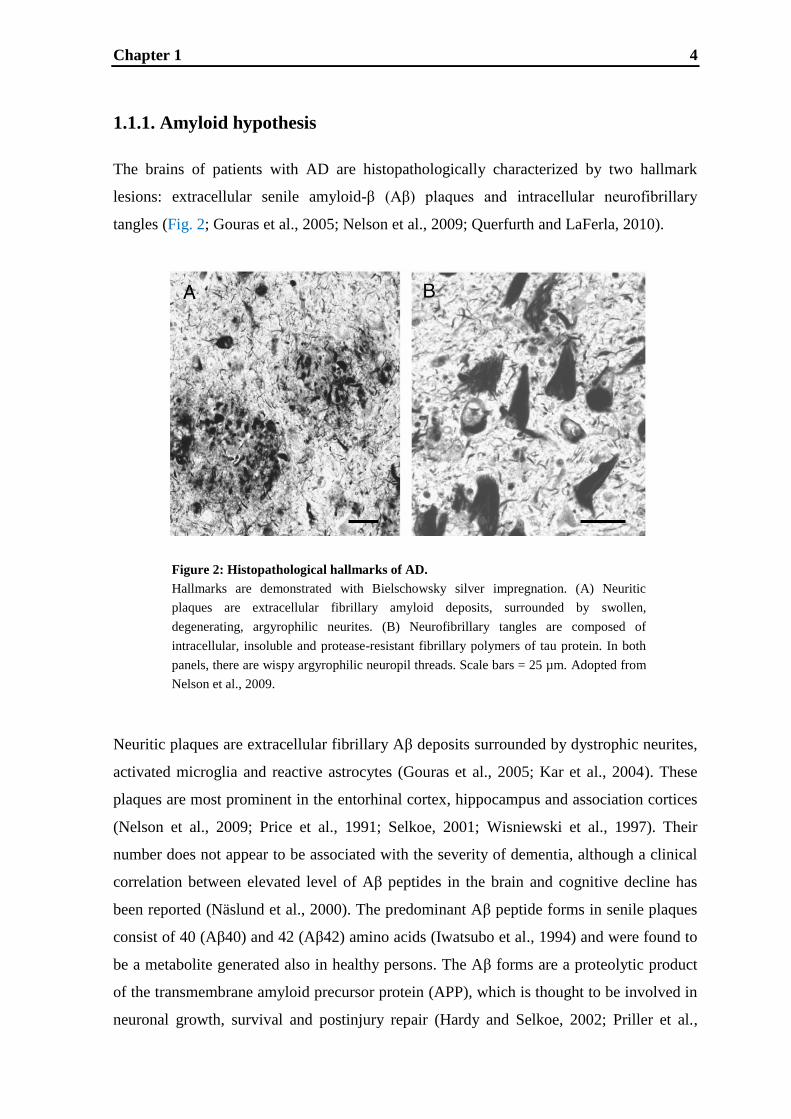
The brains of patients with AD are histopathologically characterized by two hallmark
lesions: extracellular senile amyloid-β (Aβ) plaques and intracellular neurofibrillary
tangles (Fig. 2; Gouras et al., 2005; Nelson et al., 2009; Querfurth and LaFerla, 2010).
Neuritic plaques are extracellular fibrillary Aβ deposits surrounded by dystrophic neurites,
activated microglia and reactive astrocytes (Gouras et al., 2005; Kar et al., 2004). These
plaques are most prominent in the entorhinal cortex, hippocampus and association cortices
(Nelson et al., 2009; Price et al., 1991; Selkoe, 2001; Wisniewski et al., 1997). Their
number does not appear to be associated with the severity of dementia, although a clinical
correlation between elevated level of Aβ peptides in the brain and cognitive decline has
been reported (Näslund et al., 2000). The predominant Aβ peptide forms in senile plaques
consist of 40 (Aβ40) and 42 (Aβ42) amino acids (Iwatsubo et al., 1994) and were found to
be a metabolite generated also in healthy persons. The Aβ forms are a proteolytic product
of the transmembrane amyloid precursor protein (APP), which is thought to be involved in
neuronal growth, survival and postinjury repair (Hardy and Selkoe, 2002; Priller et al.,
Figure 2: Histopathological hallmarks of AD.
Hallmarks are demonstrated with Bielschowsky silver impregnation. (A) Neurit ic p laques
are extracellular fibrillary amyloid deposits, surrounded by swollen, degenerating,
argyrophilic neurites. (B) Neurofibrillary tangles are composed of intracellular, insoluble
and protease-resistant fibrillary polymers of tau protein. In both panels, there are wispy
argyrophilic neuropil threads. Scale bars = 25 µm. Adopted from Nelson et al., 2009.
Figure 2: Histopathological hallmarks of AD.
Hallmarks are demonstrated with Bielschowsky silver impregnation. (A) Neuritic
plaques are extracellular fibrillary amyloid deposits, surrounded by swollen,
degenerating, argyrophilic neurites. (B) Neurofibrillary tangles are composed of
intracellular, insoluble and protease-resistant fibrillary polymers of tau protein. In both
panels, there are wispy argyrophilic neuropil threads. Scale bars = 25 µm. Adopted from
Nelson et al., 2009.
Chapter 1 5
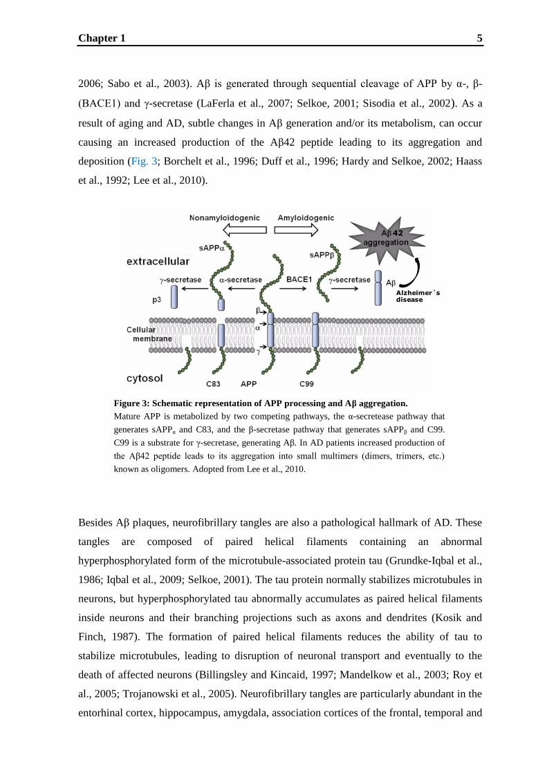
Figure 3: Schematic representation of APP processing and Aβ aggregation.
Mature APP is metabolized by two competing pathways, the α-secretease pathway that
generates sAPPα and C83, and the β-secretase pathway that generates sAPPβ and C99.
C99 is a substrate for γ-secretase, generating Aβ. In AD patients increased production of
the Aβ42 peptide leads to its aggregation into small multimers (dimers, trimers, etc.)
known as oligomers. Adopted from Lee et al., 2010.
Alzheimer`sdisease
42
2006; Sabo et al., 2003). Aβ is generated through sequential cleavage of APP by α-, β-
(BACE1) and γ-secretase (LaFerla et al., 2007; Selkoe, 2001; Sisodia et al., 2002). As a
result of aging and AD, subtle changes in Aβ generation and/or its metabolism, can occur
causing an increased production of the Aβ42 peptide leading to its aggregation and
deposition (Fig. 3; Borchelt et al., 1996; Duff et al., 1996; Hardy and Selkoe, 2002; Haass
et al., 1992; Lee et al., 2010).
Besides Aβ plaques, neurofibrillary tangles are also a pathological hallmark of AD. These
tangles are composed of paired helical filaments containing an abnormal
hyperphosphorylated form of the microtubule-associated protein tau (Grundke-Iqbal et al.,
1986; Iqbal et al., 2009; Selkoe, 2001). The tau protein normally stabilizes microtubules in
neurons, but hyperphosphorylated tau abnormally accumulates as paired helical filaments
inside neurons and their branching projections such as axons and dendrites (Kosik and
Finch, 1987). The formation of paired helical filaments reduces the ability of tau to
stabilize microtubules, leading to disruption of neuronal transport and eventually to the
death of affected neurons (Billingsley and Kincaid, 1997; Mandelkow et al., 2003; Roy et
al., 2005; Trojanowski et al., 2005). Neurofibrillary tangles are particularly abundant in the
entorhinal cortex, hippocampus, amygdala, association cortices of the frontal, temporal and

Chapter 1 6
parietal lobes and certain subcortical nuclei that project to these regions (Haroutunian et
al., 1999; Schifilliti et al., 2010; Selkoe, 2001). The extent of neurofibrillary pathology,
and particularly the number of cortical neurofibrillary tangles, correlates positively with
the severity of dementia, and thus has been used to classify the stage of the disease
(Arriagada et al., 1992; Bierer et al., 1995; Braak and Braak, 1997).
Regarding the relationship between Aβ and tau in AD, experimental evidence indicates
that Aβ can induce an increased phosphorylation of tau at the disease-relevant sites
(Busciglio et al., 1995; De Felice et al., 2008; Greenberg and Kosik, 1995; Rank et al.,
2002; Zheng et al., 2002). Indeed, studies carried out in transgenic mice suggest a
modulatory link between Aβ and tau. APP/tau double-mutant mice and tau transgenic mice
intracranially injected with synthetic Aβ developed enhanced neurofibrillary pathology as
compared to the single mutant tau mice (Götz et al., 2001; Lewis et al., 2001).
Additionally, tau pathology in three fold transgenic AD mice (Oddo et al., 2003a,b) can be
diminished by clearing Aβ due to passive immunotherapy (Oddo et al., 2004). These
studies indicate that the development of tau pathology is a downstream consequence of the
Aβ pathology (Hardy and Selkoe, 2002; Oddo et al., 2008; Tseng et al., 2008). Moreover,
as other types of neurodegenerative diseases besides AD also exhibit neurofibrillary
tangles (Hong et al., 1998; Mailliot et al., 2000), the formation of tau filaments seems to be
a more general event in neurodegenerative diseases leading to neuronal death, whereas Aβ
seems to be the specific initiator in AD.
As to the apparent involvement of Aβ in AD, the “amyloid cascade hypothesis” was
postulated, which originally links the pathological process of AD and neuronal cell death
to aggregation and deposition of Aβ (Hardy and Higgins, 1992). This hypothesis has led to
a huge number of studies concerning the effects of Aβ onto neurons and has received
abundant verification (Fig. 4; Armstrong, 2011; Forlenza et al., 2010; Pimplikar, 2009). Aβ
has both neurotrophic and neurotoxic effects, depending on the duration of exposure, the
dose and the degree of aggregation of the peptide (Kowall et al., 1991; May et al., 1992;
Youssef et al., 2008). Investigations in animal models and human brain samples have put a
special emphasis on soluble Aβ forms (Dahlgren et al., 2002; Klein, 2002; Lue et al., 1999;
Tabaton and Piccini, 2005; Walsh et al., 1999). In fact, it was demonstrated that acute
injection of soluble Aβ into the brain of rats and mice impairs learning and memory
(Balducci et al., 2010; Poling et al., 2008; Youssef et al., 2008). Hence, several lines of
evidence suggest that accumulation of Aβ peptide in the brain may initiate and/or
contribute to the pathogenesis of AD. Overproduction and/or reduced clearance of Aβ

Chapter 1 7
peptides are likely key to amyloid aggregation, which in turn contributes to the
development of senile plaques and neurofibrillary tangles (Hardy and Higgins, 1992;
Forlenza et al., 2010; Selkoe, 2001).
Due to the amyloid cascade hypothesis, approaches for a disease modifying treatment have
focused on methods to reduce Aβ through selective Aβ-lowering agents. Several active and
passive vaccines are under clinical investigation for their efficacy in reducing the level of
Aβ in the plasma and brain of subjects with mild-to-moderately advanced disease state
(Okura et al., 2006). Furthermore, having identified all the components required for
secretase function, the development of selective β- and/or -secretase inhibitors or
approaches for α-secretase enhancement are being pursued in order to lower Aβ production
in the brain (Ganjei, 2010; Geling et al., 2002; Hong et al., 2000; Luo and Yan, 2010;
Vassar, 2001). An alternative approach would be to use small molecules to bind Aβ
monomers and prevent their assembly into cytotoxic oligomers (Ganjei, 2010, Woo et al.,
2011).
Figure 4: The amyloid cascade hypothesis.
Accumulation of Aβ (pre-clinical AD) contributes to the development of fibrillary Aβ
and Aβ oligomers (pre-dementia) and eventually to development of neuritic plaques
and neurofibrillary tangles (clinical dementia). (Dotted arrows indicate possible or
secondary mechanisms affecting core pathological processes within the amyloid
cascade; PS1/2 = presenilin 1/2) From Forlenza et al., 2010.
Figure 4: The amyloid cascade hypothesis.
Accumulation of Aβ (pre-clinical AD) contributes to the development of fibrillary Aβ and Aβ
oligomers (pre-dementia) and eventually to development of neuritic plaques and neurofibrillary
tangles (clinical dementia). (Dotted arrows indicate possible or secondary mechanisms affecting core
pathologicalprocesses within the amyloid cascade; PS1/2 = presenilin 1/2) From Forlenza et al., 2010.

Chapter 1 8
The cortex shrivels up, damaging areas involved in thinking, planning and remembering
Ventricles (fluid-filled spaceswithin the brain) grow larger.
Shrinkage is especially severe in the hippocampus, an area of the cortex that plays a key role in
formation of new memories.
Healthy brain Advanced Alzheimer`s
1.1.2. Synaptic and neuronal dysfunction
As a result of the Aβ pathology, AD is correlated with a profound loss of synapses and
with neuronal dysfunction (Fig. 5; Scheff et al., 2006, 2007; Selkoe, 2002). Degenerating
neurons and synapses in the brains of individuals with AD are located predominantly
within regions that project to or from areas that display high densities of plaques and
tangles. Biochemical investigations from biopsy and autopsy of tissues from AD patients
indicate that various neurotransmitters and modulators including acetylcholine, serotonin,
noradrenalin, somatostatin and glutamate are differentially altered in the brains of
individuals with AD (Francis et al., 1993, 1999; Mann and Yates, 1986). The loss of
synapses and neuronal function are supposed to be the reason for cognitive decline in
patients (Larson et al., 1992; Terry et al., 1991). In particular, pronounced dysfunction of
the cholinergic and the glutamatergic system can be observed in AD patients and will be
discussed in the next paragraphs.
Figure 5: Schematic representation of massive cell loss in advanced AD.
From Lee et al., 2010.
Chapter 1 9
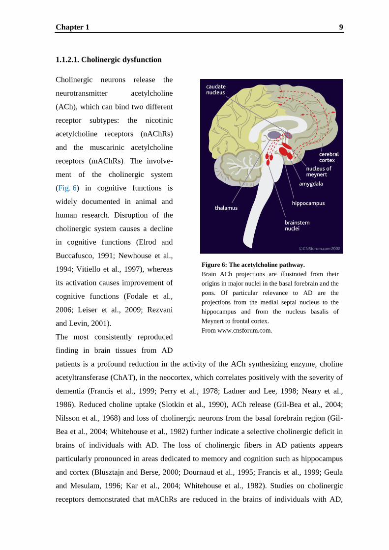
Figure 6: The acetylcholine pathway.
Brain ACh projections are illustrated from their
origins in major nuclei in the basal forebrain and the
pons. Of particular relevance to AD are the
projections from the medial septal nucleus to the
hippocampus and from the nucleus basalis of
Meynert to frontal cortex.
From www.cnsforum.com.
1.1.2.1. Cholinergic dysfunction
Cholinergic neurons release the
neurotransmitter acetylcholine
(ACh), which can bind two different
receptor subtypes: the nicotinic
acetylcholine receptors (nAChRs)
and the muscarinic acetylcholine
receptors (mAChRs). The involve-
ment of the cholinergic system
(Fig. 6) in cognitive functions is
widely documented in animal and
human research. Disruption of the
cholinergic system causes a decline
in cognitive functions (Elrod and
Buccafusco, 1991; Newhouse et al.,
1994; Vitiello et al., 1997), whereas
its activation causes improvement of
cognitive functions (Fodale et al.,
2006; Leiser et al., 2009; Rezvani
and Levin, 2001).
The most consistently reproduced
finding in brain tissues from AD
patients is a profound reduction in the activity of the ACh synthesizing enzyme, choline
acetyltransferase (ChAT), in the neocortex, which correlates positively with the severity of
dementia (Francis et al., 1999; Perry et al., 1978; Ladner and Lee, 1998; Neary et al.,
1986). Reduced choline uptake (Slotkin et al., 1990), ACh release (Gil-Bea et al., 2004;
Nilsson et al., 1968) and loss of cholinergic neurons from the basal forebrain region (Gil-
Bea et al., 2004; Whitehouse et al., 1982) further indicate a selective cholinergic deficit in
brains of individuals with AD. The loss of cholinergic fibers in AD patients appears
particularly pronounced in areas dedicated to memory and cognition such as hippocampus
and cortex (Blusztajn and Berse, 2000; Dournaud et al., 1995; Francis et al., 1999; Geula
and Mesulam, 1996; Kar et al., 2004; Whitehouse et al., 1982). Studies on cholinergic
receptors demonstrated that mAChRs are reduced in the brains of individuals with AD,
Figure 6: The acetylcholine pathway.
Brain ACh projections are illustrated from their origins in
major nuclei in the basal forebrain and the pons. Of
particular relevance to AD are the projections from the
medial septal nucleus to the hippocampus and from the
nucleus basalis of Meynert to frontal cortex. From
www.cnsforum.com.
Chapter 1 10
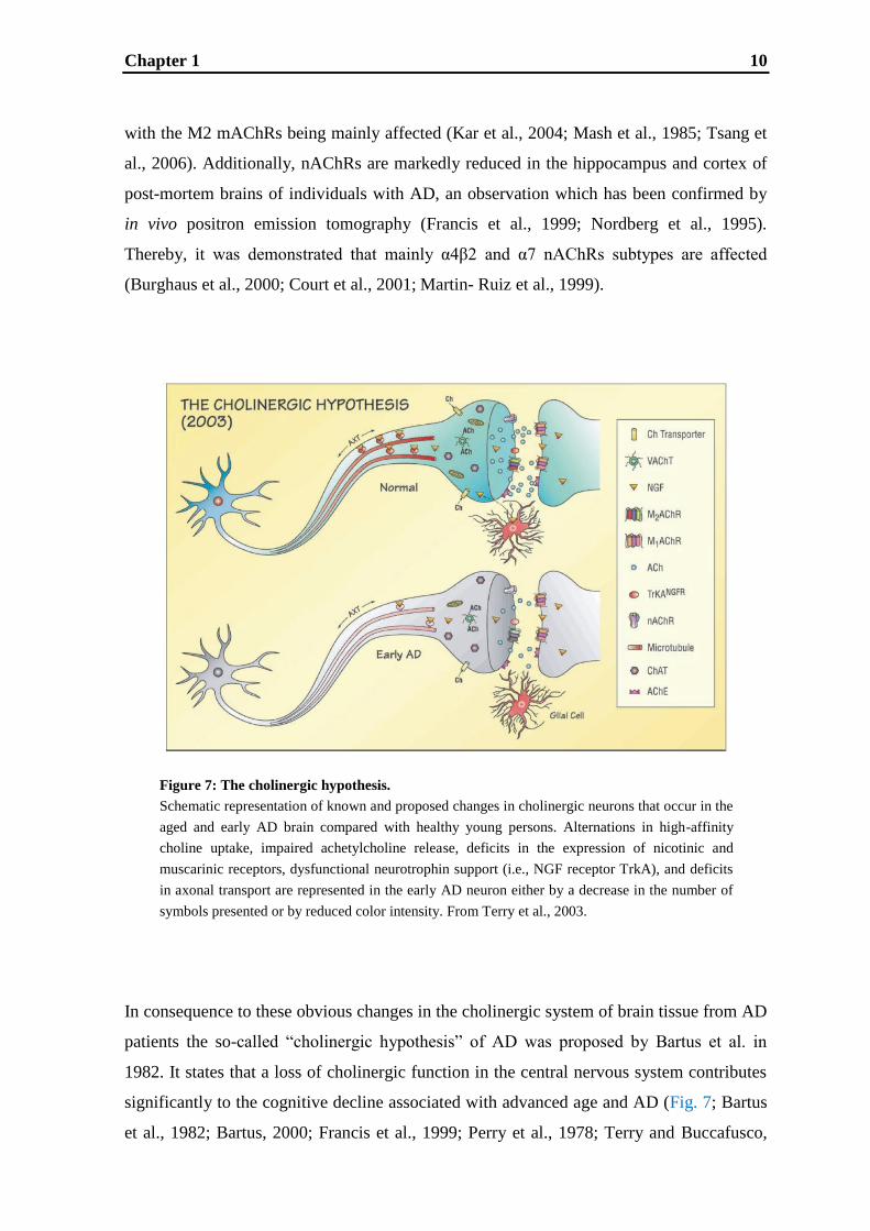
Figure 7: The cholinergic hypothesis.
Schematic representation of known and proposed changes in cholinergic neurons that occur in the
aged and early AD brain compared with healthy young persons. Alternations in high-affinity
choline uptake, impaired achetylcholine release, deficits in the expression of nicotinic and
muscarinic receptors, dysfunctional neurotrophin support (i.e., NGF receptor TrkA), and deficits
in axonal transport are represented in the early AD neuron either by a decrease in the number of
symbols presented or by reduced color intensity. From Terry et al., 2003.
Figure 7: The cholinergic hypothesis.
Schematic representation of known and proposed changes in cholinergic neurons that occur in the
aged and early AD brain compared with healthy young persons. Alternations in h igh-affinity
choline uptake, impaired achetylcholine release, deficits in the expression of nicotinic and
muscarinic receptors, dysfunctional neurotrophin support (i.e., NGF receptors), and deficits in
axonal transport are represented in the early AD neuron either by a decrease in the number of
symbols presented or by reduced color intensity. From Terry et al., 2003.
with the M2 mAChRs being mainly affected (Kar et al., 2004; Mash et al., 1985; Tsang et
al., 2006). Additionally, nAChRs are markedly reduced in the hippocampus and cortex of
post-mortem brains of individuals with AD, an observation which has been confirmed by
in vivo positron emission tomography (Francis et al., 1999; Nordberg et al., 1995).
Thereby, it was demonstrated that mainly α4β2 and α7 nAChRs subtypes are affected
(Burghaus et al., 2000; Court et al., 2001; Martin- Ruiz et al., 1999).
In consequence to these obvious changes in the cholinergic system of brain tissue from AD
patients the so-called “cholinergic hypothesis” of AD was proposed by Bartus et al. in
1982. It states that a loss of cholinergic function in the central nervous system contributes
significantly to the cognitive decline associated with advanced age and AD (Fig. 7; Bartus
et al., 1982; Bartus, 2000; Francis et al., 1999; Perry et al., 1978; Terry and Buccafusco,

Chapter 1 11
2003). Several in vivo imaging studies conducted in AD patients support the cholinergic
hypothesis. For example, positron emission tomography studies indicate that nAChR
deficits are an early phenomenon in AD, which significantly correlate with the level of
cognitive impairment (Nordberg, 2001). Other positron emission tomography studies
indicate both age- and AD-related decreases in the binding of nonselective muscarinic
ligands in neocortical regions (Zubieta et al., 2001). Moreover, single-photon emission
computed tomography studies indicate that the vesicular acetylcholine transporter is
reduced in early onset AD patients (Efange et al., 1997).
Recent studies demonstrated that Aβ causes the cholinergic hypofunction in AD patients
(Fig. 8). For example, several steps of ACh synthesis and release are reduced by Aβ and
there is a significant increase in the rate of high-affinity choline uptake (Auld et al., 1998;
Bales et al., 2006; Harkany et al., 1995a,b; Kar et al., 1998; Kristofiková et al., 2001;
Pedersen et al., 1996; Pedersen and Blusztajn, 1997). Additionally, it was found that Aβ
reduces the loading of ACh into vesicles by inhibiting the fast axonal transport of the
vesicular acetylcholine transporter in the sciatic nerve of the rat (Kasa et al., 2000, 2004).
Moreover, a redistribution of the acetylcholine esterase (AChE) within the cells is caused
by Aβ42 (Kasa et al., 1998) supporting the suggestion that neuronal degeneration is
initiated by a failure in axonal transport. Furthermore, in cortical cultures, the G-protein
coupling of mAChR activation is impaired by Aβ (Kelly et al., 1996). Similarly, in rats, the
density of total mAChRs was dose dependently decreased through repeated in vivo
intracerebroventricular administration of Aβ (Pavia et al., 2000). Moreover, there is further
evidence that neuronal nAChRs interact with Aβ42 (Nagele et al., 2002; Oddo et al., 2005;
Wang et al., 2000b). Aβ42 can bind with high affinity to the α7 nAChR and with lower
affinity to the α4β2 nAChR and hence might block the functional interaction of nicotinic
agonists with their receptors (Wang et al., 2000a). It has been suggested that the high-
affinity binding of Aβ42 to α7 nAChRs might cause the internalization and gradual
accumulation of Aβ42 in the neurons of AD brains. This suggestion is supported by a
study which demonstrated a substantial intraneuronal Aβ42 accumulation only in cells that
express relatively high levels of the α7 nAChR (Nagele et al., 2002). These data might
provide a plausible explanation for the selective vulnerability of neurons expressing the α7
nAChR in AD brains.

Chapter 1 12
Aβ
High affinity
Ch uptake
ACh release
AChE redistribution
VAChT density
Muscarinic/G
protein coupling
nAChRs
ChAT
-+
-
--
-
-
+
M1, M2 mAChRs
Figure 8: Effect of Aß on cholinergic neurons.
Schematic representation of the known changes in cholinergic neurons
due to activation of Aß.
In the light of the cholinergic hypothesis, drugs that enhance cholinergic transmission, i.e.
acetylcholine esterase (AChE) inhibitors, were developed as therapy of AD patients. These
drugs increase and prolong the synaptic levels of available ACh by preventing its
degradation and are currently the most widely prescribed drugs for the treatment of AD.
They include tacrine hydrochloride (Cognex®; approved in 1993), donepezil hydrochloride
(Aricept®; approved in 1996; current gold standard therapy), rivastigmine tartrate
(Exelon®; approved in 2000) and galantamine hydrobromide (Razadyne®/Reminyl®;
approved in 2001) (Birks and Harvey, 2003; Geerts and Grossberg, 2006; Kaduszkiewicz
et al., 2005; Trinh et al., 2003). AChE inhibitors are therapeutically used for patients
diagnosed with mild-to-moderate AD, but their efficacy in patients is only moderate as
demonstrated in a recent meta-analysis (Courtney et al., 2004; Raschetti et al., 2007).
Moreover, this treatment only alleviates symptoms of the disease, but is not curative
(Raschetti et al., 2007; Rountree et al., 2009). Alternative ways to target the cholinergic
system for memory improvement in dementia by highly selective nicotinic or muscarinic
agonism are still in early stages of clinical testing (Dunbar et al., 2007; Geerts, 2006).
Figure 8: Effect of Aβ on cholinergic neurons.
Schematic representation of the known changes in cholinergic neurons
due to activation of Aβ.

Chapter 1 13
Figure 9: The glutamate pathways.
In the normal brain the prominent glutamatergic
pathways are: the cortico-cortical pathways
(thalamus – cortex) and the extrapyramidal pathway
(cortex - striatum). Other glutamate projections exist
between the cortex, substantia nigra, subthalmic
nucleus and pallidum. Glutamate-containing
neuronal terminals are ubiquitous in the central
nervous system. From Carlsson, 1995.
1.1.2.2. Glutamatergic dysfunction
Glutamate is the primary excitatory
neurotransmitter of the central
nervous system (Fig. 9). It is used by
approximately two-thirds of
synapses in the neocortex and
hippocampus, while ACh is found in
approximately 5% of the synapses/
neurons (Fonnum, 1984). Glutamate
is involved in most aspects of
cognition and higher mental function
(Danbolt, 2001; Fonnum, 1984).
Thus, it is not surprising that loss of
cortical neurons, especially the large
pyramidal neurons in the hippo-
campus utilizing glutamate for
neurotransmission, correlate closely
with cognitive impairment and
progression of AD (Braak and Braak,
1997; Francis et al., 1993).
The most convincing evidence to
date of a glutamatergic dysfunction
in AD are histological findings of both cell and synapse loss of presumed glutamatergic
neurons (Francis 2003; Morrison and Hof, 1997; Terry et al., 1991). Moreover,
glutamatergic transmission is severely altered by early degeneration of corticocortical
connections and hippocampal projections in AD (Francis et al., 1993). In AD patients,
numerous studies have implicated reduced concentration of glutamate (Antuono et al.,
2001; Rupsingh et al., 2011; Procter et al., 1988), decreased binding to receptors (Cross et
al., 1987; Greenamyre et al., 1985, 1987; Maragos et al., 1987) and reduced glutamate
uptake into vesicles of synaptosome preparations (Procter et al., 1988; Scott et al., 2011).
Furthermore, a large decrease in glutamate receptors has been observed in the cortex and
hippocampus (Greenamyre et al., 1987; Procter et al., 1989). In general, two types of
glutamate receptors can be distinguished: ionotropic glutamate receptors, which form an
Figure 9: The glutamate pathways.
In the normal brain the prominent glutamatergic
pathways are the cortico-cortical pathways (thalamus -
cortex) and the extrapyramidal pathway (cortex -
striatum). Other glutamate projections exist between
the cortex, substantia nigra, subthalmic nucleus and
pallidum. Glutamate-containing neuronal terminals
are ubiquitous in the central nervous system. From
Carlsson, 1995.
Chapter 1 14
ion channel pore opening upon activation, and metabotropic glutamate receptors, which are
G-protein coupled receptors indirectly activating ion-channels in the plasma membrane
through a signaling cascade. Ionotropic glutamate receptors are sub-classified into α-
amino-3-hydroxy-5-methyl-4-isoxazolepropionic acid (AMPA), kainate and N-Methyl-D-
Aspartate (NMDA) receptors. Concerning AMPA receptors, especially their GluR1 and
GluR2/3 subunits are reduced in AD patients and this reduction seems to be correlated with
neurofibrillary tangle formation (Ikonomovic et al., 1995; Wakabayashi et al., 1999;
Yasuda et al., 1995). Only few studies regarding changes of kainate receptors in AD exist,
which show that kainate binding is significantly reduced in the parahippocampal gyrus
(Dewar et al., 1991). Concerning NMDA receptors and their changes in AD patients, there
is still an ongoing debate. Most reports indicated a reduction of NMDA receptors in AD
patients (Hynd et al., 2001, 2004; Panegyres et al., 2002; Penney et al., 1990; Ulas and
Cotman, 1997; Wang et al., 2000c) and thus it was proposed that the selective decrease of
NMDA receptors and/or their hypoactivity may affect the memory dysfunction in AD.
Controversially, Ikonomovic et al. (1999) reported that the NMDAR subunit NR1 is
markedly increased in vulnerable neurons of AD and thus excitotoxicity was proposed to
be responsible for neuronal loss. Excitotoxic cell death may result from excessive
activation of NMDA receptors, leading to raised intracellular calcium levels and
subsequent activation of a cascade of enzymes, resulting in cell death by necrosis and
apoptosis (Lipton, 1999). Despite the controversies in the literature, it seems to be more
likely that NMDA receptors may contribute significantly to the pathophysiology in AD via
degeneration of synaptic activity rather than cell death via excitotoxicity. However, more
research has to be done to clarify this issue (Lee et al., 2002). In comparison to ionotropic
glutamate receptors, there are much less data on the role and expression of metabotropic
glutamate receptors (mGluRs) in AD; however, dysfunction of metabotropic glutamate
receptors was demonstrated to be associated with abnormal tau phosphorylation (Boldyrev
et al., 2004).
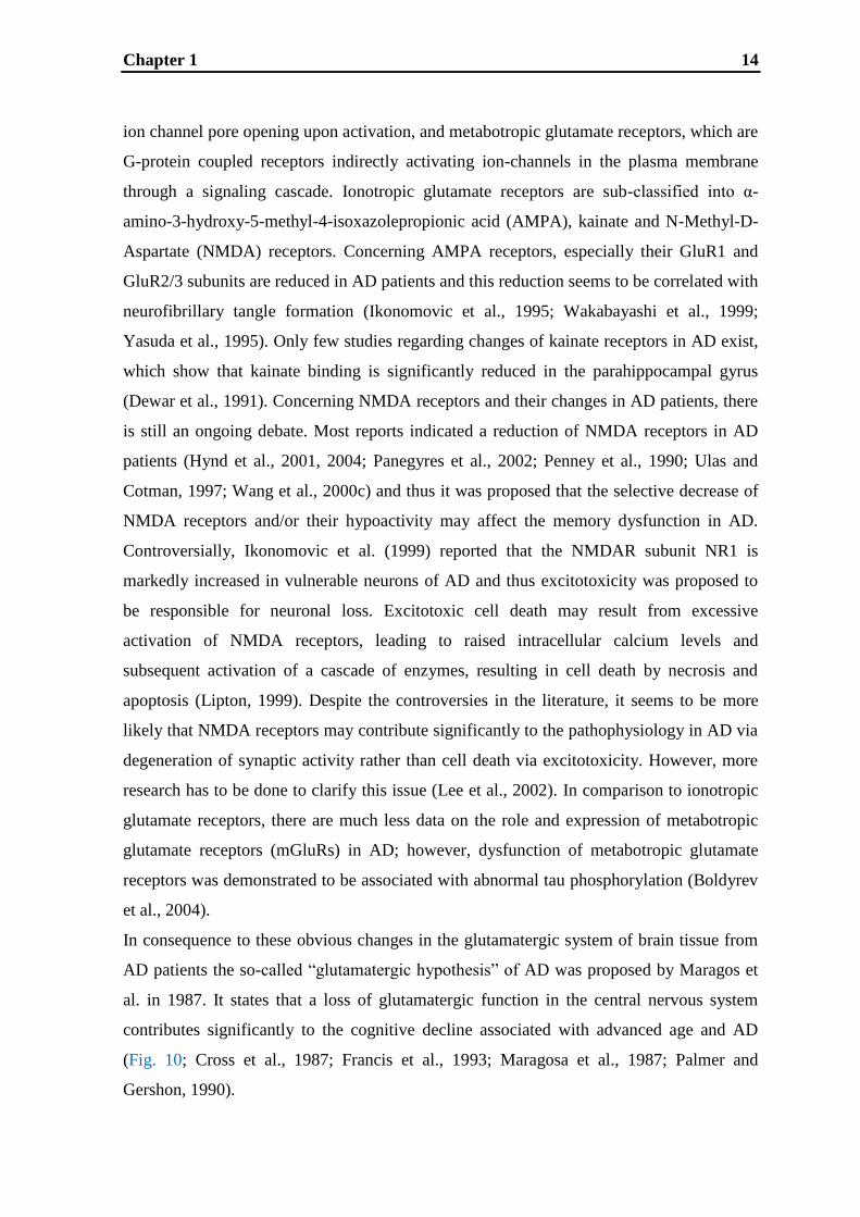
In consequence to these obvious changes in the glutamatergic system of brain tissue from
AD patients the so-called “glutamatergic hypothesis” of AD was proposed by Maragos et
al. in 1987. It states that a loss of glutamatergic function in the central nervous system
contributes significantly to the cognitive decline associated with advanced age and AD
(Fig. 10; Cross et al., 1987; Francis et al., 1993; Maragosa et al., 1987; Palmer and
Gershon, 1990).
Chapter 1 15
Presynaptic
neuron
Presynaptic
neuron
Postsynaptic
neuronPostsynaptic
neuron
AMPAR NMDAR Kainate AMPAR NMDAR Kainate
Gluatamte
Gluatamte
EAAT
EAAT
Figure 10: The glutamatergic hypothesis.
Schematic representation of known and proposed changes in glutamatergic neurons that occur in the
aged and early AD brain compared with healthy young persons. Alternations in glutamate transmission,
glutamate concentration, glutamate uptake, decreased binding of glutamate to receptors and decrease in
receptors are represented in the AD neuron by a decrease in the number of symbols.
HealthyAlzheimer`s
disease
Figure 10: The glutamatergic hypothesis.
Schematic representation of known and proposed changes in glutamatergic neurons that occur in the aged
and early AD brain compared with healthy young persons. Alterations in glutamate transmission,
glutamate concentration, glutamate uptake, decreased binding of glutamate to receptors and a decrease in
receptors are represented in the AD neuron by a decrease in the number of symbols.
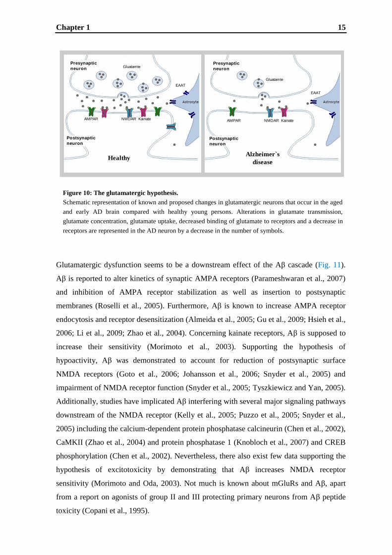
Glutamatergic dysfunction seems to be a downstream effect of the Aβ cascade (Fig. 11).
Aβ is reported to alter kinetics of synaptic AMPA receptors (Parameshwaran et al., 2007)
and inhibition of AMPA receptor stabilization as well as insertion to postsynaptic
membranes (Roselli et al., 2005). Furthermore, Aβ is known to increase AMPA receptor
endocytosis and receptor desensitization (Almeida et al., 2005; Gu et al., 2009; Hsieh et al.,
2006; Li et al., 2009; Zhao et al., 2004). Concerning kainate receptors, Aβ is supposed to
increase their sensitivity (Morimoto et al., 2003). Supporting the hypothesis of
hypoactivity, Aβ was demonstrated to account for reduction of postsynaptic surface
NMDA receptors (Goto et al., 2006; Johansson et al., 2006; Snyder et al., 2005) and
impairment of NMDA receptor function (Snyder et al., 2005; Tyszkiewicz and Yan, 2005).
Additionally, studies have implicated Aβ interfering with several major signaling pathways
downstream of the NMDA receptor (Kelly et al., 2005; Puzzo et al., 2005; Snyder et al.,
2005) including the calcium-dependent protein phosphatase calcineurin (Chen et al., 2002),
CaMKII (Zhao et al., 2004) and protein phosphatase 1 (Knobloch et al., 2007) and CREB
phosphorylation (Chen et al., 2002). Nevertheless, there also exist few data supporting the
hypothesis of excitotoxicity by demonstrating that Aβ increases NMDA receptor
sensitivity (Morimoto and Oda, 2003). Not much is known about mGluRs and Aβ, apart
from a report on agonists of group II and III protecting primary neurons from Aβ peptide
toxicity (Copani et al., 1995).
Chapter 1 16
Aβ NMDAR surface number
NMDAR sensitivity
mGluRs
AMPAR endocytosis /
desensitization
AMPAR kinetics
Kainate
sensitivity
--
+
AMPAR stabilisation /
insertion
+
+
-
NMDAR function +
downstream effects
protective
-
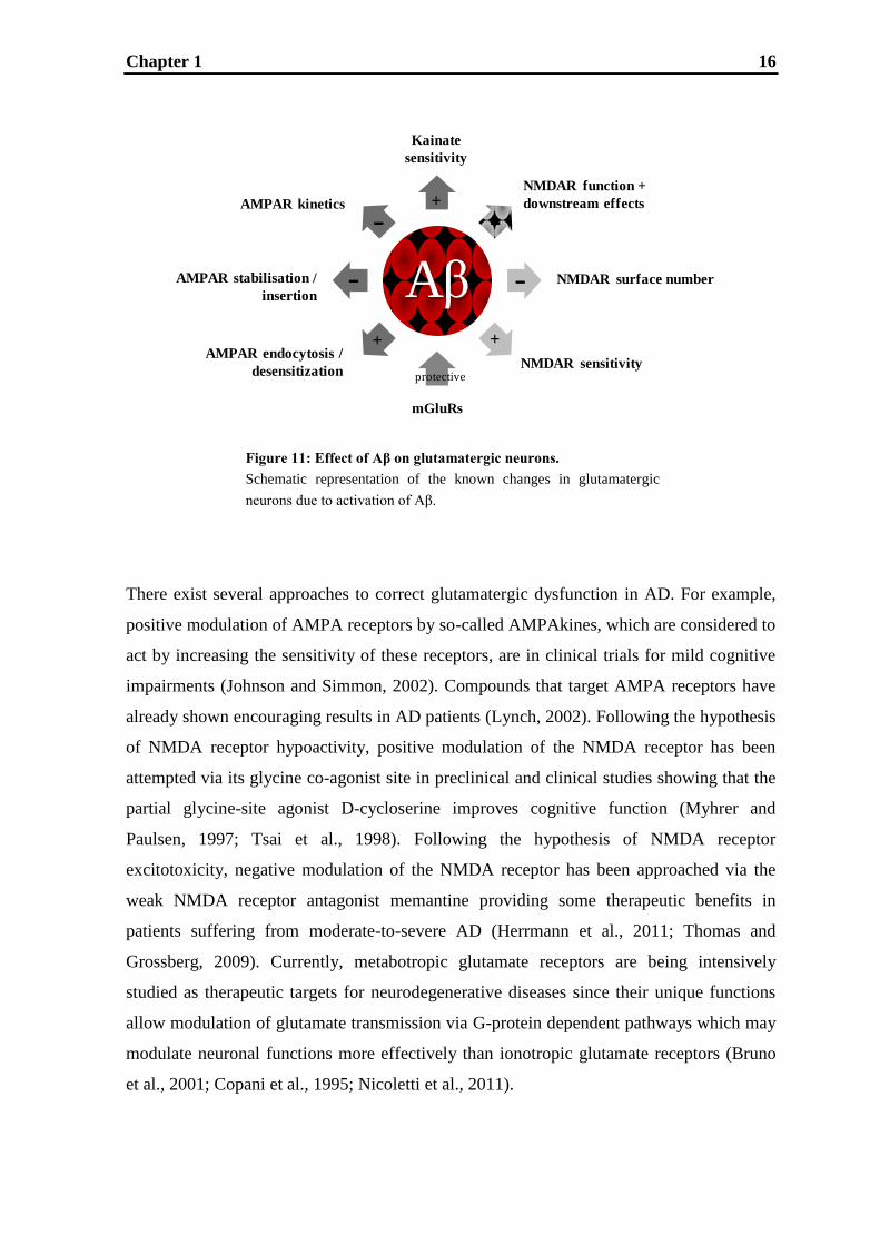
There exist several approaches to correct glutamatergic dysfunction in AD. For example,
positive modulation of AMPA receptors by so-called AMPAkines, which are considered to
act by increasing the sensitivity of these receptors, are in clinical trials for mild cognitive
impairments (Johnson and Simmon, 2002). Compounds that target AMPA receptors have
already shown encouraging results in AD patients (Lynch, 2002). Following the hypothesis
of NMDA receptor hypoactivity, positive modulation of the NMDA receptor has been
attempted via its glycine co-agonist site in preclinical and clinical studies showing that the
partial glycine-site agonist D-cycloserine improves cognitive function (Myhrer and
Paulsen, 1997; Tsai et al., 1998). Following the hypothesis of NMDA receptor
excitotoxicity, negative modulation of the NMDA receptor has been approached via the
weak NMDA receptor antagonist memantine providing some therapeutic benefits in
patients suffering from moderate-to-severe AD (Herrmann et al., 2011; Thomas and
Grossberg, 2009). Currently, metabotropic glutamate receptors are being intensively
studied as therapeutic targets for neurodegenerative diseases since their unique functions
allow modulation of glutamate transmission via G-protein dependent pathways which may
modulate neuronal functions more effectively than ionotropic glutamate receptors (Bruno
et al., 2001; Copani et al., 1995; Nicoletti et al., 2011).
Figure 11: Effect of Aβ on glutamatergic neurons.
Schematic representation of the known changes in glutamatergic
neurons due to activation of Aβ.

Chapter 1 17
1.2. Hippocampal learning and memory
The hippocampus is a major component of the
mammalian brain. It is a paired structure, with
mirror-image halves in the left and right
hemispheres of the brain being located inside the
so-called medial temporal lobe (Fig. 12). The
hippocampus belongs to the limbic system also
consisting of amygdala, anterior thalamic nuclei,
septum, limbic cortex and fornix. In 1957 Scoville
and Milner showed that bilateral hippocampal
removal, as a treatment for epilepsy suffered by
patient H.M., resulted in anterograde amnesia.
Thus, for the first time, the importance of the hippocampus and temporal lobe structures
for memory was identified. Since then, studies in humans and animals (Jarrard, 1993;
Squire et al., 1984) have consolidated the essential finding of that study. More recently,
noninvasive methods using direct brain imaging techniques, characterized blood flow and
oxygen consumption identified fluctuations in these parameters during learning tasks in the
hippocampus (Squire, 1992; Squire et al., 1990). Specifically, the hippocampus is involved
in episodic memory, i.e. memory for personally experienced events set in a spatio-temporal
context, and in spatial or topographical memory (Burgess et al., 2002; Smith and
Mizumori, 2006). In AD, the hippocampus is one of the first regions of the brain to be
affected causing first symptoms in patients, including memory problems and disorientation
(Burgess et al., 2002; Frisoni et al., 2008). Thus, improvement of hippocampal learning
and memory is an attractive approach to influence cognitive symptoms of the disease. The
research of learning and memory requires experimental model systems that can be utilized
both to characterize the underlying mechanisms associated with memory and to explore
drug candidates for the treatment of memory deficits. Although genetically engineered
mouse models for AD-like pathology exist, the following discussion will be limited to
hippocampal rodent model systems with wildtype animals, because the studies described in
this work utilized wildtype rats. In fact, cognitive enhancing drugs are able to improve
memory performance even in wildtype animals, thereby addressing those memory domains
and brain regions known to be affected in AD (Buccafusco et al., 2004; Prickaerts et al.,
2005).
Hippocampus
Figure 12: Schematic representation of
the hippocampus in the brain.
From www.macalester.edu/psychology

Chapter 1 18
1.2.1. Behavioral rodent models
A variety of experimental paradigms are available for the investigation of learning and
memory in behavioral studies. Some of these cognition tasks are mainly or even
exclusively hippocampus-dependent and therefore represent attractive experimental
approaches for AD research.
The Morris water maze task is the perhaps most commonly used model for hippocampal
spatial learning. In this test an animal‟s capacity to remember spatial cues is required to
locate a hidden submerged platform (Morris et al., 1982; Morris, 1984). Using this test,
numerous studies have proven an essential role for the hippocampus in spatial learning. In
addition, several studies have built on the original observation of O‟Keefe which identified
the involvement of specific hippocampal pyramidal cells in encoding information about the
location of an animal in a particular space (O‟Keefe, 1979). Apparently the key role of the
hippocampus in spatial learning consists in constructing and storing spatial maps (Teng
and Squire, 1999).
Another widely used model to investigate hippocampal learning is the T-maze task, which
is a behavioral test to assess spatial working memory performance of animals. This test is
based on the natural willingness of rodents to explore a new environment. The natural
tendency of rats and mice in a T-maze is to alternate their choice of the goal arm. The
response on each trial varies according to what they have previously experienced (Hudon
et al., 2002). Typical alternation rates are around 90% (Deacon et al., 2003). The T-maze is
a suitable test to investigate hippocampal (dys)function, because full lesions of the
hippocampus (Deacon and Rawlins, 2005; Lalonde, 2002) and already deletions of AMPA
receptor subunits (Reisel et al., 2002) impair performance of rodents in this task.
Another behavioral task for hippocampal learning and memory is contextual fear
conditioning, which is based on the capacity of mammals, including rodents, to associate
environmental cues with an aversive stimulus. In this model, conditioned emotional
responses are elicited by placing an animal in a chamber in which an aversive experience
has previously been made (Blanchard and Blanchard, 1972). There is good evidence
indicating that contextual fear conditioning is mostly hippocampus-dependent. This
evidence is based on lesion studies, selective infusion of pharmacological agents into the
hippocampus and animals genetically engineered to have hippocampal deficits (Chen et al.,
1996; Kim et al., 1993; Saxe et al., 2006).

Chapter 1 19
1.2.2. Long-term potentiation
A long time of research has been spent to find a model system for the cellular and
molecular mechanisms of learning and memory. Originally, it was hypothesized that
information storage relies on changes in strength of synaptic connections between neurons
that are active (Cajal, 1912 reviewed by Stahnisch and Nitsch, 2002). This hypothesis was
supported by Hebb in 1949, who proposed that if two neurons are active at the same time,
their synaptic efficiency onto a common target neuron will be strengthened (Hebb, 1949
reviewed by Cooper, 2005; Morris and Hebb, 1999). In artificial neuronal networks, such
as attention–language interactions and learning-input correlations, Hebbian learning was
proven to be successful (Garagnani et al., 2008; Gütig et al., 2003). An enormous effort has
been put into understanding the mechanism by which strengthening of synaptic
connections can be achieved and into finding a model for this process. This research has
led to the model of long-term potentiation (LTP) which was first published in 1973 (Bliss
and Lomo, 1973). They reported that trains of high frequency stimulation to the rabbit
perforant path caused a sustained increase in efficiency of synaptic transmission in the
granule cells of the dentate gyrus. This report, and others which followed during the 1970s,
confirmed the Hebbian nature of this form of synaptic plasticity. Cooperativity (Lee et al.,
1983; McNaughton, 2003), associativity (Barrionuevo and Brown, 1983; Levy and
Steward, 1979) and input specificity (Dunwiddie and Lynch 1978; Nishiyama et al., 2000),
being the characteristics of LTP, as well as the durability of LTP (Abraham et al., 1995;
Reymann et al., 1985) support the hypothesis that LTP may be a biological mechanism for
at least some forms of memory. LTP has not only been found in the hippocampus (Bliss
and Lomo, 1973; Dunwiddie and Lynch, 1978), but in many brain regions, including
piriform (Larson et al., 2005), entorhinal (Alonso et al., 1990) and prefrontal cortices
(Auclair et al., 2000), the septum (Garcia et al., 1997) and superior cervical ganglia
(Brown and McAfee, 1982) as well as in the spinal cord (Pedersen and Gjerstad, 2007).
Furthermore, LTP is not limited to the mammalian brain, but has been described in other
vertebrates such as the goldfish (Schmidt, 1990), bullfrog (Koyano et al., 1985), bird (Scott
and Bennett, 1993) and even in some invertebrates (Antonov et al., 2003; Glanzman, 2008;
Menzel and Manz, 2005; Oleskevich et al., 1997; Walters and Byrne, 1985). From the
mechanistic point of view, the opposing process to LTP is long-term depression (LTD)
described firstly in the cerebellar cortex (Ito, 1989). It is defined as an activity-dependent
reduction in the efficacy of neuronal synapses which can - like LTP - last for hours
Chapter 1 20
(Collingridge et al., 2010). LTD selectively weakens specific synapses in order to make
constructive use of synaptic strengthening caused by LTP. This is necessary because, if
allowed to continue increasing in strength, synapses would ultimately reach a ceiling level
of efficiency, which would inhibit the encoding of new information. Therefore, LTD itself
is also believed to be involved in learning and memory processes (Ito, 1989). However,
there are still remaining questions to be addressed regarding its exact contribution in
learning and memory in vitro and in vivo. Thus, in this work only LTP will be discussed,
because it is a well established and widely accepted in vitro model of synaptic plasticity,
which can even be used for profiling
putative memory enhancing
substances.
As already mentioned above, the
hippocampus is specifically attractive
for studying learning and memory.
Therefore, in this work, LTP
measurements were exclusively made
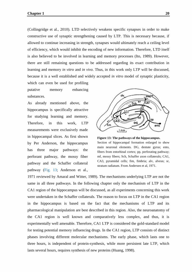
in hippocampal slices. As first shown
by Per Anderson, the hippocampus
has three major pathways: the
perforant pathway, the mossy fiber
pathway and the Schaffer collateral
pathway (Fig. 13; Anderson et al.,
1971 reviewed by Amaral and Witter, 1989). The mechanisms underlying LTP are not the
same in all three pathways. In the following chapter only the mechanism of LTP in the
CA1 region of the hippocampus will be discussed, as all experiments concerning this work
were undertaken in the Schaffer collaterals. The reason to focus on LTP in the CA1 region
in the hippocampus is based on the fact that the mechanisms of LTP and its
pharmacological manipulation are best described in this region. Also, the neuroanatomy of
the CA1 region is well known and comparatively less complex, and thus, it is
experimentally well amenable. Therefore, CA1 LTP is considered the gold-standard model
for testing potential memory influencing drugs. In the CA1 region, LTP consists of distinct
phases involving different molecular mechanisms. The early phase, which lasts one to
three hours, is independent of protein-synthesis, while more persistent late LTP, which
lasts several hours, requires synthesis of new proteins (Huang, 1998).
Figure 13: The pathways of the hippocampus.
Section of hippocampal formation enlarged to show
main neuronal elements. DG, dentate gyrus; ento,
fibers from entorhinal cortex; pp, perforating pathway;
mf, mossy fibers; Sch, Schaffer axon collaterals; CA1,
CA3, pyramidal cells; fim, fimbria; alv, alveus; sr,
stratum radiatum. From Andersen et al, 1971.
Figure 13: The pathways of the hippocampus.
Section of hippocampal formation enlarged to show
main neuronal elements. ento, Fibers from entorhinal
cortex; pp, perforating pathway; mf, mossy fibers; Sch,
Schaffer axon collaterals; CA1, CA3, pyramidal cells;
DG, dentate gyrus; sr, stratum radiatum; fim, fimbria ;
alv, alveus. From Andersen et al, 1971
DG
sr
Chapter 1 21
1.2.2.1. Mechanisms underlying early and late LTP in the CA1 region
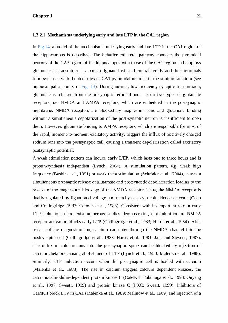
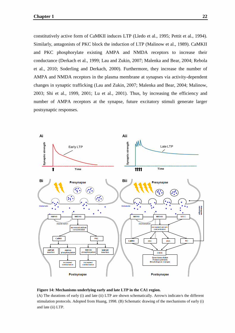
In Fig.14, a model of the mechanisms underlying early and late LTP in the CA1 region of
the hippocampus is described. The Schaffer collateral pathway connects the pyramidal
neurons of the CA3 region of the hippocampus with those of the CA1 region and employs
glutamate as transmitter. Its axons originate ipsi- and contralaterally and their terminals
form synapses with the dendrites of CA1 pyramidal neurons in the stratum radiatum (see
hippocampal anatomy in Fig. 13). During normal, low-frequency synaptic transmission,
glutamate is released from the presynaptic terminal and acts on two types of glutamate
receptors, i.e. NMDA and AMPA receptors, which are embedded in the postsynaptic
membrane. NMDA receptors are blocked by magnesium ions and glutamate binding
without a simultaneous depolarization of the post-synaptic neuron is insufficient to open
them. However, glutamate binding to AMPA receptors, which are responsible for most of
the rapid, moment-to-moment excitatory activity, triggers the influx of positively charged
sodium ions into the postsynaptic cell, causing a transient depolarization called excitatory
postsynaptic potential.
A weak stimulation pattern can induce early LTP, which lasts one to three hours and is
protein-synthesis independent (Lynch, 2004). A stimulation pattern, e.g. weak high
frequency (Bashir et al., 1991) or weak theta stimulation (Schröder et al., 2004), causes a
simultaneous presnaptic release of glutamate and postsynaptic depolarization leading to the
release of the magnesium blockage of the NMDA receptor. Thus, the NMDA receptor is
dually regulated by ligand and voltage and thereby acts as a coincidence detector (Coan
and Collingridge, 1987; Cotman et al., 1988). Consistent with its important role in early
LTP induction, there exist numerous studies demonstrating that inhibition of NMDA
receptor activation blocks early LTP (Collingridge et al., 1983; Harris et al., 1984). After
release of the magnesium ion, calcium can enter through the NMDA channel into the
postsynaptic cell (Collingridge et al., 1983; Harris et al., 1984; Jahr and Stevens, 1987).
The influx of calcium ions into the postsynaptic spine can be blocked by injection of
calcium chelators causing abolishment of LTP (Lynch et al., 1983; Malenka et al., 1988).
Similarly, LTP induction occurs when the postsynaptic cell is loaded with calcium
(Malenka et al., 1988). The rise in calcium triggers calcium dependent kinases, the
calcium/calmodulin-dependent protein kinase II (CaMKII; Fukunaga et al., 1993; Ouyang
et al., 1997; Sweatt, 1999) and protein kinase C (PKC; Sweatt, 1999). Inhibitors of
CaMKII block LTP in CA1 (Malenka et al., 1989; Malinow et al., 1989) and injection of a
Chapter 1 22
Figure 14: Mechanisms underlying early and late LTP in the CA1 region.
(A) The durations of early (i) and late (ii) LTP are shown schematically. Arrow/s indicate/s the different
stimulation protocols. Adopted from Huang, 1998. (B) Schematic drawing of the mechanisms of early (i)
and late (ii) LTP.
Early LTP Late LTP
Time Time
Syn
ap
tic
str
en
gth
Ai
Bi
Syn
ap
tic s
tren
gth
Aii
Bii
constitutively active form of CaMKII induces LTP (Lledo et al., 1995; Pettit et al., 1994).
Similarly, antagonists of PKC block the induction of LTP (Malinow et al., 1989). CaMKII
and PKC phosphorylate existing AMPA and NMDA receptors to increase their
conductance (Derkach et al., 1999; Lau and Zukin, 2007; Malenka and Bear, 2004; Rebola
et al., 2010; Soderling and Derkach, 2000). Furthermore, they increase the number of
AMPA and NMDA receptors in the plasma membrane at synapses via activity-dependent
changes in synaptic trafficking (Lau and Zukin, 2007; Malenka and Bear, 2004; Malinow,
2003; Shi et al., 1999, 2001; Lu et al., 2001). Thus, by increasing the efficiency and
number of AMPA receptors at the synapse, future excitatory stimuli generate larger
postsynaptic responses.
Chapter 1 23
The use of a stronger stimulation pattern, e.g. repeated high frequency stimulation (Lu et
al., 1999) or strong theta burst stimulation (Rönicke et al., 2009), causes the induction of a
more persistent phase of LTP, namely late LTP. Late LTP is the natural extension of early
LTP being defined as lasting longer than three hours and being protein-synthesis dependent
(Frey et al., 1988, 1996; Lu et al., 1999). Due to stronger stimulation and thus stronger
depolarization, besides NMDA receptors, also voltage-dependent calcium channels
(VDCCs), which are embedded in the postsynaptic membrane, may reach the open state
(Grover and Tyler, 1990; Kullman et al., 1992). Thus, even more calcium enters the cell
not only activating CaMKII and PKC, but also the NO-sGC-cGMP (nitric oxide - soluble
guanylate cyclase - cyclic guanosine monophosphate) pathway and the cAMP-PKA-
MAPK-CREB (cyclic adenosine monophosphate - protein kinase A - mitogen-activated
protein kinase - cAMP response element-binding) pathway. Calcium causes NO
production by stimulating calcium/calmodulin-dependent neuronal NO synthases (Arancio
et al., 2001; Christopherson et al., 1999; Son et al., 1998; Zhuo et al., 1994). The diffusible
NO is believed to act as a retrograde messenger (Böhme et al., 1991; O‟Dell et al., 1991;
Schuman and Madison, 1991), which can cause changes in the presynaptic
neurotransmitter release machinery (Feil and Kleppisch, 2008). Yet, reports about NO
acting postsynaptically also exist (Garthwaite, 2008; Ko and Kelly, 1999). Most
postsynaptically effects of NO are mediated by NO-GCs, NO-sensitive enzymes that form
the second messenger cGMP and whose blockade abolishes late LTP (Boulton et al., 1995;
Son et al., 1998; Zhuo et al., 1999). The generated cGMP causes an induction/activation of
effector molecules and/or transcription factors, e.g. protein kinases, ion channels,
phosphodiesterases, and/or CREB (Garthwaite, 2008). Beneath the NO-sGC-cGMP
pathway, several studies have indicated that late LTP is also dependent on the cAMP-
PKA-MAPK-CREB pathway. Among the downstream consequences of an increase in
cAMP concentration is the activation of PKA (Huang and Kandel, 1994; Frey et al., 1993),
whose inhibition blocks LTP (Huang and Kandel, 1994), and MAPK (also called ERK)
(Impey et al., 1998; Roberson et al., 1999). The important role of MAPK in expression of
LTP was first underlined by the finding that its inhibition resulted in suppression of late
LTP in CA1 (English and Sweatt, 1997; Impey et al., 1998). The downstream
consequences of MAPK activation are wide ranging including phosphorylation of
cytoskeletal proteins, signaling proteins and nuclear proteins/transcription factors
(Jovanovic et al., 1996; Lynch, 2004; Matsubara et al., 1996) with CREB being the most
important. In fact, mutant mice lacking CREB isoforms α and δ exhibit attenuated late LTP
Chapter 1 24
(Bourtchuladze et al., 1994). Remarkably, not only activation of the MAPK pathway is
able to activate CREB by its phosphorylation, but also PKA, PKC, CaMKII as well as
other kinases are involved in CREB activation. CREB activation due to its phosphorylation
is the first step in gene transcription (Alberini et al., 2005; Silva et al., 1998) resulting in
translation of immediate early genes, which can also act as transcription factors to induce
late-response genes. Increased protein levels of certain immediate early genes, like
zif268/Erg1, Egr2, Egr3, AP-1, c-jun, jun-B, Arc and junD (Abraham et al., 1991; Cheval
et al., 2011; Poirier et al., 2008; Yin et al., 2002), are associated with late LTP in the way
that all these gene products are thought to lead to functional and structural changes of the
synapse. In the case of zif268 one gene product was identified as a nerve growth factor
response gene product, which has been shown to stimulate cell growth and differentiation
(Milbrandt, 1987). Results from a study using zif268 knockout mice demonstrated that this
gene is required for late LTP (Cheval et al., 2011; Poirier et al., 2008). Recent attention has
focused on analysis of changes in Arc (activity-regulated cytoskeleton-associated protein;
also called as Arg3.1) in LTP (Lyford et al., 1995; Yin et al., 2002; Yilmaz-Rastoder et al.,
2010). Because Arc protein binds to actin, it has been proposed that it participates in
cytoskeletal remodeling after synaptic activation. Consistent with the observed LTP-
associated increase in Arc expression, disruption of Arc inhibits late LTP (Bramham et al.,
2008). It is likely that the increase in protein-synthesis that accompanies late LTP
contributes to the establishment of the morphological changes that have been reported, for
example, the increases in postsynaptic surface area (Desmond and Levy, 1990), spine
number (Lee et al., 1980) and spine area (Fifkova and Vanharreveld, 1977). Late LTP has
also been shown to increase the number of large spines (Desmond and Levy, 1990),
axospinous perforated synapses (Geinisman et al., 1991) and perforated synaptic densities
with larger apposition zones between pre- and postsynaptic structures (Buchs and Muller,
1996). Changes in distribution (Applegate et al., 1987) and numbers (Meshul and Hopkins,
1990) of synaptic vesicles and changes in synaptic curvature (Desmond and Levy, 1990)
have also been reported. So far however, it is unclear whether protein-synthesis takes place
in the postsynaptic cell body or in its dendrites (Kang and Schuman, 1996; Kelleher et al.,
2004; Steward and Worley, 2001).
Chapter 1 25
1.2.2.2. Natural modulation of LTP
As glutamate is the primary excitatory neurotransmitter of the central nervous system, the
backbone of LTP is based on the glutamatergic system. Animals with alterations in the
glutamatergic system, for example the NMDA receptors (Single et al., 2000), will not
survive either being stillborn or die right after birth. Even though the glutamatergic system
is ubiquitary essential for LTP, there are many other factors which are necessary to
regulate the induction and maintenance of LTP. For example neurotransmitters, like D-
serine (Henneberger et al., 2010; Yang et al., 2003), γ-aminobutyric acid (Davies et al.,
1991; Fujii et al., 2000), glycine (Martina et al., 2004), norepinephrine (Katsuki et al.,
1997), epinephrine (Korol and Gold, 2008), histamine (Brown et al., 1995; Luo and Leung,
2010), serotonine (Corradetti et al., 1992), acetylcholine (Leung et al., 2003; Ovsepian et
al., 2004), dopamine (Li et al., 2003; Swant and Wagner, 2006) and adenosine (Asztely et
al., 1994; Huang et al., 1999), and even hormones, like insulin (Stranahan et al., 2008; van
der Heide et al., 2005) or peptides like brain-derived neurotrophic factor (Lu et al., 2008),
are known to influence LTP. Thus, many different types of neuronal transmitter systems
can modulate LTP making it flexible to allow reaction to environmental changes.
However, this metaplasticity makes LTP also vulnerable to any changes concerning its
modulators, e.g. to a loss of a specific neuron type. In general, alterations of modulators are
mainly responsible for changes in LTP making them interesting targets for treatment of
memory deficits. Nevertheless, caution is required in generalizing across species,
stimulation paradigms or brain regions with regard to specific molecular processes that
occur following LTP induction (Steward et al., 2007). For example, a stronger stimulation
protocol is needed to induce LTP in mouse hippocampal slices than in rat hippocampal
slices.
1.2.2.3. LTP and memory
The concept, that LTP is a cellular model for learning and memory, is supported by a wide
variety of evidence. These include: phases, strengthening by repetition, saturation, rhythms
of the hippocampus during learning and memory tasks, inhibition, biochemical changes
and correlations of LTP decay with the time course associated with natural forgetting.
These links between LTP and memory will be discussed in the following paragraphs.
Chapter 1 26
Memory and LTP have similar phases. There is short-term memory, which endures for a
few hours, and long-term memory, which persists for several days and often much longer
(Goelet et al., 1986). At the cellular level, the storage of long-term memory is associated
with gene expression, de novo protein-synthesis and formation of new synaptic
connections (Costa-Mattioli et al., 2009; Goelet et al., 1986; Izquierdo et al., 2002; Pang
and Lu, 2004). Consistently, protein-synthesis inhibitors can block persistent memory but
leave short-term memory unaffected (Alkon et al., 2005; Goelet et al., 1986; Grecksch and
Matthies, 1980). Thus, there is an obvious parallel between memory and LTP, since LTP
also consists of distinct phases involving different molecular mechanisms (Huang, 1998).
The early phase, which lasts one to three hours, is independent of protein-synthesis, while
more persistent late LTP, which lasts several hours in vitro and weeks in vivo, requires de
novo protein-synthesis (Fazeli et al., 1993; Frey et al., 1988; Huang, 1998; Malenka and
Nicoll, 1999; Nguyen et al., 1994; Reyman and Frey, 2007).
Although memory induction can certainly be complete within a single trial (Rock, 1956),
there are numerous instances in which memory is strengthened by repeated exposure to the
learning event (Lee et al., 1991). Thus, if LTP is involved in memory formation, it should
be strengthened through repetition. Indeed, synaptic efficacy can be strengthened through
repeated exposure to the tetanizing stimulus. For example, whereas a single 100 Hz train
leads to early LTP lasting about one to three hours, three trains of 100 Hz stimulation
induce late LTP lasting longer than three hours (Albensi et al., 2006; Huang, 1998).
If it is argued that the same set of synapses is activated and modified in the same way by
LTP and spatial learning, then one could conclude that saturation of LTP would impair
learning. Indeed, some studies showed that saturating LTP impaired spatial learning
(Castro et al., 1989; Moser et al., 1998). The authors suggested that LTP and spatial
learning relay on the same cellular mechanisms.
The two most often used LTP stimulation protocols, high frequency stimulation and theta
burst stimulation (Albensi et al., 2006), are both known to naturally occur in living animals
and to be associated with learning and memory. Theta burst activity can be observed in the
hippocampus during episodes of learning and memory in animals (Buzsáki, 2002;
Hasselmo, 2005; Lubenov and Siapas, 2009; Mitchell et al., 1982; Winson, 1978). High
frequency oscillations, referred to as sharp wave/ripples, occur within the hippocampus,
are associated with synchronous discharge of a large neuronal population in multiple
hippocampal sites (Jackson et al., 2006; Ylinen et al., 1995) and are known to be important
Chapter 1 27
for consolidation of associative memory (Behrens et al., 2005; Ramadan et al., 2009;
Ylinen et al., 1995).
A major focus of several groups has been to identify the cell signaling cascades and
biochemical changes that might be involved in consolidation of memory and/or expression
of late LTP. In a study by Whitlock et al. (2006) it was found that one-trial inhibitory
avoidance learning in rats produced the same changes in hippocampal glutamate receptors
as did LTP. A consensus has emerged indicating that both late LTP and spatial learning are
dependent on increased glutamate release (Böhme et al., 1991; Daisley et al., 1998; Feil
and Kleppisch, 2008; O‟Dell et al., 1991; Richter-Levin et al., 1991; Schuman and
Madison, 1991) and subsequent activation of NMDA receptors (Coan and Collingridge
1987; Collingridge et al., 1983; Harris et al., 1984; Morris et al., 1986; Sakimura et al.,
1995; Tsien et al., 1996) causing influx of calcium, which recruits intracellular pathways.
In fact, it is known that CaMKII (Fukunaga et al., 1993; Frankland et al., 2001; Giese et
al., 1998; Malenka et al., 1989; Malinow et al., 1989; Ouyang et al., 1997; Silva et al.,
1992a,b; Sweatt, 1999; Weeber et al., 2000), PKA (Abel et al., 1997; Frey et al., 1993;
Huang and Kandel, 1994; Schafe et al., 2001), MAPK (Atkins et al., 1998; Blum et al.,
1999; English and Sweatt, 1997; Impey et al., 1998; Schafe et al., 2000; Selcher et al.,
1999) and PKMδ activation (Pastalkova et al., 2006) are involved in late LTP as well as in
spatial memory performance. Activation of immediate early genes (zif268: Bozon et al.,
2002; Jones et al., 2001; Arc: Guzowski et al., 2000), and transcription factors (CREB:
Bourtchuladze et al., 1994; Guzowski and McGaugh, 1997; Impey et al., 1996; Lamprecht
et al., 1997; Nguyen et al., 1994; Nguyen and Kandel, 1997; Silva, 1998) as well as
increased protein-synthesis (Alkon et al., 2005; Fazeli et al., 1993; Frey et al., 1988; Goelet
et al., 1986; Grecksch and Matthies, 1980; Huang, 1998; Malenka and Nicoll, 1999;
Nguyen et al., 1994; Reyman and Frey, 2007) have also been demonstrated.
Last, but not least, decay of LTP correlates with the time course associated with natural
forgetting in animals (Huang, 1998; Otto et al., 1991).
Taken together, all this evidence supports the hypothesis that LTP can be considered as a
biological substrate for at least some forms of memory and thus, that it can be readily used
as an in vitro model for learning and memory.
Chapter 1 28
1.3. Thesis Outline
My PhD-project focuses on the establishment and validation of CA1 LTP in rat
hippocampal slices to characterize memory enhancing drugs acting on specific molecular
targets as potential treatment options of AD. As the brains of AD patients are characterized
by a profound loss of mainly cholinergic and glutamatergic synapses/neurons, I focused on
testing drugs potentially being able to enhance cholinergic and glutamatergic neuronal
functions. The idea was to compare these drugs based on their effects on LTP in order to
elucidate whether glutamatergic or cholinergic drugs might be more efficacious in
enhancing learning and memory. Furthermore, I analyzed the potential of some of these
drugs to rescue LTP being impaired by Amyloid-beta derived diffusible ligands. Amyloid-
beta derived diffusible ligands – also described more generally as A -oligomers - are
believed to be the direct or indirect cause for formation of senile plaques, neurofibrillary
tangles and synaptic loss leading to cognitive dysfunction in AD patients.
In detail, I focused on the following aspects (Fig. 15):
1. A system to measure LTP was established. Therefore, a commercially available multi-
slice recording system was set up and improved by two stacked grids enabling
absolutely stable measurements of LTP for up to seven hours from several slices
simultaneously. The software Notocord® was included for on-line data acquisition and
a Pumped Perfusion System was incorporated allowing recycling of drug solutions
applied to the slice.
2. In order to analyze the effect of a certain drug on LTP precisely, it is mandatory to use
well defined and standardized protocols. Therefore, clearly defined and validated
protocols were established for the induction of early and late LTP. The established
protocols were validated for involvement of NMDA receptors and L-VDCCs by using
MK-801 and nifedipine, respectively. Then the protocols were used to elucidate the
effect of a mGlu5 receptor positive allosteric modulator (ADX-47273) on early and
late LTP (Fig. 15, shown in bluish).
3. Standard of care treatment of AD patients are acetylcholine esterase (AChE)
inhibitors, which aim to increase the neurotransmitter acetylcholine. Here, an
alternative and more direct approach to increase cholinergic neurotransmission was

Chapter 1 29
Figure 15: Outline.
Shown in blue: AMPAR, NMDAR and VDCC as well as their respective blockers/antagonists
CNQX, MK-801 and nifedipine. The mGlu5 receptor and its positive allosteric
modulator ADX-47273.
Shown in green: The α4β2 nAChR and α7 nAChR as well as their respective (partial) agonists
TC-1827 and SSR180711.
Shown in red: AChE and PDE9 as well as their respective inhibitors donepezil and BAY73-
6691.
Shown in yellow: Amyloid-beta derived diffusible ligands.
Postsynaptic
neuron
ACh-ergic
presynapse
Glutamatergic
presynapse
VDCCAMPARNMDAR
mGluR5
α7 nAChR
α4β2 nAChR
α7 nAChR
Glutamate
ACh
AChE
Donepezil
Ca2+
nNOS
NO
cGMP
CREBNucleus
GMP
BAY 73-6691
NifedipineCNQXMK-801
PDE9
ADX-47273
SSR180711
TC-1827
Aβ derived
diffusible ligands
investigated. Thus, the effect of direct activation of specific cholinergic receptors on
early and late LTP was analyzed using selective α4β2 and α7 nAChR agonists, TC-
1827 and SSR180711, respectively (Fig. 15, shown in greenish).
4. Clinical experience demonstrates only moderate efficacy for donepezil, the current
standard AChE inhibitor prescribed to treat AD patients. Candidates for alternative
treatments of cognitive dysfunction might be phosphodiesterase (PDE) inhibitors,
which influence the glutamatergic system. Thus, the effects of donepezil and a PDE9A
inhibitor (BAY 73-6691) on early and late LTP were analyzed and compared (Fig. 15,
shown in reddish).
5. Amyloid-beta derived diffusible ligands (ADDLs) are believed to be the putative
cause of AD. Therefore, the effects of synthetic ADDLs on early and late LTP were
analyzed (Fig. 15, shown in yellowish). Then, the potential of TC-1827, SSR180711,
BAY 73-6691 and donepezil to rescue ADDLs impaired LTP were evaluated.
Chapter 1 30
CHAPTER 2
A multi-slice recording system for stable
hippocampal LTP experiments
Chapter 2 31
Abstract
A major challenge in neuroscience is identifying the cellular and molecular processes
underlying learning and memory formation. In the past decades, significant progress has
been made in understanding cellular and synaptic mechanisms underlying hippocampal
learning and memory using long-term potentiation (LTP) experiments in brain slices as a
model system. To expedite LTP measurements it is helpful to further optimize such
recording systems. Here, we describe a modification of a multi-slice recording system
(SliceMaster, Scientifica Limited, East Sussex, UK) that allows absolutely stable
measurements of field excitatory postsynaptic potentials (fEPSPs) for up to eight hours in
up to eight slices simultaneously. The software Notocord® was used for on-line data
acquisition and to control the digital pattern generator which can generate different patterns
for slice stimulation, inducing different types of LTP. Moreover, in contrast to common
gravity-driven perfusion systems, a Pumped Perfusion System was employed to recycle
drug solutions applied to the hippocampal slice. In addition, slices were positioned on two
stacked grids for optimal recording of fEPSPs. These two stacked grids were placed in the
measuring chambers allowing recordings for several hours without any perturbances. In
summary, this modified slice-recording system improves throughput and allows for better
statistical design, increases number of used slices per animal and enables very robust LTP
measurements for up to seven hours. Hence, this system is suitable not only to investigate
molecular mechanisms underlying the late phase of LTP, but also to screen candidate
compounds in the context of drug discovery.

Chapter 2 32
2.1. Introduction
One of the most prominent features of the brain is its ability to acquire and store
information. Although substantial progress has been made in the past few decades in
understanding certain forms of acquisition and recall, the phenomenon of learning and
memory is still not fully understood. Mammalian brains are flexible, being able to change
their neuronal function and microstructures in response to internal and external stimuli.
These forms of neuronal plasticity correspond to long-lasting changes in the strengths of
synapses between neurons. Especially the hippocampus, a brain region crucial for the
formation of episodic memory, is the preferred system to study changes of synaptic and
neuronal plasticity. In vivo, hippocampal learning and memory can be assessed with certain
cognition tests, for example the Morris water-maze (Morris, 1984) or the T-maze (Gerlai,
1998). In vitro, cellular and synaptic mechanisms underlying hippocampal learning and
memory can be investigated with long-term potentiation (LTP) experiments in brain slices.
In the last decades, in vitro studies, especially in hippocampal slices, gained increasing
popularity as they allow more detailed investigations of physiology and pharmacology on
the cellular and molecular level as compared to in vivo experiments.
LTP is the increase of the strength of synapses between neurons for prolonged periods
following brief but intense synaptic activation (Bliss and Gradner-Medwin, 1973; Bliss and
Lomo, 1973). Currently, the majority of LTP measurements are performed in the CA1
region of hippocampal slices. Although LTP has been extensively studied in the past years,
the major hurdles for establishing LTP measurements are the following. First, LTP
experiments are highly time-consuming resulting in a low throughput. Second, although
researchers are committed to reduce the number of animals, not all prepared brain slices
(i.e. about eight hippocampal slices of 400 μm thickness from one rat brain) can be used
for experiments, since commonly used recording systems only permit measurements from
one slice at a time (Haas et al., 1979). Third, it is difficult to robustly measure LTP over a
longer period of time which is required for measuring late phase LTP, an equivalent of
synaptic plasticity underlying long-term memory. This difficulty might be due to the
decreasing vitality of the slice or, more often, due to movements of the slice or the
electrodes caused by mechanical instability of the recording system. The first two points
were already addressed by Stopps et al. (2004) who established a system allowing
electrophysiological recordings from up to eight brain slices simultaneously and hence
increasing throughput and optimizing tissue use from animals. In this study, mechanical

Chapter 2 33
instability compromising recording quality especially over a longer period of time is
addressed.
Here, we describe the improvement of the measuring chambers of an already existing
multi-slice recording system with two stacked grids enabling absolutely stable
measurements of LTP for up to seven hours from several slices simultaneously. Further
modification includes the software Notocord® used for on-line data acquisition and a
Pumped Perfusion System allowing recycling of drug solutions applied to the slice.

Chapter 2 34
2.2. Materials and methods
2.2.1. Multi brain slice system
The SliceMaster multi-slice recording system (Scientifica Limited, East Sussex, UK) was
used to record field excitatory postsynaptic potentials (fEPSPs). The system is semi-
automated allowing one operator to record simultaneously from an assembly of up to eight
brain slices. The system has been described in detail by Stopps et al. (2004). Briefly, the
SliceMaster used for this report consists of three temperature-controlled integrated brain
slice chambers and can be upgraded with five more. Each chamber is equipped with one
stimulation electrode, one recording electrode and a camera system allowing a
visualisation of each brain slice on a shared video monitor. A central control panel makes
the positioning of all electrodes and cameras possible, allowing a single operator to run all
experiments simultaneously.
2.2.2. Perfusion
A separate inflow and outflow for each integrated brain slice chamber allows individual
perfusion of each brain slice. Each integrated brain slice chamber has independent
reservoirs for buffer and drug solutions. These independent reservoirs allow random
assignment of treatments across slices. The tube lengths are minimal and identical for each
integrated brain slice chamber. A suction pump discards the used liquid via the outflow
into a waste box or recycles it back to the original reservoir. Unlike the gravity-fed system
described previously (Stopps et al., 2004), a Pumped Perfusion System (PPS; Scientifica
Limited, East Sussex, UK) was used in this study. Gassing and recirculation of solutions is
possible and a flow rate between 0.1 and 20 mL min−1
can be chosen. The PPS combines
full computer control of eight two-way, two three-way pinch valves and a peristaltic pump.
It allows easy configuration of a series of valve functions and was modified to enable four
two-way valves being controlled simultaneously (Fig. 16). There are two sets of valves,
each equipped with four two-way valves: valve set one and valve set two. All tubes
connected with valve set one arise from the buffer reservoirs, whereas all tubes coming
from the different drug solutions are linked to valve set two. As soon as an experiment is
started, valve set one is opened and automatically valve set two is closed. Hence, from

Chapter 2 35
three different buffer reservoirs artificial cerebrospinal fluid (ACSF) passes the open valve
set one, the peristaltic pump and is applied directly to the individual integrated brain slice
chamber. The valve set two can be opened by software control, simultaneously closing
valve set one, applying the different drug solutions to each of the individual integrated
brain slice chambers. The remaining two three-way pinch valves can be used to recycle
drug solutions at two individual chambers, with 20 mL being the minimum recycle
volume. For documentation, the system can be configured to send a TTL pulse for each
opening event. Furthermore, for each experiment a data log is created for all parameters of
the valves and the peristaltic pump.
Figure 16: Pumped Perfusion System.
Schematic drawing showing the flow of ACSF and drug solutions (ACSF + A and ACSF + B) from the
independent reservoirs (ACSF shown in dark blue, control and drug solutions shown in light blue) via the
two-way pinch valves and the peristaltic pump to the single integrated brain slice chambers. After passing
the integrated brain slice chambers the fluid is discarded by a suction pump (shown for the integrated slice
chambers 1 and 2) or recycled via a three-way pinch valve (shown in black-white, shown for the integrated
slice chamber 3). Opening of valve set 1, consisting of 4 two-way pinch valves (shown in white), results in
application of ACSF. Two different drug solutions and ACSF, as control, are applied to the individual
integrated brain slice chambers as soon as valve set 2 is opened, which consists of 4 two-way pinch valves
(shown in black). The eight two-way pinch valves, the two three-way pinch valves and the peristaltic pump
are controlled logged by the software LinLab. This figure shows the perfusion of 3 chambers as in this
study 3 chambers were used. However, this Pumped Perfusion System can perfuse up to 5 chambers
without any dismounting as one two-way pinch and one three-way pinch valves are free to use.
Input Signal Data Aquisition
Peristaltic Pump
Liquid Waste
Container
ACSF
Integrated Brain
Slice Chamber 1
Integrated Brain
Slice Chamber 2
Integrated Brain
Slice Chamber 3
Suction Pump
PC: LinLab
ACSF ACSF ACSF ACSF ACSF ACSF ACSF
+ A + B + B
Pumped Perfusion System
Three Way Pinch Valves
2
Two Way Pinch Valves
1 1 1 12 2 2 2

Chapter 2 36
Figure 17: Principle of the stacked grids.
(A) The stacked grids in the integrated brain slice
chamber are shown schematically. The lower grid has a
very fine mesh (20 µm mesh size, polyamide) making it
impenetrable for bubbles. The upper grid is rigid and
coarse (500 µm mesh size, polypropylene) supporting
the slice. (B) Photograph showing the two grids
mounted into the integrated brain slice chamber from
up above. The four openings at the edges of the upper
grid allow remaining bubbles to pass the slice without
destabilizing it.
2.2.3. Minimization of fluidic turbulences
As all reservoirs for buffer and drug
solutions have to be saturated with
95% O2 and 5% CO2, they need to be
constantly bubbled with carbogen.
This causes small bubbles being
transported via the PPS to the
integrated brain slice chambers.
Bubbles in the PPS cause movements
of the grid supporting the slice and
result in unstable recordings. To
minimize the movement of the slice,
stacked grids were integrated. The
original single grid was replaced by a
very fine mesh (20 μm mesh size,
polyamide; VWR International GmbH,
Darmstadt, Germany) almost
impenetrable for bubbles. On top of
this mesh a spacer with a second rigid
and coarse grid (500 μm mesh size,
polypropylene; VWR International
GmbH, Darmstadt, Germany) was
attached supporting the slice (Fig. 17).
The spacer between the upper and the
lower grid was optimized in a way that
bubbles moving the lower grid leave the upper grid unaffected (1.2 mm spacing). In the
rare event of a bubble passing through the lower grid, wide openings at the edges of the
upper grid allow these bubbles to pass. The movement of the passing bubble is minimized
by the rigidity of the upper grid.
To demonstrate the impact of this improvement of recording stability on statistical power a
sub-maximal concentration of MK-801 (0.1 μM; (−)-MK-801 hydrogen maleate, Sigma–
Aldrich Corporation, St. Louis, USA; Frankiewicz et al., 1996) was used to slightly reduce
LTP magnitude (Fig. 20B).

Chapter 2 37
2.2.4. Preparation of brain slices
Procedures involving animals and their care were conducted in conformity with
institutional and European Union guidelines (EEC Council Directive 86/609) and were
approved by the Ethical Committee of the respective regional councils. Male Wistar rats
(Janvier, Le Genest Saint Isle, France) aged six to seven weeks were shortly anaesthetized
with isoflurane and sacrificed by decapitation. The brains were quickly removed and
immersed in ice-cold ACSF (containing in mM NaCl 124, KCl 4.9, MgSO4 8, CaCl2 2.5,
KH2PO4 1.2, NaHCO3 25.6, D-glucose 10, pH 7.4, saturated with 95% O2 and 5% CO2).
Transverse hippocampal brain slices (400 μm thickness) were cut using a Vibratome
(Vibratome 3000 Deluxe, Hugo Sachs Elektronik – Harvard Apparatus GmbH, March-
Hugstetten, Germany). Both hemispheres were prepared resulting in approximately eight
slices per brain. Slices were allowed to recover in a holding chamber containing ACSF
bubbled with 95% O2 and 5% CO2 at room temperature (approximately 20 °C) for at least
one hour. After recovering in the holding chamber, one slice was transferred to each
integrated brain slice chamber, placed on the upper grid, immobilized with a platinum wire
and continuously superfused (at a flow rate of 2.5 mL min−1
, 25 ± 0.2 °C) with the same
ACSF composition used before except for 0.5 mM MgSO4. Prior to doing any
electrophysiological recordings, the slices were allowed to equilibrate for at least
30 minutes.
2.2.5. Extracellular recording and stimulation
fEPSPs were elicited in the CA1 region by stimulation of the Schaffer collateral–
commissural fibres in the stratum radiatum using glass microelectrodes (borosilicate glass,
wall thickness 0.225 mm, Ø outside 1.5 mm; Hilgenberg GmbH, Malsfeld, Germany;
pulled with a DMZ-Universal Puller, Zeitz-Instruments GmbH, Müchen, Germany) with
broken tips (filled with ACSF). For recording, the same glass microelectrodes (with intact
tips, 2–6 MΩ, filled with ACSF) were placed in the apical dendritic layer. The amplitudes
of fEPSPs were used as the parameter of interest (Collingridge et al., 1983). To generate
fEPSPs at a constant sub-maximal stimulus, as in previous studies (Seabrook et al., 1997),
the stimulus strength of the pulses was adjusted to 20–30% of the fEPSP maximum and
this voltage was used for the experiment. During baseline recording each slice was

Chapter 2 38
stimulated every 30 seconds for at least one hour. LTP was induced by different
stimulation protocols. A “weak theta burst stimulation” consistent of four paired pulses
with an interpulse interval of 10 ms that were applied at the theta frequency of 5 Hz
(Schröder et al., 2004). A “strong theta burst stimulation” composed of ten times four
pulses with an interpulse interval of ten milliseconds and an interval between bursts of
200 milliseconds, repeated two times in ten minutes intervals (Rönicke et al., 2009). A
“weak high frequency stimulation” made up of 20 pulses at the frequency of 100 Hz
(Bashir et al., 1991). An “intermediate high frequency stimulation” consisting of 50 pulses
at the frequency of 100 Hz (Sastry et al., 1987). A “strong high frequency stimulation”
composed of 100 pulses at the frequency of 100 Hz (Lu et al., 1999) and a “repeated high
frequency stimulation” consisting of 100 pulses at the frequency of 100 Hz, repeated two
times in five minutes intervals (Lu et al., 1999).
2.2.6. Recording and data acquisition
A modular electrophysiology system, supplied by npi electronic GmbH (Tamm, Germany),
conducts the low noise recordings of extracellular signals. AC coupled signals are
amplified 1000× and internally filtered with a five kHz low-pass filter as well as a three Hz
high-pass filter. In our study, a customized system (programmable pattern generator) was
used allowing simultaneous stimulation of all slices with either a single stimulus or a
specific stimulus pattern. The pattern generator is built with a Single-Board Remote
Input/Output (board: NI sbRIO-9631, National Instruments, TX, USA;
http://sine.ni.com/nips/cds/view/p/lang/en/nid/205894) and programmed with LabVIEW
(National Instruments, TX, USA). Triggering and timing of each pulse is done in the
FPGA (Field-Programmable Gate Arrays). The accuracy is one microsecond. A
customized webpage is used to configure the pattern generator via its built-in ftp server. A
simultaneous sampling rate stimulation is created by the pattern generator, if it is triggered
either manually or by the Digital Output One of the software Notocord®
(NOTOCORD
Systems, Croissy Sur Seine, France). If the pattern generator is triggered by the Digital
Output Two of Notocord®, it creates a stimulation pattern. The programmable pattern
generator allows the creation of customized stimulation patterns (high frequency
stimulation, strong and weak theta burst stimulation, etc.). The stimulus isolators (10 V
maximum voltage output, SLICE-ISO-01, npi electronic GmbH, Tamm, Germany) receive
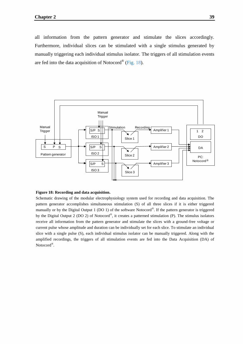
Chapter 2 39
Figure 18: Recording and data acquisition.
Schematic drawing of the modular electrophysiology system used for recording and data acquisition. The
pattern generator accomplishes simultaneous stimulation (S) of all three slices if it is either triggered
manually or by the Digital Output 1 (DO 1) of the software Notocord®. If the pattern generator is triggered
by the Digital Output 2 (DO 2) of Notocord®, it creates a patterned stimulation (P). The stimulus isolators
receive all information from the pattern generator and stimulate the slices with a ground-free voltage or
current pulse whose amplitude and duration can be individually set for each slice. To stimulate an individual
slice with a single pulse (S), each individual stimulus isolator can be manually triggered. Along with the
amplified recordings, the triggers of all stimulation events are fed into the Data Acquisition (DA) of
Notocord®.
all information from the pattern generator and stimulate the slices accordingly.
Furthermore, individual slices can be stimulated with a single stimulus generated by
manually triggering each individual stimulus isolator. The triggers of all stimulation events
are fed into the data acquisition of Notocord® (Fig. 18).
Pattern generatorPC:
Notocord®
Slice 1
Amplif ier 1
Amplif ier 2
Amplif ier 3
S
S/P
S
Manual
Trigger
P
ISO 1
S
S/P
ISO 2
S
S/P
ISO 3
S
Manual
Trigger
Slice 2
Slice 3
Stimulation Recording
DO
DA
1 2
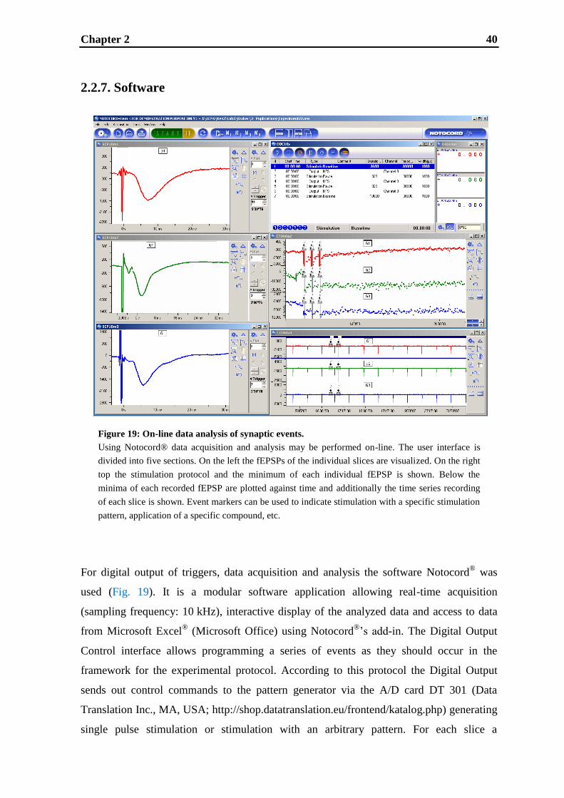
Chapter 2 40
2.2.7. Software
For digital output of triggers, data acquisition and analysis the software Notocord® was
used (Fig. 19). It is a modular software application allowing real-time acquisition
(sampling frequency: 10 kHz), interactive display of the analyzed data and access to data
from Microsoft Excel® (Microsoft Office) using Notocord
®‟s add-in. The Digital Output
Control interface allows programming a series of events as they should occur in the
framework for the experimental protocol. According to this protocol the Digital Output
sends out control commands to the pattern generator via the A/D card DT 301 (Data
Translation Inc., MA, USA; http://shop.datatranslation.eu/frontend/katalog.php) generating
single pulse stimulation or stimulation with an arbitrary pattern. For each slice a
Figure 19: On-line data analysis of synaptic events.
Using Notocord® data acquisition and analysis may be performed on-line. The user interface is
divided into five sections. On the left the fEPSPs of the individual slices are visualized. On the right
top the stimulation protocol and the minimum of each individual fEPSP is shown. Below the
minima of each recorded fEPSP are plotted against time and additionally the time series recording
of each slice is shown. Event markers can be used to indicate stimulation with a specific stimulation
pattern, application of a specific compound, etc.
Figure 20: On-line data analysis of synaptic events.
Using Notocord® data acquisition and analysis may be performed on-line. The user interface is
divided into five sections. On the left the fEPSPs of the individual slices are visualised. On the
right top the stimulation protocol and the minimum of each individual fEPSP is shown. Below the
minima of each recorded fEPSP are plotted against time and additionally the time series recording
of each slice is shown. Event markers can be used to indicate stimulation with a specific
stimulation pattern, application of a specific compound, etc.
Chapter 2 41
continuous display is shown and additional modules allow display of fEPSPs in a triggered
oscilloscope-like manner. A digital filter removes spikes or high frequency noise. The
minimum of the fEPSPs is analyzed in real-time and shown in a digital window as well as
plotted on a graph.
2.2.8. UV-spectrometry and drug exposure
To assess the effect of a drug on LTP, the knowledge whether the drug reaches the slices in
its intended concentration is indispensable, especially in pharmacological research. In this
study, UV-spectrometry (at a wavelength of 290 nm) was used to verify exposure to an
agent reducing the AMPA response (1 μM CNQX, Sigma–Aldrich Corporation, St. Louis,
USA). Directly after leaving each integrated brain slice chamber the perfusion solution was
sampled and analyzed in a UV spectrophotometer (Smartline 2500, Knauer Advanced
Scientific Instruments, Berlin, Germany).

Chapter 2 42
2.3. Results
2.3.1. Effects of the stacked grids
Extracellular recordings of evoked synaptic activity in the CA1 region of rat hippocampal
slices were utilized to assess whether the two stacked grids in the integrated brain slice
chambers enhance the stability of the system making recordings for several hours possible.
For comparison, fEPSPs were elicited from slices supported by the original single grid and
from slices positioned on the two stacked grids. Fig. 20A shows recordings from three
slices for each condition. fEPSP recordings from slices positioned on the stacked grids
were found to be much more stable than the ones from slices positioned on the original
single grid. The recordings were terminated after seven hours, although the fEPSPs were
still found to be of consistent amplitude. Statistical analysis comparing the average squared
deviations of both groups to the averages of the baselines, shows that the average squared
deviation from slices recorded on stacked grids is significantly lower than the one from
slices recorded on single grids (p < 0.001).
To determine whether this low variability of the system allows detection of small
differences between two groups and hence provides a high statistical power for
experiments, LTP recordings (stimulation protocol: 1 s 100 Hz) of slices positioned either
on the original single grid or on the two stacked grids were made. For each condition one
group of slices (n = 6) was recorded for control and the other group (n = 6) was recorded
under the influence of 0.1 μM of the NMDA-receptor antagonist MK-801 (Fig. 20B).
Comparison of the two groups recorded on the original single grid (Fig. 20Bi) shows no
significant effect (2 way-ANOVA, Dunnett's Test: not significant), whereas comparison of
the two groups recorded on the two stacked grids (Fig. 20Bii) shows a significant effect
(2 way-ANOVA, Dunnett's Test: *p < 0.05). These results can be reproduced by a
statistical analysis using two group t-test (group 1 mean = μ1; group 2 mean = μ2; test
significance level α = 0.05, two sided test) of equal means (equal n„s). This analysis
indicates, that with the two stacked grids used in this study, a significant difference in
means of 20% can be detected with recordings of only six slices (difference in means
(μ1 − μ1) = 20, common standard deviation (σ) = 10, effect size (δ = |μ1 − μ2|/σ) = 2.0,
power (%) = 80; n per group = 6). Hence, due to the two stacked grids data variability is
reduced and therefore a smaller number of slices is required to achieve statistical
significance.
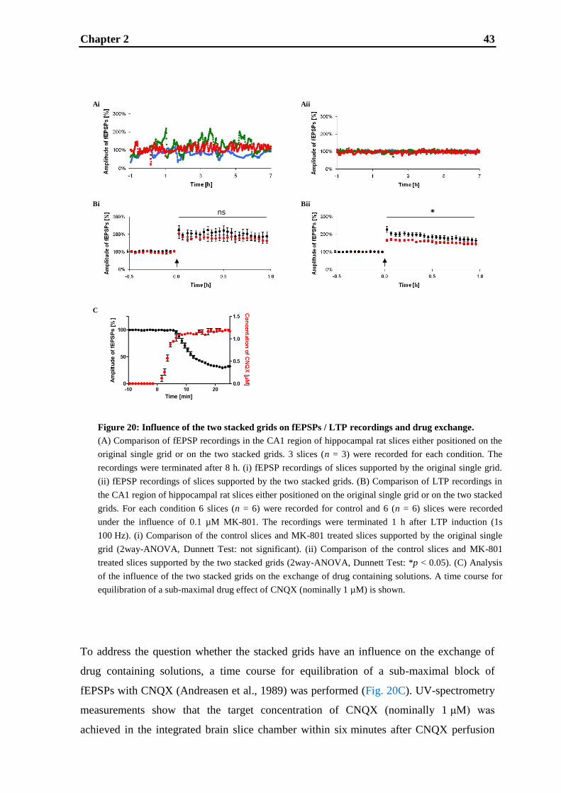
Chapter 2 43
To address the question whether the stacked grids have an influence on the exchange of
drug containing solutions, a time course for equilibration of a sub-maximal block of
fEPSPs with CNQX (Andreasen et al., 1989) was performed (Fig. 20C). UV-spectrometry
measurements show that the target concentration of CNQX (nominally 1 μM) was
achieved in the integrated brain slice chamber within six minutes after CNQX perfusion
Figure 20: Influence of the two stacked grids on fEPSPs / LTP recordings and drug exchange.
(A) Comparison of fEPSP recordings in the CA1 region of hippocampal rat slices either positioned on the
original single grid or on the two stacked grids. 3 slices (n = 3) were recorded for each condition. The
recordings were terminated after 8 h. (i) fEPSP recordings of slices supported by the original single grid.
(ii) fEPSP recordings of slices supported by the two stacked grids. (B) Comparison of LTP recordings in
the CA1 region of hippocampal rat slices either positioned on the original single grid or on the two stacked
grids. For each condition 6 slices (n = 6) were recorded for control and 6 (n = 6) slices were recorded
under the influence of 0.1 µM MK-801. The recordings were terminated 1 h after LTP induction (1s
100 Hz). (i) Comparison of the control slices and MK-801 treated slices supported by the original single
grid (2way-ANOVA, Dunnett Test: not significant). (ii) Comparison of the control slices and MK-801
treated slices supported by the two stacked grids (2way-ANOVA, Dunnett Test: *p < 0.05). (C) Analysis
of the influence of the two stacked grids on the exchange of drug containing solutions. A time course for
equilibration of a sub-maximal drug effect of CNQX (nominally 1 µM) is shown.
ns *
Ai Aii
Bii
C
Bi
FIGURE 21

Chapter 2 44
had started. Interestingly, the decrease of fEPSPs shows a five minutes delay after CNQX
containing ACSF starts to perfuse the slice with respect to drug concentration and the
equilibration is reached 15 minutes later. Therefore, the rate-limiting step is the penetration
of the slice and the pharmacological action and not the perfusion itself.
2.3.2. Long-term potentiation
To address whether this customized system, upgraded with the PPS, the two stacked grids
and the pattern generator, allows investigations of synaptic plasticity, LTP was generated
in the CA1 region of rat hippocampal slices. As a baseline recording, fEPSPs were elicited
at an intensity evoking 20–30% of the maximum response for one hour. Subsequently,
different stimulation protocols created by the pattern generator were used to induce LTP: a
weak theta burst stimulation, a strong theta burst stimulation, a weak high frequency
stimulation, an intermediate high frequency stimulation, a strong high frequency
stimulations and a repeated high frequency stimulation. All stimulation protocols used
were found to potentiate fEPSP (Fig. 21). After weak theta burst stimulation, synaptic
transmission increased up to a maximum of 150% and lasted about one hour (early phase
LTP; see Reymann and Frey, 2007) (Fig. 21A). Strong theta burst stimulation potentiated
fEPSPs up to a maximum of 200% lasting about 3.5 hours (late phase LTP; see Reymann
and Frey, 2007) (Fig. 21B). Synaptic transmission was enhanced to a maximum of 180%
lasting about two hours after weak high frequency stimulation (early phase LTP)
(Fig. 21C). Intermediate high frequency stimulation induced fEPSPs potentiation of a
maximal effect of 190% lasting for about three hours (presumably late phase LTP)
(Fig. 21D). The conventional strong high frequency stimulation caused an enhancement of
synaptic transmission up to 200% lasting longer than four hours (late phase LTP)
(Fig. 21E). The strongest fEPSP potentiation was generated by the repeated high frequency
stimulation having a maximal effect of 300% and lasting longer than seven hours (late
phase LTP) (Fig. 21F). For all protocols used, only little variability in the fEPSPs was
observed. In summary, very robust LTP may be induced by stimulation with a specific
pattern created by the pattern generator and maintained using the PPS in combination with
the two stacked grids.
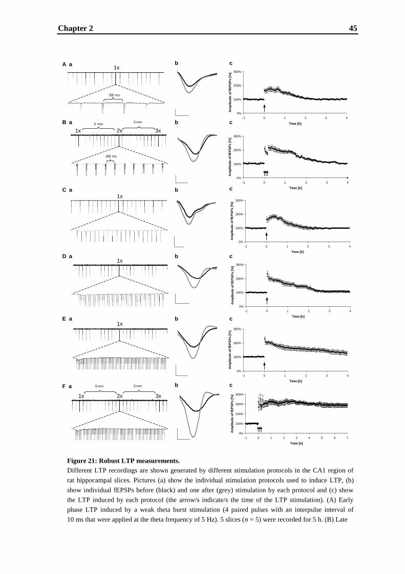
Chapter 2 45
Figure 21: Robust LTP measurements.
Different LTP recordings are shown generated by different stimulation protocols in the CA1 region of
rat hippocampal slices. Pictures (a) show the individual stimulation protocols used to induce LTP, (b)
show individual fEPSPs before (black) and one after (grey) stimulation by each protocol and (c) show
the LTP induced by each protocol (the arrow/s indicate/s the time of the LTP stimulation). (A) Early
phase LTP induced by a weak theta burst stimulation (4 paired pulses with an interpulse interval of
10 ms that were applied at the theta frequency of 5 Hz). 5 slices (n = 5) were recorded for 5 h. (B) Late
0%
100%
200%
300%
-1 0 1 2 3 4
Time [h]
Am
plitu
de
of
fEP
SP
s [
%]
0%
100%
200%
300%
-1 0 1 2 3 4
Time [h]
Am
plitu
de
of
fEP
SP
s [
%]
0%
100%
200%
300%
-1 0 1 2 3 4
Time [h]
Am
plitu
de
of
fEP
SP
s [
%]
0%
100%
200%
300%
-1 0 1 2 3 4
Time [h]
Am
plitu
de
of
fEP
SP
s [
%]
0%
100%
200%
300%
-1 0 1 2 3 4
Time [h]
Am
plitu
de
of
fEP
SP
s [
%]
0%
100%
200%
300%
-1 0 1 2 3 4
Time [h]
Am
plitu
de
of
fEP
SP
s [
%]
0%
100%
200%
300%
-1 0 1 2 3 4
Time [h]
Am
plitu
de
of
fEP
SP
s [
%]
0%
100%
200%
300%
-1 0 1 2 3 4
Time [h]
Am
plitu
de
of
fEP
SP
s [
%]
0%
100%
200%
300%
400%
-1 0 1 2 3 4 5 6 7
Time [h]
Am
plitu
de
of
fEP
SP
s [
%]
1x
1x 2x 3x
1x
1x
1x
1x 2x 3x
5 min5 min
5 min 5 min
200 ms
200 ms
A a
B a
C a
D a
E a
F a
b
b
b
b
b
b
c
c
c
c
c
c

Chapter 2 46
continued:
phase LTP induced by a strong theta burst stimulation (10 times 4 pulses with an interpulse interval of
10 ms and an interval between bursts of 200 ms, repeated 2 times in 10 min intervals). 5 slices (n = 5)
were recorded for 5 h. (C) Early phase LTP induced by a weak high frequency stimulation (20 pulses at
the frequency of 100 Hz). 5 slices (n = 5) were recorded for 5 h. (D) Presumably late phase LTP induced
by an intermediate high frequency stimulation (50 pulses at the frequency of 100 Hz). 5 slices (n = 5)
were recorded for 5 h. (E) Late phase LTP induced by the conventional strong high frequency stimulation
(100 pulses at the frequency of 100 Hz). 5 slices (n = 5) were recorded for 5 h. (F) Late phase LTP
induced by a repeated high frequency stimulation (100 pulses at the frequency of 100 Hz, repeated 2
times in 5 min intervals). 5 slices (n = 5) were measured for 8 h.

Chapter 2 47
2.4. Discussion
In this study, we describe the modification of a multi-slice recording system that allows
absolutely stable measurements of fEPSPs, enabling recording of LTP for up to seven
hours in up to eight slices simultaneously. This system overcomes the main hurdles for
efficient routine LTP measurements: (1) system instability, (2) low throughput and (3)
incomplete use of slices per animal.
A multi-slice recording system (SliceMaster, Scientifica Limited, East Sussex, UK) was
modified in several ways. Firstly, the software Notocord® was employed. So far, in
combination with the SliceMaster only Spike2 (Stopps et al., 2004; West et al., 2009;
www.ced.co.uk) had been used. Among these software other programs can be utilized for
LTP experiments (for an overview see Bortolotto et al., 2001) including the ones which
need no programming - for example Molecular Devices pClamp
(www.moleculardevices.com/pages/software/pclamp.html), Heka's Patch-Master
(www.heka.com/physio/acquisition/patchmaster.html), AxoGraph (www.axographx.com),
NClamp (/www.neuromatic.thinkrandom.com), Theta Burst's NAC Gather
(www.thetaburst.com), the Strathclyde Electrophysiology Software program WinWCP
(spider.science.strath.ac.uk/sipbs/page.php?show=software_winWCP), the LTP Program
(Anderson and Collingridge, 2001; www.winltp.com/Ltp24/indexLtp24.htm) and WinLTP
(Anderson and Collingridge, 2007; www.winltp.com) - and the ones which are customized
- for example written with Wave-Metrics‟ Igor (www.wavemetrics.com) or National
Instruments‟ LabView (www.ni.com). In this study, Notocord® was used, since it may be
individually configured to acquire a multitude of biological signals, analyze them and
create a report quickly. Furthermore, it is often employed in industry, because it is able to
generate a protocol of the users‟ actions. In this report, Notocord® was not only used to
visualize generated data files on-line or later, but also to control a digital pattern generator
which generates different patterns for slice stimulation inducing different types of LTP.
Secondly, a Pumped Perfusion System (PPS) was used. In contrast to gravity-fed systems
(Stopps et al., 2004; Easter et al., 2007), where flow is a subject to changes in solution
height, the PPS controls the flow precisely over the whole course of the experiment. The
very low noise electronics of the PPS allows its placing inside the Faraday cage and hence
results in a reduction of solution dead volumes. Additionally, the PPS can be used to
recycle drug solutions minimizing amount of drugs applied to the slices. Thirdly, the
original single grids in the isolated slice chambers were replaced by two stacked grids. The
Chapter 2 48
two stacked grids were shown not to delay the pharmacological effect of CNQX and make
the flow of ACSF very stable, hence allowing fEPSP recordings for several hours without
any perturbances. This stability enables very robust LTP measurements for up to seven
hours depending on the stimulation protocol used. Furthermore, it allows detection of a
statistically significant differences of 20% between two groups using only six slices (n per
group = 6), whereas as described in literature an average of ten slices (n per group = 10) is
necessary (Schröder et al., 2008; van der Staay et al., 2008; Rosenbrock et al., 2010).
This modified multi-slice system not only allows measuring LTP for up to seven hours and
improves statistical design, but also increases throughput and the rate of used slices per
animal. The throughput is increased as up to eight slices per animal may be studied
simultaneously permitting numerous controls or drug treatments to be performed in one
experimental run. Conventional brain slice systems only permit recording from one or two
brain slices at a time (Haas et al., 1979), even though it is often possible to prepare eight
hippocampal slices (of 400 μm thickness) from a single rat. Hence, six or seven slices are
not used for experiments. Since eight slices can be studied simultaneously with the multi-
slice system, the number of unused slices is decreased and thereby the number of animals
necessary to conduct a certain number of experiments is reduced. Therefore, this modified
system helps researchers comply with the “three Rs” (Flecknell, 2002), requested by many
sides with increasing intensity.
In conclusion, as the modified multi-slice system used in this study allows LTP recordings
for up to seven hours in up to eight slices simultaneously, it is an attractive approach for
routine late phase LTP measurements when high statistical power is needed. Thus, this
system is both suitable to investigate molecular mechanisms underlying the late phase of
LTP and it can be utilized to screen candidate compounds in the context of drug discovery.
Chapter 3 49
CHAPTER 3
Validation of early and late LTP and the effect of
the mGlu5 receptor positive allosteric modulator
ADX-47273
Chapter 3 50
Abstract
Conflicting findings are reported in the literature about the involvement of the mGlu5
receptor in hippocampal long-term potentiation (LTP), which might be a consequence of
different sub-types of LTP induced by the investigators due to the specific experimental
conditions used. A comparable controversy came up in the past concerning the influence of
different experimental conditions on the involvement of L-type voltage dependent calcium
channels (L-VDCCs) and NMDA receptors in hippocampal LTP. In this study, two
stimulation protocols under identical conditions were used to probe modulatory effects of
mGlu5 receptor activation in NMDA receptor and L-VDCCs dependent CA1 LTP: weak
high frequency stimulation (20 stimuli at 100 Hz) to induce early LTP and repeated strong
high frequency stimulation (3 times 100 stimuli at 100 Hz with 5 minutes interval) to
induce late LTP, which - in contrast to early LTP - was shown to be protein-synthesis
dependent. Using the NMDA receptor antagonist MK-801 and the L-type calcium channel
blocker nifedipine, early LTP was shown to be dependent on NMDA receptors only,
whereas late LTP was demonstrated to be dependent on NMDA receptors and L-VDCCs in
about equal parts. Moreover, late LTP, but not early LTP, was increased by the mGlu5
receptor positive allosteric modulator ADX-47273, indicating that artificial augmentation
of mGlu5 receptor activation by endogenous glutamate may boost the protein-synthesis
dependent form of LTP but not the protein-synthesis independent form.

Chapter 3 51
3.1. Introduction
Metabotropic glutamate 5 (mGlu5) receptors are expressed throughout the CNS including
the cortex and the hippocampus (Abe et al., 1992; Kerner et al., 1997; Lujan et al., 1996;
Mannaioni et al., 2001; Shigemoto et al., 1997; Spooren et al., 2003), brain structures
known to be intrinsically involved in learning and memory. The role of the mGlu5 receptor
in learning and memory has been investigated in several studies addressing long-term
potentiation (LTP) of synaptic transmission (Anwyl, 2009), which is a fundamental
experimental model of mammalian learning and memory (Bliss and Collingridge, 1993).
However, conflicting reports exist about the role of the mGlu5 receptor in hippocampal
LTP even to the point that some studies indicate that mGlu5 receptors are not important for
LTP at all (Anwyl, 1999). For example, studies in mutant mice showing that CA1 LTP
could be readily induced in mGlu5 receptor (−/+), mGlu5 receptor (−/−) or mGlu1/mGlu5
receptor double knockout mice (Bortolotto et al., 2005). In contrast, other studies have
demonstrated a prominent role of the mGlu5 receptor in LTP. Indeed, the mGlu5 receptor
antagonist MPEP was found to block LTP in the CA1 region of hippocampal slices
(Francesconi et al., 2004; Shalin et al., 2006) as well as to lead to an impairment in
hippocampal LTP in vivo (Manahan-Vaughan and Braunewell, 2005), whereas the
mGlu1/5 receptor agonist DHPG was shown to facilitate induction of CA1 LTP (Cohen et
al., 1998; Raymond et al., 2000). Likewise, mGlu5 receptor knockout mice showed an
impairment of LTP in the CA1 region and exhibited deficits in memory tasks (Jia et al.,
1998; Lu et al., 1997). It was suggested that these apparently controversial findings
regarding mGlu5 receptor‟s involvement in hippocampal CA1 LTP were due to the
examination of different types of LTP employed by the experimenters (Anwyl, 2009).
Interestingly, in the past a similar reasoning was made regarding the role of L-type voltage
dependent calcium channels (L-VDCCs) and NMDARs in LTP (Grover and Teyler, 1990;
Morgan and Teyler, 2001).
In the CA1 region of the hippocampus, a general differentiation can be made between an
early phase of LTP („early LTP‟) being induced by a ‟weak‟ stimulation and a late phase of
LTP (‟late LTP‟) being induced by a ‟strong‟ stimulation (Albensi et al., 2007). Early LTP
has been found to be independent of protein-synthesis, lasts between one and three hours
and it was shown to rely on post-translational modifications of preexisting proteins
(Huang, 1998; Reymann and Frey, 2007). Late LTP, on the other hand, has been found to
be protein-synthesis dependent as demonstrated by use of inhibitors of protein transcription
Chapter 3 52
as well as translation. This type of LTP lasts longer than three hours (Huang, 1998;
Reymann and Frey, 2007). However, in late LTP protein-synthesis already contributes to
potentiation at a point in time at which protein-synthesis independent potentiation is still
present in an early LTP paradigm (Frey et al., 1988; Krug et al., 1984).
Clearly defined and validated protocols for the induction of early and late LTP on a single
experimental platform under identical conditions are essential to test whether a certain
receptor might have differential roles concerning the two types of LTP. Therefore, two
protocols were examined in detail: weak high frequency stimulation (weak HFS; 20 stimuli
at 100 Hz) to induce early LTP and repeated strong high frequency stimulation (repeated
strong HFS; 3 times 100 stimuli at 100 Hz with 5 minutes interval) to induce late LTP. We
analyzed not only the duration and the protein-synthesis dependency, but also examined
the role of NMDA receptors and L-VDCCs on early and late LTP using their specific
antagonists MK-801 and nifedipine, respectively. The mGlu5 receptor positive allosteric
modulator ADX-47273 was used to test whether artificial augmentation of mGlu5 receptor
activation by endogenous glutamate during LTP induction may influence the expression of
the two basic forms of CA1 LTP.
Chapter 3 53
3.2. Materials and methods
3.2.1. Preparation of brain slices
Procedures involving animals and their care were conducted in conformity with
institutional and European Union guidelines (EEC Council Directive 86/609) and were
approved by the Ethical Committee of the responsible regional council (Tübingen). Male
Wistar rats (Janvier, Le Genest Saint Isle, France) aged six to seven weeks were shortly
anaesthetized with isoflurane and sacrificed by decapitation. The brains were quickly
removed and immersed in ice-cold ACSF (containing in mM NaCl 124, KCl 4.9, MgSO4
8, CaCl2 2.5, KH2PO4 1.2, NaHCO3 25.6, D-glucose 10, pH 7.4, saturated with 95% O2
and 5% CO2). Transverse hippocampal brain slices (400 μm thickness) were cut using a
Vibratome. Slices were allowed to recover in a holding chamber containing ACSF bubbled
with 95% O2 and 5% CO2 at room temperature for at least one hour. Slices were then
transferred to integrated brain slice chambers and continuously superfused (at a flow rate
of 2.5 mL min−1
, 25 ± 0.2 °C) with the same ACSF composition used before except for
0.5 mM MgSO4. Prior to doing any electrophysiological recordings, the slices were
allowed to equilibrate for at least 30 minutes.
3.2.2. Multi brain slice recording
The SliceMaster multi-slice recording system (Scientifica Limited, East Sussex, UK) was
used to record field excitatory postsynaptic potentials (fEPSPs). The system is semi-
automated allowing one operator to record simultaneously from an assembly of up to eight
brain slices. For the present study, an optimized system described by Kroker et al. (2011)
was used which enables absolutely stable fEPSPs measurements for up to eight hours.
Briefly, the SliceMaster system consists of three temperature-controlled integrated brain
slice chambers and can be upgraded with five more. Each chamber is equipped with one
stimulation electrode, one recording electrode and a camera system allowing a
visualization of each brain slice on a shared video monitor. A central control panel makes
the positioning of all electrodes and cameras possible, allowing a single operator to run all
experiments simultaneously. A separate inflow using a Pumped Perfusion System
(Scientifica Limited, East Sussex, UK) and separate outflow for each integrated brain slice

Chapter 3 54
chamber allows individual perfusion of each brain slice. To minimize movement of the
slices, stacked grids were integrated in each brain slice chamber.
fEPSPs were elicited in the CA1 region by stimulation of the Schaffer collateral–
commissural fibres in the stratum radiatum using glass electrodes with broken tips (filled
with ACSF). For recording, glass electrodes (2–6 MΩ, filled with ACSF) were placed in
the apical dendritic layer. The amplitudes of fEPSPs were used as the parameter of interest
(Collingridge et al., 1983). To generate fEPSPs at a constant sub-threshold stimulus, as in
previous studies (Seabrook et al., 1997), the stimulus strength of the pulses was adjusted to
20–30% of the fEPSP maximum and this voltage was used for the experiment. During
baseline recording each slice was stimulated every 30 seconds for at least one hour. Early
LTP was induced by weak HFS made up of 20 pulses at the frequency of 100 Hz (Bashir et
al., 1991; Kroker et al., 2011), whereas late LTP was induced by repeated strong HFS
consistent of 100 pulses at the frequency of 100 Hz, repeated two times in 5 minutes
intervals (Kroker et al., 2011; Lu et al., 1999).
3.2.3. Data acquisition, software and analysis
A modular electrophysiology system, supplied by npi electronic GmbH (Tamm, Germany),
conducts the low noise recordings of extracellular signals. AC coupled signals are
amplified 1000-fold and internally filtered with a five kHz low-pass filter as well as a three
Hz high-pass filter. In our study, a customized system (programmable pattern generator)
was used allowing simultaneous stimulation of all slices either with a single stimulus or a
specific stimulus pattern (Kroker et al., 2011). For data acquisition and analysis the
software Notocord® was used.
Data are shown as mean percent (± SEM) of the baseline fEPSP amplitude. Data were
analyzed using either t tests to compare two conditions or one-way ANOVA (post-hoc test:
Dunnett-Test) to compare multiple conditions. In both cases a p-value of ≤ 0.05 was
considered significant. Histograms show the mean amplitude (± SEM) of EPSPs measured
between 50 and 60 minutes after weak or repeated strong HFS according to Jia et al.
(2010).

Chapter 3 55
3.2.4. Drug solutions and application
The following drugs were used: 6-Cyano-7-nitroquinoxaline-2,3-dione (= CNQX),
(5S,10R)-(–)-5-Methyl-10,11-dihydro-5H-dibenzo[a,d]cyclohepten-5,10-imine hydrogen
maleate (= MK-801), (2R,3S,4S)-2-(4-Methoxybenzyl)-3,4-pyrrolidinediol-3-acetate, 2-
[(4-Methoxyphenyl)methyl]-3,4-pyrrolidinediol 3-acetate (= anisomycin) and 1,4-Dihydro-
2,6-dimethyl-4-(2-nitrophenyl)-3,5-pyridinedicarboxylic acid dimethyl ester (= nifedipine)
were purchased from Sigma-Aldrich Corporation (St. Louis, USA). The compound S-(4-
fluoro-phenyl)-{3-[3-8fluorophenyl)-[1,2,4]-oxadiazol-5-yl]-piperidin-1-yl}-methanone
(= ADX-47273) was synthesized in the Department of Chemical Research of Boehringer
Ingelheim Pharma GmbH & Co KG.
The drugs were prepared as stock solutions and diluted in ACSF immediately before
application. Nifedipine and ADX-47273 were prepared in DMSO (the final concentration
of DMSO was 0.1%). All drugs, except MK-801, were applied 30 minutes before LTP
stimulation and remained until 30 minutes after LTP stimulation. MK-801 was used in
such a way that, corresponding to Frankiewicz et al. (1996), true equilibrium blockade of
NMDA receptors can be expected: MK-801 was present immediately after preparing
slices, thereafter for an average of 6.8 ± 0.4 h prior to LTP stimulation and remained until
30 minutes after LTP stimulation.
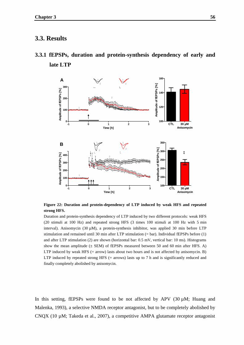
Chapter 3 56
Figure 22: Duration and protein-dependency of LTP induced by weak HFS and repeated
strong HFS.
Duration and protein-synthesis dependency of LTP induced by two different protocols: weak HFS
(20 stimuli at 100 Hz) and repeated strong HFS (3 times 100 stimuli at 100 Hz with 5 min
interval). Anisomycin (30 µM), a protein-synthesis inhibitor, was applied 30 min before LTP
stimulation and remained until 30 min after LTP stimulation (= bar). Individual fEPSPs before (1)
and after LTP stimulation (2) are shown (horizontal bar: 0.5 mV, vertical bar: 10 ms). Histograms
show the mean amplitude (± SEM) of fEPSPs measured between 50 and 60 min after HFS. A)
LTP induced by weak HFS (= arrow) lasts about two hours and is not affected by anisomycin. B)
LTP induced by repeated strong HFS (= arrows) lasts up to 7 h and is significantly reduced and
finally completely abolished by anisomycin.
-1 0 1 2 30
100
200
300
Time [h]
Am
plit
ud
e o
f fE
PS
Ps [
%]
CTL 30 µM100
120
140
160
Am
plitu
de o
f fE
PS
Ps [
%]
Anisomycin
-1 0 1 2 30
100
200
300
400
Time [h]
Am
plit
ud
e o
f fE
PS
Ps [
%]
CTL 30 µM100
150
200
250
300
350
Am
plitu
de o
f fE
PS
Ps [
%]
Anisomycin
A
1
1
2
1
2
B
1
1
2
1
2
2
2
**
3.3. Results
3.3.1 fEPSPs, duration and protein-synthesis dependency of early and
late LTP
In this setting, fEPSPs were found to be not affected by APV (30 µM; Huang and
Malenka, 1993), a selective NMDA receptor antagonist, but to be completely abolished by
CNQX (10 µM; Takeda et al., 2007), a competitive AMPA glutamate receptor antagonist
Chapter 3 57
(data not shown). Thus, AMPA receptors are exclusively responsible for the basal fEPSPs
generated in this setting.
LTP induced by weak HFS lasts about one and a half to two hours (Fig. 22A), whereas
LTP induced by repeated strong HFS lasts longer than three hours (Fig. 22B). The
classification of LTP into different phases is not sufficiently clear based on the duration
only, but also on protein-synthesis dependency. Therefore, the effect of anisomycin, a
reversible protein translation inhibitor, was analyzed in both settings. Anisomycin was
used at a concentration of 30 µM, a concentration reported to be sufficient to significantly
impair protein-synthesis (Lu et al., 1999). It had no effect on LTP induced by weak HFS
(= early LTP; Fig. 22A) (one hour after weak HFS; control: 141 ± 3%, n = 10; anisomycin:
145 ± 3%, n = 5, not significant (ns)). In contrast, LTP induced by repeated strong HFS
was significantly decreased up to a complete abolishment (= late LTP; Fig. 22B) (one hour
after repeated strong HFS; control: 306 ± 10%, n = 10; anisomycin: 236 ± 11%, n = 5,
p < 0.01).
3.3.2 Involvement of NMDA receptors and L-VDCCs in the induction of
early and late LTP
To assess the role of NMDA receptors in early LTP generated by weak HFS, the effect of
MK-801, a potent antagonist of NMDA receptors, was analyzed (Fig. 23A). Early LTP
was decreased by MK-801 (0.1 – 100 µM) in a concentration dependent manner with a
complete blockade at a concentration of 10 µM (one hour after weak HFS; control:
138 ± 6%, n = 10; 0.1 µM MK-801: 136 ± 7%, n = 5, ns; 1 µM MK-801: 113 ± 5%, n = 5,
p < 0.01; 10 µM MK-801: 100 ± 2%, n = 5, p < 0.001; 100 µM MK-801: 100 ± 1%, n = 5,
p < 0.001). To assess the role of L-VDCCs in LTP, the L-type calcium channel blocker
nifedipine (Grover and Teyler, 1990) was applied at different concentrations (0.1 - 500
µM). As shown in Fig. 23B, nifedipine had no effect on early LTP (one hour after weak
HFS; control: 137 ± 5%, n = 10; 0.1 µM nifedipine: 137 ± 3%, n = 5, ns; 1 µM nifedipine:
140 ± 5%, n = 5, ns; 10 µM nifedipine: 139 ± 3%, n = 5, ns; 100 µM nifedipine: 138 ± 5%,
n = 5, ns; 500 µM nifedipine: 139 ± 4%, n = 5, ns).

Chapter 3 58
Figure 23: Role of NMDA receptor and L-VDCC in early LTP.
Effects of MK-801, a potent antagonist of NMDA receptors, and nifedipine, an L-type calcium
channel blocker, on early LTP induced by weak HFS. A) Early LTP was reduced by MK-801 in a
concentration dependent manner with a complete abolishment at 10 µM. MK-801 (0.1 – 100 µM)
was applied 6.8 ± 0.4 h before LTP stimulation (= arrow) and was washed out 30 min after LTP
stimulation (= bar). Individual fEPSPs before (1) and after LTP stimulation (2) are shown
(horizontal bar: 0.5 mV, vertical bar: 10 ms). Histogram shows the (mean ± SEM) amplitude of
fEPSPs measured 50–60 min after delivery of LTP stimulation. B) Nifedipine had no effect on
early LTP. Nifedipine (0.1 – 500 µM) was applied 30 min before LTP stimulation (= arrow) and
remained until 30 min after LTP stimulation (= bar). Histogram shows the mean amplitude
(± SEM) of EPSPs measured between 50 and 60 min after HFS.
-1 0 1 2 30
100
200
300
Time [h]
Am
plit
ud
e o
f fE
PS
Ps [
%]
0 0.1 1 10 100100
120
140
160
Am
plitu
de o
f fE
PS
Ps [
%]
µM MK-801
-1 0 1 2 30
100
200
300
Time [h]
Am
plit
ud
e o
f fE
PS
Ps [
%]
0 0.1 1 10 100100
120
140
160
Am
plitu
de o
f fE
PS
Ps [
%]
500
µM Nifedipine
2
1
21 1 1 1
2 22
1
2
1 1
2 2 2 2 2 2
1 1 1 1
A
B
1
2
***
***
**
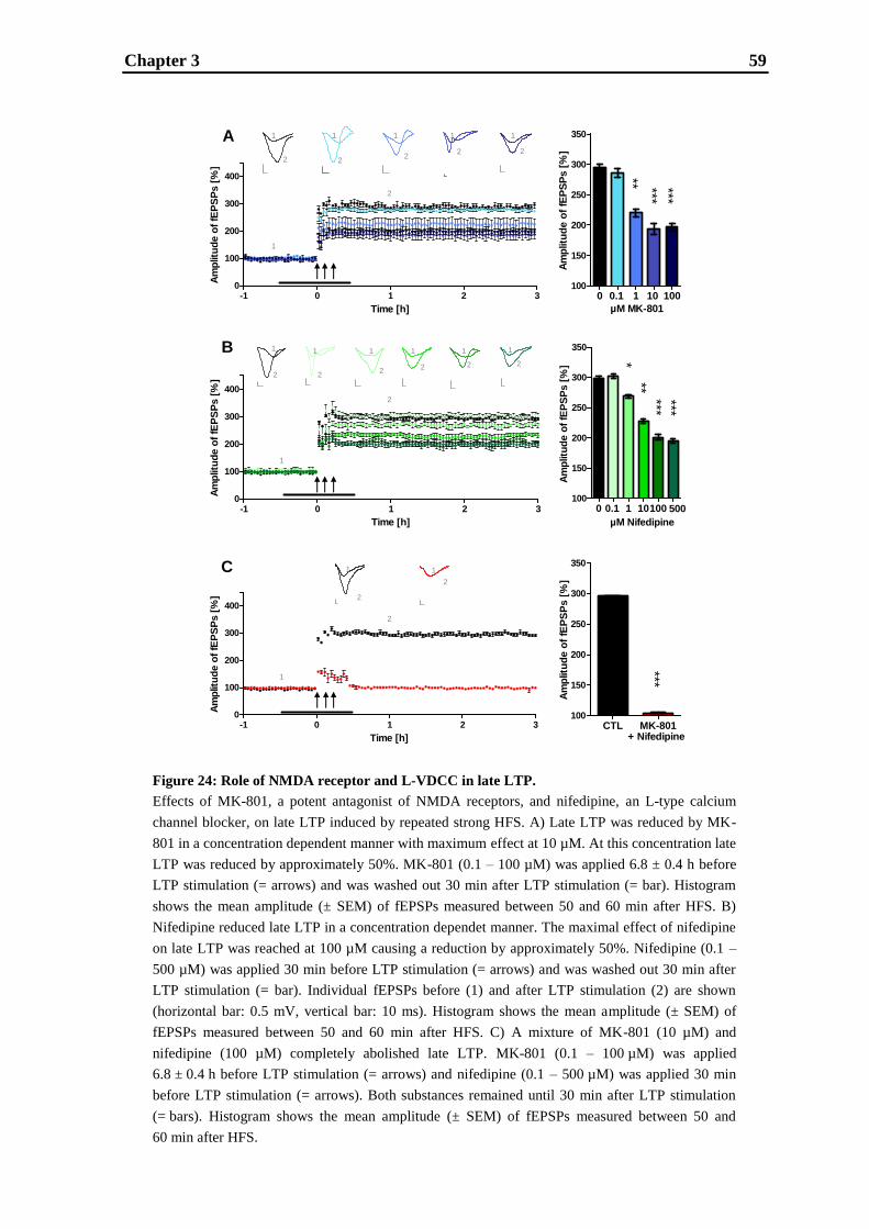
Chapter 3 59
Figure 24: Role of NMDA receptor and L-VDCC in late LTP.
Effects of MK-801, a potent antagonist of NMDA receptors, and nifedipine, an L-type calcium
channel blocker, on late LTP induced by repeated strong HFS. A) Late LTP was reduced by MK-
801 in a concentration dependent manner with maximum effect at 10 µM. At this concentration late
LTP was reduced by approximately 50%. MK-801 (0.1 – 100 µM) was applied 6.8 ± 0.4 h before
LTP stimulation (= arrows) and was washed out 30 min after LTP stimulation (= bar). Histogram
shows the mean amplitude (± SEM) of fEPSPs measured between 50 and 60 min after HFS. B)
Nifedipine reduced late LTP in a concentration dependet manner. The maximal effect of nifedipine
on late LTP was reached at 100 µM causing a reduction by approximately 50%. Nifedipine (0.1 –
500 µM) was applied 30 min before LTP stimulation (= arrows) and was washed out 30 min after
LTP stimulation (= bar). Individual fEPSPs before (1) and after LTP stimulation (2) are shown
(horizontal bar: 0.5 mV, vertical bar: 10 ms). Histogram shows the mean amplitude (± SEM) of
fEPSPs measured between 50 and 60 min after HFS. C) A mixture of MK-801 (10 µM) and
nifedipine (100 µM) completely abolished late LTP. MK-801 (0.1 – 100 µM) was applied
6.8 ± 0.4 h before LTP stimulation (= arrows) and nifedipine (0.1 – 500 µM) was applied 30 min
before LTP stimulation (= arrows). Both substances remained until 30 min after LTP stimulation
(= bars). Histogram shows the mean amplitude (± SEM) of fEPSPs measured between 50 and
60 min after HFS.
-1 0 1 2 30
100
200
300
400
Time [h]
Am
plitu
de o
f fE
PS
Ps [
%]
0 0.1 1 10100100
150
200
250
300
350
Am
plitu
de o
f fE
PS
Ps [
%]
500
µM Nifedipine
-1 0 1 2 30
100
200
300
400
Time [h]
Am
plit
ud
e o
f fE
PS
Ps [
%]
0 0.1 1 10 100100
150
200
250
300
350
Am
plitu
de o
f fE
PS
Ps [
%]
µM MK-801
-1 0 1 2 30
100
200
300
400
Time [h]
Am
plit
ud
e o
f fE
PS
Ps [
%]
CTL MK-801100
150
200
250
300
350A
mp
litu
de o
f fE
PS
Ps [
%]
+ Nifedipine
A
B
C
2
1 1 1 1 1
222 2
1 1 1 1 1 1
2 222
2 2
1
2
1
2
1 1
2
2
1
2
** *** ***
*** ***
***
***
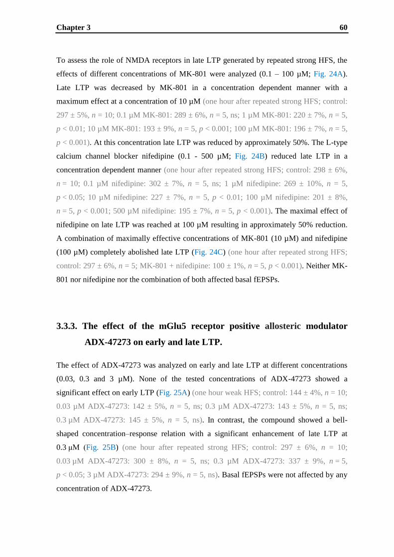
Chapter 3 60
To assess the role of NMDA receptors in late LTP generated by repeated strong HFS, the
effects of different concentrations of MK-801 were analyzed (0.1 – 100 µM; Fig. 24A).
Late LTP was decreased by MK-801 in a concentration dependent manner with a
maximum effect at a concentration of 10 µM (one hour after repeated strong HFS; control:
297 ± 5%, n = 10; 0.1 µM MK-801: 289 ± 6%, n = 5, ns; 1 µM MK-801: 220 ± 7%, n = 5,
p < 0.01; 10 µM MK-801: 193 ± 9%, n = 5, p < 0.001; 100 µM MK-801: 196 ± 7%, n = 5,
p < 0.001). At this concentration late LTP was reduced by approximately 50%. The L-type
calcium channel blocker nifedipine (0.1 - 500 µM; Fig. 24B) reduced late LTP in a
concentration dependent manner (one hour after repeated strong HFS; control: 298 ± 6%,
n = 10; 0.1 µM nifedipine: 302 ± 7%, n = 5, ns; 1 µM nifedipine: 269 ± 10%, n = 5,
p < 0.05; 10 µM nifedipine: 227 ± 7%, n = 5, p < 0.01; 100 µM nifedipine: 201 ± 8%,
n = 5, p < 0.001; 500 µM nifedipine: 195 ± 7%, n = 5, p < 0.001). The maximal effect of
nifedipine on late LTP was reached at 100 µM resulting in approximately 50% reduction.
A combination of maximally effective concentrations of MK-801 (10 µM) and nifedipine
(100 µM) completely abolished late LTP (Fig. 24C) (one hour after repeated strong HFS;
control: 297 ± 6%, n = 5; MK-801 + nifedipine: 100 ± 1%, n = 5, p < 0.001). Neither MK-
801 nor nifedipine nor the combination of both affected basal fEPSPs.
3.3.3. The effect of the mGlu5 receptor positive allosteric modulator
ADX-47273 on early and late LTP.
The effect of ADX-47273 was analyzed on early and late LTP at different concentrations
(0.03, 0.3 and 3 µM). None of the tested concentrations of ADX-47273 showed a
significant effect on early LTP (Fig. 25A) (one hour weak HFS; control: 144 ± 4%, n = 10;
0.03 µM ADX-47273: 142 ± 5%, n = 5, ns; 0.3 µM ADX-47273: 143 ± 5%, n = 5, ns;
0.3 µM ADX-47273: 145 ± 5%, n = 5, ns). In contrast, the compound showed a bell-
shaped concentration–response relation with a significant enhancement of late LTP at
0.3 μM (Fig. 25B) (one hour after repeated strong HFS; control: 297 ± 6%, n = 10;
0.03 µM ADX-47273: 300 ± 8%, n = 5, ns; 0.3 µM ADX-47273: 337 ± 9%, n = 5,
p < 0.05; 3 µM ADX-47273: 294 ± 9%, n = 5, ns). Basal fEPSPs were not affected by any
concentration of ADX-47273.
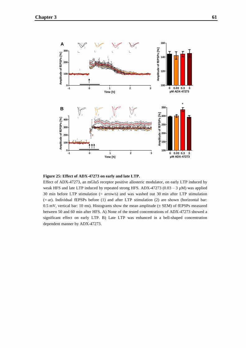
Chapter 3 61
-1 0 1 2 30
100
200
300
Time [h]
Am
plitu
de o
f fE
PS
Ps [
%]
0 0.03 0.3 3100
120
140
160
Am
plitu
de o
f fE
PS
Ps [
%]
µM ADX-47273
-1 0 1 2 30
100
200
300
400
Time [h]
Am
plit
ud
e o
f fE
PS
Ps [
%]
0 0.03 0.3 3100
150
200
250
300
350
Am
plitu
de o
f fE
PS
Ps [
%]
µM ADX-47273
A
B
2
1
2
1
2
2
1 1
2
1
1
2
1111
2
22
2
*
Figure 25: Effect of ADX-47273 on early and late LTP.
Effect of ADX-47273, an mGlu5 receptor positive allosteric modulator, on early LTP induced by
weak HFS and late LTP induced by repeated strong HFS. ADX-47273 (0.03 – 3 µM) was applied
30 min before LTP stimulation (= arrow/s) and was washed out 30 min after LTP stimulation
(= ar). Individual fEPSPs before (1) and after LTP stimulation (2) are shown (horizontal bar:
0.5 mV, vertical bar: 10 ms). Histograms show the mean amplitude (± SEM) of fEPSPs measured
between 50 and 60 min after HFS. A) None of the tested concentrations of ADX-47273 showed a
significant effect on early LTP. B) Late LTP was enhanced in a bell-shaped concentration
dependent manner by ADX-47273.

Chapter 3 62
3.4. Discussion
In vitro LTP measurements represent a fundamental experimental model of mammalian
learning and memory (Bliss and Collingridge, 1993) and have been widely used for
decades, however, certain limitations apply (Eichenbaum, 1996). Traditionally, different
labs employ slightly different conditions, implying limited comparability. However,
qualitative mechanistic statements usually have general validity. As it is known that the
details of the experimental conditions in LTP measurements are important (Larkman and
Jack, 1995), we made a point of setting up a standardized LTP setup (Kroker et al., 2011)
with the stimulation protocol being the only variable parameter. Thus, the results obtained
in this study are perfectly comparable, whereas alignment with other studies is warranted
only after careful comparison of experimental conditions.
3.4.1. Duration and protein-synthesis dependency of early and late LTP
In the present work, the capabilities of two different stimulation protocols to induce early
and late CA1 LTP were analyzed and validated by investigating the duration and protein-
synthesis dependency of both types of LTP. To analyze protein-synthesis dependency,
anisomycin was used, because it is known to successfully block late LTP (Ahmed and
Frey, 2005; Frey et al., 1988; Krug et al., 1984; Lu et al., 1999; Mullany and Lynch, 1997;
Reymann and Frey, 2007). LTP induced by weak HFS lasted about two hours and was
demonstrated not to be affected by anisomycin. In contrast, LTP induced by repeated
strong HFS lasted longer than three hours and was completely abolished within three hours
by anisomycin. Thus, LTP induced by weak HFS can be considered to be early LTP,
whereas LTP induced by repeated strong HFS is late LTP by definition. The discrimination
between early and late LTP by protein-synthesis dependency, as it is made here, is
commonly used and is based on the need for de novo protein-synthesis to establish LTP
that lasts longer than a few hours (Frey et al., 1988; Huang, 1998; Huang and Kandel,
1994; Huang et al., 1996; Nguyen et al., 1994; Nguyen and Kandel, 1997; Reymann and
Frey, 2007; Stanton and Sarvey, 1984). It is postulated that early LTP, which lasts for up to
maximum three hours, relies only on post-translational modifications of pre-existing
proteins, whereas late LTP is believed to depend on protein-synthesis triggered by the
LTP-inducing stimulation. Besides the commonly used differentiation between early and

Chapter 3 63
late LTP, a further categorization can be found in literature describing LTP1, LTP2 and
LTP3 (Raymond, 2007). Thereby, LTP1 is considered to be equivalent to early LTP, in
that it is rapidly decaying, protein-synthesis independent and probably involves post-
translational modifications of various synaptic proteins. LTP2 is considered to be an
intermediate phase of late LTP that requires protein-synthesis but is independent of gene
transcription. Finally, LTP3 represents the durable, translation- and transcription-
dependent component of late LTP, for which L-VDCCs play an important role besides the
NMDA receptors. Using this definition, the LTP induced by weak HFS can also be
classified as LTP1, whereas LTP induced by repeated strong HFS is considered to be
LTP3.
3.4.2 NMDA receptors and L-VDCCs involved in the induction of early
and late LTP
For decades in LTP research, there was a strong emphasis on the role of NMDA receptors
in LTP. In this study, we show that different protocols induce not only different types of
LTP concerning their duration and protein-synthesis dependency, but also regarding
involvement of NMDA receptors and L-VDCCs. Early LTP induced by weak HFS is
shown to be dependent on NMDA receptors only. These results are in agreement with
many studies demonstrating that LTP is dependent on activation of NMDA receptors
(Collingridge et al., 1983; Harris et al., 1984; Collingridge, 2003). Late LTP induced by
repeated strong HFS is demonstrated to be equally dependent on NMDA receptors and L-
VDCCs. This result confirms the very few studies on calcium channels showing that,
depending on the stimulation protocols, not only NMDA receptors, but also L-VDCCs can
play an important role in LTP (Cavus and Teyler, 1996; Freir and Harron, 2003; Grover
and Teyler, 1990; Morgan and Teyler, 1999, 2001). Since hippocampal pyramidal neurons
express Cav1.2 as well as Cav1.3 channels (Davare et al., 2001; Hell et al., 1993), but only
a knock-out mouse model lacking the Cav1.2 channel showed deficits in hippocampal LTP
and hippocampus-dependent learning tasks (Clark et al., 2003; Moosmang et al., 2005), it
is assumed that Cav1.2 channels are the L-VDCCs involved in LTP. Interestingly, Shankar
et al. (1998) found that LTP induced in young rats was largely NMDA receptor dependent
while LTP in slices of aged animals was more dependent on L-VDCC activation.
Chapter 3 64
Furthermore, it seems that NMDA receptor and L-VDCC driven LTP are two different
processes that involve distinct molecular downstream mechanisms (Cavus and Teyler
1996; Bayazitov et al., 2007; Moosmang et al., 2005; Zakharenko et al., 2003). The
presence of two forms of LTP in hippocampal slices should not be considered to be a
marginal phenomenon; rather it has important behavioral consequences: blockade of either
NMDA receptors or L-VDCC driven LTP impairs spatial learning in animals (Borroni et
al., 2000; Moosmang et al., 2005; Morris et al., 1986; Tsien et al., 1996). Thus, both forms
of LTP are important for learning and memory.
3.4.3 The effect of the mGlu5 receptor positive allosteric modulator ADX-
47273 on early and late LTP
To elucidate the potential for mGlu5 receptor modulation in LTP (Bortolotto et al., 2005;
Fitzjohn et al., 1998; Francesconi et al., 2004; Jia et al., 1998; Lu et al., 1997; Manahan-
Vaughan and Braunewell, 2005; Raymond et al., 2000; Rush et al., 2002; Shalin et al.,
2006), we decided to reinvestigate this issue by examining the effect of the mGlu5 receptor
positive allosteric modulator ADX-47273 on our early and late CA1 LTP settings. The use
of selective allosteric modulators may help defining the physiological role and therapeutic
potential of mGlu receptors in neurological disorders (Kew, 2004; Kew and Kemp, 2005;
Marino and Conn, 2006; Nicoletti et al., 2010; Ritzen et al., 2005). mGlu5 receptor
positive allosteric modulators do not activate mGlu5 receptors directly but act at an
allosteric site to potentiate their (endogenous) activation by glutamate (Chen and Conn,
2008; Conn et al., 2009a). ADX-47273 is described as a potent mGlu5 receptor positive
allosteric modulator in vivo and in vitro (Ayala et al., 2009; Le Poul et al., 2005; Liu et al.,
2008; Rosenbrock et al., 2010). The findings in this work demonstrate that late LTP
induced by repeated strong HFS is increased in a bell-shaped manner by ADX-47273
which is in good agreement with Rosenbrock et al., 2010. The lack of effectiveness at high
concentrations might result from activation of compensatory mechanisms leading to
alleviation of LTP enhancement. Here, we analyzed the effect of ADX-47273 on early
LTP. Surprisingly, ADX-47273 has no effect on LTP induced by weak HFS.
In general, there exist multiple factors controlling the reliable induction of early and late
LTP, e.g. the age, strain and developmental stage of the animals used, pre-incubation time
Chapter 3 65
and temperature (Raymond, 2007). However, as shown recently, in our setting the decisive
parameter can only be the induction protocol as this is the only variable factor (Kroker et
al., 2011). However, it cannot be assumed, for example, that late LTP induced with the
same protocol as ours in an older animal shares all traits with the late LTP discussed here.
Taking a closer look at the conflicting reports mentioned above, all studies demonstrating a
significant role of the mGlu5 receptor in CA1 LTP used strong or repeated stimulation
protocols (Francesconi et al., 2004; Jia et al., 1998; Lu et al., 1997; Raymond et al., 2000;
Shalin et al., 2006), which are believed to induce late LTP (Albensi et al., 2007; Huang and
Kandel, 1994). Therefore, the findings of this study and the examination of other studies
dealing with mGlu5 receptors, suggest that mGlu5 receptors have a differential role in
early and late LTP.
Why positive modulation of the mGlu5 receptor is only efficacious in late LTP remains
unknown. One possible explanation is that active NMDA receptors only potentiate mGlu5
receptor responses - via serine/threonine protein phosphatase and dephosphorylation
(Alagarsamy et al., 1999) - if a specific activation threshold is reached, which is only the
case with strong or repeated stimulation. Another possibility is that only intense
stimulation leads to spillover of synaptically released glutamate to the perisynaptic location
of mGlu5 receptors (Lujan et al., 1996) and hence to their activation. Last but not least,
another explanation might be that only the enormous increase of calcium via NMDA
receptors and L-VDCCs is sufficient to induce a relevant number of mGlu5 receptors at the
surface, as CaM stabilizes the surface expression of mGlu5 receptors via the PKC-
dependent regulation of trafficking (Lee et al., 2008). The first and the last alternatives
seem to be more likely, since despite presence of ADX-47273 (0.3 µM) late LTP is
completely abolished by a mixture of MK-801 (10 µM) and nifedipine (100 µM) (data not
shown), even though all compounds were used in concentrations having the maximal effect
on LTP (see Fig. 24, 25 and 26). This indicates that mGlu5 receptor per se is not able to
cause synaptic potentiation, even at high concentrations, and rather requires NMDA
receptor and L-VDCC function to extend its modulary action.

Chapter 3 66
3.4.4. Conclusion
We used distinct protocols to induce early and late CA1 LTP which were validated by
addressing the hallmarks accepted for these forms of LTP: protein-synthesis independence
and NMDA receptor dependence without contribution of L-VDCCs for early LTP, as
opposed to protein-synthesis dependence and NMDA / L-VDCCs dependence for late
LTP. For these forms of LTP the modulatory role of mGlu5 receptors was probed using the
positive allosteric modulator ADX-47273. ADX-47273 had only an effect on late CA1
LTP (i.e. enhancement) and none on early CA1 LTP, which corroborates previous findings
on the facilitatory role of the mGlu5 receptor for late LTP in particular (Balschun and
Wetzel, 2002; Bikbaev et al., 2008; Rosenbrock et al., 2010; Wu et al., 2008). We
conclude that the stimulus intensity sufficient for induction of early LTP does not reach a
threshold for functionally relevant stimulation of mGlu5 receptors, whereas a strong
stimulus suitable for late LTP is sufficient to do so.
Chapter 3 67
CHAPTER 4 Differential effects of subtype-specific nAChR
agonists on early and late LTP
Chapter 4 68
Abstract
Brain nicotinic acetylcholine receptors (nAChRs) are involved in several neuropsychiatric
disorders, e.g. Alzheimer's and Parkinson's diseases, Tourette's syndrome, schizophrenia,
depression, autism, attention deficit hyperactivity disorder, and anxiety. Currently,
approaches selectively targeting the activation of specific nAChRs are in clinical
development for treatment of memory impairment of Alzheimer‟s disease patients. These
are α4β2 and α7 nAChR agonists which are believed to enhance cholinergic and
glutamatergic neurotransmission, respectively. In order to gain a better insight into the
mechanistic role of these two nAChRs in learning and memory, we investigated the effects
of the α4β2 nAChR agonist TC-1827 and the α7 nAChR receptor partial agonist
SSR180711 on hippocampal long-term potentiation (LTP), a widely accepted cellular
experimental model of memory formation. Generally, LTP is distinguished in an early and
a late form, the former being protein-synthesis independent and the latter being protein-
synthesis dependent. TC-1827 was found to increase early LTP in a bell-shaped dose-
dependent manner, but did not affect late LTP. In contrast, the α7 nAChR partial agonist
SSR180711 showed enhancing effects on both early and late LTP in a bell-shaped manner.
Furthermore, SSR180711 not only increased early LTP, but transformed it into late LTP,
which was not observed with the α4β2 nAChR agonist. Therefore, based on these findings
α7 nAChR (partial) agonists appear to exhibit stronger efficacy on memory improvement
than α4β2 nAChR agonists.
Chapter 4 69
4.1. Introduction
Brain nicotinic acetylcholine receptors (nAChRs) participate in complex cognitive
functions such as attention as well as learning and memory. Clinical data suggest their
involvement in several neuropsychiatric disorders, e.g. Alzheimer's and Parkinson's
diseases, Tourette's syndrome, schizophrenia, depression, autism, attention deficit
hyperactivity disorder, and anxiety (Lindstrom et al., 1997; Philip et al., 2010; Taly et al.,
2009; Woodruff-Pak and Gould, 2003; Quik et al., 2009). For the majority of these
disorders, the use of nAChR agonists may represent a symptomatic treatment. In this study,
we focus on nAChRs as potential targets for improvement of cognitive function (Haydar
and Dunlop, 2010; Holladay et al., 1997; Lloyd and Williams, 2000; Mudo et al., 2007;
Sarter et al., 2009). In fact, nicotine was shown to exhibit pro-cognitive effects in animals
and humans (Herman and Sofuoglu, 2010; Newhouse et al., 1988; Poorthuis et al., 2009;
Rezvani and Levin, 2001; Sahakian et al., 1989; Wilson et al., 1995) and PET studies
indicated that the number of radiolabeled nicotine binding sites decrease in proportion with
the cognitive alteration in Alzheimer‟s disease (AD) patients (Paterson and Nordberg,
2000). Different nAChR subtypes are expressed in the brain, of which homopentameric α7
and heteropentameric α4β2 nAChRs are the predominant neuronal subtypes (Flores et al.,
1992; Paterson and Nordberg, 2000; Tribollet et al., 2004). For both, α4β2 and α7 nAChR
agonists, memory enhancing efficacy was demonstrated in several animal cognition tests
(Bohme et al., 2004; Chan et al., 2007; Pichat et al., 2007; Wallace et al., 2011). Thus,
specific α4β2 and α7 nAChR agonists are gaining more and more interest as clinical targets
and some are already being evaluated in clinical trials for treatment of AD and/or cognitive
impairment in schizophrenia (Freedman et al., 2001; Grassi et al., 2003; Hajós and Rogers,
2010; Leiser et al., 2009; Nordberg, 2001; Radek et al., 2010; Sarter et al., 2009; Thomsen
et al., 2010).
In order to gain more insight into the mechanistic role of these two nAChRs in a cellular
model of learning and memory, we analyzed the effects of the selective α4β2 nAChR
agonist TC-1827 and the selective α7 nAChR partial agonist SSR180711 on hippocampal
long-term potentiation (LTP). LTP is a widely-used cellular experimental model of
memory formation (Bliss and Collingridge, 1993), which can be distinguished into early
and late forms of LTP being protein-synthesis independent and protein synthesis
dependent, respectively (Kroker et al., 2011b; Reymann and Frey, 2007). Therefore, in this
study, we compared the effects of the selective α4β2 and α7 nAChR (partial) agonists on

Chapter 4 70
early and late LTP. The results provide a more detailed insight into the potential clinical
use of selective α4β2 and α7 nAChR agonists for LTP modulation and hence give a
rationale for a ranking of approaches to enhance cognitive function in patients.

Chapter 4 71
4.2. Materials and methods
4.2.1. Preparation of brain slices
Procedures involving animals and their care were conducted in conformity with
institutional and European Union guidelines (EEC Council Directive 86/609) and were
approved by the Ethical Committee of the responsible regional council (Tübingen). Male
Wistar rats (Janvier, Le Genest Saint Isle, France) aged six to seven weeks were shortly
anaesthetized with isoflurane and sacrificed by decapitation. The brains were quickly
removed and immersed in ice-cold ACSF (containing in mM NaCl 124, KCl 4.9, MgSO4
8, CaCl2 2.5, KH2PO4 1.2, NaHCO3 25.6, D-glucose 10, pH 7.4, saturated with 95% O2
and 5% CO2). Transverse hippocampal brain slices (400 µm thickness) were cut using a
Vibratome. Slices were allowed to recover in a holding chamber containing ACSF bubbled
with 95% O2 and 5% CO2 at room temperature for at least one hour. Slices were then
transferred to integrated brain slice chambers and continuously superfused (at a flow rate
of 2.5 mL min−1
, 25 ± 0.2 °C) with the same ACSF composition used before except for
0.5 mM MgSO4. Prior to doing any electrophysiological recordings, the slices were
allowed to equilibrate for at least 30 minutes.
4.2.2. Multi-slice recording
To record field excitatory postsynaptic potentials (fEPSPs) the SliceMaster multi-slice
recording system (Scientifica Limited, East Sussex, UK) was used. The system is semi-
automated allowing one operator to record simultaneously from an assembly of up to eight
brain slices. For the present study, an optimized system described by Kroker et al. (2011a)
was used. fEPSPs were elicited in the CA1 region by stimulation of the Schaffer
collateral–commissural fibres in the stratum radiatum using glass electrodes with broken
tips (filled with ACSF). For recording, glass electrodes (2 – 6 MΩ, filled with ACSF) were
placed in the apical dendritic layer. The amplitudes of fEPSPs were used as the parameter
of interest (Collingridge et al., 1983). To generate fEPSPs at a constant sub-threshold
stimulus, as in previous studies (Seabrook et al., 1997), the stimulus strength of the pulses
was adjusted to 20 - 30% of the fEPSP maximum and this voltage was used for the
experiment. During baseline recording each slice was stimulated every 30 seconds for at

Chapter 4 72
least one hour. Early LTP was induced by weak high frequency stimulation (HFS) made up
of 20 pulses at the frequency of 100 Hz (Bashir et al., 1991; Kroker et al., 2011a,b),
whereas late LTP was induced by repeated strong HFS consisting of 100 pulses at the
frequency of 100 Hz, repeated two times in five minute intervals (Kroker et al., 2011a,b;
Lu et al., 1999).
4.2.3. Data acquisition, software and analysis
A modular electrophysiology system, supplied by npi electronic GmbH (Tamm, Germany),
conducts the low noise recordings of extracellular signals. AC coupled signals are
amplified 1000-fold and internally filtered with a five kHz low-pass filter as well as a three
Hz high-pass filter. In our study, a customized system (programmable pattern generator)
was used allowing simultaneous stimulation of all slices either with a single stimulus or a
specific stimulus pattern (Kroker et al., 2011a). For data acquisition and analysis the
software Notocord® was used.
Data are shown as mean percent (± S.E.M.) of the baseline fEPSP amplitude. Data were
analyzed using either t tests to compare two conditions or one-way ANOVA (post-hoc test:
Dunnett-Test) to compare multiple conditions. In both cases a p-value of ≤ 0.05 was
considered significant. Histograms show the mean amplitude (± S.E.M.) of fEPSPs
measured between 50 and 60 minutes after weak or repeated strong HFS according to Jia et
al. (2010) and Kroker et al. (2011b).
4.2.4. Drug solutions and application
The following drugs were used: (5S,10R)-(–)-5-Methyl-10,11-dihydro-5H-dibenzo[a,d]-
cyclohepten-5,10-imine hydrogen maleate (= MK-801), (2R,3S,4S)-2-(4-Methoxybenzyl)-
3,4-pyrrolidinediol-3-acetate, 2-[(4-Methoxyphenyl)methyl]-3,4-pyrrolidinediol 3-acetate
(= anisomycin) and 1,4-Dihydro-2,6-dimethyl-4-(2-nitrophenyl)-3,5-pyridinedicarboxylic
acid dimethyl ester (= nifedipine) were purchased from Sigma-Aldrich Corporation (St.
Louis, USA). The compounds (S)-N-methyl-5-(5-pyrimidinyl)-4-penten-2-amine
hemigalactarate (= TC-1827) and 1,4-Diazabicyclo[3.2.2]nonane-4-carboxylic acid, 4-
Chapter 4 73
bromophenyl ester (= SSR180711) were synthesized in the Department of Medicinical
Chemistry of Boehringer Ingelheim Pharma GmbH & Co KG.
The drugs were prepared as stock solutions and diluted in ACSF immediately before
application. Nifedipine and SSR180711 were prepared in DMSO (the final concentration
of DMSO was 0.1%). All drugs, except MK-801, were applied 30 minutes before LTP
stimulation and remained until 30 minutes after LTP stimulation. MK-801 was used in
such a way that, corresponding to Frankiewicz et al. (1996), true equilibrium blockade of
N-methyl D-aspartate excitotoxic amino acid (NMDA) receptors can be expected: MK-801
was present immediately after preparing slices, thereafter for an average of 6.8 ± 0.4 hours
prior to LTP stimulation and remained until 30 minutes after LTP induction.
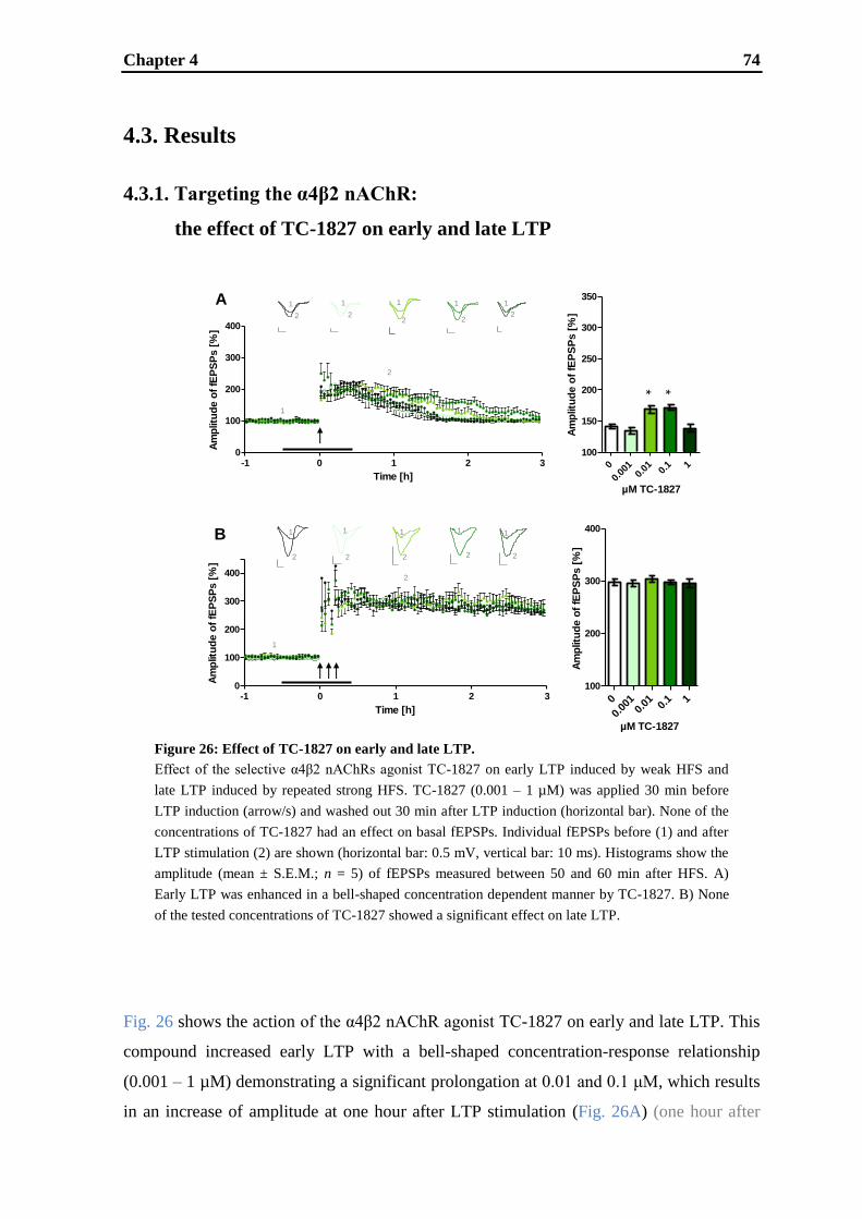
Chapter 4 74
-1 0 1 2 30
100
200
300
400
Time [h]
Am
plit
ud
e o
f fE
PS
Ps [
%]
0
0.00
10.
01 0.1 1
100
200
300
400
Am
plitu
de o
f fE
PS
Ps [
%]
µM TC-1827
-1 0 1 2 30
100
200
300
400
Time [h]
Am
plitu
de o
f fE
PS
Ps [
%]
0
0.00
10.
01 0.1 1
100
150
200
250
300
350
Am
plitu
de o
f fE
PS
Ps [
%]
µM TC-1827
1
1
2
1
2
1
2
1
2
1
2
1
2
1
2
1
2
1
2
1
2
1
2
2
* *
A
B
Figure 26: Effect of TC-1827 on early and late LTP.
Effect of the selective α4β2 nAChRs agonist TC-1827 on early LTP induced by weak HFS and
late LTP induced by repeated strong HFS. TC-1827 (0.001 – 1 µM) was applied 30 min before
LTP induction (arrow/s) and washed out 30 min after LTP induction (horizontal bar). None of the
concentrations of TC-1827 had an effect on basal fEPSPs. Individual fEPSPs before (1) and after
LTP stimulation (2) are shown (horizontal bar: 0.5 mV, vertical bar: 10 ms). Histograms show the
amplitude (mean ± S.E.M.; n = 5) of fEPSPs measured between 50 and 60 min after HFS. A)
Early LTP was enhanced in a bell-shaped concentration dependent manner by TC-1827. B) None
of the tested concentrations of TC-1827 showed a significant effect on late LTP.
4.3. Results
4.3.1. Targeting the α4β2 nAChR:
the effect of TC-1827 on early and late LTP
Fig. 26 shows the action of the α4β2 nAChR agonist TC-1827 on early and late LTP. This
compound increased early LTP with a bell-shaped concentration-response relationship
(0.001 – 1 µM) demonstrating a significant prolongation at 0.01 and 0.1 μM, which results
in an increase of amplitude at one hour after LTP stimulation (Fig. 26A) (one hour after
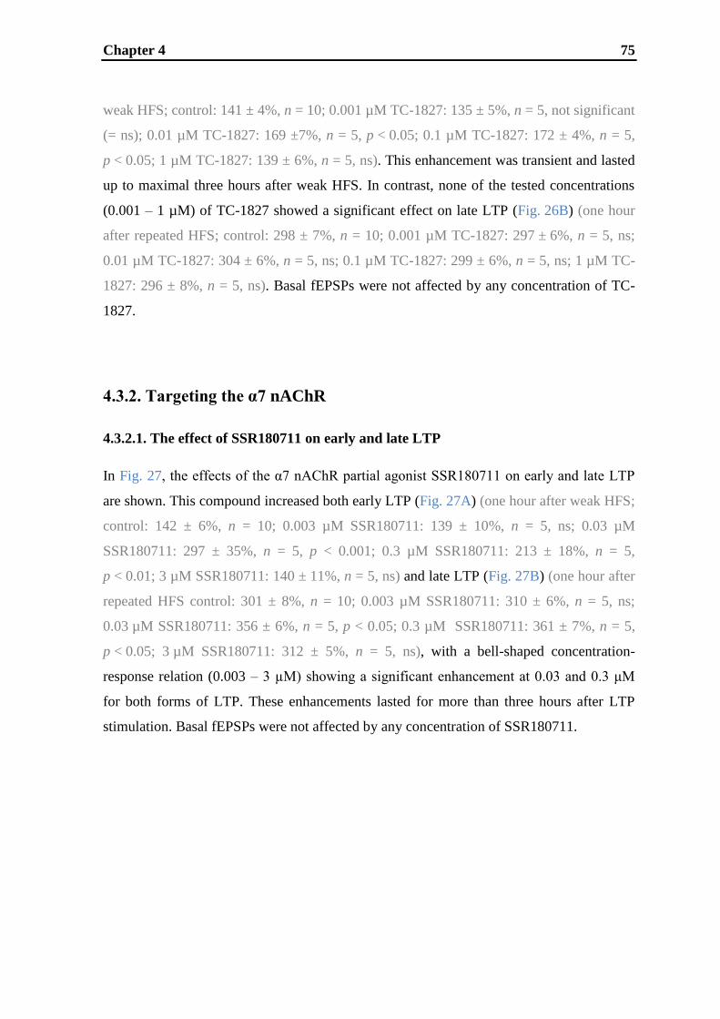
Chapter 4 75
weak HFS; control: 141 ± 4%, n = 10; 0.001 µM TC-1827: 135 ± 5%, n = 5, not significant
(= ns); 0.01 µM TC-1827: 169 ±7%, n = 5, p < 0.05; 0.1 µM TC-1827: 172 ± 4%, n = 5,
p < 0.05; 1 µM TC-1827: 139 ± 6%, n = 5, ns). This enhancement was transient and lasted
up to maximal three hours after weak HFS. In contrast, none of the tested concentrations
(0.001 – 1 µM) of TC-1827 showed a significant effect on late LTP (Fig. 26B) (one hour
after repeated HFS; control: 298 ± 7%, n = 10; 0.001 µM TC-1827: 297 ± 6%, n = 5, ns;
0.01 µM TC-1827: 304 ± 6%, n = 5, ns; 0.1 µM TC-1827: 299 ± 6%, n = 5, ns; 1 µM TC-
1827: 296 ± 8%, n = 5, ns). Basal fEPSPs were not affected by any concentration of TC-
1827.
4.3.2. Targeting the α7 nAChR
4.3.2.1. The effect of SSR180711 on early and late LTP
In Fig. 27, the effects of the α7 nAChR partial agonist SSR180711 on early and late LTP
are shown. This compound increased both early LTP (Fig. 27A) (one hour after weak HFS;
control: 142 ± 6%, n = 10; 0.003 µM SSR180711: 139 ± 10%, n = 5, ns; 0.03 µM
SSR180711: 297 ± 35%, n = 5, p < 0.001; 0.3 µM SSR180711: 213 ± 18%, n = 5,
p < 0.01; 3 µM SSR180711: 140 ± 11%, n = 5, ns) and late LTP (Fig. 27B) (one hour after
repeated HFS control: 301 ± 8%, n = 10; 0.003 µM SSR180711: 310 ± 6%, n = 5, ns;
0.03 µM SSR180711: 356 ± 6%, n = 5, p < 0.05; 0.3 µM SSR180711: 361 ± 7%, n = 5,
p < 0.05; 3 µM SSR180711: 312 ± 5%, n = 5, ns), with a bell-shaped concentration-
response relation (0.003 – 3 µM) showing a significant enhancement at 0.03 and 0.3 μM
for both forms of LTP. These enhancements lasted for more than three hours after LTP
stimulation. Basal fEPSPs were not affected by any concentration of SSR180711.

Chapter 4 76
Figure 27: Effect of SSR180711 on early and late LTP.
Effect of the selective α7 nAChR partial agonist SSR180711 on early LTP induced by weak HFS
and late LTP induced by repeated strong HFS. SSR180711 (0.003 – 3 µM) was applied 30 min
before LTP induction (arrow/s) and washed out 30 min after LTP induction (horizontal bar). None
of the concentrations of SSR180711 had an effect on basal fEPSPs. Individual fEPSPs before (1)
and after LTP stimulation (2) are shown (horizontal bar: 0.5 mV, vertical bar: 10 ms). Histograms
show the amplitude (mean ± S.E.M.; n = 5) of fEPSPs measured between 50 and 60 min after
HFS. A) Early LTP was enhanced in a bell-shaped concentration dependent manner by
SSR180711. B) SSR180711 increased late LTP in a bell-shaped concentration dependent manner.
-1 0 1 2 30
100
200
300
400
Time [h]
Am
plit
ud
e o
f fE
PS
Ps [
%]
0
0.00
30.
03 0.3 3
100
150
200
250
300
350
Am
plitu
de o
f fE
PS
Ps [
%]
µM SSR180711
-1 0 1 2 30
100
200
300
400
Time [h]
Am
plitu
de o
f fE
PS
Ps [
%]
0
0.00
3
0.03
0.
3 3 100
200
300
400
Am
plit
ud
e o
f fE
PS
Ps [
%]
µM SSR180711
A
B
1
2
1
2
1
2
1
2
1
2
1
2
1
2
1
2
1
2
1
2
**
*****
1
1
2
2
4.3.2.1. Transformation of early into late LTP
To analyze whether the long-lasting increase of early LTP by SSR180711 could be
attributed to the transformation of protein-synthesis independent early LTP into protein-
synthesis dependent late LTP, the reversible protein translation inhibitor anisomycin was
used at a concentration of 30 µM, which is known to be sufficient to significantly impair
protein-synthesis dependent late LTP (Lu et al., 1999; Kroker et al., 2011b). The addition
of 30 µM anisomycin to 0.3 μM SSR180711, a concentration shown to increase early LTP
(see Fig. 27A), caused a significant decrease of SSR180711 enhanced LTP up to a
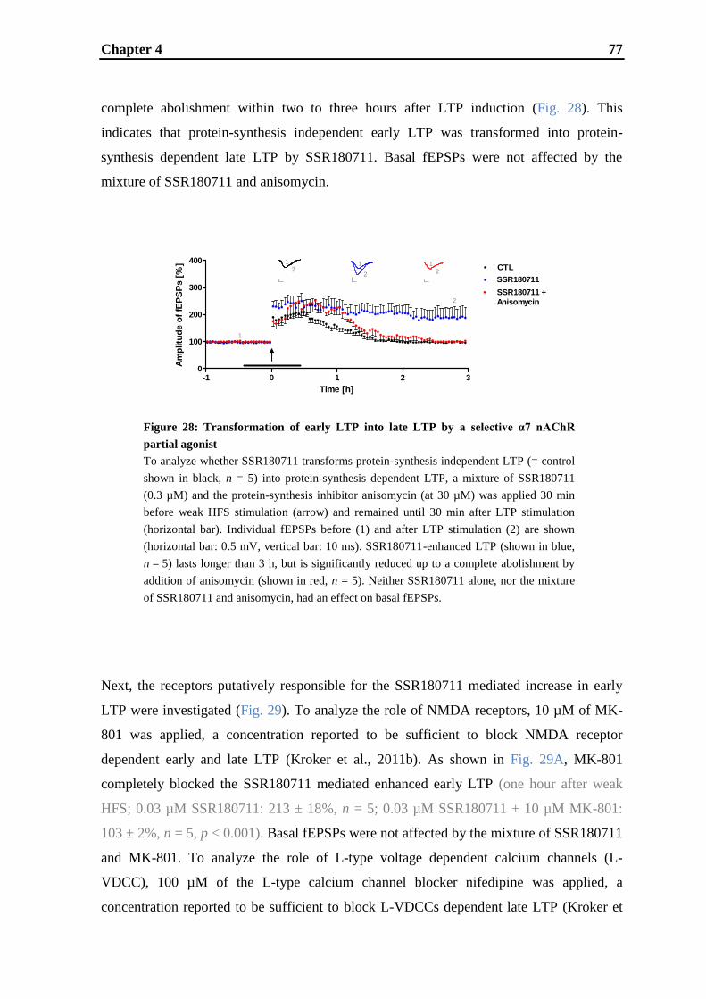
Chapter 4 77
Figure 28: Transformation of early LTP into late LTP by a selective α7 nAChR
partial agonist
To analyze whether SSR180711 transforms protein-synthesis independent LTP (= control
shown in black, n = 5) into protein-synthesis dependent LTP, a mixture of SSR180711
(0.3 µM) and the protein-synthesis inhibitor anisomycin (at 30 µM) was applied 30 min
before weak HFS stimulation (arrow) and remained until 30 min after LTP stimulation
(horizontal bar). Individual fEPSPs before (1) and after LTP stimulation (2) are shown
(horizontal bar: 0.5 mV, vertical bar: 10 ms). SSR180711-enhanced LTP (shown in blue,
n = 5) lasts longer than 3 h, but is significantly reduced up to a complete abolishment by
addition of anisomycin (shown in red, n = 5). Neither SSR180711 alone, nor the mixture
of SSR180711 and anisomycin, had an effect on basal fEPSPs.
-1 0 1 2 30
100
200
300
400
Time [h]
Am
plitu
de o
f fE
PS
Ps [
%] CTL
Legend
SSR180711 +
Anisomycin
1
2
1
21
2
1
2
-1 0 1 2 30
100
200
300
400
Time [h]
Am
plit
ud
e o
f fE
PS
Ps [
%] CTL
Legend
SSR180711 +
Anisomycin
complete abolishment within two to three hours after LTP induction (Fig. 28). This
indicates that protein-synthesis independent early LTP was transformed into protein-
synthesis dependent late LTP by SSR180711. Basal fEPSPs were not affected by the
mixture of SSR180711 and anisomycin.
Next, the receptors putatively responsible for the SSR180711 mediated increase in early
LTP were investigated (Fig. 29). To analyze the role of NMDA receptors, 10 µM of MK-
801 was applied, a concentration reported to be sufficient to block NMDA receptor
dependent early and late LTP (Kroker et al., 2011b). As shown in Fig. 29A, MK-801
completely blocked the SSR180711 mediated enhanced early LTP (one hour after weak
HFS; 0.03 µM SSR180711: 213 ± 18%, n = 5; 0.03 µM SSR180711 + 10 µM MK-801:
103 ± 2%, n = 5, p < 0.001). Basal fEPSPs were not affected by the mixture of SSR180711
and MK-801. To analyze the role of L-type voltage dependent calcium channels (L-
VDCC), 100 µM of the L-type calcium channel blocker nifedipine was applied, a
concentration reported to be sufficient to block L-VDCCs dependent late LTP (Kroker et
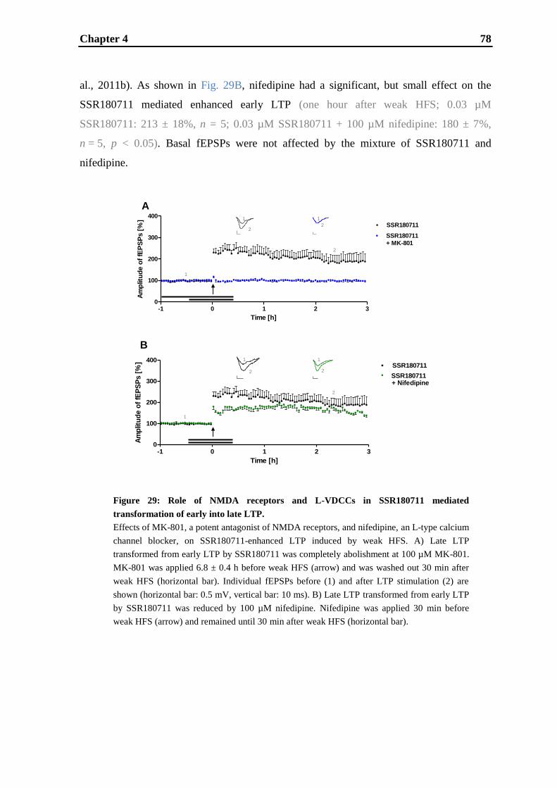
Chapter 4 78
-1 0 1 2 30
100
200
300
400
Time [h]
Am
plitu
de o
f fE
PS
Ps [
%]
SSR180711
SSR180711
+ MK-801
1
2
1
2
1
2
A
-1 0 1 2 30
100
200
300
400
Time [h]
Am
plit
ud
e o
f fE
PS
Ps [
%]
SSR180711
Legend
1
2
1
2 2
1
B
-1 0 1 2 30
100
200
300
400
Time [h]
Am
plitu
de o
f fE
PS
Ps [
%] + Nifedipine
Legend
-1 0 1 2 30
100
200
300
400
Time [h]
Am
plit
ud
e o
f fE
PS
Ps [
%]
SSR180711
Legend
al., 2011b). As shown in Fig. 29B, nifedipine had a significant, but small effect on the
SSR180711 mediated enhanced early LTP (one hour after weak HFS; 0.03 µM
SSR180711: 213 ± 18%, n = 5; 0.03 µM SSR180711 + 100 µM nifedipine: 180 ± 7%,
n = 5, p < 0.05). Basal fEPSPs were not affected by the mixture of SSR180711 and
nifedipine.
Figure 29: Role of NMDA receptors and L-VDCCs in SSR180711 mediated
transformation of early into late LTP.
Effects of MK-801, a potent antagonist of NMDA receptors, and nifedipine, an L-type calcium
channel blocker, on SSR180711-enhanced LTP induced by weak HFS. A) Late LTP
transformed from early LTP by SSR180711 was completely abolishment at 100 µM MK-801.
MK-801 was applied 6.8 ± 0.4 h before weak HFS (arrow) and was washed out 30 min after
weak HFS (horizontal bar). Individual fEPSPs before (1) and after LTP stimulation (2) are
shown (horizontal bar: 0.5 mV, vertical bar: 10 ms). B) Late LTP transformed from early LTP
by SSR180711 was reduced by 100 µM nifedipine. Nifedipine was applied 30 min before
weak HFS (arrow) and remained until 30 min after weak HFS (horizontal bar).
Chapter 4 79
4.4. Discussion
Acetylcholinesterase inhibitors, like donepezil, rivastigmine or galantamine, are used for
symptomatic treatment of mild-to-moderate AD disease patients. However, experience
from clinical practice with acetylcholinesterase inhibitors has raised concerns about the
clinical relevance of the only moderate treatment effect, the appearance of tolerance after
long-term use, interaction of cholinergic and non-cholinergic effects and possible effects of
amyloid precursor protein release (Courtney et al., 2004; Diniz et al., 2009; Helou and
Rhalimi, 2010; Hernandez et al., 2009; Kaduszkiewicz et al., 2005; Rosenbloom et al.,
2010; Rozzini et al., 2004). Currently, an alternative approach targeting cholinergic
neurotransmission through direct activation of nAChRs has come into the focus of interest
for the treatment of AD and other cognitive diseases (Arneric et al., 2007; Buccafusco et
al., 2004; D'hoedt and Bertrand, 2009; Melnikova, 2007; Nordberg, 2001; Perry et al.,
2001; Woodruff-Pak and Gould, 2002). Non-selective nAChR agonists like nicotine and
epibatidine, but also sub-type selective nAChR agonists lacking the side effects caused by
stimulation of peripheral nAChR such as TC-1827, ispronicline, TC-5619, ABT-089 and
SSR180711 have demonstrated memory enhancing effects in various animal cognition
tasks (Barak et al., 2009; Bohme et al., 2004; Decker et al., 1994; Dukat and Glennon,
2003; Lippiello et al., 2006; Pichat et al., 2007; Rezvani and Levin, 2001). Furthermore,
the α4β2 nAChR agonists ispronicline and AZD-1446 as well as the α7 nAChR agonists
TC-5619 and RO5313534 (formerly R3487/MEM3454) were progressed into phase II
clinical development (Geerts, 2006; Hauser et al., 2009; Haydar and Dunlop, 2010;
Toyohara and Hashimoto, 2010). Thus, the α4β2 and α7 nAChR represent attractive
potential drug targets for treatment of diseases linked to cognitive dysfunction. However,
their exact mode-of-action on the molecular and cellular processes underlying memory
formation, i.e. synaptic plasticity and LTP, are not fully understood.
The potent α4β2 nAChR full agonist TC-1827 (Bohme et al., 2004; Grinevich et al., 2009)
and the selective α7 nAChR partial agonist SSR180711 (Barak et al., 2009; Biton et al.,
2007; Hashimoto et al., 2008; Kristensen et al., 2007; Pichat et al., 2007) were previously
described to enhance hippocampal LTP (Biton et al., 2007; Bohme et al., 2004). However,
in these studies, only a single drug concentration was tested and no distinction was made
between effects on early and late LTP. This distinction is of importance for the evaluation
of downstream effects associated with these targets. Early LTP is independent of protein-
synthesis and involves modifications of pre-existing synapses as a result of rapid calcium
Chapter 4 80
influx through NMDA receptors and subsequent protein phosphorylation events (Bliss and
Collingridge, 1993; Malenka and Nicoll, 1999). Late LTP is clearly separated from early
LTP by its dependence on de novo protein-synthesis (Frey et al., 1988; Huang, 1998;
Huang and Kandel, 1994; Huang et al., 1996; Kroker et al., 2011b; Nguyen et al., 1994;
Nguyen and Kandel, 1997; Reymann and Frey, 2007). Protein-synthesis may contribute
both to the modification of existing synapses and to the establishment of new synaptic
contacts (Engert and Bonhoeffer, 1999; Lisman, 2003). Moreover, long-lasting forms of
memory are generally believed to be protein-synthesis dependent (Pang and Lu, 2004).
The findings in the present study demonstrate that early LTP can be enhanced in a bell-
shaped manner by the α4β2 nAChR full agonist TC-1827 with efficacious concentrations
of 0.01 and 0.1 µM confirming published results (Bohme et al., 2004) which showed that
0.1 µM TC-1827 increased LTP induced by weak stimulation supposed to induce early
LTP (Albensi et al., 2007; Huang and Kandel, 1994). Contrary to the effects on early LTP,
TC-1827 did not show any effects on late LTP. Hence, in our setting the activation of the
α4β2 nAChR seems to be able to enhance early LTP, but not late LTP. For the α7 nAChR
partial agonist SSR180711, we show that both early and late LTP are increased in a bell-
shaped manner with efficacious concentrations of 0.03 and 0.3 µM. The result on late LTP
confirms published data (Biton et al., 2007) showing that 0.3 µM SSR180711 increased
LTP induced by repeated stimulation thought to induce late LTP (Albensi et al., 2007;
Huang and Kandel, 1994). Furthermore, for the first time, it was demonstrated that
SSR180711 not only increases protein-synthesis independent LTP (early LTP) in a bell-
shaped manner, but transforms it into protein-synthesis dependent LTP (late LTP). This
transformation of early into late LTP might result from a lowered induction threshold of
late LTP. Further investigations revealed that the SSR180711 mediated increase and
transformation was dependent mainly on NMDA receptors and in part on L-VDCCs. Thus,
as late LTP induced by repeated HFS is dependent on NMDA receptors and L-VDCCs to
equal parts (Kroker et al., 2011b), a different type of late LTP seems to be induced by
administration of SSR180711 together with weak HFS.
Both the α4β2 and the α7 nAChR (partial) agonists showed a bell-shaped concentration
dependence with respect to their enhancement of LTP. An explanation might be provided
by the finding that full activation of nAChRs using high agonist concentrations can lead to
receptor endocytosis (St. John and Gordon, 2001). Another explanation concerning the
lack of effect at high concentrations might be an overstimulation of the nAChRs, not
strong enough to cause long-term depression, but sufficient to alleviate the enhancement of
Chapter 4 81
LTP. In fact, it has been shown that nicotine is able to induce long-term depression at high
concentrations (Maggi et al., 2004).
Regarding receptor specificity, both TC-1827 and SSR180711 were proven to be specific
(partial) agonists, since their enhancing effects on LTP were reversed by addition of the
respective specific nAChR antagonists dihydro-beta-erythroidine (for the α4β2 nAChR)
and methyllycaconitine (for the α7 nAChR; data not shown).
What might be the explanation for α4β2 and α7 nAChR agonists having differential effects
on early and late LTP? Both α4β2 and α7 nAChRs are localized on GABAergic
interneurons (Alkondon et al., 1999, 2004; Fabian-Fine et al., 2001; Jones and Yakel,
1997; Khiroug et al., 2003). Disinhibition can take place due to activation of α4β2 and/or
α7 nAChRs on GABAergic interneurons having inhibitory connections to other inhibitory
neurons producing a net increase in glutamatergic principal cell activity (Alkondon et al.,
2004; Ji and Dani, 2000). Furthermore, both receptors are reported to be located on the
presynapse of acetylcholinergic neurons (McGehee et al., 1995; Wonnacott, 1997) and,
due to the significant calcium permeability of these receptors (Role and Berg, 1996;
Séguéla et al., 1993; Vernino et al., 1994), can provide positive feedback by enhancing the
release probability of acetylcholine (Bohme et al., 2004; McGehee et al., 1995; Role and
Berg, 1996; Wonnacott, 1997). Therefore, activation of α4β2 and/or α7 nAChRs may
cause dishinhibition and enhancement of acetylcholine release - mechanisms which
presumably are able to enhance early, but not late LTP. The decisive difference between
α4β2 and α7 nAChR function is the additional location of α7 nAChRs on Schaffer
collateral terminals (Fabian-Fine et al., 2001; Buccafusco et al., 2005) causing
strengthening of the glutamatergic system through the facilitation of glutamate release
(Aramakis and Metherate, 1998; Biton et al., 2007; Gray et al., 1996; Jones et al., 1999;
Mansvelder and McGehee, 2000; McGehee et al., 1995; Pidoplichko et al., 2004; Radcliffe
and Dani, 1998). Due to their high calcium permeability α7 nAChRs can enhance the
synaptic release of glutamate, increase the coincidence between presynaptic release and
postsynaptic depolarization and therefore enhance the strength of LTP (Ge and Dani, 2005;
Ji et al., 2001). This might explain the enhancement of early as well as late LTP and the
transformation of early into late LTP by activation of α7 nAChRs, but not α4β2 nAChRs
lacking this feature on glutamatergic transmission. Thus, concerning cognitive functions,
activation of α7 nAChRs might be more effective than activation of α4β2 nAChRs as also
suggested by Graef et al. (2011).

Chapter 4 82
In conclusion, this study demonstrates that activation of α7 nAChRs is more efficient in
enhancing and prolonging LTP than activation of α4β2 nAChRs. We hypothesize that the
better performance of the α7 nAChR agonist is not due to disinhibition or cholinergic
transmission as these mechanisms are activated by both receptors, but due to the additional
strengthening of the glutamatergic system via the α7 nAChR. Therefore, the direct
comparison of the effects of selective α4β2 and α7 nAChR (partial) agonists on early and
late LTP suggests that α7 nAChR activation might be the more attractive approach for
enhancing cognitive function in patients.

Chapter 4 83
CHAPTER 5
Inhibition of AChE and PDE9A has differential
effects on early and late LTP
Chapter 5 84
Abstract
Donepezil is the current standard symptomatic treatment of mild-to-moderate Alzheimer‟s
disease (AD) patients. It aims to compensate for the deficit in cholinergic
neurotransmission by blocking acetylcholinesterase (AChE) and thus increases the
concentration of extracellular acetylcholine. However, experience from clinical practice
demonstrated that AChE inhibitors only have moderate treatment effects. As a potential
new approach for memory enhancement, inhibition of specific phosphodiesterases (PDEs)
has gained attention. Among those are PDE9A inhibitors which increase the levels of the
second messenger cyclic guanosine monophosphate (cGMP) intracellularly. In order to
gain more insight into the potential impact of extracellularly acting AChEs and
intracellularly acting PDE9A inhibitors on synaptic plasticity, we analyzed the effects of
the AChE inhibitor donepezil and the PDE9A inhibitor BAY 73-6691 on long-term
potentiation (LTP) in rat hippocampal slices, a widely accepted cellular experimental
model of memory formation. Generally, LTP can be differentiated into an early and a late
form, being protein-synthesis independent and protein-synthesis dependent, respectively.
Donepezil was found to increase early LTP, but did not affect late LTP. In contrast, BAY
73-6691 demonstrated enhancing effects on both early and late LTP and even transformed
early into late LTP. Furthermore, it was shown that this transformation into late LTP was
dependent on the NO-cGMP-PKG pathway. In conclusion, this study demonstrates that
BAY 73-6691 exhibits a stronger effect in enhancing and prolonging LTP than donepezil
suggesting that PDE9 inhibition might be more efficacious in enhancing learning and
memory.
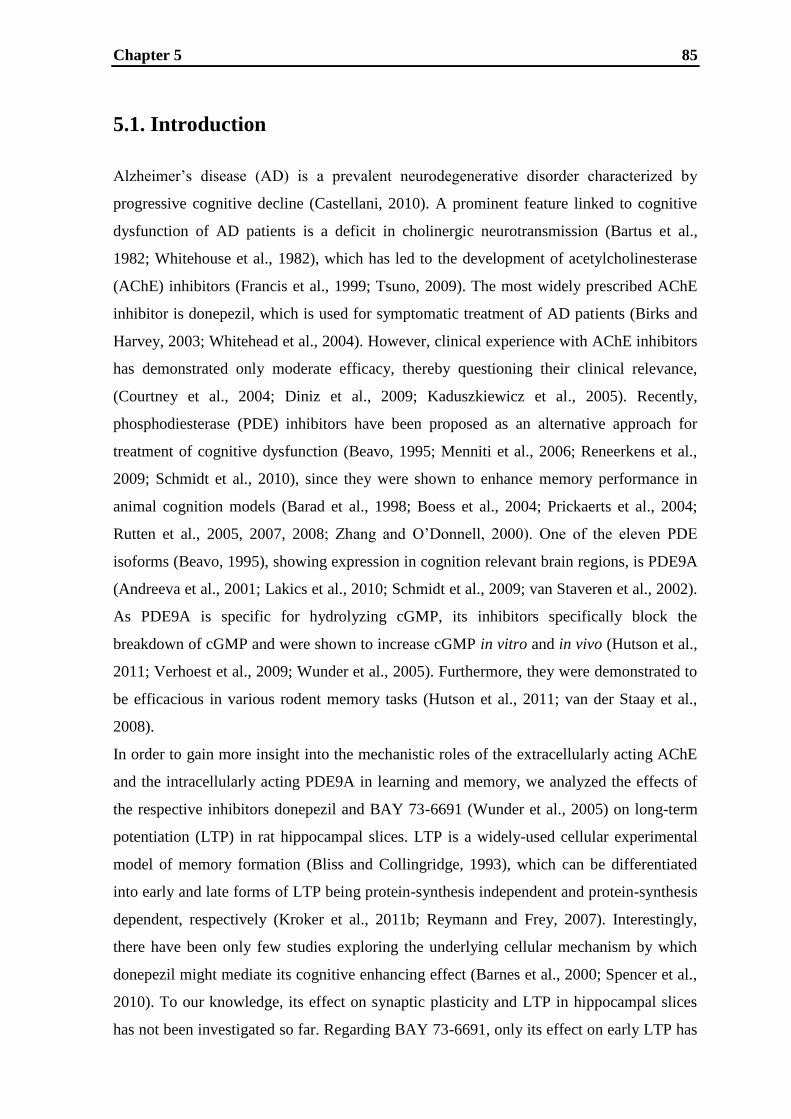
Chapter 5 85
5.1. Introduction
Alzheimer‟s disease (AD) is a prevalent neurodegenerative disorder characterized by
progressive cognitive decline (Castellani, 2010). A prominent feature linked to cognitive
dysfunction of AD patients is a deficit in cholinergic neurotransmission (Bartus et al.,
1982; Whitehouse et al., 1982), which has led to the development of acetylcholinesterase
(AChE) inhibitors (Francis et al., 1999; Tsuno, 2009). The most widely prescribed AChE
inhibitor is donepezil, which is used for symptomatic treatment of AD patients (Birks and
Harvey, 2003; Whitehead et al., 2004). However, clinical experience with AChE inhibitors
has demonstrated only moderate efficacy, thereby questioning their clinical relevance,
(Courtney et al., 2004; Diniz et al., 2009; Kaduszkiewicz et al., 2005). Recently,
phosphodiesterase (PDE) inhibitors have been proposed as an alternative approach for
treatment of cognitive dysfunction (Beavo, 1995; Menniti et al., 2006; Reneerkens et al.,
2009; Schmidt et al., 2010), since they were shown to enhance memory performance in
animal cognition models (Barad et al., 1998; Boess et al., 2004; Prickaerts et al., 2004;
Rutten et al., 2005, 2007, 2008; Zhang and O‟Donnell, 2000). One of the eleven PDE
isoforms (Beavo, 1995), showing expression in cognition relevant brain regions, is PDE9A
(Andreeva et al., 2001; Lakics et al., 2010; Schmidt et al., 2009; van Staveren et al., 2002).
As PDE9A is specific for hydrolyzing cGMP, its inhibitors specifically block the
breakdown of cGMP and were shown to increase cGMP in vitro and in vivo (Hutson et al.,
2011; Verhoest et al., 2009; Wunder et al., 2005). Furthermore, they were demonstrated to
be efficacious in various rodent memory tasks (Hutson et al., 2011; van der Staay et al.,
2008).
In order to gain more insight into the mechanistic roles of the extracellularly acting AChE
and the intracellularly acting PDE9A in learning and memory, we analyzed the effects of
the respective inhibitors donepezil and BAY 73-6691 (Wunder et al., 2005) on long-term
potentiation (LTP) in rat hippocampal slices. LTP is a widely-used cellular experimental
model of memory formation (Bliss and Collingridge, 1993), which can be differentiated
into early and late forms of LTP being protein-synthesis independent and protein-synthesis
dependent, respectively (Kroker et al., 2011b; Reymann and Frey, 2007). Interestingly,
there have been only few studies exploring the underlying cellular mechanism by which
donepezil might mediate its cognitive enhancing effect (Barnes et al., 2000; Spencer et al.,
2010). To our knowledge, its effect on synaptic plasticity and LTP in hippocampal slices
has not been investigated so far. Regarding BAY 73-6691, only its effect on early LTP has

Chapter 5 86
been published (van der Staay et al., 2008), but neither a full concentration-response curve
nor an assessment of its effect on late LTP were reported. Therefore, in this study, we
analyzed and compared the effects of donepezil and BAY 73-6691 on early and late LTP.
The results provide a deeper insight into the mechanism of action of AChE inhibitors and
the PDE9A inhibitor for LTP modulation and hence into their potential roles in learning
and memory.
Chapter 5 87
5.2. Materials and methods
5.2.1. Tissue preparation
5.2.1.1. Animals
Procedures involving animals and their care were conducted in conformity with
institutional and European Union guidelines (EEC Council Directive 86/609) and were
approved by the Ethical Committee of the respective regional council. For all experiments
male Wistar rats (Janvier, Le Genest Saint Isle, France) aged six to seven weeks were used.
5.2.1.2. Preparation of brain slices
Rats were shortly anaesthetized with isoflurane and euthanized by decapitation. The brains
were quickly removed and immersed in ice-cold ACSF (containing in mM NaCl 124, KCl
4.9, MgSO4 8, CaCl2 2.5, KH2PO4 1.2, NaHCO3 25.6, D-glucose 10, pH 7.4, saturated with
95% O2 and 5% CO2). Transverse hippocampal brain slices (400 µm thickness) were cut
using a Vibratome. Slices were allowed to recover in a holding chamber containing ACSF
bubbled with 95% O2 and 5% CO2 at room temperature for at least three hours. For
estimation of protein concentration, western blot analysis and measurement of AChE
activity the slices were homogenated by douncing in PBS (Phosphate Buffered Saline, Bio-
Rad Laboratories, Inc.) supplemented with a cocktail of protease inhibitors (Roche
Diagnostic GmbH), partitioned into three tubes and stored at -80 °C until use.
For electrophysiological recordings, slices were transferred into the integrated brain slice
chambers and continuously superfused (at a flow rate of 2.5 mL min−1
, 25 ± 0.2 °C) with
the same ACSF composition used before except for 0.5 mM MgSO4. Prior to doing any
electrophysiological recordings, the slices were allowed to equilibrate for at least
30 minutes.
5.2.1.3. Perfusion of rats and brain homogenate preperation
Perfusion and homogenate preparation were performed as described by Das et al. (2001)
with minor modifications. Briefly, rats were perfused through the heart with saline (0.9%
NaCl) under ketamine (70 mg/kg) and xylazine (6 mg/kg) anesthesia. The brain was

Chapter 5 88
removed and a 10% (w/v) homogenate of the hippocampus was prepared by douncing in
PBS supplemented with a cocktail of protease inhibitors. 3 x 33 µl of homogenate were
collected, diluted in the ratio of 1:10 and stored at -80 °C. The rest of the homogenate was
centrifuged at 100,000 x g at 4 °C for 60 minutes. The supernatant as well as the pellet
were each collected in separate tubes, diluted in the ratio of 1:10, stored at -80 °C and used
as a source of estimation of protein concentration and measurement of acetylcholinesterase
activity, respectively. Jackisch et al. (2009) verified that choline esterase activity does not
decrease under these storage conditions.
5.2.2. Electrophysiological measurements
5.2.2.1. Extracellular recording and stimulation
An optimized SliceMaster multi-slice recording system (Scientifica Limited, East Sussex,
UK; Kroker et al., 2011a) was used to record field excitatory postsynaptic potentials
(fEPSPs). fEPSPs were elicited in the CA1 region by stimulation of the Schaffer
collateral–commissural fibres in the stratum radiatum using glass electrodes with broken
tips (filled with ACSF). For recording, glass electrodes (2 – 6 MΩ, filled with ACSF) were
placed in the apical dendritic layer. The amplitudes of fEPSPs were used as the parameter
of interest (Collingridge et al., 1983; Kroker et al., 2011a). To generate fEPSPs at a
constant sub-threshold stimulus, as in previous studies (Kroker et al., 2011a; Seabrook et
al., 1997), the stimulus strength of the pulses was adjusted to 20 - 30% of the fEPSP
maximum and this voltage was used for the experiment. During baseline recording, each
slice was stimulated every 30 seconds for at least one hour. Early LTP was induced by
weak high frequency stimulation (HFS) made up of 20 pulses at the frequency of 100 Hz
(Bashir et al., 1991; Kroker et al., 2011b), whereas late LTP was induced by repeated
strong HFS consisting of 100 pulses at the frequency of 100 Hz, repeated two times in five
minute intervals (Kroker et al., 2011b; Lu et al., 1999).
5.2.2.2. Recording, data acquisition and analysis
A modular electrophysiology system, supplied by npi electronic GmbH (Tamm, Germany),
conducts the low noise recordings of extracellular signals. AC coupled signals are

Chapter 5 89
amplified 1000 x and internally filtered with a five kHz low-pass filter as well as a three
Hz high-pass filter. In our study, a customized system (programmable pattern generator)
was used allowing simultaneous stimulation of all slices either with a single stimulus or a
specific stimulus pattern (Kroker et al., 2011a). For data acquisition and analysis the
software Notocord®
was used. Data are shown as mean (± SEM) percent of the baseline
fEPSP amplitude. Data were analyzed using either t tests to compare two conditions or
one-way ANOVA (post-hoc test: Dunnett-Test) to compare multiple conditions. In both
cases a p-value of 0.05 was considered significant. Histograms show the (mean ± SEM)
amplitude of EPSPs measured 50 – 60 min after delivery of LTP stimulation according to
Jia et al. (2010) and Kroker et al. (2011b).
5.2.2.3. Drug application
Drugs were prepared as stock solutions and diluted in ACSF immediately before
application. ODQ and BAY 73-6691 were prepared in DMSO (the final concentration of
DMSO in ACSF was 0.1%). All drugs were applied 30 minutes before LTP stimulation
and remained until 30 minutes after LTP stimulation
5.2.3. Determination of protein concentration
Samples of the slice homogenate, the hippocampal homogenate, supernatant and pellet
were used for protein measurement. Protein concentration was determined by using a
Pierce BCA protein assay kit according to the manufacture‟s procedure (Thermo Scientific
Pierce BCA Protein Assay Kit).
5.2.4. Western blot analysis
A total of 20 μg of protein (from samples of hippocampal homogenate, slice homogenate,
supernant and pellet) per lane was run on 4 – 12% Bis-Tris gels (NuPAGE® Novex Bis-
Tris Gels) and then transferred onto PVDF membranes for western blotting (Trans-Blot
Transfer Medium; Bio-Rad). The membranes were blocked for one hour in Odyssey

Chapter 5 90
blocking buffer (Licor). The blots were then probed with affinity purified goat polyclonal
anti-AChE antibodies over night (E-19, dilution 1:200, Santa Cruz Bioctechnology Inc.;
Fischer et al., 2002; Jevsek et al., 2004; Santos et al., 2007; Sternfeld et al., 2000).
Following three washes, the blots were incubated for one hour with secondary anti-goat
antibodies made in donkey purchased from Licor (926-32223, dilution 1:2000, Odyssey®
,
Licor). After additional washing, immunoreactive bands were detected using a Licor
Odyssey imaging system according to the manufacture‟s procedure.
5.2.5. Measurement of achetylcholinesterase activity
Choline esterase activity was determined according to Ellman et al. (1961), as modified by
Ashour et al. (1987) and Jackisch et al. (2009). In brief, 250 μl of the reagent buffer
(0.01% DTNB in 0.05 M sodium diphosphate buffer, pH 7.4) and 6 μl tissue homogenate
(corresponding to about 24 – 42 μg protein) were added to the wells of a 96-well microtiter
plate cooled on ice. Each concentration was determined in triplicate. The enzymatic
reaction was started at room temperature by the addition of 50 μl acetylthiocholine iodide
solutions at various concentrations (final concentrations of acetylthiocholine in the wells
(μM): 31.25, 62.5, 125, 250, 500, 1000 and 2000). Absorbance change at 405 nm was
measured at room temperature using a Flex Station 3 (Molecular Devices). Values were
recorded every 30 seconds for one hour. Negative controls (in triplicate, without tissue
homogenate) were always run in parallel. For determination of the inhibitory potency of
donepezil, the assay was performed as described above, except that only one concentration
of acetylthiocholine iodide (500 µM) was used. The inhibitory potency of donepezil was
then determined in the presence of its increasing concentrations (1 nM to 100 µM). Each
concentration was determined in triplicate.
Enzyme kinetic parameters were determined according to Jackisch et al. (2009). Data were
analyzed using either t tests to compare two conditions or one-way ANOVA (post-hoc test:
Dunnett-Test) to compare multiple conditions. In both cases a p-value of 0.05 was
considered significant.

Chapter 5 91
5.2.6. Drugs
The following drugs were used: (2R,3S,4S)-2-(4-Methoxybenzyl)-3,4-pyrrolidinediol-3-
acetate, 2-[(4-Methoxyphenyl)methyl]-3,4-pyrrolidinediol 3-acetate (= anisomycin), 1H-
[1,2,4]Oxadiazolo[4,3-a]quinoxalin-1-one (= ODQ), KT 5823, donepezil hydrochloride
monohydrate (= donepezil), 5,5′-Dithiobis(2-nitrobenzoic acid) (= DTNB), 5,11-Dihydro-
11-[(4-methyl-1-piperazinyl)acetyl]-6H-pyrido[2,3-b][1,4]benzodiazepin-6-one
dihydrochloride (= pirenzepine) and acetylthiocholine iodide were purchased from Sigma-
Aldrich Corporation (St. Louis, USA). The compound BAY 73-6691 (1-(2-Chlorophenyl)-
6-[(2R)-3,3,3-trifluoro-2-methylpropyl]-1,5-dihydro-4H-pyrazolo[3,4-d]pyrimidine-4-one)
was synthesized as a racemate in the Department of Medicinical Chemistry of Boehringer
Ingelheim Pharma GmbH & Co KG.
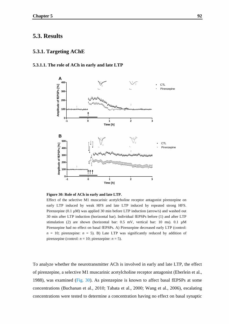
Chapter 5 92
-1 0 1 2 30
100
200
300
400
Time [h]
Am
plitu
de o
f fE
PS
Ps [
%] CTL
LegendPirenzepine
-1 0 1 2 30
100
200
300
400
500
Time [h]
Am
plitu
de o
f fE
PS
Ps [
%] CTL
LegendPirenzepine
1
1
2
A
B
1
22
1
1 1
22
2
5.3. Results
5.3.1. Targeting AChE
5.3.1.1. The role of ACh in early and late LTP
To analyze whether the neurotransmitter ACh is involved in early and late LTP, the effect
of pirenzepine, a selective M1 muscarinic acetylcholine receptor antagonist (Eberlein et al.,
1988), was examined (Fig. 30). As pirenzepine is known to affect basal fEPSPs at some
concentrations (Buchanan et al., 2010; Tabata et al., 2000; Wang et al., 2006), escalating
concentrations were tested to determine a concentration having no effect on basal synaptic
Figure 30: Role of ACh in early and late LTP.
Effect of the selective M1 muscarinic acetylcholine receptor antagonist pirenzepine on
early LTP induced by weak HFS and late LTP induced by repeated strong HFS.
Pirenzepine (0.1 µM) was applied 30 min before LTP induction (arrow/s) and washed out
30 min after LTP induction (horizontal bar). Individual fEPSPs before (1) and after LTP
stimulation (2) are shown (horizontal bar: 0.5 mV, vertical bar: 10 ms). 0.1 µM
Pirenzepine had no effect on basal fEPSPs. A) Pirenzepine decreased early LTP (control:
n = 10; pirenzepine: n = 5). B) Late LTP was significantly reduced by addition of
pirenzepine (control: n = 10; pirenzepine: n = 5).

Chapter 5 93
45678910110
20
40
60
80
100
120
- log conc [M]
AC
hE
acti
vit
y (
% o
f co
ntr
ol)
B
C
HH HS0.0
0.5
1.0
nm
ole
s/m
in
/mg
pro
tein
60 Kd
A 1 2 3 4
HH HS S P
transmission (data not shown). In fact, 0.1 µM was found to be the highest concentration of
pirenzepine not compromising basal fEPSPs which is supported by two other studies
(Calabresi et al., 1999; Bonsi et al., 2008). Therefore, 0.1 µM pirenzepine was used for
LTP experiments, and this concentration caused a significant impairment of early LTP
(Fig. 30A) (one hour after weak HFS; control: 141 ± 10%, n = 10; 0.1 µM pirenzepine:
1 ± 3, n = 5, p < 0.05) and late LTP (Fig. 30B) (one hour after repeated HFS control:
308 ± 15%, n = 10; 0.1 µM pirenzepine: 214 ± 27%, n = 5, p < 0.01) indicating that the
neurotransmitter ACh is involved in both forms of LTP.
5.3.1.2. Expression and activity of AChE in hippocampal slices and inhibitory potency
of donepezil
Figure 31: AChE in hippocampal slices.
Analysis of the expression and activity of AChE and the inhibitory potency of donepezil
in hippocampal slices. A) Western blot analysis of the AChE expression of different
samples. Lane 1 shows AChE expression in freshly prepared hippocampal homogenate
(HH), lane 2 shows AChE expression in hippocampal slices homogenated 5 h after cutting
(HS), lane 3 shows AChE expression in the supernatant of the hippocampal homogenate
representing the cytosolic fraction (S), and lane 4 shows AChE expression in the pellet of
the hippocampal homogenate representing the membrane fraction (P). B) Comparison of
the AChE activity of the freshly prepared hippocampal homogenate (HH) and the
hippocampal slices homogenated 5 h after cutting (HS). C) Inhibitory potency of
donepezil on the AChE activity of hippocampal slice homogenate indicating that AChE
activity can be completely abolished.
Chapter 5 94
As shown by western blotting (Fig. 31A), AChE expression can be detected in freshly
prepared hippocampal homogenate (HH, lane 1) and in hippocampal slices homogenated
five hours after cutting (HS, lane 2). The supernatant (S) of the hippocampal homogenate
after its centrifugation (i.e. the cytosolic fraction), shown in lane three, exhibits only a
weak AChE immunoreactive band compared to the pellet (P) (i.e. the membrane fraction),
shown in lane four, indicating that AChE is mainly attached to the membrane of cells. In
Fig. 31B the AChE activity of HH and HS is compared demonstrating that both probes
have a specific activity of about 1 nmol/min/mg protein (HH: 1.029 ± 0.01288; HS:
1.032 ± 0.01100). Fig. 31C shows the inhibitory potency of donepezil on the AChE
activity in HS indicating that AChE activity can be completely abolished. The IC50 for
donepezil was determined to be 55 nM.
5.3.1.3. The effect of donepezil on early and late LTP
Fig. 32 shows the effects of donepezil on early and late LTP. This compound affects early
LTP in a concentration dependent manner (0.01 – 10 µM) demonstrating a significant
prolongation at 0.1 – 10 μM. This prolongation results in an increased early LTP at one
hours after LTP stimulation (Fig. 32A) (one hour after weak HFS control: 142 ± 11%,
n = 10; 0.01 µM donepezil: 146 ± 14%, n = 5, not significant (= ns); 0.1 µM donepezil:
192 ± 17%, n = 5, p < 0.05; 1 µM donepezil: 189 ± 16%, n = 5, p < 0.05; 10 µM donepezil:
185 ± 23%, n = 5, p < 0.05). This enhancement was transient and lasted up to maximally
three hours after weak HFS. In contrast, none of the tested concentrations (0.01 – 10 µM)
of donepezil showed a significant effect on late LTP (Fig. 32B) (one hour after repeated
HFS control: 312 ± 17%, n = 10; 0.01 µM donepezil: 296 ± 21%, n = 5, ns; 0.1 µM
donepezil: 312 ± 18%, n = 5, ns; 1 µM donepezil: 313 ± 30%, n = 5, ns; 10 µM donepezil:
296 ± 39%, n = 5, ns). Basal fEPSPs were not affected by any concentration of donepezil.
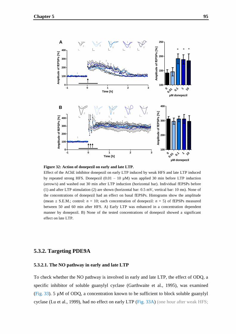
Chapter 5 95
-1 0 1 2 30
100
200
300
400
Time [h]
Am
plit
ud
e o
f fE
PS
Ps [
%]
0
0.01
0.
1 1 10
100
150
200
250
Am
plitu
de o
f fE
PS
Ps [
%]
µM donepezil
-1 0 1 2 30
100
200
300
400
Time [h]
Am
plitu
de o
f fE
PS
Ps [
%]
0
0.01
0.
1 1 10
100
200
300
400
Am
plitu
de o
f fE
PS
Ps [
%]
µM donepezil
A
B
1
2
1 1
2
1 1
22 2
1
2
1
2
1
2
1
2
1 1
2 2
2
1
* * *
5.3.2. Targeting PDE9A
5.3.2.1. The NO pathway in early and late LTP
To check whether the NO pathway is involved in early and late LTP, the effect of ODQ, a
specific inhibitor of soluble guanylyl cyclase (Garthwaite et al., 1995), was examined
(Fig. 33). 5 µM of ODQ, a concentration known to be sufficient to block soluble guanylyl
cyclase (Lu et al., 1999), had no effect on early LTP (Fig. 33A) (one hour after weak HFS;
Figure 32: Action of donepezil on early and late LTP.
Effect of the AChE inhibitor donepezil on early LTP induced by weak HFS and late LTP induced
by repeated strong HFS. Donepezil (0.01 – 10 µM) was applied 30 min before LTP induction
(arrow/s) and washed out 30 min after LTP induction (horizontal bar). Individual fEPSPs before
(1) and after LTP stimulation (2) are shown (horizontal bar: 0.5 mV, vertical bar: 10 ms). None of
the concentrations of donepezil had an effect on basal fEPSPs. Histograms show the amplitude
(mean ± S.E.M.; control: n = 10; each concentration of donepezil: n = 5) of fEPSPs measured
between 50 and 60 min after HFS. A) Early LTP was enhanced in a concentration dependent
manner by donepezil. B) None of the tested concentrations of donepezil showed a significant
effect on late LTP.
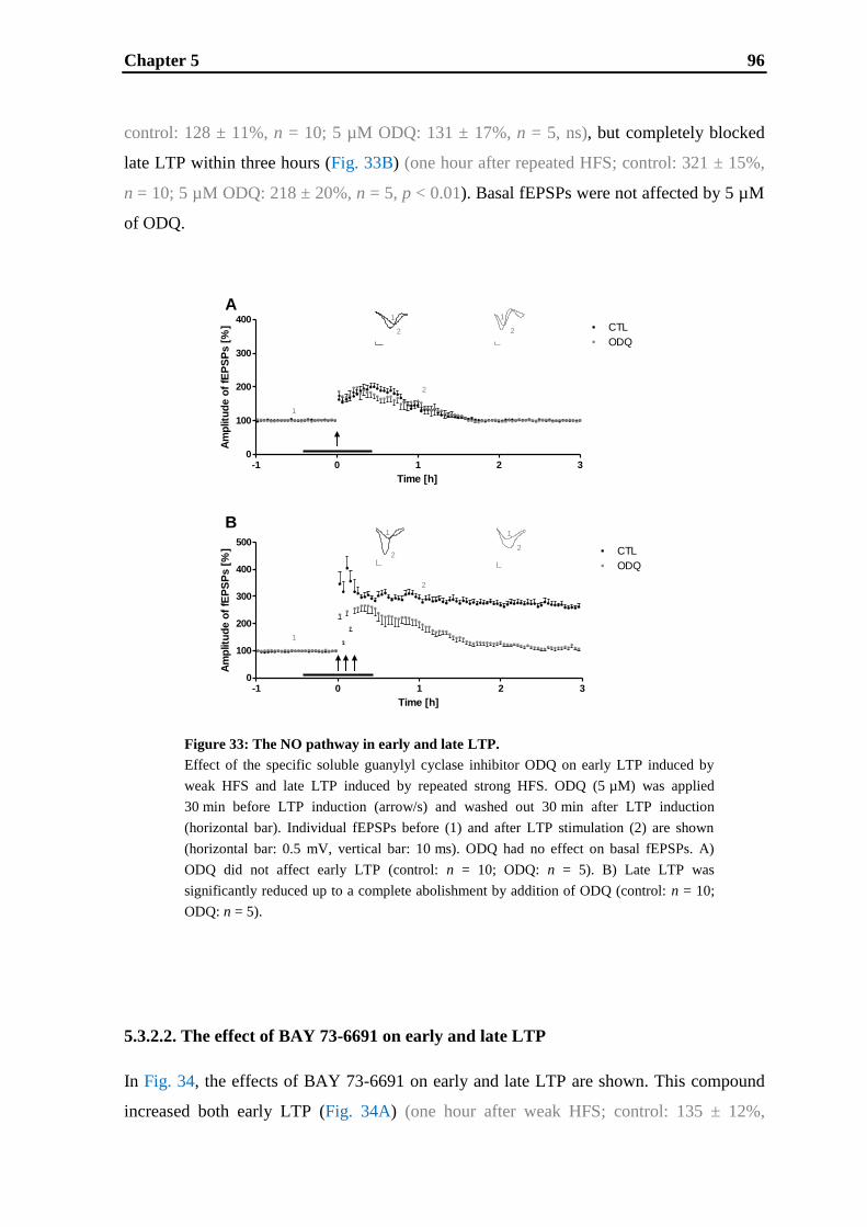
Chapter 5 96
-1 0 1 2 30
100
200
300
400
500CTL
ODQ (5 µM)
Time [h]
Am
plitu
de o
f fE
PS
Ps [
%]
-1 0 1 2 30
100
200
300
400
Time [h]
Am
plitu
de o
f fE
PS
Ps [
%]
-1 0 1 2 30
100
200
300
400
500CTL
ODQ (5 µM)
Time [h]
Am
plitu
de o
f fE
PS
Ps [
%]
1
1
A
B
2
1
1
2
1
2
2
2
1
2
control: 128 ± 11%, n = 10; 5 µM ODQ: 131 ± 17%, n = 5, ns), but completely blocked
late LTP within three hours (Fig. 33B) (one hour after repeated HFS; control: 321 ± 15%,
n = 10; 5 µM ODQ: 218 ± 20%, n = 5, p < 0.01). Basal fEPSPs were not affected by 5 µM
of ODQ.
5.3.2.2. The effect of BAY 73-6691 on early and late LTP
In Fig. 34, the effects of BAY 73-6691 on early and late LTP are shown. This compound
increased both early LTP (Fig. 34A) (one hour after weak HFS; control: 135 ± 12%,
Figure 33: The NO pathway in early and late LTP.
Effect of the specific soluble guanylyl cyclase inhibitor ODQ on early LTP induced by
weak HFS and late LTP induced by repeated strong HFS. ODQ (5 µM) was applied
30 min before LTP induction (arrow/s) and washed out 30 min after LTP induction
(horizontal bar). Individual fEPSPs before (1) and after LTP stimulation (2) are shown
(horizontal bar: 0.5 mV, vertical bar: 10 ms). ODQ had no effect on basal fEPSPs. A)
ODQ did not affect early LTP (control: n = 10; ODQ: n = 5). B) Late LTP was
significantly reduced up to a complete abolishment by addition of ODQ (control: n = 10;
ODQ: n = 5).

Chapter 5 97
Figure 34: Effect of BAY 73-6691 on early and late LTP.
Effect of the PDE9A inhibitor BAY 73-6691 on early LTP induced by weak HFS and late LTP
induced by repeated strong HFS. BAY 73-6691 (0.1 – 10 µM) was applied 30 min before LTP
induction (arrow/s) and washed out 30 min after LTP induction (horizontal bar). None of the
concentrations of BAY 73-6691 had an effect on basal fEPSPs. Individual fEPSPs before (1) and
after LTP stimulation (2) are shown (horizontal bar: 0.5 mV, vertical bar: 10 ms). Histograms
show the amplitude (mean ± S.E.M.; control: n = 10; each concentration of BAY 73-6691: n = 5)
of fEPSPs measured between 50 and 60 min after HFS. A) Early LTP was enhanced by BAY 73-
6691 in a concentration dependent manner. B) BAY 73-6691 increased late LTP concentration
dependently.
-1 0 1 2 30
100
200
300
400
Time [h]
Am
plitu
de o
f fE
PS
Ps [
%]
00.
1 1 10 100
100
150
200
250
Am
plitu
de o
f fE
PS
Ps [
%]
µM BAY 73-6691
-1 0 1 2 30
100
200
300
400
Time [h]
Am
plit
ud
e o
f fE
PS
Ps [
%]
00.
11
0 1010
0 100
200
300
400
Am
plitu
de o
f fE
PS
Ps [
%]
µM BAY 73-6691
A
B
1
2
1
21 1 1
222
**
*** *
1
2
1
2
1
2
1
2
1 1 1
22 2
n = 10; 0.1 µM BAY 73-6691: 128 ± 18, n = 5, ns; 1 µM BAY 73-6691: 220 ± 20, n = 5,
p < 0.01; 10 µM BAY 73-6691: 195 ± 11%, n = 5, p < 0.01) and late LTP (Fig. 34B) (one
hour after repeated HFS; control: 316 ± 16%, n = 10; 0.1 µM BAY 73-6691: 308 ± 14%,
n = 5, ns; 1 µM BAY 73-6691: 357 ± 14%, n = 5, p < 0.05; 10 µM BAY 73-6691:
345 ± 20%, n = 5, p < 0.05) in a concentration dependent manner (0.01 – 10 µM) showing
a significant enhancement at 1 - 10 μM for both forms of LTP. In both cases, these
enhancements lasted for more than three hours after LTP stimulation. Basal fEPSPs were
not affected by any concentration of BAY 73-6691.
Chapter 5 98
5.3.2.3. Transformation of early into late LTP due to BAY 73-6691
To analyze whether the long-lasting increase of early LTP by BAY 73-6691 is attributed to
the transformation of protein-synthesis independent early LTP into protein-synthesis
dependent late LTP, the reversible protein translation inhibitor anisomycin was used at a
concentration of 30 µM, which is known to be sufficient to significantly impair protein-
synthesis dependent late LTP (Kroker et al., 2011b; Lu et al., 1999). As shown in Fig. 35A,
the addition of 30 µM anisomycin to 1 μM BAY 73-6691, a concentration shown to
increase early LTP (see Fig. 34A), caused a significant decrease of BAY 73-6691-
mediated enhancement of early LTP up to a complete abolishment within two to three
hours after LTP induction (one hour after weak HFS; control: 135 ± 12%, n = 10; 1 µM
BAY 73-6691: 220 ± 20%, n = 5, p < 0.01; 1 µM BAY 73-6691 + 30 µM anisomycin:
150 ± 14%, n = 5, ns). This indicates that protein-synthesis independent early LTP was
transformed into protein-synthesis dependent late LTP by BAY 73-6691. Basal fEPSPs
were not affected by the mixture of BAY 73-6691 and anisomycin.
To test whether the transformation from early into late LTP by BAY 73-6691 is attributed
to the NO pathway, the effect of the soluble guanylyl cyclase inhibitor ODQ was analyzed.
5 µM of ODQ, a concentration shown to be sufficient to significantly impair protein-
synthesis dependent late LTP (see Fig. 34B), was used. As shown in Fig. 35B, the addition
of 5 µM ODQ to 1 μM BAY 73-6691, a concentration shown to increase early LTP (see
Fig. 5A), caused a reversal of the BAY 73-6691-mediated enhancement of early LTP (one
hour after weak HFS; control: 135 ± 12%, n = 10; 1 µM BAY 73-6691: 220 ± 20%, n = 5,
p < 0.01; 1 µM BAY 73-6691 + 5 µM ODQ: 139 ± 16%, n = 5, ns). This indicates that the
transformation from early into late LTP by BAY 73-6691 is based on the NO pathway.
Basal fEPSPs were not affected by the mixture of BAY 73-6691 and ODQ.
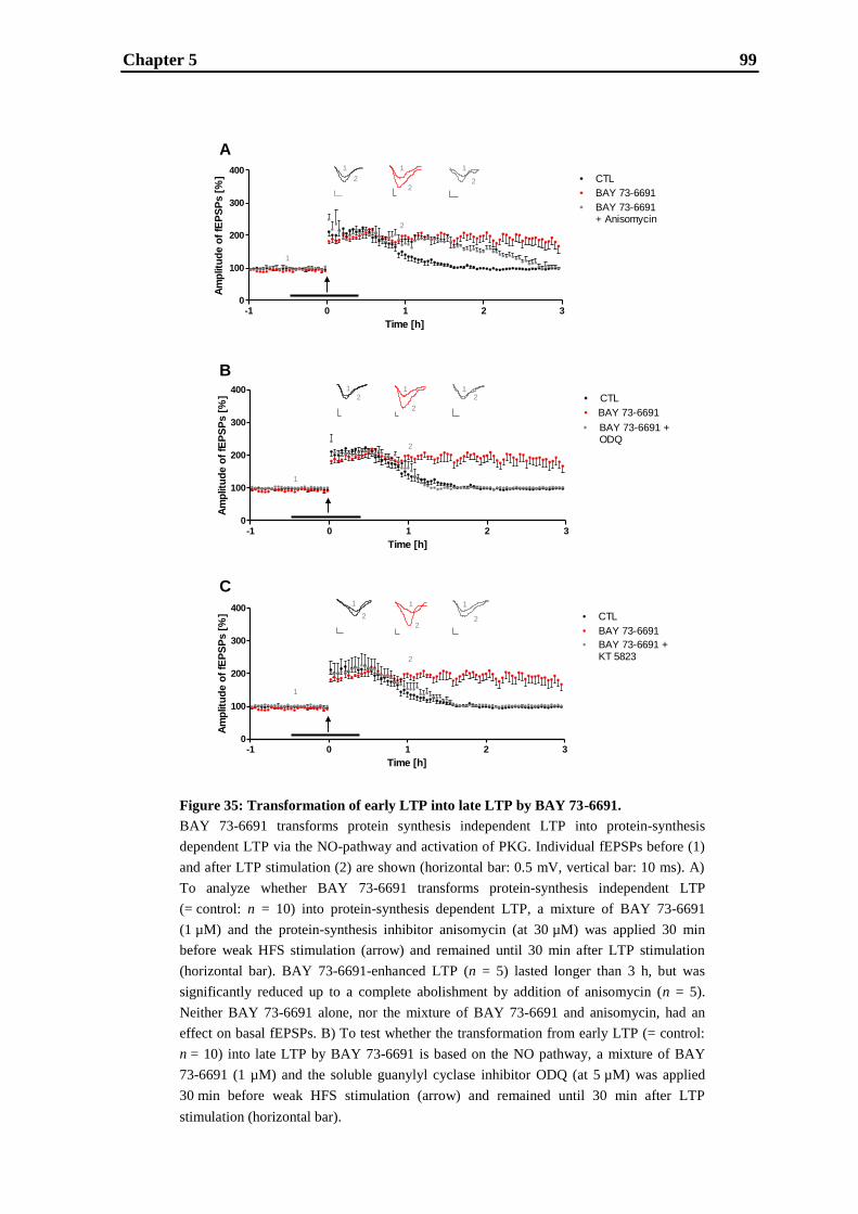
Chapter 5 99
Figure 35: Transformation of early LTP into late LTP by BAY 73-6691.
BAY 73-6691 transforms protein synthesis independent LTP into protein-synthesis
dependent LTP via the NO-pathway and activation of PKG. Individual fEPSPs before (1)
and after LTP stimulation (2) are shown (horizontal bar: 0.5 mV, vertical bar: 10 ms). A)
To analyze whether BAY 73-6691 transforms protein-synthesis independent LTP
(= control: n = 10) into protein-synthesis dependent LTP, a mixture of BAY 73-6691
(1 µM) and the protein-synthesis inhibitor anisomycin (at 30 µM) was applied 30 min
before weak HFS stimulation (arrow) and remained until 30 min after LTP stimulation
(horizontal bar). BAY 73-6691-enhanced LTP (n = 5) lasted longer than 3 h, but was
significantly reduced up to a complete abolishment by addition of anisomycin (n = 5).
Neither BAY 73-6691 alone, nor the mixture of BAY 73-6691 and anisomycin, had an
effect on basal fEPSPs. B) To test whether the transformation from early LTP (= control:
n = 10) into late LTP by BAY 73-6691 is based on the NO pathway, a mixture of BAY
73-6691 (1 µM) and the soluble guanylyl cyclase inhibitor ODQ (at 5 µM) was applied
30 min before weak HFS stimulation (arrow) and remained until 30 min after LTP
stimulation (horizontal bar).
-1 0 1 2 30
100
200
300
400
Time [h]
Am
plit
ud
e o
f fE
PS
Ps [
%] CTL
Legend
BAY 73-6691+ Anisomycin
BAY 73-6691
-1 0 1 2 30
100
200
300
400
Time [h]
Am
plit
ud
e o
f fE
PS
Ps [
%] CTL
Legend
BAY 73-6691 +ODQ
BAY 73-6691
-1 0 1 2 30
100
200
300
400
Time [h]
Am
plitu
de o
f fE
PS
Ps [
%] CTL
Legend
BAY 73-6691 +KT 5823
BAY 73-6691
A
B
C
1
2
1 1
22
1
2
12
1 12
2
2
1
1
2
1 1
22
1
2

Chapter 5 100
continued:
BAY 73-6691-enhanced LTP (n = 5) was significantly reduced and the effect of BAY 73-
6691 was completely reversed (n = 5). Neither BAY 73-6691 alone, nor the mixture of
BAY 73-6691 and ODQ, had an effect on basal fEPSPs. C) To analyze whether BAY 73-
6691 transforms early LTP (= control: n = 10) into late LTP through the activation of
PKG, a mixture of BAY 73-6691 (1 µM) and the PKG-inhibitor KT 5823 (at 2 µM) was
applied 30 min before weak HFS stimulation (arrow) and remained until 30 min after LTP
stimulation (horizontal bar). BAY 73-6691-enhanced LTP (n = 5) was significantly
reduced and the effect of BAY 73-6691 was completely reversed (n = 5). Neither BAY 73-
6691 alone, nor the mixture of BAY 73-6691 and KT 5823, had an effect on basal fEPSPs.
To analyze whether BAY 73-6691 transforms early into late LTP through the activation of
PKG, the PKG-inhibitor KT 5823 was used at a concentration of 2 µM, which is known to
be sufficient to significantly impair protein-synthesis dependent late LTP (Lu et al., 1999).
As shown in Fig. 35C, the addition of 2 µM KT 5823 to 1 μM BAY 73-6691, a
concentration shown to increase early LTP (see Fig. 34A), caused a reversal of the BAY
73-6691-mediated enhancement of early LTP (one hour after weak HFS; control:
135 ± 12%, n = 10; 1 µM BAY 73-6691: 220 ± 20, n = 5, p < 0.01; 1 µM BAY 73-6691 +
2 µM KT 5823: 138 ± 19%, n = 5, ns). This indicates that the transformation from early
into late LTP by BAY 73-6691 is attributed to the activation of PKG. Basal fEPSPs were
not affected by the mixture of BAY 73-6691 and KT 5823.

Chapter 5 101
5.4. Discussion
The AChE inhibitor donepezil is the current gold standard for symptomatic treatment of
mild-to-moderate AD patients (Birks and Harvey, 2003; Whitehead et al., 2004). By
blocking AChE, it increases and prolongs the concentration of ACh in the synaptic cleft in
order to compensate for the cholinergic deficit in AD patients. These increased
extracellular levels of ACh may activate metabotropic muscarinic and ionotropic nicotinic
acetylcholine receptors, which finally lead to the pro-cognitive effects of donepezil.
However, experience from clinical practice with AChE inhibitors has shown only moderate
efficacy, emergence of tolerance after long-term use, interaction of cholinergic and non-
cholinergic effects and possible effects of amyloid precursor protein release (Courtney et
al., 2004; Diniz et al., 2009; Helou and Rhalimi, 2010; Hernandez et al., 2009;
Kaduszkiewicz et al., 2005; Rosenbloom et al., 2010; Rozzini et al., 2004). As a potential
new approach, inhibition of specific PDEs has come into the focus of interest for the
treatment of memory impairment (Menniti et al., 2006; Reneerkens et al., 2009; Schmidt,
2010). One of these PDEs is PDE9A, which contributes to the intracellular NO-sGC-
cGMP signalling cascade by its cGMP hydrolytic activity (Arancio et al., 2001; Son et al.,
1998; Zhuo et al., 1994). Under physiological conditions, cGMP is formed by NO-
sensitive soluble guanylyl cyclases (NO-GCs) (Garthwaite, 2008). These NO-GCs are
activated by NO, which is generated by calcium/calmodulin-dependent neuronal NO
synthases (Christopherson et al., 1999). NO is believed to act as a retrograde messenger
and has been implicated as a neuromodulator in synaptic transmission (Böhme et al., 1991;
Boehning and Snyder, 2003; Garthwaite, 2008; O‟Dell et al., 1991; Schuman and Madison,
1991). Indeed, presynaptic cGMP facilitates glutamate release (Neitz et al., 2011).
Furthermore, cGMP acts postsynaptically as part of the sGC-cGMP-PKG pathway, known
to indirectly activate the transcription factor CREB (Ko and Kelly, 1999; Lu et al., 1999).
Thus, overall elevation of cGMP results in increased glutamate release presynaptically and
increased phosphorylation of CREB postsynaptically, both being important mechanisms
for learning and memory (Blokland et al., 2006; Prickaerts et al., 2004; Rutten et al., 2007;
Silva, et al., 1998; Son et al., 1998). Indeed, it was shown that PDE9A inhibitors can
increase cGMP levels in the brain of animals (Verhoest et al., 2009) and in CSF of animals
and humans (Hutson et al., 2011; Nicholas et al., 2009), and furthermore, memory
enhancing efficacy of the PDE9A inhibitor BAY 73-6691 was demonstrated in various
Chapter 5 102
animal cognition tasks (Hutson et al., 2011; van der Stay et al., 2008). In order to gain
more insight into the mechanistic roles of AChE inhibitors and PDE9A inhibitors in
learning and memory, we analyzed the effects of donepezil and the potent, selective
PDE9A inhibitor BAY 73-6691 (Wunder et al., 2005) on LTP measured in rat
hippocampal slices.
We report here that donepezil is capable of prolonging and hence enhancing early LTP in
rat hippocampal slices in a concentration dependent manner with a minimal efficacious
concentration of 0.1 µM (see Fig. 32A). This supports previous findings showing that
donepezil is able to delay the decay of LTP in vivo (Barnes et al., 2000) - the only LTP
study published so far using donepezil - and to enhance cognitive function in animals
(McCarthy et al., 2011; Prickaerts et al., 2005; van der Staay and Bouger, 2005) and
humans (Jacobson and Sabbagh, 2008; Rogers et al., 1998; Seltzer et al., 2004; Tsao and
Heilman, 2005). In our study, donepezil had no effect on late LTP (see Fig. 32B) which is
in agreement with a previous report on the failure of the AChE inhibitor galantamine to
increase LTP induced by two tetani (100 Hz for 1 s, 5 s time interval; Lagostena et al.,
2008), a stimulation protocol which is believed to induce late LTP (Albensi et al., 2007;
Huang and Kandel, 1994). Donepezil having no effect on late LTP cannot be attributed to
an insufficient increase of ACh by LTP stimulation, since the muscarinic acetylcholine
receptor antagonist pirenzepine caused an impairment of late LTP (see Fig. 30) suggesting
that the cholinergic system is indeed involved in late LTP and functional in the slice
preparation. Moreover, AChE expression, AChE activity and blockade of AChE activity
by donepezil were confirmed in hippocampal slices (see Fig. 31A-C). It also cannot be
attributed to changes of AChE expression in hippocampal slices (see Fig. 31A), nor to a
decrease of AChE activity in hippocampal slices versus freshly prepared homogenate
(Fig. 31B), nor a failure of donepezil to block AChE activity in hippocampal slices
(Fig. 31C: the IC50 of donepezil is roughly in the range of published data (Geerts et al.,
2005; Jackisch et al., 2009; Naik et al., 2009)). Thus, even though donepezil has the
potential to augment early LTP, we show that it has only limited impact on synaptic
plasticity, since it cannot enhance late LTP.
The differentiation between early and late LTP is of importance for the evaluation of
downstream effects. Early LTP is independent of protein-synthesis (Bliss and Collingridge,
1993; Kroker et al., 2011b; Malenka and Nicoll, 1999; Reyman and Frey, 2007) and
involves modifications of pre-existing synapses as a result of rapid calcium influx through
NMDA receptors and subsequent phosphorylation events (Derkach et al., 1999; Malenka

Chapter 5 103
and Bear, 2004) as well as insertion of additional AMPA receptors into the postsynaptic
membrane (Davies et al., 1989; Liao et al., 2001; Lu et al., 2001; Malenka and Bear, 2004;
Malinow, 2003; Shi et al., 1999, 2001). The time-window of early LTP corresponds with
the duration of short-term memory as defined by some researchers, i.e. short-term memory
is a form of memory which does not require gene expression and protein-synthesis
(Grecksch and Matthies, 1980; Izquierdo et al., 2002). Late LTP is clearly separated from
early LTP by its dependence on de novo protein-synthesis (Frey et al., 1988; Huang, 1998,
1996; Huang and Kandel, 1994; Kroker et al., 2011b; Nguyen et al., 1991; Nguyen and
Kandel, 1997; Reymann and Frey, 2007). Protein-synthesis may contribute to both the
modification of existing synapses and to the establishment of new synaptic contacts (Buchs
and Muller, 1996; Chang and Greenough, 1984; Desmond and Levy, 1988; Lee et al.,
1980; Lisman, 2003). Moreover, long-lasting forms of memory are generally believed to
be protein-synthesis dependent (Costa-Mattioli et al., 2009; Grecksch and Matthies, 1980;
Pang and Lu, 2004).
In this study, the role of the NO pathway in LTP was assessed using the sGC inhibitor
ODQ and the results demonstrate that this pathway is important for late, but not for early
LTP (see Fig. 33). These results confirm other studies showing that the involvement of the
NO pathway is strongly dependent on the LTP stimulation protocol (Chetkovich et al.,
1993; Gribkoff and Lum-Ragan, 1992; Haley et al., 1996; Lu et al., 1999; Malen and
Chapman, 1997; O‟Dell et al., 1994; Williams et al., 1993; Zhuo et al., 1999). For the
PDE9A inhibitor BAY 73-6691, we show that both early and late LTP are increased in a
bell-shaped manner with efficacious concentrations of 1 and 10 µM. The lack of efficacy
of BAY 73-6691 at the highest concentration might result from an excessive increase in
cGMP relative to the augmentation in the cAMP level as suggested by van der Staay et al.
(2008). This might cause activation of compensatory mechanisms leading to alleviation of
LTP enhancement. Our early LTP data confirm previous findings (van der Staay et al.,
2008) showing an increase of early LTP in a bell-shaped manner. Compared to this study,
we observed a slight left shift of effective concentrations, which may be due to the
different protocols used to induce early LTP. The present study further demonstrates that
BAY 73-6691 not only increases protein-synthesis independent LTP (early LTP) in a
concentration dependent manner, but transforms it into protein-synthesis dependent LTP
(late LTP) due to the activation of the NO pathway and particularly PKG (see Fig. 35).
This transformation into late LTP is consistent with findings regarding the transformation
of early into late LTP in mice hippocampal slices by Br-cGMP (Lu et al., 1999) and with
Chapter 5 104
the basic idea that information in the short-term memory can be transferred into long term
memory (Baddeley 2003; Pang et al., 2004). Hence, this study demonstrates that donepezil
showed no enhancement of late LTP and only a moderate increase in early LTP, whereas
inhibition of PDE9A led to a strong enhancement of early LTP with its transformation into
late LTP. Furthermore, PDE9A inhibition was even able to increase protein-synthesis
dependent late LTP.
In conclusion, this study demonstrates that BAY 73-6691 is more efficacious than
donepezil in enhancing and prolonging LTP in rat hippocampal slices. This direct
comparison of the effects the AChE inhibitor and the PDE9A inhibitor on early and late
LTP indicates that PDE9A inhibition might also be more efficacious in enhancing
cognitive function in vivo. These results may also provide a mechanistic rationale for the
apparently modest efficacy of donepezil for treating memory impairment in AD patients,
and suggest that there is a potential for alternative or adjunctive therapies with greater
impact on synaptic plasticity, which may be superior to AChE inhibition alone.
Chapter 5 105
CHAPTER 6
Effects of cognitive enhancing drugs on amyloid-
beta oligomer induced impairment of LTP
Chapter 6 106
Abstract
Brains of Alzheimer‟s disease (AD) patients are histopathologically characterized by
extracellular senile plaques consisting of fibrillary amyloid-β (Aβ) deposits and by
intracellular neurofibrillary tangles composed of hyperphosphorylated tau protein. Soluble
Aβ oligomers, also referred to as Aβ derived diffusible ligands (ADDLs), are thought to be
neuro- or synaptotoxic Aβ-species causative for AD. In order to gain more insight into the
mechanistic role of ADDLs in a cellular model of learning and memory, we analyzed their
effect on hippocampal long-term potentiation (LTP) in the CA1 region of rat hippocampal
slices. This study demonstrates that early LTP, being protein-synthesis independent, and
late LTP, being protein-synthesis dependent, are affected to different extents by ADDLs.
Furthermore, ADDLs were shown to exclusively target NMDA receptors and/or their
signaling cascades. These results corroborate the hypothesis that soluble Aβ oligomers
cause synaptic dysfunction which might lead to cognitive decline seen in AD patients.
Furthermore, cognitive enhancing (pre-)clinical drugs acting on different targets tested at
their efficacious concentration regarding LTP enhancement were evaluated for their ability
to ameliorate ADDLs induced LTP deficits with the aim to determine the potential
therapeutic value of these targets as alternative strategies for the treatment of AD. The
current gold standard for treatment of AD patients is the acetylcholine esterase inhibitor
donepezil, which was demonstrated to slightly restore ADDLs induced impairment of early
LTP, but to have no effect on ADDLs induced impairment of late LTP. The same was
shown for the α4β2 nicotinic acetylcholine receptor (nAChR) agonist TC-1827. Both, the
α7 nAChR partial agonist SSR180711 and the phosphodiesterase (PDE) 9A inhibitor BAY
73-6691 completely rescued early as well as late LTP impaired by ADDLs. The
metabotropic glutamate 5 receptor positive allosteric modulator ADX-47273 had no effect
on ADDLs impaired early LTP, but partly restored ADDLs impaired late LTP. Thus,
activating α7 nAChRs and inhibiting PDE9 were found to be most effective in
ameliorating ADDLs induced LTP deficits, which suggests that targeting α7 nAChRs and
PDE9 might represent powerful alternative approaches for the treatment of AD.
Chapter 6 107
6.1. Introduction
Alzheimer‟s disease (AD) is a neurodegenerative disorder characterized by progressive
cognitive decline and specific pathological findings in patients. Histopathological
hallmarks of AD brains are extracellular senile plaques of amyloid- (A ) peptides and
intracellular neurofibrillary tangles of tau protein (Gouras et al., 2005; LaFerla and Oddo,
2005; Querfurth and LaFerla, 2010). The predominant types of Aβ peptides in senile
plaques are Aβ42 and Aβ40 (Fodale et al., 2006). As a result of aging and AD, subtle
changes in Aβ generation, or its metabolism, may occur causing an increased production
especially of the Aβ42 peptide leading to its accumulation and deposition in the brain
(Borchelt et al., 1996; Duff et al., 1996; Hardy and Selkoe, 2002). As for the relationship
between Aβ and neurofibrillary tangles in AD, it is suggested that the development of tau
is a downstream consequence of the Aβ pathology (Hardy and Selkoe, 2002; Kitazawa et
al., 2005; Oddo et al., 2008; Tseng et al., 2008). Due to the apparent involvement of Aβ in
AD, the “amyloid cascade hypothesis” was postulated, which originally links the
pathological process of AD and neuronal cell death to the aggregation and deposition of
Aβ (Hardy and Higgins, 1992). Current investigations in animal models and human brain
samples have placed a special emphasis on soluble Aβ aggregates (Dahlgren et al., 2002;
Klein, 2002; Lue et al., 1999; McLean et al., 1999; Tabaton and Piccini, 2005; Walsh et al.,
1999; Youssef et al., 2008), also referred to as amyloid-β derived diffusible ligands
(ADDLs). Indeed, these soluble oligomers are currently believed to be the putative cause
of AD, rather than insoluble plaques (Hardy and Selkoe, 2002; Small, 1998).
In order to gain more insight into the mechanistic role of ADDLs in a cellular model of
learning and memory, we analyzed the effects of synthetic ADDLs on hippocampal long-
term potentiation (LTP). LTP is a widely-used cellular experimental model of memory
formation (Bliss and Collingridge, 1993), which can be subclassified into early and late
forms of LTP having different mechanisms (Kroker et al., 2011b; Reymann and Frey,
2007). Some studies already demonstrated that ADDLs elicit an impairment of LTP in the
CA1 region of the hippocampus (Chen et al., 2000; Kim et al., 2001; Klybin et al., 2004;
Raymond et al., 2003; Shankar et al., 2008; Walsh et al., 2002), but the effects of ADDLs
on the different forms of LTP have not been addressed so far. Therefore, in this study, we
analyzed the effects of synthetic ADDLs on validated early and late hippocampal CA1
LTP. These results provide a more detailed insight into the mechanistic role of soluble Aβ
oligomers in CA1 LTP and their effect on NMDA receptor signaling. Furthermore, we
Chapter 6 108
evaluated several cognitive enhancing (pre-)clinical drugs acting on different targets - the
acetylcholine esterase (AChE) inhibitor donepezil, the α4β2 nicotinic acetylcholine
receptor (nAChR) agonist TC-1827, the α7 nAChR partial agonist SSR180711, the
phosphodiesterase (PDE) 9A inhibitor BAY 73-6691 and the metabotropic glutamate 5
(mGlu5) receptor positive modulator ADX-47273 - for their ability to ameliorate ADDLs
induced LTP deficits in order to determine the potential therapeutic value of these targets
for the treatment of AD. All these drugs were tested at their efficacious concentration
regarding LTP enhancement (Kroker et al., 2011b,c,d).
Chapter 6 109
6.2. Materials and methods
6.2.1. Preparation of brain slices
Procedures involving animals and their care were conducted in conformity with
institutional and European Union guidelines (EEC Council Directive 86/609) and were
approved by the Ethical Committee of the responsible regional council (Tübingen). Male
Wistar rats (Janvier, Le Genest Saint Isle, France) aged six to seven weeks were shortly
anaesthetized with isoflurane and sacrificed by decapitation. The brains were quickly
removed and immersed in ice-cold ACSF (containing in mM NaCl 124, KCl 4.9, MgSO4
8, CaCl2 2.5, KH2PO4 1.2, NaHCO3 25.6, D-glucose 10, pH 7.4, saturated with 95% O2
and 5% CO2). Transverse hippocampal brain slices (400 µm thickness) were cut using a
Vibratome. Slices were allowed to recover in a holding chamber containing ACSF bubbled
with 95% O2 and 5% CO2 at room temperature for at least one hour. Slices were then
transferred to integrated brain slice chambers and continuously superfused (at a flow rate
of 2.5 mL min−1
, 25 ± 0.2 °C) with the same ACSF composition used before except for
0.5 mM MgSO4. Prior to doing any electrophysiological recordings, the slices were
allowed to equilibrate for at least 30 minutes.
6.2.2. Multi-slice recording
To record field excitatory postsynaptic potentials (fEPSPs) the SliceMaster multi-slice
recording system (Scientifica Limited, East Sussex, UK) was used. The system is semi-
automated allowing one operator to record simultaneously from an assembly of up to eight
brain slices. For the present study, an optimized system described by Kroker et al. (2011a)
was used. fEPSPs were elicited in the CA1 region by stimulation of the Schaffer
collateral–commissural fibres in the stratum radiatum using glass electrodes with broken
tips (filled with ACSF). For recording, glass electrodes (2 – 6 MΩ, filled with ACSF) were
placed in the apical dendritic layer. The amplitudes of fEPSPs were used as the parameter
of interest (Collingridge et al., 1983). To generate fEPSPs at a constant sub-threshold
stimulus, as in previous studies (Seabrook et al., 1997), the stimulus strength of the pulses
was adjusted to 20 - 30% of the fEPSP maximum and this voltage was used for the
experiment. During baseline recording each slice was stimulated every 30 seconds for at
Chapter 6 110
least one hour. Early LTP was induced by weak high frequency stimulation (HFS) made up
of 20 pulses at the frequency of 100 Hz (Bashir et al., 1991; Kroker et al., 2011a,b),
whereas late LTP was induced by repeated strong HFS consisting of 100 pulses at the
frequency of 100 Hz, repeated two times in five minute intervals (Kroker et al., 2011a,b;
Lu et al., 1999).
6.2.3. Data acquisition, software and analysis
A modular electrophysiology system, supplied by npi electronic GmbH (Tamm, Germany),
conducts the low noise recordings of extracellular signals. AC coupled signals are
amplified 1000-fold and internally filtered with a five kHz low-pass filter as well as a three
Hz high-pass filter. In our study, a customized system (programmable pattern generator)
was used allowing simultaneous stimulation of all slices either with a single stimulus or a
specific stimulus pattern (Kroker et al., 2011a). For data acquisition and analysis the
software Notocord® was used.
Data are shown as mean percent (± S.E.M.) of the baseline fEPSP amplitude. Data were
analyzed using either t tests to compare two conditions or one-way ANOVA (post-hoc test:
Dunnett-Test) to compare multiple conditions. For rescue experiments, the effect of
ADDLs on LTP was compared with the effect of ADDLs and the compound on LTP. In all
cases a p-value of ≤ 0.05 was considered significant. Histograms show the mean amplitude
(± S.E.M.) of fEPSPs measured between 25 and 35 minutes after weak or repeated strong
HFS according to Jia et al. (2010) and Kroker et al. (2011b).
6.2.4. Aβ (ADDLs) preparation and application
ADDLs were prepared according to Lambert et al. (1998) with slight modifications
(Moreth et al., 2011). Briefly, Aβ42 (purchased from Bachem) was solubilized in
1,1,1,3,3,3-hexafluoro-2-propanol by sonication (10 minutes) to a final concentration of
1 mM. Then, it was flash-frozen in liquid nitrogen and lyophilized. The lyophilized Aβ42
was solubilized in 10 mM NaOH and brought up in ice cold Ham‟s F12 medium (w/o
phenol red) to a final concentration of 100 µM (pH 7.4 at 4 °C for at least 14 hours). The
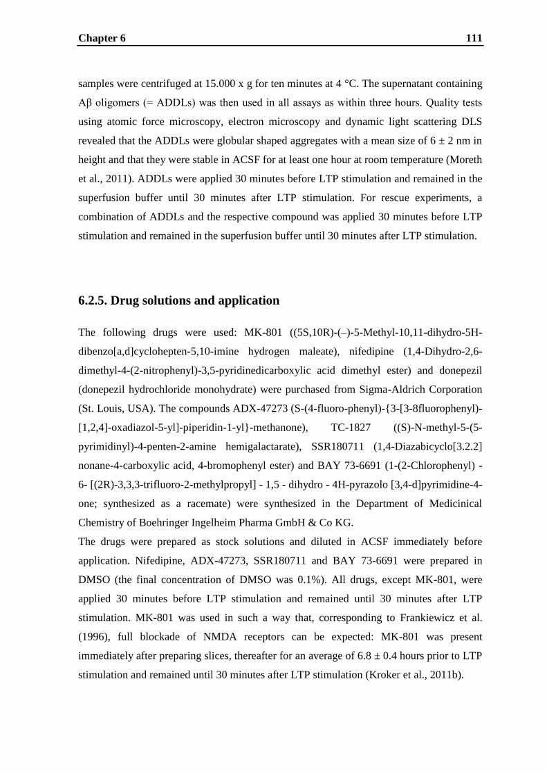
Chapter 6 111
samples were centrifuged at 15.000 x g for ten minutes at 4 °C. The supernatant containing
Aβ oligomers (= ADDLs) was then used in all assays as within three hours. Quality tests
using atomic force microscopy, electron microscopy and dynamic light scattering DLS
revealed that the ADDLs were globular shaped aggregates with a mean size of 6 ± 2 nm in
height and that they were stable in ACSF for at least one hour at room temperature (Moreth
et al., 2011). ADDLs were applied 30 minutes before LTP stimulation and remained in the
superfusion buffer until 30 minutes after LTP stimulation. For rescue experiments, a
combination of ADDLs and the respective compound was applied 30 minutes before LTP
stimulation and remained in the superfusion buffer until 30 minutes after LTP stimulation.
6.2.5. Drug solutions and application
The following drugs were used: MK-801 ((5S,10R)-(–)-5-Methyl-10,11-dihydro-5H-
dibenzo[a,d]cyclohepten-5,10-imine hydrogen maleate), nifedipine (1,4-Dihydro-2,6-
dimethyl-4-(2-nitrophenyl)-3,5-pyridinedicarboxylic acid dimethyl ester) and donepezil
(donepezil hydrochloride monohydrate) were purchased from Sigma-Aldrich Corporation
(St. Louis, USA). The compounds ADX-47273 (S-(4-fluoro-phenyl)-{3-[3-8fluorophenyl)-
[1,2,4]-oxadiazol-5-yl]-piperidin-1-yl}-methanone), TC-1827 ((S)-N-methyl-5-(5-
pyrimidinyl)-4-penten-2-amine hemigalactarate), SSR180711 (1,4-Diazabicyclo[3.2.2]
nonane-4-carboxylic acid, 4-bromophenyl ester) and BAY 73-6691 (1-(2-Chlorophenyl) -
6- [(2R)-3,3,3-trifluoro-2-methylpropyl] - 1,5 - dihydro - 4H-pyrazolo [3,4-d]pyrimidine-4-
one; synthesized as a racemate) were synthesized in the Department of Medicinical
Chemistry of Boehringer Ingelheim Pharma GmbH & Co KG.
The drugs were prepared as stock solutions and diluted in ACSF immediately before
application. Nifedipine, ADX-47273, SSR180711 and BAY 73-6691 were prepared in
DMSO (the final concentration of DMSO was 0.1%). All drugs, except MK-801, were
applied 30 minutes before LTP stimulation and remained until 30 minutes after LTP
stimulation. MK-801 was used in such a way that, corresponding to Frankiewicz et al.
(1996), full blockade of NMDA receptors can be expected: MK-801 was present
immediately after preparing slices, thereafter for an average of 6.8 ± 0.4 hours prior to LTP
stimulation and remained until 30 minutes after LTP stimulation (Kroker et al., 2011b).
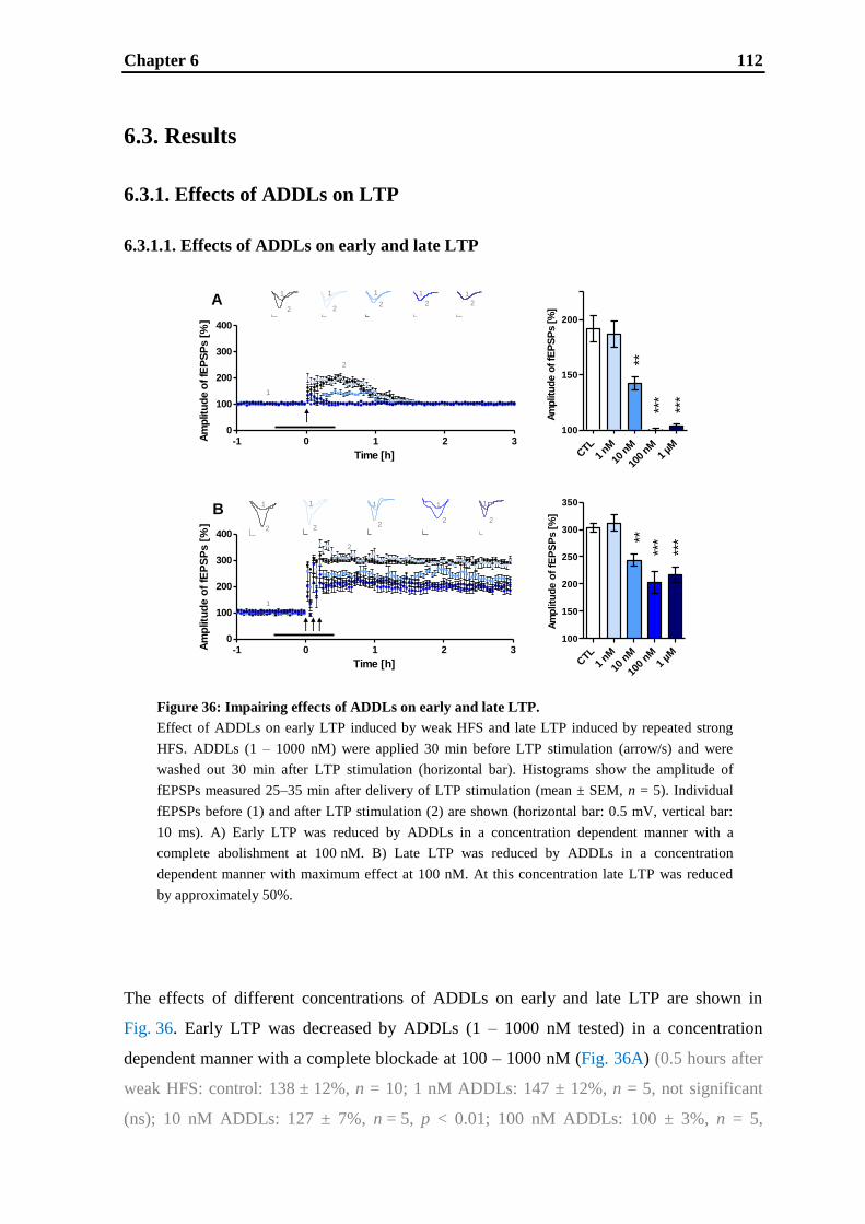
Chapter 6 112
A
B
-1 0 1 2 30
100
200
300
400
Time [h]
Am
plitu
de o
f fE
PS
Ps [
%]
Am
plitu
de
of
fEP
SP
s [
%]
CTL
1 nM
10 n
M
100
nM1
µM
100
150
200
250
300
350
***
***
**
***
*****
-1 0 1 2 30
100
200
300
400
Time [h]
Am
plit
ud
e o
f fE
PS
Ps [
%]
Am
plit
ud
e o
f fE
PS
Ps [%
]
CTL
1 nM
10 n
M
100
nM1
µM
100
150
200
1
2
1
2
1
2
1
2
1
2
1
2
1
2
1
2
1
2
1
2
1
2
1
2
6.3. Results
6.3.1. Effects of ADDLs on LTP
6.3.1.1. Effects of ADDLs on early and late LTP
The effects of different concentrations of ADDLs on early and late LTP are shown in
Fig. 36. Early LTP was decreased by ADDLs (1 – 1000 nM tested) in a concentration
dependent manner with a complete blockade at 100 – 1000 nM (Fig. 36A) (0.5 hours after
weak HFS: control: 138 ± 12%, n = 10; 1 nM ADDLs: 147 ± 12%, n = 5, not significant
(ns); 10 nM ADDLs: 127 ± 7%, n = 5, p < 0.01; 100 nM ADDLs: 100 ± 3%, n = 5,
Figure 36: Impairing effects of ADDLs on early and late LTP.
Effect of ADDLs on early LTP induced by weak HFS and late LTP induced by repeated strong
HFS. ADDLs (1 – 1000 nM) were applied 30 min before LTP stimulation (arrow/s) and were
washed out 30 min after LTP stimulation (horizontal bar). Histograms show the amplitude of
fEPSPs measured 25–35 min after delivery of LTP stimulation (mean ± SEM, n = 5). Individual
fEPSPs before (1) and after LTP stimulation (2) are shown (horizontal bar: 0.5 mV, vertical bar:
10 ms). A) Early LTP was reduced by ADDLs in a concentration dependent manner with a
complete abolishment at 100 nM. B) Late LTP was reduced by ADDLs in a concentration
dependent manner with maximum effect at 100 nM. At this concentration late LTP was reduced
by approximately 50%.

Chapter 6 113
p < 0.001; 1000 nM ADDLs: 104 ± 2%, n = 5, p < 0.001). ADDLs (1 – 1000 nM tested)
also decreased late LTP in a concentration dependent manner with a maximum effect at
100 – 1000 nM (Fig., 36B) (0.5 hours after repeated strong HFS; control: 299 ± 8%,
n = 10; 1 nM ADDLs: 303 ± 10%, n = 5, ns; 10 nM ADDLs: 254 ± 11, n = 5, p < 0.01;
100 nM ADDLs: 217 ± 10%, n = 5, p < 0.001; 1000 nM ADDLs: 214 ± 11%, n = 5,
p < 0.001). At 100 – 1000 nM ADDLs concentration, late LTP was not completely
abolished, but reduced by approximately 50%. None of the concentrations of ADDLs
affected basal fEPSPs.
6.3.1.2. ADDLs impair late LTP by targeting NMDA receptor signaling
The signaling pathways putatively being impaired by ADDLs in late LTP were
investigated (Fig. 37). To analyze the potential effects of ADDLs on NMDA receptor
signaling, 10 µM of the NMDA receptor antagonist MK-801 was applied in addition to
100 nM ADDLs. This concentration of MK-801 was reported to be sufficient to block the
NMDA receptor dependent part of late LTP (Kroker et al., 2011b). As shown in Fig. 37A,
MK-801 had no further effect on ADDLs mediated impairment of late LTP (0.5 hours after
weak HFS; control: 299 ± 8%, n = 10; 100 nM ADDLs: 217 ± 10%, n = 5, p < 0.001;
100 nM ADDLs + 10 µM MK-801: 212 ± 11%, n = 5, p < 0.001). Basal fEPSPs were not
affected by the mixture of ADDLs and MK-801.
To analyze the role of L-type voltage dependent calcium channel (L-VDCC) signaling,
100 µM of the L-VDCC blocker nifedipine was applied in addition to 100 nM ADDLs.
This nifedipine concentration was reported to be sufficient to block the L-VDCCs
dependent part of late LTP (Kroker et al., 2011b). As shown in Fig. 37B, nifedipine
completely blocked the ADDLs impaired late LTP (0.5 hours after weak HFS; control:
299 ± 8%, n = 10; 100 nM ADDLs: 217 ± 10%, n = 5, p < 0.001; 100 nM ADDLs +
100 µM nifedipine: 99 ± 2%, n = 5, p < 0.001). Basal fEPSPs were not affected by the
mixture of ADDLs and nifedipine. Thus, NMDA receptors and/or NMDA receptor
downstream signaling, but not L-VDCCs and/or their downstream signaling, were
apparently targeted by ADDLs.

Chapter 6 114
A
B
-1 0 1 2 30
100
200
300
400
500
Time [h]
Am
plitu
de o
f fE
PS
Ps [
%] CTL
Legend
ADDLs +MK-801
-1 0 1 2 30
100
200
300
400
500
Time [h]
Am
plitu
de o
f fE
PS
Ps [
%] CTL
ADDLs
ADDLs +Nifedipine
-1 0 1 2 30
100
200
300
400
500
Time [h]
Am
plitu
de o
f fE
PS
Ps [
%] CTL
Legend
ADDLs +MK-801
1
2
1
2
1
2
1
2
1
2
1
2
1
2
1
2
Figure 37: ADDLs impair late LTP by exclusively targeting NMDA receptors and/or
their downstream signaling.
Effects of MK-801, a potent antagonist of NMDA receptors, and nifedipine, a L-VDCC
blocker, on ADDLs impaired late LTP. The histograms in A and B show the amplitude
(mean ± S.E.M; n = 5) of fEPSPs measured 25–35 min after LTP stimulation. Individual
fEPSPs before (1) and after LTP stimulation (2) are shown (horizontal bar: 0.5 mV,
vertical bar: 10 ms). A) 10 µM MK-801 had no effect on ADDLs mediated impairment of
late LTP (100 nM). MK-801 was applied 6.8 ± 0.4 h before LTP stimulation (arrows) and
was washed out 30 min after LTP stimulation (horizontal bar). ADDLs were applied
30 min before LTP stimulation (arrows) and remained until 30 min after weak HFS
(horizontal bar). B) ADDLs mediated impairment of late LTP (100 nM) was completely
abolished by 100 µM nifedipine. The mixture of ADDLs and nifedipine was applied
30 min before LTP stimulation (arrows) and remained until 30 min after weak HFS
(horizontal bar).
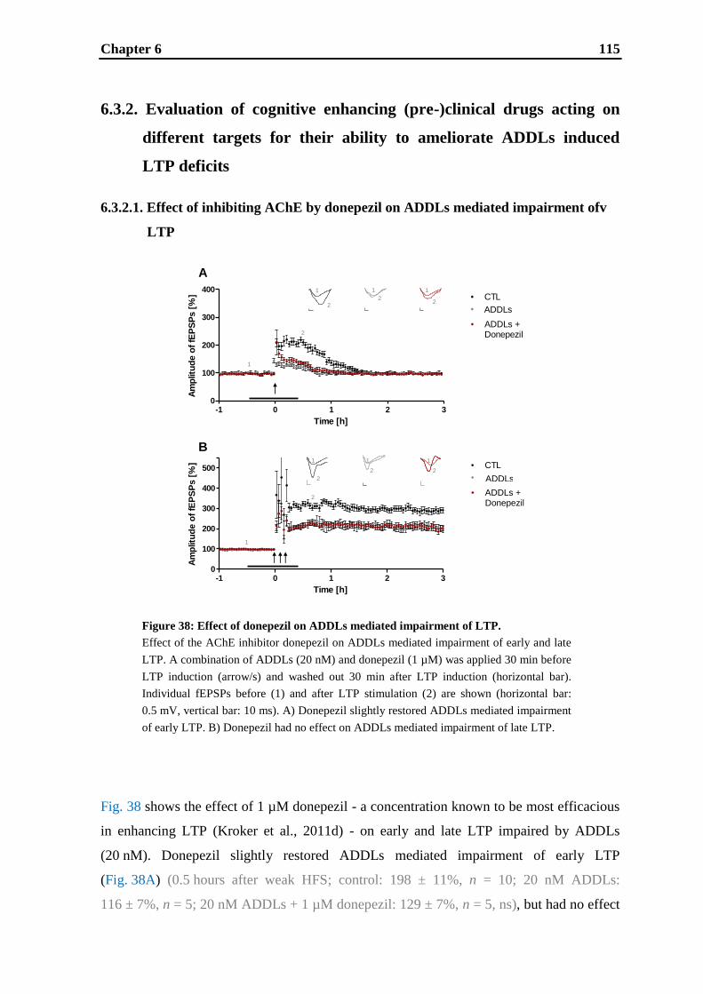
Chapter 6 115
A
B
-1 0 1 2 30
100
200
300
400
500
Time [h]
Am
plitu
de o
f fE
PS
Ps [
%] CTL
Legend
ADDLs +Donepezil
-1 0 1 2 30
100
200
300
400
Time [h]
Am
plitu
de o
f fE
PS
Ps [
%] CTL
Legend
ADDLs +Donepezil
1
2
1
2
1
2
1
2
1
2
1
2
1
2
1
2
-1 0 1 2 30
100
200
300
400
Time [h]
Am
plitu
de o
f fE
PS
Ps [
%] CTL
Legend
ADDLs +Donepezil
-1 0 1 2 30
100
200
300
400
500
Time [h]
Am
plitu
de o
f fE
PS
Ps [
%] CTL
Legend
ADDLs +Donepezil
6.3.2. Evaluation of cognitive enhancing (pre-)clinical drugs acting on
different targets for their ability to ameliorate ADDLs induced
LTP deficits
6.3.2.1. Effect of inhibiting AChE by donepezil on ADDLs mediated impairment ofv
LTP
Fig. 38 shows the effect of 1 µM donepezil - a concentration known to be most efficacious
in enhancing LTP (Kroker et al., 2011d) - on early and late LTP impaired by ADDLs
(20 nM). Donepezil slightly restored ADDLs mediated impairment of early LTP
(Fig. 38A) (0.5 hours after weak HFS; control: 198 ± 11%, n = 10; 20 nM ADDLs:
116 ± 7%, n = 5; 20 nM ADDLs + 1 µM donepezil: 129 ± 7%, n = 5, ns), but had no effect
Figure 38: Effect of donepezil on ADDLs mediated impairment of LTP.
Effect of the AChE inhibitor donepezil on ADDLs mediated impairment of early and late
LTP. A combination of ADDLs (20 nM) and donepezil (1 µM) was applied 30 min before
LTP induction (arrow/s) and washed out 30 min after LTP induction (horizontal bar).
Individual fEPSPs before (1) and after LTP stimulation (2) are shown (horizontal bar:
0.5 mV, vertical bar: 10 ms). A) Donepezil slightly restored ADDLs mediated impairment
of early LTP. B) Donepezil had no effect on ADDLs mediated impairment of late LTP.

Chapter 6 116
-1 0 1 2 30
100
200
300
400
500
Time [h]
Am
plitu
de o
f fE
PS
Ps [
%] CTL
Legend
ADDLs +TC-1827
A
B
-1 0 1 2 30
100
200
300
400
Time [h]
Am
plitu
de o
f fE
PS
Ps [
%] CTL
Legend
ADDLs +TC-1827
1
2
1
2
1
2
1
2
1
2
1
2
1
2
1
2
-1 0 1 2 30
100
200
300
400
Time [h]
Am
plitu
de o
f fE
PS
Ps [
%] CTL
Legend
ADDLs +TC-1827
-1 0 1 2 30
100
200
300
400
500
Time [h]
Am
plit
ud
e o
f fE
PS
Ps [
%] CTL
Legend
ADDLs +TC-1827
on ADDLs mediated impairment of late LTP (Fig. 38B) (0.5 hours after repeated strong
HFS; control: 315 ± 14%, n = 10; 20 nM ADDLs: 223 ± 11%, n = 5, 20 nM ADDLs +
1 µM donepezil: 218 ± 11%, n = 5, ns). Neither 20 nM ADDLs, nor 1 µM donepezil or a
combination of both affected basal fEPSPs.
6.3.2.2. Effect of the a4β2 nAChR agonist TC-1827 on ADDLs mediated impairment
of LTP
Figure 39: Effect of TC-1827 on ADDLs mediated impairment of LTP.
Effect of the α4β2 nAChR agonist TC-1827 on ADDLs mediated impairment of early
and late LTP. A combination of ADDLs (20 nM) and TC-1827 (0.1 µM) was applied
30 min before LTP induction (arrow/s) and washed out 30 min after LTP induction
(horizontal bar). Individual fEPSPs before (1) and after LTP stimulation (2) are shown
(horizontal bar: 0.5 mV, vertical bar: 10 ms). A) TC-1827 slightly restored ADDLs
mediated impairment of early LTP. B) TC-1827 had no effect on ADDLs mediated
impairment of late LTP.

Chapter 6 117
Fig. 39 shows the effect of 0.1 µM TC-1827 - a concentration known to be most
efficacious in enhancing LTP (Kroker et al., 2011c) - on early and late LTP impaired by
ADDLs (20 nM). TC-1827 slightly restored ADDLs induced impairment of early LTP
(Fig. 39A) (0.5 hours after weak HFS; control: 197 ± 12%, n = 10; 20 nM ADDLs:
117 ± 8%, n = 5; 20 nM ADDLs + 0.1 µM TC-1827: 131 ± 9%, n = 5, ns), but had no
effect on ADDLs induced impairment of late LTP (Fig. 39B) (0.5 hours after repeated
strong HFS; control: 313 ± 15%, n = 10; 20 nM ADDLs: 222 ± 10%, n = 5; 20 nM ADDLs
+ 0.1 µM TC-1827: 232 ± 16%, n = 5, ns). Neither 20 nM ADDLs, nor 0.1 µM TC-1827
or a combination of both affected basal fEPSPs.
6.3.2.3. Effect of the α7 nAChR partial agonist SSR180711 on ADDLs mediated
impairment of LTP
Fig. 40 shows the effect of 0.3 µM SSR180711 - a concentration known to be most
efficacious in enhancing LTP (Kroker et al., 2011c) - on early and late LTP impaired by
ADDLs (20 nM). SSR180711 completely rescued ADDLs mediated impairment of early
LTP (Fig. 40A) (0.5 hours after weak HFS; control: 200 ±13%, n = 10; 20 nM ADDLs:
115 ± 6%, n = 5; 20 nM ADDLs + 0.3 µM SSR180711: 198 ± 26%, n = 5, p < 0.05). Also,
ADDLs induced impairment of late LTP was fully restored by SSR180711 (Fig. 40B)
(0.5 hours after repeated strong HFS; control: 310 ± 14%, n = 10; 20 nM ADDLs:
219 ± 9%, n = 5; 20 nM ADDLs + 0.3 µM SSR180711: 293 ± 18%, n = 5, p < 0.01).
Neither 20 nM ADDLs, nor 0.3 µM SSR180711 or a combination of both affected basal
fEPSPs.
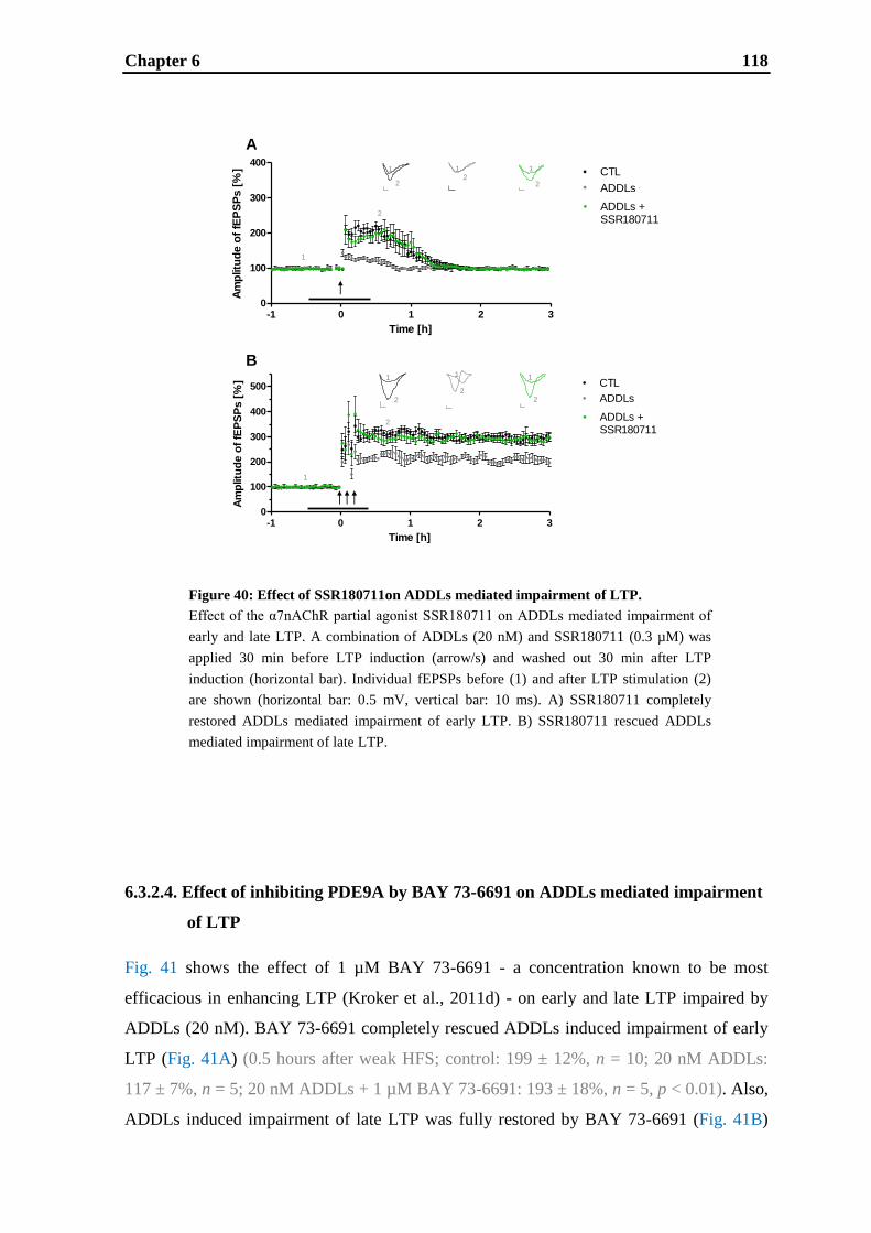
Chapter 6 118
A
B
-1 0 1 2 30
100
200
300
400
500
Time [h]
Am
plitu
de o
f fE
PS
Ps [
%] CTL
Legend
ADDLs +SSR180711
-1 0 1 2 30
100
200
300
400
Time [h]
Am
plitu
de o
f fE
PS
Ps [
%] CTL
Legend
ADDLs +SSR180711
1
2
12
1
2
1
2
1
2
1
2
1
2
2
1
-1 0 1 2 30
100
200
300
400
Time [h]
Am
plit
ud
e o
f fE
PS
Ps [
%] CTL
Legend
ADDLs +SSR180711
-1 0 1 2 30
100
200
300
400
500
Time [h]
Am
plit
ud
e o
f fE
PS
Ps [
%] CTL
Legend
ADDLs +SSR180711
6.3.2.4. Effect of inhibiting PDE9A by BAY 73-6691 on ADDLs mediated impairment
of LTP
Fig. 41 shows the effect of 1 µM BAY 73-6691 - a concentration known to be most
efficacious in enhancing LTP (Kroker et al., 2011d) - on early and late LTP impaired by
ADDLs (20 nM). BAY 73-6691 completely rescued ADDLs induced impairment of early
LTP (Fig. 41A) (0.5 hours after weak HFS; control: 199 ± 12%, n = 10; 20 nM ADDLs:
117 ± 7%, n = 5; 20 nM ADDLs + 1 µM BAY 73-6691: 193 ± 18%, n = 5, p < 0.01). Also,
ADDLs induced impairment of late LTP was fully restored by BAY 73-6691 (Fig. 41B)
Figure 40: Effect of SSR180711on ADDLs mediated impairment of LTP.
Effect of the α7nAChR partial agonist SSR180711 on ADDLs mediated impairment of
early and late LTP. A combination of ADDLs (20 nM) and SSR180711 (0.3 µM) was
applied 30 min before LTP induction (arrow/s) and washed out 30 min after LTP
induction (horizontal bar). Individual fEPSPs before (1) and after LTP stimulation (2)
are shown (horizontal bar: 0.5 mV, vertical bar: 10 ms). A) SSR180711 completely
restored ADDLs mediated impairment of early LTP. B) SSR180711 rescued ADDLs
mediated impairment of late LTP.

Chapter 6 119
-1 0 1 2 30
100
200
300
400
500
Time [h]
Am
plitu
de o
f fE
PS
Ps [
%] CTL
Legend
ADDLs +EX735
A
B
-1 0 1 2 30
100
200
300
400
Time [h]
Am
plitu
de o
f fE
PS
Ps [
%] CTL
Legend
ADDLs +EX734
1
2
1
2
1
2
1
2
1
2
2
1
2
1
2
1
-1 0 1 2 30
100
200
300
400
Time [h]
Am
plitu
de o
f fE
PS
Ps [
%] CTL
Legend
ADDLs +EX734
-1 0 1 2 30
100
200
300
400
500
Time [h]
Am
plitu
de o
f fE
PS
Ps [
%] CTL
Legend
ADDLs +EX735
(0.5 hours after repeated strong HFS; control: 315 ± 15%, n = 10; 20 nM ADDLs:
225 ± 12%, n = 5; 20 nM ADDLs + 1 µM BAY 73-6691: 303 ± 17%, n = 5, p < 0.01).
Neither 20 nM ADDLs, nor 1 µM BAY 73-6691 or a combination of both affected basal
fEPSPs.
Figure 41: Effect of BAY 73-6691 on ADDLs mediated impairment of LTP.
Effect of inhibiting PDE9A by BAY 73-6691 on ADDLs mediated impairment of early
and late LTP. A combination of ADDLs (20 nM) and BAY 73-6691 (1 µM) was applied
30 min before LTP induction (arrow/s) and washed out 30 min after LTP induction
(horizontal bar). Individual fEPSPs before (1) and after LTP stimulation (2) are shown
(horizontal bar: 0.5 mV, vertical bar: 10 ms). A) BAY 73-6691 completely restored
ADDLs mediated impairment of early LTP. B) BAY 73-6691 rescued ADDLs mediated
impairment of late LTP.
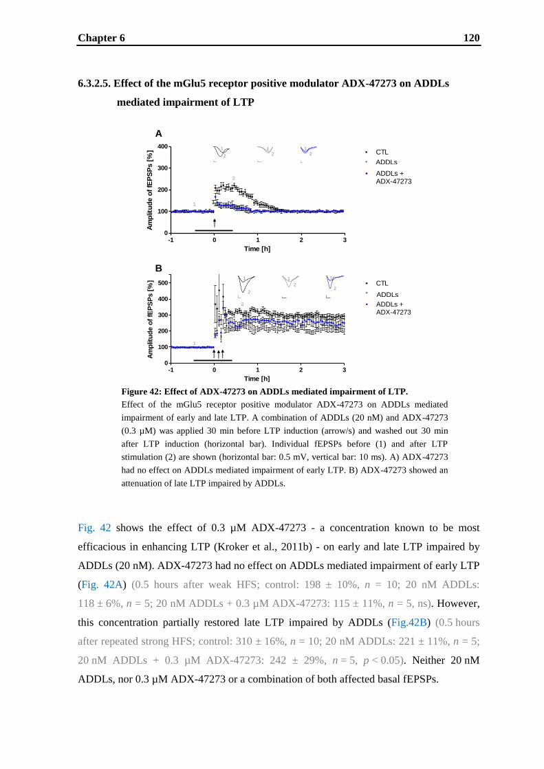
Chapter 6 120
-1 0 1 2 30
100
200
300
400
Time [h]
Am
plitu
de o
f fE
PS
Ps [
%] CTL
Legend
ADDLs +ADX-47273
A
B
-1 0 1 2 30
100
200
300
400
Time [h]
Am
plitu
de o
f fE
PS
Ps [
%] CTL
Legend
ADDLs +Donepezil
-1 0 1 2 30
100
200
300
400
500
Time [h]
Am
plit
ud
e o
f fE
PS
Ps [
%] CTL
Legend
ADDLs +ADX-47273
-1 0 1 2 30
100
200
300
400
Time [h]
Am
plit
ud
e o
f fE
PS
Ps [
%] CTL
Legend
ADDLs +Donepezil
1
2
12
12
1
2
1
2
1
2
1
2
1
2
6.3.2.5. Effect of the mGlu5 receptor positive modulator ADX-47273 on ADDLs
mediated impairment of LTP
Fig. 42 shows the effect of 0.3 µM ADX-47273 - a concentration known to be most
efficacious in enhancing LTP (Kroker et al., 2011b) - on early and late LTP impaired by
ADDLs (20 nM). ADX-47273 had no effect on ADDLs mediated impairment of early LTP
(Fig. 42A) (0.5 hours after weak HFS; control: 198 ± 10%, n = 10; 20 nM ADDLs:
118 ± 6%, n = 5; 20 nM ADDLs + 0.3 µM ADX-47273: 115 ± 11%, n = 5, ns). However,
this concentration partially restored late LTP impaired by ADDLs (Fig.42B) (0.5 hours
after repeated strong HFS; control: 310 ± 16%, n = 10; 20 nM ADDLs: 221 ± 11%, n = 5;
20 nM ADDLs + 0.3 µM ADX-47273: 242 ± 29%, n = 5, p < 0.05). Neither 20 nM
ADDLs, nor 0.3 µM ADX-47273 or a combination of both affected basal fEPSPs.
Figure 42: Effect of ADX-47273 on ADDLs mediated impairment of LTP.
Effect of the mGlu5 receptor positive modulator ADX-47273 on ADDLs mediated
impairment of early and late LTP. A combination of ADDLs (20 nM) and ADX-47273
(0.3 µM) was applied 30 min before LTP induction (arrow/s) and washed out 30 min
after LTP induction (horizontal bar). Individual fEPSPs before (1) and after LTP
stimulation (2) are shown (horizontal bar: 0.5 mV, vertical bar: 10 ms). A) ADX-47273
had no effect on ADDLs mediated impairment of early LTP. B) ADX-47273 showed an
attenuation of late LTP impaired by ADDLs.

Chapter 6 121
6.4. Discussion
AD is the most common form of dementia in elderly people, accounting for around 50 –
60% of all cases of mental deterioration among persons over 65 years of age (Blennow et
al., 2006). Estimates foresee that more than 80 million individuals will be affected by the
disease in 2040 due to population aging worldwide (Brookmeyer et al., 2007; Forlenza et
al., 2010). Thus, there is a growing need of efficacious and safe medication for AD
patients. Since soluble Aβ oligomers, also called ADDLs, are currently discussed as the
putative cause of AD, rather than insoluble plaques (Hardy and Selkoe, 2002; Small,
1998), several research groups have reported on artificially generated Aβ oligomers and
their effects in vitro and in vivo (Catalano et al., 2006; Dahlgren et al., 2002; Klein, 2002;
Lue et al., 1999; Tabaton and Piccini, 2005; Youssef et al., 2008). For example, correlation
of elevated levels of ADDLs in the AD brain and cognitive decline has been demonstrated
(Kar et al., 2004; Näslund et al., 2000). Moreover, ADDLs are known to be responsible for
abnormal dendritic spine morphology, changed synaptic receptor composition and spine
loss (Lacor et al., 2007). It was reported that acute injection of ADDLs into the
hippocampus of rats and mice induces impairments of learning and memory (Li et al.,
2005; McDonald et al., 1994). Some studies already demonstrated that ADDLs impair LTP
in the CA1 region of the hippocampus in general (Chen et al., 2000; Kim et al., 2001;
Klybin et al., 2004; Raymond et al., 2003; Shankar et al., 2008; Walsh et al., 2002), but the
effects of ADDLs on early and late LTP have not been addressed so far. Therefore, in this
study, we analyzed the effects of synthetic ADDLs on validated early and late hippocampal
CA1 LTP. Furthermore, we evaluated several cognitive enhancing drugs acting on
different targets for their ability to ameliorate ADDLs induced LTP deficits in order to
determine the potential therapeutic value of these targets as new approaches in the
treatment of AD.
6.4.1. Effect of ADDLs on early and late LTP
Here we demonstrate for the first time that early and late LTP are affected by ADDLs to a
different extent. Early LTP is decreased by ADDLs in a concentration dependent manner
to a complete abolishment (see Fig. 36A). Late LTP is also decreased in a concentration
dependent manner, but at maximum concentrations it is only reduced by about 50% (see

Chapter 6 122
Fig. 36B). As already shown in a previous study, in this setting early LTP is dependent on
NMDA receptors only, whereas late LTP is equally dependent on NMDA receptors and L-
type voltage depended calcium channels (L-VDCCs) (Kroker et al., 2011b). Hence,
ADDLs can completely abolish NMDA receptor dependent early LTP. Furthermore, by the
use of MK-801 and nifedipine, it was demonstrated that the portion of late LTP being
impaired by ADDLs was dependent on NMDA receptors and/or NMDA receptor
downstream signaling (see Fig. 37). The results confirm published data showing that
ADDLs reduce postsynaptic surface NMDA receptors (Goto et al., 2006; Johansson et al.,
2006; Snyder et al., 2005) and impair NMDA receptor function (Snyder et al., 2005;
Tyszkiewicz and Yan, 2005). Additionally, studies have implicated ADDLs to interfere
with several major signaling pathways downstream of the NMDA receptor (Snyder et al.,
2005) including the calcium-dependent protein phosphatase calcineurin (Chen et al., 2002),
CaMKII (Zhao et al., 2004), protein phosphatase 1 (Knobloch et al., 2007) and CREB
(Chen et al., 2002). Indeed, post-mortem tissue analysis of the brains of AD patients
indicated a reduction of NMDA receptors (Hynd et al., 2001; Penney et al., 1990; Ulas and
Cotman, 1997), especially of the subunits NR1, NR2A and NR2B in the hippocampus
(Hynd et al., 2004). Similarly, a trend towards a reduction in NR1 mRNA was found in the
hippocampus in AD by in situ hybridization (Panegyres et al., 2002).
The differentiation between early and late LTP, as it is made in this study, is of importance
for the evaluation of downstream effects. Early LTP is independent of protein-synthesis
(Bliss and Collingridge, 1993; Kroker et al., 2011b; Malenka and Nicoll, 1999; Reyman
and Frey, 2007) and involves modifications of pre-existing synapses as a result of rapid
calcium influx through NMDA receptors and subsequent phosphorylation events (Derkach
et al., 1999; Malenka and Bear, 2004) as well as insertion of additional AMPA receptors
into the postsynaptic membrane (Davies et al., 1989; Liao et al., 2001; Lu et al., 2001;
Malenka and Bear, 2004; Malinow, 2003; Shi et al., 1999, 2001). Late LTP is clearly
separated from early LTP by its dependence on de novo protein-synthesis (Frey et al.,
1988; Huang, 1996, 1998; Huang and Kandel, 1994; Kroker et al., 2011b; Nguyen et al.,
1991; Nguyen and Kandel, 1997; Reymann and Frey, 2007). Protein-synthesis may
contribute both to the modification of existing synapses and to the establishment of new
synaptic contacts (Buchs and Muller, 1996; Lee et al., 1980; Lisman, 2003). Thus, like
memory has different phases, namely short-term and long-term memory, LTP consists of
different phases as well. The time-window of early LTP corresponds with the duration of
short-term memory as defined by some researchers, i.e. short-term memory is a form of

Chapter 6 123
memory which does not require gene expression and protein-synthesis (Grecksch and
Matthies, 1980; Izquierdo et al., 2002). Moreover, long-lasting forms of memory are
generally believed to be protein-synthesis dependent (Costa-Mattioli et al., 2009; Grecksch
and Matthies, 1980; Pang and Lu, 2004), which is also the case for late LTP.
6.4.2. Cognitive enhancing (pre-)clinical drugs and their ability to
ameliorate ADDLs induced LTP deficits
The potential therapeutic value of the different approaches currently pursued in clinical
development for the symptomatic treatment of AD was determined by testing (tool)
compounds addressing these targets for their ability to ameliorate ADDLs induced LTP
deficits.
The AChE inhibitor donepezil is the current gold standard for symptomatic treatment of
mild-to-moderate AD patients (Birks and Harvey, 2003; Whitehead et al., 2004). In this
study we show that donepezil only slightly restores ADDLs induced LTP impairment in
early LTP and has no effect in late LTP (see Fig. 38). The small effect in early LTP might
contribute to explain the only moderate therapeutic efficacy of donepezil in AD patients
(Courtney et al., 2004; Diniz et al., 2009; Kaduszkiewicz et al., 2005).
Further promising targets for improvement of cognitive function and hence candidate
targets for AD treatment are nAChRs (Haydar and Dunlop, 2010; Holladay et al., 1997;
Lloyd and Williams, 2000; Mudo et al., 2007; Sarter et al., 2009). For both, α4β2 and α7
nAChR agonists, memory enhancing efficacy was demonstrated in several animal
cognition tests (Bohme et al., 2004; Chan et al., 2007; Pichat et al., 2007; Wallace et al.,
2011). Hence, these targets are gaining increasing interest and some specific modulators
are already being evaluated in clinical trials for treatment of AD and/or cognitive
impairment in schizophrenia (Freedman et al., 2001; Grassi et al., 2003; Hajós and Rogers,
2010; Leiser et al., 2009; Nordberg, 2001; Radek et al., 2010; Sarter et al., 2009; Thomsen
et al., 2010). In this study, we demonstrate for the first time that the α4β2 nAChR agonist
TC-1827 is capable to slightly restore early LTP impaired by ADDLs, but has no effect on
late LTP (see Fig. 39). Furthermore, it was shown that complete rescues of ADDLs
mediated impairment of early and late LTP are possible using the α7 nAChR partial
agonist SSR180711 (see Fig. 40). These results confirm previous data, showing that α7
Chapter 6 124
nAChR agonists can rescue Aβ oligomer induced impairment of LTP (Chen et al., 2006,
2010; Welsby et al., 2007). Therefore, α7 nAChR agonism seems to be more efficacious in
ameliorating ADDLs induced LTP deficits than α4β2 nAChR agonism.
Another potential new approach, inhibition of specific PDEs, has come into the focus of
interest for the treatment of memory impairment (Menniti et al., 2006; Reneerkens et al.,
2009; Schmidt, 2010). It was shown that PDE9A inhibitors can increase cGMP levels in
the brain of animals (Verhoest et al., 2009) and in CSF of animals and humans (Nicholas et
al., 2009). Furthermore, memory enhancing efficacy of the PDE9A inhibitors PF-
04447943 and BAY 73-6691 was demonstrated in various animal cognition tasks (Hutson
et al., 2011; van der Stay et al., 2008). Here, using BAY 73-6691, we show for the first
time that ADDLs induced impairment of early as well as late LTP can be completely
rescued by PDE9-inhibition (see Fig. 41).
As evidence accumulates that amplification of mGlu5 receptor function could be beneficial
in enhancing cognitive function (Cleva and Olive, 2011; Simonyi et al., 2010), a series of
selective mGlu5-receptor positive allosteric modulators (Conn et al., 2009b) were
generated. Among these, CDPPB and ADX-47273 were shown to be active in preclinical
models that might predict efficacy for the treatment of positive symptoms and cognitive
dysfunction associated with schizophrenia (Liu et al., 2008; Rosenbrock et al., 2010;
Schlumberger et al., 2009, 2010; Vardigan et al., 2010). As mGlu5 receptor allosteric
modulators might also be candidates for AD treatment, in this study, the ability of ADX-
47273 to ameliorate ADDLs induced LTP deficits was evaluated. Here, we show for the
first time that the mGlu5 receptor positive modulator ADX-47273 has no effect on ADDLs
impaired early LTP, but can partially restore ADDLs induced impairment of late LTP (see
Fig. 42).
6.4.3. Conclusion
This study demonstrates that early and late LTP are affected to different extents by
ADDLs. Due to the differentiation between early and late LTP and the pharmacological
dissection of the relevant receptors, ADDLs were shown to exclusively target NMDA
receptors and/or their signaling cascade. These results further contribute to the hypothesis
that soluble Aβ oligomers cause NMDA receptor related synaptic dysfunction which might
lead to cognitive decline seen in AD patients. Furthermore, several cognitive enhancing
Chapter 6 125
(pre-)clinical drugs addressing different targets were evaluated for their ability to
ameliorate ADDLs induced LTP deficits in order to assess their potential therapeutic value
for symptomatic treatment of AD. The current gold standard for treatment of AD patients
is the AChE inhibitor donepezil, which was demonstrated to slightly restore ADDLs
induced impairment of early LTP, but to have no effect on ADDLs induced impairment of
late LTP. The same was shown for the α4β2 nAChR agonist TC-1827. Both, the α7nAChR
partial agonist SSR180711 and the PDE9 inhibitor BAY 73-6691 completely rescued early
as well as late LTP impaired by ADDLs. The mGlu5 receptor positive allosteric modulator
ADX-47273 had no effect on ADDLs impaired early LTP, but slightly restored ADDLs
impaired late LTP. Thus, α7 nAChRs agonism and PDE9 inhibition were found to be most
effective in ameliorating ADDLs induced LTP deficits. Therefore, targeting α7 nAChRs
and PDE9 might represent powerful approaches for symptomatic treatment of AD. In
general, this study shows that targets/drugs acting on glutamatergic NMDAR-related
pathways seem to be very promising to translate into clinical benefit.

Chapter 6 126
CHAPTER 7
General discussion and outlook
Chapter 7 127
The present thesis focuses on the establishment and validation of LTP in rat hippocampal
slices to characterize memory enhancing drugs as potential treatment of AD. First, a multi-
slice recording system was set up enabling stable measurements of LTP for up to seven
hours from several slices simultaneously. Then, distinct protocols to induce early and late
CA1 LTP, resembling short-term and long-term memory, were established. They were then
validated addressing the hallmarks accepted for these forms of LTP: protein-synthesis
independence and NMDA receptor dependence without contribution of L-VDCCs for early
LTP, as opposed to protein-synthesis dependence and additive NMDA/L-VDCCs
dependence for late LTP. As a loss of mainly cholinergic and glutamatergic synapses has
been described in AD patients, the validated forms of LTP were used to study drugs
potentially enhancing cholinergic and glutamatergic neuronal function. Thus, the potential
therapeutic value of approaches aiming at enhancement of cholinergic and glutamatergic
neuronal function was determined by analyzing their potential to improve early and late
LTP and to restore LTP impaired by soluble Aβ oligomers (Fig. 43). Soluble Aβ
oligomers, also referred to as amyloid-β derived diffusible ligands (ADDLs), are thought to
be one cause of AD. In this thesis, they were demonstrated to impair early and late LTP to
different extents by exclusively targeting NMDA receptors and/or their signaling. These
results further contribute to the hypothesis that soluble Aβ oligomers cause synaptic
dysfunction which might lead to cognitive decline seen in AD patients. The AChE
inhibitor donepezil (Fig. 43 shown in dark red) is the current gold standard for
symptomatic treatment of patients suffering from mild-to-moderate AD. It facilitates
exclusively cholinergic function by increasing and prolonging the concentration of ACh in
the synaptic cleft. The increased extracellular levels of the transmitter ACh can activate
cholinergic receptors such as nAChRs, which are thought to cause the cognition enhancing
effects of donepezil. In this study, the effect of donepezil on LTP was demonstrated to be
only moderate: donepezil was found to increase early LTP, but to not affect late LTP.
Similarly, it slightly restored ADDLs induced deficits in early LTP, but had no effect on
late LTP impaired by ADDLs. Interestingly, donepezil showed quantitatively the same
effects on LTP and ADDLs induced LTP deficits as the α4β2 nAChR agonist TC-1827
(Fig. 43 shown in red). This similarity may indicate that the levels of ACh increased by
donepezil activate the α4β2 nAChR, but not sufficiently the α7 nAChR. The reason for the
α4β2 nAChR being indirectly targeted by donepezil might be based on the much higher
binding affinity of ACh to the α4β2 nAChR as compared to the α7 nAChR (Ki for α4β2:
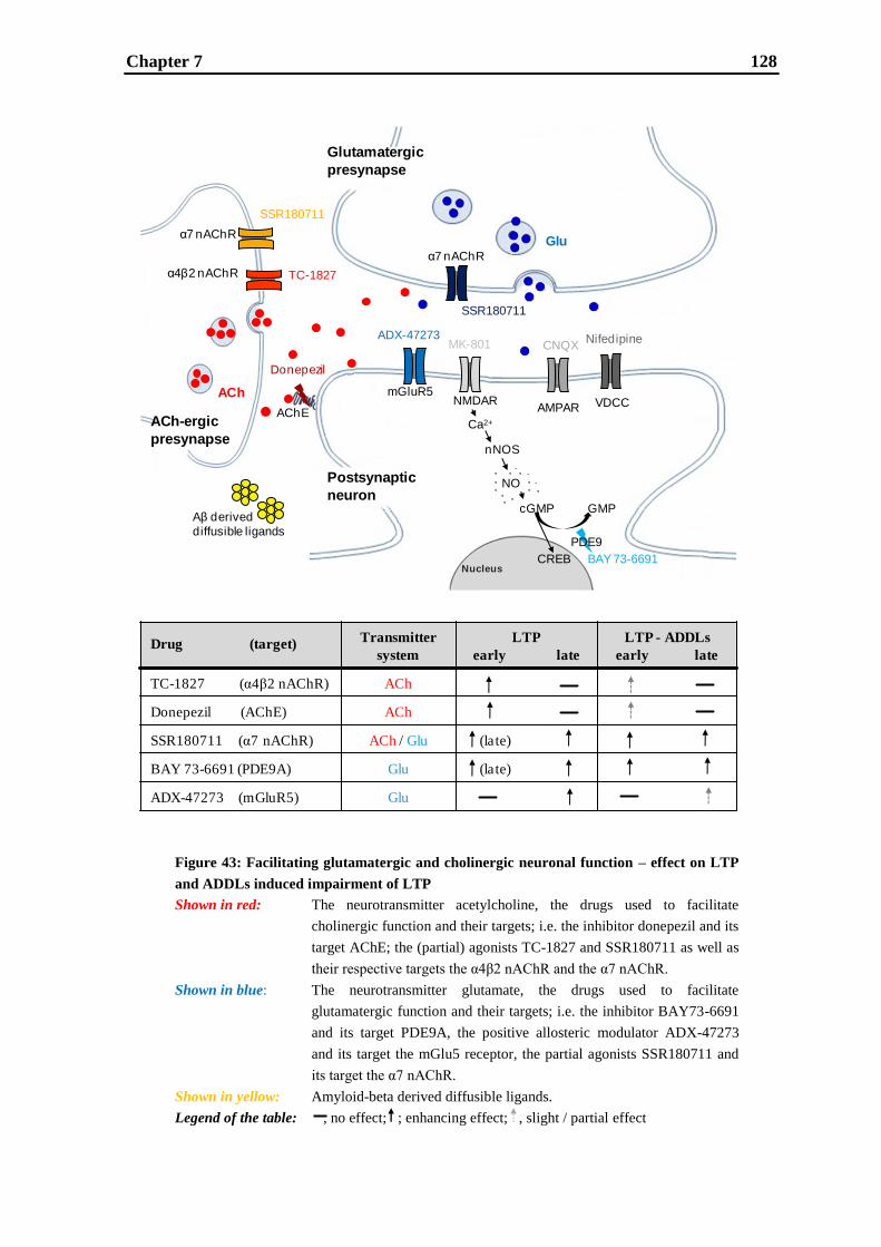
Chapter 7 128
Figure 43: Facilitating glutamatergic and cholinergic neuronal function – effect on LTP
and ADDLs induced impairment of LTP
Shown in red: The neurotransmitter acetylcholine, the drugs used to facilitate
cholinergic function and their targets; i.e. the inhibitor donepezil and its
target AChE; the (partial) agonists TC-1827 and SSR180711 as well as
their respective targets the α4β2 nAChR and the α7 nAChR.
Shown in blue: The neurotransmitter glutamate, the drugs used to facilitate
glutamatergic function and their targets; i.e. the inhibitor BAY73-6691
and its target PDE9A, the positive allosteric modulator ADX-47273
and its target the mGlu5 receptor, the partial agonists SSR180711 and
its target the α7 nAChR.
Shown in yellow: Amyloid-beta derived diffusible ligands.
Legend of the table: , no effect; ; enhancing effect; , slight / partial effect
Postsynaptic
neuron
ACh-ergic
presynapse
Glutamatergic
presynapse
VDCCAMPARNMDAR
mGluR5
α7 nAChR
α4β2 nAChR
α7 nAChR
Glu
ACh
AChE
Donepezil
Ca2+
nNOS
NO
cGMP
CREBNucleus
GMP
BAY 73-6691
NifedipineCNQXMK-801
PDE9
ADX-47273
SSR180711
TC-1827
Aβ derived
diffusible ligands
SSR180711
Drug (target)Transmitter
system
LTP
early late
LTP - ADDLs
early late
TC-1827 (α4β2 nAChR) ACh
Donepezil (AChE) ACh
SSR180711 (α7 nAChR) ACh / Glu (late)
BAY 73-6691 (PDE9A) Glu (late)
ADX-47273 (mGluR5) Glu

Chapter 7 129
6.8-57 nM; Ki for α7: 4000-10830 nM; Sharples and Wonnacott, 2001). Activation of the
α4β2 nAChR is able to enhance and slightly rescue early LTP impaired by ADDLs, but it
has no effect on late LTP. The mechanism responsible for this effect seems likely to be
disinhibition, which may take place by activation of α4β2 and/or α7 nAChRs located on
GABAergic interneurons having inhibitory connections to other inhibitory neurons, thus
producing a net increase in glutamatergic principal neuron activity. The decisive difference
between α4β2 and α7 nAChR function is the additional location of α7 nAChRs on Schaffer
collateral terminals causing strengthening of the glutamatergic system by facilitation of
glutamate release. Hence, the α7 nAChR partial agonist SSR180711 facilitates cholinergic
and glutamatergic function (Fig. 43 shown in light red and dark blue depending on the
location and hence function) and most likely thereby enhances both LTP types and even
transforms early into late LTP. Furthermore, a complete rescue of ADDLs induced
impairment of early and late LTP was observed by SSR180711. As the PDE9A inhibitor
BAY 73-6691 (Fig. 43 shown in light blue), exclusively targeting glutamatergic function,
shows the same effect as SSR180711 on LTP and LTP impaired by ADDLs, the crucial
factor for an effective LTP enhancement and rescue seems to be the activation of
glutamatergic function. PDE9 is part of the intracellular signaling pathway NO-sGC-
cGMP influencing the levels of the second messenger cGMP by its cGMP-specific
hydrolytic activity. Elevation of cGMP caused by PDE9 inhibition most likely results in
strengthening of the NMDA receptor related postsynaptical pathway NO-sGC-cGMP-PKG
leading to CREB phosphorylation being a crucial cellular mechanism for learning and
memory. Another drug exclusively interfering with glutamatergic function is the mGlu5
receptor positive allosteric modulator ADX-47273 (Fig. 43 shown in blue), which was
found to increase late LTP and to partly rescue late LTP impaired by ADDLs. It had no
effect on early LTP, no matter whether it was intact or impaired by ADDLs. The reason for
this is likely, that the stimulus intensity sufficient for induction of early LTP does not reach
a threshold for functionally relevant stimulation of mGlu5 receptors.
Hence, drugs facilitating glutamatergic function not only seem to be more efficacious in
enhancing LTP than drugs facilitating solely cholinergic function, but they are also
superior in ameliorating soluble Aβ oligomer induced LTP deficits. Consequently, the
findings of this thesis might provide an explanation for the rather moderate therapeutic
effects of donepezil in AD patients. Moreover, based on the results of the present thesis, it
can be concluded that drugs interfering with glutamatergic function seem to have a high
therapeutic value as alternative, and probably more efficacious, approaches for the
Chapter 7 130
symptomatic treatment of AD. Further investigations have to show whether this finding
also holds true for animal models addressing learning and memory, and - ultimately - for
AD patients in clinical trials.
Even though this thesis comes to an end, “this is not the end. It is not even the beginning of
the end. But it is, perhaps, the end of the beginning.” (Winston Churchill)
For example, one might not only analyze a single concentration for the rescuing
experiments of the ADDLs induced impairment of LTP, but perform a concentration series
for each drug. In this thesis, only one concentration of each drug was tested, namely the
concentration which showed the best effect in enhancing early and late LTP. To detect the
unlikely event of an effectiveness shift of concentration in the rescuing experiments, a
concentration series of all drugs would be desirable.
A question, which arose during the thesis, deals with the site of action of some of the tested
drugs. For example, it is not completely known whether BAY 73-6691 acts presynaptically
and/or postsynaptically. Paired-pulse facilitation, a short-term form of synaptic plasticity,
could shed light on the involvement of BAY 73-6691 in the presynapse. The experimental
setting is identical to the one used for LTP measurements, but instead of an LTP inducing
stimulation pattern, two stimulations are delivered at a very short interval, i.e. 20 - 50
milliseconds. The responses to both stimulations are analyzed and the second fEPSP can be
up to five times the size of the first. Facilitation has been shown to occur solely at the
presynaptic side. It is thought to be the result of an increase in probability of vesicle
release. If BAY 73-6691 acted presynaptically by an increase in the probability of vesicle
release, the facilitating effect of the second pulse should exceed the one of the control
conditions. Not only electrophysiological measurements could be used to address the site
of action of some of the tested drugs, but also immunohistochemistry having a very high
resolution going down to the sub-cellular level would be a suitable method to identify the
localization of a target protein.
Another very interesting question deals with the potential effect of some drugs on
interneurons. For example, donepezil and TC-1827 are thought to increase early LTP by
disinhibition. A patch clamp study to address the effect of doenepezil and TC-1827 on
interneurons could shed some light on this issue.
It is known that late LTP requires de novo protein-synthesis (Fazeli et al., 1993; Frey et al.,
1988; Huang, 1998; Malenka and Nicoll, 1999; Nguyen et al., 1994; Reyman and Frey,
2007). Furthermore, transcription of different immediate early genes like zif268 and Arc

Chapter 7 131
after late LTP induction is described (Bozon et al., 2002; Jones et al., 2001; Guzowski et
al., 2000). Nevertheless, the knowledge about the processes and detailed signaling
pathways remains to be elucidated. One way to shed light onto these downstream
mechanisms could be high-resolution transcriptome profiling of the respective neurons
after LTP induction, using e.g. deep sequencing technologies. This approach has the
potential to further clarify the mode of late LTP induction and might also provide
information of further molecular sites of action for its modulation.
In this thesis, LTP experiments were exclusively used to analyze drugs targeting AD. As
not only AD patients are impaired in their learning and memory abilities, but also patients
suffering from schizophrenia, LTP experiments are not exclusively a model for AD. LTP
could also be used as a model to screen for potential drugs for the treatment of impaired
cognitive function in schizophrenia patients.
Finally, all LTP experiments in this thesis were conducted in rat hippocampal slices.
Transferring this technique onto mouse hippocampal slices could pave the way to new and
more disease-specific model systems: genetically engineered mouse models for AD or
schizophrenia-like pathology could be used to address the effectiveness of drugs in a more
disease relevant context.

Chapter 7 132
REFERENCES
References 133
Abe T, Sugihara H, Nawa H, Shigemoto R, Mizuno N, Nakanishi S (1992) Molecular characterization of a
novel metabotropic glutamate receptor mGluR5 coupled to inositol phosphate/Ca2+
signal transduction. J
Biol Chem 267:13361-13368.
Abel T, Nguyen PV, Barad M, Deuel TA, Kandel ER, Bourtchouladze R (1997) Genetic demonstration of a
role for PKA in the late phase of LTP and in hippocampus-based long-term memory. Cell 88(5):615-626.
Abraham WC, Dragunow M, Tate WP (1991) The role of immediate early genes in the stabilization of long-
term potentiation. Mol Neurobiol 5(2-4):297-314.
Abraham WC, Mason-Parker SE, Williams J, Dragunow M (1995) Analysis of the decremental nature of
LTP in the dentate gyrus. Brain Res Mol Brain Res 30(2):367-372.
Ahmed T, Frey JU (2005) Plasticity-specific phosphorylation of CaMKII, MAPkinases and CREB during
late-LTP in rat hippocampal slices in vitro. Neuropharmacology 49:477-492.
Alagarsamy S, Marino MJ, Rouse ST, Gereau RW 4th, Heinemann SF, Conn PJ (1999) Activation of
NMDA receptors reverses desensitization of mGluR5 in native and recombinant systems. Nat Neurosci
2(3):234-240.
Albensi BC, Oliver DR, Toupin J, Odero G (2007) Electrical stimulation protocols for hippocampal synaptic
plasticity and neuronal hyper-excitability: are they effective or relevant? Exp Neurol 204(1):1-13.
Alberini CM, Taubenfeld SM, Garcia-Osta A (2005) CREB and the CREB-C/EBP-dependent gene
expression cascade in long-term memory. Cellscience Reviews 2(2).
Alkon DL, Epstein H, Kuzirian A, Bennett MC, Nelson TJ (2005) Protein synthesis required for long-term
memory is induced by PKC activation on days before associative learning. Proc Natl Acad Sci U S A
102(45):16432-16437.
Alkondon M, Albuquerque EX (2004) The nicotinic acetylcholine receptor subtypes and their function in
the hippocampus and cerebral cortex. Prog Brain Res 145:109-120.
Alkondon M, Pereira EFR, Eisenberg HM, Albuquerque EX (1999) Choline and selective antagonists
identify two subtypes of nicotinic acetylcholine receptors that modulate GABA release from CA1
interneurons in rat hippocampal slices. J Neurosci 19:2693–2705.
Allen JW, Ivanova SA, Fan L, Espey MG, Basile AS, Faden AI (1999) Group II metabotropic glutamate
receptor activation attenuates traumatic neuronal injury and improves neurological recovery after traumatic
brain injury. J Pharmacol Exp Ther 290(1):112-120.
Almeida CG, Tampellini D, Takahashi RH, Greengard P, Lin MT, Snyder EM, Gouras GK (2005) Beta-
amyloid accumulation in APP mutant neurons reduces PSD-95 and GluR1 in synapses. Neurobiol Dis
20(2):187-198.
Alonso A, de Curtis M, Llinás R (1990) Postsynaptic Hebbian and non-Hebbian long-term potentiation of
synaptic efficacy in the entorhinal cortex in slices and in the isolated adult guinea pig brain. Proc Natl Acad
Sci U S A 87(23):9280-9284.
References 134
Alzheimer A: Über eine eigenartige Erkrankung der Hirnrinde. Vortrag in der Versammlung
Südwestdeutscher Irrenärzte in Tübingen am 3. November 1906. Referiert als Eigenbericht: Allgemeine
Zeitschrift für Psychiatrie und psychisch-gerichtliche Medizin 64. Bd. (1907) S. 146-148.
Alzheimer's Association (2011) 2011 Alzheimer's disease facts and figures. Alzheimers Dement 7(2):208-
244.
Amaral DG, Witter MP (1989) The three-dimensional organization of the hippocampal formation: a review
of anatomical data. Neuroscience 31(3):571-591.
Andersen P, Bliss TVP, Skrede KK (1971) Lamellar organization of hippocampal excitatory pathways. Exp
Brain Res 13:222–238.
Anderson WW, Collingridge GL (2001) The LTP Program: a data acquisition program for on-line analysis
of long-term potentiation and other synaptic events. J Neurosci Methods 108(1):71-83.
Anderson WW, Collingridge GL (2007) Capabilities of the WinLTP data acquisition program extending
beyond basic LTP experimental functions. J Neurosci Methods 162(1-2):346-356.
Andreasen M, Lambert JD, Jensen MS (1989) Effects of new non-N-methyl-D-aspartate antagonists on
synaptic transmission in the in vitro rat hippocampus. J Physiol 414:317-336.
Andreeva SG, Dikkes P, Epstein PM, Rosenberg PA (2001) Expression of cGMP-specific
phosphodiesterase 9A mRNA in the rat brain. J Neurosci 21(22):9068-9076.
Antonov I, Antonova I, Kandel ER, Hawkins RD (2003) Activity-dependent presynaptic facilitation and
hebbian LTP are both required and interact during classical conditioning in Aplysia. Neuron 37(1):135-147.
Antuono PG, Jones JL, Wang Y, Li SJ (2001) Decreased glutamate + glutamine in Alzheimer's disease
detected in vivo with (1)H-MRS at 0.5 T. Neurology 56(6):737-742.
Anwyl R (1999) Metabotropic glutamate receptors: electrophysiological properties and role in plasticity.
Brain Res Rev 29(1):83-120.
Anwyl R (2009) Metabotropic glutamate receptor-dependent long-term potentiation. Neuropharmacology
56(4):735-740.
Applegate MD, Kerr DS, Landfield PW (1987) Redistribution of synaptic vesicles during long-term
potentiation in the hippocampus. Brain Res 401(2):401-406.
Aramakis VB, Metherate R (1998) Nicotine selectively enhances NMDA receptor-mediated synaptic
transmission during postnatal development in sensory neocortex. J Neurosci 18:8485-8495.
Arancio O, Antonova I, Gambaryan S, Lohmann SM, Wood JS, Lawrence DS, Hawkins RD (2001)
Presynaptic role of cGMP-dependent protein kinase during long-lasting potentiation. J Neurosci 21(1):143-
149.
Arehart-Treichel J (2011) Dramatic cognitive decline seen years before Alzheimer's diagnosis. Psychiatric

References 135
News 46(9):18.
Armstrong RA (2011) The pathogenesis of Alzheimer's disease: a reevaluation of the "amyloid cascade
hypothesis". Int J Alzheimers Dis 2011:630865.
Arneric SP, Holladay M, Williams M (2007) Neuronal nicotinic receptors: a perspective on two decades of
drug discovery research. Biochem Pharmacol 74(8):1092-1101.
Arriagada PV, Growdon JH, Hedley-Whyte ET, Hyman BT (1992) Neurofibrillary tangles but not senile
plaques parallel duration and severity of Alzheimer's disease. Neurology 42(3 Pt 1):631-639.
Ashford JW, Schmitt FA (2001) Modeling the time-course of Alzheimer dementia. Curr Psychiatry Rep
3(1):20-28.
Ashour MB, Gee SJ, Hammock BD (1987) Use of a 96-well microplate reader for measuring routine
enzyme activities. Anal Biochem 166(2):353-360.
Asztely F, Xiao MY, Wigström H, Gustafsson B (1994) Effect of adenosine-induced changes in presynaptic
release probability on long-term potentiation in the hippocampal CA1 region. J Neurosci 14(11 Pt 1):6707-
6714.
Atkins CM, Selcher JC, Petraitis JJ, Trzaskos JM, Sweatt JD (1998) The MAPK cascade is required for
mammalian associative learning. Nat Neurosci 1(7):602-609.
Auclair N, Otani S, Soubrie P, Crepel F (2000) Cannabinoids modulate synaptic strength and plasticity at
glutamatergic synapses of rat prefrontal cortex pyramidal neurons J Neurophysiol 83(6):3287-3293.
Auld DS, Kar S, Quirion R (1998) Beta-amyloid peptides as direct cholinergic neuromodulators: a missing
link? Trends Neurosci 21(1):43-49.
Ayala JE, Chen Y, Banko JL, Sheffler DJ, Williams R, Telk AN, Watson NL, Xiang Z, Zhang Y, Jones PJ,
Lindslay CW, Olive MF, Conn PJ (2009) mGluR5 positive allosteric modulators facilitate both hippocampal
long-term potentiation and long-term depression and enhance spatial learning. Neuropsychopharmacology
34:2057-2071.
Baddeley A (2003) Working memory: looking back and looking forward. Nat Rev Neurosci 4(10):829-839.
Balducci C, Beeg M, Stravalaci M, Bastone A, Sclip A, Biasini E, Tapella L, Colombo L, Manzoni C,
Borsello T, Chiesa R, Gobbi M, Salmona M, Forloni G (2010) Synthetic amyloid-beta oligomers impair
long-term memory independently of cellular prion protein. Proc Natl Acad Sci U S A 107(5):2295-2300.
Bales KR, Tzavara ET, Wu S, Wade MR, Bymaster FP, Paul SM, Nomikos GG (2006) Cholinergic
dysfunction in a mouse model of Alzheimer disease is reversed by an anti-A beta antibody. J Clin Invest
116(3):825-832.
Ballard C, Gauthier S, Corbett A, Brayne C, Aarsland D, Jones E (2011) Alzheimer's disease. Lancet
377(9770):1019-1031.
References 136
Balschun D, Wetzel W (2002) Inhibition of mGluR5 blocks hippocampal LTP in vivo and spatial learning in
rats. Pharmacol Biochem Behav 73:375–380.
Barad M, Bourtchouladze R, Winder DG, Golan H, Kandel E (1998) Rolipram, a type IV-specific
phosphodiesterase inhibitor, facilitates the establishment of long-lasting long-term potentiation and
improves memory. Proc Natl Acad Sci U S A 95(25):15020-15025.
Barak S, Arad M, De Levie A, Black MD, Griebel G, Weiner I (2009) Pro-cognitive and antipsychotic
efficacy of the alpha7 nicotinic partial agonist SSR180711 in pharmacological and neurodevelopmental
latent inhibition models of schizophrenia. Neuropsychopharmacology 34(7):1753-1763.
Barnes CA, Meltzer J, Houston F, Orr G, McGann K, Wenk GL (2000) Chronic treatment of old rats with
donepezil or galantamine: effects on memory, hippocampal plasticity and nicotinic receptors. Neuroscience
99(1):17-23.
Barrionuevo G, Brown TH (1983) Associative long-term potentiation in hippocampal slices. Proc Natl Acad
Sci U S A 80(23):7347-7351.
Bartus RT (2000) On neurodegenerative diseases, models, and treatment strategies: lessons learned and
lessons forgotten a generation following the cholinergic hypothesis. Exp Neurol 163(2):495-529.
Bartus RT, Dean RL 3rd, Beer B, Lippa AS (1982) The cholinergic hypothesis of geriatric memory
dysfunction. Science 217(4558):408-414.
Bashir ZI, Alford S, Davies SN, Randall AD, Collingridge GL (1991) Long-term potentiation of NMDA
receptor-mediated synaptic transmission in the hippocampus. Nature 349(6305):156-158.
Bayazitov IT, Richardson R, Fricke RG, Zakharenko SS (2007) Slow presynaptic and fast postsynaptic
components of compound long-term potentiation. J Neurosci 27(43):11510-11521.
Beavo JA (1995) Cyclic nucleotide phosphodiesterases: functional implications of multiple isoforms.
Physiol Rev 75(4):725-748.
Behrens CJ, van den Boom LP, de Hoz L, Friedman A, Heinemann U (2005) Induction of sharp wave-ripple
complexes in vitro and reorganization of hippocampal networks. Nat Neurosci 8(11):1560-1567.
Bertram L, Tanzi RE (2005) The genetic epidemiology of neurodegenerative disease. J Clin Invest
115(6):1449-1457.
Bierer LM, Hof PR, Purohit DP, Carlin L, Schmeidler J, Davis KL, Perl DP (1995) Neocortical
neurofibrillary tangles correlate with dementia severity in Alzheimer's disease. Arch Neurol 52(1):81-88.
Bikbaev A, Neyman S, Ngomba RT, Conn J, Nicoletti F, Manahan-Vaughan D (2008) mGluR5 mediates
the interaction between late-LTP, network activity, and learning. PloS One 3(5):e2155.
Billingsley ML, Kincaid RL (1997) Regulated phosphorylation and dephosphorylation of tau protein: effects
on microtubule interaction, intracellular trafficking and neurodegeneration. Biochem J 323(Pt3):577-591.

References 137
Birks JS, Harvey R (2003) Donepezil for dementia due to Alzheimer's disease. Cochrane Database Syst Rev
(1):CD001190.
Biton B, Bergis OE, Galli F, Nedelec A, Lochead AW, Jegham S, Godet D, Lanneau C, Santamaria R,
Chesney F, Léonardon J, Granger P, Debono MW, Bohme GA, Sgard F, Besnard F, Graham D, Coste A,
Oblin A, Curet O, Vigé X, Voltz C, Rouquier L, Souilhac J, Santucci V, Gueudet C, Françon D, Steinberg
R, Griebel G, Oury-Donat F, George P, Avenet P, Scatton B (2007) SSR180711, a novel selective alpha7
nicotinic receptor partial agonist: (1) binding and functional profile. Neuropsychopharmacology 32(1):1-16.
Blanchard RJ, Blanchard DC (1969) Crouching as an index of fear. J Comp Physiol Psychol 67(3):370-375.
Blennow K, de Leon MJ, Zetterberg H (2006) Alzheimer's disease. Lancet 368(9533):387-403.
Bliss TVP, Collingridge GL (1993) A synaptic model of memory: long-term potentiation in the
hippocampus. Nature 361(6407):31-39.
Bliss TVP, Gradner-Medwin AR (1973) Long-lasting potentiation of synaptic transmission in the dentate
area of the unanaesthetized rabbit following stimulation of the perforant path. J Physiol 232(2):357-374.
Bliss TVP, Lomo T (1973) Long-lasting potentiation of synaptic transmission in the dentate area of the
anaesthetised rabbit following stimulation of the perforant path. J Physiol (London) 232:331-356.
Blokland A, Schreiber R, Prickaerts J (2006) Improving memory: a role for phosphodiesterases. Curr Pharm
Des 12(20):2511-2523.
Blum S, Moore AN, Adams F, Dash PK (1999) A mitogen-activated protein kinase cascade in the CA1/CA2
subfield of the dorsal hippocampus is essential for long-term spatial memory. J Neurosci 19(9):3535-3544.
Blusztajn JK, Berse B (2000) The cholinergic neuronal phenotype in Alzheimer's disease. Metab Brain Dis
15(1):45-64.
Boehning D, Snyder SH (2003) Novel neural modulators. Annu Rev Neurosci 26:105-131.
Boess FG, Hendrix M, van der Staay FJ, Erb C, Schreiber R, van Staveren W, de Vente J, Prickaerts J,
Blokland A, Koenig G (2004) Inhibition of phosphodiesterase 2 increases neuronal cGMP, synaptic
plasticity and memory performance. Neuropharmacology 47(7):1081-1092.
Böhme GA, Bon C, Stutzmann JM, Doble A, Blanchard JC (1991) Possible involvement of nitric oxide in
long-term potentiation. Eur J Pharmacol 199(3):379-381.
Bohme GA, Letchworth SR, Piot-Grosjean O, Gatto GJ, Obinu M-C, Caldwell WS, Laville M, Brunel P,
Pellerin R, Leconte J-P, Genevois-Borella A, Dubedat P, Mazadier M, Pradier L, Bencherif M, Benavides J
(2004) In Vitro and In Vivo Characterization of TC-1827, a Novel Brain a4b2 Nicotinic Receptor Agonist
with Pro-Cognitive Activity. Drug Dev Res 62:26–40.
Boldyrev A, Bulygina E, Makhro A (2004) Glutamate receptors modulate oxidative stress in neuronal cells.
A mini-review. Neurotox Res 6(7-8):581-587.

References 138
Bonsi P, Martella G, Cuomo D, Platania P, Sciamanna G, Bernardi G, Wess J, Pisani A (2008) Loss of
muscarinic autoreceptor function impairs long-term depression but not long-term potentiation in the
striatum. J Neurosci 28(24):6258-6263.
Borchelt DR, Thinakaran G, Eckman CB, Lee MK, Davenport F, Ratovitsky T, Prada CM, Kim G, Seekins
S, Yager D, Slunt HH, Wang R, Seeger M, Levey AI, Gandy SE, Copeland NG, Jenkins NA, Price DL,
Younkin SG, Sisodia SS (1996) Familial Alzheimer's disease-linked presenilin 1 variants elevate Abeta1-
42/1-40 ratio in vitro and in vivo. Neuron 17(5):1005-1013.
Borroni AM, Fichtenholtz H, Woodside BL, Teyler TJ (2000) Role of voltage-dependent calcium channel
longterm potentiation (LTP) and NMDA LTP in spatial memory. J Neurosci 20:9272-9276.
Bortolotto ZA, Anderson WW, Isaac JT, Collingridge GL (2001) Synaptic plasticity in the hippocampal
slice preparation. Curr Protoc Neurosci Chapter 6:Unit 6.13.
Bortolotto ZA, Collett VJ, Conquet F, Jia Z, van der Putten H, Collingridge GL (2005) The regulation of
hippocampal LTP by the molecular switch, a form of metaplasticity, requires mGlu5 receptors.
Neuropharmacology 49(1):13-25.
Boulton CL, Southam E, Garthwaite J (1995) Nitric oxide-dependent long-term potentiation is blocked by a
specific inhibitor of soluble guanylyl cyclase. Neuroscience 69(3):699-703.
Bourtchuladze R, Frenguelli B, Blendy J, Cioffi D, Schutz G, Silva AJ (1994) Deficient long-term memory
in mice with a targeted mutation of the cAMP-responsive element-binding protein. Cell 79(1):59-68.
Boyle PA, Wilson RS, Aggarwal NT, Tang Y, Bennett DA (2006) Mild cognitive impairment: risk of
Alzheimer disease and rate of cognitive decline. Neurology 67(3):441-445.
Bozon B, Davis S, Laroche S (2002) Regulated transcription of the immediate-early gene Zif268:
mechanisms and gene dosage-dependent function in synaptic plasticity and memory formation.
Hippocampus 12(5):570-577.
Braak H, Braak E (1997) Frequency of stages of Alzheimer-related lesions in different age categories.
Neurobiol Aging 18(4):351-357.
Bramham CR, Worley PF, Moore MJ, Guzowski JF (2008) The immediate early gene arc/arg3.1: regulation,
mechanisms, and function. J Neurosci 28(46):11760-11767.
Brookmeyer R, Gray S, Kawas C (1998) Projections of Alzheimer's disease in the United States and the
public health impact of delaying disease onset. Am J Public Health 88(9):1337-1342.
Brookmeyer R, Johnson E, Ziegler-Graham K, Arrighi HM (2007) Forecasting the global burden of
Alzheimer's disease. Alzheimers Dement 3(3):186-191.
Brown RE, Fedorov NB, Haas HL, Reymann KG (1995) Histaminergic modulation of synaptic plasticity in
area CA1 of rat hippocampal slices. Neuropharmacology 34(2):181-190.
Brown TH, McAfee DA (1982) Long-term synaptic potentiation in the superior cervical ganglion. Science
References 139
215(4538):1411-1413.
Bruno V, Battaglia G, Copani A, D'Onofrio M, Di Iorio P, De Blasi A, Melchiorri D, Flor PJ, Nicoletti F
(2001) Metabotropic glutamate receptor subtypes as targets for neuroprotective drugs. J Cereb Blood Flow
Metab 21(9):1013-1033.
Buccafusco JJ (2004) Cognitive enhancing drugs. Birkhäuser Verlag, Basel, Switzerland.
Buccafusco JJ, Beach JW, Terry Jr AV, Doad GS, Sood A, Arias E, Misawa H, Masai M, Fujii T,
Kawashima K (2004) Novel analogs of choline as potential neuroprotective agents. J Alzheimer's Dis 6:85–
92.
Buccafusco JJ, Letchworth SR, Bencherif M, Lippiello PM (2005) Long-lasting cognitive improvement with
nicotinic receptor agonists: mechanisms of pharmacokinetic-pharmacodynamic discordance. Trends
Pharmacol Sci 26:352-360.
Buchanan KA, Petrovic MM, Chamberlain SE, Marrion NV, Mellor JR (2010) Facilitation of long-term
potentiation by muscarinic M(1) receptors is mediated by inhibition of SK channels. Neuron 68(5):948-963.
Buchs PA, Muller D (1996) Induction of long-term potentiation is associated with major ultrastructural
changes of activated synapses. Proc Natl Acad Sci U S A 93(15):8040-8045.
Burgess N, Maguire EA, O'Keefe J (2002) The human hippocampus and spatial and episodic memory.
Neuron 35(4):625-641.
Burghaus L, Schütz U, Krempel U, de Vos RA, Jansen Steur EN, Wevers A, Lindstrom J, Schröder H
(2000) Quantitative assessment of nicotinic acetylcholine receptor proteins in the cerebral cortex of
Alzheimer patients. Brain Res Mol Brain Res 76(2):385-388.
Busciglio J, Lorenzo A, Yeh J, Yankner BA (1995) beta-amyloid fibrils induce tau phosphorylation and loss
of microtubule binding. Neuron 14(4):879-888.
Buzsáki G (2002) Theta oscillations in the hippocampus. Neuron 33(3):325-340.
Cai XD, Golde TE, Younkin SG (1993) Release of excess amyloid beta protein from a mutant amyloid beta
protein precursor. Science 259(5094):514-516.
Cajal YR (1913) Histologie du Systeme Nerveux de l‟Homme et des Vertebras. Paris: Maloine.
Calabresi P, Centonze D, Gubellini P, Bernardi G (1999) Activation of M1-like muscarinic receptors is
required for the induction of corticostriatal LTP. Neuropharmacology 38(2):323-326.
Castellani RJ, Rolston RK, Smith MA (2010) Alzheimer disease. Dis Mon 56(9):484-546.
Castro CA, Silbert LH, McNaughton BL, Barnes CA (1989) Recovery of spatial learning deficits after decay
of electrically induced synaptic enhancement in the hippocampus. Nature 342(6249):545-548.
Catalano SM, Dodson EC, Henze DA, Joyce JG, Krafft GA, Kinney GG (2006) The role of amyloid-beta
References 140
derived diffusible ligands (ADDLs) in Alzheimer's disease. Curr Top Med Chem 6(6):597-608.
Cavus I, Teyler T (1996) Two forms of long-term potentiation in area CA1 activate different signal
transduction cascades. J Neurophysiol 76:3038-3047.
Chan WK, Wong PT, Sheu FS (2007) Frontal cortical alpha7 and alpha4beta2 nicotinic acetylcholine
receptors in working and reference memory. Neuropharmacology 52:1641-1649.
Chang FL, Greenough WT (1984) Transient and enduring morphological correlates of synaptic activity and
efficacy change in the rat hippocampal slice. Brain Res 309(1):35-46.
Chen C, Kim JJ, Thompson RF, Tonegawa S (1996) Hippocampal lesions impair contextual fear
conditioning in two strains of mice. Behav Neurosci 110(5):1177-1180.
Chen L, Wang H, Zhang Z, Li Z, He D, Sokabe M, Chen L (2010) DMXB (GTS-21) ameliorates the
cognitive deficits in beta amyloid(25-35(-)) injected mice through preventing the dysfunction of alpha7
nicotinic receptor. J Neurosci Res 88(8):1784-1794.
Chen L, Yamada K, Nabeshima T, Sokabe M (2006) alpha7 nicotinic acetylcholine receptor as a target to
rescue deficit in hippocampal LTP induction in beta-amyloid infused rats. Neuropharmacology 50(2):254-
268.
Chen QS, Kagan BL, Hirakura Y, Xie CW (2000) Impairment of hippocampal long-term potentiation by
Alzheimer amyloid beta-peptides. J Neurosci Res 60(1):65-72.
Chen QS, Wei WZ, Shimahara T, Xie CW (2002) Alzheimer amyloid beta-peptide inhibits the late phase of
long-term potentiation through calcineurin-dependent mechanisms in the hippocampal dentate gyrus.
Neurobiol Learn Mem 77(3):354-371.
Chen Y, Conn PJ (2008) mGluR5 positive allosteric modulators. Drugs of the Future 33:355–360.
Chetkovich DM, Sweatt JD (1993) Nitric oxide synthase-independent long-term potentiation in area CA1 of
hippocampus. J Neurochem 61(5):1933-1942.
Cheval H, Chagneau C, Levasseur G, Veyrac A, Faucon-Biguet N, Laroche S, Davis S (2011) Distinctive
features of Egr transcription factor regulation and DNA binding activity in CA1 of the hippocampus in
synaptic plasticity and consolidation and reconsolidation of fear memory. Hippocampus doi:
10.1002/hipo.20926. [Epub ahead of print].
Christopherson KS, Hillier BJ, Lim WA, Bredt DS (1999) PSD-95 assembles a ternary complex with the N-
methyl-D-aspartic acid receptor and a bivalent neuronal NO synthase PDZ domain. J Biol Chem
274(39):27467-27473.
Citron M, Oltersdorf T, Haass C, McConlogue L, Hung AY, Seubert P, Vigo-Pelfrey C, Lieberburg I,
Selkoe DJ (1992) Mutation of the beta-amyloid precursor protein in familial Alzheimer's disease increases
beta-protein production. Nature 360(6405):672-674.
Citron M, Vigo-Pelfrey C, Teplow DB, Miller C, Schenk D, Johnston J, Winblad B, Venizelos N, Lannfelt

References 141
L, Selkoe DJ (1994) Excessive production of amyloid beta-protein by peripheral cells of symptomatic and
presymptomatic patients carrying the Swedish familial Alzheimer disease mutation. Proc Natl Acad Sci U S
A 91(25):11993-11997.
Clark NC, Nagano N, Kuenzi FM, Jarolimek W, Huber I, Walter D, Wietzorrek G, Boyce S, Kullmann DM,
Striessnig J, Seabrook GR (2003) Neurological phenotype and synaptic function in mice lacking the CaV1.3
alpha subunit of neuronal L-type voltage-dependent Ca2+
channels. Neuroscience 120:435-442.
Cleva RM, Olive MF (2011) Positive allosteric modulators of type 5 metabotropic glutamate receptors
(mGluR5) and their therapeutic potential for the treatment of CNS disorders. Molecules 16(3):2097-2106.
Coan EJ, Collingridge GL (1987) Characterization of an N-methyl-D-aspartate receptor component of
synaptic transmission in rat hippocampal slices. Neuroscience 22(1):1-8.
Cohen AS, Raymond CR, Abraham WC (1998) Priming of long-term potentiation induced by activation of
metabotropic glutamate receptors coupled to phospholipase C. Hippocampus 8(2):160-170.
Collingridge GL (2003) The induction of N-methyl-D-aspartate receptor-dependent long-term potentiation.
Phil Trans R Soc Lond B 358:635–641.
Collingridge GL, Kehl SJ, McLennan H (1983) Excitatory amino acids in synaptic transmission in the
schaffer collateral-commissural pathway of the rat hippocampus. J Physiol 334:33-46.
Collingridge GL, Peineau S, Howland JG, Wang YT (2010) Long-term depression in the CNS. Nat Rev
Neurosci 11(7):459-473.
Conn PJ, Christopoulos A, Lindsley CW (2009a) Allosteric modulators of GPCRs: a novel approach for the
treatment of CNS disorders. Nat Rev Drug Discov 8(1):41-54.
Conn PJ, Lindsley CW, Jones CK (2009b) Activation of metabotropic glutamate receptors as a novel
approach for the treatment of schizophrenia. Trends Pharmacol Sci 30(1):25-31.
Cooper SJ (2005) Donald O. Hebb's synapse and learning rule: a history and commentary. Neurosci
Biobehav Rev 28(8):851-874.
Copani A, Bruno V, Battaglia G, Leanza G, Pellitteri R, Russo A, Stanzani S, Nicoletti F (1995) Activation
of metabotropic glutamate receptors protects cultured neurons against apoptosis induced by beta-amyloid
peptide. Mol Pharmacol 47(5):890-897.
Corder EH, Saunders AM, Strittmatter WJ, Schmechel DE, Gaskell PC, Small GW, Roses AD, Haines JL,
Pericak-Vance MA (1993) Gene dose of apolipoprotein E type 4 allele and the risk of Alzheimer's disease in
late onset families. Science 261(5123):921-923.
Corradetti R, Ballerini L, Pugliese AM, Pepeu G (1992) Serotonin blocks the long-term potentiation induced
by primed burst stimulation in the CA1 region of rat hippocampal slices. Neuroscience 46(3):511-518.
Costa-Mattioli M, Sossin WS, Klann E, Sonenberg N (2009) Translational control of long-lasting synaptic
plasticity and memory. Neuron 61(1):10-26.

References 142
Cotman CW, Monaghan DT, Ganong AH (1988) Excitatory amino acid neurotransmission: NMDA
receptors and Hebb-type synaptic plasticity. Annu Rev Neurosci 11:61-80.
Court J, Martin-Ruiz C, Piggott M, Spurden D, Griffiths M, Perry E (2001) Nicotinic receptor abnormalities
in Alzheimer's disease. Biol Psychiatry 49(3):175-184.
Courtney C, Farrell D, Gray R, Hills R, Lynch L, Sellwood E, Edwards S, Hardyman W, Raftery J, Crome
P, Lendon C, Shaw H, Bentham P, AD2000 Collaborative Group (2004) Long-term donepezil treatment in
565 patients with Alzheimer's disease (AD2000): randomised double-blind trial. Lancet 363(9427):2105-
2115.
Cross AJ, Slater P, Candy JM, Perry EK, Perry RH (1987) Glutamate deficits in Alzheimer's disease. J
Neurol Neurosurg Psychiatry 50(3):356-358.
Cummings JL (2004) Alzheimer's disease. N Engl J Med 351(1):56-67.
Dahlgren KN, Manelli AM, Stine WB Jr, Baker LK, Krafft GA, LaDu MJ (2002) Oligomeric and fibrillar
species of amyloid-beta peptides differentially affect neuronal viability. J Biol Chem 277(35):32046-32053.
Daisley JN, Gruss M, Rose SP, Braun K (1998) Passive avoidance training and recall are associated with
increased glutamate levels in the intermediate medial hyperstriatum ventrale of the day-old chick. Neural
Plast 6(3):53-61.
Danbolt NC (2001) Glutamate uptake. Prog Neurobiol 65(1):1-105.
Das A, Dikshit M, Nath C (2001) Profile of acetylcholinesterase in brain areas of male and female rats of
adult and old age. Life Sci 68(13):1545-1555.
Davare MA, Avdonin V, Hall DD, Peden EM, Burette A, Weinberg RJ, Horne MC, Hoshi T, Hell JW
(2001) A beta2 adrenergic receptor signalling complex assembled with the Ca2+
channel Cav1.2. Science
293:98-101.
Davies CH, Starkey SJ, Pozza MF, Collingridge GL (1991) GABA autoreceptors regulate the induction of
LTP. Nature 349(6310):609-611.
Davies SN, Lester RA, Reymann KG, Collingridge GL (1989) Temporally distinct pre- and post-synaptic
mechanisms maintain long-term potentiation. Nature 338(6215):500-503.
De Felice FG, Wu D, Lambert MP, Fernandez SJ, Velasco PT, Lacor PN, Bigio EH, Jerecic J, Acton PJ,
Shughrue PJ, Chen-Dodson E, Kinney GG, Klein WL (2008) Alzheimer's disease-type neuronal tau
hyperphosphorylation induced by A beta oligomers. Neurobiol Aging 29(9):1334-1347.
Deacon RM, Penny C, Rawlins JN (2003) Effects of medial prefrontal cortex cytotoxic lesions in mice.
Behav Brain Res 139(1-2):139-155.
Deacon RM, Rawlins JN (2005) Hippocampal lesions, species-typical behaviours and anxiety in mice.
Behav Brain Res 156(2):241-249.
References 143
Decker MW, Brioni JD, Sullivan JP, Buckley MJ, Radek RJ, Raszkiewicz JL, Kang CH, Kim DJB, Giardina
WJ, Wasicak JT, Garvey DS, Williams M, Arneric SP (1994) (S)-3-methyl-5 (1-methyl-2-
pyrrolidinyl)Isoxazole (ABT-418): a novel cholinergic ligand with cognition-enhancing and anxiolytic
activities: II. In vivo characterization. J Pharmacol Exp Ther 270:319–328.
DeKosky ST, Scheff SW, Styren SD (1996) Structural correlates of cognition in dementia: quantification
and assessment of synapse change. Neurodegeneration 5(4):417-421.
Derkach V, Barria A, Soderling TR (1999) Ca2+
/calmodulin-kinase II enhances channel conductance of
alpha-amino-3-hydroxy-5-methyl-4-isoxazolepropionate type glutamate receptors. Proc Natl Acad Sci U S A
96(6):3269-3274.
Desmond NL, Levy WB (1988) Synaptic interface surface area increases with long-term potentiation in the
hippocampal dentate gyrus. Brain Res 453(1-2):308-314.
Desmond NL, Levy WB (1990) Morphological correlates of long-term potentiation imply the modification
of existing synapses, not synaptogenesis, in the hippocampal dentate gyrus. Synapse 5(2):139-143.
Dewar D, Chalmers DT, Graham DI, McCulloch J (1991) Glutamate metabotropic and AMPA binding sites
are reduced in Alzheimer's disease: an autoradiographic study of the hippocampus. Brain Res 553(1):58-64.
D'hoedt D, Bertrand D (2009) Nicotinic acetylcholine receptors: an overview on drug discovery. Expert
Opin Ther Targets 13(4):395-411.
Diniz BS, Pinto JA Jr, Gonzaga ML, Guimarães FM, Gattaz WF, Forlenza OV (2009) To treat or not to
treat? A meta-analysis of the use of cholinesterase inhibitors in mild cognitive impairment for delaying
progression to Alzheimer's disease. Eur Arch Psychiatry Clin Neurosci 259(4):248-256.
Dournaud P, Delaere P, Hauw JJ, Epelbaum J (1995) Differential correlation between neurochemical
deficits, neuropathology, and cognitive status in Alzheimer's disease. Neurobiol Aging 16(5):817-823.
Duff K, Eckman C, Zehr C, Yu X, Prada CM, Perez-tur J, Hutton M, Buee L, Harigaya Y, Yager D, Morgan
D, Gordon MN, Holcomb L, Refolo L, Zenk B, Hardy J, Younkin S (1996) Increased amyloid-beta42(43) in
brains of mice expressing mutant presenilin 1. Nature 383(6602):710-713.
Dukat M, Glennon RA (2003) Epibatidine: impact on nicotinic receptor research. Cell Mol Neurobiol
23(3):365-378.
Dunbar G, Boeijinga PH, Demazières A, Cisterni C, Kuchibhatla R, Wesnes K, Luthringer R (2007) Effects
of TC-1734 (AZD3480), a selective neuronal nicotinic receptor agonist, on cognitive performance and the
EEG of young healthy male volunteers. Psychopharmacology (Berl) 191(4):919-929.
Dunwiddie T, Lynch G (1978) Long-term potentiation and depression of synaptic responses in the rat
hippocampus: localization and frequency dependency. J Physiol 276, 353-367.
Easter A, Sharp TH, Valentin JP, Pollard CE (2007) Pharmacological validation of a semi-automated in
vitro hippocampal brain slice assay for assessment of seizure liability. J Pharmacol Toxicol Methods
56(2):223-233.
References 144
Eberlein WG, Engel WW, Trummlitz G, Schmidt G, Hammer R (1988) Tricyclic compounds as selective
antimuscarinics. 2. Structure-activity relationships of M1-selective antimuscarinics related to pirenzepine. J
Med Chem 31(6):1169-1174.
Efange SM, Garland EM, Staley JK, Khare AB, Mash DC (1997) Vesicular acetylcholine transporter
density and Alzheimer's disease. Neurobiol Aging 18(4):407-413.
Eichenbaum H (1996) Learning from LTP: a comment on recent attempts to identify cellular and molecular
mechanisms of memory. Learn Mem 3(2-3):61-73.
Ellman GL, Courtney KD, Andres V Jr, Feather-Stone RM (1961) A new and rapid colorimetric
determination of acetylcholinesterase activity. Biochem Pharmacol 7:88-95.
Elrod K, Buccafusco JJ (1991) Correlation of the amnestic effects of nicotinic antagonists with inhibition of
regional brain acetylcholine synthesis in rats. J Pharmacol Exp Ther 258(2):403-409.
Engert F, Bonhoeffer T (1999) Dendritic spine changes associated with hippocampal long-term synaptic
plasticity. Nature 399:66-70.
English JD, Sweatt JD (1997) A requirement for the mitogen-activated protein kinase cascade in
hippocampal long term potentiation. J Biol Chem 272(31):19103-19106.
Fabian-Fine R, Skehel P, Errington ML, Davies HA, Sher E, Stewart MG, Fine A (2001) Ultrastructural
distribution of the alpha7 nico tinic acetylcholine receptor subunit in rat hippocampus. J Neurosci 21:7993–
8003.
Fazeli MS, Corbet J, Dunn MJ, Dolphin AC, Bliss TV (1993) Changes in protein synthesis accompanying
long-term potentiation in the dentate gyrus in vivo. J Neurosci 13(4):1346-1353.
Feil F, Kleppisch T (2008) NO/cGMP-Dependent Modulation of Synaptic Transmission. T.C. Südhof, K.
Starke (eds.), Pharmacology of Neurotransmitter Release. 529 Handbook of Experimental Pharmacology
184. Springer-Verlag Berlin Heidelberg.
Fifková E, Van Harreveld A (1977) Long-lasting morphological changes in dendritic spines of dentate
granular cells following stimulation of the entorhinal area. J Neurocytol 6(2):211-230.
Fischer A, Sananbenesi F, Schrick C, Spiess J, Radulovic J (2002) Cyclin-dependent kinase 5 is required for
associative learning. J Neurosci 22(9):3700-3707.
Fitzjohn SM, Bortolotto ZA, Palmer MJ, Doherty AJ, Ornstein PL, Schoepp DD, Kingston AE, Lodge D,
Collingridge GL (1998) The potent mGlu receptor antagonist LY341495 identifies roles for both cloned and
novel mGlu receptors in hippocampal synaptic plasticity. Neuropharmacology 37(12), 1445-1458.
Flecknell P (2002) Replacement, reduction and refinement. ALTEX 19(2):73-78.
Flores CM, Rogers SW, Pabreza LA, Wolfe BB, Kellar KJ (1992) A subtype of nicotinic cholinergic
receptors in the rat brain is composed of α4 and β2 subunits and is up-regulated by chronic nicotine
treatment. Mol Pharmacol 41:31–37.
References 145
Fodale V, Quattrone D, Trecroci C, Caminiti V, Santamaria LB (2006) Alzheimer's disease and anaesthesia:
implications for the central cholinergic system. Br J Anaesth 97(4):445-452.
Fonnum F (1984) Glutamate: a neurotransmitter in mammalian brain. J Neurochem 42(1):1-11.
Forlenza OV, Diniz BS, Gattaz WF (2010) Diagnosis and biomarkers of predementia in Alzheimer's disease.
BMC Med 8:89.
Francesconi W, Cammalleri M, Sanna PP (2004) The metabotropic glutamate receptor 5 is necessary for
late-phase long-term potentiation in the hippocampal CA1 region Brain Res 1022:12-18.
Francis PT (2003) Glutamatergic systems in Alzheimer's disease. Int J Geriatr Psychiatry 18(Suppl 1):15-
21.
Francis PT, Palmer AM, Snape M, Wilcock GK (1999) The cholinergic hypothesis of Alzheimer's disease: a
review of progress. J Neurol Neurosurg Psychiatry 66(2):137-147.
Francis PT, Sims NR, Procter AW, Bowen DM (1993) Cortical pyramidal neurone loss may cause
glutamatergic hypoactivity and cognitive impairment in Alzheimer's disease: investigative and therapeutic
perspectives. J Neurochem 60(5):1589-1604.
Frankiewicz T, Potier B, Bashir ZI, Collingridge GL, Parsons CG (1996) Effects of memantine and MK-801
on NMDA-induced currents in cultured neurones and on synaptic transmission and LTP in area CA1 of rat
hippocampal slices. Br J Pharmacol 117(4):689-697.
Frankland PW, O'Brien C, Ohno M, Kirkwood A, Silva AJ (2001) Alpha-CaMKII-dependent plasticity in
the cortex is required for permanent memory. Nature 411(6835):309-313.
Freedman R, Leonard S, Gault JM, Hopkins J, Cloninger CR, Kaufmann CA, Tsuang MT, Farone SV,
Malaspina D, Svrakic DM, Sanders A, Gejman P (2001) Linkage disequilibrium for schizophrenia at the
chromosome 15q13-14 locus of the alpha7- nicotinic acetylcholine receptor subunit gene (CHRNA7). Am J
Med Genet 105:20–22.
Freir DB, Herron CE (2003) Inhibition of L-type voltage dependent calcium channels causes impairment of
long-term potentiation in the hippocampal CA1 region in vivo. Brain Res 967(1-2):27-36.
Frey U, Frey S, Schollmeier F, Krug M (1996) Influence of actinomycin D, a RNA synthesis inhibitor, on
long-term potentiation in rat hippocampal neurons in vivo and in vitro. J Physiol 490(Pt3):703-711.
Frey U, Huang YY, Kandel ER (1993) Effects of cAMP simulate a late stage of LTP in hippocampal CA1
neurons. Science 260(5114):1661-1664.
Frey U, Krug M, Reymann KG, Matthies H (1988) Anisomycin, an inhibitor of protein synthesis, blocks late
phases of LTP phenomena in the hippocampal CA1 region in vitro. Brain Res 452(1-2):57-65.
Frisoni GB, Ganzola R, Canu E, Rüb U, Pizzini FB, Alessandrini F, Zoccatelli G, Beltramello A,
Caltagirone C, Thompson PM (2008) Mapping local hippocampal changes in Alzheimer's disease and
normal ageing with MRI at 3 Tesla. Brain 131(Pt12):3266-3276.
References 146
Fujii S, Jia Y, Yang A, Sumikawa K (2000) Nicotine reverses GABAergic inhibition of long-term
potentiation induction in the hippocampal CA1 region. Brain Res 863(1-2):259-265.
Fukunaga K, Stoppini L, Miyamoto E, Muller D (1993) Long-term potentiation is associated with an
increased activity of Ca2+
/calmodulin-dependent protein kinase II. J Biol Chem 268(11):7863-7867.
Ganjei JK (2010) Targeting amyloid precursor protein secretases: Alzheimer's disease and beyond. Drug
News Perspect 23(9):573-584.
Garagnani M, Wennekers T, Pulvermüller F (2008) A neuroanatomically grounded Hebbian-learning model
of attention-language interactions in the human brain. Eur J Neurosci 27(2):492-513.
Garcia R, Vouimba RM, Jaffard R (1997) Contextual conditioned fear blocks the induction but not the
maintenance of lateral septal LTP in behaving mice. J Neurophysiol 78(1):76-81.
Garthwaite J (2008) Concepts of neural nitric oxide-mediated transmission. Eur J Neurosci 27(11):2783-
2802.
Garthwaite J, Southam E, Boulton CL, Nielsen EB, Schmidt K, Mayer B (1995) Potent and selective
inhibition of nitric oxide-sensitive guanylyl cyclase by 1H-[1,2,4]oxadiazolo[4,3-a]quinoxalin-1-one. Mol
Pharmacol 48(2):184-188.
Ge S, Dani JA (2005) Nicotinic acetylcholine receptors at glutamate synapses facilitate long-term
depression or potentiation. J Neurosci 25:6084–6091.
Geerts H (2006) Ispronicline (Targacept). Curr Opin Investig Drugs 7(1):60-69.
Geerts H, Grossberg GT (2006) Pharmacology of acetylcholinesterase inhibitors and N-methyl-D-aspartate
receptors for combination therapy in the treatment of Alzheimer's disease. J Clin Pharmacol 46(7 Suppl
1):8S-16S.
Geerts H, Guillaumat PO, Grantham C, Bode W, Anciaux K, Sachak S (2005) Brain levels and
acetylcholinesterase inhibition with galantamine and donepezil in rats, mice, and rabbits. Brain Res
1033(2):186-193.
Geinisman Y, deToledo-Morrell L, Morrell F (1991) Induction of long-term potentiation is associated with
an increase in the number of axospinous synapses with segmented postsynaptic densities. Brain Res 1991
566(1-2):77-88.
Geling A, Steiner H, Willem M, Bally-Cuif L, Haass C (2002) A gamma-secretase inhibitor blocks Notch
signaling in vivo and causes a severe neurogenic phenotype in zebrafish. EMBO Rep 3(7):688-694.
Gerlai R (1998) A new continuous alternation task in T-maze detects hippocampal dysfunction in mice. A
strain comparison and lesion study. Behav Brain Res 95(1):91-101.
Geula C, Mesulam MM (1996) Systematic regional variations in the loss of cortical cholinergic fibers in
Alzheimer's disease. Cereb Cortex 6(2):165-177.
References 147
Giese KP, Fedorov NB, Filipkowski RK, Silva AJ (1998) Autophosphorylation at Thr286 of the alpha
calcium-calmodulin kinase II in LTP and learning. Science 279(5352):870-873.
Gil-Bea FJ, García-Alloza M, Domínguez J, Marcos B, Ramírez MJ (2005) Evaluation of cholinergic
markers in Alzheimer's disease and in a model of cholinergic deficit. Neurosci Lett 375(1):37-41.
Glanzman DL (2008) Octopus conditioning: a multi-armed approach to the LTP-learning question. Curr
Biol 18(12):R527-530.
Goate A, Chartier-Harlin MC, Mullan M, Brown J, Crawford F, Fidani L, Giuffra L, Haynes A, Irving N,
James L, Mant R, Newton P, Rooke K, Roques P,Talbot C, Pericak-Vance M, Roses A, Williamson R,
Rossor M, Owen M, Hardy J (1991) Segregation of a missense mutation in the amyloid precursor protein
gene with familial Alzheimer's disease. Nature 349(6311):704-706.
Goelet P, Castellucci VF, Schacher S, Kandel ER (1986) The long and the short of long-term memory - a
molecular framework. Nature 322(6078):419-422.
Goto Y, Niidome T, Akaike A, Kihara T, Sugimoto H (2006) Amyloid beta-peptide preconditioning reduces
glutamate-induced neurotoxicity by promoting endocytosis of NMDA receptor. Biochem Biophys Res
Commun 351(1):259-265.
Götz J, Chen F, van Dorpe J, Nitsch RM (2001) Formation of neurofibrillary tangles in P301l tau transgenic
mice induced by Abeta 42 fibrils. Science 293(5534):1491-1495.
Gouras GK, Almeida CG, Takahashi RH (2005) Intraneuronal Abeta accumulation and origin of plaques in
Alzheimer's disease. Neurobiol Aging 26(9):1235-1244.
Graef S, Schönknecht P, Sabri O, Hegerl U (2011) Cholinergic receptor subtypes and their role in cognition,
emotion, and vigilance control: An overview of preclinical and clinical findings. Psychopharmacology
(Berl) 215:205-229.
Grassi F, Palma E, Tonini R, Amici M, Ballivet M, Eusebi F (2003) Amyloid beta(1-42) peptide alters the
gating of human and mouse alpha-bungarotoxin-sensitive nicotinic receptors. J Physiol 547:147–157.
Gray R, Rajan AS, Radcliffe KA, Yakehiro M, Dani JA (1996) Hippocampal synaptic transmission
enhanced by low concentrations of nicotine. Nature 83:713–716.
Grecksch G, Matthies H (1980) Two sensitive periods for the amnesic effect of anisomycin. Pharmacol
Biochem Behav 12(5):663-665.
Greenamyre JT, Penney JB, D'Amato CJ, Young AB (1987) Dementia of the Alzheimer's type: changes in
hippocampal L-[3H]glutamate binding. J Neurochem 48(2):543-551.
Greenamyre JT, Penney JB, Young AB, D'Amato CJ, Hicks SP, Shoulson I (1985) Alterations in L-
glutamate binding in Alzheimer's and Huntington's diseases. Science 227(4693):1496-1499.
Greenberg SM, Kosik KS (1995) Secreted beta-APP stimulates MAP kinase and phosphorylation of tau in
neurons. Neurobiol Aging 16(3):403-407; discussion 407-408.

References 148
Gribkoff VK, Lum-Ragan JT (1992) Evidence for nitric oxide synthase inhibitor-sensitive and insensitive
hippocampal synaptic potentiation. J Neurophysiol 68(2):639-642.
Grinevich VP, Papke RL, Lippiello PM, Bencherif M (2009) Atypical antipsychotics as noncompetitive
inhibitors of alpha4beta2 and alpha7 neuronal nicotinic receptors. Neuropharmacology 57(2):183-191.
Grover LM, Teyler TJ (1990) Two components of long-term potentiation induced by different patterns of
afferent activation. Nature 347(6292):477-479.
Grundke-Iqbal I, Iqbal K, Tung YC, Quinlan M, Wisniewski HM, Binder LI (1986) Abnormal
phosphorylation of the microtubule-associated protein tau (tau) in Alzheimer cytoskeletal pathology. Proc
Natl Acad Sci U S A 83(13):4913-4917.
Gu Z, Liu W, Yan Z (2009) {beta}-Amyloid impairs AMPA receptor trafficking and function by reducing
Ca2+
/calmodulin-dependent protein kinase II synaptic distribution. J Biol Chem 284(16):10639-10649.
Gütig R, Aharonov R, Rotter S, Sompolinsky H (2003) Learning input correlations through nonlinear
temporally asymmetric Hebbian plasticity. J Neurosci 23(9):3697-3714.
Guzowski JF, Lyford GL, Stevenson GD, Houston FP, McGaugh JL, Worley PF, Barnes CA (2000)
Inhibition of activity-dependent arc protein expression in the rat hippocampus impairs the maintenance of
long-term potentiation and the consolidation of long-term memory. J Neurosci 20(11):3993-4001.
Guzowski JF, McGaugh JL (1997) Antisense oligodeoxynucleotide-mediated disruption of hippocampal
cAMP response element binding protein levels impairs consolidation of memory for water maze training.
Proc Natl Acad Sci U S A 94(6):2693-1698.
Haas HL, Schaerer B, Vosmansky M (1979) A simple perfusion chamber for the study of nervous tissue
slices in vitro. J Neurosi Methods 1(4):323-325.
Haass C, De Strooper B (1999) The presenilins in Alzheimer's disease-proteolysis holds the key. Science
286(5441):916-919.
Hajós M, Rogers BN (2010) Targeting alpha7 nicotinic acetylcholine receptors in the treatment of
schizophrenia. Curr Pharm Des 16(5):538-554.
Haley JE, Schaible E, Pavlidis P, Murdock A, Madison DV (1996) Basal and apical synapses of CA1
pyramidal cells employ different LTP induction mechanisms. Learn Mem 3(4):289-295.
Hardy J, Selkoe DJ (2002) The amyloid hypothesis of Alzheimer's disease: progress and problems on the
road to therapeutics. Science 297(5580):353-356.
Hardy JA, Higgins GA (1992) Alzheimer's disease: the amyloid cascade hypothesis. Science 256(5054):184-
185.
Harkany T, De Jong GI, Soós K, Penke B, Luiten PG, Gulya K (1995a) Beta-amyloid (1-42) affects
cholinergic but not parvalbumin-containing neurons in the septal complex of the rat. Brain Res 698(1-
2):270-274.
References 149
Harkany T, Lengyel Z, Soós K, Penke B, Luiten PG, Gulya K (1995b) Cholinotoxic effects of beta-amyloid
(1-42) peptide on cortical projections of the rat nucleus basalis magnocellularis. Brain Res 695(1):71-75.
Haroutunian V, Purohit DP, Perl DP, Marin D, Khan K, Lantz M, Davis KL, Mohs RC (1999)
Neurofibrillary tangles in nondemented elderly subjects and mild Alzheimer disease. Arch Neurol
56(6):713-718.
Harris EW, Ganong AH, Cotman CW (1984) Long-term potentiation in the hippocampus involves activation
of N-methyl-D-aspartate receptors. Brain Res 323(1):132-137.
Hashimoto K, Ishima T, Fujita Y, Matsuo M, Kobashi T, Takahagi M, Tsukada H, Iyo M (2008)
Phencyclidine-induced cognitive deficits in mice are improved by subsequent subchronic administration of
the novel selective alpha7 nicotinic receptor agonist SSR180711. Biol Psychiatry 63(1):92-97.
Hasselmo ME (2005) What is the function of hippocampal theta rhythm? Linking behavioral data to phasic
properties of field potential and unit recording data. Hippocampus 15(7):936-949.
Hauser TA, Kucinski A, Jordan KG, Gatto GJ, Wersinger SR, Hesse RA, Stachowiak EK, Stachowiak MK,
Papke RL, Lippiello PM, Bencherif M (2009) TC-5619: an alpha7 neuronal nicotinic receptor-selective
agonist that demonstrates efficacy in animal models of the positive and negative symptoms and cognitive
dysfunction of schizophrenia. Biochem Pharmacol 78:803-812.
Haydar SN, Dunlop J (2010) Neuronal nicotinic acetylcholine receptors - targets for the development of
drugs to treat cognitive impairment associated with schizophrenia and Alzheimer's disease. Curr Top Med
Chem 10(2):144-152.
Hebb DO (1929) The Organization of Behaviour. New York: Wiley.
Hell JW, Westenbroek RE, Warner C, Ahlijanian MK, Prystay W, Gilbert MM, Snutch TP, Catterall WA
(1993) Identification and differential subcellular localization of the neuronal class C and class D L-type
calcium channel alpha 1 subunits. J Cell Biol 123:949-962.
Helou R, Rhalimi M (2010) Cholinesterase inhibitors and the risk of pulmonary disorders in hospitalized
dementia patients. J Popul Ther Clin Pharmacol 17(3):e379-89.
Henneberger C, Papouin T, Oliet SH, Rusakov DA (2010) Long-term potentiation depends on release of D-
serine from astrocytes. Nature 463(7278):232-236.
Herman AI, Sofuoglu M (2010) Cognitive effects of nicotine: genetic moderators. Addict Biol 15(3):250-
265.
Hernandez RK, Farwell W, Cantor MD, Lawler EV (2009) Cholinesterase inhibitors and incidence of
bradycardia in patients with dementia in the veterans affairs new England healthcare system. J Am Geriatr
Soc 57(11):1997-2003.
Herrmann N, Li A, Lanctôt K (2011) Memantine in dementia: a review of the current evidence. Expert Opin
Pharmacother 12(5):787-800.

References 150
Holladay MW, Dart MJ, Lynch JK (1997) Neuronal nicotinic receptors as targets for drug discovery. J Med
Chem 40:4169–4194.
Holmes C (2002) Genotype and phenotype in Alzheimer's disease. Br J Psychiatry 180:131-134.
Hong L, Koelsch G, Lin X, Wu S, Terzyan S, Ghosh AK, Zhang XC, Tang J (2000) Structure of the
protease domain of memapsin 2 (beta-secretase) complexed with inhibitor. Science 290(5489):150-153.
Hong M, Zhukareva V, Vogelsberg-Ragaglia V, Wszolek Z, Reed L, Miller BI, Geschwind DH, Bird TD,
McKeel D, Goate A, Morris JC, Wilhelmsen KC, Schellenberg GD, Trojanowski JQ, Lee VM (1998)
Mutation-specific functional impairments in distinct tau isoforms of hereditary FTDP-17. Science
282(5395):1914-1917.
Hsieh H, Boehm J, Sato C, Iwatsubo T, Tomita T, Sisodia S, Malinow R (2006) AMPAR removal underlies
Abeta-induced synaptic depression and dendritic spine loss. Neuron 52(5):831-843.
Huang CC, Liang YC, Hsu KS (1999) A role for extracellular adenosine in time-dependent reversal of long-
term potentiation by low-frequency stimulation at hippocampal CA1 synapses. J Neurosci 19(22):9728-
9738.
Huang EP (1998) Synaptic plasticity: going through phases with LTP. Curr Biol 8(10):R350-352.
Huang YY, Kandel ER (1994) Recruitment of long-lasting and protein kinase A-dependent long-term
potentiation in the CA1 region of hippocampus requires repeated tetanization. Learn Mem 1(1):74-82.
Huang YY, Malenka RC (1993) Examination of TEA-induced synaptic enhancement in area CA1 of the
hippocampus: the role of voltage-dependent Ca2+
channels in the induction of LTP. J Neurosci 13(2):568-
576.
Huang YY, Nguyen PV, Abel T, Kandel ER (1996) Long-lasting forms of synaptic potentiation in the
mammalian hippocampus. Learn Mem 3(2-3):74-85.
Hudon C, Doré FY, Goulet S (2002) Spatial memory and choice behavior in the radial arm maze after fornix
transection. Prog Neuropsychopharmacol Biol Psychiatry 26(6):1113-1123.
Hutson PH, Finger EN, Magliaro BC, Smith SM, Converso A, Sanderson PE, Mullins D, Hyde LA, Eschle
BK, Turnbull Z, Sloan H, Guzzi M, Zhang X, Wang A, Rindgen D, Mazzola R, Vivian JA, Eddins D,
Uslaner JM, Bednar R, Gambone C, Le-Mair W, Marino MJ, Sachs N, Xu G, Parmentier-Batteur S (2011)
The selective phosphodiesterase 9 (PDE9) inhibitor PF-04447943 (6-[(3S,4S)-4-methyl-1-(pyrimidin-2-
ylmethyl)pyrrolidin- 3-yl]-1- (tetrahydro-2H-pyran-4-yl)- 1,5-dihydro-4H-pyrazolo [3,4-d]pyrimidin -4-one)
enhances synaptic plasticity and cognitive function in rodents. Neuropharmacology 61(4):665-676.
Hynd MR, Scott HL, Dodd PR (2001) Glutamate(NMDA) receptor NR1 subunit mRNA expression in
Alzheimer's disease. J Neurochem 78(1):175-182.
Hynd MR, Scott HL, Dodd PR (2004) Differential expression of N-methyl-D-aspartate receptor NR2
isoforms in Alzheimer's disease. J Neurochem 90(4):913-919.
References 151
Ikonomovic MD, Mizukami K, Davies P, Hamilton R, Sheffield R, Armstrong DM (1997) The loss of
GluR2(3) immunoreactivity precedes neurofibrillary tangle formation in the entorhinal cortex and
hippocampus of Alzheimer brains. J Neuropathol Exp Neurol 56(9):1018-1027.
Ikonomovic MD, Mizukami K, Warde D, Sheffield R, Hamilton R, Wenthold RJ, Armstrong DM (1999)
Distribution of glutamate receptor subunit NMDAR1 in the hippocampus of normal elderly and patients
with Alzheimer's disease. Exp Neurol 160(1):194-204.
Impey S, Mark M, Villacres EC, Poser S, Chavkin C, Storm DR (1996) Induction of CRE-mediated gene
expression by stimuli that generate long-lasting LTP in area CA1 of the hippocampus. Neuron 16(5):973-
982.
Impey S, Obrietan K, Wong ST, Poser S, Yano S, Wayman G, Deloulme JC, Chan G, Storm DR (1998)
Cross talk between ERK and PKA is required for Ca2+
stimulation of CREB-dependent transcription and
ERK nuclear translocation. Neuron 21(4):869-883.
Iqbal K, Liu F, Gong CX, Alonso Adel C, Grundke-Iqbal I (2009) Mechanisms of tau-induced
neurodegeneration. Acta Neuropathol 118(1):53-69.
Ito M (1989) Long-term depression. Annu Rev Neurosci 12, 85-102.
Iwatsubo T, Odaka A, Suzuki N, Mizusawa H, Nukina N, Ihara Y (1994) Visualization of A beta 42(43) and
A beta 40 in senile plaques with end-specific A beta monoclonals: evidence that an initially deposited
species is A beta 42(43). Neuron 13(1):45-53.
Izquierdo LA, Barros DM, Vianna MR, Coitinho A, deDavid e Silva T, Choi H, Moletta B, Medina JH,
Izquierdo I (2002) Molecular pharmacological dissection of short- and long-term memory. Cell Mol
Neurobiol 22(3):269-287.
Jackisch R, Förster S, Kammerer M, Rothmaier AK, Ehret A, Zentner J, Feuerstein TJ (2009) Inhibitory
potency of choline esterase inhibitors on acetylcholine release and choline esterase activity in fresh
specimens of human and rat neocortex. J Alzheimers Dis 16(3):635-647.
Jackson JC, Johnson A, Redish AD (2006) Hippocampal sharp waves and reactivation during awake states
depend on repeated sequential experience. J Neurosci 26(48):12415-12426.
Jacobson SA, Sabbagh MN (2008) Donepezil: potential neuroprotective and disease-modifying effects.
Expert Opin Drug Metab Toxicol 4(10):1363-1369.
Jahr CE, Stevens CF (1987) Glutamate activates multiple single channel conductances in hippocampal
neurons. Nature 325(6104):522-525.
Jarrard LE (1993) On the role of the hippocampus in learning and memory in the rat. Behav Neural Biol
60(1):9-26.
Jevsek M, Mars T, Mis K, Grubic Z (2004) Origin of acetylcholinesterase in the neuromuscular junction
formed in the in vitro innervated human muscle. Eur J Neurosci 20(11):2865-2871.
References 152
Ji D, Dani JA (2000) Inhibition and disinhibition of pyramidal neurons by activation of nicotinic receptors
on hippocampal interneurons. J Neurophysiol 83:2682–2690.
Ji D, Lape R, Dani JA (2001) Timing and location of nicotinic activity enhances or depresses hippocampal
synaptic plasticity. Neuron 31:131–141.
Jia Y, Yamazaki Y, Nakauchi S, Ito K, Sumikawa K (2010) Nicotine facilitates long-term potentiation
induction in oriens-lacunosum moleculare cells via Ca2+
entry through non-alpha7 nicotinic acetylcholine
receptors. Eur J Neurosci 31(3):463-476.
Jia Z, Lu Y, Henderson J, Taverna F, Romano C, Abramow-Newerly W, Wojtowicz JM, Roder J (1998)
Selective abolition of the NMDA component of long-term potentiation in mice lacking mGluR5. Learn Mem
5:331–343.
Johansson S, Radesäter AC, Cowburn RF, Thyberg J, Luthman J (2006) Modelling of amyloid beta-peptide
induced lesions using roller-drum incubation of hippocampal slice cultures from neonatal rats. Exp Brain
Res 168(1-2):11-24.
Johnson SA, Simmon VF (2002) Randomized, double-blind, placebo-controlled international clinical trial of
the Ampakine CX516 in elderly participants with mild cognitive impairment: a progress report. J Mol
Neurosci 19(1-2):197-200.
Jones MW, Errington ML, French PJ, Fine A, Bliss TV, Garel S, Charnay P, Bozon B, Laroche S, Davis S
(2001) A requirement for the immediate early gene Zif268 in the expression of late LTP and long-term
memories. Nat Neurosci 4(3):289-296.
Jones S, Sudweeks S, Yakel JL (1999) Nicotinic receptors in the brain: correlating physiology with function.
Trends Neurosci 22:555–561.
Jones S, Yakel JL (1997) Functional nicotinic ACh receptors on interneurones in the rat hippocampus. J
Physiol 504:603-610.
Jovanovic JN, Benfenati F, Siow YL, Sihra TS, Sanghera JS, Pelech SL, Greengard P, Czernik AJ (1996)
Neurotrophins stimulate phosphorylation of synapsin I by MAP kinase and regulate synapsin I-actin
interactions. Proc Natl Acad Sci U S A 93(8):3679-3683.
Kaduszkiewicz H, Zimmermann T, Beck-Bornholdt HP, van den Bussche H (2005) Cholinesterase
inhibitors for patients with Alzheimer's disease: systematic review of randomised clinical trials. BMJ
331(7512):321-327.
Kang H, Schuman EM (1996) A requirement for local protein synthesis in neurotrophin-induced
hippocampal synaptic plasticity. Science 273(5280):1402-1406.
Kar S, Issa AM, Seto D, Auld DS, Collier B, Quirion R (1998) Amyloid beta-peptide inhibits high-affinity
choline uptake and acetylcholine release in rat hippocampal slices. J Neurochem 70(5):2179-2187.
Kar S, Slowikowski SP, Westaway D, Mount HAT (2004) Interactions between beta-amyloid and central
cholinergic neurons: implications for Alzheimer's disease. J Psychiatry Neurosci 29(6):427-441.

References 153
Kasa P Sr, Papp H, Kasa P Jr, Pakaski M, Balaspiri L (2004) Effects of amyloid-beta on cholinergic and
acetylcholinesterase-positive cells in cultured basal forebrain neurons of embryonic rat brain. Brain Res
998(1):73-82.
Kasa P, Papp H, Kovacs I, Forgon M, Penke B, Yamaguchi H (2000) Human amyloid-beta1-42 applied in
vivo inhibits the fast axonal transport of proteins in the sciatic nerve of rat. Neurosci Lett 278(1-2):117-119.
Kasa P, Penke B, Pakaski M (1998) Amyloid beta-peptide treatment induces a redistribution of
acetylcholinesterase within the enzyme-containing neurons in in vitro tissue cultures. Neurobiology (Bp)
6(3):369-371.
Katsuki H, Izumi Y, Zorumski CF (1997) Noradrenergic regulation of synaptic plasticity in the hippocampal
CA1 region. J Neurophysiol 77(6):3013-3020.
Kelleher RJ 3rd, Govindarajan A, Tonegawa S (2004) Translational regulatory mechanisms in persistent
forms of synaptic plasticity. Neuron 44(1):59-73.
Kelly BL, Vassar R, Ferreira A (2005) Beta-amyloid-induced dynamin 1 depletion in hippocampal neurons.
A potential mechanism for early cognitive decline in Alzheimer disease. J Biol Chem 280(36):31746-31753.
Kelly JF, Furukawa K, Barger SW, Rengen MR, Mark RJ, Blanc EM, Roth GS, Mattson MP (1996)
Amyloid beta-peptide disrupts carbachol-induced muscarinic cholinergic signal transduction in cortical
neurons. Proc Natl Acad Sci U S A 93(13):6753-6758.
Kerner JA, Standaert DG, Penney JB Jr, Young AB, Landwehrmeyer GB (1997) Expression of group one
metabotropic glutamate receptor subunit mRNAs in neurochemically identified neurons in the rat
neostriatum, neocortex, and hippocampus. Brain Res Mol Brain Res 48(2):259-269.
Kew JN (2004) Positive and negative allosteric modulation of metabotropic glutamate receptors: emerging
therapeutic potential. Pharmacol Ther 104(3):233-244.
Kew JN, Kemp JA (2005) Ionotropic and metabotropic glutamate receptor structure and pharmacology.
Psychopharmacology (Berl) 179(1):4-29.
Khiroug L, Giniatullin R, Klein RC, Fayuk D, Yakel JL (2003) Functional mapping and Ca2+
regulation of
nicotinic acetylcholine receptor channels in rat hippocampal CA1 neurons. J Neurosci 23:9024 –9031.
Kim JH, Anwyl R, Suh YH, Djamgoz MB, Rowan MJ (2001) Use-dependent effects of amyloidogenic
fragments of (beta)-amyloid precursor protein on synaptic plasticity in rat hippocampus in vivo. J Neurosci
21(4):1327-1333.
Kim JJ, Rison RA, Fanselow MS (1993) Effects of amygdala, hippocampus, and periaqueductal gray lesions
on short- and long-term contextual fear. Behav Neurosci 107(6):1093-1098.
Kitazawa M, Oddo S, Yamasaki TR, Green KN, LaFerla FM (2005) Lipopolysaccharide-induced
inflammation exacerbates tau pathology by a cyclin-dependent kinase 5-mediated pathway in a transgenic
model of Alzheimer's disease. J Neurosci 25(39):8843-8853.

References 154
Klein WL (2002) Abeta toxicity in Alzheimer's disease: globular oligomers (ADDLs) as new vaccine and
drug targets. Neurochem Int 41(5):345-352.
Klyubin I, Walsh DM, Cullen WK, Fadeeva JV, Anwyl R, Selkoe DJ, Rowan MJ (2004) Soluble Arctic
amyloid beta protein inhibits hippocampal long-term potentiation in vivo. Eur J Neurosci 19(10):2839-2846.
Knobloch M, Farinelli M, Konietzko U, Nitsch RM, Mansuy IM (2007) Abeta oligomer-mediated long-term
potentiation impairment involves protein phosphatase 1-dependent mechanisms. J Neurosci 27(29):7648-
7653.
Ko GY, Kelly PT (1999) Nitric oxide acts as a postsynaptic signaling molecule in calcium/calmodulin-
induced synaptic potentiation in hippocampal CA1 pyramidal neurons. J Neurosci 19(16): 6784-6794.
Korol DL, Gold PE (2008) Epinephrine converts long-term potentiation from transient to durable form in
awake rats. Hippocampus 18(1):81-91.
Kosik KS, Finch EA (1987) MAP2 and tau segregate into dendritic and axonal domains after the elaboration
of morphologically distinct neurites: an immunocytochemical study of cultured rat cerebrum. J Neurosci
7(10):3142-3153.
Kowall NW, Beal MF, Busciglio J, Duffy LK, Yankner BA (1991) An in vivo model for the
neurodegenerative effects of beta amyloid and protection by substance P. Proc Natl Acad Sci U S A
88(16):7247-7251.
Koyano K, Kuba K, Minota S (1985) Long-term potentiation of transmitter release induced by repetitive
presynaptic activities in bull-frog sympathetic ganglia. J Physiol 359:219-233.
Kristensen SE, Thomsen MS, Hansen HH, Timmermann DB, Hay-Schmidt A, Mikkelsen JD (2007) The
alpha7 nicotinic receptor agonist SSR180711 increases activity regulated cytoskeleton protein (Arc) gene
expression in the prefrontal cortex of the rat. Neurosci Lett 418(2):154-158.
Kristofiková Z, Tejkalová H, Klaschka J (2001) Amyloid beta peptide 1-40 and the function of rat
hippocampal hemicholinium-3 sensitive choline carriers: effects of a proteolytic degradation in vitro.
Neurochem Res 26(3):203-212.
Kroker KS, Rast G, Giovannini R, Marti A, Dorner-Ciossek C, Rosenbrock H (2011d) Inhibition of
acetylcholinesterase and phosphodiesterase-9A has differential effects on hippocampal early and late LTP.
Neuropharmacology: submitted. This manuscript is presented as chapter 5 in the thesis.
Kroker KS, Rast G, Rosenbrock H (2011b) Differential effect of the mGlu5 receptor positive allosteric
modulator ADX-47273 on early and late hippocampal LTP. Neuropharmacology 61, 707-714. This
manuscript is presented as chapter 3 in the thesis.
Kroker KS, Rast G, Rosenbrock H (2011c) Differential effects of subtype-specific nicotinic
acetylcholinergic receptor agonists on early and late hippocampal LTP. European Journal of Pharmacology:
under minor revision. This manuscript is presented as chapter 4 in the thesis.
Kroker KS, Rosenbrock H, Rast G (2011a) A multi-slice recording system for stable late phase hippocampal
References 155
long-term potentiation experiments. J Neurosci Methods 194(2), 394-401. This manuscript is presented as
chapter 2 in the thesis.
Krug M, Lossner B, Ott T (1984) Anisomycin blocks the late phase of long-term potentiation in the dentate
gyrus of freely moving rats. Brain Res Bull 13:39-42.
Kullmann DM, Perkel DJ, Manabe T, Nicoll RA (1992) Ca2+
entry via postsynaptic voltage-sensitive Ca2+
channels can transiently potentiate excitatory synaptic transmission in the hippocampus. Neuron 9(6):1175-
1183.
Lacor PN, Buniel MC, Furlow PW, Clemente AS, Velasco PT, Wood M, Viola KL, Klein WL (2007) Abeta
oligomer-induced aberrations in synapse composition, shape, and density provide a molecular basis for loss
of connectivity in Alzheimer's disease. J Neurosci 27(4):796-807.
LaFerla FM, Green KN, Oddo S (2007) Intracellular amyloid-beta in Alzheimer's disease. Nat Rev Neurosci
8(7):499-509.
LaFerla FM, Oddo S (2005) Alzheimer's disease: Abeta, tau and synaptic dysfunction. Trends Mol Med
11(4):170-176.
Lagostena L, Trocme-Thibierge C, Morain P, Cherubini E (2008) The partial alpha7 nicotine acetylcholine
receptor agonist S 24795 enhances long-term potentiation at CA3-CA1 synapses in the adult mouse
hippocampus. Neuropharmacology 54(4):676-685.
Lakics V, Karran EH, Boess FG (2010) Quantitative comparison of phosphodiesterase mRNA distribution
in human brain and peripheral tissues. Neuropharmacology 59(6):367-374.
Lalonde R (2002) The neurobiological basis of spontaneous alternation. Neurosci Biobehav Rev 26(1):91-
104.
Lambert MP, Barlow AK, Chromy BA, Edwards C, Freed R, Liosatos M, Morgan TE, Rozovsky I,
Trommer B, Viola KL, Wals P, Zhang C, Finch CE, Krafft GA, Klein WL (1998) Diffusible, nonfibrillar
ligands derived from Abeta1-42 are potent central nervous system neurotoxins. Proc Natl Acad Sci U S A
95(11):6448-6453.
Lamprecht R, Hazvi S, Dudai Y (1997) cAMP response element-binding protein in the amygdala is required
for long- but not short-term conditioned taste aversion memory. J Neurosci 17(21):8443-8450.
Larkman AU, Jack JJ (1995) Synaptic plasticity: hippocampal LTP. Curr Opin Neurobiol 5(3):324-334.
Larson EB, Kukull WA, Katzman RL (1992) Cognitive impairment: dementia and Alzheimer's disease.
Annu Rev Public Health 13,431-449.
Larson J, Jessen RE, Kim D, Fine AK, du Hoffmann J (2005) Age-dependent and selective impairment of
long-term potentiation in the anterior piriform cortex of mice lacking the fragile X mental retardation
protein. J Neurosci 25(41):9460-9469.
Lau CG, Zukin RS (2007) NMDA receptor trafficking in synaptic plasticity and neuropsychiatric disorders.

References 156
Nat Rev Neurosci 8(6):413-426.
Le Poul E, Bessis AS, Lutgens R, Bonnet B, Rocher JP, Epping-Jordan M, Mutel V (2005) In vitro
pharmacological characterization of selective mGluR5 positive allosteric modulators. Neuropharmacology
49(Suppl. 1):252.
Lee HG, Zhu X, Ghanbari HA, Ogawa O, Raina AK, O'Neill MJ, Perry G, Smith MA (2002) Differential
regulation of glutamate receptors in Alzheimer's disease. Neurosignals 11(5):282-292.
Lee JH, Lee J, Choi KY, Hepp R, Lee JY, Lim MK, Chatani-Hinze M, Roche PA, Kim, DG, Ahn YS, Kim
CH, Roche KW (2008) Calmodulin dynamically regulates the trafficking of the metabotropic glutamate
receptor mGlu5 receptor. Proc Natl Acad Sci U S A 105(34):12575-12580.
Lee KS (1983) Cooperativity among afferents for the induction of long-term potentiation in the CA1 region
of the hippocampus. J Neurosci 3(7):1369-1372.
Lee KS, Schottler F, Oliver M, Lynch G (1980) Brief bursts of high-frequency stimulation produce two
types of structural change in rat hippocampus. J Neurophysiol 44(2):247-258.
Lee TD, Swanson LR, Hall AL (1991) What is repeated in a repetition? Effects of practice conditions on
motor skill acquisition. Phys Ther 71(2):150-156.
Leiser SC, Bowlby MR, Comery TA, Dunlop J (2009) A cog in cognition: how the alpha 7 nicotinic
acetylcholine receptor is geared towards improving cognitive deficits. Pharmacol Ther 122(3):302-311.
Leung LS, Shen B, Rajakumar N, Ma J (2003) Cholinergic activity enhances hippocampal long-term
potentiation in CA1 during walking in rats. J Neurosci 23(28):9297-9304.
Levy WB, Steward O (1979) Synapses as associative memory elements in the hippocampal formation. Brain
Res 175(2):233-245.
Levy-Lahad E, Wasco W, Poorkaj P, Romano DM, Oshima J, Pettingell WH, Yu CE, Jondro PD, Schmidt
SD, Wang K, Crowley AC, Ying-Hui F, Suzanne YG, Galas D, Nemens E, Wijsman EM, Bird TD,
Schellenberg GD, Tanzi RE (1995) Candidate gene for the chromosome 1 familial Alzheimer's disease
locus. Science 269(5226):973-977.
Lewis J, Dickson DW, Lin WL, Chisholm L, Corral A, Jones G, Yen SH, Sahara N, Skipper L, Yager D,
Eckman C, Hardy J, Hutton M, McGowan E (2001) Enhanced neurofibrillary degeneration in transgenic
mice expressing mutant tau and APP. Science 293(5534):1487-1491.
Li S, Cullen WK, Anwyl R, Rowan MJ (2003) Dopamine-dependent facilitation of LTP induction in
hippocampal CA1 by exposure to spatial novelty. Nat Neurosci 6(5):526-531.
Li S, Hong S, Shepardson NE, Walsh DM, Shankar GM, Selkoe D (2009) Soluble oligomers of amyloid
Beta protein facilitate hippocampal long-term depression by disrupting neuronal glutamate uptake. Neuron
62(6):788-801.
Li Y, Qin HQ, Chen QS, Wang JJ (2005) Neurochemical and behavioral effects of the intrahippocampal co-
References 157
injection of beta-amyloid protein1-40 and ibotenic acid in rats. Life Sci 76(11):1189-1197.
Liao D, Scannevin RH, Huganir R (2001) Activation of silent synapses by rapid activity-dependent synaptic
recruitment of AMPA receptors. J Neurosci 21(16):6008-6017.
Lindstrom J (1997) Nicotinic acetylcholine receptors in health and disease. Mol Neurobiol 15(2):193-222.
Lippiello P, Letchworth SR, Gatto GJ, Traina VM, Bencherif M (2006) Ispronicline: a novel alpha4beta2
nicotinic acetylcholine receptor-selective agonist with cognition-enhancing and neuroprotective properties. J
Mol Neurosci 30(1-2):19-20.
Lipton P (1999) Ischemic cell death in brain neurons. Physiol Rev 79(4):1431-1568.
Lisman J (2003) Actin's actions in LTP-induced synapse growth. Neuron 38(3):361-362.
Liu F, Grauer S, Kelley C, Navarra R, Graf R, Zhang G, Atkinson PJ, Popiolek M, Wantuch C, Khawaja X,
Smith D, Olsen M, Kouranova E, Lai M, Pruthi F, Pulicicchio C, Day M, Gilbert A, Pausch MH, Brandon
NJ, Beyer CE, Comery TA, Logue S, Rosenzweig-Lipson S, Marquis KL (2008) ADX47273 [S-(4-fluoro-
phenyl)-{3-[3-(4-fluoro-phenyl)-[1,2,4]-oxadiazol-5-yl]-piperidin-1-yl}-methanone]: a novel metabotropic
glutamate receptor 5-selective positive allosteric modulator with preclinical antipsychotic-like and
procognitive activities. J Pharmacol Exp Ther 327(3):827-839.
Lledo PM, Hjelmstad GO, Mukherji S, Soderling TR, Malenka RC, Nicoll RA (1995) Calcium/calmodulin-
dependent kinase II and long-term potentiation enhance synaptic transmission by the same mechanism. Proc
Natl Acad Sci U S A 92(24):11175-1119.
Lloyd GK, Williams M (2000) Neuronal nicotinic acetylcholine receptors as novel drug targets. J
Pharmacol Exp Ther 292(2):461-467.
Lu W, Man H, Ju W, Trimble WS, MacDonald JF, Wang YT (2001) Activation of synaptic NMDA
receptors induces membrane insertion of new AMPA receptors and LTP in cultured hippocampal neurons.
Neuron 29(1):243-254.
Lu Y, Christian K, Lu B (2008) BDNF: a key regulator for protein synthesis-dependent LTP and long-term
memory? Neurobiol Learn Mem 89(3):312-323.
Lu YF, Kandel ER, Hawkins RD (1999) Nitric oxide signaling contributes to late-phase LTP and CREB
phosphorylation in the hippocampus. J Neurosci 19(23):10250-10261.
Lu YM, Jia Z, Janus C, Henderson JT, Gerlai R, Wojtowicz JM, Roder JC (1997) Mice lacking
metabotropic glutamate receptor 5 show impaired learning and reduced CA1 long-term potentiation (LTP)
but normal CA3 LTP. J Neurosci 17:5196–5205.
Lubenov EV, Siapas AG (2009) Hippocampal theta oscillations are travelling waves. Nature
459(7246):534-539.
Lue LF, Kuo YM, Roher AE, Brachova L, Shen Y, Sue L, Beach T, Kurth JH, Rydel RE, Rogers J (1999)
Soluble amyloid beta peptide concentration as a predictor of synaptic change in Alzheimer's disease. Am J
References 158
Pathol 155(3):853-862.
Lujan R, Nusser Z, Roberts JD, Shigemoto R, Somogyi P (1996) Perisynaptic location of metabotropic
glutamate receptors mGluR1 and mGluR5 on dendrites and dendritic spines in the rat hippocampus. Eur J
Neurosci 8(7):1488-1500.
Luo T, Leung LS (2010) Endogenous histamine facilitates long-term potentiation in the hippocampus during
walking. J Neurosci 30(23):7845-7852.
Luo X, Yan R (2010) Inhibition of BACE1 for therapeutic use in Alzheimer's disease. Int J Clin Exp Pathol
3(6):618-628.
Lyford GL, Yamagata K, Kaufmann WE, Barnes CA, Sanders LK, Copeland NG, Gilbert DJ, Jenkins NA,
Lanahan AA, Worley PF (1995) Arc, a growth factor and activity-regulated gene, encodes a novel
cytoskeleton-associated protein that is enriched in neuronal dendrites. Neuron 14(2):433-445.
Lynch G (2002) Memory enhancement: the search for mechanism-based drugs. Nat Neurosci 5 Suppl:1035-
1038.
Lynch G, Larson J, Kelso S, Barrionuevo G, Schottler F (1983) Intracellular injections of EGTA block
induction of hippocampal long-term potentiation. Nature 305(5936):719-721.
Lynch MA (2004) Long-term potentiation and memory. Physiol Rev 84(1):87-136.
Maggi L, Sola E, Minneci F, Le Magueresse C, Changeux JP, Cherubini E (2004) Persistent decrease in
synaptic efficacy induced by nicotine at Schaffer collateral-CA1 synapses in the immature rat hippocampus.
J Physiol 559(Pt 3):863-874.
Mahley RW (1988) Apolipoprotein E: cholesterol transport protein with expanding role in cell biology.
Science 240(4852):622-630.
Mailliot C, Bussière T, Hamdane M, Sergeant N, Caillet ML, Delacourte A, Buée L (2000) Pathological tau
phenotypes. The weight of mutations, polymorphisms, and differential neuronal vulnerabilities. Ann N Y
Acad Sci 920:107-114.
Malen PL, Chapman PF (1997) Nitric oxide facilitates long-term potentiation, but not long-term depression.
J Neurosci 17(7):2645-2651.
Malenka RC, Bear MF (2004) LTP and LTD: an embarrassment of riches. Neuron 44(1):5-21.
Malenka RC, Kauer JA, Perkel DJ, Mauk MD, Kelly PT, Nicoll RA, Waxham MN (1989) An essential role
for postsynaptic calmodulin and protein kinase activity in long-term potentiation. Nature 340(6234):554-
557.
Malenka RC, Kauer JA, Zucker RS, Nicoll RA (1988) Postsynaptic calcium is sufficient for potentiation of
hippocampal synaptic transmission. Science 242(4875):81-84.
Malenka RC, Nicoll RA (1999) Long-term potentiation - a decade of progress? Science 285(5435):1870-

References 159
1874.
Malinow R (2003) AMPA receptor trafficking and long-term potentiation. Philos Trans R Soc Lond B Biol
Sci 358(1432):707-714.
Malinow R, Schulman H, Tsien RW (1989) Inhibition of postsynaptic PKC or CaMKII blocks induction but
not expression of LTP. Science 245(4920):862-866.
Manahan-Vaughan D, Braunewell KH (2005) The metabotropic glutamate receptor, mGluR5, is a key
determinant of good and bad spatial learning performance and hippocampal synaptic plasticity. Cereb
Cortex 15:1703–1713.
Mandelkow EM, Stamer K, Vogel R, Thies E, Mandelkow E (2003) Clogging of axons by tau, inhibition of
axonal traffic and starvation of synapses. Neurobiol Aging 24(8):1079-1085.
Mann DM, Yates PO (1986) Neurotransmitter deficits in Alzheimer's disease and in other dementing
disorders. Hum Neurobiol 5(3):147-158.
Mannaioni G, Marino MJ, Valenti O, Traynelis SF, Conn PJ (2001) Metabotropic glutamate receptors 1 and
5 differentially regulate CA1 pyramidal cell function. J Neurosci 21(16):5925-5934.
Mansvelder HD, McGehee DS (2000) Long-term potentiation of excitatory inputs to brain reward areas by
nicotine. Neuron 27:349 –357.
Maragos WF, Chu DC, Young AB, D'Amato CJ, Penney JB Jr (1987) Loss of hippocampal [3H]TCP
binding in Alzheimer's disease. Neurosci Lett 74(3):371-376.
Marino MJ, Conn PJ (2006) Glutamate-based therapeutic approaches: allosteric modulators of metabotropic
glutamate receptors. Curr Opin Pharmacol 6:98 –102.
Martina M, Gorfinkel Y, Halman S, Lowe JA, Periyalwar P, Schmidt CJ, Bergeron R (2004) Glycine
transporter type 1 blockade changes NMDA receptor-mediated responses and LTP in hippocampal CA1
pyramidal cells by altering extracellular glycine levels. J Physiol 557(Pt2):489-500.
Martin-Ruiz CM, Court JA, Molnar E, Lee M, Gotti C, Mamalaki A, Tsouloufis T, Tzartos S, Ballard C,
Perry RH, Perry EK (1999) Alpha4 but not alpha3 and alpha7 nicotinic acetylcholine receptor subunits are
lost from the temporal cortex in Alzheimer's disease. J Neurochem 73(4):1635-1640.
Mash DC, Flynn DD, Potter LT (1985) Loss of M2 muscarine receptors in the cerebral cortex in Alzheimer's
disease and experimental cholinergic denervation. Science 228(4703):1115-1117.
Masters CL, Beyreuther K (1998) Alzheimer's disease. BMJ 316(7129):446-448.
Matarredona ER, Santiago M, Venero JL, Cano J, Machado A (2001) Group II metabotropic glutamate
receptor activation protects striatal dopaminergic nerve terminals against MPP+-induced neurotoxicity along
with brain-derived neurotrophic factor induction. J Neurochem 76(2):351-360.
Matsubara M, Kusubata M, Ishiguro K, Uchida T, Titani K, Taniguchi H (1996) Site-specific
References 160
phosphorylation of synapsin I by mitogen-activated protein kinase and Cdk5 and its effects on physiological
functions. J Biol Chem 271(35):21108-21113.
May PC, Gitter BD, Waters DC, Simmons LK, Becker GW, Small JS, Robison PM (1992) beta-Amyloid
peptide in vitro toxicity: lot-to-lot variability. Neurobiol Aging 13(5):605-607.
McCarthy AD, Owens IJ, Bansal AT, McTighe SM, Bussey TJ, Saksida LM (2011) FK962 and donepezil
act synergistically to improve cognition in rats: Potential as an add-on therapy for Alzheimer's disease.
Pharmacol Biochem Behav 98(1):76-80.
McDonald MP, Dahl EE, Overmier JB, Mantyh P, Cleary J (1994). Effects of an exogenous beta-amyloid
peptide on retention for spatial learning. Behav Neurol Biol 62:60–67.
McGehee DS, Heath MJ, Gelber S, Devay P, Role LW (1995) Nicotine enhancement of fast excitatory
synaptic transmission in CNS by presynaptic receptors. Science 269:1692–1696.
McLean CA, Cherny RA, Fraser FW, Fuller SJ, Smith MJ, Beyreuther K, Bush AI, Masters CL (1999)
Soluble pool of Abeta amyloid as a determinant of severity of neurodegeneration in Alzheimer‟s disease.
Ann Neurol 46:860-866.
McNaughton BL (2003) Long-term potentiation, cooperativity and Hebb's cell assemblies: a personal
history. Philos Trans R Soc Lond B Biol Sci 358(1432):629-634.
Melnikova I (2007) Therapies for Alzheimer's disease. Nat Rev Drug Discov 6(5):341-342.
Menniti FS, Faraci WS, Schmidt CJ (2006) Phosphodiesterases in the CNS: targets for drug development.
Nat Rev Drug Discov 5(8):660-670.
Menzel R, Manz G (2005) Neural plasticity of mushroom body-extrinsic neurons in the honeybee brain. J
Exp Biol 208(Pt22):4317-4332.
Meshul CK, Hopkins WF (1990) Presynaptic ultrastructural correlates of long-term potentiation in the CA1
subfield of the hippocampus. Brain Res 514(2):310-319.
Milbrandt J (1987) A nerve growth factor-induced gene encodes a possible transcriptional regulatory factor.
Science 238(4828):797-799.
Mitchell SJ, Rawlins JN, Steward O, Olton DS (1982) Medial septal area lesions disrupt theta rhythm and
cholinergic staining in medial entorhinal cortex and produce impaired radial arm maze behavior in rats. J
Neurosci 2(3):292-302.
Moosmang S, Haider N, Klugbauer N, Adelsberger H, Langwieser N, Müller J, Stiess M, Marais E, Schulla
V, Lacinova L, Goebbels S, Nave KA, Storm DR, Hofmann F, Kleppisch T (2005) Role of hippocampal
Cav1.2 Ca2+
channels in NMDA receptor-independent synaptic plasticity and spatial memory. J Neurosci
25:9883–9892.
Moreth J, Kroker KS, Schwanzar D, Rosenbrock H, Kussmaul L (2011) Hunting for the most toxic Aβ-
oligomer - one species or a plethora of intermediates? Neurodegenerative Dis 8(Suppl.1):1
References 161
(DOI:10.1159/000327701)
Morgan SL, Teyler TJ (1999) VDCCs and NMDA receptors underlie two forms of LTP in CA1
hippocampus in vivo. J Neurophysiol 82(2):736-740.
Morgan SL, Teyler TJ (2001) Electrical stimuli patterned after the thetarhythm induce multiple forms of
LTP. J Neurophysiol 86:1289-1296.
Morimoto K, Oda T (2003) Kainate exacerbates beta-amyloid toxicity in rat hippocampus. Neurosci Lett
340(3):242-244.
Morris R (1984) Developments of a water-maze procedure for studying spatial learning in the rat. J
Neurosci Methods 11(1):47-60.
Morris RG, Anderson E, Lynch GS, Baudry M (1986) Selective impairment of learning and blockade of
long-term potentiation by an N-methyl-D-aspartate receptor antagonist, AP5. Nature 319(6056):774-776.
Morris RG, Garrud P, Rawlins JN, O'Keefe J (1982) Place navigation impaired in rats with hippocampal
lesions. Nature 297(5868):681-683.
Morris RG, Hebb DO (1999) The Organization of Behavior, Wiley: New York, 1949. Brain Res Bull 50(5-
6):437.
Morrison JH, Hof PR (1997) Life and death of neurons in the aging brain. Science 278(5337):412-419.
Moser EI, Krobert KA, Moser MB, Morris RG (1998) Impaired spatial learning after saturation of long-term
potentiation. Science 281(5385):2038-2042.
Mudo G, Belluardo N, Fuxe K (2007) Nicotinic receptor agonists as neuroprotective/neurotrophic drugs.
Progress in molecular mechanisms. J Neural Transm 114(1):135-147.
Mullany P, Lynch MA (1997) Changes in protein synthesis and synthesis of the synaptic vesicle protein,
synaptophysin, in entorhinal cortex following induction of long-term potentiation in dentate gyrus: an age-
related study in the rat. Neuropharmacology 36:973–980.
Myhrer T, Paulsen RE (1997) Infusion of D-cycloserine into temporal-hippocampal areas and restoration of
mnemonic function in rats with disrupted glutamatergic temporal systems. Eur J Pharmacol 328(1):1-7.
Nagele RG, D'Andrea MR, Anderson WJ, Wang HY (2002) Intracellular accumulation of beta-amyloid(1-
42) in neurons is facilitated by the alpha 7 nicotinic acetylcholine receptor in Alzheimer's disease.
Neuroscience 110(2):199-211.
Naik RS, Hartmann J, Kiewert C, Duysen EG, Lockridge O, Klein J (2009) Effects of rivastigmine and
donepezil on brain acetylcholine levels in acetylcholinesterase-deficient mice. J Pharm Pharm Sci 12(1):79-
85.
Näslund J, Haroutunian V, Mohs R, Davis KL, Davies P, Greengard P, Buxbaum JD (2000) Correlation
between elevated levels of amyloid beta-peptide in the brain and cognitive decline. JAMA 283(12):1571-
References 162
1577.
Neary D, Snowden JS, Mann DM, Bowen DM, Sims NR, Northen B, Yates PO, Davison AN (1986)
Alzheimer's disease: a correlative study. J Neurol Neurosurg Psychiatry 49(3):229-237.
Neitz A, Mergia E, Eysel UT, Koesling D, Mittmann T (2011) Presynaptic nitric oxide/cGMP facilitates
glutamate release via hyperpolarization-activated cyclic nucleotide-gated channels in the hippocampus. Eur
J Neurosci 33(9):1611-1621.
Nelson PT, Braak H, Markesbery WR (2009) Neuropathology and cognitive impairment in Alzheimer
disease: a complex but coherent relationship. J Neuropathol Exp Neurol 68(1):1-14.
Nestor SM, Rupsingh R, Borrie M, Smith M, Accomazzi V, Wells JL, Fogarty J, Bartha R, Alzheimer's
Disease Neuroimaging Initiative (2008) Ventricular enlargement as a possible measure of Alzheimer's
disease progression validated using the Alzheimer's disease neuroimaging initiative database. Brain
131(Pt9):2443-2354.
Newhouse PA, Potter A, Corwin J, Lenox R (1994) Age-related effects of the nicotinic antagonist
mecamylamine on cognition and behavior. Neuropsychopharmacology 10(2):93-107.
Newhouse PA, Sunderland T, Tariot PN, Blumhardt CL, Weingartner H, Mellow A, Murphy DL (1988)
Intravenous nicotine in Alzheimer‟s disease: a pilot study. Psychopharmacology 95:171–175.
Nguyen PV, Abel T, Kandel ER (1994) Requirement of a critical period of transcription for induction of a
late phase of LTP. Science 265:1104-1107.
Nguyen PV, Kandel ER (1997) Brief theta-burst stimulation induces a transcription-dependent late phase of
LTP requiring cAMP in area CA1 of the mouse hippocampus. Learn Mem 4(2):230-243.
Nicholas T, Evans R, Styren S, Qiu R, Wang EQ, Nelson F, Le V, Grimwood S, Christoffersen C, Banerjee
S, Corrigan B , Kocan G, Geoghegan K, Carrieri C, Raha N, Verhoest P, Soares H (2009) PF-04447943, a
novel PDE9A inhibitor, inreasescGMP levels in cerebrospinal fluid: translation from non-clinical species to
healthy human volunteers. Alzheimer Dementia 5:330-331.
Nicoletti F, Bockaert J, Collingridge GL, Conn PJ, Ferraguti F, Schoepp DD, Wroblewski JT, Pin JP (2011)
Metabotropic glutamate receptors: from the workbench to the bedside. Neuropharmacology 60(7-8):1017-
1041.
Nilsson L, Nordberg A, Hardy J, Wester P, Winblad B (1986) Physostigmine restores 3H-acetylcholine
efflux from Alzheimer brain slices to normal level. J Neural Transm 67(3-4):275-285.
Nishiyama M, Hong K, Mikoshiba K, Poo MM, Kato K (2000) Calcium stores regulate the polarity and
input specificity of synaptic modification. Nature 408(6812):584-588.
Nordberg A (2001) Nicotinic receptor abnormalities of Alzheimer‟s disease: therapeutic implications. Biol
Psychiatry 49:200–210.
Nordberg A, Lundqvist H, Hartvig P, Lilja A, Långström B (1995) Kinetic analysis of regional (S)(-)11C-
References 163
nicotine binding in normal and Alzheimer brains--in vivo assessment using positron emission tomography.
Alzheimer Dis Assoc Disord 9(1):21-27.
Oddo S, Billings L, Kesslak JP, Cribbs DH, LaFerla FM (2004) Abeta immunotherapy leads to clearance of
early, but not late, hyperphosphorylated tau aggregates via the proteasome. Neuron 43(3):321-332.
Oddo S, Caccamo A, Kitazawa M, Tseng BP, LaFerla FM (2003a) Amyloid deposition precedes tangle
formation in a triple transgenic model of Alzheimer‟s disease. Neurobiol Aging 24:1063–1070.
Oddo S, Caccamo A, Green KN, Liang K, Tran L, Chen Y, Leslie FM, LaFerla FM (2005) Chronic nicotine
administration exacerbates tau pathology in a transgenic model of Alzheimer's disease. Proc Natl Acad Sci
U S A 102(8):3046-3051.
Oddo S, Caccamo A, Shepherd JD, Murphy MP, Golde TE, Kayed R, Metherate R, Mattson MP, Akbari Y,
LaFerla FM (2003b) Triple-transgenic model of Alzheimer‟s disease with plaques and tangles: intracellular
Aβ and synaptic dysfunction. Neuron 39:409–421.
O'Dell TJ, Hawkins RD, Kandel ER, Arancio O (1991) Tests of the roles of two diffusible substances in
long-term potentiation: evidence for nitric oxide as a possible early retrograde messenger. Proc Natl Acad
Sci U S A 88(24):11285-11289.
O'Dell, TJ, Huang PL, Dawson TM, Dinerman JL, Snyder SH, Kandel ER, Fishman MC (1994) Endothelial
NOS and the blockade of LTP by NOS inhibitors in mice lacking neuronal NOS. Science 265(5171):542-
546.
O'Keefe J (1979) A review of the hippocampal place cells. Prog Neurobiol 13(4):419-439.
Okura Y, Miyakoshi A, Kohyama K, Park IK, Staufenbiel M, Matsumoto Y (2006) Nonviral Abeta DNA
vaccine therapy against Alzheimer's disease: long-term effects and safety. Proc Natl Acad Sci U S A
103(25):9619-9624.
Oleskevich S, Clements JD, Srinivasan MV (1997) Long-term synaptic plasticity in the honeybee. J
Neurophysiol 78(1):528-532.
Otto T, Eichenbaum H, Wiener SI, Wible CG (1991) Learning-related patterns of CA1 spike trains parallel
stimulation parameters optimal for inducing hippocampal long-term potentiation. Hippocampus 1(2):181-
192.
Ouyang Y, Kantor D, Harris KM, Schuman EM, Kennedy MB (1997) Visualization of the distribution of
autophosphorylated calcium/calmodulin-dependent protein kinase II after tetanic stimulation in the CA1
area of the hippocampus. J Neurosci 17(14):5416-5427.
Ovsepian SV, Anwyl R, Rowan MJ (2004) Endogenous acetylcholine lowers the threshold for long-term
potentiation induction in the CA1 area through muscarinic receptor activation: in vivo study. Eur J Neurosci
20(5):1267-1275.
Palmer AM, Gershon S (1990) Is the neuronal basis of Alzheimer's disease cholinergic or glutamatergic?
FASEB J 4(10):2745-2752.
References 164
Panegyres PK, Zafiris-Toufexis K, Kakulas BA (2002) The mRNA of the NR1 subtype of glutamate
receptor in Alzheimer's disease. J Neural Transm 109(1):77-89.
Pang PT, Lu B (2004) Regulation of late-phase LTP and long-term memory in normal and aging
hippocampus: role of secreted proteins tPA and BDNF. Ageing Res Rev 3(4):407-430.
Parameshwaran K, Sims C, Kanju P, Vaithianathan T, Shonesy BC, Dhanasekaran M, Bahr BA,
Suppiramaniam V (2007) Amyloid beta-peptide Abeta(1-42) but not Abeta(1-40) attenuates synaptic AMPA
receptor function. Synapse 61(6):367-374.
Pastalkova E, Serrano P, Pinkhasova D, Wallace E, Fenton AA, Sacktor TC (2006) Storage of spatial
information by the maintenance mechanism of LTP. Science 313(5790):1141-1144.
Paterson D, Nordberg A (2000) Neuronal nicotinic receptors in the human brain. Prog Neurobiol 61:75–
111.
Pavía J, Alberch J, Alvárez I, Toledano A, de Ceballos ML (2000) Repeated intracerebroventricular
administration of beta-amyloid(25-35) to rats decreases muscarinic receptors in cerebral cortex. Neurosci
Lett 278(1-2):69-72.
Pedersen LM, Gjerstad J (2008) Spinal cord long-term potentiation is attenuated by the NMDA-2B receptor
antagonist Ro 25-6981. Acta Physiol (Oxf) 192(3):421-427.
Pedersen WA, Blusztajn JK (1997) Characterization of the acetylcholine-reducing effect of the amyloid-beta
peptide in mouse SN56 cells. Neurosci Lett 239(2-3):77-80.
Pedersen WA, Kloczewiak MA, Blusztajn JK (1996) Amyloid beta-protein reduces acetylcholine synthesis
in a cell line derived from cholinergic neurons of the basal forebrain. Proc Natl Acad Sci U S A
93(15):8068-8071.
Penney JB, Maragos WF, Greenamyre JT, Debowey DL, Hollingsworth Z, Young AB (1990) Excitatory
amino acid binding sites in the hippocampal region of Alzheimer's disease and other dementias. J Neurol
Neurosurg Psychiatry 53(4):314-320.
Perry EK, Martin-Ruiz CM, Court JA (2001) Nicotinic receptor subtypes in human brain related to aging
and dementia. Alcohol 24:63–68.
Perry EK, Tomlinson BE, Blessed G, Bergmann K, Gibson PH, Perry RH (1978) Correlation of cholinergic
abnormalities with senile plaques and mental test scores in senile dementia. Br Med J 2(6150):1457-1459.
Pettit DL, Perlman S, Malinow R (1994) Potentiated transmission and prevention of further LTP by
increased CaMKII activity in postsynaptic hippocampal slice neurons. Science 266(5192):1881-1885.
Philip NS, Carpenter LL, Tyrka AR, Price LH (2010) Nicotinic acetylcholine receptors and depression: a
review of the preclinical and clinical literature. Psychopharmacology (Berl) 212(1):1-12.
Pichat P, Bergis OE, Terranova JP, Urani A, Duarte C, Santucci V, Gueudet C, Voltz C, Steinberg R,
Stemmelin J, Oury-Donat F, Avenet P, Griebel G, Scatton B (2007) 80711, a novel selective alpha7
References 165
nicotinic receptor partial agonist: (II) efficacy in experimental models predictive of activity against cognitive
symptoms of schizophrenia. Neuropsychopharmacology 32(1):17-34.
Pidoplichko VI, Noguchi J, Areola OO, Liang Y, Peterson J, Zhang T, Dani JA (2004) Nicotinic cholinergic
synaptic mechanisms in the ventral tegmental area contribute to nicotine addiction. Learn Mem 11:60–69.
Pimplikar SW (2009) Reassessing the amyloid cascade hypothesis of Alzheimer's disease. Int J Biochem
Cell Biol 41(6):1261-1268.
Poirier R, Cheval H, Mailhes C, Garel S, Charnay P, Davis S, Laroche S (2008) Distinct functions of egr
gene family members in cognitive processes. Front Neurosci 2(1):47-55.
Poling A, Morgan-Paisley K, Panos JJ, Kim EM, O'Hare E, Cleary JP, Lesné S, Ashe KH, Porritt M, Baker
LE (2008) Oligomers of the amyloid-beta protein disrupt working memory: confirmation with two
behavioral procedures. Behav Brain Res 193(2):230-234.
Poorthuis RB, Goriounova NA, Couey JJ, Mansvelder HD (2009) Nicotinic actions on neuronal networks
for cognition: general principles and long-term consequences. Biochem Pharmacol 78(7):668-676.
Price JL, Davis PB, Morris JC, White DL (1991) The distribution of tangles, plaques and related
immunohistochemical markers in healthy aging and Alzheimer's disease. Neurobiol Aging 12(4):295-312.
Prickaerts J, Sik A, van der Staay FJ, de Vente J, Blokland A (2005) Dissociable effects of
acetylcholinesterase inhibitors and phosphodiesterase type 5 inhibitors on object recognition memory:
acquisition versus consolidation. Psychopharmacology (Berl) 177(4):381-390.
Priller C, Bauer T, Mitteregger G, Krebs B, Kretzschmar HA, Herms J (2006) Synapse formation and
function is modulated by the amyloid precursor protein. J Neurosci 26(27):7212-7221.
Procter AW, Palmer AM, Francis PT, Lowe SL, Neary D, Murphy E, Doshi R, Bowen DM (1988) Evidence
of glutamatergic denervation and possible abnormal metabolism in Alzheimer's disease. J Neurochem
50(3):790-802.
Puzzo D, Vitolo O, Trinchese F, Jacob JP, Palmeri A, Arancio O (2005) Amyloid-beta peptide inhibits
activation of the nitric oxide/cGMP/cAMP-responsive element-binding protein pathway during hippocampal
synaptic plasticity. J Neurosci 25(29):6887-6897.
Querfurth HW, LaFerla FM (2010) Alzheimer's disease. N Engl J Med 362(4):329-344.
Quik M, Huang LZ, Parameswaran N, Bordia T, Campos C, Perez XA (2009) Multiple roles for nicotine in
Parkinson's disease. Biochem Pharmacol 78(7):677-685.
Radcliffe K, Dani JA (1998) Nicotinic stimulation produces multiple forms of glutamatergic synaptic
enhancement. J Neurosci 18:7075–7083.
Radek RJ, Kohlhaas KL, Rueter LE, Mohler EG (2010) Treating the cognitive deficits of schizophrenia with
alpha4beta2 neuronal nicotinic receptor agonists. Curr Pharm Des 16(3):309-322.

References 166
Ramadan W, Eschenko O, Sara SJ (2009) Hippocampal sharp wave/ripples during sleep for consolidation of
associative memory. PLoS One 4(8):e6697.
Rank KB, Pauley AM, Bhattacharya K, Wang Z, Evans DB, Fleck TJ, Johnston JA, Sharma SK (2002)
Direct interaction of soluble human recombinant tau protein with Abeta 1-42 results in tau aggregation and
hyperphosphorylation by tau protein kinase II. FEBS Lett 514(2-3):263-268.
Raschetti R, Albanese E, Vanacore N, Maggini M (2007) Cholinesterase inhibitors in mild cognitive
impairment: a systematic review of randomised trials. PLoS Med 4(11):e338.
Raymond CR (2007) LTP forms 1, 2 and 3: different mechanisms for the "long" in long-term potentiation.
Trends Neurosci 30(4):167-175.
Raymond CR, Thompson VL, Tate WP, Abraham WC (2000). Metabotropic glutamate receptors trigger
homosynaptic protein synthesis to prolong long-term potentiation. J Neurosci 20:969-976.
Rebola N, Srikumar BN, Mulle C (2010) Activity-dependent synaptic plasticity of NMDA receptors. J
Physiol 588(Pt1):93-99.
Reisel D, Bannerman DM, Schmitt WB, Deacon RM, Flint J, Borchardt T, Seeburg PH, Rawlins JN (2002)
Spatial memory dissociations in mice lacking GluR1. Nat Neurosci 5(9):868-873.
Reneerkens OA, Rutten K, Steinbusch HW, Blokland A, Prickaerts J (2009) Selective phosphodiesterase
inhibitors: a promising target for cognition enhancement. Psychopharmacology (Berl) 202(1-3):419-443.
Reymann KG, Frey JU (2007) The late maintenance of hippocampal LTP: requirements, phases, 'synaptic
tagging', 'late-associativity' and implications. Neuropharmacology 52(1):24-40.
Reymann KG, Malisch R, Schulzeck K, Brödemann R, Ott T, Matthies H (1985) The duration of long-term
potentiation in the CA1 region of the hippocampal slice preparation. Brain Res Bull 15(3), 249-255.
Rezvani AH, Levin ED (2001) Cognitive effects of nicotine. Biol Psychiatry 49:258–267.
Richter-Levin G, Canevari L, Bliss TV (1991) Decreased glutamate release correlates with elevated
dynorphin content in the hippocampus of aged rats with spatial learning deficits. Hippocampus 1(4):391-
397.
Ritzén A, Mathiesen JM, Thomsen C (2005) Molecular pharmacology and therapeutic prospects of
metabotropic glutamate receptor allosteric modulators. Basic Clin Pharmacol Toxicol 97(4):202-213.
Roberson ED, English JD, Adams JP, Selcher JC, Kondratick C, Sweatt JD (1999) The mitogen-activated
protein kinase cascade couples PKA and PKC to cAMP response element binding protein phosphorylation
in area CA1 of hippocampus. J Neurosci 19(11):4337-4348.
Rock I (1957) The role of repetition in associative learning. Am J Psychol 70(2):186-193.
Rogers SL, Doody RS, Mohs RC, Friedhoff LT (1998) Donepezil improves cognition and global function in
Alzheimer disease: a 15-week, double-blind, placebo-controlled study. Donepezil Study Group. Arch Intern
Med 158(9):1021-1031.
References 167
Role LW, Berg DK (1996) Nicotinic receptors in the development and modulation of CNS synapses.
Neuron 16(6):1077-1085.
Rönicke R, Mikhaylova M, Rönicke S, Meinhardt J, Schröder UH, Fändrich M, Reiser G, Kreutz MR,
Reymann KG Early neuronal dysfunction by amyloid beta oligomers depends on activation of NR2B-
containing NMDA receptors. Neurobiol Aging. 2010 Feb 2. [Epub ahead of print]
Rönicke R, Schröder UH., Böhm K, Reymann KG (2009) The Na+/H+ exchanger modulates long-term
potentiation in rat hippocampal slices. Naunyn Schmiedebergs Arch Pharmacol 379(3):233-239.
Roselli F, Tirard M, Lu J, Hutzler P, Lamberti P, Livrea P, Morabito M, Almeida OF (2005) Soluble beta-
amyloid1-40 induces NMDA-dependent degradation of postsynaptic density-95 at glutamatergic synapses. J
Neurosci 25(48):11061-11070.
Rosenbloom MH, Finley R, Scheinman MM, Feldman MD, Miller BL, Rabinovici GD (2010) Donepezil-
associated bradyarrhythmia in a patient with dementia with Lewy bodies (DLB). Alzheimer Dis Assoc
Disord 24(2):209-211.
Rosenbrock H, Kramer G, Hobson S, Koros E, Grundl M, Grauert M, Reymann KG, Schröder UH (2010)
Functional interaction of metabotropic glutamate receptor 5 and NMDA-receptor by a metabotropic
glutamate receptor 5 positive allosteric modulator. Eur J Pharmacol 639(1-3):40-46.
Roses AD, Lutz MW, Amrine-Madsen H, Saunders AM, Crenshaw DG, Sundseth SS, Huentelman MJ,
Welsh-Bohmer KA, Reiman EM (2010) A TOMM40 variable-length polymorphism predicts the age of late-
onset Alzheimer's disease. Pharmacogenomics J 10(5):375-384.
Rountree SD, Chan W, Pavlik VN, Darby EJ, Siddiqui S, Doody RS (2009) Persistent treatment with
cholinesterase inhibitors and/or memantine slows clinical progression of Alzheimer disease. Alzheimers Res
Ther 1(2):7.
Roy S, Zhang B, Lee VM, Trojanowski JQ (2005) Axonal transport defects: a common theme in
neurodegenerative diseases. Acta Neuropathol 109(1):5-13.
Rozzini L, Ghianda D, Trabucchi M, Padovani A (2004) Severe worsening of parkinsonism in Lewy body
dementia due to donepezil. Neurology 63(8):1543-1544.
Rupsingh R, Borrie M, Smith M, Wells JL, Bartha R (2011) Reduced hippocampal glutamate in Alzheimer
disease. Neurobiol Aging 32(5):802-810.
Rush AM, Wu J, Rowan MJ, Anwyl R (2002) Group I metabotropic glutamate receptor (mGluR)-dependent
long-term depression mediated via p38 mitogen-activated protein kinase is inhibited by previous high-
frequency stimulation and activation of mGluRs and protein kinase C in the rat dentate gyrus in vitro. J
Neurosci 22(14):6121-6128.
Rusinek H, de Leon MJ, George AE, Stylopoulos LA, Chandra R, Smith G, Rand T, Mourino M, Kowalski
H (1991) Alzheimer disease: measuring loss of cerebral gray matter with MR imaging. Radiology
178(1):109-114.
References 168
Rutten K, Basile JL, Prickaerts J, Blokland A, Vivian JA (2008) Selective PDE inhibitors rolipram and
sildenafil improve object retrieval performance in adult cynomolgus macaques. Psychopharmacology (Berl)
196(4):643-648.
Rutten K, Prickaerts J, Hendrix M, van der Staay FJ, Sik A, Blokland A (2007) Time-dependent
involvement of cAMP and cGMP in consolidation of object memory: studies using selective
phosphodiesterase type 2, 4 and 5 inhibitors. Eur J Pharmacol 558(1-3):107-112.
Rutten K, Vente JD, Sik A, Ittersum MM, Prickaerts J, Blokland A (2005) The selective PDE5 inhibitor,
sildenafil, improves object memory in Swiss mice and increases cGMP levels in hippocampal slices. Behav
Brain Res 164(1):11-16.
Sabo SL, Ikin AF, Buxbaum JD, Greengard P (2003) The amyloid precursor protein and its regulatory
protein, FE65, in growth cones and synapses in vitro and in vivo. J Neurosci 23(13):5407-5415.
Sahakian B, Jones G, Levy R, Gray J, Warburton D (1989) The effects of nicotine on attention, information
processing, and shortterm memory in patients with dementia of the Alzheimer type. Br J Psychiatry
154:797–800.
Sakimura K, Kutsuwada T, Ito I, Manabe T, Takayama C, Kushiya E, Yagi T, Aizawa S, Inoue Y,
Sugiyama H, Mishina M (1995) Reduced hippocampal LTP and spatial learning in mice lacking NMDA
receptor epsilon 1 subunit. Nature 373(6510):151-155.
Santos SC, Vala I, Miguel C, Barata JT, Garção P, Agostinho P, Mendes M, Coelho AV, Calado A, Oliveira
CR, e Silva JM, Saldanha C (2007) Expression and subcellular localization of a novel nuclear
acetylcholinesterase protein. J Biol Chem 282(35):25597-25603.
Sarter M, Parikh V, Howe WM (2009) nAChR agonist-induced cognition enhancement: integration of
cognitive and neuronal mechanisms. Biochem Pharmacol 78(7):658-667.
Sastry BR, Goh JW, May BY. Chirwa SS (1987) The involvement of nonspiking cells in long-term
potentiation of synaptic transmission in the hippocampus. Can J Physiol Pharmacol 66:841-844.
Saxe MD, Battaglia F, Wang JW, Malleret G, David DJ, Monckton JE, Garcia AD, Sofroniew MV, Kandel
ER, Santarelli L, Hen R, Drew MR (2006) Ablation of hippocampal neurogenesis impairs contextual fear
conditioning and synaptic plasticity in the dentate gyrus. Proc Natl Acad Sci U S A 103(46):17501-17506.
Scatena R, Martorana GE, Bottoni P, Botta G, Pastore P, Giardina B (2007) An update on pharmacological
approaches to neurodegenerative diseases. Expert Opin Investig Drugs 16(1):59-72.
Schafe GE, Atkins CM, Swank MW, Bauer EP, Sweatt JD, LeDoux JE (2000) Activation of ERK/MAP
kinase in the amygdala is required for memory consolidation of pavlovian fear conditioning. J Neurosci
20(21):8177-8187.
Schafe GE, Nader K, Blair HT, LeDoux JE (2001) Memory consolidation of Pavlovian fear conditioning: a
cellular and molecular perspective. Trends Neurosci 24(9):540-546.
Scheff SW, Price DA, Schmitt FA, DeKosky ST, Mufson EJ (2007) Synaptic alterations in CA1 in mild

References 169
Alzheimer disease and mild cognitive impairment. Neurology 68(18):1501-1508.
Scheff SW, Price DA, Schmitt FA, Mufson EJ (2006) Hippocampal synaptic loss in early Alzheimer's
disease and mild cognitive impairment. Neurobiol Aging 27(10):1372-1384.
Schifilliti D, Santamaria LB, Rosa G, Di Nino G, Mandal PK, Fodale V (2010) Cholinergic central system,
Alzheimer's disease, and anesthetics liaison: a vicious circle? J Alzheimers Dis 22(3):35-41.
Schlumberger C, Pietraszek M, Gravius A, Danysz W (2010) Effects of a positive allosteric modulator of
mGluR5 ADX47273 on conditioned avoidance response and PCP-induced hyperlocomotion in the rat as
models for schizophrenia. Pharmacol Biochem Behav 95(1):23-30.
Schlumberger C, Pietraszek M, Gravius A, Klein KU, Greco S, Morè L, Danysz W (2009) Comparison of
the mGlu(5) receptor positive allosteric modulator ADX47273 and the mGlu(2/3) receptor agonist
LY354740 in tests for antipsychotic-like activity. Eur J Pharmacol 623(1-3):73-83.
Schmidt CJ (2010) Phosphodiesterase inhibitors as potential cognition enhancing agents. Curr Top Med
Chem 10(2):222-230.
Schmidt CJ, Harms JF, Tingley FD, Schmidt K, Adamowicz WO, Romegialli A, Kleiman RJ, Barry CJ,
Coskran TM, O'Neill SM, Stephenson DT, Menniti FS (2009) PDE9A-mediated regulation of cGMP:
Developing a biomarker for a novel therapy for Alzheimer's disease. Alzheimers Dement 5(Suppl.):P331.
Schmidt JT (1990) Long-term potentiation and activity-dependent retinotopic sharpening in the regenerating
retinotectal projection of goldfish: common sensitive period and sensitivity to NMDA blockers. J Neurosci
10(1):233-246.
Schröder UH, Hock FJ, Wirth K, Englert HC, Reymann KG (2004) The ATP-regulated K+-channel inhibitor
HMR-1372 affects synaptic plasticity in hippocampal slices. Eur J Pharmacol 502:99-104.
Schröder UH, Müller T, Schreiber R, Stolle A, Zuschratter W, Balschun D, Jork R, Reymann KG (2008)
The potent non-competitive mGlu1 receptor antagonist BAY 36-7620 differentially affects synaptic
plasticity in area cornu ammonis 1 of rat hippocampal slices and impairs acquisition in the water maze task
in mice. Neuroscience 157(2):385-395.
Schuman EM, Madison DV (1991) A requirement for the intercellular messenger nitric oxide in long-term
potentiation. Science 254(5037):1503-1506.
Scott HA, Gebhardt FM, Mitrovic AD, Vandenberg RJ, Dodd PR (2011) Glutamate transporter variants
reduce glutamate uptake in Alzheimer's disease. Neurobiol Aging 32(3):553.e1-553.e11.
Scott TR, Bennett MR (1993) The effect of nitric oxide on the efficacy of synaptic transmission through the
chick ciliary ganglion. Br J Pharmacol 110(2):627-632.
Scoville WB, Milner B (2000) Loss of recent memory after bilateral hippocampal lesions. J
Neuropsychiatry Clin Neurosci 12(1):103-113.
Seabrook GR, Easter A, Dawson GR, Bowery BJ (1997) Modulation of long-term potentiation in CA1
References 170
region of mouse hippocampal brain slices by GABAA receptor benzodiazepine site ligands.
Neuropharmacology 36(6):823-830.
Séguéla P, Wadiche J, Dineley-Miller K, Dani JA, Patrick JW (1993) Molecular cloning, functional
properties, and distribution of rat brain alpha 7: a nicotinic cation channel highly permeable to calcium. J
Neurosci 13(2):596-604.
Selcher JC, Atkins CM, Trzaskos JM, Paylor R, Sweatt JD (1999) A necessity for MAP kinase activation in
mammalian spatial learning. Learn Mem 6(5):478-490.
Selkoe DJ (2001) Alzheimer's disease: genes, proteins, and therapy. Physiol Rev 81(2):741-766.
Selkoe DJ (2002) Alzheimer's disease is a synaptic failure. Science 298(5594):789-791.
Seltzer B, Zolnouni P, Nunez M, Goldman R, Kumar D, Ieni J, Richardson S (2004) Donepezil "402" Study
Group. Efficacy of donepezil in early-stage Alzheimer disease: a randomized placebo-controlled trial. Arch
Neurol 61(12):1852-1856.
Shalin SC, Hernandez CM, Dougherty MK, Morrison DK, Sweatt JD (2006) Kinase suppressor of Ras1
compartmentalizes hippocampal signal transduction and subserves synaptic plasticity and memory
formation. Neuron 50:765–779.
Shankar GM, Li S, Mehta TH, Garcia-Munoz A, Shepardson NE, Smith I, Brett FM, Farrell MA, Rowan
MJ, Lemere CA, Regan CM, Walsh DM, Sabatini BL, Selkoe DJ (2008) Amyloid-beta protein dimers
isolated directly from Alzheimer's brains impair synaptic plasticity and memory. Nat Med 14(8):837-842.
Shankar S, Teyler TJ, Robbins N (1998) Aging differentially alters forms of long-term potentiation in rat
hippocampal area CA1. J Neurophysiol 79(1):334-341.
Sharples CGV, Wonnacott S (2001) Neuronal nocotinic receptors. Tocris Reviews No. 19.
Sherrington R, Rogaev EI, Liang Y, Rogaeva EA, Levesque G, Ikeda M, Chi H, Lin C, Li G, Holman K,
Tsuda T, Mar L, Foncin JF, Bruni AC, Montesi MP, Sorbi S, Rainero I, Pinessi L, Nee L, Chumakov I,
Pollen D, Brookes A, Sanseau P, Polinsky RJ, Wasco W, Da Silva HA, Haines JL, Perkicak-Vance MA,
Tanzi RE, Roses AD, Fraser PE, Rommens JM, St George-Hyslop PH (1995) Cloning of a gene bearing
missense mutations in early-onset familial Alzheimer's disease. Nature 375(6534):754-760.
Shi S, Hayashi Y, Esteban JA, Malinow R (2001) Subunit-specific rules governing AMPA receptor
trafficking to synapses in hippocampal pyramidal neurons. Cell 105(3):331-343.
Shi SH, Hayashi Y, Petralia RS, Zaman SH, Wenthold RJ, Svoboda K, Malinow R (1999) Rapid spine
delivery and redistribution of AMPA receptors after synaptic NMDA receptor activation. Science
284(5421):1811-1816.
Shigemoto R, Kinoshita A, Wada E, Nomura S, Ohishi H, Takada M, Flor PJ, Neki A, Abe T, Nakanishi S,
Mizuno N (1997) Differential presynaptic localization of metabotropic glutamate receptor subtypes in the rat
hippocampus. J Neurosci 17(19):7503-7522.

References 171
Silva AJ, Kogan JH, Frankland PW, Kida S (1998). CREB and memory. Annu Rev Neurosci 21:127-148.
Silva AJ, Paylor R, Wehner JM, Tonegawa S (1992a) Impaired spatial learning in alpha-calcium-calmodulin
kinase II mutant mice. Science 257(5067):206-211.
Silva AJ, Stevens CF, Tonegawa S, Wang Y (1992b) Deficient hippocampal long-term potentiation in
alpha-calcium-calmodulin kinase II mutant mice. Science 257(5067):201-206.
Simonyi A, Schachtman TR, Christoffersen GR (2010) Metabotropic glutamate receptor subtype 5
antagonism in learning and memory. Eur J Pharmacol 639(1-3):17-25.
Single FN, Rozov A, Burnashev N, Zimmermann F, Hanley DF, Forrest D, Curran T, Jensen V, Hvalby O,
Sprengel R, Seeburg PH (2000) Dysfunctions in mice by NMDA receptor point mutations NR1(N598Q) and
NR1(N598R). J Neurosci 20(7):2558-2566.
Sisodia SS, St George-Hyslop PH (2002) gamma-Secretase, Notch, Abeta and Alzheimer's disease: where
do the presenilins fit in? Nat Rev Neurosci 3(4):281-290.
Slotkin TA, Seidler FJ, Crain BJ, Bell JM, Bissette G, Nemeroff CB (1990) Regulatory changes in
presynaptic cholinergic function assessed in rapid autopsy material from patients with Alzheimer disease:
implications for etiology and therapy. Proc Natl Acad Sci U S A 87(7):2452-2455.
Small DH (1998) The Sixth International Conference on Alzheimer's disease, Amsterdam, The Netherlands,
July 1998. The amyloid cascade hypothesis debate: emerging consensus on the role of A beta and amyloid
in Alzheimer's disease. Amyloid 5(4):301-304.
Smith DM, Mizumori SJ (2006) Hippocampal place cells, context, and episodic memory. Hippocampus
16(9):716-729.
Snyder EM, Nong Y, Almeida CG, Paul S, Moran T, Choi EY, Nairn AC, Salter MW, Lombroso PJ, Gouras
GK, Greengard P (2005) Regulation of NMDA receptor trafficking by amyloid-beta. Nat Neurosci
8(8):1051-1058.
Soderling TR, Derkach VA (2000) Postsynaptic protein phosphorylation and LTP. Trends Neurosci
23(2):75-80.
Son H, Lu YF, Zhuo M, Arancio O, Kandel ER, Hawkins RD (1998) The specific role of cGMP in
hippocampal LTP. Learn Mem 5(3):231-245.
Sonkusare SK, Kaul CL, Ramarao P (2005) Dementia of Alzheimer's disease and other neurodegenerative
disorders--memantine, a new hope. Pharmacol Res 51(1):1-17.
Spencer JP, Middleton LJ, Davies CH (2010) Investigation into the efficacy of the acetylcholinesterase
inhibitor, donepezil, and novel procognitive agents to induce gamma oscillations in rat hippocampal slices.
Neuropharmacology 59(6):437-443.
Spooren W, Ballard T, Gasparini F, Amalric M, Mutel V, Schreiber R (2003) Insight into the function of
Group I and Group II metabotropic glutamate (mGlu) receptors: behavioural characterization and
References 172
implications for the treatment of NS. Behav Pharmacol 14:257–277.
Squire LR (1992) Memory and the hippocampus: a synthesis from findings with rats, monkeys, and humans.
Psychol Rev 99(2):195-231.
Squire LR, Amaral DG, Press GA (1990) Magnetic resonance imaging of the hippocampal formation and
mammillary nuclei distinguish medial temporal lobe and diencephalic amnesia. J Neurosci 10(9):3106-3117.
Squire LR, Cohen NJ, Zouzounis JA (1984) Preserved memory in retrograde amnesia: sparing of a recently
acquired skill. Neuropsychologia 22(2):145-152.
Stahnisch FW, Nitsch R (2002) Santiago Ramón y Cajal's concept of neuronal plasticity: the ambiguity lives
on. Trends Neurosci 25(11):589-591.
Stanton PK, Sarvey JM (1984) Blockade of long-term potentiation in rat hippocampal CA1 region by
inhibitors of protein synthesis. J Neurosci 4:3080–3088.
Sternfeld M, Shoham S, Klein O, Flores-Flores C, Evron T, Idelson GH, Kitsberg D, Patrick JW, Soreq H
(2000) Excess "read-through" acetylcholinesterase attenuates but the "synaptic" variant intensifies
neurodeterioration correlates. Proc Natl Acad Sci U S A 97(15):8647-8652.
Steward O, Huang F, Guzowski JF (2007) A form of perforant path LTP can occur without ERK1/2
phosphorylation or immediate early gene induction. Learn Mem 14(6):433-445.
Steward O, Worley PF (2001) A cellular mechanism for targeting newly synthesized mRNAs to synaptic
sites on dendrites. Proc Natl Acad Sci U S A 98(13):7062-7068.
St John PA, Gordon H (2001) Agonists cause endocytosis of nicotinic acetylcholine receptors on cultured
myotubes. J Neurobiol 49(3):212-223.
Stopps M, Allen N, Barrett R, Choudhury HI, Jarolimek W, Johnson M., Kuenzi FM, Maubach KA, Nagano
N, Seabrook GR (2004) Design and application of a novel brain slice system that permits independent
electrophysiological recordings from multiple slices. J Neurosci Meth 132:137-148.
Stranahan AM, Norman ED, Lee K, Cutler RG, Telljohann RS, Egan JM, Mattson MP (2008) Diet-induced
insulin resistance impairs hippocampal synaptic plasticity and cognition in middle-aged rats. Hippocampus
18(11):1085-1088.
Strittmatter WJ, Saunders AM, Schmechel D, Pericak-Vance M, Enghild J, Salvesen GS, Roses AD (1993)
Apolipoprotein E: high-avidity binding to beta-amyloid and increased frequency of type 4 allele in late-
onset familial Alzheimer disease. Proc Natl Acad Sci U S A 90(5):1977-1981.
Swant J, Wagner JJ (2006) Dopamine transporter blockade increases LTP in the CA1 region of the rat
hippocampus via activation of the D3 dopamine receptor. Learn Mem 13(2):161-167.
Sweatt JD (1999) Toward a molecular explanation for long-term potentiation. Learn Mem 6(5), 399-416.
Tabata K, Matsumoto K, Watanabe H (2000) Paeoniflorin, a major constituent of peony root, reverses
muscarinic M1-receptor antagonist-induced suppression of long-term potentiation in the rat hippocampal

References 173
slice. Jpn J Pharmacol 83(1):25-30.
Tabaton M, Piccini A (2005) Role of water-soluble amyloid-beta in the pathogenesis of Alzheimer's disease.
Int J Exp Pathol 86(3):139-145.
Takeda A, Fuke S, Minami A, Oku N (2007) Role of zinc influx via AMPA/kainate receptor activation in
metabotropic glutamate receptor-mediated calcium release. J Neurosci 85(6):1310-1317.
Taly A, Corringer PJ, Guedin D, Lestage P, Changeux JP (2009) Nicotinic receptors: allosteric transitions
and therapeutic targets in the nervous system. Nat Rev Drug Discov 8(9):733-750.
Teng E, Squire LR (1999) Memory for places learned long ago is intact after hippocampal damage. Nature
400(6745):675-677.
Terry AV Jr, Buccafusco JJ (2003) The cholinergic hypothesis of age and Alzheimer's disease-related
cognitive deficits: recent challenges and their implications for novel drug development. J Pharmacol Exp
Ther 306(3):821-827.
Terry RD, Masliah E, Salmon DP, Butters N, DeTeresa R, Hill R, Hansen LA, Katzman R (1991) Physical
basis of cognitive alterations in Alzheimer's disease: synapse loss is the major correlate of cognitive
impairment. Ann Neurol 30(4):572-580.
Thomas SJ, Grossberg GT (2009) Memantine: a review of studies into its safety and efficacy in treating
Alzheimer's disease and other dementias. Clin Interv Aging 4:367-377.
Thompson PM, Hayashi KM, de Zubicaray G, Janke AL, Rose SE, Semple J, Herman D, Hong MS, Dittmer
SS, Doddrell DM, Toga AW (2003) Dynamics of gray matter loss in Alzheimer's disease. J Neurosci
23(3):994-1005.
Thomsen MS, Hansen HH, Timmerman DB, Mikkelsen JD (2010) Cognitive improvement by activation of
alpha7 nicotinic acetylcholine receptors: from animal models to human pathophysiology. Curr Pharm Des
16(3):323-343.
Toyohara J, Hashimoto K (2010) α7 Nicotinic Receptor Agonists: Potential Therapeutic Drugs for
Treatment of Cognitive Impairments in Schizophrenia and Alzheimer's Disease. Open Med Chem J 4:37-
56.
Tribollet E, Bertrand D, Marguerat A, Raggenbass M (2004) Comparative distribution of nicotinic receptor
subtypes during development, adulthood and aging: an autoradiographic study in the rat brain. Neuroscience
124:405–420.
Trinh NH, Hoblyn J, Mohanty S, Yaffe K (2003) Efficacy of cholinesterase inhibitors in the treatment of
neuropsychiatric symptoms and functional impairment in Alzheimer disease: a meta-analysis. JAMA
289(2):210-216.
Trojanowski JQ, Smith AB, Huryn D, Lee VM (2005) Microtubule-stabilising drugs for therapy of
Alzheimer's disease and other neurodegenerative disorders with axonal transport impairments. Expert Opin
Pharmacother 6(5):683-686.
References 174
Tsai GE, Falk WE, Gunther J (1998) A preliminary study of D-cycloserine treatment in Alzheimer's disease.
J Neuropsychiatry Clin Neurosci 10(2):224-226.
Tsang SW, Lai MK, Kirvell S, Francis PT, Esiri MM, Hope T, Chen CP, Wong PT (2006) Impaired
coupling of muscarinic M1 receptors to G-proteins in the neocortex is associated with severity of dementia
in Alzheimer's disease. Neurobiol Aging 27(9):1216-1223.
Tsao JW, Heilman KM (2005) Donepezil improved memory in multiple sclerosis in a randomized clinical
trial. Neurology 64(10):1823.
Tseng BP, Green KN, Chan JL, Blurton-Jones M, LaFerla FM (2008) Abeta inhibits the proteasome and
enhances amyloid and tau accumulation. Neurobiol Aging 29(11):1607-1618.
Tsien JZ, Huerta PT, Tonegawa S (1996) The essential role of hippocampal CA1 NMDA receptor-
dependent synaptic plasticity in spatial memory. Cell 87(7):1327-1338.
Tsuno N (2009) Donepezil in the treatment of patients with Alzheimer's disease. Expert Rev Neurother
9(5):591-598.
Tyszkiewicz JP, Yan Z (2005) beta-Amyloid peptides impair PKC-dependent functions of metabotropic
glutamate receptors in prefrontal cortical neurons. J Neurophysiol 93(6):3102-3111.
Ulas J, Cotman CW (1997) Decreased expression of N-methyl-D-aspartate receptor 1 messenger RNA in
select regions of Alzheimer brain. Neuroscience 79(4);973-982.
van der Heide LP, Kamal A, Artola A, Gispen WH, Ramakers GM (2005) Insulin modulates hippocampal
activity-dependent synaptic plasticity in a N-methyl-d-aspartate receptor and phosphatidyl-inositol-3-kinase-
dependent manner. J Neurochem 94(4):1158-1166.
van der Staay FJ, Bouger PC (2005) Effects of the cholinesterase inhibitors donepezil and metrifonate on
scopolamine-induced impairments in the spatial cone field orientation task in rats. Behav Brain Res
156(1):1-10.
van der Staay FJ, Rutten K, Bärfacker L, Devry J, Erb C, Heckroth H, Karthaus D, Tersteegen A, van
Kampen M, Blokland A, Prickaerts J, Reymann KG, Schröder UH, Hendrix M (2008) The novel selective
PDE9 inhibitor BAY 73-6691 improves learning and memory in rodents. Neuropharmacology 55(5):908-
918.
van Staveren WC, Glick J, Markerink-van IM, Shimizu M, Beavo JA, Steinbusch HW, de Vente J (2002)
Cloning and localization of the cGMP-specific phosphodiesterase type 9 in the rat brain. J Neurocytol 31(8-
9):729-741.
Vardigan JD, Huszar SL, McNaughton CH, Hutson PH, Uslaner JM (2010) MK-801 produces a deficit in
sucrose preference that is reversed by clozapine, D-serine, and the metabotropic glutamate 5 receptor
positive allosteric modulator CDPPB: relevance to negative symptoms associated with schizophrenia?
Pharmacol Biochem Behav 95(2):223-229.
Vassar R (2001) The beta-secretase, BACE: a prime drug target for Alzheimer's disease. J Mol Neurosci

References 175
17(2):157-170.
Verhoest PR, Proulx-Lafrance C, Corman M, Chenard L, Helal CJ, Hou X, Kleiman R, Liu S, Marr E,
Menniti FS, Schmidt CJ, Vanase-Frawley M, Schmidt AW, Williams RD, Nelson FR, Fonseca KR, Liras S
(2009) Identification of a brain penetrant PDE9A inhibitor utilizing prospective design and chemical
enablement as a rapid lead optimization strategy. J Med Chem 52(24):7946-7949.
Vernino S, Rogers M, Radcliffe KA, Dani JA (1994) Quantitative measurement of calcium flux through
muscle and neuronal nicotinic acetylcholine receptors. J Neurosci 14(9):5514-5524.
Vishal S, Sourabh A, Harkirat S (2011) Alois Alzheimer (1864-1915) and the Alzheimer syndrome. J Med
Biogr 19(1):32-33.
Vitiello B, Martin A, Hill J, Mack C, Molchan S, Martinez R, Murphy DL, Sunderland T (1997) Cognitive
and behavioral effects of cholinergic, dopaminergic, and serotonergic blockade in humans.
Neuropsychopharmacology 16(1):15-24.
Wakabayashi K, Narisawa-Saito M, Iwakura Y, Arai T, Ikeda K, Takahashi H, Nawa H (1999) Phenotypic
down-regulation of glutamate receptor subunit GluR1 in Alzheimer's disease. Neurobiol Aging 20(3):287-
295.
Waldemar G, Dubois B, Emre M, Georges J, McKeith IG, Rossor M, Scheltens P, Tariska P, Winblad B,
EFNS (2007) Recommendations for the diagnosis and management of Alzheimer's disease and other
disorders associated with dementia: EFNS guideline. Eur J Neurol 14(1):e1-26.
Wallace TL, Callahan PM, Tehim A, Bertrand D, Tombaugh G, Wang S, Xie W, Rowe WB, Ong,V,
Graham E, Terry AV Jr, Rodefer JS, Herbert B, Murray M, Porter R, Santarelli L, Lowe DA (2011)
RG3487, a novel nicotinic α7 receptor partial agonist, improves cognition and sensorimotor gating in
rodents. J Pharmacol Exp Ther 336:242-253.
Walsh DM, Hartley DM, Kusumoto Y, Fezoui Y, Condron MM, Lomakin A, Benedek GB, Selkoe DJ,
Teplow DB (1999) Amyloid beta-protein fibrillogenesis. Structure and biological activity of protofibrillar
intermediates. J Biol Chem 274(36):25945-25952.
Walsh DM, Klyubin I, Fadeeva JV, Cullen WK, Anwyl R, Wolfe MS, Rowan MJ, Selkoe DJ (2002)
Naturally secreted oligomers of amyloid beta protein potently inhibit hippocampal long-term potentiation in
vivo. Nature 416(6880):535-539.
Walters ET, Byrne JH (1985) Long-term enhancement produced by activity-dependent modulation of
Aplysia sensory neurons. J Neurosci 5(3): 662-672.
Wang HY, Lee DH, D'Andrea MR, Peterson PA, Shank RP, Reitz AB (2000b) beta-Amyloid(1-42) binds to
alpha7 nicotinic acetylcholine receptor with high affinity. Implications for Alzheimer's disease pathology. J
Biol Chem 275(8):5626-5632.
Wang HY, Lee DH, Davis CB, Shank RP (2000a) Amyloid peptide Abeta(1-42) binds selectively and with
picomolar affinity to alpha7 nicotinic acetylcholine receptors. J Neurochem 75(3):1155-1161.

References 176
Wang Y, TesFaye E, Yasuda RP, Mash DC, Armstrong DM, Wolfe BB (2000c) Effects of post-mortem
delay on subunits of ionotropic glutamate receptors in human brain. Brain Res Mol Brain Res 80(2):123-
131.
Wang Z, Kai L, Day M, Ronesi J, Yin HH, Ding J, Tkatch T, Lovinger DM, Surmeier DJ (2006)
Dopaminergic control of corticostriatal long-term synaptic depression in medium spiny neurons is mediated
by cholinergic interneurons. Neuron 50(3):443-452.
Weeber EJ, Atkins CM, Selcher JC, Varga AW, Mirnikjoo B, Paylor R, Leitges M, Sweatt JD (2000) A role
for the beta isoform of protein kinase C in fear conditioning. J Neurosci 20(16):5906-5914.
Welsby PJ, Rowan MJ, Anwyl R (2007) Beta-amyloid blocks high frequency stimulation induced LTP but
not nicotine enhanced LTP. Neuropharmacology 53(1):188-195.
West PJ, Marcy VR, Marino MJ, Schaffhauser H (2009) Activation of the 5-HT(6) receptor attenuates long-
term potentiation and facilitates GABAergic neurotransmission in rat hippocampus. Neuroscience
164(2):692-701.
Whitehead A, Perdomo C, Pratt RD, Birks J, Wilcock GK, Evans JG (2004) Donepezil for the symptomatic
treatment of patients with mild to moderate Alzheimer's disease: a meta-analysis of individual patient data
from randomised controlled trials. Int J Geriatr Psychiatry 19(7):624-633.
Whitehouse PJ, Price DL, Struble RG, Clark AW, Coyle JT, Delon MR (1982) Alzheimer's disease and
senile dementia: loss of neurons in the basal forebrain. Science 215(4537):1237-1239.
Whitlock JR, Heynen AJ, Shuler MG, Bear MF (2006) Learning induces long-term potentiation in the
hippocampus. Science 313:1093–1097.
Williams JH, Li YG, Nayak A, Errington ML, Murphy KP, Bliss TV (1993) The suppression of long-term
potentiation in rat hippocampus by inhibitors of nitric oxide synthase is temperature and age dependent.
Neuron 11(5):877-884.
Wilson AL, Langley LK, Monley J, Bauer T, Rottunda S, McFalls E, Kovera C, McCarten JR (1995)
Nicotine patches in Alzheimer‟s disease: pilot study on learning, memory and safety. Pharmacol Biochem
Behav 51:509–514.
Winson J (1978) Loss of hippocampal theta rhythm results in spatial memory deficit in the rat. Science
201(4351):160-163.
Wisniewski T, Ghiso J, Frangione B (1997) Biology of A beta amyloid in Alzheimer's disease. Neurobiol
Dis 4(5):313-328.
Wonnacott S (1997) Presynaptic nicotinic ACh receptors. Trends Neurosci 20:92–98.
Woo HN, Baik SH, Park JS, Gwon AR, Yang S, Yun YK, Jo DG (2011) Secretases as therapeutic targets for
Alzheimer's disease. Biochem Biophys Res Commun 404(1):10-15.
Woodruff-Pak DS, Gould TJ (2002) Neuronal nicotinic acetylcholine receptors: involvement in Alzheimer's
References 177
disease and schizophrenia. Behav Cogn Neurosci Rev 1(1):5-20.
Wu J, Harney S, Rowan MJ, Anwyl R (2008) Involvement of group I mGluRs in LTP induced by strong
high frequency stimulation in the dentate gyrus in vitro. Neurosci Lett 436(2):235-238.
Wunder F, Tersteegen A, Rebmann A, Erb C, Fahrig T, Hendrix M (2005) Characterization of the first
potent and selective PDE9 inhibitor using a cGMP reporter cell line. Mol Pharmacol 68(6):1775-1781.
Yang Y, Ge W, Chen Y, Zhang Z, Shen W, Wu C, Poo M, Duan S (2003) Contribution of astrocytes to
hippocampal long-term potentiation through release of D-serine. Proc Natl Acad Sci U S A 100(25):15194-
15199.
Yasuda RP, Ikonomovic MD, Sheffield R, Rubin RT, Wolfe BB, Armstrong DM (1995) Reduction of
AMPA-selective glutamate receptor subunits in the entorhinal cortex of patients with Alzheimer's disease
pathology: a biochemical study. Brain Res 678(1-2):161-167.
Yilmaz-Rastoder E, Miyamae T, Braun AE, Thiels E (2010) LTP- and LTD-inducing stimulations cause
opposite changes in arc/arg3.1 mRNA level in hippocampal area CA1 in vivo. Hippocampus doi:
10.1002/hipo.20838. [Epub ahead of print]
Yin Y, Edelman GM, Vanderklish PW (2002) The brain-derived neurotrophic factor enhances synthesis of
Arc in synaptoneurosomes. Proc Natl Acad Sci U S A 99(4):2368-2373.
Ylinen A, Bragin A, Nádasdy Z, Jandó G, Szabó I, Sik A, Buzsáki G (1995) Sharp wave-associated high-
frequency oscillation (200 Hz) in the intact hippocampus: network and intracellular mechanisms. J Neurosci
15(1 Pt 1):30-46.
Youssef I, Florent-Béchard S, Malaplate-Armand C, Koziel V, Bihain B, Olivier JL, Leininger-Muller B,
Kriem B, Oster T, Pillot T (2008) N-truncated amyloid-beta oligomers induce learning impairment and
neuronal apoptosis. Neurobiol Aging 29(9):1319-1333.
Zakharenko SS, Patterson SL, Dragatsis I, Zeitlin SO, Siegelbaum SA, Kandel ER, Morozov A (2003)
Presynaptic BDNF required for a presynaptic but not postsynaptic component of LTP at hippocampal CA1-
CA3 synapses. Neuron 39:975–990.
Zhang HT, O'Donnell JM (2000) Effects of rolipram on scopolamine-induced impairment of working and
reference memory in the radial-arm maze tests in rats. Psychopharmacology (Berl) 150(3):311-316.
Zhao D, Watson JB, Xie CW (2004) Amyloid beta prevents activation of calcium/calmodulin-dependent
protein kinase II and AMPA receptor phosphorylation during hippocampal long-term potentiation. J
Neurophysiol 92(5):2853-2858.
Zheng WH, Bastianetto S, Mennicken F, Ma W, Kar S (2002) Amyloid beta peptide induces tau
phosphorylation and loss of cholinergic neurons in rat primary septal cultures. Neuroscience 115(1):201-
211.
Zhuo M, Hu Y, Schultz C, Kandel ER, Hawkins RD (1994) Role of guanylyl cyclase and cGMP-dependent
protein kinase in long-term potentiation. Nature 368(6472):635-639.

References 178
Zhuo M, Laitinen JT, Li XC, Hawkins RD (1999) On the respective roles of nitric oxide and carbon
monoxide in long-term potentiation in the hippocampus. Learn Mem 5(6), 467-480. Corrected and
republished in: Learn Mem 6(1):63-76.
Zubieta JK, Koeppe RA, Frey KA, Kilbourn MR, Mangner TJ, Foster NL, Kuhl DE (2001) Assessment of
muscarinic receptor concentrations in aging and Alzheimer disease with [11C]NMPB and PET. Synapse
39(4):275-287.

References 179
ACKNOWLEDGEMENTS
List of congress contributions iv
I can no other answer make, but, thanks, and thanks.
William Shakespeare
It is a great pleasure to thank those who made this thesis possible.
I would like to express my gratitude to Dr. Cornelia Dorner-Ciossek and Dr. Bernd
Sommer for generous support and giving me permission to commence this thesis in the
first instance.
I wish to express my deepest appreciation to my supervisors, whose help and stimulating
suggestions encouragement me in my research. I would like to express my sincere
gratitude to Dr. Martin Lenter for discussions and useful comments on my research.
Furthermore, I would like to thank Prof. Dr. Wolfgang Rössler, who supported me in my
entire research carrier and always showed great interest in my work.
I am heartily thankful to my tutors for encouragement, guidance and support from the
initial to the final level. I would like to thank Dr. Georg Rast, who supported me with help
and advice concerning the system and technical details. When needed - which was quite
often at the beginning of my thesis with all the new and defective equipment - he was
always on the spot at once. He not only laid out the methodological foundations of my
thesis, but also accounted for constructive ideas concerning my research. I also wish to
thank Dr. Holger Rosenbrock, who broadened my perspective on pharmacology itself and
manipulation of neuronal function by the use of various compounds (receptor modulators,
agonists, antagonists, etc.). He laid out the theoretical foundations of my research by
letting me participate from his empirical knowledge on numerous aspects of treating
neurodegenerative diseases. Being cared for by only one of my tutors would already have
been great, but being cared for by the both of them was excellent, as they complemented
one another forming a perfect team. There is no telling how much my thesis profited from
working in such a network being professional and amicable at the same time.
I wish to extend my warmest thanks to all those who helped me with my work. Especially,
I would like to thank Dr. Brian Guth for discussions, useful comments on my research and

List of congress contributions v
his untiringly commitment concerning English style and grammar. Furthermore, I wish to
thank Jürgen Weber for technical support, Werner Meyer for the introduction into
Notocord®
and Carina Ittrich for statistical support. Moreover, I offer my regards to all my
co-authors, Jens Moreth, Dr. Lothar Kussmaul, Dr. Cornelia Dorner-Ciossek, Dr. Anelise
Lazaris and Dr. Riccardo Giovannini for good cooperation.
In general, I want to thank all my colleagues for creating such a friendly and inspiring
atmosphere. I always felt comfortable and we had a lot of fun. In particular, I am much
obliged to Nancy Kötteritzsch and Klaus Rüdiger Jentzsch, who always took care of my
ACSF to be frozen in time. Furthermore, I would like to thank Sandra Kades, Maike
Knäple and Renate Zimmermann for the warm and cordial welcome into the DDS.
Last, but not least, I owe my loving thanks to my parents and my brother for their
understanding and support throughout the thesis. Especially, I wish to thank my boyfriend
Patrick for giving me encouragement and being interested in my research.