established in 1871 swiss medical weekly - current … · the swiss medical weekly is an open...
TRANSCRIPT

Swiss Medical WeeklyFormerly: Schweizerische Medizinische Wochenschrift
An open access, online journal • www.smw.ch
49th Annual Meeting Swiss Society of Nephrology Granges-Paccot (Switzerland), December 7–8, 2017
SMW Established in 1871
Supplementum 227ad Swiss Med Wkly2017;147December 4, 2017
Abstracts

© EMH Swiss Medical Publishers Ltd. (EMH), 2017. The Swiss Medical Weekly is an open access publication of EMH. Accordingly, EMH grants to all users on the basis of the Creative Commons license “Attribution – Non commercial – No Derivative Works” for an unlimited period the right to copy, distribute, dis-play, and perform the work as well as to make it publicly available on condition that (1) the work is clearly attributed to the author or licensor (2) the work is not used for commercial purposes and (3) the work is not altered, transformed, or built upon. Any use of the work for com-mercial purposes needs the explicit prior authorisation of EMH on the basis of a written agreement.
ImpressumEditorial board: Prof. Adriano Aguzzi, Zurich (ed. in chief) Prof. Manuel Battegay, Basel Prof. Jean-Michel Dayer, Geneva Prof. Douglas Hanahan, Lausanne Prof. André P. Perruchoud, Basel (senior editor) Prof. Christian Seiler, Berne Prof. Peter Suter, Genève (senior editor)
Head of publications Natalie Marty, MD ([email protected])
Papers administrator Gisela Wagner ([email protected])
All communications to: EMH Swiss Medical Publishers Ltd. Swiss Medical Weekly Farnsburgerstrassse 8 CH-4132 Muttenz, Switzerland Phone +41 61 467 85 55 Fax +41 61 467 85 56 [email protected]
Cover photo:© Elena Duvernay | Dreamstime.com
Listed in: Index Medicus / MEDLINE Web of science Current Contents Science Citation Index EMBASE
Guidelines for authors The Guidelines for authors are published on our website www.smw.ch Submission to this journal proceeds totally on-line: www.smw.ch
ISSN printed supplement: 1661-6855 ISSN online supplement: 2504–1622
Oral communications
2 S OC 1 – OC 6 Clinical Nephrology / Hypertension / Mineral / Electrolytes
3 S OC 7 – OC 12 Basic Science / Genetics / Experimental Nephrology
5 S OC 13 – OC 18 Hemodialysis / Peritoneal Dialysis
7 S OC 19 – OC 24 Transplantation
Poster Presentations
9 S P 1 – P 23 Clinical Nephrology / Hypertension / Mineral / Electrolytes
16 S P 24 – P 39 Basic Science / Genetics / Experimental Nephrology
21 S P 40 – P 46 Hemodialysis / Peritoneal Dialysis
24 S P 47 – P 56 Transplantation
Index of first authors
28 S
TABLE OF CONTENTS 1 S

ORAL COMMUNICATIONS – CLINICAL NEPHROLOGY / HYPERTENSION / MINERAL / ELECTROLYTES 2 S
OC 1
Reduced cortical oxygenation predicts progressive renal function decline in humans: results of a prospective studyDr. Menno Pruijm1, Mr. Bastien Milani1, Dr. Edward Pivin1, Prof. Matthias Stuber1, Prof. Bruno Vogt2, Prof. Michel Burnier1 1CHUV Lausanne, Lausanne, Switzerland; 2Inselspital, Bern, SwitzerlandBackground: Renal tissue hypoxia is generally considered as the final pathway in the development and progression of chronic kidney disease (CKD), but whether renal oxygenation predicts renal function decline in humans has not been proven.Methods: We performed a prospective study and measured renal tissue oxygenation with Blood oxygenation leveldependent MR imaging (BOLD-MRI) in 112 CKD patients, 47 hypertensives and 24 controls. Images were analyzed with the twelve-layer concentric objects method that divides renal parenchyma in 12 layers of equal thickness, and reports the mean R2* value of each layer (high R2* corresponding to low oxygenation), along with the change in R2* between layers called the R2* slope. Creatinine values were collected to calculate the yearly change in estimated glomerular function rate (eGFRmdrd).Results: Follow up was 3.0 ± 1.1 years. The change in eGFR in CKD, hypertensive and controls was –2.0 ± 6.0, 0.5 ± 4.9 and –0.2 ± 5.3 ml/min/1.73 m²/year. In multivariable regression analysis adjusted for age, gender, diabetes, RASblockers, eGFR and proteinuria, the yearly eGFR change correlated negatively with baseline 24h proteinuria and mean R2* value of the cortical layers, and positively with the R2* slope, but not with the other above mentioned covariates. CKD patients with high outer R2* or a flat R2* slope were three times more likely to develop an adverse renal outcome (renal replacement therapy or >30% increase in creatinine).Conclusions: we demonstrate that low cortical oxygenation is an independent predictor of renal function decline. These data stimulate studies exploring the impact of treatments improving renal oxygenation on renal disease progression.
OC 2
An MRI based score for assessment of fibrosis in CKD patientsDr. Lena Berchtold1, Dr. Iris Friedli2, Dr. Lindsey Crowe2, Dr. Solange Moll3, Dr. Karine Hadaya2, Dr. Thomas De Perrot2, Prof. Pierre-Yves Martin4, Prof. Jean-paul Vallée2, Prof. Sophie De Seigneux1 1University Hospital Geneva, Geneva, Switzerland; 2Geneva University Hospitals, Geneva, Switzerland; 3Geneva University Hospital and Medical School; 4AMC/Geneva University Hospitals, Geneva, SwitzerlandBackground: Renal interstitial fibrosis (IF) is a process common to kidney diseases and is predictive of renal prognosis. IF can currently only be assessed by biopsy, an invasive procedure. Diffusion-weighted Magnetic Resonance Imaging (DW-MRI) is a promising tool to evaluate kidney fibrosis non invasively. The aim of this study was to validate, in a mixed CKD population, a novel renal MRI diffusion sequence and to create a new non-invasive score for IF assessment.Methods: We prospectively included 130 CKD patients, both native and allograft patients. Optimized Diffusion-Weighted Imaging (DWI), and T1 sequences were compared to histological assessment of IF. The cortico-medullary difference for Apparent Diffusion Coefficient (ΔADC) and T1 (ΔT1) values were assessed and compared to histopathology. We then combined routinely measured serum markers and ΔADC to create a new score for assessment of IF.Results: ΔADC correlated well with IF (r = –0.57, p <0.001). This good correlation was observed in both native and allograft patients, with a better discrimination in native kidneys. ΔADC showed better discrimination of IF than cortical ADC values, cortical T1 values and ΔT1. To optimize fibrosis prediction, we combined ΔADC values with routinely obtained seric markers (phosphate, hemoglobin) to obtain a score of predicted fibrosis where each variable added significant information. We observed a strong correlation between our score and histological IF (r = 0.75, p <0.001). We further built ROC curves and AUC to discriminate patients with high levels of fibrosis (>= 40%). Analysis revealed that our score was predictive of fibrosis >= 40% with an AUC of 0.91.Conclusions: We validated the use of ΔADC to predict IF non invasively in CKD patients. We have derived a scoring system from ΔADC and commonly obtained laboratory values that improved fibrosis prediction and showed a high specificity to identify patient harboring extensive IF.
OC 3
Prevalences of distal renal tubular acidosis and other metabolic abnormalities among 534 non-selected kidney stone formers – a single center studyProf. Bernhard Hess1, Dr. Jerzy Sromicki2 1KidneyStone Center Zurich, Klinik Im Park, Zurich, Switzerland; 2University Hospital, Zurich, SwitzerlandBackground: Background: Impaired renal H+ ion secretion (distal RTA) can be associated with elevated urine pH, hypercalciuria and hypocitraturia.Methods: Methods: From January 1, 2006, until December 31, 2016, 534 SF (381 men, 153 women) were evaluated (454 calcium, 63 uric acid, 9 struvite and 8 cystine SF), with a mean of 8 stones. Fasting venous blood, fasting urine as well as two 24-h-urines on free-choice diet were analysed. Fasting urine pH (U-pH) in 2nd morning urines was measured by test strips and pH-meter. If U-pH remained >5.80, SF underwent 1-day acid loading by ammonium chloride (NH4Cl, 50 mg/kg BW, 3 oral doses, Urolithiasis 45, 263-, 2017), and U-pH and venous blood were remeasured the next morning. Values are mean + SD.Results: The most frequent abnormalities were protein consumption >1.0 g/kg normal BW (68.7%), urine volume <2.0 L/d (49.3%) and hypocitraturia (25.1%). 80 SF (15.0%) had distal tubular acidosis (dRTA), 11.3% males, 24.2% females. Only 1 SF had overt distal RTA. Compared with idiopathic calcium SF (ICSF), fasting S-K+ was lower in dRTA-SF (3.88 ± 0.32 vs. 4.00 ± 0.29 mM, p = 0.002). Urine pH was higher in dRTA-SF in fasting (6.48 ± 0.2 vs. 6.15 ± 0.48, p <0.0001) as well as 24h urines (6.36 ± 0.35 vs. 5.75 ± 0.61, p <0.0001). 24h urine calcium was 6.80 ± 3.50 in dRTA-SF vs. 5.87 ± 2.52 mmol/d in ICSF, p = 0.009). Ca/Cit ratio was higher in dRTA-SF (3.10 ± 2.75 vs. 2.46 ± 2.19, p = 0.033).Conclusions: 1) Most frequent abnormalities in SF are protein overconsumption and low urine volume. 2) Type 1 dRTA occurs in 15%,more frequently in females than males. 3) Key features of dRTA are female gender, lower S-K+ and elevations in U-pHs, U-calcium and U-Ca/Cit (relative hypocitraturia).
OC 4
Dietary sodium intake modulates urinary potassium excretion in humansProf. Antoinette Pechere1, Dr. Khalil Udwan2, Ms. Marinette Chiki1, Dr. Lena Berchtold1, Prof. Sophie De Seigneux1, Prof. Pierre-Yves Martin3, Prof. Reto Krapf4, Prof. Johannes Loffing5, Prof. Eric Feraille2 1University Hospital Geneva, Geneva, Switzerland; 2University of Geneva, Geneva, Switzerland; 3AMC/Geneva University Hospitals; 4University of Basel, Basel, Switzerland; 5University of Zurich, Zurich, SwitzerlandBackground: The effect of dietary sodium intake on urinary potassium excretion is poorly understood. Low sodium intake is expected to increase urinary potassium excretion via activation of the renin-angiotensin-aldosterone system while effect of high sodium intake on urinary potassium excretion remains to be determined. Moreover, the inverse relationship between the activity of the thiazide-sensitive cotransporter and urinary potassium excretion described in mice remains to be demonstrated in humans.Methods: Sixteen male volunteers aged 18–30 were allocated to receive 3 sequences of 1 week of a diet with a fixed amount of saltin two groups with different order of sequences. Eight volunteers were assigned to group 1: low (3 g/day), high (12 g/day) and normal (6 g/day) salt diet, and eight to group 2: high, low and normal salt diets. Dietary intake and urinary excretion of sodium and potassium were recorded daily. Activity of the distal tubule NaCl cotransporter was assessed at steady-state by the natriuretic response to 100 mg of hydrochlorothiazide. Results: Results showed that steady state sodium balance was reached after 3 to 4 days for each salt intakes. In group 1, urinary potassium excretion was lower under high salt and unchanged under low salt diet as compared to normal salt diet while in group 2, urinary potassium excretion increased to the same extent under high and low salt diets. The natriuretic response to thiazides was the highest under high salt diet and the lowest under low salt diet.Conclusions: This study shows that in sodium-repleted subjects, kaliuresis increases in response to high and low sodium intakes while in sodium-depleted subjects kaliuresis decreases in response to high sodium intake. We also show that the thiazide-sensitive sodium reabsorption is proportional to dietary salt intake. This result suggests that high salt intake induces a redistribution of sodium reabsorption from proximal to distal tubules.
SWISS MEDICAL WEEKLY 2017;147 (SUPPL 227) WWW.SMW.CH
ORAL COMMUNICATIONS – CLINICAL NEPHROLOGY / HYPERTENSION / MINERAL / ELECTROLYTES 3 S
OC 5
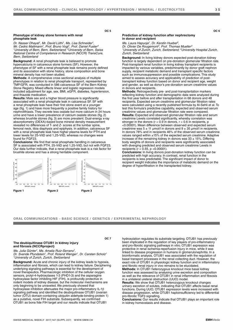
Phenotype of kidney stone formers with renal phosphate leakDr. Nasser Dhayat1, Mr. David Lüthi1, Ms. Lisa Schneider1, Mr. Cedric Mattmann1, Prof. Bruno Vogt1, Prof. Daniel Fuster2 1University of Bern, Bern, Switzerland; 2University of Bern, Swiss National Centre of Competence in Research (NCCR) TransCure, Bern, SwitzerlandBackground: A renal phosphate leak is believed to promote hypercalciuria in calcareous stone formers (SF). However, the phenotype of SF with a renal phosphate leak remains poorly defined and its association with stone history, stone composition and bone mineral density has not been studied.Methods: A comprehensive cross sectional analysis of multiple phenotypes in relation to renal phosphate transport, represented by TMP/GFR, was conducted in 586 calcareous SF of the Bern Kidney Stone Registry. Mixed effects linear and logistic regression models included adjustment for age, sex, BMI, eGFR, diabetes, hypertension, and thiazide medication.Results: Male sex and a higher blood pressure is significantly associated with a renal phosphate leak in calcareous SF. SF with a renal phosphate leak have their first stone event at a younger age (fig. 1) and have more frequently a positive family history for nephrolithiasis. They excrete less oxalate but more calcium in 24 hours urine and have a lower prevalence of calcium oxalate stones (fig. 2) whereas brushite stones (fig. 3) are more prevalent. Dual-energy x-ray absorptiometry (DEXA)-based bone mineral density measurement revealed no differences of T- and Z- scores at the lumbar spine, femoral neck, tibia diaphysis and epiphysis. In addition, calcareous SF with a renal phosphate leak have higher plasma levels for PTH and lower levels for 25-VitD and 1,25-VitD, whereas no changes were found for FGF23.Conclusions: We find that renal phosphate handling in calcareous SF is associated with PTH, 25-VitD and 1,25-VitD, but not with FGF23. Our data further indicate, that a renal phosphate leak is a risk factor for brushite stones and has a strong heritable component.
OC 6
Prediction of kidney function after nephrectomy in donor and recipientMs. Laura Hapuoja1, Dr. Kerstin Huebel2, Dr. Olivier De Rougemont2, Prof. Thomas Mueller2 1University of Zurich, Zurich, Switzerland; 2University Hospital Zurich, Zurich, SwitzerlandBackground: In living kidney donors expected post-donation kidney function is largely dependent on pre-donation glomerular filtration rate. Post-transplant renal function in living kidney transplant recipients is impacted by various variables, predominantly by donor graft nephron supply, recipient metabolic demand and transplant specific factors such as immunosuppression and possible complications. This study aimed to assess accuracy and applicability of prediction of post-transplant kidney function based on donor and recipient age, weight and gender, as well as donor’s pre-donation serum creatinine values in donors and recipients.Methods: Retrospectively pre- and post-transplantation markers reflecting kidney function and demographic data were analyzed during the first year before and after transplantation in 68 donors and 48 recipients. Expected serum creatinine and glomerular filtration rates were calculated using a recently published formula by Al-Sehli et al. To test this formula’s predictive capability, expected and observed serum creatinine values and glomerular filtration rate were compared.Results: Expected and observed glomerular filtration rate and serum creatinine Levels correlated significantly, whereby correlation was stronger in the donors (r = 0.9 in donors, r = 0.6 in recipients, p <0.00001). Mean difference between observed and expected serum creatinine was 5 ± 11 μmol/l in donors and 9 ± 35 μmol/l in recipients. In donors 78% and in recipients 46% of the observed serum creatinine values ranged within ±15% of the expected serum creatinine. Adaptive increase of the remaining kidney in donors was 33 ± 15%. Differing body weights of donors and recipients were significantly associated with diverging predicted and observed serum creatinine Levels in recipients (r = 0.55, p <0.00001).Conclusions: In living donors post-donation kidney function can be predicted with high accuracy. In contrast, renal function in the recipients is less predictable. The significant impact of donor to recipient weight indicates the importance of metabolic demand on the degree of hyperfiltration in the transplanted kidney.
Figure 1
OC 7
The deubiquitinase OTUB1 in kidney injury and fibrosis (NCCRproject)Ms. Julia Günter1, Ms. Amalia Ruiz-Serrano1, Dr. Svende Pfundstein1, Prof. Roland Wenger1, Dr. Carsten Scholz1 1University of Zurich, Zurich, SwitzerlandBackground: Acute and chronic injury of the kidney leads to hypoxia, inflammation and fibrosis, which can lead to kidney failure. Deciphering underlying signaling pathways is essential for the development of novel therapeutics. Pharmacologic inhibition of the cellular oxygen sensors, prolyl-4-hydroxylases 1-3 (PHD1-3) and the asparagine hydroxylase factor inhibiting HIF (FIH), is profoundly protective in rodent models of kidney disease, but the molecular mechanisms are only beginning to be unraveled. We previously showed that hydroxylase inhibition attenuates the major pro-inflammatory IL-1β signaling pathway and identified the deubiquitinase OTUB1 (ovarian tumor (OTU) domain-containing ubiquitin aldehyde-binding protein 1) as a putative, novel FIH substrate. Subsequently, we confirmed OTUB1 as bona fide FIH target and our results indicate that OTUB1
hydroxylation regulates its substrate targeting. OTUB1 has previously been implicated in the regulation of key players of pro-inflammatory and pro-fibrotic signaling pathways in vitro. OTUB1 expression was protective in renal ischemia-reperfusion injury in mice, while it was linked to disease progression in humans in glomerulonephritis. In a bioinformatic analysis, OTUB1 was associated with the regulation of basal transport processes in the renal collecting duct. However, the exact role of OTUB1 in physiologic kidney function and in inflammatory and fibrotic renal injury in vivo remains to be elucidated.Methods: In OTUB1 heterozygous knockout mice basal kidney function was assessed by analysing urine excretion and composition as well as the relevance of OTUB1 in renal inflammation and fibsosis in a unilateral ureteral obstruction (UUO) model.Results: We show that OTUB1 heterozygous knockout changes urinary excretion of solutes, indicating that OTUB1 affects basal renal functions. During UUO, OTUB1 expression levels were increased with disease progression, while OTUB1 haploinsufficiency led to differential pro-fibrotic TGFβ signalling.Conclusions: Our results indicate that OTUB1 plays an important role in kidney homeostasis and disease.
ORAL COMMUNICATIONS – BASIC SCIENCE / GENETICS / EXPERIMENTAL NEPHROLOGY
Figure 2 Figure 3
SWISS MEDICAL WEEKLY 2017;147 (SUPPL 227) WWW.SMW.CH

ORAL COMMUNICATIONS – BASIC SCIENCE / GENETICS / EXPERIMENTAL NEPHROLOGY 4 S
OC 8
Low β-catenin expression levels during development alter renal morphology and function (NCCR Project)Dr. Stefan Rudloff1, Dr. Delphine Lambert1, Ms. Andrea Karolin1, Ms. Andrea Bileck1, Prof. Uyen Huynh-Do1 1University Hospital Bern, Bern, SwitzerlandBackground: During kidney development, segmental identity of the future nephron is laid down by a proximal-distal β-catenin gradient. Ex-vivo chemical stimulation of β-catenin activity leads to an expansion of distal segment identity, whereas suppression of β-catenin activity promotes proximal positional identity. In this project, we reduced β-catenin expression levels along the entire nephron during nephrogenesis in-vivo or in murine distal convoluted tubule (DCT) cell lines in-vitro. We hypothesize that upon this restriction the DCT adapts properties of more proximal nephron segments, which further affects renal function.Methods: β-catenin expression in embryonic kidneys was reduced to 12.5% or 25% compared to wild type using 2 different Cre-lines. Kidneys were isolated before (Cdx1::Cre) or after birth (Pax8::Cre) and analyzed histologically, by qRTPCR or Western blot. Furthermore, biochemical parameters in urine and serum, as well as blood pressure were determined of Pax8::Cre mice. In vitro, β-catenin was knocked down in murine DCT cells with siRNA and the expression of specific nephron markers was assessed.Results: β-catenin knock-down kidneys were smaller in size, displaying multiple cysts. The expression of DCT markers was significantly reduced, whereas transcription of TAL markers was enhanced. Furthermore, Pax8::Cre mice displayed polyuria with an elevated protein:creatinine ratio, and had higher blood pressure than controls. Lastly, knock-down of β-catenin in DCT cells increased the expression of proximal nephron marker genes in these cells.Conclusions: Reducing β-catenin profoundly alters renal morphology and function, which is reflected by multiple phenotypes including cysts, polyuria or hypertension. Furthermore, the altered specification of intermediate nephron segments (DCT and TAL) provides a possible explanation for polyuria and hypertension in these mice. Whether the cystic transformation is also a consequence of the distorted pattern of nephron segments remains to be investigated.
OC 9
Protein phosphatase 1 inhibitor 1 mediates cAMP- dependent stimulation of the renal NaCl cotransporterDr. David Penton Ribas1, Ms. Sandra Moser1, Ms. Agnieszka Wengi1, Dr. Jan Czogalla1, Dr. Lena Lindtoft Ronsenbaek2, Dr. Nourdine Faresse1, Prof. Robert Fenton2, Dr. Dominique Loffing1, Prof. Johannes Loffing1 1University of Zurich, Zurich, Switzerland; 2University of Aarhus, Aarhus, DenmarkBackground: The thiazide-sensitive NaCl cotransporter (NCC) in the distal convoluted tubule (DCT) is critical for renal Na+ reabsorption and blood pressure control. Several cAMP-elevating hormones, including epinephrine, stimulate NCC activity. Here, we tested the hypothesis that the DCT-enriched protein phosphatase 1 inhibitor 1 (I1) mediates the effects of cAMP on NCC.Methods: In addition to MDCK cells stably transfected with NCC, several ex vivo approaches such as isolated mouse DCTs, mouse kidney slices and isolated perfused mouse kidneyswere used. The expression and phosphorylation of NCC and I1 were assessed by immunobloting and immunohystochemistry.
Results: Exposure of isolated DCTs to the cAMP-elevating agents forskolin and IBMX rapidly increased the phosphorylation of NCC via a protein kinase A (PKA)-dependent pathway. The forskolin/IBMX-induced NCC phosphorylation was paralleled by phosphorylation of I1 at its PKA-consensus phosphorylation site (T35). Forskolin/IBMXinduced phosphorylation of NCC was diminished in kidney slices from I1-knockout mice (I1-KO), while transgenic overexpression of a phosphomimetic I1 mutant (T35D) in kidneys of I1-KO mice restored NCC phosphorylation, but made NCC resistant to forskolin/IBMX stimulation. Yeast two-hybrid and co-immunoprecipitation experiments in MDCK cells stably transfected with NCC indicated a physical interaction between NCC and the I1-target PP1. Pharmacological inhibition of PP1 by calyculin A increased NCC phosphorylation. Finally, studies on kidney slices and isolated perfused kidneys from control and I1-KO mice demonstrated that I1 is critical for the beta-adrenergic stimulation of NCC.Conclusions: Our data establish a complete signal transduction pathway by which cAMP, via a PKA-dependent phosphorylation of I1 and subsequent inhibition of PP1, increases NCC phosphorylation. This pathway likely accounts for beta-adrenergic NCC activation and may hence contribute to salt-sensitive hypertension in patients with sympathetic hyperactivity.
OC 10
Localization and role of uromodulin in the distal convoluted tubuleDr. Natsuko Tokonami1, Dr. Tomoaki Takata1, Mr. Jan Beyeler1, Prof. Johannes Loffing1, Prof. Olivier Devuyst1, Dr. Eric Olinger1 1University of Zurich, Zurich, SwitzerlandBackground: The classical view is that uromodulin, the most abundant urinary protein, is exclusively produced by the cells lining the thick ascending limb (TAL), where it regulates the activity of the cotransporter NKCC2. It has been suggested that uromodulin may also be expressed in cells lining the distal convoluted tubule (DCT), where its role remains unknown.Methods: Detailed expression profiles of uromodulin were performed by RT-qPCR on microdissected tubular segments, in-situ hybridization (ISH), and co-immunostaining. Functional analysis of the DCT in Umod-/- vs. Umod+/+ mice was performed at baseline and after 5-day furosemide infusion to increase distal Na+ and Ca2+ delivery.Results: RT-qPCR and ISH confirmed peak expression levels of Umod in the cortical TAL, closely followed by the medullary TAL. We evidenced a significant Umod expression in mouse DCT, amounting to ~10% of TAL expression. Immunostaining in mouse and human kidney confirmed apical uromodulin localization in the DCT, largely restricted to the early DCT (DCT1). Compared to controls, Umod-/- mice exhibited 2-fold increased NCC phosphorylation (T53 & T58) at baseline, with no changes in plasma and urine electrolyte levels. Chronic furosemide infusion increased the levels of total NCC expression and phosphorylation in both Umod-/-and Umod+/+ kidneys. However, the fold change of NCC phosphorylation between furosemide non-treated and treated group was 2-fold smaller in Umod-/- mice (Umod+/+ 513% vs. Umod-/- 240%). Furthermore, Umod-/- mice displayed an exaggerated hypercalciuria with polyuria after 3 days of furosemide infusion, suggesting altered DCT electrolyte handling. Immunostaining for T53 phospho-NCC revealed a shift in NCC activity from DCT1 pools to downstream DCT2 segments in Umod-/- mice, both at baseline and after furosemide infusion.Conclusions: These results strongly corroborate uromodulin expression in the DCT1 in mouse und human kidney and suggest a role for uromodulin in the DCT homeostasis and NCC activity.
OC 11
Sex-hormone regulation of uric acid homeostasisDr. Muriel Auberson1, Ms. Fanny Durussel1, Prof. Olivier Bonny2 1University of Lausanne, Lausanne, Switzerland; 2University Hospital (CHUV) and University of Lausanne, Lausanne, SwitzerlandBackground: Men have significantly higher serum uric acid concentration (SUA) than women, leading to more frequent gout flairs and uric acid kidney stones. The difference can be largely accounted for by sex hormones, the mechanisms underlying this sex-specific regulation remains largely unknown.Methods: We used C57BL/6 mice as a model to study the role of sex hormones on uric acid homeostasis. Results: We showed that male mice have 30% higher SUA than females. After castration, this difference disappeared. We further explored the role of the kidney and measured the fractional excretion of urate, which was lower in male mice. Next, we determined the Figure 1
SWISS MEDICAL WEEKLY 2017;147 (SUPPL 227) WWW.SMW.CH

Results: 11 patients were started on RPM up to August 2017. PD team had to face a learning curve to instaure a systematic analysis of the transmitted data. In the first patient, RPM helped to recognize prolonged drained times (fig. 1) and led to early clinical evaluation with ensuing diagnosis and correction of PD catheter migration. Identification of <90% adherence to prescribed PD therapy was then documented with the RPM system (fig. 2), alerting the clinical staff to address this important issue given its association with significant negative clinical outcomes1. RPM also allows clinicians to remotely alter PD prescription, which proved very useful in one peculiar oliguric patient with ultrafiltration and subsequent overhydration problems necessitating weekly therapy adaptation. Some specific populations such as people with reduced mobility could also particularly benefit from RPM.Conclusions: RPM of APD patients with a two-way cloud-based connectivity platform allows for monitoring and quick adjustment of therapy, as well as early recognition and timely management of adverse clinical issues. It is therefore a promising new tool that may help clinicians to improve PD therapy outcomes and both patient and clinician confidence in embracing home dialysis.1 Jotterand Drepper et al., PDI, in press.
ORAL COMMUNICATIONS – BASIC SCIENCE / GENETICS / EXPERIMENTAL NEPHROLOGY 5 S
expression of the main renal uric acid transporters. SLC22A12 (URAT1), involved in urate reabsorption, was strongly down regulated in the female kidney, while SLC2A9 (GLUT9) was unchanged. In the liver, we observed a significant increase of SLC2A9 in female mice, allowing urate intake into the hepatocyte and its degradation by the intracellular uricase which was unchanged by istself. Preliminary results also showed decreased xanthine oxidase activity in the liver of females. In the intestine, uric acid is rapidly degraded in the lumen by the uricase expressed by the intestinal microbiota. We showed that females had a higher intestinal uricase activity than males, particularly in the caecum.Conclusions: Overall, these results showed co-operation between multiple organs to differentially regulate urate homeostasis between males and females. In females, (i) urate renal reabsorption is decreased, potentially through lower URAT1 expression, favoring renal excretion; (ii) GLUT9-mediated urate transport in the hepatocyte is increased, facilitating urate degradation by the intracellular uricase; (iv) uric acid synthesis by xanthine oxidase is decreased; (v) finally, intestinal uricase activity is higher in females. These observations will be of particular interest to personalize treatments for hyperuricemia, gout and kidney stones between genders.
OC 12
Absence of HIF pathway activation during chronic kidney disease (CKD): a pathway to anemia (NCCR project)Mr. Romain Dissard1, Ms. Frederique Ino1, Ms. Vasiliki Delitsikou1, Prof. Sophie De Seigneux2 1University of Geneva, Geneva, Switzerland; 2University Hospital Geneva, Geneva, SwitzerlandBackground: Anemia is prevalent in chronic kidney disease (CKD). Renal hypoxia and HIF activation have been proposed to play a role in CKD progression, whereas this was never clearly demonstrated. We
hypothesize that hypoxia and HIF activation are not prevalent in fibrotic areas of CKD, in relation to loss of tubular workload, explaining the anemia of CKD. Our aim was to analyze regional hypoxia and HIF activation at different CKD stages.Methods: We use of a model of glomerular disease induced by dose dependent genetic podocyte deletion, a model of tubular lesion (folic acid nephropathy), and a toxic model of proteinuria. We determine regional hypoxia by pimonidazole, HIF activation and Epo production by western blotting, PCR and immunohistochemistry. Results: HIF activation and hypoxia were not observed in early CKD stages characterized by mild fibrosis and proteinuria. Anemia was not observed in these stages, whereas kidney Epo production was mildly increased. In advanced CKD stages characterized by extensive fibrosis, proteinuria, anemia and low GFR, HIF protein and HIF targets genes were all downregulated. Hypoxia was restricted to some remnant tubuli and absent in fibrotic areas. This observation was confirmed in folic acid induced fibrosis. In parallel to HIF downregulation, a massive loss of mitochondrial mass and markers of decreased fatty acid oxidation were observed in tubular cells. In addition, in two models of proteinuric CKD, FIH protein expression was increased from early CKD stages, likely participating to HIF pathway inhibition. Epo expression was not increased in the diseased kidneys despite anemia, whereas we observe de novo synthesis of liver Epo.Conclusions: In advanced CKD, in opposition to the common belief, we observe a decrease in HIF pathway activation in the kidney, likely participating to anemia. The mechanisms of HIF pathway inhibition in advanced CKD include decreased energy consumption by fibrotic areas, mitochondrial loss, and FIH stabilization.
OC 13
Implementation of remote patient management in the care of automated peritoneal dialysis patients in Switzerland: 18 months experienceDr. Valérie Jotterand Drepper1, Prof. Pierre-Yves Martin1, Dr. Thomas Ernandez1, Dr. Catherine Stoermann-Chopard1 1AMC/Geneva University Hospitals, Geneva, SwitzerlandBackground: Given the remote nature of peritoneal dialysis (PD), nephrologist visibility to patient and therapy-related issues can be problematic. Remote patient management (RPM) enables monitoring of patients outside of conventional clinical settings and has already been implemented in many other medical fields including oncology, cardiology, diabetology and neurology with significant positive impact on patient outcomes but only scarce data exist up to now in nephrology.Methods: A newly available automated peritoneal dialysis (APD) RPM system (Claria Sharesource) with cloud-based connectivity was implemented in our department in December 2015. We present here our 18 months experience (up to August 2017).
ORAL COMMUNICATIONS – HEMODIALYSIS / PERITONEAL DIALYSIS
Figure 1
Figure 2
SWISS MEDICAL WEEKLY 2017;147 (SUPPL 227) WWW.SMW.CH
ORAL COMMUNICATIONS – HEMODIALYSIS / PERITONEAL DIALYSIS 6 S
OC 14
Experience with rivaroxaban treatment in 10 haemodialysis patientsMr. Davide Spica1, Dr. Beatrice Paul1, Prof. Stephan Segerer1, Prof. Andreas Bock1 1Kantonsspital Aarau, Aarau, SwitzerlandBackground: Using a direct factor Xa inhibitor such as rivaroxaban in haemodialysis patients has theoretical advantages, because in contrast to the vitamin K antagonists it lacks the risk of promoting arterial calcification and calciphylaxis. No data on efficacy and safety of this therapy exist in haemodialysis patients, and their use in this group is therefore discouraged.Methods: We retrospectively collected data on indication, duration of treatment, adverse events, and reasons for discontinuation in 10 haemodialysis patients treated with rivaroxaban (10 mg morning dose) over the past two years at our unit. Peak rivaroxaban concentrations (approximately 4 hours after intake) were measured at the beginning of afternoon haemodialysis sessions using the BIOPHEN DiXal kit (HYPHEN BioMed France), which measures anti-factor Xa activity using a chromogenic method.Results: Drug exposure time, mean rivaroxaban plasma concentration (±SD), indications for rivaroxaban, reasons for discontinuation and adverse events are listed in table 1. No patient had a major bleeding. One of 10 patients suffered from a cardioembolic stroke. The two deaths were unobserved sudden death episodes at home. Two patients were switched to vitamin K antagonists at the time of listing for kidney transplantation, where rivaroxaban is impractical, because there currently is no antagonist available.Conclusions: In this small group of haemodialysis patients exposed to rivaroxaban 10 mg/d during a total of 1817 days, the drug was well tolerated without any bleeding complication. If the mean rivaroxaban levels in this small group of haemodialysis patients are considered as CMax levels, they fall between the mean CMax levels found in phase 2 studies for VTE prevention after hip replacement (125 μg/l) and for stroke prevention in atrial fibrillation (229–249 μg/l). This may be appropriate for this group at high risk for bleeding but obviously requires prospective studies.
OC 15
Do measurements of serum ferritin and TSAT as performed in clinical practice accurately guide iv iron therapy with ferrum carboxymaltose in hemodialysis- patients?Mr. Matthias Diebold1, Dr. Andreas Kistler1 1Kantonsspital Frauenfeld, Frauenfeld, SwitzerlandBackground: Most chronic hemodialysis patients are in negative iron balance due various reasons. Iron is supplemented with doses of 100–200 mg every 2–4 weeks. While no laboratory parameters have been shown to reliably reflect total body iron stores, serum ferritin and transferrin saturation (TSAT) are generally used to assess the iron status of dialysis patients and to adjust maintenance iron dosing. The rationale of this study is to evaluate whether variations in the timing of blood sampling relative to the iron dosing schedule influences these laboratory parameters to a relevant degree and whether a certain amount of time should elapse between the last maintenance FCM dose and evaluation of iron Status.Methods: Patients are recruited from the outpatient hemodialysis population treated in the dialysis units in Frauenfeld and Münsterlingen. At the beginning of a dialysis session during which patients receive their FCM dose as well as at days 2, 4, 7, 14, 21 and 28, the following laboratory parameters are assessed: serum ferritin, TSAT, hematogram, CRP, reticulocyte count. The values will be compared to their baseline value using a two-sided paired t-test. Main inclusion criteria are stable dosing of FCM for the last 12 weeks, of epo (+/-25%) for the last 2 months, stable Hb values and normal CRP.
Results: By September 05, 2017, 13 patients have been included and 37 are planned to be enrolled by November. A first interim data analysis revealed a great interpatient variability in the rise of ferritin. Maximum peak values are reached between 4–7 days after FCM injection. In particular a dose of 200 mg FCM led to a significant transient rise of serum ferritin values.Conclusions: The timing of iron status evaluation has to be coordinated to FCM injection. An analysis of all patients completing the study by November will be presented at the Meeting.
OC 16
The role of ISO-9001:2008 certification for management and quality control in dialysis: the experience of a Swiss centreDr. Claudia Ferrier1, Prof. Stefano Calciolari2, Prof. Mario Bianchetti3 1Nefrocentro Clinica Luganese, NetMEGS, Università della Svizzera Italiana, Lugano, Switzerland; 2Istituto IdEP, Facoltà di Scienze Economiche, Università della Svizzera Italiana, Lugano, Switzerland; 3Facoltà di Biomedicina, Università della Svizzera Italiana, Lugano, SwitzerlandBackground: Worldwide government and health insurances are striving to contain increasing healthcare costs. In front of an aging population, providing high-quality haemodialysis therapy could be challenging in the near future. Therefore, defining a cost-effective organization able to remove inefficiencies by maintaining the quality of care is of major importance. The aim of this study was to analyse patients outcome and dialysis unit performance at different reorganization stages aimed to implement ISO-9001:2008 during the period 2000–2015.Methods: The “clinical-process indicators” (patients outcome) were measured using the yearly mean values of systolic blood pressure (SBP), urea reduction rate (URR%), haemoglobin (Hb), serum phosphate (P), calcium/phosphate product (Ca*P), albumin, ferritin and transferrin saturation (STrans). The targets were defined as SBP <160 mm Hg, URR>65%, Hb range 10–12 g/dl, P <1.8 mmol/L, Ca*P <4.5, albumin >34 g/L, ferritin 150 mg/L, STrans >20%. The clinical quality goal is the achievement of the target in 80% of the patients. “Structure-results indicators” (dialysis unit performance) were analysed in terms of mortality and growth over 12 months. The target for mortality was <15% and for growth >4%. The collected outcome and performance data were analysed according to each reorganization design model (e.g. elementary), following Mintzberg’s typology.Results: Our data, collected over 15 years, were compared with the “Swiss renal registry and quality assessment program” (srrqap). All clinical-process and structure-results targets were reached with the adhocracy model. Compared to other models, adhocracy reached an higher number of patients with a lower employment rate and no increase in mortality. Notwithstanding the older age of our patients, we found a similar mortality rate (12%) to srrqap analysis.Conclusions: Our results suggest that the organization developments pursued to implement ISO-9001:2008 had a positive influence on the management and quality control in dialysis. Adhocracy model displayed the highest efficiency. Whether this model is applicable to large centres needs further evaluation.
OC 17
Risk factors for community-acquired Acute Kidney Injury in patients with and without chronic kidney injury and impact of its initial management on prognosis: a prospective observational studyDr. Patrick Saudan1, Dr. Fabien Stucker2, Dr. Belen Ponte1, Dr. Cyrielle Alves1, Prof. Thomas Perneger1, Prof. Pierre-Yves Martin1 1Hôpitaux universitaires de Genève, Geneva, Switzerland; 2Hôpital de la Providence, Neuchâtel, SwitzerlandBackground: We aimed to describe clinical characteristics of patients with community-acquired acute kidney injury (CA-AKI), the effectiveness of initial management of CA-AKI, its prognosis and the impact of medication on its occurrence in patients with previous chronic kidney injury (CKI).Methods: We undertook a prospective observational study within the Emergency Department (ED) of a University Hospital, screening for any patient >16 years admitted with an eGFR <60 ml/mn/1.73 m2 and a rise in serum creatinine as compared to previous values. Patients’ medical files were reviewed by a panel of nephrologists in the subsequent days and at one and three-years follow-up.Results: From May 1st to June 21st 2013, there were 8464 admissions in the ED, of which 653 had an eGFR <60 ml/mn/1.73 m2. Of these, 352 had previous CKI, 341 had CA-AKI, and 104 had
Table 1
SWISS MEDICAL WEEKLY 2017;147 (SUPPL 227) WWW.SMW.CH

OC 20
Aryl hydrocarbon receptor expression by macrophages and lymphocytes within infiltrates in BK Polyomavirus associated nephropathyDr. Yassine Bouatou1, Dr. Geurt Stokman2, Dr. Nike Claessen2, Dr. Joris Roelofs2, Prof. Frederike Bemelman2, Dr. Jesper Kers2, Prof. Sandrine Florquin2 1AMC/Geneva University Hospitals, Geneva, Switzerland; 2AMC, Amsterdam, NetherlandsBackground: BK virus nephropathy (BKPyVN) is a major complication after renal transplantation. Little is known about the intra renal immune response during BKPyVN. The role of macrophages remains elusive. The activation of aryl hydrocarbon receptor (AHR) – a transcription factor involved in drug metabolism – plays a key role in inflammation and viral tolerance through modulation of macrophages polarization. Since AHR has not been studied in kidney transplantation, our aim was to compare the AHR expression within renal grafts in BKPyVN with T-cell mediated rejection (TCMR) as a control.Methods: We evaluated AHR expression in kidney grafts from BKPyVN (n = 8) with TCMR as control (n = 6) among cases with available frozen material for AHR gene intragraft transcription measurement and stainings for AHR, CD68 and CD45.Results: AHR transcription was higher in BKPyVN grafts versus TCMR (p = 0.03). While CD68+ or CD45+ cell expression did not differ within infiltrates (median score = 3 in both groups; p = 1.0 and 0.69, respectively), a higher proportion of nuclear AHR expression was found in BKPyVN for CD68+ and CD45+ cells when compared with TCMR (score median 2 vs 0; P = 0.007 and 1 vs 0; p = 0.013, respectively).Conclusions: We describe for the first time a higher expression of AHR in inflammatory cell infiltrates from BKPyVN versus TCMR renal biopsies. Further studies are required to explore AHR as a potential target in the modulation of inflammatory response in BKPyVN with known modulating ligands.
ORAL COMMUNICATIONS – HEMODIALYSIS / PERITONEAL DIALYSIS 7 S
CA-ACKI (community-acquired acute on chronic kidney injury). Occurrence of CA-ACKI was associated with male gender and with use of diuretics, but not with use of ARBs or ACEIs. Adequate management of CA-AKI defined as identification, diagnostic procedures and therapeutic intervention within 24 hours, was recorded in 45% of the cases and was not associated with improved outcomes. Three-year mortality was 21 and 48% in CKI patients, respectively, without or with CA-AKI, and 40% in patients with only CA-AKI (p <0.001). Mortality was significantly associated with age, hypertension, ischemic heart disease and CA-AKI. Progression of renal insufficiency was associated with male gender and age.Conclusions: CA-AKI is more frequently encountered in male patients and those treated with diuretics and is an independent risk factor for long-term mortality. Its initial adequate management failed to improve outcomes.
OC 18
Toward a better understanding of chronic kidney disease using metabolomicsMr. Yoric Gagnebin1, Mr. Julian Pezzati1, Dr. Pierre Lescuyer2, Prof. Sophie De Seigneux3, Dr. Julien Broccard1, Prof. Serge Rudaz1, Dr. Belen Ponte3 1University of Geneva and Lausanne, Geneva and Lausanne, Switzerland; 2Geneva University Hospitals, Geneva, Switzerland; 3Geneva University Hospitals and Medical School, Geneva, SwitzerlandBackground: Metabolomics aims to analyse comprehensively the metabolic complexity of biological systems and constitutes a potent method for assessing phenotype modifications caused environmental influences or pathologies. An extensive coverage of metabolites (mass
<1000 Da) is required for relevant untargeted metabolomics. As no technique offers an exhaustive monitoring of all metabolites in a biofluid, the use of multiple analytical platforms is needed. This methodology was implemented in the context of a clinical study to detect alterations in plasma metabolomic profiles due to chronic kidney disease (CKD) and allow a better understanding of the pathology.Methods: Reversed-phase chromatography (RPLC) and hydrophilic interaction chromatography (HILIC) coupled to high resolution mass spectrometry (HRMS) are complementary techniques commonly used for their coverage of apolar and polar metabolites, respectively. A strategy based on the combination of these two analytical approaches was applied to plasma samples collected from a clinical study designed to evaluate the metabolic impact of CKD. The cohort was composed of 56 control samples, and 104 patients at several disease stages, including 35 dialysed patients before their mid-week dialysis session. Each sample and quality control (QC) was analysed by RPLC and HILIC coupled to QTOF-MS in negative and positive ESI mode.Results: More than 230 annotated compounds were investigated thanks to the fusion of datasets generated from multiple platforms using an in-house database of 600 metabolites. The major sources of variability observed in the dataset were related to biological alterations due to the pathology that could be related to the Glomerular Filtration Rate. The multivariate analysis of the dataset showed a strong ability to stratify patients. Metabolite enrichment analysis was performed on discriminant metabolites to evaluate pathways potentially involved in the pathology.Conclusions: The workflow developed in this study allowed patient stratification according to CKD stages and helped to generate biological hypotheses based on the metabolomic profiles.
OC 19
Clinical long-term outcomes of kidney transplantation from pediatric donorsDr. Yvonne Holzmann1, Dr. Argyrios Georgalis1, Dr. Caroline Wehmeier1, Dr. Patricia Hirt-Minkowski1, Mr. Gideon Hönger1, Dr. Helmut Hopfer1, Prof. Lorenz Gürke1, Prof. Jürg Steiger1, Prof. Stefan Schaub1, Dr. Patrizia Amico1 1University Hospital Basel, Basel, SwitzerlandBackground: The aim was to compare principal long-term outcomes such as the risk of rejection, long-term graft function, and survival of kidney transplants deriving from pediatric deceased donors with transplants of adult kidneys in normal risk kidney transplantations.Methods: Of 352 kidney transplantations from deceased donors performed between 2005–2016, 47 transplants were from pediatric donors after excluding recipients with pre-transplant HLA-DSA (n = 86 in adult transplants and n = 2 in pediatric transplants). According to our policy, surveillance biopsies were taken at 3 and 6 months posttransplant, and indication biopsies performed in case of increase of serum creatinine or proteinuria.Results: At a median follow-up of 4 years post-transplant, death-censored graft survival of pediatric transplants was comparable to transplants from deceased adult donor (94% vs 93%; p = 0.54), while patient survival was significantly superior among recipients of pediatric transplants (96% vs 88%, p = 0.044), probably due to the younger age at transplantation (52 vs 59 yrs, p = 0.002). The cumulative incidence of (sub)clinical antibody-mediated rejection was significantly lower in the pediatric transplants (p = 0.0037), mainly driven by a lower incidence of clinical antibody-mediated rejection (p = 0.01). Furthermore, allograft function at last follow-up (eGFR-MDRD) was significantly different in pediatric transplant (87 vs 47 ml/min/1.73 m2, p <0.0001).Conclusions: Currently,pediatric kidneys constitute approximately 11–13% of deceased donor kidneys in our transplant unit. Despite the very young donor age, small kidney size, and surgical vascular challenges at transplantation, kidneys from pediatric donors show excellent clinical long-term outcome and therefore represent a valid expansion of the donor pool.
ORAL COMMUNICATIONS – TRANSPLANTATION
SWISS MEDICAL WEEKLY 2017;147 (SUPPL 227) WWW.SMW.CH
ORAL COMMUNICATIONS – TRANSPLANTATION 8 S
OC 21
Adherence and tolerability of prolonged-release tacrolimus in stable kidney and liver transplant patients after conversion from immediate-release tacrolimus in routine clinical practice: the IMPROVE studyDr. Marco Bonani1, Dr. Alexandre Balaphas2, Dr. Giulia Bedino3, Prof. Léo Bühler2, Dr. Philipp Dutkowski1, Dr. Kathrin Fausch4, Dr. Silvia Gluderer5, Dr. Nicole Graf6, Dr. Patricia Hirt-Minkowski7, Prof. Beat Müllhaupt1, Dr. Carlo Schönholzer3, Dr. Martin Schulz5, Dr. Reto Venzin4, Prof. Rudolf P. Wüthrich1 1University Hospital Zurich, Zurich, Switzerland; 2University Hospital Geneva, Geneva, Switzerland; 3Lugano Regional Hospital, Lugano, Switzerland; 4Cantonal Hospital Graubünden, Chur, Switzerland; 5Astellas Pharma AG, Wallisellen, Switzerland; 6Graf Biostatistics, Winterthur, Switzerland; 7University Hospital Basel, Basel, SwitzerlandBackground: Reduced dosing frequency may improve non-adherence with immunosuppression, a cause of graft rejection. Thus, the aim of this study was to evaluate adherence at 1 year in stable kidney and liver transplant patients converted from twice-daily, immediate-release tacrolimus (IR-T) to once-daily, prolonged-release tacrolimus (PR-T).Methods: This was a multicentre, non-interventional, observational, 12-month study conducted at five sites in Switzerland. Adult stable liver and kidney transplant patients, converted from IRT to PR-T in routine clinical practice, were included. Data were collected pre-conversion (Visit (V) 1), 2 weeks, 6 and 12 months post-conversion (V2–4). Primary composite endpoint was non-adherence by the Basel Assessment of Adherence to Immunosuppressive Medication Scale (BAASIS; V4), any investigator adherence rating of ‘poor’ (V2–4), or a subtherapeutic (investigator-defined) or over-therapeutic (>15 ng/mL) tacrolimus trough level (V3–4). Secondary endpoints included: components of the composite, pill burden, patient satisfaction, and adverse drug reactions (ADRs).Results: Seventy-eight patients were enrolled; 75 received PR-T, and 68 (46 kidney, 22 liver) completed the study. Most (81.8%, 36/44) patients were non-adherent for the composite endpoint. Overall non-adherence by BAASIS was similar at V1 (30.7%, 23/75) and V4 (28.3%, 17/60). During follow-up, investigators rated two patients as nonadherent; 62.0% (31/50) of patients had sub-therapeutic tacrolimus trough levels. PR-T decreased tacrolimus pill burden in 66.7% (40/60) of patients; median daily number of tacrolimus capsules decreased in kidney recipients (from 3.0 to 2.0) and liver recipients (4.0 to 2.0). All patients were very satisfied/satisfied with PR-T administration; 75% (48/64) of patients found it easier to remember to take PR-T versus IR-T. Overall, 20.0% (15/75) of patients reported ADRs, most frequently infections (9.3%; 7/75).Conclusions: For the kidney and liver transplant population combined, 1-year non-adherence rates were similar following conversion from IR-T to PR-T; however, PR-T intake was more convenient. PR-T was well tolerated over 1 year of treatment.
OC 22
Long-term outcomes after BK Polyomavirus replication in renal allograft recipientsMs. Nicole Bischof1, Prof. Hans Hirsch1, Dr. Caroline Wehmeier1, Dr. Patrizia Amico1, Prof. Michael Dickenmann1, Dr. Patricia Hirt-Minkowski1, Prof. Jürg Steiger1, Dr. Thomas Menter1, Dr. Helmut Hopfer1, Prof. Stefan Schaub1 1University Hospital Basel, Basel, SwitzerlandBackground: BK polyomavirus (BKPyV) replication defined by BKPyV-viremia occurs in 10–20% after kidney transplantation. Reduction of immunosuppression is the preferred treatment option for BKPyV-viremia, but there are very limited data on long-term outcomes of this strategy.Methods: We investigated 644 consecutive transplantations from 01/2005 to 08/2015, which had a standardized screening for BKPyV replication and a standardized treatment strategy consisting of reduction of immunosuppression in case of sustained BKPyV-viremia. Based on the presence of ‘Decoy-cells’ in the urine and BKPyV -viremia, transplantations were classified as ‘no Decoy-cells’ (n = 432; 67%), ‘Decoy-cells, no viremia’ (n = 107; 17%), and ‘viremia’ (n = 105; 16%). The investigated outcomes were graft/patient survival, occurrence of rejection, and evolution of allograft function.Results: There were no major differences regarding the baseline characteristics in the three groups. The median followup time was 6.5 years. Among the 105 ‘viremia’ cases, BKPyV-viremia was first detected at a median of 79 days (IQR 50–133) and resolved in 99/105 cases (94%) after a median of 137 days (IQR 72–397). Six-year graft survival was not different between the ‘no Decoy-cells’, ‘Decoy-cells, no viremia’, and ‘viremia’ groups (79%; 83%; 81%; p = 0.13). No graft loss
occurred due to BKPyV-associated nephropathy. Six-year incidence of clinical rejection was similar between 24% and 27% (p = 0.92). Median eGFR at last follow-up were between 47 and 50 ml/min (p = 0.30). Patients with a high BKPyV-burden (highest third of calculated BKPyV AUC) had a significantly lower eGFR at last follow-up compared to the other patients with BKPyV-viremia (40 vs 50 ml/min; p = 0.04).Conclusions: Reduction of immunosuppression is a successful long-term treatment strategy for BKPyV replication. A high BKPyV-burden seems to induce persisting allograft damage. Therefore, early detection of ongoing BKPyV replication and limiting the infectious burden are key components for good outcomes.
OC 23
Immunosuppressive drugs used to treat acute antibody-mediated rejection in kidney transplant recipients of the Swiss Transplant Cohort Study (STCS)Dr. Nancy Perrottet Ries1, Dr. Dela Golshayan2, Dr. Samuel Rotman1, Dr. Solange Moll 3, Dr. Helmut Hopfer4, Mrs. Emmanuelle Catana1, Dr. Michael Koller4, Dr. Jean-Pierre Venetz2, Dr. Vincent Aubert1, Dr. Oriol Manuel2, Prof. Léo Bühler5, Dr. Karine Hadaya6, Prof. Thomas Mueller7, Prof. Uyen Huynh-Do8, Dr. Isabelle Binet9, Prof. Michael Dickenmann4, Prof. Jürg Steiger4, Prof. Manuel Pascual2 1University Hospital (CHUV), Lausanne, Switzerland; 2University Hospital (CHUV) and University of Lausanne, Lausanne, Switzerland; 3Geneva University Hospital and Medical School, Geneva, Switzerland; 4University Hospital Basel, Basel, Switzerland, 5Geneva University Hospitals and Medical School, Geneva, Switzerland; 6Geneva University Hospitals, Geneva, Switzerland; 7Universitätsspital Zürich, Zurich, Switzerland; 8Inselspital Bern, Bern, Switzerland; 9Cantonal Hospital St Gallen, St. Gallen, SwitzerlandBackground: Acute antibody-mediated rejection (AMR) remains an important challenge after kidney transplantation. Various therapeutic strategies are used to treat acute AMR, however limited data from randomized trials are available. Methods: In this retrospective observational study, we included all kidney transplant recipients from the Swiss Transplant Cohort Study (STCS) from 2008 to 2014 who received a treatment for an acute AMR episode occurring in the first year posttransplantation (post-Tx). The primary objective was to analyze the use of immunosuppressive (IS) drugs to treat acute AMR in a “real life” cohort setting. The secondary objectives were to analyze the efficacy (improvement in renal function at 3 months post-acute AMR) and the safety (infectious complications occurring in the following 6 months) of the IS treatment used.Results: 64/1669 (3.8%) patients were treated for an acute AMR occurring in the first year post-Tx in the STCS (74 episodes in total). The median number of therapies used per acute AMR episode was two (range: 1–5 therapies). The most common bitherapy was Plasmapheresis (PPh) with methylprednisolone, and most common tritherapy was PPh, methylprednisolone and IVIG. The treatments used were effective in most cases, with full recovery of renal function in 68% of episodes. At 1-year, graft survival was 91%, and ongoing rejection was the main cause of graft loss. Four patients (4/64) died during the first year post-Tx (6.3%), two because of severe infectious complications. Overall, the incidence of any infectious complication was 42% in the following 6 months post AMR treatment.Conclusions: We found an heterogeneity in the IS drugs used to treat acute AMR within the first year post-Tx in the STCS, with an overall satisfactory response to therapy. Acute AMR remains a serious event post kidney-Tx with 9% graft loss at 1-year and with potentially severe infectious complications associated with its therapy.
OC 24
Review of studies on changes in renal physiology induced by nephrectomy in living donors (NCCR project)Dr. Andreja Figurek1, Prof. Valerie Luyckx1, Dr. Seraina Von Moos1, Prof. Thomas Mueller1 1University Spital Zürich, Zurich, SwitzerlandBackground: Safety is of utmost importance for living kidney donors (LKD). A number of studies indicate a low risk for healthy donors, however they often lack well matched control groups and sufficient long-term follow up. Hence, a better understanding of the basic physiological changes induced by donation would be most helpful regarding longterm risk assessment for potential donors, in particular when ‘borderline donors’ (BD) want to donate.Methods: In order to collect existing data on donor nephrectomy induced changes we performed a literature review on studies concerning the change in key nephrophysiology markers that occurs after kidney donation in LKD: preand post-donation glomerular filtration
SWISS MEDICAL WEEKLY 2017;147 (SUPPL 227) WWW.SMW.CH

ORAL COMMUNICATIONS – TRANSPLANTATION 9 S
rate (GFR), renal reserve capacity (RRC), effective renal plasma flow (ERPF). The additional aim was to focus on these changes in donors with medical risks.Results: We could identify a total of 35 physiology studies in LKD performed between 1956 to 2017 including a total of 4107 patients. Only two studies covered a followed-up period of more than 10 years. More than half of the studies assessed only GFR, and six studies were analysing post-nephrectomy changes in BD. The studies showed an adaptive increase in ERPF (106.64 ± 51.56 ml/min/1.73 m2) and measured GFR (16.15 ± 10.39 ml/min/1.73 m2) in the remaining kidney
after donation. Only 11 studies investigated RRC before and after donation, here a decrease in RRC post-nephrectomy could be seen (6.00 ± 1.41%). Very few studies analysed changes in BD, indicating a significant impact of age and body weight on GFR and RRC.Conclusions: Better understanding and more studies on physiology changes induced by kidney donation on key physiology markers are needed to further improve the safety of donation and a robust risk assessment for the potential LKD and to facilitate decision making in donor selection.
P 1
Impact of citrate supplementation on urinary risk profile in Swiss recurrent calcium stone formers (NCCR Project)Ms. Gioia Fischer1, Ms. Anna Wiegand1, Dr. Harald Seeger1, Prof. Daniel Fuster2, Prof. Olivier Bonny3, Dr. Thomas Ernandez4, Dr. Min Jeong KIM5, Prof. Carsten Wagner6, Dr. Nilufar Mohebbi1 1University Hospital Zurich, Zurich, Switzerland; 2University Hospital Bern, Bern, Switzerland; 3CHUV Lausanne, Lausanne, Switzerland; 4University Hospital Geneva, Geneva, Switzerland; 5University Hospital Basel, Basel, Switzerland; 6University of Zurich, Zurich, SwitzerlandBackground: Urolithiasis is common in developed countries with a significant recurrence rate. Hypocitraturia and hypercalciuria have been reported as the most prevalent risk factors. Citrate is a strong crystallization inhibitor and citrate supplementation has been introduced for metaphylaxis in recurrent kidney stone formers (rKSF) with hypocitraturia and normocitraturia. However, only few studies have investigated the impact of citrate on urinary stone risk profile parameters. Thus, the aim of this study was to investigate the changes of urinary stone risk profile after citrate supplementation in Swiss rKSF.Methods: This study is a retrospective analysis of prospectively collected data from the Swiss kidney stone cohort. 24hurine parameters were measured at baseline, after 3 months and one year of therapy. The primary endpoint of this study is the change of urinary parameters after citrate supplementation.Results: 446 participants (mean age 47 ± 14 years, 70% male) were evaluated. 95% of stones were calcium-containing, 88% consisted of oxalate, followed by 47% phosphate, 8% uric acid and 2% cysteine. Potassium citrate was administered to 52 patients (11.7%) at a mean dosage of 2523 ± 1173 mg citrate/d. Mean 24h-urine parameters at baseline were as follows: citrate 2.79 ± 1.54 mmol/d, potassium 60.12 ± 24.86 mmol/d, calcium 5.71 ± 3.27 mmol/d, sodium 164.81 ± 80.16 mmol/d, oxalate 0.21 ± 0.17 mmol/d, ammonium 19.42 ± 10.8 mmol/d, magnesium 3.84 ± 1.84 mmol/d, pH 5.99 ± 1.23, volume 1.82 ± 0.83 l/d. Treatment with potassium citrate was associated with significant changes after 3 months in the following parameters: pH (p = 0.047), citrate (p = 0.002), magnesium (p = 0.0248) and volume (p = 0.012). Interestingly, no significant changes were found between baseline and after 1 year, however, 1-y follow-up data were only available in a small subset of patients.Conclusions: Citrate supplementation in Swiss rKSF resulted in a significant increase of urinary citrate excretion, urinary magnesium excretion and urinary pH resulting in a beneficial change of urinary risk profile parameters. In addition, 24h urine volume increased significantly according to dietary recommendations.
P 2
Zinc deficiency in chronic kidney disease is not related to increased urinary excretionDr. Philippe Braconnier1, Dr. Sébastien Lenglet1, Dr. Marc Augsburger1, Prof. Michel Burnier2, Prof. Aurélien Thomas3, Dr. Menno Pruijm1 1University Hospital (CHUV), Lausanne, Switzerland; 2CHUV Lausanne, Lausanne, Switzerland; 3University of Lausanne, Lausanne, SwitzerlandBackground: Zinc (Zn) is an essential element in human physiology. Several studies have shown a high prevalence of Zn deficiency in patients with ESRD; however data on Zn in earlier stages of chronic
kidney disease (CKD) are scarce. The aim of this study was therefore to assess the Zn status in CKD patients in Switzerland.Methods: Serum Zn levels and Zn excretion in 24 hour urine were measured in 220 participants of the LauBOLDcohort that includes stage 1–5 CKD patients, hypertensives without CKD and healthy controls. A total of 117 patients with CKD (eGFR [mean ± SD] 55.3 ± 2.7 ml/min/1.73 m2) and 103 participants without CKD (eGFR 94.1 ± 1.5 ml/min/1.73 m2) were included.Results: Serum Zn levels were significantly lower in CKD patients compared to no-CKD patients (610.3 ± 105.4 μg/l versus 672.9 ± 97.4 μg/l, p <0.05) whereas urinary zinc excretion was not statistically
POSTER PRESENTATIONS – CLINICAL NEPHROLOGY / HYPERTENSION / MINERAL / ELECTROLYTES
SWISS MEDICAL WEEKLY 2017;147 (SUPPL 227) WWW.SMW.CH

POSTER PRESENTATIONS – CLINICAL NEPHROLOGY / HYPERTENSION / MINERAL / ELECTROLYTES 10 S
different between the two groups (611.6 ± 450.4 μg/24h versus 596.7 ± 493.7 μg/24h, p = 0.473). The decrease in serum Zn level started in the early stages of kidney disease (CKD stage 1: 626.2 ± 101.5 μg/l; stage 3: 593.8 ± 98.8 μg/l; stage 5: 558.2 ± 67.0 μg/l) and correlated significantly with eGFR (CKD-EPI) (Spearman’s rho: 0.28, p <0.05) (fig. 1). Zinc deficiency defined as serum zinc <658 μg/l in men and <638 μg/l in women was present in 64.7% of CKD patients and 35.3% of no-CKD patients (p <0.05). There was no significant correlation between urinary Zn excretion and eGFR (Spearman’s rho: 0.04, p = 0.6) (fig. 2). Multivariate linear regression showed a significant association between serum Zn level and eGFR (coef. β = 0.95 [95%CI: 0.3–1.6], p <0.05) (table 1).Conclusions: Zinc deficiency in CKD patients starts as early as CKD stage 1 and correlates with eGFR. However, urinary Zn excretion remains stable in CKD patients suggesting that Zn deficiency is rather a consequence of decreased dietary Zn intake and intestinal absorption than increased urinary excretion.
P 3
Essential trace elements in chronic kidney disease patients in SwitzerlandDr. Philippe Braconnier1, Dr. Sébastien Lenglet1, Dr. Marc Augsburger1, Prof. Michel Burnier2, Prof. Aurélien Thomas3, Dr. Menno Pruijm1 1University Hospital (CHUV), Lausanne, Switzerland; 2CHUV Lausanne, Switzerland; 3University of Lausanne, SwitzerlandBackground: Trace elements have important metabolic properties. Several studies suggest alterations in the levels of certain trace element in patients suffering from chronic kidney disease (CKD), which might contribute to the progression of kidney disease. The aim of this study was to determine the status of essential trace elements in CKD patients in Switzerland as compared to individuals without CKD.
Methods: Serum levels and 24 hour urine excretion of 23 trace elements (lithium, beryllium, aluminum, vanadium, chromium, manganese, cobalt, nickel, copper, arsenic, selenium, molybdenum, palladium, silver, cadmium, tin, antimony, iodine, platinum, mercury, thallium, lead, bismuth) were measured in 220 participants of the LauBOLD cohort. A total of 117 patients with CKD (eGFR [mean ± SD] 55.3 ± 2.7 ml/min/1.73 m2) and 103 participantswithout CKD (eGFR 94.1 ± 1.5 ml/min/1.73 m2) were included.Results: Demographic patient characteristics are shown in table 1. In CKD patients, serum levels of aluminum, arsenic, molybdenum, palladium, mercury, lithium and iodine were significantly increased compared to no-CKD patients, whereas no differences were seen in urinary excretion, lithium excepted (63.8 ± 60.2 μg/24h versus 44.7 ± 37.0 μg/24h, p <0.05) (table 2). Serum levels of nickel and bismuth were significantly reduced in CKD patients without increase in urinary excretion.Conclusions: Serum levels of biologically important trace elements were substantially different in CKD patients compared with controls. The majority of serum levels were higher in CKD, at equal urinary excretion. This possibly points towards disturbed renal elimination in CKD patients. However, most serum levels of trace elements were in reference ranges for both CKD and no-CKD patients.
P 4
Longitudinal follow-up of stone formers in Switzerland – NCCR Kidney.CH projectProf. Olivier Bonny1, Prof. Daniel Fuster2, Dr. Nilufar Mohebbi3, Dr. Thomas Ernandez4, Dr. Min Jeong KIM5, Dr. Florian Buchkremer6, Dr. Beat Roth2, Dr. Grazia Cereghetti7, Prof. Carsten Wagner8 1University Hospital (CHUV), Lausanne, Switzerland; 2Inselspital Bern, Bern, Switzerland; 3Universitätsspital Zürich, Zurich, Switzerland; 4Hôpitaux universitaires de Genève, Geneva, Switzerland; 5University Hospital Basel, Basel, Switzerland; 6Kantonsspital Aarau, Aarau, Switzerland; 7NCCR Kidney.CH, Zurich, Switzerland; 8University of Zurich, Zurich, SwitzerlandBackground: Kidney stones are highly prevalent and represent a significant burden to the health system, while neglected by research and support. The Swiss Kidney Stone Cohort is meant to fill this gap and collects epidemiological and biological data from recurrent stone formers in Switzerland. One strength of the Cohort is the longitudinal follow-up and the extensive phenotype of the patients.Methods: Adult patients were recruited in the five Swiss University Clinics of Nephrology (Basel, Bern, Geneva, Lausanne and Zurich) and Kantonspital Aarau if they were recurrent stone formers or had a single episode with predetermined risk factors. Work-ups are standardized between the centers and include 2×24 h urine collection, food and activity questionnaires and standardized 24h recall interviews of food intake by trained dieticians. Samples of urine, blood and DNA are stored in a biobank. All lab analysis are centralized. Follow-up visits are organized at 3 months and annually. We report here on patients who performed complete work-ups at baseline, 3 months and first year visit.Results: From 493 patients recruited so far in the SKSC, we obtained full set of data from 113 of them for baseline and follow-up visit at 3 months and at first year. 77 males and 35 females were analyzed. Mean age is 49.0 ± 14.7 years. BMI is 26.4 ± 4.8 Kg/m2. In the blood, only albumine and PTH were significantly different between baseline and 1-year-visit (lower albumine and PTH at 1 year). In 24h urine collection (under oil), urine volume and citrate excretion were significantly increased at 3 months, a difference still present at 1 year, attenuated though.Conclusions: Our data show that in stone formers followed over time, life style changes (increased hydration, fruits) as well as therapeutic intervention (citrate) can be succesfully implemented and effects are sustained over time. We encourage long term follow-up of stone formers.
P 5
Pre-existing, Perioperative and Postoperative Chronic Kidney Disease in Patients Receiving Orthotopic Liver TransplantationMr. Fabian Hauenstein1, Dr. Guido Beldi1, Prof. Bruno Vogt1, Prof. Uyen Huynh-Do1, Dr. Vanessa Banz1, Dr. Daniel Sidler1 1University Hospital Bern, Bern, SwitzerlandBackground: Chronic Kidney Disease is associated with increased morbidity and mortality in the general population and a strong predictor for adverse outcome in patients with concomitant diseases, including heart, lung and liver diseases.
Table 1 : Demographic patient characteristics
Total(n=220) CKD(n=117) No-CKD(n=103) pAge(y)1 54.0±0.9 56.4±1.3 51.3±1.3 <0.05Male(n) 143(65%) 82(70.1%) 61(59.2%) <0.05Hypertension(n) 153(69.5%) 92(78.6%) 61(59.2%) <0.05Diabetesmellitus(n) 39(17.7%) 29(24.7%) 10(9.7%) <0.05eGFR(CKD-EPI)(ml/min)1 73.5±2.0 55.3±2.7 94.1±1.5 <0.05Smoker(n) 78(34.5%) 43(36.7%) 35(33.9%) 0.564BMI(kg/m2)1 27.5±4.9 27.7±4.6 27.3±5.2 0.531Dataexpressedasmean±SD
Table 2 : Biochemical parameters of the study groups* (Data expressed as mean±SD)
Reference(range)1,2 CKD(n=117) No-CKD(n=103) pAluminumserum(µg/l) 5–24 4.8±7.7 3.0±1.4 <0.05Aluminiumexcretion(µg/24h) 10.4±9.3 11.3±13.1 0.576Nickelserum(µg/l) 0.04–5.3 0.86±0.48 1.05±0.79 <0.05Nickelexcretion(µg/24h) 2.2±1.6 1.9±1.1 0.096Arsenicserum(µg/l) 0.7–16 2.5±3.8 1.0±1.9 <0.05Arsenicexcretion(µg/24h) 64.9±117.7 42.1±96.0 0.167Molybdenumserum(µg/l) 0.4–1.9 1.6±2.4 0.9±0.4 <0.05Molybdenumexcretion(µg/24h) 48.0±38.5 60.6±66.8 0.119Palladiumserum(µg/l) 0.05–0.17 0.08±0.04 0.05±0.02 <0.05Palladiumexcretion(µg/24h) 0.28±0.25 0.24±0.19 0.200Mercuryserum(µg/l) 0.16–2.3 0.96±0.58 0.80±0.40 <0.05Mercuryexcretion(µg/24h) 0.93±0.75 0.94±0.98 0.925Bismuthserum(µg/l) 0.002–0.04 0.011±0.006 0.013±0.009 <0.05Bismuthexcretion(µg/24h) 0.06±0.25 0.04±0.059 0.355Lithiumserum(µg/l) 0.7–9.1 6.9±6.1 4.9±5.8 <0.05Lithiumexcretion(µg/24h) 63.8±60.2 44.7±37.0 <0.05Iodineserum(µg/l) 53–110 61.0±21.0 55.5±10.7 <0.05Iodineexcretion(µg/24h) 196.5±162.9 195.8±373.6 0.987Berylliumserum(µg/l) 0.0039±0.0035 0.0037±0.0027 0.724Vanadiumserum(µg/l) 0.21–0.70 0.29±0.069 0.29±0.043 0.162Chromiumserum(µg/l) 0.5–1.2 0.74±0.13 0.75±0.13 0.639Manganeseserum(µg/l) 0.6–2.6 0.51±0.17 0.49±0.11 0.347Cobaltserum(µg/l) 0.14–0.40 0.16±0.42 0.12±0.12 0.367Copperserum(µg/l) 770–2500 907.0±231.8 897.0±236.1 0.751Seleniumserum(µg/l) 72–160 102.0±31.3 100.9±15.6 0.749Silverserum(µg/l) 0.01–0.88 0.29±0.40 0.27±0.41 0.680Cadmiumserum(µg/l) 0.01–0.16 0.047±0.016 0.046±0.015 0.397Tinserum(µg/l) 0.15–0.69 0.25±0.34 0.20±0.25 0.206Antimonyserum(µg/l) 0.03–0.15 0.024±0.011 0.023±0.009 0.650Platinumserum(µg/l) <0.015 0.051±0.11 0.045±0.039 0.604Thalliumserum(µg/l) 0.002–0.02 0.037±0.014 0.036±0.012 0.925Leadserum(µg/l) 0.014–0.89 0.69±0.55 0.60±0.36 0.107*Urinaryexcretionwasmeasuredinasubgroupof97CKDpatientsand79no-CKDpatients1GoulléJ-P,etal.,Themetallicprofile:anewbiologicalconcept.AnnBiolClin68(2010)429-440
2BaseltRC,Dispositionoftoxicdrugsandchemicalsinman,8thEdition,BiomedicalPublications(2008),FosterCity,California
SWISS MEDICAL WEEKLY 2017;147 (SUPPL 227) WWW.SMW.CH
alterations of kidney fatty acid metabolism are risk factors for CKD progression. We hypothesized that the abnormal fat distribution found in HIV positive patients with LD may be an independent risk factor to develop CKD.Methods: All patients from the Swiss HIV Cohort Study with an estimated glomerular filtration rate (eGFR) >60 ml/min/1.73 m2 at entry in the cohort from 2002 to 2015 with a minimal follow-up of 3 months were included. The primary endpoint was defined as a sustained eGFR <60 ml/min/1.73 m2. Cox regression models were used to measure the risk to develop CKD associated with different patterns of LD.Results: Among the 5’384 patients included, 4’246 did not have LD at entry in the cohort. 31.0% developed LD during their follow-up after a median time of 17.1 months (IQR: 0–45.2 months) and 252 (4.7%) reached the studied endpoint after a median follow-up time of 43.7 months from baseline (IQR: 18.5–89.3 months). Overall LD increased significantly the risk of an eGFR <60 ml/min/1.73 m2 in univariate analysis with a hazard ratio (HR) 2.25 (95% confidence interval (CI): 1.68–3.00; p <0.001). After adjustment for main confounders (such as age, sex, hypertension, diabetes, baseline eGFR and viral load), LD increased the risk of eGFR <60 ml/min/1.73 m2 by a HR 1.98 (95% CI: 1.31–2.99; p = 0.001).Conclusions: LD might be a risk factor for eGFR decline in HIV positive patients independently of previously reported CKD risk factors.
P 8
Diagnostic accuracy of immunofluorescence versus immunoperoxidase staining to distinguish immune complex-mediated glomerulonephritis and C3 dominant glomerulopathyDr. Yassine Bouatou1, Dr. Jesper Kers2, Ms. Marie Chevalier Florquin2, Dr. Nike Claessen2, Dr. Tri Nguyen3, Dr. Jeffrey Damman2, Dr. Henrique Proenca4, Dr. Joris Roelofs2, Prof. Sandrine Florquin2 1AMC/Geneva University Hospitals, Geneva, Switzerland; 2AMC, Amsterdam, Netherlands; 3UMCU, Utrecht, Netherlands; 4Universidade Metropolitana de Santos (UNIMES), Santos, BrazilBackground: Membranoproliferative glomerulonephritis (MPGN) has been reclassified from an electron microscopy to an immunofluorescence (IF) based semi quantitative classification with immunoperoxidase (IP) technique as a backup option when IF is not possible. However, no data are available on the interobserver variability, the correlation and the reclassification of MPGN based on these two techniques.Methods: We retrospectively analyzed cases of type 1 MPGN. We repeated IF and performed IP for IgG, kappa, lambda, C3c, and C4d in 35 renal biopsies among which 19 biopsies having a matched IP and IF.Results: We observed a substantial to near perfect agreement among the 7 observers for both IF and IP (W coefficients from 0.66 for IF lambda to 0.89 for IF C4d). Of the 19 cases with matching IP and IF, 5 (26%) turned out to have a different diagnosis on IF as compared to IP. Also, C4d ability to discriminate immune complex-mediated GN (ICGN) from C3 glomerulopathy (C3G) was poor with an area under the curve of 0.44 (95% CI = 0.24–0.63) and 0.66 (95% CI = 0.50–0.81) for the receiver operating characteristic curves of IF and IP respectively. Limitations include that no clinical data regarding complement activation were available.Conclusions: The diagnosis of ICGN versus C3GN depends on the immunochemical technique used. Also, the use of C4d failed to discriminate ICGN from C3G in our study. Further validation studies are required to avoid misdiagnosis based on kidney biopsy.
P 9
Medication adherence during work-up for Conn syndromeDr. Jürgen Bohlender1, Dr. Friederike Sandbaumhüter1, Prof. Bruno Vogt1, Prof. Manuel Haschke1 1Inselspital, Bern University Hospital, Bern, SwitzerlandBackground: Non-adherence to prescribed medication may lead to misinterpretations of aldosterone-to-renin ratios during the work-up of resistant hypertension and Conn syndrome. We compared plasma concentrations of antihypertensive drugs with actual prescriptions in routine patients referred to our hypertension clinic for evaluation of difficult-to-treat hypertension on at least 2 antihypertensives.Methods: Plasma concentrations of individually prescribed irbesartan, valsartan, olmesartan, candesartan, perindopril, metoprolol, amlodipine, lercanidipine and hydrochlorothiazide were determined in all patients by mass spectroscopy on the 1st clinical visit and again after stopping renin-angiotensin system inhibitors, beta-blockers and
Methods: We evaluated the frequency, cause and outcome of kidney disease in patients undergoing orthotopic liver transplantations (OLT). We evaluated 279 OLT in 262 patients at the University Hospital in Bern from 01.01.2005 to 18.07.2017.Results: Preexisting kidney disease was infrequent in patients during evaluation for OLT with a prevalence of 15% of patients suffering from CDK3 and higher. Perioperative AKINIII was present in 38% of patients and among those, 69% required renal replacement therapy. Preexisting CKD, perioperative AKINIII and perioperative RRT were strong predictors for inferior patient outcome after OLT. Among the patients with at least one month of follow-up, median Creatinine at the end of follow up was 106 umol/l (range 24–779) with a eGFR-EPI of 55 ml/min/1.73 m2 (range 16–88 ml/min/1.73 m2). 51.7% of patients suffered from CKD3 and higher. Pre-existing eGFR <90 ml/min/1.73 m2, older age at transplantation, perioperative AKIN III and hypertension were strongly associated with the development of chronic CKD in the OLT cohort.Conclusions: In conclusion, concomitant kidney disease in the pre- or post-transplant period is a highly relevant co-morbidity in the OLT cohort in need of more specific attention.
P 6
Change in V-ATPase B1 but not B2 subunit abundance in human urinary exosomes in response to acute acid/alkali loading and distal renal tubular acidosisDr. Ganesh Pathare1, Dr. Nasser Dhayat1, Dr. Nilufar Mohebbi2, Prof. Carsten Wagner3, Dr. Alexandru Bobulescu4, Prof. Orson Moe4, Prof. Daniel Fuster1 1University of Bern, Bern, Switzerland; 2University Hospital Zurich, Zurich, Switzerland; 3University of Zurich, Zurich, Switzerland; 4University of Texas Southwestern Medical Center, Dallas, Texas, USABackground: In the kidney, final urinary acidification is achieved by V-ATPases expressed in type A intercalated cells. The B1 subunit of the V-ATPase is required for maximal urinary acidification, while the role of the homologous B2 subunit is less clear.Methods: We examined the effect of acute acid/alkali loading in humans on B1 and B2 subunit abundance in urinary exosomes in normal subjects and of acid loading in patients with distal renal tubular acidosis (dRTA). Specificities of B1 and B2 subunit antibodies were verified by yeast heterologously expressing human B1 and B2 subunits, and murine WT and B1-deleted kidney lysates.Results: Acute NH4Cl loading elicited systemic acidemia, drop in urinary pH, and increase in urinary NH4 excretion. Nadir urinary pH was achieved in 4–5 hrs, and exosomal B1 abundance was significantly increased at 2, 3 and 4 hrs after NH4Cl loading. After acute equimolar NaHCO3 loading, blood and urinary pH rose rapidly, with concomitant reduction of exosomal B1 abundance within 2 hrs and B1 abundance remained lower throughout the test. In contrast, no changes in exosomal B2 abundance were observed following acid or alkali loading. In patients with inherited or acquired dRTA, urinary B1 subunit was extremely low or undetectable and did not respond to acid loading in urine, whereas no change in B2 subunit was observed.Conclusions: In summary, both B1 and B2 subunits of the V-ATPase are detected in human urinary exosomes, and acid and alkali loading or dRTA cause changes in the B1 but not B2 subunit abundance in urinary exosomes.
P 7
Lipodystrophy increases the risk of developing chronic kidney disease in HIV- positive patients in Switzerland: the LIPOKID studyDr. Yassine Bouatou1, Dr. Angèle Gayet-Ageron1, Dr. Enos Bernasconi2, Prof. Manuel Battegay3, Dr. Matthias Hoffmann4, Dr. Cornelia Staehelin5, Dr. Laurent Merz6, Dr. Helen Kovari7, Dr. Christoph Fux8, Prof. Alexandra Calmy1, Prof. Sophie De Seigneux1 1University Hospital Geneva, Geneva, Switzerland; 2Regional Hospital Lugano, Lugano, Switzerland; 3University Hospital Basel, Basel, Switzerland; 4Kantonsspital St. Gallen, St. Gallen, Switzerland; 5Bern University Hospital, Bern, Switzerland; 6CHUV Lausanne, Lausanne, Switzerland; 7University Hospital Zurich, Zurich, Switzerland; 8Kantonsspital Aarau, Aarau, SwitzerlandBackground: Antiretroviral therapy (ART) improved HIV positive patient survival. However, those patients developed metabolic complications such as lipodystrophy (LD) which is the hallmark of first generation ART. LD, defined as body shape abnormalities such as central fat accumulation and peripheral fat loss, has been associated with modifications in adipokines and may be related to alterations of mitochondrial metabolism. Abdominal obesity, adipokines and
POSTER PRESENTATIONS – CLINICAL NEPHROLOGY / HYPERTENSION / MINERAL / ELECTROLYTES 11 S
SWISS MEDICAL WEEKLY 2017;147 (SUPPL 227) WWW.SMW.CH

P 11
Renal Amyloidosis is not sufficiently prevented in patients with FMF in ArmeniaDr. Sofi Sarinyan1, Dr. Helen Nazaryan2, Dr. Armen Sanamyan2, Prof. Gayane Amaryan1, Prof. Ernst Leumann3, Prof. Ara Babloyan2, Prof. Ashot Sarkissian2 1Yerevan State Medical University, Yerevan, Armenia; 2Arabkir Medical Centre, Yerevan, Armenia; 3University Children’s Hospital, Zurich, SwitzerlandBackground: Familial Mediterranean fever (FMF) is a serious health problem in Armenia. Although amyloidosis – a potentially fatal complication of FMF – can largely be prevented by colchicine administration, we are still confronted with renal amyloidosis. The aim was to analyze the demography and reasons for biopsy-proven amyloid nephropathy.Methods: The National Pediatric Center for FMF (NPC FMF) was established in 1998 to allow early diagnosis, treatment, follow-up and wide dissemination of information on FMF. Since 2003 NPC FMF has implemented the long term programm and provided regularly colchicine to over 3000 children <18 years, but not to adults. Diagnosis of FMF is based on Tel-Hashomer criteria and molecular genetic analysis (since 1998). Amyloid nephropathy was confirmed by renal biopsy (Congo Red). Patients with biopsies 1993–2004 (group 1; n = 206) are compared with those 2005–2016 (group 2; n = 475).Results: Amyloid nephropathy due to FMF in group 1 was detected in 47 pts (38 children, 9 adults) = 23% of all biopsies, as compared to 42 pts (22 children, 20 adults) in group 2 (= 9% of biopsies; p <0.05). The second group was further analyzed: On admission 6 children versus 7 adults had proteinuria, 14 versus 10 were nephrotic and 2 versus 3 had CKD (stage 3). FMF was diagnosed late in 18 pediatric patients, one was noncompliant, three were partially resistent to colchicine. In adults, FMF was diagnosed late in 16, whereas in 4 colchicine was not sufficiently effective (low dose?). Late administration of colchicine could not reverse the course.Conclusions: In contrast to the absolute number of patients the proportion of biopsies showing amyloid nephropathy has significantly dropped. The National FMF program in Armenia is only partly effective in prevention of renal amyloidosis and requires additional efforts. Colchicine is not able to reverse advanced amyloid nephropathy.
P 12
Microbiological analysis of hemodialysis water at the university teaching hospital of Yaounde, CameroonDr. Cedric Gueguim1, Dr. Alain Ragon2, Prof. Francois Kaze1, Prof. Hortense Gonsu Kamga1 1University of Yaounde I, Yaounde, Cameroon; 2Hospital of Conception, Marseille, FranceBackground: Rigorous control of the microbiological quality of water in hemodialysis services is important because the immune system of patients with chronic renal failure is weakened. The objective of this study was to determine the microbiological quality of water for hemodialysis in the hemodialysis department of the University Teaching Hospital of Yaoundé in order to improve the disinfection strategy.Methods: Twelve water samples were collected each month at different sites of the hemodialysis circuits A (inlet of filters), B (Outlet of filters / inlet of Reverse Osmosis (RO) device) and C (outlet of the RO device / close to the generator) between July and October 2015 to be analyzed. The bacteria were isolated after filtration of 100 ml of water at each site through nitrocellulose membrane with 0.45 μm microporosity deposited on the surface of the Tryptone Glucose Extract Agar (TGEA) and then incubated at room temperature (20 to 22 °C) for 7 days. After transplanting to different environments, pure bacterial isolates were identified by their cultural characters and marketed biochemical galleries.Results: The colony count was well above the required international standards (>100 CFU / ml), for the hemodialysis water with a percentage of 83.3% (10/12) of non-compliance. Amongthe bacteria identified, nine (09) were Gram-negative bacilli including Pasteurella haemolytica, Pseudomonas fluorescens, Pseudomonas paucimobilis, Aeromonas salmonicidaand Klebsiella pneumoniae subsp ozaenae, three (03) Gram-positive bacilli all Bacillus sp and six (06) Gram-positivecocci all of coagulase-negative staphylococci. The most frequently isolated bacterial genera were Pseudomonas (30.4%), Staphylococcus (26.1%), Aeromonas (13%), Bacillus (13%), Klebsiella(13%) and Pasteurella (4.3%).Conclusions: The detection of a variety of bacteria in the hemodialysis water indicates the need for regular monitoring of the water for hemodialysis by the CHUY hemodialysis center to ensure a better quality of life for patients undergoing this treatment.
POSTER PRESENTATIONS – CLINICAL NEPHROLOGY / HYPERTENSION / MINERAL / ELECTROLYTES 12 S
diuretics before aldosterone-to-renin ratio determinations (2nd visit). Ca-antagonists and doxazosin were allowed on 2nd visits to control hypertension. Patients on aliskiren or aldosterone, chronic renal insufficiency stage >3, heart failure NYHA IV and pregnancy were excluded.Results: Twenty-four consecutive patients were included: 42% female, 21% diabetics; mean age 54 ± 13y, BMI 28 ± 7, s.-creatinine 87 ± 26 μmol/l). Mean 1st to 2nd visit interval was 14 ± 6 days. Prescribed angiotensin receptor-blockers had (1st/2nd visit) 54.2/0%, converting enzyme inhibitors 29.2/0%, Ca-antagonists 79.2/79.2%, diuretics 66.7/0%, doxazosin 0/25.0%, beta-blockers 50,0/0%, thiazides 62.5/0, loop diuretics 4.2/0%. Antihypertensives not determined were lisinopril, azilsartan, nebivolol, carvedilol, torasemide (10 concentrations/visit). Discrepancy of measured plasma levels (absence/presence) with prescribed and tested medication was detected in 25% on 1st visit (non-adherence 12.5%/unprescribed presence 12.5%) and 21% (0%/21%) on 2nd visit concerning 41.7% of patients for both visits (10/24, 95% confidence interval 22.1 to 61.4%). Prohibited medication on 2nd visit was detectable in 16.7% (4/24; 95% confidence interval 4.8 to 37.4%).Conclusions: Non-adherence to prescribed medication is frequent during work-up of difficult-to-treat hypertension and Conn syndrome and may interfere with aldosterone/renin determinations in a significant number. Physicians should interpret results cautiously in view of possible medication bias and seek confirmation in unclear cases (funded by Swiss Kidney Foundation/Bär-Spycher Foundation).
P 10
An algorithm to prevent overly rapid correction of hyponatremia by urine volume monitoringDr. Florian Buchkremer1, Prof. Stephan Segerer1, Prof. Andreas Bock1 1Kantonsspital Aarau, Aarau, SwitzerlandBackground: Inadvertent overcorrection of chronic hyponatremia is the most important risk factor for osmotic demyelination, a devastating complication with huge morbidity and mortality. Excessive increases of the plasma sodium concentration (P-Na) are usually caused by an unanticipated, substantial water diuresis. A simple formula for monitoring urine volume may thus increase the safety of corrective therapy in hyponatremia.Methods: Using a derivation of the Edelman equation we analyzed the impact on the P-Na of four different parameters: (a) urine volume, (b) total body water, (c) urine sodium and potassium concentration and (d) baseline P-Na. P-Na increases with higher urine volume, higher total body water, more dilute urine and higher baseline P-Na.Results: By choosing values for parameters b-d, that would lead to the highest increase in P-Na, and allowing for a maximal increase of the P-Na of 8 mmol/l per day, we arrived at a an upper “safe” limit of urine volume of 24 ml per kg of body weight per 24h (=1 ml/kg per hour) or a urine volume of 2400 ml/d (=100 ml/h), whichever is smaller. Since the relationship between changes in P-Na and urine volume per body weight is nearly proportional (fig. 1), bedside adjustements are easy.Conclusions: We suggest monitoring the urine volume of hyponatremic patients as an additional safety measure to prevent overcorrection and osmotic demyelination. Using an upper urine flow limit of 1 ml/kg/h (or 100 ml/h, whichever is smaller) has the potential to identify patients at risk of inadvertent overcorrection earlier and more reliably than serial P-Na determinations alone.
Figure 1
SWISS MEDICAL WEEKLY 2017;147 (SUPPL 227) WWW.SMW.CH
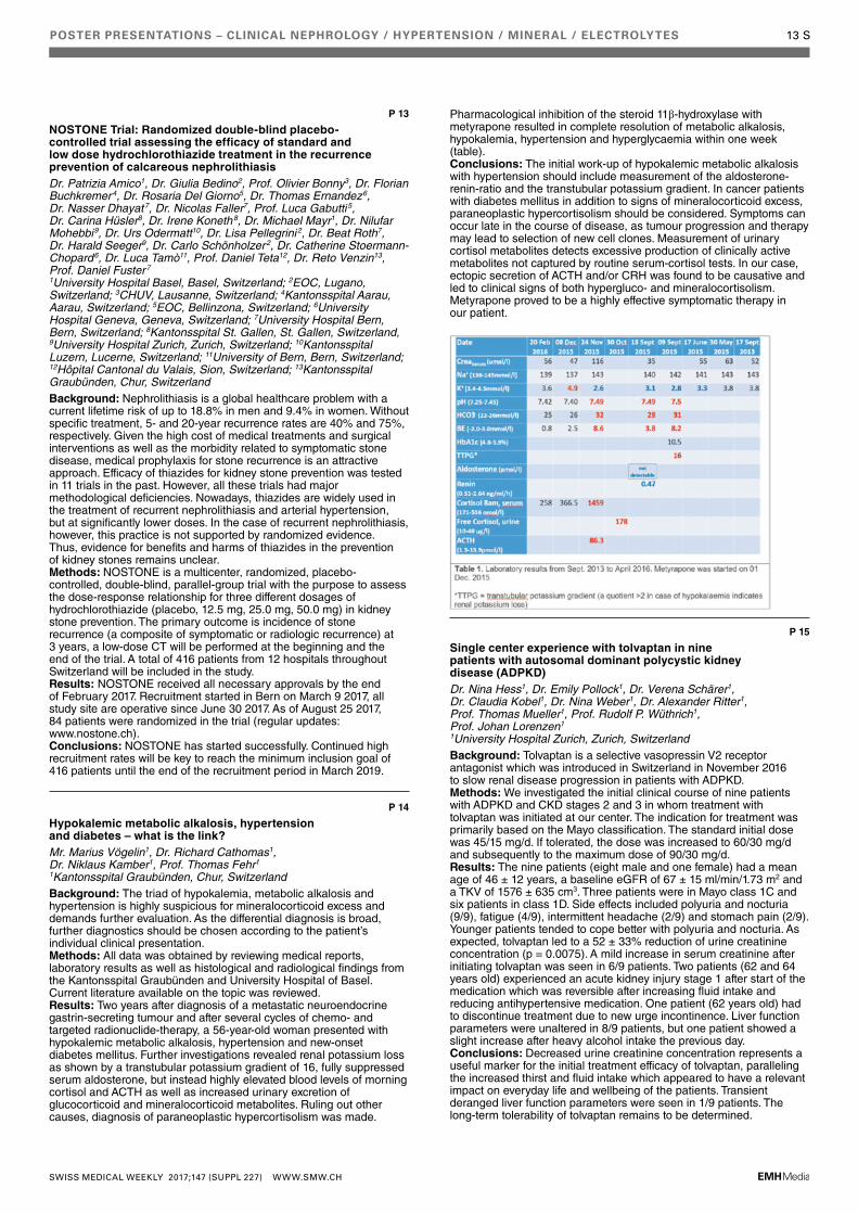
P 13
NOSTONE Trial: Randomized double-blind placebo- controlled trial assessing the efficacy of standard and low dose hydrochlorothiazide treatment in the recurrence prevention of calcareous nephrolithiasisDr. Patrizia Amico1, Dr. Giulia Bedino2, Prof. Olivier Bonny3, Dr. Florian Buchkremer4, Dr. Rosaria Del Giorno5, Dr. Thomas Ernandez6, Dr. Nasser Dhayat7, Dr. Nicolas Faller7, Prof. Luca Gabutti 5, Dr. Carina Hüsler8, Dr. Irene Koneth8, Dr. Michael Mayr1, Dr. Nilufar Mohebbi9, Dr. Urs Odermatt10, Dr. Lisa Pellegrini2, Dr. Beat Roth7, Dr. Harald Seeger9, Dr. Carlo Schönholzer2, Dr. Catherine Stoermann-Chopard6, Dr. Luca Tamò11, Prof. Daniel Teta12, Dr. Reto Venzin13, Prof. Daniel Fuster7 1University Hospital Basel, Basel, Switzerland; 2EOC, Lugano, Switzerland; 3CHUV, Lausanne, Switzerland; 4Kantonsspital Aarau, Aarau, Switzerland; 5EOC, Bellinzona, Switzerland; 6University Hospital Geneva, Geneva, Switzerland; 7University Hospital Bern, Bern, Switzerland; 8Kantonsspital St. Gallen, St. Gallen, Switzerland, 9University Hospital Zurich, Zurich, Switzerland; 10Kantonsspital Luzern, Lucerne, Switzerland; 11University of Bern, Bern, Switzerland; 12Hôpital Cantonal du Valais, Sion, Switzerland; 13Kantonsspital Graubünden, Chur, SwitzerlandBackground: Nephrolithiasis is a global healthcare problem with a current lifetime risk of up to 18.8% in men and 9.4% in women. Without specific treatment, 5- and 20-year recurrence rates are 40% and 75%, respectively. Given the high cost of medical treatments and surgical interventions as well as the morbidity related to symptomatic stone disease, medical prophylaxis for stone recurrence is an attractive approach. Efficacy of thiazides for kidney stone prevention was tested in 11 trials in the past. However, all these trials had major methodological deficiencies. Nowadays, thiazides are widely used in the treatment of recurrent nephrolithiasis and arterial hypertension, but at significantly lower doses. In the case of recurrent nephrolithiasis, however, this practice is not supported by randomized evidence. Thus, evidence for benefits and harms of thiazides in the prevention of kidney stones remains unclear.Methods: NOSTONE is a multicenter, randomized, placebo-controlled, double-blind, parallel-group trial with the purpose to assess the dose-response relationship for three different dosages of hydrochlorothiazide (placebo, 12.5 mg, 25.0 mg, 50.0 mg) in kidney stone prevention. The primary outcome is incidence of stone recurrence (a composite of symptomatic or radiologic recurrence) at 3 years, a low-dose CT will be performed at the beginning and the end of the trial. A total of 416 patients from 12 hospitals throughout Switzerland will be included in the study.Results: NOSTONE received all necessary approvals by the end of February 2017. Recruitment started in Bern on March 9 2017, all study site are operative since June 30 2017. As of August 25 2017, 84 patients were randomized in the trial (regular updates: www.nostone.ch).Conclusions: NOSTONE has started successfully. Continued high recruitment rates will be key to reach the minimum inclusion goal of 416 patients until the end of the recruitment period in March 2019.
P 14
Hypokalemic metabolic alkalosis, hypertension and diabetes – what is the link?Mr. Marius Vögelin1, Dr. Richard Cathomas1, Dr. Niklaus Kamber1, Prof. Thomas Fehr1 1Kantonsspital Graubünden, Chur, SwitzerlandBackground: The triad of hypokalemia, metabolic alkalosis and hypertension is highly suspicious for mineralocorticoid excess and demands further evaluation. As the differential diagnosis is broad, further diagnostics should be chosen according to the patient’s individual clinical presentation.Methods: All data was obtained by reviewing medical reports, laboratory results as well as histological and radiological findings from the Kantonsspital Graubünden and University Hospital of Basel. Current literature available on the topic was reviewed.Results: Two years after diagnosis of a metastatic neuroendocrine gastrin-secreting tumour and after several cycles of chemo- and targeted radionuclide-therapy, a 56-year-old woman presented with hypokalemic metabolic alkalosis, hypertension and new-onset diabetes mellitus. Further investigations revealed renal potassium loss as shown by a transtubular potassium gradient of 16, fully suppressed serum aldosterone, but instead highly elevated blood levels of morning cortisol and ACTH as well as increased urinary excretion of glucocorticoid and mineralocorticoid metabolites. Ruling out other causes, diagnosis of paraneoplastic hypercortisolism was made.
POSTER PRESENTATIONS – CLINICAL NEPHROLOGY / HYPERTENSION / MINERAL / ELECTROLYTES 13 S
Pharmacological inhibition of the steroid 11β-hydroxylase with metyrapone resulted in complete resolution of metabolic alkalosis, hypokalemia, hypertension and hyperglycaemia within one week (table).Conclusions: The initial work-up of hypokalemic metabolic alkalosis with hypertension should include measurement of the aldosterone-renin-ratio and the transtubular potassium gradient. In cancer patients with diabetes mellitus in addition to signs of mineralocorticoid excess, paraneoplastic hypercortisolism should be considered. Symptoms can occur late in the course of disease, as tumour progression and therapy may lead to selection of new cell clones. Measurement of urinary cortisol metabolites detects excessive production of clinically active metabolites not captured by routine serum-cortisol tests. In our case, ectopic secretion of ACTH and/or CRH was found to be causative and led to clinical signs of both hypergluco- and mineralocortisolism. Metyrapone proved to be a highly effective symptomatic therapy in our patient.
P 15
Single center experience with tolvaptan in nine patients with autosomal dominant polycystic kidney disease (ADPKD)Dr. Nina Hess1, Dr. Emily Pollock1, Dr. Verena Schärer1, Dr. Claudia Kobel1, Dr. Nina Weber1, Dr. Alexander Ritter1, Prof. Thomas Mueller1, Prof. Rudolf P. Wüthrich1, Prof. Johan Lorenzen1 1University Hospital Zurich, Zurich, SwitzerlandBackground: Tolvaptan is a selective vasopressin V2 receptor antagonist which was introduced in Switzerland in November 2016 to slow renal disease progression in patients with ADPKD.Methods: We investigated the initial clinical course of nine patients with ADPKD and CKD stages 2 and 3 in whom treatment with tolvaptan was initiated at our center. The indication for treatment was primarily based on the Mayo classification. The standard initial dose was 45/15 mg/d. If tolerated, the dose was increased to 60/30 mg/d and subsequently to the maximum dose of 90/30 mg/d.Results: The nine patients (eight male and one female) had a mean age of 46 ± 12 years, a baseline eGFR of 67 ± 15 ml/min/1.73 m2 and a TKV of 1576 ± 635 cm3. Three patients were in Mayo class 1C and six patients in class 1D. Side effects included polyuria and nocturia (9/9), fatigue (4/9), intermittent headache (2/9) and stomach pain (2/9). Younger patients tended to cope better with polyuria and nocturia. As expected, tolvaptan led to a 52 ± 33% reduction of urine creatinine concentration (p = 0.0075). A mild increase in serum creatinine after initiating tolvaptan was seen in 6/9 patients. Two patients (62 and 64 years old) experienced an acute kidney injury stage 1 after start of the medication which was reversible after increasing fluid intake and reducing antihypertensive medication. One patient (62 years old) had to discontinue treatment due to new urge incontinence. Liver function parameters were unaltered in 8/9 patients, but one patient showed a slight increase after heavy alcohol intake the previous day.Conclusions: Decreased urine creatinine concentration represents a useful marker for the initial treatment efficacy of tolvaptan, paralleling the increased thirst and fluid intake which appeared to have a relevant impact on everyday life and wellbeing of the patients. Transient deranged liver function parameters were seen in 1/9 patients. The long-term tolerability of tolvaptan remains to be determined.
SWISS MEDICAL WEEKLY 2017;147 (SUPPL 227) WWW.SMW.CH

P 16
Could polymerase chain reaction (pcr) avert worsening acute kidney injury? A case of leptospirosis in a regional hospitalDr. James Tataw1, Dr. Terezija Gracin1, Dr. Cecile Bassi1 1Hôpital du Jura bernois SA, Moutier, SwitzerlandBackground: Leptospirosis is a frequent zoonotic disease with high mortality from sepsis and multiorgan dysfunction including renal failure. Renal manifestation varies from subclinical disease with mild urine abnormalities to severe acute kidney injury sometimes requiring hemodialysis. Due to high case fatality rate, early diagnosis during the initial phase of the disease (first 3 to 7 days) may guide early treatment with an influence on patient outcome. We report the case of severe non-oliguric acute kidney injury in a patient with fever myalgia and severe thrombocytopenia. Despite a negative ELISA test, PCR was positive for leptospira.Methods: A 67-year-old male presented with fever and myalgia. He had hypertension managed with telmisartan. He denied swimming or water related activities. Physical examination was unremarkable except for dry oral mucosa. Laboratory findings included severe renal failure, thrombocytopenia, raised CRP and altered liver enzymes. Urinalysis showed only mild hematuria. Fractional excretion of sodium was low. With a suspicion of sepsis and pre renal AKI, intravenous fluids were commenced alongside broad spectrum antibiotics. Clinical course was marked by worsening thrombocytopenia and renal failure resulting in further imunologic work up which returned unremarkable. Further history revealed patient kept rats and mice.Results: ELISA (IgM) for Leptospira sp was negative but further serum PCR returned positive for leptopsira. Ceftriaxone was commenced followed by out patient amoxicillin with improved clinical course alongside renal function. A couple of weeks following discharge, repeat ELISA was positive suggesting seroconversion.Conclusions: The incidence of leptospirosis associated-AKI varies with disease severity. Several mechanisms of renal damage exists. However, the diagnosis of leptopsirosis can be difficult during the initial phase. Although ELISA antibody testing is highly recommended with its high sensitivity and specificity, when negative, this can present a diagnostic dilema. Our Case highlights this and underpins the role of PCR for early diagnosis to avert worsening AKI.
P 17
Vascular aging processes accelerate during the whole life following an alarming kinetic; Pulse Wave Velocity as an objective counterpart that time fliesProf. Luca Gabutti1, Dr. Rosaria Del Giorno1 1EOC, Bellinzona, SwitzerlandBackground: Arterial stiffness mainly measured by Pulse Wave Velocity (PWV) is a reliable marker of vascular aging and is considered to be the only parameter quantifying the consequences, on the vascular wall, of whole life cumulated cardiovascular risk factors. The acceleration in arterial stiffness progression related to age can be worsened by different acquired factors that translate into the concept of early vascular aging.Methods: Pulling together the data of three epidemiological studies concerning the normal population, we aimed to calculate the age related increase in acceleration of PWV and estimate the age specific relative amount of time (in days or fractions of year) equivalent to that necessary to progress one year in vascular age at 20. A population of 3724 subjects classified in 5 age groups was analysed. A polynomial function and its derivative, expressing the instantaneous PWV acceleration, were calculated.Results: The number of days (if 365 at 20 years) necessary at 30, 50 and 70 years to produce the same amount of PWV progression obtained in one year at 20 is respectively 318,170 and 69. The age related increase in PWV does not follow a kinetic of constant acceleration; this means that vascular aging processes accelerate during the whole life. Our data confirmed that acceleration in PWV has dramatic consequences producing, comparing to the age of 20, the same amount of structural changes at 50 in half of the time and at 70 in a fifth.Conclusions: The results confirm the subjective perception, that time is running faster and faster; perception that can be translated into two specular concepts: shorter times later in life to reach the same amount of vascular structural changes or more years of life if the age related changes at 20 are used as the reference to calculate the progression. Once again we can choose: half empty or half full?
POSTER PRESENTATIONS – CLINICAL NEPHROLOGY / HYPERTENSION / MINERAL / ELECTROLYTES 14 S
P 18
A rare renal complication leading to the first diagnosis of chronic pancreatitisDr. Nikolaos Drivakos1, Dr. Cédric Pulver1, Dr. Annette Marie Wons1, Dr. Jörg Neuweiler1, Dr. Françoise-Isabelle Binet1, Dr. Dimitrios Tsinalis1 1Kantonsspital St. Gallen. St. Gallen, SwitzerlandBackground: Although chronic oxalosis is a well-known complication of chronic pancreatitis there are only rare cases of acute oxalate nephropathy reported with chronic pancreatitis.Methods: An 85-year-old man was admitted for deteriorating general health and diarrhea since 4 weeks exacerbated during a vacation abroad. The patient was known for hypertension, diabetes and chronic kidney disease G3a. On admission he was hypovolemic and oliguric with BP 100/47 mm Hg. Blood tests showed creatinine 1253 μmol/l, urea 48 mmol/l, bicarbonate 4 mmol/l, CRP 9 mg/l, no signs of hemolysis, negative auto-antibodies. Urinanalysis revealed granular casts and tubular epithelial cells but no erythrocytes, FeUrea was 30%. An US showed 9 cm kidneys with normal cortico-medullar differentiation and no obstruction. Stool analysis remained negative for pathogens.Results: A pre-renal acute kidney injury was assumed and treated by intravenous hydration and bicarbonate substitution. Diuresis was restored but creatinine and urea did not improve and renal replacement therapy was initiated. The diagnostic procedure was a kidney biopsy which showed numerous intra-tubular crystals suggestive of oxalate (double refraction, Kossa-negative), tubular damage and granulomatous reaction around crystal extravasation. Endoscopic ultrasound and MRCP with secretin stimulation lead to the diagnosis of chronic pancreatitis with exocrine pancreatic insufficiency. This was considered as the underlying cause for the oxalate nephropathy acutely precipitated by the diarrhea. Treatment with pancreatic enzymes and CaCO3 for oxalate binding lead to an improvement of diarrhea but 3 months later the patient remains on dialysis.Conclusions: In chronic pancreatitis calcium binds non digested fatty acids leading to an increase of unbound oxalate in the gut lumen. Additionally free fatty acids induce a hyperpermeability of the colonic mucosa for oxalate. This results in hyperoxalemia and consecutive hyperoxaluria. The acute setting of diarrhea led to volume depletion and increased urine concentration further promoting oxalate precipitation and explaining the acute oxalate nephropathy in this Patient.
P 19
Excessive consumption of “over-the-counter” vitamin D supplements leading to severe hypercalcemia and acute kidney injury: case reportDr. Catherine Beglinger1, Dr. Dimitrios Tsinalis2, Dr. Min Jeong KIM1, Mrs. Karin Jung1, Prof. Michael Brändle1, Dr. Françoise-Isabelle Binet2 1Cantonal Hospital St. Gallen, St. Gallen, Switzerland; 2Kantonsspital St. Gallen, St. Gallen, SwitzerlandBackground: Widely available information on the potential health benefit of vitamin D supplementation and the easy accessibility to high-dose vitamin D supplements over-the-counter or via the internet increases the risk of vitamin D intoxication resulting in severe adverse effects.Methods: We report a case of a 76 year-old female suffering from rheumatoid arthritis for 50 years. Results: Inspired from a YouTube video promising beneficial effects of vitamin D on her disease, she stopped all her regular medicines and started taking daily an excessive amount of over-the-internet purchased calcium and vitamin D supplements 4 months before the admission. Two days before admission, she consulted her family doctor due to slowly progressive fatigue, lethargy, gate disturbance and cognitive dysfunction. The blood chemistry revealed elevated calcium level of 4.2 mmol/l and she was therefore referred to our hospital. On admission, the calcium level was 3.5 and the ionized calcium 1.77 mmol/l. In addition, increased creatinine of 234 μmol/l was observed. While the levels of both 25(OH)D & 1,25(OH)2D were markedly elevated (309 nmol/l [75–250], and 242 pmol/l [36.5–216.2]), we were able to rule out other aetiologies of hypercalcemia, such as primary hyperparathyroidism, granulomatous diseases, or malignancy. In addition to intensive hydration, administration of denosumab in total 240 mg s.c., pamidronate 60 mg i.v. as well as prednisone were required to lower the calcium levels to the normal range during the hospitalisation for 32 days. An heterozygous mutation of
SWISS MEDICAL WEEKLY 2017;147 (SUPPL 227) WWW.SMW.CH
POSTER PRESENTATIONS – CLINICAL NEPHROLOGY / HYPERTENSION / MINERAL / ELECTROLYTES 15 S
24-Hydroxylase, a potential cause of an impaired degradation of 25(OH)D and 1,25(OH)2D, should be suspected due to the therapy-resistant hypercalcemia with markedly elevated concentrations of 1,25(OH)2D. The kidney biopsy revealed mild nephrocalcinosis, explaining the lack of renal function improvement.Conclusions: Our case underscores the importance of information on potential hazardous effects of vitamin D overconsumption. Despite the easy accessibility, seeking medical advice before starting a supplementation is highly recommended.
P 20
mTOR Inhibitor: a new indication for a rare disease?Dr. Ann-Kathrin Schwarzkopf1 1Hirslanden Bern, Salem-Spital, Bern, SwitzerlandBackground: Morbus Ormond is characterized by inflammatory fibrotic mass in the retroperitoneal space often involving vessels and ureters causing obstructive nephropathy with or without aneurysm of the abdominal aorta. Systemic steroids, azathioprine or cyclophosphamide are used as immunosuppressive therapy. In order to have a more specific and antiproliferative treatment mTOR-inihibitor everolimus was used in a patient with this fibrotic disorder.Methods: case reportResults: A 71 yr old black african presented with postrenal kidney failure. Abdominal computertomography showed an aneurysm of the abdominal aorta (3.4 × 3.4 cm) including iliac arteries and fibrotic mass in the retroperitoneal space with consecutive obstructive nephropathy. Diagnosis of chronic aortitis, retroperitoneal fibrosis (M. Ormond) was made. The patient underwent urologic procedure with nephrostomy and ureteral catheterism (double j guide), respectively. An immunosuppressive therapy with systemic steroids was installed and ureteral obstruction resolved. Under steroid therapy the patient developed a severe insulin-dependent diabetes mellitus and was therefore switched to azathioprine. Due to cholestasis azathioprine had to be stopped and steroids were continued. 2015 mTOR inhibitor everolimus was started as an off label use after written informed consent. Steroids could be tapered successfully without any need for further antidiabetic drug. Trough levels were aimed 2–3 ng/ml. Kidney function in terms of eGFR by CKD-EPI formula remained stable between 55 and 65 ml/min (Serum creatinine 110 to 120 umol/l). Fibrosis is well controlled. After interruption due to vacation in Nigeria disease relapsed but mTOR inbitor was successfully reinstalled.Conclusions: We have therefore a proofe of principle for mTOR inhibition in this individual case of fibrotic disorder.
P 21
Diabetic muscle infarction: a diagnostic challenge for clinicians and radiologistsMrs. Jennifer Scotti Gerber, Dr. Davide Giunzioni, Dr. Silvio Pianca, Prof. Jos C. Van Den Berg, Dr. Carlo Schönholzer 1Regional Hospital Lugano, Lugano, SwitzerlandBackground: Diabetic muscle infarction (DMI) is a rare and probably underdiagnosed complication of diabetes mellitus. Risk factors are poor glycemic control and the presence of diabetic complications, especially diabetic nephropathy.Methods: Case report.Results: Case 1: A 44-year old female patient with insulin dependent diabetes mellitus (IDDM) type 2 for 11 years and diabetic nephropathy with chronic kidney disease (CKD) stage 5 presented with acute, excruciating pain localized at the left hip. Laboratory findings showed a slightly elevated C-reactive Protein (CRP) with no elevation of leukocytes, Creatinin-Kinase (CK) or Lactat-Dehydrogenase (LDH). MRI of the hip, however, demonstrated hyperintensity of the T-2and STIR (short tau inversion recovery) sequences with enhancement after gadolinium administration, compatible with necrosis of the adductor muscles due to DMI. The condition was managed conservatively with rest and analgesics. Case 2: A 59-years old female with a 15-year history of IDDM type 2 and diabetic nephropathy with CKD stage 5 presented with severe and disabling pain in the left thigh. Laboratory findings showed an elevation of CRP and LDH with no leukocytosis or elevation in CK. CT and MRI showed edema of the quadriceps muscle as well as fluid over the fascia lata with features suggesting necrotizing fasciitis. Intravenous antibiotics were initiated immediately after diagnosis and a fasciotomy was performed. Histologic evaluation revealed non-specific muscular atrophy. The postoperative course was complicated by a hematoma at the surgical site necessitating an evacuation. A muscular biopsy was repeated and histologic examination revealed necrosis of muscle and fascia due to DMI. Pain improved with analgesics and rest.
Conclusions: DMI is an important differential diagnosis in patients with longstanding diabetes who present with unexplained pain at the lower limbs. MRI is the imaging study of choice.
P 22
Combined primary and secondary hyperparathyroidism in a liver transplant patient: a case reportDr. Curzio Solca1, Dr. Benedetta Terziroli 2, Dr. Fabio Cattaneo3, Dr. Claudia Ferrier1 1Nefrocentro Clinica Luganese Moncucco, Lugano, Switzerland; 2Epatocentro, Lugano, Switzerland; 3Clinica Luganese Moncucco, Lugano, SwitzerlandBackground: Primary hyperparathyroidism (PHPT) is characterized by hypercalcemia, hypophosphatemia and elevated serum PTH levels. Common clinical manifestations of PHPT include fatigue, polyuria, hypertension and kidney stones1. By contrast secondary hyperparathyroidism (SHPT), induced by chronic renal failure (CRF), results in high iPTH, but decreased calcium and elevated serum phosphate levels2. Therefore, superimposed CRF with SHPT may mask and delay the diagnosis of an underlying PHPT.Methods: –Results: We report a case of a 70 yrs old women, with H/O liver transplant (1990), surgical treatment for breast cancer (2012) and moderate hypercalcemia from January 2013 (range 2.65–2.95 mmol/l) with an increased PTH level (89 ng/l) and a declining renal function (eGFR-EPI 40 ml/min/1.73 m2). Vitamin D3 level was low (20 nmol/l) and PTHrp was not detectable. In March 2017, during a treatment with ribavirin for acute hepatitis E, she suffered from polyuria with a weight loss of 4 kg, worsening of the hypercalcemia (3.10 mmol/l) and consequent acute decline of kidney function (eGFR-EPI 20 ml/min/1.73 m2). With intravenous hydration kidney function improved, followed by an increase of the eGFR-EPI to 30 ml/min/1.73 m2 and a decrease of total serum calcium level to 2.73 mmol/l. Further evaluation for suspected PHPT showed a single parathyroid adenoma on the lower right lap on ultrasound. After surgical removal of the adenoma, iPTH dropped and serum calcium levels remained within normal range.Conclusions: In CRF the presence of SHPT, may mask and delay the diagnosis of primary hyperparathyroidism, thus increasing the risk clinical complications.
P 23
Unexplained severe lactic acidosis in a non-diabetic chronic hemodialysis patient: do you consider to make a phone call to the pharmacy?Ms. Céline Aeberhard1, Dr. Ould Maouloud Hemett1, Dr. Yvan Fleury1, Dr. Benoit Fellay1, Dr. Jean Luc Magnin1, Dr. Pierre Lescuyer2, Prof. Daniel Hayoz1, Dr. Eric Descombes1 1HFR, Hôpital Cantonal Fribourg, Fribourg, Switzerland; 2Geneva University Hospitals, Geneva, SwitzerlandBackground: Metformin is acknowledged worldwide as having a central role in the primary treatment of type 2 diabetes mellitus. The drug has been used since 1957. The mean plasma elimination half-life of metformin after oral administration is 4.0–8.7 h, and since it is cleared almost exclusively by the kidneys, its elimination is prolonged in persons with CKD. Among these adverse effects, the lactate acidosis defined as an elevated lactatemia (>5 mmol/L) along with a decreased blood pH (<7.35) and an increase anion gap, is most dangerous. This condition has a mortality of up to 50% per episode. The incidence of lactate acidosis varied between 2,5 to 10 cases/100 000 patient-years1. In Switzerland Bailey and Nattrass have reported 2cases until 1972 to 1977 means an incidence of 6.7/100 000 patient-years1. The majority of nephrologysocieties2 proposed that metformin use be reevaluated when GFR is <45 mL/min/1.73 m2 and stopped when <30 mL/min/1.73 m2.Methods: Hemodialysis is an efficient method to treat metformin intoxication and correct the metabolic abnormalities. Lalau and al.1 have reported two unusual cases: metformin accumulation in the absence of hyperlactatemia, and metformin- induced hyperlactatemia in the absence of metformin accumulation. Results: We report a case of metformin-associated severe lactic acidosis (pH = 7.20, lactate 11.4 mmol/l) in a 33-years old non-diabetic HD patient that remained initially unexplained. In this patient aluminium hydroxide (Phosphonorm 3 × 300 mg/day) had been prescribed but the pharmacy, by mistake, delivered instead metformin that the patient took at a dose of 3 × 500 mg/day for 10 days before hospitalisation.Conclusions: Toxicology blood analysis was negative in our patient, but it is important to know that depending on the analytical technique used metformin is not always detectable in blood.
SWISS MEDICAL WEEKLY 2017;147 (SUPPL 227) WWW.SMW.CH
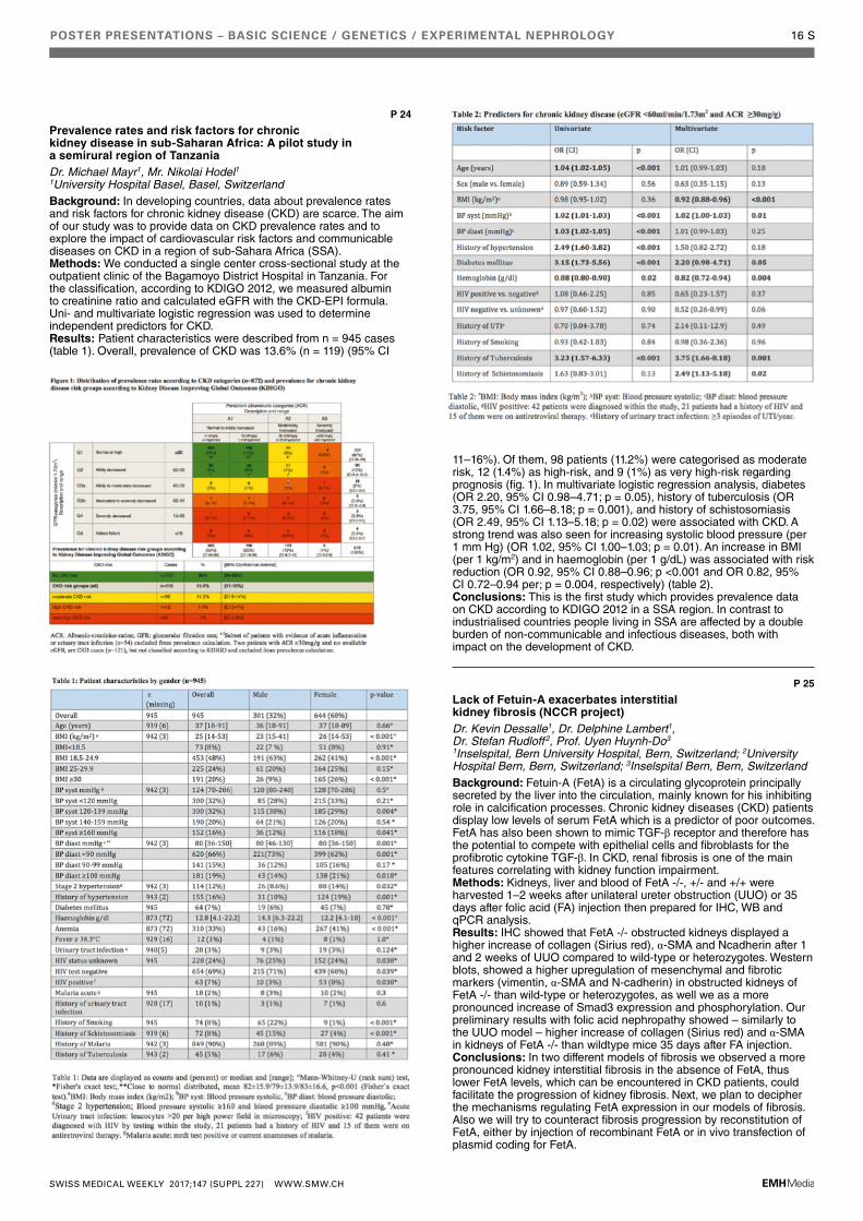
POSTER PRESENTATIONS – BASIC SCIENCE / GENETICS / EXPERIMENTAL NEPHROLOGY 16 S
P 24
Prevalence rates and risk factors for chronic kidney disease in sub-Saharan Africa: A pilot study in a semirural region of TanzaniaDr. Michael Mayr1, Mr. Nikolai Hodel1 1University Hospital Basel, Basel, SwitzerlandBackground: In developing countries, data about prevalence rates and risk factors for chronic kidney disease (CKD) are scarce. The aim of our study was to provide data on CKD prevalence rates and to explore the impact of cardiovascular risk factors and communicable diseases on CKD in a region of sub-Sahara Africa (SSA).Methods: We conducted a single center cross-sectional study at the outpatient clinic of the Bagamoyo District Hospital in Tanzania. For the classification, according to KDIGO 2012, we measured albumin to creatinine ratio and calculated eGFR with the CKD-EPI formula. Uni- and multivariate logistic regression was used to determine independent predictors for CKD.Results: Patient characteristics were described from n = 945 cases (table 1). Overall, prevalence of CKD was 13.6% (n = 119) (95% CI
11–16%). Of them, 98 patients (11.2%) were categorised as moderate risk, 12 (1.4%) as high-risk, and 9 (1%) as very high-risk regarding prognosis (fig. 1). In multivariate logistic regression analysis, diabetes (OR 2.20, 95% CI 0.98–4.71; p = 0.05), history of tuberculosis (OR 3.75, 95% CI 1.66–8.18; p = 0.001), and history of schistosomiasis (OR 2.49, 95% CI 1.13–5.18; p = 0.02) were associated with CKD. A strong trend was also seen for increasing systolic blood pressure (per 1 mm Hg) (OR 1.02, 95% CI 1.00–1.03; p = 0.01). An increase in BMI (per 1 kg/m2) and in haemoglobin (per 1 g/dL) was associated with risk reduction (OR 0.92, 95% CI 0.88–0.96; p <0.001 and OR 0.82, 95% CI 0.72–0.94 per; p = 0.004, respectively) (table 2).Conclusions: This is the first study which provides prevalence data on CKD according to KDIGO 2012 in a SSA region. In contrast to industrialised countries people living in SSA are affected by a double burden of non-communicable and infectious diseases, both with impact on the development of CKD.
P 25
Lack of Fetuin-A exacerbates interstitial kidney fibrosis (NCCR project)Dr. Kevin Dessalle1, Dr. Delphine Lambert1, Dr. Stefan Rudloff2, Prof. Uyen Huynh-Do3 1Inselspital, Bern University Hospital, Bern, Switzerland; 2University Hospital Bern, Bern, Switzerland; 3Inselspital Bern, Bern, SwitzerlandBackground: Fetuin-A (FetA) is a circulating glycoprotein principally secreted by the liver into the circulation, mainly known for his inhibiting role in calcification processes. Chronic kidney diseases (CKD) patients display low levels of serum FetA which is a predictor of poor outcomes. FetA has also been shown to mimic TGF-β receptor and therefore has the potential to compete with epithelial cells and fibroblasts for the profibrotic cytokine TGF-β. In CKD, renal fibrosis is one of the main features correlating with kidney function impairment.Methods: Kidneys, liver and blood of FetA -/-, +/- and +/+ were harvested 1–2 weeks after unilateral ureter obstruction (UUO) or 35 days after folic acid (FA) injection then prepared for IHC, WB and qPCR analysis.Results: IHC showed that FetA -/- obstructed kidneys displayed a higher increase of collagen (Sirius red), α-SMA and Ncadherin after 1 and 2 weeks of UUO compared to wild-type or heterozygotes. Western blots, showed a higher upregulation of mesenchymal and fibrotic markers (vimentin, α-SMA and N-cadherin) in obstructed kidneys of FetA -/- than wild-type or heterozygotes, as well we as a more pronounced increase of Smad3 expression and phosphorylation. Our preliminary results with folic acid nephropathy showed – similarly to the UUO model – higher increase of collagen (Sirius red) and α-SMA in kidneys of FetA -/- than wildtype mice 35 days after FA injection.Conclusions: In two different models of fibrosis we observed a more pronounced kidney interstitial fibrosis in the absence of FetA, thus lower FetA levels, which can be encountered in CKD patients, could facilitate the progression of kidney fibrosis. Next, we plan to decipher the mechanisms regulating FetA expression in our models of fibrosis. Also we will try to counteract fibrosis progression by reconstitution of FetA, either by injection of recombinant FetA or in vivo transfection of plasmid coding for FetA.
SWISS MEDICAL WEEKLY 2017;147 (SUPPL 227) WWW.SMW.CH
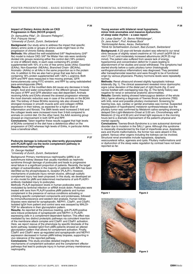
POSTER PRESENTATIONS – BASIC SCIENCE / GENETICS / EXPERIMENTAL NEPHROLOGY 17 S
P 26
Impact of Dietary Amino Acids on CKD Progression in Rats (NCCR project)Dr. Samyuktha Pillai1, Dr. Giovanni Pellegrini1, Prof. François Verrey1 1University of Zurich, Zurich, SwitzerlandBackground: Our study aims to address the impact that specific dietary amino acids or groups of amino acids might have on the progression of chronic kidney disease.Methods: We utilised the well-established 5/6th Nephrectomy (5/6th Nx) model to induce CKD. 5/6th Nx Wistar Han rats were randomly divided into groups receiving either the control diet (18% protein) or one of different diets, in each case containing 8% protein supplemented with 10% of a mix of free amino acids (AAs): Essential- (EAAs), Non-Essential- (NEAAs), Branched Chain- (BCAAs), Aromatic- (AAAs) or all AAs in the same proportion as in the protein mix. In addition to this we also had a group that was fed a diet containing 18% protein supplemented with 1.82% L-arginine. Both GFR and RPF were measured in free moving animals, GFR transcutaneously using FITC-sinistrin, and RPF by using radiolabelled para-aminohippurate (PAH).Results: None of the modified diets did cause any decrease in body weight, food and water consumption in the different groups. However the pace of RPF and GFR alteration was diet-dependent. Animals receiving AAAs and EAAs showed the slowest progression whereas the most dramatic reduction was in case of the animals on the BCAA diet. The kidney of these BCAAs receiving rats also showed the strongest increase in smooth muscle actin and collagen mRNA expression in their kidney, as expected for a higher level of inflammation and fibrosis. The SMA levels were increased 2-fold and the collagen levels were increased more than 3-fold compared to animals on control diet. On the other hand, the AAA receiving group showed an improvement in both GFR and RPF. Conclusions: Taken together these results suggest that high levels of BCAAs contained in the diet have a deleterious effect on the progression of CKD whereas high levels of EAAs, in particular AAAs have a beneficial effect.
P 27
Podocyte damage is induced by aberrantly glycosylated anti-PLA2R-IgG4 via the lectin complement pathway in membranous nephropathyDr. George Haddad1 1University of Zurich, Zurich, SwitzerlandBackground: Primary membranous nephropathy (pMN) is an autoimmune kidney disease that usually manifests as nephrotic syndrome through damage of podocytes and leads to progressive renal failure in a significant proportion of patients. Recently, the target antigen of autoantibodies in the majority of patients with iMN has been identified as the phospholipase A2 receptor (PLA2R1). However, mechanisms of podocyte injury remain elusive, although sublytic complement injury has been proposed. In this study, we developed an in vitro model for pMN and determined mechanisms of anti-PLA2R1-antibody mediated injury to podocytes.Methods: PLA2R expression levels in human podocytes were modulated by lentiviral infection or siRNA knock down. Podocytes were treated with sera from PLA2R1-positive pMN patients and human complement in the presence of various conditions activating or inhibiting specific complement components and proteinases, followed by immunofluorescence and western blot analysis. Human kidney biopsies were stained for synaptopodin, NEPH1, C3aR1, and C5aR1. Isolated IgG4 from patient and control sera was assayed by MALDI-TOF for alterations in their glycosylation pattern.Results: As reported at the previous meeting, anti-PLA2R-positive sera induce proteolysis of synaptopodin and NEPH1 in PLA2R-expressing cells in a complement-dependent fashion. This effect was mediated by two distinct proteolytic pathways via coincident insertion of the membrane attack complex and C3aR1 and C5aR1 activation. Here, we report rescue of the phenotype by specific blockade of the lectin pathway. Isolated IgG4 from pMN patients showed an altered glycosylation pattern that allows for complement activation. Finally, C3aR1 and C5aR1 were up-regulated and synaptopodin and NEPH1 expression decreased in human pMN kidney biopsies, supporting in vivo relevance of the reported pathway.Conclusions: This study provides detailed insights into the mechanisms of complement activation and the complement effector pathways that lead to podocyte injury in human primary membranous nephropathy.
P 28
Young woman with bilateral renal hypoplasia, minor limb anomalies and massive dysfunction of sleep wake rhythm – a case reportDr. Lukas Gerber1, Dr. Benno Röthlisberger1, Dr. Jens Acker2, Prof. Andreas Bock1 1Kantonsspital Aarau, Aarau, Switzerland; 2Klinik für Schlafmedizin Zurzach, Bad Zurzach, SwitzerlandBackground: A 22-year-old female student was referred to our renal clinic because of slightly raised creatinine at 110 umol/l (eGFR 59 m/min/1.73 m2) and moderately increased albuminuria (ACR 25 mg/mmol). The patient also suffered from severe lack of energy, hypersomnia and concentration deficit for 3 years leading to abandonment of her studies and social withdrawal. The symptoms had started shortly before a cystic pituitary tumor (histologically xanthogranulomatous inflammation) was diagnosed. They persisted after transsphenoidal resection and were thought to be of functional origin by various physicians. Pituitary hormone levels were repeatedly normal.Methods: Renal ultrasound showed slightly hypoplastic kidneys bilaterally (fig. 1). The clinical assessment revealed minor dysmorphic signs (ulnar deviation of the distal part of right thumb (fig. 2) and narrow forefeet with overlapping toes (fig. 3). The family history was negative for renal or extrarenal anatomic abnormalities. Results: Genetic testing showed heterozygous deletion of the whole SALL1gene allowing a diagnosis of Townes Brock syndrome (TBS) with limb, renal and possible pituitary involvement. Screening for hearing loss, eye, cardiac or genital anomalies was normal. Suspected dysregulation of hypothalamic melatonin synthesis/release due the pituitary tumor was confirmed by Melatonin saliva sampling showing a delayed Dim-Light-Melatonin-Onset at 5:00 am. Chronotherapy with Melatonin (2 mg at 8:30 pm) and timed light exposure in the morning hours led to a dramatic improvement of the patient’s physical and mental capacity.Conclusions: Townes-Brock-Syndrome is a rare autosomal dominant disorder due to mutation in the SALL1 gene. Although this syndrome is classically characterized by the triad of imperforate anus, dysplastic ears and thumb malformations, the former two were absent in this patient. Various other organs including the kidneys can be involved. Structural renal anomalies include hypoplasia, dysplasia, vesicoureteral reflux, agenesis and cysts. Pituitary malformations and/or dysfunction of the sleep wake regulation by contrast have not been reported so far.
Figure 1 Figure 2
Figure 3
SWISS MEDICAL WEEKLY 2017;147 (SUPPL 227) WWW.SMW.CH

POSTER PRESENTATIONS – BASIC SCIENCE / GENETICS / EXPERIMENTAL NEPHROLOGY 18 S
P 29
Role of the serine protease CAP2/Tmprss4 in renal adaptation to potassium depletion (NCCR project)Dr. Anna Keppner1, Dr. Darko Maric2, Dr. Denise Kratschmar3, Dr. Petra Klusonova3, Dr. Jérémie Canonica2, Prof. Alex Odermatt3, Dr. Gilles Crambert4, Prof. David Hoogewijs5, Prof. Edith Hummler2 1University of Lausanne and University of Fribourg, Lausanne and Fribourg, Switzerland; 2University of Lausanne, Lausanne, Switzerland; 3University of Basel, Basel, Switzerland; 4French National Institute of Health and Medical Research (Inserm), Paris, France; 5University of Fribourg, Fribourg, SwitzerlandBackground: The membrane-bound serine protease CAP2/Tmprss4 was previously identified as in vitro activator of the epithelial sodium channel (ENaC) (Vuagniaux et al. 2002), but its genetic ablation in mice revealed that CAP2/Tmprss4 is not required for ENaC-mediated sodium homeostasis in vivo (Keppner et al. 2015), leading us to explore its implication in renal potassium handling.Methods: CAP2/Tmprss4 knockout mice were kept in metabolic cages and fed a K+-deficient diet. Urine and plasma parameters were subsequently analysed, and kidneys recovered for protein and mRNA quantifications.Results: In this study, we show that CAP2Tmprss4 is regulated by dietary potassium specifically in the kidney, and that upon K+-deficient diet, CAP2Tmprss4 knockout mice display altered sodium and water handling, along with changes in the urinary osmolality and pH, whereas urinary potassium excretion was preserved. We could link these parameters to differential expression of several transporters and channels, including the renal H+,K+-ATPase (HKA2), NKCC2 and AQP2, as well as abnormal sensitivity to vasopressin (AVP).Conclusions: Taken together, these results suggest a new role for the serine protease CAP2/Tmprss4 in renal potassium handling, by regulating the expression of the HKA2.
P 30
Salt-sensitive hypertension in a new rat model for primary generalized glucocorticoid resistance (NCCR project)Dr. Sofia Verouti1,3, Dr. Qing Wang3, Mr. David Ancín Del Olmo2, Mrs. Anne-marie Merillat4, Dr. Veronica Ponce De Leon4, Dr. Denise Kratschmar5, Prof. Alex Odermatt5, Prof. Edith Hummler4 1Inselspital, Bern, Switzerland; 2University of Lausanne, Lausanne, Switzerland; 3Centre Hospitalier Universitaire Vaudois (CHUV), Lausanne, Switzerland; 4University of Lausanne, Lausanne, Switzerland; 5University of Basel, Basel, SwitzerlandBackground: Glucocorticoids mainly act through the glucocorticoid receptor (GR) that functions as transcription factor. Glucocorticoid resistance is a condition characterized by generalized, partial target tissue resistance to glucocorticoids. Compensatory mechanisms lead to elevation in circulating adrenal steroids with mineralocorticoid and/or androgenic activity, and the clinical spectrum of this condition is broad ranging from asymptomatic to severe cases of hyperandrogenism and/or mineralocorticoid excess. So far, no animal model existed that mimics all clinical symptoms as observed in human generalized glucocorticoid resistance.Methods: We generated the first TALEN-engineered rats carrying knockout and in-frame mutations on the GR gene locus within its dimerization domain.Results: Heterozygous mutant GR+/em2rats reproduce all clinical features of glucocorticoid resistance and show increased size of the adrenal gland (adrenal hyperplasia), increase of mineralocorticoids (hypermineralocorticoidism), corticosterone (hypercorticosteronism) and androgens (hyperandrogenism). Heterozygous mutant GR+/em4 rats, carrying deletion of the exon 3 exhibit hypercorticosteronism, but lack hypermineralocorticoidism and hyperandrogenism. In summary, these rats reproduce most of the clinical signs of primary generalized glucocorticoid resistance syndrome.Moreover, all mutants develop salt-sensitive hypertension.Conclusions: In conclusion, we show that (1) glucocorticoids induce salt-sensitive hypertension and (2) dimerization domain of the GR is likely implicated in this mechanism.
P 31
Rebore the kidney – a novel model of back diffusion for acute shock related damage and acute tubular obstruction in anuric/oliguric kidneys: analysis from mathematical modelsProf. Mark Christopher Arokiaraj1 1Pondicherry Institute of Medical Sciences, Pondicherry, IndiaBackground: It is a very common clinical observation that cardiogenic and septic shocks as well as acute tubular damages result in anuria and acute kidney injury. This condition is associated with high mortality. The model was developed to salvage the kidneys from acute damages and recover to its partial functional potential. Methods: The governing equations by the renal modelers across the world for renal function, which includes glomerular and tubular functions, were studied. A theoretical model for a single kidney was developed based on the governing equations.Results: The SNGFR can be modified by transient occlusion. The backpressure transiently reduces filtration, however, the permeability (k) and surface area (S) of the filtration and nephron recruitment (n) increases. By altering the chloride levels (Σ) in the macula densa the tubuloglomerular feedback (g) and the afferent arteriolar diameter can be modified or dilated. By increasing the back diffusion, the hydrostatic pressure and the diameters of the ascending as well the descending tubular sizes can be increased. By altering the pH of the back diffusion fluid, the obstructed tubules can be opened. By addition of proteolytic enzymes the inflammasomes and inflammatory protiens could be modified or degraded and the tubular recovery may be achieved. By increasing the back diffusion pressure to hydro-nephrotic range the glomerular filtration pores could be cleansed and opened. Hence, the final model is transient pulsatile occlusion of the pelvicalyceal ureter and back diffusion by fluids of low chloride and pH near 5.5, and pressure of about 30 mm Hg with proteolytic enzymes in the fluids. The propsed time for occlusion of the ureter could be about 30 to 45 mins.Conclusions: There is potential for a novel back-diffusion method for recovery of anuric or oliguric kidneys. The model needs to be validated by experimental studies.
P 32
Aldosterone controls primary cilium length and Ift88 abundance via mineralocorticoid receptor in the distal segments of the kidney tubule *NCCR project*Ms. Olga Komarynets1, Dr. Eva Bernabeu-Dizin1, Dr. Jan Czogalla2, Dr. Khalil Udwan1, Ms. Alexandra Chassot1, Mr. Isabelle Roth1, Prof. Johannes Loffing2, Prof. Eric Feraille1 1University of Geneva, Geneva, Switzerland; 2University of Zurich, Zurich, SwitzerlandBackground: The kidneys are major players in the control of body fluid homeostasis. At the cellular level, the apical single non-motile primary cilium is a signalling hub and may function as a urinary chemosensor. We hypothesized that aldosterone, a mineralocorticoid hormone that stimulates sodium reabsorption in the distal nephron, modulates cilium length and thereby its functional properties.Methods: Experiments were performed in cultured mCCDcl1 cells, a model of aldosterone-responsive collecting duct principal cells, transgenic mice with inducible kidney tubule-specific knockout (KO) of the mineralocorticoid receptor (MR) and mice with randomized deletion of the MR in renal tubule cells (MR/X mice). Primary cilia were detected by indirect immunofluorescence using anti-acetylated α-tubulin antibodies.Results: In mCCDcl1 cells, aldosterone-stimulated Na+ transport was correlated with lengthening of primary cilia. In contrast, in MR KO mice displaying decreased sodium reabsorption along the aldosterone-sensitive distal nephron, primary cilia of distal tubules and collecting ducts were shorter compared to WT mice. The primary cilium length in
Figure 1
SWISS MEDICAL WEEKLY 2017;147 (SUPPL 227) WWW.SMW.CH

POSTER PRESENTATIONS – BASIC SCIENCE / GENETICS / EXPERIMENTAL NEPHROLOGY 19 S
MR/X mice was reduced in MR-KO cells in comparison to MR-WT cells, indicating that MR-dependent cilium growth relies on cell autonomous mechanisms. In mCCDcl1 cells, the primary cilium lengthening in response to aldosterone was associated with increased Ift88 abundance, a major component of the intraflagellar transport machinery responsible for cilium building and maintenance. This effect relies on decreased Ift88 degradation. We observed that aldosterone treatment lowered autophagic activity assessed by decreased LC3-I/LC3-II ratio. This effect was associated with activation of the mTORC1 signaling pathway. Rapamycin treatment strongly inhibit mTORC1 as shown by blunted p70-Rsk phosphorylation but did not prevent the inhibition of autophagy by aldosterone, indicating that MR controls autophagy via a mTORC1-independent pathway.Conclusions: Thus, we have shown that aldosterone controls primary cilium length via MR in a cell autonomous manner. This process may rely on increased Ift88 abundance via inhibition of autophagy.
P 33
Identification of a novel hepatitis E virus-genotype 3 strain from a chronic hepatitis E virus infection in a kidney transplant recipient in SwitzerlandDr. Agnes Kneubühl1, Mr. Bo Wang2, Mr. Dominik Harms2, Dr. Diana Ciardo3, Prof. Jörg Hofmann4, Prof. Claus-Thomas Bock2 1Hospital Lachen, Lachen, Switzerland; 2Robert Koch Institute, Berlin, Germany; 3Corelab Immunology Viollier AG, Allschwil, Switzerland; 4Charité, University Hospital, Berlin, GermanyBackground: The causal pathogen of chronic hepatitis E in humans is hepatitis E Virus (HEV)-genotype 3. Transmission occurs with raw meat. Organ recipients are at risk for chronic courses. We report the identification of a HEVgenotype strain from a kidney transplant recipient in Switzerland with viral hepatitis E that showed less than 88% homology compared to known HEV-genotype 3 strains suggesting a new HEV subgenotype 3. The HEV strain is called SW/16-0282.Methods: Viral RNA extraction from patient serum and feces was performed using the High Pure Viral Nucleic Acid Kit (Roche Diagnostics, Germany) according to the manufacturer’s instructions. The complete viral genome was amplified using KAPA HiFi HotStart
ReadyMix PCR (Kapa Biosystems, USA). 5’- and 3’-sequences were determined using 5’-and 3’- rapid amplification of cDNA ends (Roche Diagnostics, Germany). Whole genome sequence and phylogenetic analyses were done using Geneious-10.0.5 and Mega-7 software.Results: The virus was isolated from a male 57 year old kidney transplant recipient from nephrology departement of hospital Lachen, Switzerland in 2016. He presented four months after living kidney donor transplantation due to end stage kidney disease because of autosomal dominant polycystic kidney disease with itching and elevated liver enzymes while treated with immunosuppressive drugs tacrolimus and mycophenolate and newly with a chinolone for urinary tract infection. No recovery was seen of liver enzyme elevation despite of stop of chinolones. Search for viral hepatitis resulted in positiv anti-HEV IgM/IgG and HEV-RNA in serum and feces. Further analyses showed less than 88% homology compared to known HEV-genotype 3 strains. Patient got ribavirin for treatment seeing chronic course.Conclusions: There was poor homology to any HEV-genotype 3 subtype usually isolated. The SW/16-0282 strain represents a new HEV subgenotype 3 by comprehensive genetic analyses of HEV strains from human and animal reservoirs. Ribavirin successfully treated this new subgenotype without reccurence within 1 year.
P 34
Renal erythropoietin producing cells in vivo (NCCR Project)Dr. Karen Nolan1, Mr. Willy Kuo1, Dr. Faik Imeri1, Dr. Svende Pfundstein1, Dr. Irene Abreu-rodríguez1, Mr. Patrick Spielmann1, Prof. Edith Hummler2, Dr. Claus-dieter Schuh1, Prof. Andrew Hall1, Prof. Vartan Kurtcuoglu1, Dr. Carsten Scholz1, Prof. David Hoogewijs3, Prof. Roland Wenger1 1University of Zurich, Zurich, Switzerland; 2University of Lausanne, Lausanne, Switzerland; 3University of Fribourg, Fribourg, SwitzerlandBackground: The adult kidney is an important sensor of molecular oxygen (O2), however, the molecular mechanisms involved in hypoxia-induced erythropoietin (Epo) expression in vivo remain poorly understood.Methods: We developed a novel transgenic mouse model (Epo-CreERT2) with inducible Cre recombinase expression under the control of a 220 kb DNA fragment containing the mouse Epogene locus which, when crossed with the Rosa26-flStopfl-tdTomato reporter, allows us to permanently tag renal erythropoietin producing (REP) cells in response to Epo inducing stimuli.Results: Whole genome sequencing identified the integration loci of the Epo-CreERT2recombined bacterial artificial chromosome in two founder lines. A non-coding integration site was found for founder 1 (chr14:56,337,060-56,337,390) and integration was found within the Pou2f3 gene in founder 241 (chr9:43,159,262-43,211,463). We demonstrate that REP cells are clustered predominantly in the interstitium of the juxtamedullary cortex and that few cells are needed to maintain Epo production under physiological conditions. Under conditions of reduced O2 availability, recruitment of additional REP cells, as well as an increase in the Epo mRNA produced per cell is required to meet the demand for increased Epo, demonstrated using RNAscope in situ hybridisation. Tissue clearing techniques allowed for 3-dimensional visualisation of REP cell clustering and interaction with blood vessels in vitro and 2-photon microscopy using a custom build microscope stage enabled imaging of REP cells in the live mouse.Conclusions: Our data indicate that REP cells are a unique and dynamic population of cells which respond with exquisite sensitivity to changes in O2 availability and proliferation of these cells suggests a physiological adaptation to hypoxia.
P 35
Hypoxia-induced long non-coding RNA Malat1 is dispensible for renal ischemia/reperfusion injuryMr. Malte Kölling1, Dr. Celina Schauerte2, Dr. Tamas Kaucsar3, Ms. Anika Hübner2, Dr. Song Rong2, Dr. Roland Schmitt2, Dr. Inga Sörensen-Zender2, Prof. Hermann Haller2, Prof. Rudolf P. Wüthrich1, Prof. Martin Zörnig4, Prof. Thomas Thum2, Prof. Johan Lorenzen1 1University Hospital Zurich, Zurich, Switzerland; 2Hannover Medical School, Hannover, Germany; 3Semmelweis University, Budapest, Hungary; 4Georg-Speyer-Haus, Frankfurt, GermanyBackground: Renal ischemia/reperfusion (I/R) injury represents a major socioeconomic health problem. Non-coding RNA are crucially involved in pathophysiology. Long non-coding RNA Malat1 (Metastasis Associated Lung Adenocarcinoma Transcript 1) was upregulated in renal I/R injury. We elucidated the functional role of Malat1 in vitro and its potential contribution to kidney injury in vivo.
SWISS MEDICAL WEEKLY 2017;147 (SUPPL 227) WWW.SMW.CH

POSTER PRESENTATIONS – BASIC SCIENCE / GENETICS / EXPERIMENTAL NEPHROLOGY 20 S
Methods: In patients,kidney biopsies and plasma samples were collected. In vivo, Malat1 knockout- (KO) and wild-type (WT) mice were subjected to unilateral and bilateral I/R. Histopathology-, gene expression-, kidney function- and survival studies were performed. In vitro, Malat1 was silenced by antisense oligonucleotides in endothelial cells (EC) and tubular epithelial cells (TEC) subjected to hypoxia/reoxygenation. Transcriptional activation- and functional studies were implemented. Genome-wide RNA analysis was performed.Results: Malat1 was upregulated in human kidney biopsies and plasma and in murine kidney tissue, EC and TEC and mainly nuclear-chromatin associated. In vitro, Malat1 inhibition reduced EC in the S-phase of the cell cycle. Proliferation decreased. Less EC were apoptotic after Malat1 silencing. TEC were not functionally altered. Malat1 was transcriptionally activated in EC and TEC by Hypoxia-inducible factor 1-alpha. In vivo, Malat1 KO- and WT mice showed similar degrees of tubular epithelial injuries and proliferating cells. Capillary rarefaction was not affected. Kidneys of Malat1 KO- and WT mice expressed more pro-inflammatory (IL-1beta, IL-6, MIP2a, MCP-1) and pro-fibrotic (Col1a2, Col III, TGF-beta) genes. Similar amounts of macrophage-, T-cell infiltration and tubulointerstitial collagen were detected. mRNA- and smallRNA expressions showed only minor differences. The reduced kidney function was not altered by Malat1 KO. Malat1 KO mice showed no survival benefit.Conclusions: Malat1 plays a pivotal role in hypoxia/reoxygenation induced endothelial cell pathology. Even though previous studies have suggested a prominent role of Malat1 in the induction of disease, we did not confirm an effect of Malat1 loss on the progression of renal I/R-inury.
P 36
Anti-oxidative role of cytoglobin in podocytes and its association with chronic kidney disease (NCCR project)Dr. Elisa Randi1, Dr. Benjamin Vervaet2, Mr. Stijn Vermeylen2, Dr. Maria Tsachaki3, Ms. Elena Porto4, Dr. Maja T. Lindenmeyer5, Dr. Clemens D. Cohen5, Prof. Thomas Hankeln4, Dr. Andreas Kistler6, Prof. Sylvia Dewilde2, Prof. Olivier Devuyst5, Prof. Alex Odermatt3, Prof. Roland Wenger5, Prof. David Hoogewijs7 1University of Fribourg & University of Zurich, Fribourg and Zurich, Switzerland; 2University of Antwerp, Antwerp, Belgium; 3University of Basel, Basel, Switzerland; 4University of Mainz, Mainz, Germany; 5University of Zurich, Zurich, Switzerland; 6Kantonsspital Frauenfeld, Frauenfeld, Switzerland; 7University of Fribourg, Fribourg, SwitzerlandBackground: Cytoglobin (Cygb) is an anti-oxidative protein that belongs to the mammalian globin family. Despite extensive research efforts, little is known about its physiological role in the kidney. Accumulating evidence suggests that oxidative stress plays a crucial role in podocyte detachment and/or apoptosis during diabetic nephropathy. In the present study we investigated if Cygb has a protective role in the kidney.Methods: We generated stable CYGB knock-down and overexpressing cellular models in 2 independent human podocyte cell lines (AB8/13 and LY), in order to investigate the Cygb-dependent transcriptome, cell viability and oxidative stress response. Additionally, we validated the results in vivo, by comparing renal function, apoptosis and gene expression of Cygb–/– and Cygb+/+ mice.Results: Cygb-deficient podocytes displayed increased cell death and accumulation of ROS as assessed by H2-DCF-DA assays and the redox sensitive probe roGFP2-Orp1. Transcriptome analysis of control and Cygb-depleted cells identified dysregulation of multiple genes involved in apoptosis, oxidative stress and podocyte injury. Gene array data from human patients showed that CYGB is upregulated in diabetic nephropathy and GWAS analysis identified a SNP in the 3’ intergenic region of Cygb that is potentially associated with chronic kidney disease (CKD). Cygb–/– mice displayed impaired renal function and increased apoptosis compared to Cygb+/+ mice under basal conditions. Analysis of Cygb-dependent gene expression in mice is currently ongoing.Conclusions: Data of our study demonstrate that Cygb protects podocytes from oxidative stress and apoptosis in vitro and may be involved in CKD, particularly in diabetic nephropathy. In vivo data show that Cygb deficiency is associated with worse renal clearance and increase in apoptosis, consistent with a protective role of Cygb in the kidney.
P 37
Uromodulin excretion is modulated by the calcium-sensing receptorDr. Natsuko Tokonami1, Dr. Eric Olinger1, Prof. Pascal Houillier2, Prof. Olivier Devuyst1 1University of Zurich, Zurich; 2Paris Descartes University, Paris, FranceBackground: Uromodulin is the most abundant urinary protein and predominantly produced in the thick ascending limb (TAL) of the Henle’s loop, a nephron segment crucial for Ca2+ homeostasis under control of the basolateral calcium-sensing receptor (CaSR). Whether the CaSR is involved in the physiological regulation of uromodulin excretion remains unknown.Methods: We studied two mouse models with inactivating (BCH002) and activating (Nuf) mutations in the Casr gene as models for the chronic modulation of CaSR activation. As an acute model, we used primary mouse TAL cells (mTAL), which endogenously express uromodulin and CaSR, exposed to specific CaSR agonist (Calindol, 100 nM) and antagonist (NPS2143, 1 μM) and to variable Ca levels (1 mM and 3 mM) in the medium.Results: We confirmed the expected alterations in blood calcium and phosphorus levels in BCH002 and Nuf mice. Interestingly, urinary uromodulin excretion was significantly increased in BCH002 mice and decreased in Nuf mice, without changes in kidney expression levels. Immunostaining suggested a shift from cytoplasmic to apical localization of uromodulin pools in the kidneys of BCH002 mice contrasting with a broad cytoplasmic staining in Nuf kidneys. In mTAL cells, uromodulin secretion was abolished upon treatment with Calindol for 4 hours without changes in cellular expression levels. In contrast, 6-hour NPS2143 treatment normalized the reduced uromodulin secretion induced by high Ca (3 mM) medium in mTAL cells.Conclusions: Taken together, these results indicate that activation of CaSR modulates the release of uromodulin in the urine, probably through post-translational control of uromodulin trafficking and processing in TAL cells. Modulators of CaSR, which are used in clinical practise, are thus able to change the levels of uromodulin in urine.
P 38
The TWEAK/Fn14 pathway is required for Calcineurin Inhibitor Toxicity of the KidneysMs. Meike Claus1, Ms. Rana Herro2, Dr. Dennis Wolf2, Dr. Konrad Buscher2, Dr. Stefan Rudloff1, Prof. Uyen Huynh-Do1, Dr. Linda Burkly3, Dr. Mick Croft2, Dr. Daniel Sidler1 1University Hospital Bern, Bern, Switzerland; 2La Jolla Institute for Allergy and Immunology, La Jolla, United States; 3Biogen, Boston, United StatesBackground: Calcineurin inhibitor toxicity (CNT) is a frequent occurrence in transplanted renal grafts and autochthone kidneys from patients undergoing long-term treatment with Calcineurin inhibitors, notably Cyclosporin A (CsA) and Tacrolimus.Methods: We studied the effect of CsA on the TWEAK/Fn14 axis of the kidney in vitro and in vivo utilizing tubular epithelial cell cultures and mice sufficent or deficient for the TWEAK gene.Results: We show an indispensable role of the TNF superfamily molecule TWEAK (TNFSF12) in the pathogenesis of acute CNT lesions in mice. A deficiency in TWEAK resulted in limited tubulotoxicity after CsA exposure, which correlated with diminished expression of inflammatory cytokines and reduced intraparenchymal infiltration with immune cells. We further identified tubular epithelial cells of the kidney as major targets of CsA activity and found that Fn14 (TNFRSF12A), the receptor for TWEAK, is a highly CsA-inducible gene in these cells. Correlating with this, CsA pretreatment sensitized tubular epithelial cells specifically to the pro-inflammatory activities of recombinant TWEAK in vitro. Moreover, injection of rTWEAK alone into mice induced moderate disease similar to CsA, and rTWEAK combined with CsA resulted in synergistic nephrotoxicity.Conclusions: These findings support the importance of tubular epithelial cells as cellular targets of CsA toxicity and introduce TWEAK as a critical contributor to CNT pathogenesis.
SWISS MEDICAL WEEKLY 2017;147 (SUPPL 227) WWW.SMW.CH

P 40
ionized and total serum magnesium in hemodialysis: predictors and variability. A longitudinal cross-sectional studyDr. Rosaria Del Giorno1, Dr. Hilary Riva2, Mr. Gaetano Lucio Donato3, Prof. Luca Gabutti3 1EOC Bellinzona, Bellinzona, Switzerland; 2EOC Mendrisio, Mendrisio, Switzerland; 3EOC Locarno, Locarno, SwitzerlandBackground: Ionized Magnesium(ion-Mg)represents the active biological fraction of the serum magnesium content.The assessment of total serum Mg(tot-Mg)might not accurately identify patients with hypo-orhyper-magnesaemie. In hemodialysis,serum tot-Mg levels in the upper part of the distribution,have been associated with reduced mortality and fewer vascular calcifications;thus,resulting in the tendency to increase the Mg concentration in the dialysate, traditionally set at 0.5 mmol/LMethods: Single-center study in chronic hemodialysis patients, designed in two phases, cross-sectional and longitudinal, aimed to investigate: i) the sensitivity for pathological values of ion-Mg compared to tot-Mg ii) the predictors of ion-Mg developing ad hoc equations; iii) the inter- and intra-individual variability of ion-Mg iv)the risk factors for hypermagnesemia.Tot-Mg,ion-Mg, and covariates of42 hemodialysis sessions, in 42 patients during the cross-sectional phase and of 270 sessions in 27 patients in the longitudinal one were analysed.Results: Ion-Mg significantly correlates with tot-Mg:β = 0.52; r = 0.88, p <0.001. Multiple linear regressions in normo- and hypo-albuminemic patients gave the following results:ion-Mg=tot-Mg/2-K+/50+Ca2+/5-HCO3-/100 and ion-Mg=tot-Mg/2+Albumin/100.Ion-Mg showed a high temporal variability in the longitudinal phase(between months p <0.001;wintervs.summer, p <0.027).A high intra-individual variability was also found:coefficient of variation 0.116.Comparing patients with high and low intra-individual variability we found: age 67vs.77y; p <0.001; urea 26.3 ± 0.5 vs. 21.2 ± 0.4 mmol/L, p <0.001; nPCR0.92 ± 0.1 vs. 0.77 ± 0.1 g/Kg*day, p <0.001; PTH 46.3 ± 4 vs. 28.5 ± 3 pmol/L, p <0.001.Conclusions: Ion-Mg can be useful in unmasking unrecognized hyper- and hypo-magnesemic and false hyper-magnesemic patients. Ion-Mg is characterized by high intra- and inter-individual variability particularly in younger women and those with better nutrition.Patients with greater variability could potentially be at risk if exposed to higher concentrations of magnesium in the dialysate.An interventional study,with controlled increase of magnesium concentrations in the dialysate has been planned.
POSTER PRESENTATIONS – BASIC SCIENCE / GENETICS / EXPERIMENTAL NEPHROLOGY 21 S
P 39
Bone marrow transplantation improves proximal tubule dysfunction in mouse models of Dent diseaseMr. Alkaly Gassama1, Ms. Beatrice Paola Festa1, Dr. Sarah Gabriel2, Dr. Alessandro Luciani1, Prof. Thomas Fehr3, Prof. Olivier Devuyst1 1University of Zurich, Zurich, Switzerland; 2University of Melbourne, Victoria, Australia; 3Kantonsspital Graubünden, Chur, SwitzerlandBackground: Dent disease is a rare X-linked tubulopathy caused by mutations in the endosomal chloride-proton exchanger (ClC-5; Dent 1) and in an inositol polyphosphate 5-phosphatase (OCRL; Dent 2), resulting in defective receptormediated endocytosis and severe proximal tubule (PT) dysfunction. Bone marrow (BM) transplantation has recently been shown to preserve kidney function in cystinosis, a lysosomal storage disease causing PT dysfunction. Here we tested the effects of BM transplantation in Clcn5Y/- and OcrlY/- mice, faithful models for Dent disease 1 and 2.Methods: Mice were irradiated and subsequently transplanted at 10 weeks-old with wild-type GFP+ or Clcn5Y/- BM cells. The kidney function was monitored during 16 weeks post-transplantation via urine and plasma analyses. To substantiate our findings in vitro, we established a system of primary cultures of mouse PT cells which were co-cultivated for 2 days with BM-derived dendritic cells/macrophages.
Results: Transplantation of wild-type BM in Clcn5Y/- mice significantly improved PT dysfunction, with decreased lowmolecular-weight proteinuria, glycosuria, calciuria, and polyuria four months after transplantation, compared to Clcn5Y/- mice transplanted with ClC-5 knockout BM. BM–derived cells engrafted in the interstitium surrounding PT cells, which showed a rescue of the apical expression of ClC-5 and megalin receptors. Co-culture of Clcn5Y/- or OcrlY/-PT cells with wild-type BM–derived cells confirmed rescue of megalin resulting in improved endocytosis. Nanotubular extensions between the engrafted BM–derived cells and PT cells were observed in vivo and in vitro. No rescue was found when the formation of the tunneling nanotubes was prevented by actin depolymerization or when cells were physically separated by transwell inserts.Conclusions: Bone marrow transplantation rescues the PT phenotype in mouse models of Dent disease, through the development of tunneling nanotubes between transplanted BM-derived cells and diseased PT cells. These studies open perspectives for cell-based therapy of inherited tubulopathies.
POSTER PRESENTATIONS – HEMODIALYSIS / PERITONEAL DIALYSIS
P 41
Clostridium ramosum – a rare cause of peritoneal dialysis related peritonitisMrs. Jennifer Scotti Gerber1, Dr. Linda Berney-Meyer2, Prof. Stephan Segerer3 1EOC Lugano, Lugano, Switzerland; 2Universitätsspital Zürich, Zurich, Switzerland; 3Kantonsspital Aarau, Aarau, SwitzerlandBackground: A 74-year old patient with alcoholic liver cirrhosis was hospitalized because of abdominal pain and diarrhea for 2–3 days. He was on peritoneal dialysis (PD) for two months due to hepatorenal-syndrome. Since he never experienced spontaneous bacterial peritonitis before, he was not on antibiotic prophylaxes.Methods: Case report.Results: At presentation he was afebrile and confused with a low blood pressure. Abdominal examination was unremarkable, especially the exit site and the PD-catheter were without signs of infection. A white blood cell count in the dialysate was >7.000/mm3 with neutrophilic predominance. Empiric antibiotic therapy was initiated for peritonitis with intraperitoneal Cefazolin and Ceftazidim. Deterioration to severe sepsis led to ICU admission the next day, where intravenous therapy with Ceftazidim and Vancomycin was added. Due to further rapid clinical deterioration the therapy regime was finally switched to comfort care due to the patient’s will. The gram-stain of the peritoneal effluent showed gram-negative rods and post-mortem cultivation revealed growth of Clostridium (C.) ramosum; blood cultures remained without growth of bacteria. Clostridium species are part of normal flora of the gastrointestinal tract and oral mucosa. They can be responsible for infections, mostly in pediatric patients with otitis media, in the elderly or immunocompromised patients. It is rarely associated with severe infection or bacteremia. While there are single case reports about PD related peritonitis due to C. difficile or perfrigens, this is the first report on PD related peritonitis due to C. ramosum. C. ramosum is mostly sensitive to amoxicillin-clavulanate, piperacillin-tazobactam, metronidazole, imipenem and vancomycin. Sensitivity to penicillin and cephalosporins, however, is variable and resistances are described in the literature. Unfortunately, it is unlikely that prophylaxes with norfloxacin would have prevented the infection with C. ramosum.Conclusions: Although rare, we suggest, to include C. ramosum in the differential diagnosis of PD related peritonitis, particularly in patients with liver cirrhosis.
SWISS MEDICAL WEEKLY 2017;147 (SUPPL 227) WWW.SMW.CH
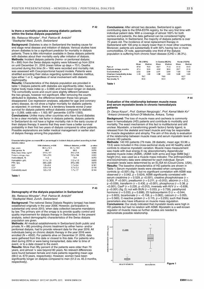
POSTER PRESENTATIONS – HEMODIALYSIS / PERITONEAL DIALYSIS 22 S
P 42
Is there a mortality paradox among diabetic patients within the Swiss dialysis population?Ms. Rebecca Winzeler1, Prof. Patrice M. Ambühl1 1Stadtspital Waid, Zurich, SwitzerlandBackground: Diabetes is also in Switzerland the leading cause for end stage renal disease and initiation of dialysis. Various studies have shown diabetes to be a significant predictor for mortality in dialysis patients. There is little information available in Swiss dialysis patients with diabetes about their mortality early after initiation of dialysis.Methods: Incident dialysis patients (hemo- or peritoneal dialysis; N = 895) from the Swiss dialysis registry were followed up from 2014 on until December 31, 2016 (mean follow up days = 751). Deaths occurred during this time (N = 189) were recorded and mortality risk was assessed with Cox-proportional hazard models. Patients were stratified according their status regarding systemic diabetes mellitus, type either 1 or 2, regardless of renal involvement with diabetic nephropathy.Results: Characteristics of the dialysis population are provided in table 1. Dialysis patients with diabetes are significantly older, have a higher body mass index (p = 0.068) and have been longer on dialysis. The comorbidity score and count were slightly different between the two groups, however not significant. After removing the counts attributed to diabetes, the difference between these two groups disappeared. Cox regression analyses, adjusted for age and coronary artery disease, do not show a higher mortality for diabetic patients as expected. In contrast, there is a tendency towards worse 2-year survival for dialysis patients without diabetes compared to diabetic (p = 0.269, odds ratio = 1.183; 95th percentile: 0.878–1.593).Conclusions: Unlike many other countries who have found diabetes to be a clear mortality risk factor in dialysis patients, dialysis patients in Switzerland do not have a higher mortality risk in the early course of their dialysis therapy. It seems that they even have a better survival probability in their first two years of dialysis compared to other patients. Possible explanations are better medical management or earlier start of dialysis therapy among this population.
P 43
Demography of the dialysis population in SwitzerlandMs. Rebecca Winzeler1, Prof. Patrice M. Ambühl1 1Stadtspital Waid, Zurich, SwitzerlandBackground: The national Swiss Dialysis Registry (srrqap) has been established originally in the year 2006. However, participation is substantial only since 2013, when data collection became mandatory by law. The primary aim of the srrqap is to provide quality control and quality improvement for dialysis therapy in Switzerland. In the present analysis, select demographic characteristics of the Swiss dialysis population are given.Methods: All medical establishments in Switzerland (both public and private; N = 88) providing chronic treatment by either hemo- and/or peritoneal dialysis, had to provide relevant data for the year 2016. All individuals being on chronic dialytic therapy in the year 2016 were enrolled (N = 4502). For patients alive on December 31 2016, data were gathered from this date or closest to this date. For patients who died during 2016 or were being transplanted, data refer to time of event, or to a date closest to the event.Results: More than fifty percent of the patients were older than 70 years, and almost ¼ was beyond 80 years. No relevant differences were found between female and male patients regarding mean age (68.0 vs. 67.9 years, respectively). However, women have been significantly longer on dialysis compared to men (51.3 vs. 45.3 months, respectively).
Conclusions: After almost two decades, Switzerland is again contributing data to the ERA-EDTA registry, for the very first time with individual patient data. With a coverage of almost 100% for both centers and patients, the data gathered can be considered highly representative. In Switzerland, the majority of dialysis patients is over 70 years old. The incidence of renal replacement therapy in Switzerland with 100 pmp is clearly lower than in most other countries. Moreover, patients are substantially ill with 50% having two or more comorbidities. Of note, approximately one third of the dialysis population is suffering from chronic heart disease (CHD = 36.3%).
P 44
Evaluation of the relationship between muscle mass and serum myostatin levels in chronic hemodialysis patientsDr. Derya Koyun1, Prof. Gokhan Nergizoglu1, Prof. Kemal Metin Kır1 1Ankara University School Of Medicine, Ankara, TurkeyBackground: The loss of muscle mass and cachexia is commonly seen in hemodialysis (HD) patients and contribute to morbidity and mortality. The exact mechanism of this fact is multifactorial and still unclear. Myostatin, a transforming growth factor-β family ligand, is released from the skeletal and heart muscle and may be responsible for muscle degradation and atrophy. The aim of this study is evaluation of the relationship between muscle mass and serum myostatin level in chronic HD patients.Methods: 140 HD patients (79 male, 28 diabetic, mean age; 53.96 ± 13.6) were included in this cross-sectional study and 40 healthy adult controls to observe myostatin variation. Muscle mass measurement was made with dual energy-X ray absorptiometry. Appendicular skeletal muscle index (ASMI), (ASMI: both arms and legs SMM [kg] / height [m2], was used as a muscle mass indicator. The anthropometric and biochemistry data were obtained for each individual. Serum myostatin levels were determined by an ELISA kit (Cloud-Clone, USA).Results: The baseline characteristics of HD patients are shown in Table 1. Serum myostatin levels were elevated when compared to controls (p <0.001) (fig. 1) but no significant correlation with ASMI was observed (r = 0.042, p = 0.624). ASMI significantly correlated with serum creatinine (r = 0.529, p <0.001), creatine phosphokinase (r = 0.305, P <0.001), prealbumin (r = 0.211, p <0.012), albümin (r = 0.2, p <0.039), transferrin (r = 0.430, p <0.001), phosphorus (r = 0.38, p <0.001), CaxP (r = 0.235, p <0.012), inversely with Kt/V (r = –0.636, p <0.001) (fig. 2); not with BUN (r = 0.033, p = 0.739), parathyroid hormone (r = 0.033, p = 0.698), 25 hydroxyvitamin D (r = –0.044, p = 0.603), bicarbonate (r = –0.158, p = 0.062), calcium (r = 0.055, p = 0.560), C-reactive protein (r = 0.115, p = 0.235); such that these parameters also have influence on muscle mass regulation.Conclusions: Our study indicated that myostatin levels were high in HD patients but had no relation with ASMI. Myostatin is a well-known regulator of muscle mass so further studies are needed to demonstrate possible relationship.
Table 1
Table 1
Figure 1
SWISS MEDICAL WEEKLY 2017;147 (SUPPL 227) WWW.SMW.CH
POSTER PRESENTATIONS – HEMODIALYSIS / PERITONEAL DIALYSIS 23 S
P 45
Prolonged disease-free survival after treatment of a bilateral breast granulocytic sarcoma with low-dose sorafenib in a chronic hemodialyzed patientMs. Julie Robert1, Dr. Ould Maouloud Hemett1, Dr. Merryl Cassat1, Dr. Gaëlle Rhyner1, Dr. Eric Descombes1, Prof. Daniel Betticher1 1HFR, Hopital Cantonal de Fribourg, Fribourg, SwitzerlandBackground: Granulocytic sarcoma (GS), or extramedullary leukemia, is a rare manifestation of acute myelogenous leukemia (AML) and often accompanies bone marrow involvement. It is not uncommon for GS to present as an isolated disease without bone marrow involvement, in particular upon disease relapse. Certain extramedullary sites such as the central nervous system and the reproductive organs are more prone to be involved at time of relapse.
Presenting signs or symptoms are mainly due to mass effect of the tumor and dysfunction of the organ or tissue affected; the diagnosis is performed by biopsy. GS occurs in 2–9% of newly diagnosed AML patients, either isolated or in combination with bone marrow involvement. Presence of GS carries a poor prognosis with a 5-years survival of 20–30%.Methods: The multi kinase inhibitor Sorafenib was found to inhibit proliferation and to induce apoptosis in FLT3-ITD+AML blasts at concentrations achievable in vivo. Almost all the clinical trials have exclude hemodialyzed patient, but sorafenib has already been used at reduced doses to treat dialysis patients with renal carcinoma Results: We report the case of 45-years old woman hemodialyzed since December 2013 for renal failure due to interstitial nephritis and nephrocalcinosis complicating the induction chemotherapy of AML (idarubicine, cytarabine). AML was classified as M4/M5 according to FAB classification with positive FLT3-ITD and NPM1mutation, and negative [AML1-ETO, t(8,21), CBFb-MYH11, inv(16) PML_RARa, t(15;17)]. After the induction cure, she was in remission on day 17 and received 2 cures of consolidation. She refused an autograft. One year later, she developed bilateral breast GS with positive FLTD3-ITD mutationConclusions: The treatment included four monthly cycles of azacitidine (120 mg from D1 to D5) alternating with Sorafenib (200 mg 2× day) with a good tolerance and involution of the tumors. At present time the patient is still in remissionwith a fairly good quality of life.
P 46
Safety first, fistula second!Dr. Sandra Edith Knych1, Dr. Georg-Wilhelm Meier-Fiorese1, Dr. Felix Burkhalter1 1Kantonsspital Baselland, Liestal, SwitzerlandBackground: The native arteriovenous (AV) fistulas and the AV graft are considered the best types of access for hemodialysis patients. Stenosis of fistulas is one of the most common complications in up to 80% of patients needing treatment with percutaneous transluminal balloon angioplasty (PTA) during follow-up. Stenosis is another common risk factor for acute thrombosis as well as coagulation disorder, hypotension or existing aneurysm of the venous vessel.Methods: no methodsResults: A 77 year old patient on chronic hemodialysis presented with acute thrombosis of his fistula after a car accident. His fistula of the upper arm was created in 2008. He had several PTA of stenosis during follow-up and developed two aneurysms. The last PTA of a stenosis of the subclavian vein was performed a year before. Since then accessflow remained stable. When he suffered the car accident, the airbag was activated. As he was not severely injured, he was not sent to the hospital and presented him two days later for his regular hemodialysis session. On physical examination he had two hematomas, one in the region of the breast bone and another on his right upper arm. There was no flow detectable in the fistula and acute thrombosis was diagnosed. A revision of the fistula with resection of the aneurysm and thrombectomy was carried out. In addition angiography showed restenosis of the subclavian vein and PTA was performed. As there was no clinical sign of restenosis of the subclavian vein prior to the acute thrombosis, we suggest that due to the strong hit by the inflated airbag on the fistula, adherent thrombotic material from the aneurysm was dislodged and occluded the Fistula.Conclusions: In the presence of an aneurysm and/or adherent thrombotic material to the vessel wall of a fistula, strong physical energy can dislodged thrombotic material leading to acute thrombosis.
Figure 2
SWISS MEDICAL WEEKLY 2017;147 (SUPPL 227) WWW.SMW.CH

POSTER PRESENTATIONS – TRANSPLANTATION 24 S
P 47
Role of Immune-Senescence in the development of de-novo Donor-Specific Antibodies after Kidney TransplantationDr. Seraina von Moos1, Dr. Gesa Schalk2, Dr. Guido F. Laube2, Prof. Thomas Mueller1 1Universitätsspital Zürich, Zurich, Switzerland; 2UniversitätsKinderspital Zürich, Zurich, SwitzerlandBackground: The concept of immune-senescence, implicating a changing immune-reactivity during lifetime, has gained increasing awareness. Higher age is associated with increased risk of infections and malignancies but reduced vaccination responses and reduced frequency of acute rejections in solid organ transplantation. Yet, the influence of immune-senescence on development of donor-specific antibodies (DSA) is unknown.Methods: In this observational study, we included all children younger than 10 years of age and all adults older than 60 years of age, who received a kidney transplant at the University Hospital of Zurich between January 2006 and February 2015. Maximum follow-up time for occurrence of de-novo DSA as measured by Luminex-assay was until March 2016.Results: Out of 160 elderly patients, a total of 12 patients (11%) developed de-novo DSAas compared to 6 patients out of 19 (32%) transplanted children. Risk ofdevelopment of de-novo DSA wassignificantly lower in elderly patients (HR 0.21) as compared to children (p = 0.0224). Median time to development of de-novoDSA was similar in both age groups (adults 720 days, children 1086 days). Likewise, de-novo DSA were predominantly of classII with similar peak MFI in elderly adults (MFI 7023) and children (MFI 6408). Immunosuppressionwas significantly more often a combination with cyclosporine than withtacrolimus in elderly patients developing denovo DSA. The same trend was observed in children.Conclusions: Risk of development of de-novo DSA is lower in elderly adults as compared to inconcordance with the concept of immune-senescence.
P 48
Postoperative Seroma after Kidney Transplantation: Identification of donor-, recipient- and procedure- associated risk factorsMr. Ruhin Parsa1, Dr. Spiridon Arampatzis1, Dr. Guido Beldi1, Dr. Suzan Dahdal1, Ms. Angela Buechel1, Prof. Bruno Vogt1, Prof. Uyen Huynh-Do1, Dr. Vanessa Banz1, Dr. Daniel Sidler1 1University Hospital Bern, Bern, SwitzerlandBackground: Kidney transplantation is an efficacious and safe treatment for end stage renal disease. The early postoperative outcome of kidney transplantation has tremendously improved in the last decades and patient and allograft survival reach nearly 100% after the first year of transplantation. Peri-renal fluid collections are frequent findings in the early phase after kidney transplantation and occur in up to 50% of patients. The pathogenesis and clinical significance of seroma is not clear. Furthermore, treatment modalities of seroma, including pre-emptive drainage and laparoscopic fenestration have never been studied in a prospective and randomized trial.Methods: We determined the incidence of seroma in 294 consecutive kidney transplantations at the University of Bern over a period of more than eight years and identified donor-derived, recipient-derived and surgical risk factors.Results: Seroma frequency was significantly higher in the Deceased Donor Transplantation (DDT, 49%) than the Living Donor Transplantation (LDT, 33%) cohort. Patient and allograft survival and function were comparable in patients with or without seroma. 65% of seroma were treated conservatively and among those 38% showed spontaneous regression. 35% of seroma were treated either by percutaneous drainage or laparoscopic fenestration. Both procedures were safe, although the former elicited 28% re-occurrence and need for secondary treatment. In the DDT cohort, we identified donor derived factors for the development of seroma, namely increased donor age which was associated with a 30% increased risk for postoperative seroma.Conclusions: These results indicate that postoperative seroma is a frequent finding after kidney transplantation, namely DDT and are associated with donor-derived risk factors. These findings suggest that seroma – irrespective of size – are a surrogate marker of allograft “wear and tear” of the renal allograft. The clinical significance of pre-emptive treatment remains unclear.
P 49
Safety of Early RAS Blockade in Kidney Transplant RecipientsMr. Luca Valente1, Dr. Spiridon Arampatzis1, Dr. Mariam Semmo1, Prof. Bruno Vogt1, Prof. Uyen Huynh-Do2, Dr. Daniel Sidler1 1University Hospital Bern, Bern, Switzerland; 2Inselspital Bern, Bern, SwitzerlandBackground: RAS blockade is often withheld from kidney transplant recipients due to feared deterioration of allograft function. Meanwhile, due to a high prevalence of cardiovascular co-morbidities, many transplant recipients could substantially profit from these compounds. The safetly of these compound is still controversal in the early phase after transplantion.Methods: In a retrospective study, we investigated the safety and outcome of RAS blockade in the early phase after kidney transplantation in 171 recipients of renal transplantations at the University Hospital in Bern. Results: At the time of allocation, 107/170 (63%) of patients hat RAS blockade implemented with a median prescription of 50% of the maximal registered dosage. Treatment was paused at time of allocation and re-implemented in 141/170 patients (82.9%) within the first six months of transplantation. 55/141 (39%) received the same RASblocker as previously, 39/141 (28%) received a different compound and 47 (33%) received a de novo prescription of RAS blockade. Median time point to start RAS blockade was 24 days post transplantation. Mean serum creatinine rose 4.9%+/-0.17% from baseline value and 11%+/-0.22% at day 7 and day 30 respectively. Serum creatinine rose more than 10% in 49/132 (37.1%) patients, more than 20% in 20/132 (15.2%) patients and more than 30% in 10/132 (7.6%) in the early phase after treatment implementation. Therapy was stopped in 24/141 (17%) patients within the first six months of transplantation, mostly due to excessive increase of serum creatinine or symptomatic orthostasis or hypotension. Patient and allograft survival was excellent in the observation period. For the composite endpoint of alive, functioning graft and eGFR >30 ml/min/1.73 m2, no significant difference was found between patients with or without RAS blockadeConclusions: We conclude, that early implementation of RAS blockade is safe in kidney transplant recipients, although treatment discontinuation is necessary in up to 20% of patients.
P 50
Antibody-mediated rejection triggered by parvovirus B19 infection? A case reportDr. Melanie Schönenberger1, Dr. Patricia Hirt-Minkowski1, Dr. Helmut Hopfer1, Dr. Thomas Menter1, Prof. Stefan Schaub1, Dr. Katrin König1 1University Hospital Basel, Basel, SwitzerlandBackground: Transplant recipients can be sensitized against allo-HLA antigens by previous transplantation, blood transfusion, or pregnancy. The role of infectious pathogens in triggering allo-HLA cross-reactive T-cells has been well described for CMV and EBV. However so far, the role of parvovirus B19 has not been considered. Methods: A 46-year old male, suffering from ESRD due to chronic interstitial nephritis was transplanted from a pediatric deceased donor. He had no previous sensitizing events and no detectable pre-transplant HLA-antibodies by Luminex SA assays. Immunosuppressive therapy consisted of tacrolimus, mycophenolate mofetil, prednisone and basiliximab for induction. Because of delayed graft function, a kidney biopsy was performed on day 8 which showed no rejection. Creatinine levels decreased slowly from 1236 mmol/l to 210 mmol/l. He developed severe reticulocytopenic anemia and parvovirus B19 primo-infection was diagnosed by PCR on day 34. The dosage of mycophenolate mofetile was reduced and IVIG initiated. On day 45, the patient had to be re-hospitalized due to acute graft failure. Kidney biopsy was performed and revealed acute mixed rejection (Banff scores t1, ptc2, v1, C4d0). Anti-rejection therapy with methylprednisolone, IVIG, and thymoglobuline was given. Creatinine values decreased to its baseline around 200 mmol/l.Results: Day 65, the patient presented again with high creatinine around 600 mmol/l. A 3-day-methylprednisolone-trial did not show any improvement of the transplant function. Due to acute graft failure and limited therapeutic options (pancytopenia), a transplant nephrectomy was performed on day 70 post-transplant. The histology revealed features of both antibody- and T-cell-mediated rejection (t2, i3, v2, g1, ptc2, C4d3). Blood evaluation by Luminex SA showed strong antibodies against 10/12 mismatched donor HLA-antigens. During the
SWISS MEDICAL WEEKLY 2017;147 (SUPPL 227) WWW.SMW.CH
POSTER PRESENTATIONS – TRANSPLANTATION 25 S
whole post-transplant course, there were no signs of other infections such as CMV, EBV, and BKV.Conclusions: Our patient developed early graft failure due to severe, therapy-resistant acute rejection, which was likely triggered by parvovirus B19 primoinfection.
P 51
Transplant Glomerulopathy and Arteriolohyalinosis in Renal Allograft Biopsies: Correlation between Histopathology and Clinical OutcomeMr. Jannick Schneuwly1, Dr. Vera Genitsch2, Prof. Bruno Vogt1, Prof. Uyen Huynh-Do1, Dr. Daniel Sidler1 1University Hospital Bern, Bern, Switzerland; 2University of Bern, Bern, SwitzerlandBackground: Kidney transplantation is the best therapeutic option for most patients with end-stage kidney disease. In spite of a marked decrease in acute rejection episodes in the early post-transplant period, the long-term graft survival is still poor due to chronic allograft pathology. Classical histopathological features of chronic allograft lesions are transplantat glomerulopathy (cg) and arteriolohyalinosis (aah), which derive from chronic antibody mediated rejection and chronic nephrotoxicity due to calcineurin inhibitor treatment.Methods: We aimed to analyze chronic allograft lesions in indication biopsies in 295 patients and 303 kidney transplantations from 01.01.09 through 29.06.17.Results: Chronic allograft lesions were infrequent in early biopsies within the first year of transplantation (<5%). Thereafter, 48/92 (52%) showed no cg>0 or aah>0 lesions (group 1), 14/92 (15%) had cg>0 (group 2) and 16/92 (18%) aah>0 (group 3). 14/92 (15%) showed composite findings (cg>0 and aah>0). At time of biopsy, serum creatinine levels were similar between the various groups. aah-lesions were associated with a further deterioration kidney function within the next six months and only limited risk for grafts loss. Meanwhile, cg lesions were associated with an increased risk for allograft loss. In composite lesions, the deteriorating kidney function was dominant.Conclusions: Histopathological and Clinical Correlation of Chronic Allograft Lesions may predict allograft outcome and guide personalized treatment options.
P 52
Relationship of serum bicarbonate levels with 1-year graft function in kidney transplant recipientsMs. Anna Wiegand1, Dr. Nicole Graf2, Dr. Marco Bonani1, Dr. Diana Frey1, Prof. Rudolf P. Wüthrich1, Dr. Nilufar Mohebbi1 1University Hospital Zurich, Zurich, Switzerland; 2Graf Biostatistics, Winterthur, SwitzerlandBackground: Metabolic acidosis (MA) is common in CKD patients and kidney transplant recipients (KTR) with a prevalence between 12% and 58% after transplantation. MA is an independent risk factor for kidney disease progression in CKD. Interestingly, a recent publication has shown for the first time that MA after transplantation was also associated with increased risk of graft loss and death. To extend these observations we tested if there is a relationship between serum bicarbonate at 3 months after transplantation and graft function after one year in Swiss KTR.Methods: This is a post-hoc analysis of a previously performed open-label randomized study testing denosumab for its effects on bone mineral density in 90 de novo KTR. MA was defined as serum bicarbonate levels at 3 months of <22 mmol/l. Graft function was determined by eGFR according to CKD-EPI equation. Values were set to missing for patients treated for MA.Results: Prevalence of MA decreased from 63% at baseline to 39% after 3 months and to 28% after one year. Bicarbonate levels and eGFR both increased in the first year. Mean bicarbonate level at baseline was 20.6 ± 3.0 mmol/l, 22.0 ± 2.8 mmol/l /l after 3 months and 22.7 ± 2.7 mmol/l after one year. Mean eGFR at baseline was 53.3 ± 15.8 ml/min/1.73 m2, 55.1 ± 15.8 ml/min/1.73 m2 after 3 months and 56.9 ± 18.5 ml/min/1.73 m2 after one year. Mean changes of eGFR after one year in KTR with and without MA are shown in table 1.
Conclusions: Prevalence of metabolic acidosis was highest at baseline and decreased within the first year after transplantation. In parallel, eGFR values increased from baseline to one year. However, mean rise in eGFR was higher in KTR without MA compared to patients with MA suggesting a potential role of MA on kidney graft function.
P 53
Preservation of kidney function in kidney transplant recipients by alkali therapy (Preserve-Transplant Study)Ms. Anna Wiegand1, Dr. Alexander Ritter1, Dr. Nicole Graf2, Dr. Karine Hadaya3, Dr. Spyridon Arampatzis4, Prof. Thomas Mueller1, Prof. Carsten Wagner5, Prof. Rudolf P. Wüthrich1, Dr. Nilufar Mohebbi1
1University Hospital Zurich, Zurich, Switzerland; 2Graf Biostatistics, Winterthur, Switzerland; 3Geneva University Hospitals, Geneva, Switzerland; 4Inselspital Bern, Bern, Switzerland; 5University of Zurich, Zurich, Switzerland,Background: Kidney transplantation is the treatment of choice for patients with ESRD. Short- and long-term graft survival after kidney transplantation have significantly improved within the last decades but declining transplant functionor even graft loss is still a common issue. Metabolic acidosis (MA) is highly prevalent in renal transplant patients and a recent study has shown that MA may be a significant risk factor for graft loss and mortality. However, no data exist yet on the role of alkali treatment in the prevention of progressive loss in renal allograft recipients. Given the expanding number of CKD patients – including former kidney transplant recipients – an alkali treatment study in kidney transplant patients is of prime importance and has the potential to show that such treatment may slow or reduce the progression towards graft failure and significantly decrease the rate of ESRD.Methods: This study is a multi-center, prospective, randomized, single-blinded, placebo-controlled interventional trial to test the superiority of alkali treatment in comparison to placebo for preservation of kidney function in 300 kidney transplant recipients. The duration of the study is 2 years for each individual participant. The patients are randomized into 2 arms: an intervention arm (sodium hydrogen carbonate) and a placebo arm. The study is supported by the Swiss National Science Foundation as an Investigator-initiated clinical Trial.Results: The Preserve-Transplant Study has received all required approvals by the end of March 2017. Patient recruitment has started on June 12th, 2017 in Zurich. Both study sites in Berne and Geneva have been operative since July 12th 2017. As of September 8th 2017, 18 patients have been enrolled.Conclusions: The Preserve-Transplant Study has been launched successfully. High recruitment rates are essential to achieve the planned number of 300 patients by the end of the recruitment period in June 2019.
P 54
The humoral long-term progression of kidney function in patients with and without humoral allograft responseDr. Seraina von Moos1, Dr. Pietro E. Cippà2, Mr. Robert Van Breemen1, Prof. Thomas Mueller1 1Universitätsspital Zürich, Zurich, Switzerland; 2University of Southern California, Los Angeles, United StatesBackground: While deterioration of long-term graft function due to insufficient control of humoral immune responses is a major focus of current research the typical evolution of kidney function in the patients without humoral immune activation is not well described. In the present longitudinal observational cohort study we analyzed the course of kidney function in patients stratified to those with a detectable antibody response and those without.Methods: All kidney transplant patients at the University Hospital of Zurich between January 2006 and February 2015 were included and the course of kidney function was determined by slope of eGFR after CKD-EPI starting from 12 months after transplantation until the last follow up visit (at latest February 2016). Slope of eGFR was compared between patients without development of humoral allograft immune responses and patients with development of donor specific antibodies as determined by Luminex single bead assays, which were performed at least annually.Results: Patients without humoral allograft immune responses present an improvement of kidney function in the long term follow up as reflected by a positive eGFR slope. Such a continuous improvement of graft function is still present even six years after transplantation. In
Table 1
SWISS MEDICAL WEEKLY 2017;147 (SUPPL 227) WWW.SMW.CH

POSTER PRESENTATIONS – TRANSPLANTATION 26 S
contrast, patients with donor specific antibodies show a decline in kidney function as reflected by a negative eGFR slope.Conclusions: The kidney allograft, transplanted in a patient without development of humoral allograft immune response as determined by development of donor specific antibodies shows an adaptive hyperfiltration, which is maintained even in the long-term.
P 55
Rituximab induces “false” positive complement- dependent cytotoxic B cells crossmatchesDr. Karine Hadaya1, Mr. Johan Geiser2, Prof. Thierry Berney2, Prof. Pierre-Yves Martin3, Prof. Jean Villard2, Dr. Sylvie Ferrari-Lacraz2 1Geneva University Hospitals, Geneva, Switzerland; 2Geneva University Hospital and Medical School, Geneva, Switzerland; 3AMC/Geneva University Hospitals, Geneva, SwitzerlandBackground: ABO incompatibilibity (ABOi) is no more considered an immunological barrier in kidney transplantation, allowing patients with a healthy living donor to undergo transplantation. ABOi kidney transplantations are routinely performed at Geneva University Hospital since May 2008 with great success and with long-term outcomes equivalent to ABO-compatible kidney transplantations.Methods: Before all kidney transplantations, a prospective cell-based CDC (complement-dependent cytotoxic) is performed. A single dose Rituximab 375 mg/m2 is required 30 days before transplantation in the ABOi transplantation therapeutic strategy. We have observed highly positive CDC B cells crossmatch following these perfusions. This positivity is due to the link of the circulating drug to B cells. Our aim was to determine for how long Rituximab may alter crossmatch results. 13 couples were analyzed 1 month before (and therefore before Rituximab perfusion), at day 0 and 3 month post-transplantation, by performing CDC and Facs crossmatches.Results: CDC and Facs crossmatches performed 1 month before transplantation were negative for both T and B cells in all analyzed couples. When crossmatches were performed with serum of the day of transplantation, CDC and Facs crossmatches were positive for B cells but negative for T cells. 3 months after transplantation, both CDC and Facs crossmatches were again negative for B cells. We also performed anti-HLA antibodies analyses in these sera and for each patient we did not observe major differences in anti-HLA antibodies specificities on the 3 time points.Conclusions: The interaction of rituximab with the classical CDC crossmatch observed 1 month after Rituximab perfusion is short-lived, as at 3 month post-transplantation (and therefore 4 month post-perfusion) the interaction of Rituximab with B cells is not observed anymore. Therefore, if a CDC or Facs crossmatch should be performed after transplantation, it could be safely performed 3 month later and thereafter.
P 56
Activablility of circulating Tfh17 predicts humoral response to thymus-dependent antigensDr. Suzan Dahdal1, Ms. Carole Saison1, Dr. Martine Valette2, Prof. Bruno Lina3, Prof. Guillaume Monneret4, Dr. Thierry Defrance5, Prof. Emmanuel Morelon6, Prof. Olivier Thaunat4 1French National Institute of Health and Medical Research (Inserm) Unit 1111, Lyon, France; 2Hospices Civils de Lyon, Croix-Rousse University Hospital, Lyonh, France; 3Hospices Civils de Lyon, Croix-Rousse University Hospital, Lyon, France; 4Hospices Civils de Lyon, Edouard Herriot University Hospital, Lyon, France; 5French National Institute of Health and Medical Research (Inserm), Lyon, France; 6Hospices Civils de Lyon, Edouard Herriot University Hospital, Department of Transplantation, Lyon, FranceBackground: Despite therapeutic immunosuppression, a significant proportion of transplanted patients develop donorspecific antibodies (DSA), which are currently recognized as the first cause of allograft failure. Generation of antibodies against protein antigens (including donor HLA) results from a thymus-dependent (TD) humoral response, which means that B cells need to receive a co-stimulation signal from activated follicular helper T cells (Tfh) to differentiate into plasmacells.Methods: In this study, we test whether profiling of circulating Tfh (cTfh) could predict the ability to mount a TD humoral response in 36 renal transplanted patients and 9 healthy controls. We took advantage of the 2015 influenza vaccination campaign, which provided a normalized setting of antigenic stimulation. The number of cTfh, their polarisation profile, and ability to up-regulate i) helper molecules (CD40L and ICOS) and ii) the activation marker CD25 following in vitro stimulation in presence of patients’ own plasma (with IS drugs) were measured prior vaccination. These parameters were then compared between responders and non-responders to influenza vaccine.Results: While most of the characteristics of cTfh profile were similar between the two groups, we observed that responders showed a significantly higher proportion of cTfh17 that upregulated CD25 expression after in vitro stimulation. We performed a posteriori analysis of the cTfh profile of 15 transplanted patients at the time of DSA appearance and found that the proportion of cTfh17 cells that upregulated CD25 after in vitro stimulation was similar to responder to influenza vaccine.Conclusions: We concluded that the ability of the cTfh17 subset to be activated in vitro predicts TD antibody response and might be used as non-invasive biomarker to identify transplanted patients at risk to develop DSA.
SWISS MEDICAL WEEKLY 2017;147 (SUPPL 227) WWW.SMW.CH


INDEX OF FIRST AUTHORS 28 S
The numbers refer to the pages of this supplement.
Aeberhard C 15 SAmico P 13 SArokiaraj M C 18 SAuberson M 4 S
Beglinger C 14 SBerchtold L 2 SBischof N 8 SBohlender J 11 SBonani M 8 SBonny O 10 SBouatou Y 7 S, 11 SBraconnier P 9 S, 10 SBuchkremer F 12 S
Claus M 20 S
Dahdal S 26 SDel Giorno R 21 SDessalle K 16 SDhayat N 3 SDiebold M 6 SDissard R 5 SDrivakos N 14 S
Ferrier C 6 SFigurek A 8 SFischer G 9 S
Gabutti L 14 SGagnebin Y 7 SGassama A 21 SGerber L 17 SGueguim C 12 SGünter J 3 S
Hadaya K 26 SHaddad G 17 SHapuoja L 3 SHauenstein F 10 SHess B 2 SHess N 13 SHolzmann Y 7 S
Jotterand Drepper V 5 S
Keppner A 18 SKneubühl A 19 SKnych S E 23 SKölling M 19 SKomarynets O 18 SKoyun D 22 S
Mayr M 16 S
Nolan K 19 S
Parsa R 24 SPathare G 11 SPechere A 2 S
Penton Ribas D 4 SPerrottet Ries N 8 SPillai S 17 SPruijm M 2 S
Randi E 20 SRobert J 23 SRudloff S 4 S
Sarinyan S 12 SSaudan P 6 SSchneuwly J 25 SSchönenberger M 24 SSchwarzkopf A-K 15 SScotti Gerber J 15 S, 21 SSolca C 15 SSpica D 6 S
Tataw J 14 STokonami N 4 S, 20 S
Valente L 24 SVerouti S 18 SVögelin M 13 S
von Moos S 24 S, 25 S
Wiegand A 25 SWinzeler R 22 S
SWISS MEDICAL WEEKLY 2017;147 (SUPPL 227) WWW.SMW.CH