essentials of biology -
TRANSCRIPT

1/25/2011
1
Essentials of
Biology
Sylvia S. Mader
Chapter 4
Lecture Outline
Copyright © The McGraw-Hill Companies, Inc. Permission required for reproduction or display.
Cells are tiny!!
Cells are tiny!!Cells are tiny—even in an Orca!!
Lots of Blood Stem Cell Division in your bone Marrow!!
Biology and Society: Cells That Cure
• During a heart attack,
– Heart muscle cells die because they are starved
for oxygen.
• Unfortunately, these kinds of cells do not
regenerate.
Copyright © 2007 Pearson Education, Inc. publishing as Pearson Benjamin Cummings

1/25/2011
2
Copyright © 2007 Pearson Education Inc., publishing as Pearson Benjamin Cummings
• Muscle stem cells
are transplanted to
the ailing heart,
facilitating
healing.
Cells Therapy
Figure 4.1
4.1 Cells Under the Microscope
• Cells
• Are extremely diverse
• Nearly all require a microscope to be seen
• Each type in our body is specialized for a particular
function
Light microscope
• Invented in 17th century
• Limited by properties of light
Electron microscope
• Invented in 1930s
• Overcomes limitation by using beam of electrons
Scientist using an electronmicroscope.
LM of leaf cells
LM of Euglena
Scientist using a light microscope.
LM of human epithelial cells
Figure 4.1
Diversity of cellsFigure 4.2 Relative sizes of some living things and their components
electron microscope
unaided eye
1 km100 m10 m1 m0.1 m1 cm1 mm100 m10 m1 m100 nm10 nm1 nm0.1 nm
mouse
frog
egg
human
egg
plant and
animal
cells
most bacteria
viruses
proteins
amino acids
atoms
ant
human
blue whalechloroplast
light microscope
Need large surface area for entry and exit of nutrients
and wastes
Small cells have a large Surface-area-to-volume-ratio
• Small cells have more surface area for exchange.
Adaptations to increase surface area
• Microvilli in the small intestine increase surface area for
absorption of nutrients.
Why does natural selection favor small cells? 4.2 The Two Main Types of Cells
• Cell theory
All organisms composed of cells
All cells come only from preexisting cells.
• All cells have a plasma membrane.
Encloses cytoplasm and genetic material
• 2 main types of cells
Based on organization of genetic material and complexity
1. ______________cells – lack membrane-bounded nucleus
and only ribosomes as organelles
2. ______________ cells – have nucleus housing DNA and
contain many different kinds of organelles

1/25/2011
3
cell
Eukaryotic cell: complex
internal structure
Prokaryotic cell: Simple internal
structure
flagella
Figure 4.3
Comparison of
prokaryotic and
eukaryotic cells
Prokaryotic
vs.
Eukaryotic
cells
• Prokaryotic cells
– From the domains Bacteria and Archaea
– Are smaller than eukaryotic cells.
– Lack internal structures surrounded by
membranes.
– Lack a nucleus.
– Reproduce very quickly
• Bacteria
– Well known because some cause disease
– Others have roles in the environment
– Some are used to manufacture chemicals, food,
drugs, etc.
capsule
gel-like coating outside
the cell wallnucleoid
location of the bacterial
chromosome
conjugation pilus
elongated, hollow
appendage used to transfer
DNA to other cells
flagellum
rotating filament that
propels the cell
ribosome
site of protein synthesis
plasma membrane
sheet that surrounds the
cytoplasm and regulates
entrance and exit of moleculescell wall
structure that provides support
and shapes the cell
fimbriae
hairlike bristles
that allow
adhesion
to surfaces cytoplasm
semifluid solution surrounded
by the plasma membrane;
contains nucleoid and
ribosomes
© Ralph A. Slepecky/Visuals Unlimited
Figure 4.4 Prokaryotic cell
• Bacterial structure
Cytoplasm surrounded by plasma membrane
and cell wall
• Sometimes a capsule – protective layer
• Plasma membrane the same as eukaryotes
Cell wall maintains shape of cell
DNA – single circular chromosome located in
nucleoid (region – not membrane enclosed)
Ribosomes – site of protein synthesis
Appendages
• Flagella – propulsion
• Fimbriae – attachment to surfaces
• Conjugation pili – DNA transfer
Bacterial Structure
1/25/2011
4
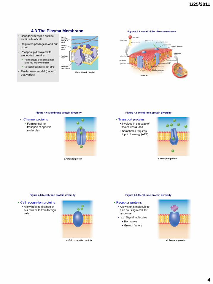
4.3 The Plasma Membrane• Boundary between outside
and inside of cell
• Regulates passage in and out
of cell
• Phospholipid bilayer with
embedded proteins
Polar heads of phospholipids
face into watery medium
Nonpolar tails face each other
• Fluid-mosaic model (pattern
that varies)Fluid Mosaic Model
Figure 4.5 A model of the plasma membrane
external membrane
surface
carbohydrate chain
glycoprotein
phospholipid
bilayer
cholesterol
cytoskeleton
filaments
internal membrane
surface
protein molecule
hydrophobic
phospholipid
hydrophilic
hydrophilic
polar head
nonpolar tail
Outside of cell
Inside of cell
• Channel proteins
Form tunnel for
transport of specific
molecules
Figure 4.6 Membrane protein diversity
a. Channel protein b. Transport protein
Transport proteins
• Involved in passage of
molecules & ions
• Sometimes requires
input of energy (ATP)
Figure 4.6 Membrane protein diversity
c. Cell recognition protein
Figure 4.6 Membrane protein diversity
Cell recognition proteins
• Allow body to distinguish
our own cells from foreign
cells.
d. Receptor protein
Receptor proteins
• Allow signal molecule to
bind causing a cellular
response
• e.g. Signal molecules
• Hormones
• Growth factors
Figure 4.6 Membrane protein diversity

1/25/2011
5
e. Enzymatic protein
Figure 4.6 Membrane protein diversity
Enzymatic proteins
• Speed up metabolic
reactions in cell
f. Junction proteins
Junction proteins
• Form points of contact
between cells
• Cell-to-cell adhesion and
communication
Figure 4.6 Membrane protein diversity
4.4 Eukaryotic cells• Protists, fungi, plants, and animals
• Have a membrane-bounded nucleus housing DNA
• Much larger than prokaryotic cells
• Compartmentalized and contain organelles
• 4 categories of organelles
1. Nucleus and ribosomes
2. Endomembrane system
3. Energy-related
4. Cytoskeleton
Figure 4.7 Structure of typical animal cell
Figure 4.8 Structure of a typical plant cell 1. The Nucleus and Ribosomes:Genetic Control of the Cell
• The nucleus is the manager of the cell.
– Genes in the nucleus store information necessary
to produce ________?????_________.

1/25/2011
6
Structure and Function of the Nucleus
• The nucleus is bordered by a
double membrane called the
nuclear envelope.
– Function of pores??
• Nucleus contains
– chromatin: diffuse DNA
surrounded by a coat of
protein,
• Prior to cell division
DNA compacts into
chromosomes.
– Nucleolus
• Makes ___???____.
Figure 4.9 Structure of the nucleus
How DNA Controls the Cell
• DNA transfers its coded
information into RNA.
• The information in the RNA
is used to make proteins.
• Types of proteins made
determine phenotype
Figure 4.10 The nucleus, ribosomes & endoplasmic reticulum (ER)
receptor
ribosome
lumen of
the ER
mRNA is produced in
the nucleus but movesthrough a nuclear pore
into the cytoplasm.
DNA
mRNA
Cytoplasm
nuclear pore
ribosome
Nucleus
In the cytoplasm, the
mRNA and ribosomalsubunits join, and
polypeptide synthesis
begins.
ribosome
polypeptide
protein
ER membrane
Endoplasmic reticulum
small
subunit
large subunitIf a ribosome attaches
to a receptor on the ER,the polypeptide enters
the lumen of the ER.
At ter mination, the polypeptide
becomes a protein. Theribosomal subunits disengage,
and the mRNA is released.
1
2 3
4
Ribosomes
• Carry out protein synthesis
• Found in both prokaryotes and eukaryotes
• Mix of proteins and ribosomal RNA (rRNA)
• Receive mRNA as instructions sequence of amino
acids in a polypeptide
• In eukaryotes,
– Some ribosomes free in cytoplasm
– Many attached to endoplasmic reticulum
2. Endomembrane System: Manufacturing and Distributing Cellular Products
• Endomembrane
system:
– Nuclear envelope
– Endoplasmic
reticulum
– Golgi apparatus,
– Vesicles
• Helps
compartmentalize cell
• Restricts certain
reactions to specific
regions
Figure 4.12 Endomembrane system
Figure 4.12 Endomembrane system
rough ER
transport
vesicle
secretory
vesicle
smooth ER
transport
vesicle
lysosomes
incoming
vesicle
Golgi
apparatus

1/25/2011
7
Please note that due to differing
operating systems, some animations
will not appear until the presentation is
viewed in Presentation Mode (Slide
Show view). You may see blank slides
in the “Normal” or “Slide Sorter” views.
All animations will appear after viewing
in Presentation Mode and playing each
animation. Most animations will require
the latest version of the Flash Player,
which is available at
http://get.adobe.com/flashplayer.
The Endoplasmic Reticulum (ER)
• System of membranous channels
and saccules
• Connected to of nuclear envelope
• Rough ER
– Studded with ribosomes
• Produce membrane proteins and
proteins exported from cell
– Forms transport vesicles going to
Golgi apparatus
• Smooth ER
– Continuous with rough ER
– No ribosomes
– Function depends on cell
• Produce lipids (e.g. steroids,
testosterone)
• Detoxify drugs (e.g. liver)
Figure 4.11 Rough ER
Figure 4.11 Endoplasmic reticulum (ER)
outer membranenuclear envelope
SEM of
freeze-fracturednuclear envelope
inner membrane
nuclear pores
nucleolus
chromatin
nucleoplasm
endoplasmic reticulum
ribosome
ER lumen
© Ron Milligan, Scripps Research Institute
• After the rough ER synthesizes a protein, it
packages the molecule into transport vesicles.
Ribosome on Rough ER Producing a Protein such as Insulin
– A ribosome reads mRNA to produce a protein molecule
ID of structures...
1. ___________________
2. ___________________
3. ___________________
4. ___________________
Rough E.R. to Golgi Apparatus

1/25/2011
8
Transport from Golgi Apparatus
Proteins modified by Golgi Apparatus are either...
Used inside cell
e.g._____________________
Or
Exported from cell
e.g. _____________________
The Golgi Apparatus
• The Golgi apparatus
– Transfer or processing station
– Receives vesicles from ER
– Modifies molecules
– Sorts and repackages for new destination
• Some are lysosomes
Lysosomes
• Vesicles that digest molecules or portions of the cell
– Have a double membrane
• Digestive enzymes
– break down macromolecules in monomers
Please note that due to differing
operating systems, some animations
will not appear until the presentation is
viewed in Presentation Mode (Slide
Show view). You may see blank slides
in the “Normal” or “Slide Sorter” views.
All animations will appear after viewing
in Presentation Mode and playing each
animation. Most animations will require
the latest version of the Flash Player,
which is available at
http://get.adobe.com/flashplayer.
The formation and functions of lysosomes (Layer 1) The formation and functions of lysosomes (Layer 2)
1/25/2011
9
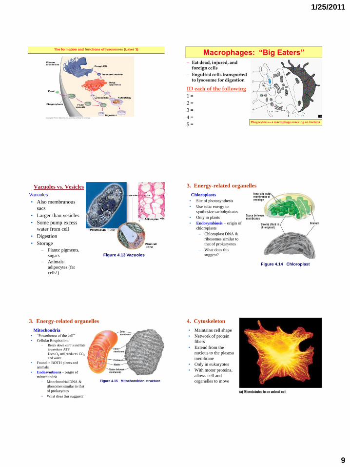
The formation and functions of lysosomes (Layer 3)
Macrophages: “Big Eaters”
– Eat dead, injured, and foreign cells
– Engulfed cells transported to lysosome for digestion
ID each of the following
1 =
2 =
3 =
4 =
5 =Phagocytosis—a macrophage snacking on bacteria
Vacuoles vs. Vesicles
Vacuoles
• Also membranous
sacs
• Larger than vesicles
• Some pump excess
water from cell
• Digestion
• Storage
– Plants: pigments,
sugars
– Animals:
adipocytes (fat
cells!)
Figure 4.13 Vacuoles
3. Energy-related organelles
Chloroplasts
• Site of photosynthesis
• Use solar energy to
synthesize carbohydrates
• Only in plants
• Endosymbiosis – origin of
chloroplasts
– Chloroplast DNA &
ribosomes similar to
that of prokaryotes
– What does this
suggest?
Figure 4.14 Chloroplast
3. Energy-related organelles
Mitochondria
• “Powerhouse of the cell”
• Cellular Respiration:
– Break down carb’s and fats
to produce ATP
– Uses O2 and produces CO2
and water
• Found in BOTH plants and
animals
• Endosymbiosis – origin of
mitochondria
– Mitochondrial DNA &
ribosomes similar to that
of prokaryotes
– What does this suggest?
Figure 4.15 Mitochondrion structure
4. Cytoskeleton
• Maintains cell shape
• Network of protein
fibers
• Extend from the
nucleus to the plasma
membrane
• Only in eukaryotes
• With motor proteins,
allows cell and
organelles to move

1/25/2011
10
4. Cytoskeleton
Microtubules
• hollow protein
cylinders
• Help maintain cell
shape
• Act as track ways to
move organelles
• Move chromosomes
in cell divisionFigure 4.16 Microtubules
•Actin filaments
2 chains of monomers twisted in a helix
Forms a dense web to support the cell
Figure 4.17 Actin filaments
Copyright © The McGraw-Hill Companies, Inc. Permission required for reproduction or display.
microvilli
nucleus
actin
filaments
• Motor proteins
Instrumental in allowing cellular movements
Myosin
• Interacts with actin
• Cells move in amoeboid fashion
• Muscle contraction
Kinesin and dynein
• Move along microtubules
• Transport vesicles from Golgi apparatus to final destination
Figure 4.18 Motor proteins
Copyright © The McGraw-Hill Companies, Inc. Permission required for reproduction or display.
ATP
vesicle
microtubule
kinesin
kinesin
receptor
vesicle moves,
not microtubule
b. Kinesin
• Centrioles
Made of 9 sets of microtubule triplets
Two centrioles lie at right angles
In animal cells, not present in plant cells

1/25/2011
11
Figure 4.19 Centrioles
Copyright © The McGraw-Hill Companies, Inc. Permission required for reproduction or display.
one pair of centrioles in a
centrosome
one microtubule
triplet
centrosome
Courtesy Kent McDonald
• Cilia and flagella
Eukaryotes
For movement of the cell or fluids past the cell
Similar construction in both
• 9+2 pattern of microtubules
Cilia shorter and more numerous than flagella
Figure 4.20 Cilia and
flagella
b.
Basal body
cross section
Flagellum
Flagellumcross section
triplets
Basal body
microtubuledoublet
centralmicrotubules
plasmamembrane
dynein side arms
TEM 350,000
TEM 101,000
cilia in bronchial wall
a.flagella of sperm
Copyright © The McGraw-Hill Companies, Inc. Permission required for reproduction or display.
a(cilia): © Manfred Kage/Peter Arnold; (sperm): © David M. Phillips/Photo Researchers, Inc.; b: (flagellum, basal body): © William L. Dentler/BPS
4.5 Outside the Eukaryotic Cell
• Plant cell walls
Primary cell walls • Cellulose fibrils and noncellulose substances
• Wall stretches when cell is growing
Secondary cell walls• Forms inside primary cell wall
• Woody plants
• Lignin adds strength
Plasmodesmata• Plant cells connected by numerous channels that pass
through cell walls
• For exchange of water and small solutes between cells
Figure 4.21 Plasmodesmata
cell wall
plasmodesmata
cell wall
Cell 1 Cell 2
plasma
membrane
cell wall
cytoplasm cytoplasm
plasmodesmata
53,000
middle lamella
Copyright © The McGraw-Hill Companies, Inc. Permission required for reproduction or display.
© E.H. Newcomb/BPS
• Exterior cell surfaces in animals
No cell wall
Extracellular matrix (ECM)
• Meshwork of fibrous proteins and polysaccharides
• Collagen and elastin well-known proteins
• Matrix varies – flexible in cartilage, hard in bone

1/25/2011
12
Figure 4.22 Animal cell extracellular matrix
receptor
protein
collagen
polysaccharides
cytoplasm
cytoskeleton
filament
elastic fiber
Copyright © The McGraw-Hill Companies, Inc. Permission required for reproduction or display.
• Junctions between cells
Adhesion junctions
• Internal cytoplasmic plaques joined by intercellular
filaments
• Sturdy but flexible sheet of cells
Figure 4.23 Junctions between cells of the intestinal wall
intercellular
space
filaments of
cytoskeleton
cytoplasmic
plaque
a. Adhesion junction
plasma
membranes
intercellular
filaments
Copyright © The McGraw-Hill Companies, Inc. Permission required for reproduction or display.
a. From Douglas E. Kelly, Journal of Cell Biology, 28 (1966:51). Reproduced by permission of The Rockefeller University Press
Tight junctions
•Plasma membrane proteins attach to each other
•Zipperlike
•Cells of tissues that serve as barriers
Figure 4.23 continuedCopyright © The McGraw-Hill Companies, Inc. Permission required for reproduction or display.
plasma
membranes
intercellular space
tight junction
proteins
b. Tight junction
Gap junctions
•Allow cells to communicate through plasma membrane channels
•Lends strength while allowing small molecules and ions to pass through
Figure 4.23 continued
plasma
membranes
membrane
channel
Intercellular
space
c. Gap junction
Copyright © The McGraw-Hill Companies, Inc. Permission required for reproduction or display.