essentials in ophthalmology vitreo-retinal surgeryessentials in ophthalmology g.k.krieglstein...
TRANSCRIPT

Essentials in Ophthalmology Vitreo-retinal Surgery
B. Kirchhof D. WongEditors

Essentials in Ophthalmology
G. K. Krieglstein R. N. WeinrebSeries Editors
Glaucoma
Cataract and Refractive Surgery
Uveitis and Immunological Disorders
Vitreo-retinal Surgery
Medical Retina
Oculoplastics and Orbit
Pediatric Ophthalmology, Neuro-Ophthalmology, Genetics
Cornea and External Eye Disease
Editors Bernd KirchhofDavid Wong
Vitreo-retinal Surgery
With 122 Figures, Mostly in Colourand 13 Tables
123
Series Editors
Günter K. Krieglstein, MDProfessor and ChairmanDepartment of OphthalmologyUniversity of CologneKerpener Straße 6250924 CologneGermany
Robert N. Weinreb, MDProfessor and DirectorHamilton Glaucoma CenterDepartment of OphthalmologyUniversity of California at San Diego9500 Gilman DriveLa Jolla, CA 92093-0946USA
Volume Editors
Bernd Kirchhof, MDProfessor and ChairmanDepartment of Vitreo-retinal SurgeryCenter for OphthalmologyUniversity of CologneKerpener Straße 6250924 CologneGermany
David Wong, MDProfessor and ChairmanDepartment of OphthalmologyFaculty of MedicineUniversity of Hong Kong21 Sassoon Road, Pok Fu LamHong Kong
ISBN-10 3-540-33669-9Springer Berlin Heidelberg NewYork
ISBN-13 978-3-540-33669-3Springer Berlin Heidelberg NewYork
ISSN 1612-3212
Library of Congress Control Number: 2006935425
This work is subject to copyright. All rights are reserved, whether the whole or part of the material is concerned, specifically the rights of translation, reprinting, reuse of illustrations, recitation, broadcasting, reproduction on microfilms or in any other way, and storage in data banks. Duplication of this publication or parts thereof is permit-ted only under the provisions of the German Copyright Law of September 9, 1965, in its current version, and per-mission for use must always be obtained from Springer-Verlag. Violations are liable for prosecution under the German Copyright Law.
Springer is a part of Springer Science + Business Media
springer.com
© Springer-Verlag Berlin Heidelberg 2007
The use of general descriptive names, registered names, trademarks, etc. in this publication does not imply, even in the absence of a specific statement, that such names are exempt from the relevant protective laws and regulations and therefore free for general use.
Product liability: The publishers cannot guarantee the ac-curacy of any information about dosage and application contained in this book. In every individual case the user must check such information by consulting the relevant literature.
Editor: Marion Philipp, Heidelberg, GermanyDesk Editor: Martina Himberger, Heidelberg, GermanyProduction: LE-TeX Jelonek, Schmidt & Vöckler GbR, Leipzig, GermanyCover Design: Erich Kirchner, Heidelberg, Germany
Printed on acid-free paper24/3100Wa 5 4 3 2 1 0
The series Essentials in Ophthalmology was initi-ated two years ago to expedite the timely trans-fer of new information in vision science and evidence-based medicine into clinical practice. We thought that this prospicient idea would be moved and guided by a resolute commitment to excellence. It is reasonable to now update our readers with what has been achieved.
The immediate goal was to transfer informa-tion through a high quality quarterly publication in which ophthalmology would be represented by eight subspecialties. In this regard, each issue has had a subspecialty theme and has been overseen by two internationally recognized volume edi-tors, who in turn have invited a bevy of experts
to discuss clinically relevant and appropriate top-ics. Summaries of clinically relevant information have been provided throughout each chapter.
Each subspecialty area now has been covered once, and the response to the first eight volumes in the series has been enthusiastically positive. With the start of the second cycle of subspecialty coverage, the dissemination of practical informa-tion will be continued as we learn more about the emerging advances in various ophthalmic subspecialties that can be applied to obtain the best possible care of our patients. Moreover, we will continue to highlight clinically relevant in-formation and maintain our commitment to ex-cellence.
G. K. Krieglstein R. N.WeinrebSeries Editors
Foreword

This second edition promises to challenge our ideas about vitreoretinal surgery. As expected, some new and innovative techniques are de-scribed, the most exciting of which is the use of free retinal pigment epithelium patch grafts in patients with atrophic macular degeneration. We are interested in the advances that have made this surgery possible and look forward to the long-term outcomes. In the last couple of years, surgeons have become increasingly convinced by the advantages conferred by smaller gauge instruments, brighter and even safer lights, com-bined with an innovative viewing system to make routine surgery quicker and less traumatic for the patients. The use of 25-gauge for tumor biopsies is an exciting new development that represents a huge advantage over needle biopsies. The indi-cation and the rationale for biopsies in cases of suspected uveal melanoma are presented.
There will always of course be difficult clinical decisions; choices have to be made despite the ab-sence of good data based on randomized trials. A good example of a dilemma is whether to perform early vitrectomy for postoperative endophthal-mitis. For this edition, we have opposing views from two surgeons. You simply have to read and decide for yourself. There are other controversies such as vitrectomy for floaters: the preparation in
terms of counseling, patient selection, and pitfalls are well covered in a chapter on the subject.
The chapter on macular holes examines the evidence behind our current practice. Despite the high success rate, a minority of patients will fail to respond to conventional surgery and a novel approach with heavy silicone oil is presented.
Sometimes, the skill of a surgeon is to know when not to operate. Asymptomatic inferior retinal detachment is another controversial area explored in a reasoned fashion, though no one knows for sure the cause of optical coherence to-mography findings of subclinical macular retinal detachments.
Then we come to the big topic of tamponad-ing. The need to apply scleral buckling, to adopt postoperative head-down posturing, or to use long-acting gas for inferior retinal breaks are be-ing challenged as more rhegmatogenous retinal detachments are treated using primary vitrec-tomy and air alone. There is renewed interest in the techniques for draining subretinal fluid and the chapter on slippage explains how unwanted retinal folds can be avoided, whilst offering an easy technique for the exchange of fluids and the injection of silicone oil.
We hope you will enjoy reading this book as much as we have preparing it.
Bernd KirchhofDavid WongEditors
Preface

Chapter 1Macular HolesJames Bainbridge, Zdenek Gregor
1.1 Epidemiology . . . . . . . . . . . . . . . 11.2 Pathogenesis . . . . . . . . . . . . . . . 11.3 Clinical Staging . . . . . . . . . . . . . 21.4 Natural History . . . . . . . . . . . . . . 31.5 Risk of Full-Thickness
Macular Hole in the Fellow Eye . . . . . . . . . . . . . . . . . . . . . . . . . 4
1.6 Symptoms and Signs . . . . . . . . 41.7 Fluorescein Angiography
and Fundus Autofluorescence 51.8 Optical Coherence
Tomography . . . . . . . . . . . . . . . . 61.9 Surgical Management of
Macular Holes . . . . . . . . . . . . . . . 61.10 Technique for Conventional
Macular Hole Surgery . . . . . . . . 71.11 Tamponade Agents
and Postoperative Posturing . . 81.12 Biological Adjuncts
to Macular Hole Surgery . . . . . 91.13 Technique for Intraoperative
Application of Biological Adjuncts . . . . . . . . . . . . . . . . . . . 10
1.14 Inner Limiting Membrane Peeling . . . . . . . . . . . . . . . . . . . . 10
1.15 Technique for Inner Limiting Membrane Peeling . . . . . . . . . 11
1.16 Vital Staining to Facilitate Inner Limiting Membrane Peeling . . . . . . . . . . . . . . . . . . . . 11
1.17 Indocyanine Green . . . . . . . . . 111.18 Trypan Blue . . . . . . . . . . . . . . . . 121.19 Technique for Inner Limiting
Membrane Staining . . . . . . . . 131.20 Complications of Macular
Hole Surgery . . . . . . . . . . . . . . . 131.21 Prognosis Following Macular
Hole Surgery . . . . . . . . . . . . . . . 13
Chapter 2Heavy Silicone Oil for PersistentMacular HolesStanislao Rizzo, Federica Genovesi-Ebert
2.1. Introduction . . . . . . . . . . . . . . . 192.1.1 Basics . . . . . . . . . . . . . . . . . . . . . 192.1.2 Therapeutic Options . . . . . . . . 192.1.2.1 Observation . . . . . . . . . . . . . . . 202.1.2.2 Laser Photocoagulation and
Outpatient FGEX . . . . . . . . . . . 202.1.2.3 Re-Operations . . . . . . . . . . . . . 202.1.3 Rationale of HSO
Endotamponading . . . . . . . . . 222.2 Treatment of Persistent
Macular Holes . . . . . . . . . . . . . . 222.2.1 Heavier Than Water Silicone
Oils . . . . . . . . . . . . . . . . . . . . . . . . 222.2.2 Surgical Technique . . . . . . . . . 242.2.3 HSO Removal . . . . . . . . . . . . . . 252.3 Conclusions . . . . . . . . . . . . . . . . 25
Chapter 3The Role of Combined Adjunctive 5-Fluorouracil and Low Molecular Weight Heparin in Proliferative Vitreoretinopathy PreventionDavid G. Charteris, D. Wong
3.1 Introduction . . . . . . . . . . . . . . . 333.2 PVR Pathobiology . . . . . . . . . . 343.2.1 PVR Development . . . . . . . . . . 343.2.2 PVR Components . . . . . . . . . . . 343.3 Adjunctive Agents . . . . . . . . . . 343.3.1 5-Fluorouracil . . . . . . . . . . . . . . 343.3.2 Low Molecular Weight
Heparin . . . . . . . . . . . . . . . . . . . . 353.3.3 Initial Clinical Studies . . . . . . . 353.4 Clinical Trials of Combined
Adjunctive 5FU and LMWH . . 353.4.1 Adjunctive Regime . . . . . . . . . 35
Contents
X Contents
3.4.2 High-Risk Retinal Detachments in Eyes Undergoing Vitrectomy and Gas Exchange (PVR 1) . . 36
3.4.3 Established PVR in Eyes Undergoing Vitrectomy and Silicone Oil Exchange (PVR 2) . . . . . . . . . . . . . . . . . . . . 36
3.4.4 Unselected Primary Retinal Detachments in Eyes Undergoing Vitrectomy and Gas Exchange (PVR 3) . . . 36
3.4.5 Macular Translocation with 360° Retinotomy . . . . . . . . . . . 37
3.5 Implications for Clinical Use . . 373.5.1 Uncomplicated Primary
Retinal Detachments . . . . . . . 373.5.2 Established PVR . . . . . . . . . . . . 373.5.3 High-Risk Retinal
Detachments . . . . . . . . . . . . . . 373.5.4 Intraocular Trauma Patients
Undergoing Vitrectomy Surgery . . . . . . . . . . . . . . . . . . . 37
Chapter 4Slippage of the Retina: What Causes It and How Can It Be Prevented?David Wong
4.1 Introduction . . . . . . . . . . . . . . . 414.2 What is Slippage? . . . . . . . . . . 414.2.1 Surface Tension
and Interfacial Tension . . . . . . 414.2.2 Shape of Endotamponade
Bubble . . . . . . . . . . . . . . . . . . . . 424.2.3 Surface Property
of the Retina . . . . . . . . . . . . . . . 434.2.4 Fluid–Air Exchange . . . . . . . . . 434.3 Setting the Scene for
Slippage . . . . . . . . . . . . . . . . . . . 444.3.1 Perfluorocarbon Liquids
and the “Doughnut” of Fluid 444.3.2 Seeing and Doing . . . . . . . . . . 464.3.3 Air Versus Silicone Oil
Exchange for PFCL . . . . . . . . . . 464.3.4 Oil on Water? No, Water
on Oil! . . . . . . . . . . . . . . . . . . . . . 474.4 Overfilling and Complete
Elimination of Aqueous . . . . . 484.5 Injection of Silicone Oil . . . . . 48
Chapter 5Complete and Early Vitrectomy for Endophthalmitis (CEVE) as Today’s Alternative to the Endophthalmitis Vitrectomy StudyFerenc Kuhn, Giampaolo Gini
5.1 Introduction and Definitions . 545.1.1 Introduction . . . . . . . . . . . . . . . 545.1.2 Definitions . . . . . . . . . . . . . . . . . 545.2 Etiology and Classification . . 565.3 Pathophysiology,
Organisms, and Diagnostics in Brief . . . . . . . . . . . . . . . . . . . . 56
5.3.1 Pathophysiology . . . . . . . . . . . 565.3.2 Organisms . . . . . . . . . . . . . . . . . 565.3.3 Diagnostics . . . . . . . . . . . . . . . . 565.3.3.1 Clinical . . . . . . . . . . . . . . . . . . . . 565.3.3.2 Other . . . . . . . . . . . . . . . . . . . . . . 575.4 Principles of Therapy . . . . . . . 575.5 The EVS . . . . . . . . . . . . . . . . . . . . 575.5.1 Study Design . . . . . . . . . . . . . . . 575.5.2 Results . . . . . . . . . . . . . . . . . . . . 585.5.3 Consequences of the EVS
Recommendations . . . . . . . . . 585.6 Rationale for Performing
Complete and Early Vitrectomy for Endophthalmitis . . . . . . . . . . . 58
5.6.1 Why Perform Vitrectomy? . . . 585.6.2 Why Perform Early
Vitrectomy? . . . . . . . . . . . . . . . . 585.6.3 Why Perform Complete
Vitrectomy? . . . . . . . . . . . . . . . . 595.7 Complete and Early
Vitrectomy for Endophthalmitis: Surgical Steps . . . . . . . . . . . . . . . . . . . . . . 59
5.7.1 Initial Steps . . . . . . . . . . . . . . . . 595.7.2 Cornea . . . . . . . . . . . . . . . . . . . . 605.7.3 Anterior Chamber . . . . . . . . . . 605.7.4 Pupil . . . . . . . . . . . . . . . . . . . . . . 615.7.5 (Intraocular) Lens . . . . . . . . . . . 615.7.6 Posterior Lens Capsule . . . . . . 615.7.7 Vitrectomy . . . . . . . . . . . . . . . . . 615.7.8 When to Stop Vitreous
Removal? . . . . . . . . . . . . . . . . . . 635.7.9 Retina . . . . . . . . . . . . . . . . . . . . . 635.7.10 Enucleation/Evisceration . . . . 645.7.11 Pharmacological Treatment . . 64
Contents XI
5.8 Surgical Decision-Making and Complications . . . . . . . . . 64
5.8.1 Decision-Making . . . . . . . . . . . 645.8.2 Complications and Their
Management . . . . . . . . . . . . . . 645.8.2.1 Retinal Break . . . . . . . . . . . . . . . 645.8.2.2 Retinal Detachment . . . . . . . . 655.8.2.3 Silicone Oil as a Long-Term
Tamponade . . . . . . . . . . . . . . . . 655.9 Results with CEVE and Their
Comparison with the EVS . . . 665.10 Summary
and Recommendations . . . . . 67
Chapter 6Treatment of Acute Bacterial Endophthalmitis After Cataract Surgery Without VitrectomyThomas Theelen, Maurits A.D. Tilanus
6.1 Introduction . . . . . . . . . . . . . . . 706.1.1 Basics . . . . . . . . . . . . . . . . . . . . . 706.1.2 Pathophysiology . . . . . . . . . . . 706.1.2.1 Phases of Infection . . . . . . . . . 706.1.3 Clinical Diagnosis . . . . . . . . . . . 716.1.3.1 Role of Ultrasonography . . . . 726.1.4 Microbial Spectrum . . . . . . . . . 726.2 Therapeutical Approaches . . 756.2.1 Basics . . . . . . . . . . . . . . . . . . . . . 756.2.2 Early Pars Plana Vitrectomy . . 756.2.3 Nonvitrectomizing
Endophthalmitis Treatment . . 766.3 Emergency Management
of Endophthalmitis After Cataract Surgery . . . . . . . . . . . 76
6.3.1 Surgical Technique . . . . . . . . . 766.3.2 Equipment for the
Emergency Management of Endophthalmitis . . . . . . . . . 77
6.3.3 Treatment Protocol . . . . . . . . . 776.3.4 Treatment Outcome
in Endophthalmitis Without Immediate Vitrectomy . . . . . . 77
6.3.4.1 Introduction . . . . . . . . . . . . . . . 776.3.4.2 Results in Our Department . . 796.3.4.3 Discussion . . . . . . . . . . . . . . . . . 806.4 Conclusion . . . . . . . . . . . . . . . . . 81
Chapter 7New Instruments in VitrectomyMasahito Ohji, Yasuo Tano
7.1 Introduction . . . . . . . . . . . . . . . 857.2 Sutureless Transconjunctival
Vitrectomy . . . . . . . . . . . . . . . . . 857.2.1 25-Gauge Vitrectomy
System . . . . . . . . . . . . . . . . . . . . 857.2.2 Instruments . . . . . . . . . . . . . . . . 867.2.2.1 Trocars . . . . . . . . . . . . . . . . . . . . 867.2.2.2 Vitreous Cutter . . . . . . . . . . . . . 867.2.2.3 Light Pipe . . . . . . . . . . . . . . . . . . 877.2.2.4 Other 25-Gauge
Instruments . . . . . . . . . . . . . . . . 877.2.2.5 Other Instruments . . . . . . . . . . 877.2.3 Surgical Procedures
of the 25-Gauge System . . . . 897.2.3.1 Insert Three Trocars . . . . . . . . . 897.2.3.2 Vitrectomy . . . . . . . . . . . . . . . . . 897.2.3.3 Closure of the Wound . . . . . . . 897.2.4 Advantage and
Disadvantage of 25-Gauge System . . . . . . . . . . . . . . . . . . . . 89
7.3 23-Gauge Vitrectomy System . . . . . . . . . . . . . . . . . . . . 90
7.4 Xenon Endo-Illumination for Vitrectomy . . . . . . . . . . . . . . 90
7.4.1 Yellow-Filtered Xenon . . . . . . 927.4.2 Wide-Angle Viewing System 927.4.3 Adjuncts . . . . . . . . . . . . . . . . . . . 957.4.3.1 Staining of Internal
Limiting Membrane with Indocyanine Green . . . . 95
7.4.3.2 Other Dyes Staining the ILM . 95
Chapter 825-Gauge Biopsy of Uveal TumorsBertil Damato, Carl Groenewald
8.1 Introduction . . . . . . . . . . . . . . . 998.1.1 Uveal Tumors . . . . . . . . . . . . . . 998.2 Diagnosis of Choroidal
Tumors . . . . . . . . . . . . . . . . . . . . 998.2.1 Ophthalmoscopy . . . . . . . . . . . 998.2.2 Angiography . . . . . . . . . . . . . . 1018.2.3 Echography . . . . . . . . . . . . . . . 1018.2.4 Other Investigations . . . . . . . 1018.3 Current Biopsy Techniques . . 102
XII Contents
8.3.1 Fine Needle AspirationBiopsy . . . . . . . . . . . . . . . . . . . . 102
8.3.2 Trans-Scleral Tumor Biopsy . 1028.3.3 20-Gauge Vitreous Cutter . . 1028.3.4 Trans-Vitreal Incisional
Biopsy . . . . . . . . . . . . . . . . . . . . 1028.3.5 25-Gauge Biopsy
Technique . . . . . . . . . . . . . . . . 1048.4 Preoperative Management . 1048.5 Surgical Technique . . . . . . . . 1048.6 Care of Specimen . . . . . . . . . . 1048.7 Postoperative
Management . . . . . . . . . . . . 1048.8 Results and Complications . 1058.8.1 Adequacy of Sample . . . . . . . 1058.8.2 Sampling Error . . . . . . . . . . . . 1058.8.3 Tumor Seeding . . . . . . . . . . . . 1058.8.4 Hemorrhage . . . . . . . . . . . . . . 1058.8.5 Rhegmatogenous Retinal
Detachment . . . . . . . . . . . . . . 1098.8.6 Other Complications . . . . . . . 1098.9 Indications
and Contraindications . . . . . 1098.9.1 Indications . . . . . . . . . . . . . . . . 1098.9.1.1 Diagnosis of Metastasis . . . . 1098.9.1.2 Differentiation Between
Metastasis and Melanoma . . 1098.9.1.3 Differentiation Between
Nevus and Melanoma . . . . . 1098.9.1.4 Confirmation of Local
Recurrence of Melanomaafter ConservativeTreatment . . . . . . . . . . . . . . . . 110
8.9.1.5 Confirmation of IntraocularLymphoma . . . . . . . . . . . . . . . 110
8.9.1.6 Prognosis . . . . . . . . . . . . . . . . . 1108.9.2 Contraindications . . . . . . . . . 1138.10 Conclusions . . . . . . . . . . . . . . . 113
Chapter 9Vitrectomy Against FloatersHans Hoerauf
9.1 Introduction . . . . . . . . . . . . . . 1159.2 Clinical Findings . . . . . . . . . . 1169.3 Symptoms and Natural
Course . . . . . . . . . . . . . . . . . . . 116
9.4 Diagnostic Methods . . . . . . . 1179.5 Surgical Treatment
for Vitreous Floaters:Important Studies . . . . . . . . . 117
9.6 Vitrectomy for VitreousFloaters Despite Full VisualAcuity . . . . . . . . . . . . . . . . . . . . 117
9.6.1 Patients . . . . . . . . . . . . . . . . . . 1189.6.2 Surgical Technique . . . . . . . . 1189.6.3 Results . . . . . . . . . . . . . . . . . . . 1199.6.4 Case Report . . . . . . . . . . . . . . 1199.7 Analysis of Clinical Studies . . 1199.8 Alternative Therapeutic
Options . . . . . . . . . . . . . . . . . . . 1209.9 Personality Traits . . . . . . . . . . 1219.10 Alternative Assessment
of Visual Function . . . . . . . . . 1219.11 Conclusions . . . . . . . . . . . . . . . 123
Chapter 10Treatment of Retinal Detachment from Inferior Breaks with Pars Plana VitrectomyVicente Martinez-Castillo, Jose Garcia-Arumi, Anna Boixadera, Miguel A. Zapata
10.1 Introduction . . . . . . . . . . . . . . 12510.1.1 Basics . . . . . . . . . . . . . . . . . . . . 12510.1.2 Importance of Inferior
Breaks When Pars PlanaVitrectomy is Performed . . . 126
10.1.3 Developmentof a Chorioretinal Scar . . . . . 126
10.1.4 Type of Tamponade Agentfor Inferior Breaks . . . . . . . . . 127
10.2 Management of InferiorBreaks with PPV: RecentPublications . . . . . . . . . . . . . . 127
10.3 PPV Alone with AirTamponade: SurgicalTechnique . . . . . . . . . . . . . . . . 129
10.3.1 Inclusion Criteria . . . . . . . . . . 12910.3.2 Vitrectomy . . . . . . . . . . . . . . . . 12910.3.3 Drainage of Subretinal
Fluid . . . . . . . . . . . . . . . . . . . . . 12910.3.4 Retinopexy . . . . . . . . . . . . . . . 13010.3.5 Tamponade Agent . . . . . . . . . 130

Contents XIII
Chapter 11Subclinical Retinal DetachmentJose Garcia-Arumi, Anna Boixadera, Vicente Martinez-Castillo, Miguel A. Zapata
11.1 Definition . . . . . . . . . . . . . . . . . 13311.2 Natural History . . . . . . . . . . . . 13511.2.1 Role of the Vitreous . . . . . . . . 13511.3 Therapeutic Options . . . . . . . 13611.3.1 Observation . . . . . . . . . . . . . . 13611.3.2 Laser Demarcation . . . . . . . . 13711.3.3 Surgery . . . . . . . . . . . . . . . . . . 13711.3.3.1 Pneumatic Retinopexy . . . . . 13711.3.3.2 Scleral Buckling . . . . . . . . . . . 13811.3.3.3 Pars Plana Vitrectomy . . . . . . 13811.3.4 Pros and Cons of Treating . . 13811.3.4.1 Pros of Treating . . . . . . . . . . . 13811.3.4.2 Cons of Treating . . . . . . . . . . . 13811.4 Current Clinical Practice/
Recommendations . . . . . . . . 13911.5 Subclinical Retinal
Detachment Diagnosedwith Optical CoherenceTomography AfterSuccessful Surgeryfor Rhegmatogenous RetinalDetachment . . . . . . . . . . . . . . 139
Chapter 12Autologous Translocation of the Choroid and RPE in Patients with Geographic AtrophyAntonia M. Joussen, Jan van Meurs, Bernd Kirchhof
12.1 Introduction: Pathologyand Epidemiology . . . . . . . . . 143
12.2 Treatment Approachesin Patients with GeographicAtrophy . . . . . . . . . . . . . . . . . . 144
12.3 Concept of Translocationof a Free Choroidal:RPE Graft (Free GraftTranslocation) . . . . . . . . . . . . . 144
12.3.1 Patients and Methods . . . . . . 14412.3.2 Free Graft Translocation
Surgery in Dry AMD . . . . . . . 14512.3.3 Results . . . . . . . . . . . . . . . . . . . 14512.3.3.1 Free Graft Translocation
Surgery and ComplicationProfile . . . . . . . . . . . . . . . . . . . . 145
12.3.3.2 Anatomical Outcome:Funduscopic Appearance,Vascularization,and Autofluorescenceof the Graft . . . . . . . . . . . . . . . 146
12.3.3.3 Functional Outcome:Distant Visual Acuity,Reading Ability, Fixation,and Microperimetry Duringthe Postoperative Course . . 147
12.4 Comment . . . . . . . . . . . . . . . . 148

James BainbridgeMoorfields Eye HospitalCity RoadLondon EC1V 2PDUK
Anna BoixaderaHospital Vall d´ HebronServicio de OftalmologiaPaseo Vall d´ Hebron, 119 – 121BarcelonaSpain
David G. CharterisMoorfields Eye HospitalCity RoadLondon EC1V 2PDUK
Bertil DamatoRoyal Liverpool University HospitalPrescot StreetLiverpool L7 8XPUK
Jose Garcia-ArumiInstituto de Microcirugía Ocular C/ Munner nº 1008022 BarcelonaSpain
Giampaolo GiniCity HospitalViale Vittorio Veneto 60Prato 59100Italy
Zdenek GregorMoorfields Eye HospitalCity RoadLondon EC1V 2PDUK
Carl GroenewaldRoyal Liverpool University HospitalPrescot StreetLiverpool L7 8XPUK
Hans HoeraufUniversity Eye Clinic, UK S-HCampus LübeckLübeckGermany
Antonia M. JoussenDepartment of OphthalmologyUniversity of DuesseldorfMoorenstraße 540225 DuesseldorfGermany
Bernd KirchhofDepartment of Vitreo-retinal SurgeryCenter for OphthalmologyUniversity of CologneKerpener Straße 6250924 CologneGermany
Ferenc KuhnUniversity of Alabama at Birmingham1201 11th Avenue South, Suite 300BirminghamAL 35205USA
Vicente Martínez-CastilloHospital Vall d´ HebronServicio de OftalmologiaPaseo Vall d´ Hebron, 119 – 121BarcelonaSpain
Contributors

XVI Contributors
Masahito OhjiShiga University of Medical ScienceOsaka University Medical School, E-7Department of Ophthalmology2-2 Yamidaoka SuitaOsaka 565-0871Japan
Stanislao RizzoChirurgica Oftalmica Ospedale S. ChiaraVia Roma 5556100 PisaItaly
Yasuo TanoShiga University of Medical ScienceOsaka University Medical School, E-7Department of Ophthalmology2-2 Yamidaoka SuitaOsaka 565-0871Japan
Thomas TheelenAcademish Ziekenhuis NijmegenPhilips van Leydenlaan 156525 EX NijmegenThe Netherlands
Maurits A.D. TilanusAcademish Ziekenhuis NijmegenPhilips van Leydenlaan 156525 EX NijmegenThe Netherlands
Jan van MeursRotterdam Eye HospitalPostbus 700303000 LM RotterdamThe Netherlands
David WongDepartment of OphthalmologyFaculty of MedicineUniversity of Hong Kong21 Sassoon Road, Pok Fu LamHong Kong
Miguel A. ZapataC/ Sta Teresa 70 2º 1ª08172 Sant CugatSpain
Core Messages
■ Idiopathic full-thickness macular holesare believed to result from tangential andantero-posterior traction exerted by theposterior vitreous cortex on the neuro-sensory retina at the fovea.
■ Only a minority of full-thickness macu-lar holes resolve spontaneously and thereis a significant risk of macular hole de-velopment in the fellow eye.
■ Surgical management by removal of theposterior cortical vitreous and long-act-ing intraocular gas tamponade results ina high rate of anatomical closure and abenefit to visual function.
■ Modifications to this surgical technique,proposed with the aim of improving ana-tomical and visual outcomes, include theuse of adjuvants to stimulate glial repairand the dissection of the inner limitingmembrane, with or without the aid ofvital stains. The value of these modifica-tions is not well-established.
■ The importance of prolonged face-downposturing to the rate of macular hole clo-sure is unproven.
1.1 EpidemiologyA macular hole is a full-thickness defect of theneurosensory retina involving the anatomical fo-vea. Idiopathic full-thickness macular holes arean important cause of central visual loss; these le-sions are twice as common in females as in males[1, 2] and three-quarters of affected individualsare over the age of 65 years [1]. The populationprevalence of macular holes is estimated as being0.1% in individuals aged 40 years or older and
0.8% in those aged over 74 years. No significantsystemic risk factor for the development of mac-ular holes has been identified although a pos-sible association with the menopause has beendescribed [1].
1.2 PathogenesisThe pathogenesis of idiopathic full-thicknessmacular holes is not well understood. Macularholes are commonly idiopathic in etiology, al-though trauma and high myopia are implicatedin a minority of cases. On the basis of clinicalobservations, ocular imaging, histological stud-ies, and the results of vitrectomy surgery, themechanism underlying idiopathic macular holeis widely believed to involve tangential as well asantero-posterior traction exerted by the posteriorvitreous cortex on the neurosensory retina at thefovea. Evidence of the role of posterior vitreousattachment in the development of macular holesincludes its association with an increased risk ofmacular hole development in the fellow eyes ofindividuals with unilateral macular holes andwith the subsequent enlargement of establishedholes [3, 4]. There is no consensus on the exactmechanism of vitreo-foveal traction. Tangentialtraction may be the result of contraction of theprefoveal vitreous cortex following invasion andproliferation of Müller cells [5]. Anteroposteriortraction may occur from dynamic tractionalforces on an abnormally persistent vitreo-fovealattachment following perifoveal vitreous separa-tion [6, 7]. The role of antero-posterior vitreo-foveal traction is supported by optical coherencetomography (OCT) studies that clearly identifyperifoveal posterior hyaloid separation with per-sistent adherence of the posterior hyaloid to thecentre of the fovea [6–8]. A cone-shaped zoneof Müller cells, the ‘Müller cell cone’ forms the
Macular HolesJames Bainbridge, Zdenek Gregor
Chapter 1
1
2 Macular Holes
1central and inner part of the fovea centralis andappears to confer structural support, serving as aplug to bind together the foveolar photoreceptorcells [9]. Vitreo-foveal traction may result in dis-insertion of the Müller cell cone from underlyingfoveolar photoreceptor cells and in the formationof a foveal schisis or “cyst” [6–8]. A dehiscencedevelops in the roof of the foveal cyst that mayextend by centric expansion, or more commonlyin a pericentric fashion, to form a crescentic holethat progresses to a horseshoe tear [6]. Completeavulsion of the cyst roof results in a fully de-tached operculum that is suspended on the pos-terior vitreous cortex in the prefoveal plane [6, 7,10]. Opercula primarily comprise vitreous cortexand glial elements with a variable amount of fo-veal tissue that includes photoreceptor cells in40% of cases [11]. The photoreceptor layer, whichis no longer anchored by the Muller cell cone atthe foveola, undergoes passive centrifugal retrac-tion to form a full-thickness retinal dehiscencewith centrifugal displacement of xanthophyll [9,12]. The edge of the hole becomes progressivelyelevated and a cuff of subretinal fluid develops.In the event of vitreo-foveal separation duringthe development of the macular hole, the reliefof traction may result in regression of a cyst, butspontaneous closure of an established full-thick-ness macular hole is relatively uncommon [13].
While the majority of age-related macularholes are idiopathic in etiology, full-thickness
macular holes may also occur in association withhigh myopia, following posterior segment sur-gery such as scleral buckling and pneumatic reti-nopexy, and following ocular trauma. Traumaticmacular holes typically result from blunt injury,but have also been reported following laser in-jury and lightning strike.
Summary for the Clinician
■ Idiopathic full-thickness macular holesare caused by traction exerted by theposterior vitreous cortex on the neuro-sensory retina at the fovea. Disinsertionof its glial plug results in the formation ofa foveal “cyst.” Avulsion of the cyst roofresults in a free operculum suspended onthe posterior vitreous cortex. The neuro-sensory retina retracts centrifugally toform a full-thickness retinal dehiscence.
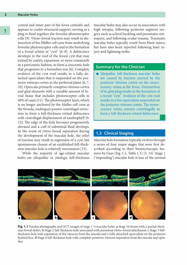
1.3 Clinical StagingMacular hole formation typically evolves througha series of four major stages that were first de-scribed according to their biomicroscopic fea-tures by Gass (Fig. 1.1, Table 1.1) [5, 14]. Stage 1(‘impending’) macular hole is loss of the normal
Fig. 1.1 Fundus photographs and OCT images of stage 1–4 macular holes. a Stage 1b lesion with a partial-thick-ness foveal defect. b Stage 2 full-thickness hole associated with persistent vitreo-foveal attachment. c Stage 3 full-thickness hole with separation of the vitreous from the macula and a fully detached operculum on the posteriorhyaloid face. d Stage 4 full-thickness hole with complete posterior vitreous separation from the macula and opticdisc

foveal depression, associated with the develop-ment of a yellow spot (stage 1a) or ring (stage1b) in the centre of the fovea, changes that re-flect the intraretinal schisis that progresses intoan intraretinal cyst. A foveal dehiscence maybe masked on biomicroscopy (stage 1-b, occulthole) by semi-opaque contracted prefoveolar vit-reous cortex bridging the yellow ring. Stage 2 isa small full-thickness hole (≤200 µm), typicallywith a pericentric configuration, associated withpersistent vitreo-foveal attachment. Stage 3 is alarger full-thickness hole (250–400 µm) with arim of elevated retina and separation of the pos-terior hyaloid from the macula. A fully detachedoperculum on the posterior hyaloid may be evi-dent on biomicroscopy. Stage 4 is a full-thicknesshole (≥450 µm) with complete posterior vitreousseparation from the optic disc, typically demon-strated by the presence of a Weiss ring.
1.4 Natural HistoryThe natural history of macular holes is typicallyto progress in size and clinical stage, with dete-rioration in visual acuity that generally stabilizesat the 6/60 to 3/60 level, redistribution of yellownodular opacities at the level of the retinal pig-ment epithelium, and the development of reti-nal pigment epithelial atrophy surrounding themacular hole, resulting in a ‘bull’s-eye’ macularappearance.
An estimated 40% of stage 1 (impending)macular holes progress to full-thickness holes[15], but up to 50% resolve spontaneously [15,16]. Stage 2 macular holes typically enlarge, withprogression to stage 3 in at least 75% of cases[10, 16, 17] and spontaneous closure has beenreported in no more than 15–21% [10, 13, 16].Stage 3 holes progress to stage 4 in approximately30% of cases over 3 years [16]. Full-thicknessholes tend to enlarge modestly; by 25% duringthe first 12 months and by 29% at 24 months,
Table 1.1 Clinical features and natural history of idiopathic macular holes
Stage 1 Stage 2 Stage 3 Stage 4
Symptoms Asymptomatic,or mild meta-morphopsia
Metamorphop-sia and loss ofcentral vision
Metamorphop-sia and loss ofcentral vision
Metamorphop-sia and loss ofcentral vision
Visual acuity 20/20–20/60 20/40–20/100 20/60–20/200 20/60–20/400
Biomicroscopy Loss of fovealdepression
Yellow spot (1a) oryellow ring (1b)
Posterior vitreousattached to foveaand optic disc
Full-thicknessretinal defect
Typically ≤200 µmin diameter
Posterior vitreousattached to foveaand optic disc
Full-thicknessretinal defect
Typically 250–400 µm in diameter
Operculum maybe evident
Posterior vitreoustypically detachedfrom fovea, but at-tached to optic disc
Full-thicknessretinal defect
Typically ≥450 µmin diameter
Operculum maybe evident
Posterior vitreousdetached from bothfovea and optic disc
Natural history 50% regress40% progress
15–21% regress75% progress
5% regress30% progress 20% enlarge
10% stabilize
1.4 Natural History 3