escort aptamers: new tools for the targeted delivery of
TRANSCRIPT
12 | ActA nAturAe | VOL. 3 № 4 (11) 2011
reVIeWS
Escort Aptamers: New Tools for the Targeted Delivery of Therapeutics into Cells
A. S. Davydova, M. A. Vorobjeva*, A. G. VenyaminovaInstitute of Chemical Biology and Fundamental Medicine, Siberian Branch, Russian Academy of Sciences*E-mail: [email protected] 08.07.2011Copyright © 2011 Park-media, Ltd. This is an open access article distributed under the Creative Commons Attribution License,which permits unrestricted use, distribution, and reproduction in any medium, provided the original work is properly cited.
ABSTRACT Escort aptamers are DNA or RNA sequences with high affinity to certain cell-surface proteins, which can be used for targeted delivery of various agents into cells of a definite type. The peculiarities of the selection of escort aptamers are discussed in this review. The methods used in selection of escort aptamers via the SELEX technique are considered, including selection against isolated cell-surface proteins, cell fragments, living eukary-otic cells, and bacteria. Particular attention is given to the design and chemical modification of escort aptamers. The different fields of application of escort aptamers are described, including the targeted delivery of siRNAs, nanoparticles, toxins, and photoagents, as well as the identification of specific cell markers and the detection or isolation of cells of a definite type. The potential for the application of escort aptamers in the development of new therapeutic agents and diagnostic systems is also discussed.KEYWORDS SELEX method; NA aptamers; escort aptamers; specific cell binding; addressed cell delivery; detec-tion of cells.ABBREVIATIONS dsDNA – double-stranded DNA; ssDNA – single-stranded DNA; siRNA – small interfering RNA; PSMA – prostate-specific membrane antigen; IC50 – therapeutical concentration required to suppress the growth of 50% cells; Kd – apparent constant of dissociation of the aptamer–target complex; LNA – locked nucleic acids; SELEX – Systematic Evolution of Ligands by EXponential Enrichment.
INTRODUCTIONAptamers (Latin aptus – suitable) are single-stranded DnA and rnA molecules that are capable of specific recognition of definite types of compounds, thanks to their unique spatial structure. In the 1990s, methods for in vitro selection, enabling one to obtain nucleic ac-ids with predetermined properties, were described by three independent research groups. A. ellington and J. Szostak [1] obtained an rnA molecule that was ca-pable of specifically binding to an organic dye. c. tuerk and L. Gold described the selection of rnA molecules that were capable of binding to phage 4 DnA polymer-ase and called the developed method SeLeX (System-atic evolution of Ligands by exponential enrichment) [2]. D. robertson and G. Joyce used in vitro selection to convert a group I ribozyme from a ribonuclease into a deoxyribonuclease [3]. throughout the subsequent two decades, this field has developed rapidly; meth-ods for the selection of aptamers and approaches to their design have been further refined. A large number of aptamers capable of binding to various targets with high specificity have already been obtained (see re-
views [4–7]). Aptamers find broad application across a wide range of research fields, thanks to their unique properties (namely their high affinity and selectivity in binding to a target molecule). In particular, aptam-ers can be used to obtain highly efficient and specific inhibitors of target proteins that can be applied in the design of new drugs. A number of aptamers are cur-rently in different stages of clinical trials [8]. Macugen (Eyetech Pharmaceuticals and Pfizer), which is based on aptamer binding a human vascular endothelial growth factor (VeGF), has been certified as an efficient drug for the treatment of age-related macular degeneration [9, 10].
One of the most interesting and promising aspects in the field is designing aptamers that are capable of specific recognition of cells of a definite type through binding with certain dominants on their surface. In the review by B. Hicke et al. [11], these compounds were referred to as escort aptamers. the use of escort aptam-ers as an addressing fragment opens wide possibilities for the targeted delivery of agents of different nature to cells of definite types. today, a large number of

reVIeWS
VOL. 3 № 4 (11) 2011 | ActA nAturAe | 13
escort aptamers directed toward various target cells have been obtained, and a wide range of applications for these aptamers for specific action on cells, diagnos-tics, and cell isolation have been described. the present review is devoted to the selection, design, and different aspects in the use of escort aptamers.
OBTAINMENT OF APTAMERS BY in vitro SELECTION
The general principle of the SELEX methodDnA and rnA aptamers are obtained via in vitro se-lection from combinatorial libraries of nucleic acid molecules. A conventional library is a set of oligonu-cleotides with the randomized region consisting of 20–60 nucleotides flanked with the constant regions that are required for binding to primers and the Pcr amplification of DnA. currently, libraries containing both ssDnA and rnA molecules are widely used for the selection of aptamers. rnA aptamers are capable of forming a greater variety of spatial structures as com-pared with DnA aptamers, as a result of the presence of 2'-OH groups. However, rnA aptamers are more sensitive to the action of cell nucleases and require the introduction of additional protective groups [12].
the ssDnA libraries are obtained via the conven-tional methods for the chemical synthesis of oligodeox-yribonucleotides using a mixture of all four monomers
when synthesizing a randomized fragment. In order to obtain an rnA library, the chemical synthesis of an ssDnA library containing the promoter sequence for t7 rnA polymerase at its 5'-terminal region is first performed. the ssDnA matrix is then used to obtain a dsDnA, which is subsequently applied for the synthesis of rnA via in vitro transcription. the general scheme of the SeLeX method for DnA libraries comprises the following stages: incubation of a library with a tar-get, separation of oligonucleotide–target complexes from the unbound oligonucleotides, disruption of the oligonucleotide–target complexes, and amplification of the bound molecules (Fig. 1A). For the selection of rnA aptamers, SeLeX also comprises the following additional stages: production of the rnA library on the DnA matrix, reverse transcription of the bound rnA molecules to produce DnA, and DnA amplification (Fig. 1B).
During the selection process, the library is enriched in sequences possessing increased affinity to the tar-get. Five to fifteen rounds of selection are typically per-formed to obtain aptamers, depending on the values of the dissociation constant of the aptamer–target com-plex. After the dissociation constant ceases to decrease (i.e., the affinity of the library to the target stops rising), the enriched library is cloned, and the sequences of the individual aptamers are determined. the homology be-
А DNA libraryb
Target
Removal of the unbound DNA moleculesDisruption of
the target–DNA complex
PCR
DNA libraryTranscription
RNA library
Target
Removal of the unbound RNA molecules
Disruption of the target–RNA complex
RT-PCR
Transcription
Fig. 1. The general scheme of the SELEX method using DNA (A) and RNA (B) libraries.
14 | ActA nAturAe | VOL. 3 № 4 (11) 2011
reVIeWS
tween the individual aptamers is then analyzed. On the basis of the results obtained, the aptamers are classified into several groups; their capability to interact with the target is assessed. the sequences with the maximum affinity to the target are selected for further studies. the secondary structure of aptamers is studied by an-alyzing their conserved motifs, by computer simula-tion, and chemical and enzymatic probing. the minimal size of an aptamer required for specific recognition of a target is determined at the next stage. For this purpose, a series of truncated variants of the aptamer is synthe-sized and the ability of these aptamers to bind to the target is determined.
Aptamers are usually characterized by high affinity to their targets. the characteristic values of the dissoci-ation constant (K
d) for protein targets lie in the nanomo-
lar and subnanomolar ranges (1 × 10⁻¹⁰–1 × 10⁻⁷ М). In terms of their affinity and specificity, aptamers are similar to monoclonal antibodies; however, aptamers have a number of distinct characterisitcs (Table 1). Among these characteristics, the possibilities to pro-duce an aptamer via chemical synthesis and to modify them chemically are the ones most worthy of note.
Chemical modifications of aptamersthe introduction of different modifications into escort aptamers enables one to considerably increase their sta-bility in biological media, as well as their functionality. the introduction of different substituents at the 2' po-sition of ribose (Fig. 2) (the most common modification used to increase an aptamer’s stability) helps to prevent the cleavage of aptamers by endonucleases. this type of modification is typically used to protect escort rnA
aptamers, whereas escort DnA aptamers are more frequently used without any additional modifications. the major pathway of degradation of rnA aptamers in biological media is cleavage by pyrimidine endoribonu-cleases; therefore, as early as at the stage of construc-tion of combinatorial rnA libraries for selecting escort aptamers, the pyrimidine nucleosides within them are substituted for their 2'-fluoro- and 2'-amino analogues by using the corresponding modified nucleoside triphos-phates for synthesizing a library. It is possible to use t7 rnA polymerase [13] or its mutant version (capable of inserting these 2'-modified nucleoside triphosphates into rnA with higher efficiency) to integrate them into a growing rnA strand [14]. there is also mutant rnA polymerase inserting 2'-O-methyl analogues of nucleo-side triphosphates [15, 16]; however, due to the problems associated with the reaction of reverse transcription of 2'-O-methyl-containing rnAs, the use of 2'-O-methyl-rnA libraries directly during the selection process has not yet become common practice [4].
In order to obtain 2'-O-methyl-containing aptamers, a quantity of ribonucleotides within a rnA aptamer are substituted for their 2'-O-methyl analogues, after the aptamers have been selected and their nucleotide sequences determined. the introduction of 2'-O,4'-c-methylene-linked bicyclic nucleotides (LnA – locked nucleic acids) is another way of increasing the stabil-ity of aptamers of a known sequence. this modifica-tion is used both in rnA [17] and DnA aptamers [18]. A number of nucleotides can be substituted for a flex-ible linker based on ethylene glycol for the purpose of minimizing the aptamer’s length and simultaneously increasing its resistance to endonucleases [18, 19]. cap-
Table 1. Comparison of the properties of NA aptamers and monoclonal antibodies
Aptamers Monoclonal antibodies
Selection method In vitro selection Hybridoma technology, including immunization of animals
Synthesis method chemical or enzymatic synthesis Produced using cell cultures or laboratory animals
Limitations imposed on the target molecules no limitations Antibodies against non-immunogenic
or toxic substances cannot be obtained
Affinity to the target Kd ≈ 10–10–10–7 M K
d ≈ 10–10–10–7 M
Specificity High High
Stability can re-naturate after heat treatment; long-term storage is possible
Irreversible denaturation after heat treatment; higher sensitivity to storing conditions
Immunogenicity not shown High
Possibility of chemical modification Wide Limited

reVIeWS
VOL. 3 № 4 (11) 2011 | ActA nAturAe | 15
ping of the 3'-terminus with an additional thymidine residue linked via the 3'-3'-phosphodiester bond is used to prevent the cleavage of aptamers [17, 19].
researchers either use the modified nucleoside tri-phosphates or modify the “ready-made” aptamers in order to introduce additional functional groups into an aptamer during the selection (see review [4]). In the case of escort aptamers, the introduction of an aliphatic amino or sulfhydryl group to the 5'- or 3'-terminus of an aptamer is the most common. It allows one to syn-thesize the various conjugates of aptamers with toxins, antibiotics, fluorescent or photoreactive groups, nano-particles, etc. (see Section Application of escort aptam-ers).
It must be kept in mind that the introduction of mod-ifications into a “ready-made” aptamer may result in considerable changes in its molecular conformation, as well as a decrease in the aptamer’s affinity to its target. therefore, in each case one must thoroughly select the type and position of these modifications. In addition, one must study the effect they have on the aptamer’s affinity to its target.
OBTAINMENT OF ESCORT APTAMERSWhen selecting escort aptamers in vitro, the individual proteins from the cell surface or whole cells are used as targets. the use of cells as targets has a number of advantages over using purified proteins:
– no need for producing a pure protein to act as a target;
– the obtained aptamers possess high affinity to the target cells;
– the selection can be performed for the entire cell, even if a particular surface target protein is not known a priori; and
– the possibility to identify new, previously un-known specific markers on the cell surface emerges.
the protocol for cell selection of escort aptamers has specific features. the high number of surface domi-nants, which can either be unique for a definite cell type or be common to cells of various types, is one of the key problems that arise when using cells as targets. In order to eliminate the nonspecific aptamers binding to the molecular targets that are common to many cell types from the selection, an additional stage of coun-ter-selection, or negative selection, is added to SeLeX. thus, to select aptamers that can bind to a certain pro-tein on the cell surface, two cell lines are used: one of these cell lines (target cells) expresses the desired pro-tein, whereas the second cell line (control “negative” cells) is represented by cells of the same type that do not express this protein. Sequential incubation of the nucleic acid library with the control cells and target cells enables one to select particular sequences that bind only to the desired protein on the cell surface .
the general scheme of cell selection of escort aptam-ers (by the example of an rnA library) is provided in Fig. 3. the initial oligonucleotide library is incubated with control cells, and the unbound molecules are iso-lated. they are then incubated with the target cells, and the unbound molecules are isolated again. After the cells have been disintegrated, the bound aptamers are extracted, amplified, and subsequently used in the next round of selection.
the usage of cells as targets for in vitro selection was first described by K. Morris et al. [20]. DnA aptamers recognizing ghosts of human erythrocytes (haemoglob-in-free cells that retain the same shape of the mem-brane as native erythrocytes) were obtained in that study. In order to produce aptamers, the ssDnA library was incubated with target cells. then, the bound se-quences were isolated via filtration through nitrocel-lulose filters. the resulting set of DnA molecules was amplified and used in the subsequent round of selec-tion. After 25 selection rounds, two aptamer motifs comprised 25% of the total number of clones. Photoac-tivatable phenyl azide groups introduced into these two aptamers were used to demonstrate that they bind to the cell surface of various molecular targets. the study was the first example of using combinatorial libraries of nucleic acids for the selection of aptamers targeted at such complex objects as the cell membrane.
2’-fluoro 2’-amino
2’-O-methyl LNA
3’-3’-phosphodiester bond
Hexaethylene glycol linker
Fig. 2. Chemical modifications of sugar-phosphate backbone increasing the resistance of escort aptamers in biological media.
16 | ActA nAturAe | VOL. 3 № 4 (11) 2011
reVIeWS
Aptamers recognizing malignant cellsthe overwhelming majority of studies devoted to the selection of aptamers targeting living cells have focused on the search for sequences that can specifi-cally bind to malignant cells. to this end, S. Lupold et al. [21] described the obtainment of 2'-F-modified rnA aptamers capable of binding to prostate-specific mem-brane antigen (PSMA). this protein is located on the cell membrane’s surface and is the marker of tumor cells of the prostate gland. the cells of healthy tissues are characterized by a very low level of PSMA, which considerably increases with the development of malig-nant tumors. A recombinant protein corresponding to the extracellular domain of PSMA, rather than whole tumor cells, was used as a target. After six rounds of selection, two rnA sequences (A9 and A10) comprised 95% of the enriched library. A 56-nt aptamer A10-3 was obtained via minimization of the length of the A10 aptamer; an additional thymidine residue was linked to the 3'-terminus of A10-3 via a 3'-3'-phosphodiester bond for the purpose of protection from exonucleases. this aptamer was shown to specifically bind to PSMA-expressing LncaP cells, and not to bind to Pc-3 pros-tate cancer cells, which do not express this protein.
c. Ferreira et al. [22–24] also described the use of the fragments of individual surface proteins in studies de-
voted to the selection of DnA aptamers against the tu-mor marker surface glycoprotein mucin (Muc1). Mucin hyperexpression is typical of cancer cells. Immunogenic synthetic peptides (mucin fragments immobilized on a column with functionalized sepharose) were used as targets for the selection. After 10 rounds of selection, 12 aptameric sequences were obtained, one of which (aptamer S1.3/S2.2) was capable of binding to mucin-producing tumor cells [23]. the same method was used to produce an additional four DnA aptamers, although recombinant mucin was used as a target [22]. the third variant selected against the mucin mimetic compound, O-glycosylated peptide, proved to be the most success-ful [24]. the DnA aptamer 5TRG2, which was obtained via this method, is characterized by the highest affinity to the target peptide (Kd
= 18.6 nM) and is capable of not only selective binding to mucin on the cell surface, but also of penetrating into the cells via receptor-me-diated endocytosis.
It is noteworthy that the selection against whole cells is considered to be the most reliable and efficient method for aptamer production. thus, D. Daniels et al. [25] described the selection of DnA aptamers ca-pable of binding to the surface of glioblastoma u251 cells. After 21 selection rounds, the resulting GBI-10 aptamer, together with its homologues, comprised ap-
RNA library
“Negative” cells
Negative selection
Removal of the RNA molecules bound to the
“negative” cells
Target cells
Positive selection
Removal of the bound RNA molecules
Transcription
RT-PCR
Fig. 3. The scheme of in vitro cell selection (includ-ing the negative selection stage) by the exam-ple of the RNA library.

reVIeWS
VOL. 3 № 4 (11) 2011 | ActA nAturAe | 17
proximately 10% of the entire quantity of the selected sequences. It was ascertained via affinity purification of the cell extract using the GBI-10 aptamer immobi-lized on magnetic particles that this aptamer is targeted against tenascin c (tn-c), the protein located mostly in the extracellular matrix. Hyperexpression of this pro-tein is typical of a wide range of tumors. Selection was carried out at 4°c in order to prevent the penetration of aptamers inside the cells and to reduce the degree of aptamer degradation. the apparent dissociation con-stant of the aptamer–tenascin c complex was equal to 150 nM under the said conditions, whereas the K
d value
increased by an order of magnitude as the temperature rose from 4 to 37°c.
the considerably more stable complexes of 2'-F-rnA aptamers against tenascin c were obtained using three in vitro simultaneous selection protocols. In the first and second cases, a recombinant protein and gliob-lastoma u251 cells, respectively, were used as targets. In the third case, cross-selection was performed, which comprised two additional selection rounds with respect to the tn-c protein following the 9 rounds of selection with respect to glioblastoma cells [19]. All pyrimidine nucleosides within the rnA library were replaced by their 2'-F-analogues, in order to enhance their stabil-ity in biological media. the aptamers selected via all three methods had an appreciably high affinity to tn-c (K
d = 1–10 nM). the aptamers obtained by selec-
tion against the individual protein and the aptamers obtained by selection against the cells contained simi-lar sequences. the 55-nt aptamer TN-9 was truncated by 16 nucleotides; it also underwent several additional chemical modifications. namely, several nucleotides were substituted for a hexaethylene glycol linker; most purine residues were substituted for their 2'-O-methyl
analogues; the 3'-terminus was capped with a thymi-dine residue linked via a 3'-3'-phosphodiester bond; and amino groups were added to the 5'-terminus to produce bioconjugates. the resulting modified ТТА1 aptamer (Fig. 4) retained a high level of affinity to the target protein (K
d = 5 nM) and was characterized by
high biological stability.W. tan et al. obtained DnA aptamers capable of
binding to t-cell leukaemia ccrF-ceM cells [26]. Burkitt lymphoma B-cells (the so-called ‘ramos cells’) were used as controls at the counter-selection stage. the resulting, highly affine aptamers were capable of not only selective binding to the ccrF-ceM target cells, but also of recognizing these cells in a mixture containing other cancer cell lines and cells from the medullary fluid of healthy individuals [27]. It turned out that the 88-nt sgc8 aptamer with the highest af-finity to the target cells (K
d = 0.8 nM) binds to protein
tyrosine kinase 7 (PtK7) on its surface [28]. PtK7 par-ticipates in signal transduction during the development and metastatic spread of malignant tumors. Moreover, the sgc8 aptamer is capable of penetrating into ccrF-ceM cells, where it localizes in endosomes [29]. the C8FL aptamer containing 33 nucleotides and a linker and possessing exceptional stability to serum nucle-ases and high affinity to target cells (K
d = 1.53 nM) was
obtained via minimization of the nucleotide sequence of the sgc8 aptamer and the introduction of chemical modifications into its structure (Fig. 5) [18].
In 2009, A. ellington et al. [30] studied the specifi-city of the binding of a number of cell aptamers. In particular, they demonstrated that aptamers targeted at ccrF-ceM cells were capable of recognizing other types of malignant cells, which are typically capable of forming a monolayer. the authors assumed that
'
' '
Fig. 4. The proposed secondary structure of the modified ТТА1 aptamer binding to tenascin C [19]. Designations: NF – 2’-fluoro-2’-deoxyribonucleotide, Nm – 2’-O-methyl-ribonucleotide, NH
2 – aminohexanol residue, L – hexaeth-
ylene glycol phosphate linker.
''
Fig. 5. The proposed second-ary structure of the modified C8FL aptamer against CCRF-CEM cells [18]. Designations: L – hexaethylene glycol phos-phate, N* – LNA nucleotides.
18 | ActA nAturAe | VOL. 3 № 4 (11) 2011
reVIeWS
the aptamers obtained in the studies conducted by W. tan et al. [26–29] could specifically recognize not a definite type of leukaemia cells, but cells capable of ad-hesion. the following arguments are given to support this assumption: 1) protein tyrosine kinase 7 partici-pates in cell adhesion, 2) the ccrF-ceM cells capable of monolayer formation were used as targets during the selection, whilst the ramos cells, which do not form a monolayer, were used as a negative control. In other words, it was possible to select aptamers against the cells capable of adhesion. However, it should be not-ed that it was the homologous sga16 aptamer, and not the sgc8 aptamer, that was used in the experiments described above. the affinity of the sga16 aptamer to the target cells is lower than that of sgc8 by an order of magnitude. Hence, one cannot unequivocally claim that all the aptamers obtained by W. tan et al. [26–29] are nonspecific to ccrF-cFM cells.
W. tan et al. [31, 32] used ramos cells not only as control cells, but also as selection targets; in the latter case, the SeLeX cycle did not comprise the counter-selection stage [31]. It was demonstrated that the im-munoglobulin M heavy chain bound to the membrane acted as a molecular target for aptamers on the surface of ramos cells [32]. the tendencies of these aptamers to bind to the target cells only at low temperatures (the selection was carried out at 4°С) and their potential to bind not only to the cell surface of IgM, but also to the soluble IgM in blood plasma were their significant drawbacks. to solve this problem, the minimization of the structure of TD05 aptamer (truncation of the nu-cleotide sequence from 48 to 37 nucleotides), combined with the substitution of four deoxyribonucleotides at the 3'-terminus for their LnA analogues, was per-formed [33]. the truncated modified aptamers TD05.17
were then used to design tri- and tetrameric constructs in which the aptameric sequences were linked by non-nucleotide polyethylene glycol insertions (e.g., see Fig. 6). the resulting multimers were specifically bound to ramos cells at 37°С (Kd
= 256 nM for the trimer and 272 nM for the tetramer); they recognized heavy IgM chains and did not interact with soluble IgM.
W. tan et al. also obtained DnA aptamers capable of distinguishing between two closely related acute my-eloid leukaemia cell lines [34], between small-cell and nonsmall-cell lung cancer cells [35], and between he-patic cancer cells and normal hepatocytes [36].
M. Blank et al. [37] obtained DnA aptamers which can selectively recognize brain tumor micro-vessels in rats and do not bind to healthy vessels. During the study, the DnA library was first incubated with the control cells represented by n9 microglial cells (brain mono-cytes), followed by incubation with the target cells rep-resented by rat YPen-1 immortalized endothelial cells transformed with the SV40 hybrid virus. Following the in vitro selection, the specificity of the binding of the target and control cells to each clone was determined and the histochemical staining of tumor vessels was performed. It turned out that the endothelial protein pigpen, whose synthesis increases in migrating and ac-tively dividing endothelial cells, is the molecular target for the most active III.1 aptamer (Fig. 7). the authors believe that this protein can be considered both as a new diagnostic angiogenesis marker and as a potential molecular target to block tumor angiogenesis.
In 2011, e. Zueva et al. published a study [38] devot-ed to the search for aptamers capable of distinguish-ing between highly mobile metastatic cells and malig-nant cells with low mobility. two lines of transformed Syrian hamster fibroblasts (the control line with low
' '
Fig. 6. The proposed secondary structure of the trimer formed by the modified TD05 .17 aptamer against Ramos cells [33]. Designations: L – hexaethylene glycol phos-phate, N* – LNA nucleotides.
Fig. 7. The proposed secondary structure of DNA aptam-er III .1 against microvessels of rat brain tumor [37].
'
'

reVIeWS
VOL. 3 № 4 (11) 2011 | ActA nAturAe | 19
mobility and the target cells with high mobility) were used to select 2'-F-containing rnA aptamers. Aptam-ers Е10 and Е37 were capable of binding to metastatic cells with high affinity and selectivity (K
d values were
37 and 50 nM, respectively), and they were capable of suppressing cell migration at a concentration of 100 nM. the E10 aptamer was also capable of suppressing cell invasion.
J. Mi et al. [39] described the selection of aptamers against colorectal cancer cells with metastasis to the liver. It is noteworthy that model animals, rather than a cell culture, were used for the selection: a 2'-F-contain-ing rnA library was intravenously injected into mice with a previously grafted liver tumor, followed by the extraction of the bound 2'-F-rnA from the liver. After it was intravenously injected to mice, the 14-16 aptamer was selectively bound to intraliver tumors. this aptamer was capable of penetrating into tumor cells and binding with helicase p68 in the nucleus and cytoplasm (hyper-expression of helicase p68 being typical of colorectal can-cer). thus far, this study is the only example of aptamer selection using multicellular organisms.
Aptamers binding to the surface receptors of cellscell receptors are considered to be an attractive thera-peutic target. they can be neutralized by the action of aptamers blocking ligand-induced activation. Design-ing aptamers capable of specific blockade of certain receptors and the subsequent “switching-off” of the corresponding signal pathways provides the opportu-nity both to study the molecular mechanisms of their function and to investigate the diagnostics and thera-py of different diseases. Several research groups have managed to obtain aptamers capable of binding to cell receptors.
recombinant proteins corresponding to the extracel-lular domains of receptors have been used as targets to obtain receptor-recognizing aptamers. the method was used to obtain 2'-fluoro-containing rnA aptamers capable of recognizing rat cD4 receptors [40], human cD4 receptors [41], and mouse ctLA-4 receptors [42]. c. chen et al. [43] described the production of aptam-ers against the mouse transferrin receptor. Fluores-cently labelled rnA and DnA aptamers conjugated to streptavidin were used to demonstrate the ability of aptamers to penetrate into cells via endocytosis. Mean-while, aptamer binding to the transferrin receptor had no effect on the interaction between this receptor and transferrin, since these aptamers can be used for the delivery of therapeutics without blocking the functions of the receptor necessary for the vital activity of the cells.
using the cell-SeLeX method with a counter-selec-tion stage, 2'-F-containing rnA aptamers against the human transforming growth factor β (tGF-β) receptor type III (tbrIII)(expressed on the surface of chinese hamster ovary (cHO) cells) were selected [44]. In the study, parent cHO cells that do not express this pro-tein were used at the negative selection stage. the А07 aptamer (Fig. 8) that is capable of selective binding to the receptor tbrIII and forms with it a stable complex (Kd
= 2.47 nM) was obtained via selection. this aptamer was also shown to be capable of inhibiting association between the receptor tbrIII and its ligand, tGF-β
2.
L. cerchia et al. [45] described the selection of 2'-F-rnA aptamers that bind to the mutant dimeric form of the human receptor tyrosine kinase retc634Y, which is typically present upon multiple endocrine neoplasia type 2. During the selection, the library was incubated with cells expressing the mutant form of receptor retc634Y (Pc12/Men2А). At the counter-selection stage, the oligonucleotide library was incu-bated with two types of control cells, the “parent” cells Pc12 that did not express the target protein and the Pc12/Men2B cells that had a morphology similar to that of Pc12/Men2А but expressed the monomer-ic form of receptor tyrosine kinase retМ918Т. the D4 and D24 aptamers obtained after 15 selection rounds were capable of efficiently binding to the target cells (K
d = 40 nM) and suppressing the activity of ret by
70% at a concentration of 200 nM. It should be noted that the D4 aptamer was also capable of binding to Pc12/Men2B cells, although the degree of binding was considerably lower (by a factor of approximately 2.5 as compared with that of the target cells Pc12/Men2А).
unfortunately, the use of cross-selection to obtain aptamers against the receptor retc634Y (seven rounds of selection for Pc12/Men2А cells and four rounds of selection for the purified recombinant protein [46])
'
Fig. 8. The proposed secondary structure of RNA aptamer А07 against the human transforming growth factor recep-tor [44]. Designations: NF – 2’-fluoro-2’-deoxyribonucle-otide.

20 | ActA nAturAe | VOL. 3 № 4 (11) 2011
reVIeWS
failed to yield better results. the obtained E38 aptamer had a considerably different structure in comparison with that of the aptamers against the target cells and was characterized by lower affinity to Pc12/Men2А cells (К
d = 100 nM). It was also incapable of inhibiting
the activity of the receptor tyrosine kinase.
Aptamers capable of recognizing undifferentiated cellsthe production of aptamers that can bind to undiffer-entiated cells is challenging. c. Wang et al. [47] selected DnA aptamers capable of distinguishing between dif-ferentiated Pc12 cells and the undifferentiated “par-ent” cells that were used as the control cells at the counter-selection stage. Six selection rounds were suf-ficient to obtain aptamers that were capable of recog-nizing the differentiated cells and did not bind to the undifferentiated cells. Aptamers against undifferen-tiated cells types, e.g., against stem cells, can be used for cell isolation and purification in regenerative medi-cine, which is a rapidly developing field of medicine. the studies conducted by K. Guo et al. [48, 49] were devoted to the creation of DnA aptamers for cell isola-tion and immobilization. In particular, DnA aptamers capable of binding to mature mesenchymal stem cells were obtained [49]. After 12 selection rounds without counter-selection, aptamers capable of selectively rec-ognizing the target cells, among other medullary cells, were obtained; the possibility of using them to iso-late stem cells from bone marrow was demonstrated. J. Hoffmann et al. [50] produced DnA aptamers capa-ble of binding to the precursors of porcine endothelial cells, which were subsequently used to immobilize the cells on the surface of polytetrafluoroethylene or poly-dimethylsiloxane disks (see Section Application of es-cort aptamers).
Identification of new biomarkers via the cell selection of aptamersthe in vitro selection of aptamers using living cells ena-bles to identify new biomarkers typical for cells of a definite type, after the aptamers have been selected and the surface proteins binding to these aptamers have been revealed. existing methods for searching for biomarkers (Western blotting, screening of mrnAs using quantitative Pcr or chips, 2D electrophoresis coupled with mass spectroscopy) are not sufficiently efficienct; their common drawback being the possibil-ity of false positive and false negative results, which often occurs. the general strategy for the search for biomarkers using aptamers (AptaBiD, Aptamer-facil-itated Biomarker Discovery) formulated by M.V. Be-rezovski et al. [51] enables to overcome these difficul-ties. the probability of obtaining false positive results
decreases through multiple selection rounds, which eliminate the impact of such random factors as the sto-chastic differences between cells of the same type and unintended variations at all stages of cell treatment. Meanwhile, the exponential enrichment of the library during the selection allows to reveal even insignificant distinctions between the control cells and the target cells, if they are retained from round to round. this re-duces the probability of obtaining false negative results. to confirm the high potential of the AptaBiD strategy, it was used to search for biomarkers determining the differences between mature and immature dendritic cells. As a result, both previously known biomarkers of dendritic cells and six new biomarkers of immature dendritic cells were revealed. A significant feature of this method worth noting is that it does not involve the cloning and sequencing stages; the enriched librar-ies, rather than individual aptamers, are used for the search for biomarkers. thus, the process becomes both quicker and cheaper.
Aptamers recognizing the surface proteins of microorganismsIn addition to cultured cells, pathogenic microorgan-isms can also be used as targets for cell selection. the aptamers obtained via this method can be subsequently used in the diagnostics and therapy of infectious dis-eases, as well as for the quantitative determination of microorganisms.
M. Homann and H. Göringer [52] obtained rnA aptamers capable of binding to the living trypanosomes Trypanosoma brucei, parasitic protozoans causing the sleeping sickness. two Tr. brucei strains were used as targets for the selection. the rnA library was incu-bated with parasitic organisms that were present at the bloodstream stage; the unbound molecules were re-moved via centrifugation. the resulting aptamers were capable of binding to the organisms of both strains at the bloodstream stage (Kd
= 60 nM), whilst being in-capable of recognizing Tr. brucei at other stages of development. the methods of photoaffinity modifica-tion and fluorescent microscopy with the fluorescent-labelled aptamer 2-16 were used to ascertain that the protein with a molecular weight of 42 kDa located in the trypanosome flagellar pocket acts as a target for this aptamer. After binding to this protein, the aptamer penetrates into the trypanosome via receptor-medi-ated endocytosis and is subsequently located in endo-somes. As shown by the example of the 2-16 aptamer conjugated with biotin, these aptamers can be used to deliver other substances into trypanosomes [53]. Py-rimidine nucleosides were replaced by their 2'-amino or 2'-fluoro analogues in order to increase the stabil-ity of the 2-16 aptamer in biological media. As a result

reVIeWS
VOL. 3 № 4 (11) 2011 | ActA nAturAe | 21
of the modification, the aptamer containing 2'-nH2
groups lost its ability to bind to trypanosomes; in con-trast, the 2'-F-modified aptamer retained its affinity to these organisms (K
d = 70 nM) and was characterized
by high resistance to serum nucleases [54]. the use of modified rnA libraries containing 2'-fluoro or 2'-amino pyrimidine nucleotides during the selection was more successful. Living trypanosomes were used as targets when selecting 2'-amino-containing rnA aptamers. the resulting aptamer possessed affinity to trypano-somes, being virtually equal to that of the 2-16 aptamer (K
d = 70 nM), and bound to these organisms within the
limited area around the flagellum [55]. Selection against the purified surface protein sVSG was used to obtain 2'-F-containing rnA aptamers capable of binding to the entire trypanosome surface [56, 57].
2'-F-rnA aptamers against another type of trypano-somes (Tr. crusi, the agent of chagas disease) were also obtained [58]. At the trypomastigote stage, Tr. cruzi bind to the host cells and penetrate into them through interaction with the extracellular matrix proteins of the host cells. 2'-F-pyrimidine-containing rnA aptam-ers at a concentration of 1 µM blocked the penetration of these parasitic organisms into the cells by 50–80%.
Aptamers capable of binding to different types of bacteria generate significant interest. thus, the selec-tion of DnA aptamers with respect to the causative agent of tuberculosis Mycobacterium tuberculosis was performed [59]. A single introduction of 0.8 µg of the re-sulting NK2 aptamer resulted in a decrease in the num-bers of microbacteria in tuberculosis-infected mice, al-leviated disease presentations, and also increased the lifetime [59]. the potential use of the NK2 aptamer in tuberculosis therapy is assumed. Aptamers capable of specific binding to the spore surface of the causative agent of anthrax Bacillus anthracis [60] and crystal-forming bacteria B. thuringiensis [61], salmonellae Sal-monella enterica [62] and S. typhi [63], staphylococci Staphylococcus aureus [64], lactic acid bacteria Lacto-bacillus acidophilus [65], Escherichia coli [66, 67], and Campylobacter jejuni bacteria [68] have also been ob-tained.
escort aptamers capable of binding to “foreign” proteins located on the surface of infected cells should be considered separately. A. Barfod et al. [69] obtained aptamers against the PfeMP1 protein, which is ex-pressed on the surface of erythrocytes infected with the malaria parasite Plasmodium falciparum. this pro-tein facilitates erythrocyte aggregation (formation of the so-called ‘rosettes’) and adhesion of the infected erythrocytes to the walls of minute blood vessels. the recombinant DBL1α protein (the semi-conserved n-terminal domain of the PfeMP1 protein responsible for rosettes formation) was used as a target for the se-
lection of 2'-F-pyrimidine-containing rnA aptamers. the resulting aptamers at a concentration of 387 nM (12 µg/ml) caused the almost complete disintegration of rosettes in a cell culture, which allows one to view them as potential anti-malaria agents.
2'-F-rnA aptamers against gp120, the human im-munodeficiency virus type 1 (HIV-1) coat protein, were obtained by selection with respect to the recombinant protein; these aptamers were capable of binding to gp120 on the surface of the infected cells (see review [7]).
F. chen et al. [70] described a procedure for obtain-ing DnA aptamers against the e2 protein expressed on the cell surface (the hepatitis c virus coat protein). the same cell line incapable of expressing this protein was used for counter-selection. Among the resulting series of aptamers, the ZE2 aptamer possessed the highest affinity to the surface protein (Kd
= 1 nM) (Fig. 9). At a concentration of 100 nM, this aptamer was both capa-ble of binding to viral particles, as well as blocking their fusion with cells. these results enable one to assume that the ZE2 aptamer can potentially be used both for the diagnostics of hepatitis c and for the treatment of patients suffering from this disease, as well as for stud-ying the virus-cell interactions.
the following section describes the design of systems centred on the cell delivery of therapeutic agents, di-agnosing various diseases, and determining the path-ogenic microorganisms in the environment and food products based on escort aptamers.
APPLICATION OF ESCORT APTAMERSnumerous multifunctional constructs have been de-signed on the basis of escort aptamers, where an aptam-er acts as a directing component ensuring the specific recognition of cells or target tissues. the aptamers that
'
'
Fig. 9. The proposed secondary structure of DNA aptam-er ZE2 against the hepatitis C virus coat protein [70].
22 | ActA nAturAe | VOL. 3 № 4 (11) 2011
reVIeWS
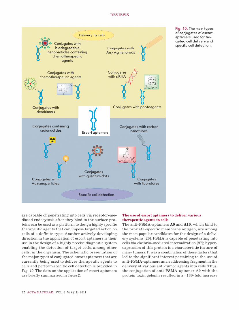
are capable of penetrating into cells via receptor-me-diated endocytosis after they bind to the surface pro-teins can be used as a platform to design highly specific therapeutic agents that can impose targeted action on cells of a definite type. Another actively developing direction in the application of escort aptamers is their use in the design of a highly precise diagnostic system enabling the detection of target cells, among other cells, in the organism. the schematic presentation of the major types of conjugated escort aptamers that are currently being used to deliver therapeutic agents to cells and perform specific cell detection is provided in Fig. 10. the data on the application of escort aptamers are briefly summarized in Table 2.
The use of escort aptamers to deliver various therapeutic agents to cellsthe anti-PSMA-aptamers A9 and A10, which bind to the prostate-specific membrane antigen, are among the most popular candidates for the design of a deliv-ery systems [20]. PSMA is capable of penetrating into cells via clathrin-mediated internalization [97]; hyper-expression of this protein is a characteristic feature of many tumors. It was a combination of these factors that led to the significant interest pertaining to the use of anti-PSMA-aptamers as an addressing fragment in the delivery of various anti-tumor agents into cells. thus, the conjugation of anti-PSMA-aptamer A9 with the protein toxin gelonin resulted in a ~180-fold increase
Delivery to cells
Conjugates with biodegradable
nanoparticles containing chemotherapeutic
agents
Conjugates with Au/Ag nanorods
Conjugates with chemotherapeutic agents
Conjugates with siRNA
Conjugates with dendrimers
Conjugates with photoagents
Conjugates containing radionuclides
Escort aptamers
Conjugates with carbon nanotubes
Conjugates with Au nanoparticles
Conjugates with quantum dots
Conjugates with fluorofores
Specific cell detection
Fig. 10. The main types of conjugates of escort aptamers used for tar-geted cell delivery and specific cell detection.

reVIeWS
VOL. 3 № 4 (11) 2011 | ActA nAturAe | 23
Table 2. Escort aptamers and their use for the delivery of various therapeutics into cells and for specific cell detection
Aptamer target Selection method Application
2'-F-rnAProstate-specific membrane anti-
gen (PSMA)
Selection against recom-binant protein – extracel-lular domain of PSMA [21]
Delivering gelonin [71] and doxorubicin [72] into LncaP tumor cells.
Delivering sirnA into LncaP cells [74–77].Delivering nanoparticles containing antitumor thera-
peutic docetaxel into LncaP cells [82–84].Imaging of LncaP cells using aptamer-conjugated
gold nanoparticles [92] or aptamer-conjugated lumi-nescent cdSe–cdte crystals [90].
electrochemical detection of prostate cancer cells using an aptamer immobilized on an Au-electrode [91].
Detection of PSMA on LncaP cell surface via proximity ligation assay [96]
DnA Protein tyrosine kinase 7 (PtK7)
Selection against the ccrF-ceM cells (the precursors
of t-cell acute myeloid leukaemia cells) [26]
Delivering doxorubicin [73], Au/Ag nanorods [80, 81], and poly(amidoamine)-based dendrimers [87] into
ccrG-ceM cells .Investigation of the distribution of PtK7 receptors
over the cell surface using aptamer–fluorescein conjugates [93].
reversible fluorescent labelling of ccrF-ceM cells using aptamer-conjugated quantum dots Qdot525 [94]
2'-F-rnA rat cD4 receptor Selection against recom-binant protein [40] Delivering sirnA into cells [78]
2'-F-rnA HIV-1 coat protein
Selection against recom-binant protein gp120 [79] Delivering sirnA into HIV-1-infected cells [7, 79]
DnA Mucin (Muc1)Selection against synthetic peptides (mucin fragments)
[23, 24]
Photodynamic therapy using photoreactive conjugates of the aptamer and chlorine e6 [24].
tumor imaging using radioactive isotopes (99tc) [88]
DnA, rnA Mouse transferrin receptor (tfr)
Selection against recom-binant protein – extracel-
lular domain of tfr
Delivering lysosomal enzymes into cells to treat lyso-somal storage diseases [43]
2'-F-rnA tenascin ccross-selection using a
recombinant protein and u251 cells [19]
tumor imaging using radioactive isotopes (99tc) [89]
DnA Membrane-bound IgM heavy chain
Selection against Burkitt lymphoma B-cells (ramos
cells) [31]
Micelles for delivering various pharmaceutics into cells [86].
test strips based on TD05 and TE02 aptamers for the detection of ramos cells in blood samples [95]
DnA B. thuringiensis bacteria
Selection against B. thuring-iensis spores
Detection of B. thuringiensis spores using aptamer-conjugated cdSe-ZnS quantum dots [61]
rnA E. coli bacteria Selection against E. coli DH5α strain [67]
Potentiometric detection of E. coli using aptamers-conjugated carbon nanotubes [67].
Detection of E. coli via quantitative rt-Pcr of rnA aptamers bound to the bacteria [98]
rnA S. typhi bacteriaSelection against the major
protein of S. typhi microvilli [63]
Potentiometric detection of S. typhi using aptamer-conjugated carbon nanotubes [97]
DnA Mesenchymal stem cells
Selection against porcine mesenchymal stem cells [49]
Isolation of stem cells from bone marrow using aptam-ers immobilized on magnetic particles; cell sorting
using fluorescent aptamer conjugates [49]
DnAPrecursors of
porcine endothe-lial cells
Selection against cD31-positive cells from porcine
blood [50]
Immobilization, growth, and differentiation of the pre-cursors of endothelial cells on the surface of disks with immobilized aptamers (a model of vascular implants)
[50]

24 | ActA nAturAe | VOL. 3 № 4 (11) 2011
reVIeWS
in cytotoxicity with respect to the target cells, in com-parison with that for unbound gelonin (Ic
50 = 27 nM for
the conjugate and 5 µM for gelonin) [71]. Meanwhile, in the case of control cells incapable of expressing the PSMA protein, the cytotoxicity of the aptamer-gelonin conjugate was lower than that of the unbound gelonin (Ic
50 = 15 µM), which attests to the fact that this con-
jugate is highly selective. V. Bagalkot et al. [72] used the A10 anti-PSMA-aptamer to deliver doxorubicin into cells. the anthracycline antibiotic agent doxoru-bicin is used for the therapy of a wide range of diseases, such as leukaemia, malignant lymphomas, sarcomas, and cancers of various etiologies. However, the draw-backs include toxic side effects, in particular, cardio-toxicity. the ability of doxorubicin to intercalate be-tween the base pairs of double-stranded nucleic acids was used to obtain the conjugates [72]. the intercala-tion of doxorubicin into a double-stranded fragment of the A10 aptamer yielded non-covalent conjugates. the cytotoxicity of these conjugates with respect to LncaP target cells was comparable to that of unbound doxo-rubicin of the same concentration (Ic
50 = 5 µM). the
cytotoxicity of conjugates with respect to Pc3 control cells was significantly lower. the sgc8с escort aptamer (“truncated” variant of the sgc8 aptamer) was used to deliver doxorubicin into human leukemic lymphob-lasts (ccrF-ceM cells). Y. Huang et al. [73] obtained the doxorubicin–sgc8c aptamer conjugate, with doxo-rubicin bound to the 5'-terminus of the aptamer via an acid-labile hydrazone bond that was hydrolyzed after the conjugate had penetrated into the cell. It has been shown that these conjugates can selectively penetrate into ccrF-ceM cells via receptor-mediated endocy-tosis; their cytotoxicity with respect to ccrF-ceM cells being comparable to that of unbound doxorubicin (Ic
50 = 0.3 µM). As opposed to unbound doxorubicin,
the aptamer-doxorubicin conjugate showed no toxicity with respect to the control ramos cells. thus, binding of escort aptamers to chemotherapeutic agents makes it possible to reduce their toxic effects on tumor cells only. It can be anticipated that these conjugates will be used to design novel agents for anti-tumor chemo-therapy with minimum adverse effects.
escort aptamers were also used to deliver small in-terfering rnA (sirnA) into cells. Several types of con-structs to deliver sirnA were designed on the basis of anti-PSMA-aptamers. the tetrameric biotin-strepta-vidin complex containing two biotinylated strands of the A9 aptamer and two biotinylated sirnA mol-ecules targeted against mrnAs of the lamin A/c or GADPH genes (Fig. 11A) was used to deliver sirnAs into PSMA-positive tumor cells [74]. these complexes could penetrate into cells without the use of transfect-ants. At a concentration of 22.5 nM, they suppressed
target gene expression by 50–80%; the efficiency of the suppression was identical to that of the corresponding sirnAs delivered into cells using Oligofectamine. the chimeric constructs were designed which consisted of a joint nucleotide sequence containing the A10 aptamer and one of the sirnA strands with the complementary second sirnA strand (Fig. 11B) [75]. At a concentration of 400 nM, these constructs can penetrate into PSMA-positive cells without transfectants and almost com-pletely suppress the expression of the bcl2 and plk1 tar-get genes. With the aim of optimizing the structure of chimeric constructs, the targeting aptamer was trun-cated from 71 down to 39 nucleotides, which simpli-fied the chemical synthesis of both components of the construct. A number of modifications increasing the specificity and efficiency of the interaction with the mrnA target were introduced into the sirnA struc-ture. A polyethylene glycol residue with a molecular weight of 20 kDa was bound to the sirnA passenger strand, which increased the half-circulation time of the chimeric constructs in mouse blood from 35 min to 30 h. the obtained preparation resulted in a considerable re-gression of the PSMA-positive tumor in mice after the injection of five 0.25 nmol doses [76].
Another variant of a chimeric construct contain-ing two molecules of anti-PSMA-aptamer A10-3 was
А
b
siRNA siRNA
siRNA
Streptavidin
Bio Bio
Bio Bio
Anti-PSMA-aptamer A9
Anti-PSMA-aptamer A10
Fig. 11. Schematic representation of chimeric constructs for siRNA delivery into PSMA-positive cells. A. The conjugate of biotinylated anti-PSMA aptamer and siRNA connected via streptavidin [74]. B. Chimeric RNA built from an anti-PSMA aptamer and siRNA [75]. Bio – biotin residue.

reVIeWS
VOL. 3 № 4 (11) 2011 | ActA nAturAe | 25
described in [77]. In this construct, one of the sirnA strands targeted against mrnA of the eukaryotic elon-gation factor 2 (eeF2) is inserted between two apta-meric sequences, the second strand being complemen-tary [77]. At a concentration of 2 µM, these conjugates caused suppression of the growth of the target cells by 95%, with no impact on the growth of PSMA-negative control cells.
An interesting construct containing the “address-ing” aptamer and sirnAs was described in [78]. Phage φ29 rnA capable of multimeric complex formation via interaction between rnA loops was used to design these constructs. A phage rnA fragment was bound to each component of the construct, i.e., to the aptamer recognizing the cD4 receptor [40], to sirnAs targeted against the mrnAs of various apoptotic factors, and to the fluorescent dye. At a concentration of 100 nM, the resulting trimers containing an aptamer, sirnA, and a reporter group were able to penetrate into the cD4-positive cells and inhibit the expression of the target genes.
chimeric constructs [7, 79] designed according to the principle proposed by J. Mcnamara et al. [75] and con-sisting of a 2'-F-rnA-aptamer recognizing the viral protein gp120 on the cell surface and sirnA targeted against the Тat/Rev of HIV-1 rnA were used to act upon HIV-1 infected cells. these constructs (at a con-centration of 400 nM) have the ability to inhibit HIV-1 replication in a cell culture [79]. the use of these con-structs to suppress HIV-1 replication in mice has also been reported [7].
the sgc8c aptamer conjugated with Au-Ag nanorods was used to impose photothermal action on leukaemia cells [80, 81]. the nanorods are heated to 50°c under laser irradiation, which results in cell death through thermal shock. Aptamers conjugated with nanorods (approximately 80 aptameric molecules were bound to a nanorod) were capable of selective penetration into the target cells; approximately 90% of the cells died af-ter exposure to radiation [81].
reactive agents for photodynamic therapy (aptam-ers covalently conjugated to chlorine e6) have been de-signed on the basis of aptamers capable of binding to the surface protein mucin [24]. these conjugates can selectively penetrate into mucin-expressing tumor cells and cause their death after exposure to radiation; the efficiency of the conjugates was 500-fold higher than that of unbound chlorine. Meanwhile, the conjugates showed no toxicity towards healthy cells.
the DnA aptamer against mouse transferrin re-ceptor was used to deliver the lysosomal enzyme α-L-iduronidase into cells [43]. It was demonstrated that the aptamer-enzyme conjugates penetrated into mouse fibroblasts deficient in this enzyme and were further
transported to lysosomes, where the introduced en-zyme was capable of both functioning and facilitating the recovery of the cell metabolism. the results ob-tained make aptamers against the transferrin receptor conjugated with lysosomal enzymes promising thera-peutic agents for the treatment of diseases associated with the lysosomal function disorder.
Various carriers bound to the addressing aptamers are also used to ensure specific delivery of therapeutic agents. thus, nanoparticles composed of the copolymer of lactic acid and glycolic acid (PLGA) with encapsulat-ed docetaxel were covalently bound to the molecule of anti-PSMA-aptamer A10 [82–84]. the resulting conju-gate was capable of specific binding to LncaP cells ex-pressing the PSMA protein and penetrating into them [82, 85]. Mice with a grafted prostate tumor were used to demonstrate that anti-PSMA-aptamers conjugated with nanoparticles based on PLGA containing docetax-el can efficiently suppress tumor growth and can even result in complete remission [83].
the construction of micelles based on the TD05 aptamer with the stearic acid bound to it was described in [86]. these micelles were characterized by increased affinity to the target cells in comparison with that of the individual TD05 aptamer, and they were capable of specific penetration into the cells. It is not anticipated that these micelles will be used further to deliver ther-apeutics into cells.
J. Zhou et al. [87] proposed using polyamidoamine (PAMAM) dendrimers as carriers for therapeutic de-livery [87]. Sgc8c aptamer-conjugated dendrimers proved capable of selective and efficient binding to ccrF-ceM cells and penetrating into them. the size of the aptamer-dendrimer conjugate is approximately 8 nm, the optimal size for using these conjugates as a platform for the delivery of therapeutic agents.
thus, the use of aptamers for targeted delivery of nanoparticles with anti-tumor agents into tumor tis-sues is a promising avenue in the development of novel anti-tumor therapeutic strategies.
The use of escort aptamers for specific cell detectionthe capability of escort aptamers to selectively recog-nize cells of a definite type enables one to use them to design highly specific detection systems. the introduc-tion of different types of labels into aptamers allows one to use them for cell detection in cultures, biological samples, and in living multi-cellular organisms. thus, radio-labeled aptamers have been used for tumor im-aging in mice. conjugates of the anti-tenascin aptamer TTA1 and anti-mucin aptamers with chelating agents capable of binding to 99tc were used for the imaging of glioblastoma and breast cancer xenografts in mice [88, 89].
26 | ActA nAturAe | VOL. 3 № 4 (11) 2011
reVIeWS
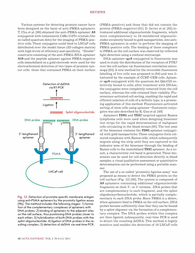
Various systems for detecting prostate cancer have been designed on the basis of anti-PSMA-aptamers. t. chu et al. [90] obtained the anti-PSMA-aptamer A9 conjugated with luminescent cdSe/cdte crystals (the so-called quantum dots) for the imaging of PSMA-pos-itive cells. these conjugates could bind to LncaP cells distributed over the model tissue (3D collagen matrix) with high levels of efficiency and specificity. “Double” constructs consisting of the anti-PSMA-rnA-aptamer A10 and the peptide aptamer against PSMA-negative cells immobilized on a gold electrode were used for the electrochemical detection of two types of prostate can-cer cells: those that contained PSMA on their surface
(PSMA-positive) and those that did not contain the protein (PSMA-negative) [91]. D. Javier et al. [92] in-troduced additional oligonucleotide fragments, which were complementary to 24-membered oligonucle-otides covalently bound to gold nanoparticles, into anti-PSMA-aptamers in order to perform the imaging of PSMA-positive cells. the binding of these complexes to PSMA on the cell surface was observed by reflected light detection using a confocal microscope.
DnA-aptamer sgc8 conjugated to fluorescein was used to study the distribution of the receptors of PtK7 over the cell surface via fluorescence correlation micro-scopy [93]. A new method for the reversible fluorescent labelling of live cells was proposed in [94] and was il-lustrated by the example of ccrF-ceM cells. Aptam-er sgc8 conjugated with the quantum dot Qdot525 se-lectively bound to cells; after treatment with DnAse, the conjugates were completely removed from the cell surface, whereas the cells retained their viability. Flu-orescence-activated cell sorting, enabling the rapid and efficient isolation of cells of a definite type, is a promis-ing application of this method. Fluorescence-activated sorting of stem cells using aptamer–fluorescein conju-gates was also described by K. Guo et al. [49]
Aptamers TD05 and TE02 targeted against ramos lymphoma cells were used when designing biosensor test strips for the rapid determination of malignant cells circulating in the blood stream [95]. the test zone of the biosensor contains the TD05 aptamer conjugat-ed with gold nanoparticles. these conjugates form col-oured complexes with ramos cells, which subsequently migrate along the strip until they are captured in the indicator zone of the biosensor through the binding of ramos cells to the immobilized TE02 aptamer. As a re-sult, a characteristic red band is generated. these bio-sensors can be used for cell detection directly in blood samples; a visual qualitative assessment or quantitative determination can be performed using a portable scan-ner.
the use of a so-called ‘proximity ligation assay’ was proposed as means to detect the PSMA protein on the cell surface (Fig. 12) [96]. the system is composed of A9 aptamers containing additional oligonucleotide fragments on their 3'- or 5'-termini, DnA probes that are complementary to each fragment, and the splint oligodeoxyribonucleotide, which is partially comple-mentary to each DnA probe. Since PSMA is a dimer, when aptamers bind to PSMA on the cell surface, DnA probes become sufficiently close that they can be bound by a splint oligomer via the formation of a complemen-tary complex. the DnA probes within this complex are then ligated; subsequently, real-time Pcr is used to detect the resulting dsDnA. this method is highly sensitive and enables the detection of 10 LncaP cells
Detection of dsDNA via RT-PCR
Ligation
Splint oligonucleotide
DNA probeDNA probe
5’-lengthened aptamer
3’-lengthened aptamer
PSMA
LNCaP cell
5' 3'
Fig. 12. Detection of prostate-specific membrane antigen using anti-PSMA aptamers by the proximity ligation assay [96]. The method includes the following stages: 1) forma-tion of the complementary complexes of aptamers with DNA probes; 2) binding of aptamers to the adjacent sites on the cell surface, thus positioning DNA probes closer to each other; 3) hybridization of both DNA probes with the splint oligonucleotide; 4) ligation of DNA probes in the re-sulting complex; 5) detection of dsDNA via real-time PCR.

reVIeWS
VOL. 3 № 4 (11) 2011 | ActA nAturAe | 27
(prostate cancer) in the presence of 105 HeLa cells that do not contain the PSMA protein on their surface.
It was recently suggested that the aptamers bind-ing to bacteria can be used for the design of biosensors for use in the detection of pathogenic microorganisms. DnA aptamers with bound cdSe-ZnS quantum dots were used for the fluorescent detection of B. thuring-iensis [61]. rnA aptamers immobilized on the surface of single-walled carbon nanotubes were used to de-sign potentiometric biosensors to determine E. coli [67] and S. typhi [97] and to demonstrate that they can be used for selective detection of these bacteria. A method based on the quantitative rt-Pcr of rnA aptamers bound to bacteria immobilized on magnetic particles was developed for the detection of E. coli [98]. novel aptamer-based biosensors could be used for highly se-lective determination of bacteria in the environment and food products and for diagnosing infectious dis-eases.
Another promising direction in the use of escort aptamers is the selective isolation of cells and cell im-mobilization. thus, a system for the selective isolation of stem cells from bone marrow was designed on the basis of magnetic particles conjugated with aptam-ers capable of binding to porcine stem cells [49]. DnA aptamers capable of binding to the precursors of por-cine endothelial cells were immobilized on the surface of polytetrafluoroethylene or polydimethylsiloxane disks [50]. It turned out that aptamer-coated disks can be used to selectively isolate endothelial precursor cells, which can be subsequently differentiated into vascular
endothelial cells while remaining bound to the disks. the assumption is that this approach could be used for the epithelialization of vascular implants, thus reducing the risk of implant rejection.
CONCLUSIONSA number of escort aptamers capable of specific and ef-ficient binding to cells of a definite type were recently obtained and approaches have been developed aimed at enhancing the stability of escort aptamers. It has been demonstrated that escort aptamers can be used to design efficient and highly specific systems for the de-livery of therapeutic agents into cells, for the detection of cells of a definite type, for cell sorting, and for the selective blockage of surface proteins. Year after year, the number of studies devoted to the selection and ap-plication of escort aptamers against various cell tar-gets (from bacteria to stem cells) steadily increases. It should be noted that the selection of aptamers against living cells has remained a more laborious and delicate process than the selection of aptamers against indi-vidual compounds. However, the increasing interest in this field and the advances in the selection methods are reasons to be hopeful that a greater variety of escort aptamers and aptamer-based therapeutic and diagnos-tic agents will appear in the near future.
This work was supported by the Russian Foundation for Basic Research (grant № 11-04-01014-a) and the Grant for Young Researchers provided by the
Novosibirsk Region Administration, 2011.
reFerenceS1. ellington A.D., Szostak J.W. // nature. 1990. V. 346.
P. 818–822.2. tuerk c., Gold L. // Science. 1990. V. 249. P. 505–510.3. robertson D.L., Joyce G.F. // nature. 1990. V. 344.
P. 467–468.4. Mayer G. // Angew. chem. Int. ed. 2009. V. 48. P. 2672–
2689.5. Stoltenburg r., reinemann c., Strehlitz B. // Biomol. eng.
2007. V. 24. P. 381–403.6. Shamah S.M., Healy J.M., cload S.t. // Acc. chem. res.
2008. V. 41. P. 130–138.7. Zhou J., rossi J.J. // Oligonucleotides. 2011. V. 21. P. 1–10.8. Syed M.A., Pervaiz S. // Oligonucleotides. 2010. V. 20.
P. 215–224.9. chapman J.A., Beckey c. // Ann. Pharmacother. 2006.
V. 40. P. 1322–1326.10. ng e.W., Shima D.t., calias P., cunningham e.t., Guyer
D.r., Adamis A.P. // nat. rev. Drug. Discov. 2006. V. 5. P. 123–132.
11. Hicke B.J., Stephens A.W. // J. clin. Invest. 2000. V. 106. P. 923–928.
12. Breaker r.r. // curr. Opin. chem. Biol. 1997. V. 1. P. 26–31.
13. Fitzwater t., Polisky B. // Meth. enzymol. 1996. V. 267. P. 275–301.
14. Sousa r. // Meth. enzymol. 2000. V. 317. P. 65–74.15. chelliserrykattil J., ellington A.D. // nat. Biotechnol.
2004. V. 22. P. 1155–1160.16. Burmeister P.e., Lewis S.D., Silva r.F., Preiss J.r.,
Horwitz L.r., Pendergrast P.S., Mccauley t.G., Kurz J.c., epstein D.M., Wilson c., et al. // chem. Biol. 2005. V. 12. P. 25–33.
17. Schmidt K.S., Borkowski S., Kurreck J., Stephens A.W., Bald r., Hecht M., Friebe M., Dinkelborg L., erdmann V.A. // nucl. Acids res. 2004. V. 32. P. 5757–5765.
18. Shangguan D., tang Z., Mallikaratchy P., Xiao Z., tan W. // chemBiochem. 2007. V. 8. P. 603–606.
19. Hicke B.J., Marion c., chang Y.F., Gould t., Lynott c.K., Parma D., Schmidt P.G., Warren S. // J. Biol. chem. 2001. V. 276. P. 48644–48654.
20. Morris K.n., Jensen K.B., Julin c.M., Weil M., Gold L. // Proc. natl. Acad. Sci. uSA. 1998. V. 95. P. 2902–2907.
21. Lupold S.e., Hicke B.J., Lin Y., coffey D.S. // cancer res. 2002. V. 62. P. 4029–4033.
22. Ferreira c., Papamichael K., Guilbault G., Schwarzacher t., Gariepy J., Missailidis S. // Anal. Bioanal. chem. 2008. V. 390. P. 1039–1050.

28 | ActA nAturAe | VOL. 3 № 4 (11) 2011
reVIeWS
23. Ferreira c.S., Matthews c.S., Missailidis S. // tumour Biol. 2006. V. 27. P. 289–301.
24. Ferreira c.S., cheung M.c., Missailidis S., Bisland S., Gariepy J. // nucl. Acids res. 2009. V. 37. P. 866–876.
25. Daniels D.A., chen H., Hicke B.J., Swiderek K.M., Gold L. // Proc. natl. Acad. Sci. uSA. 2003. V. 100. P. 15416–15421.
26. Shangguan D., Li Y., tang Z., cao Z.c., chen H.W., Mal-likaratchy P., Sefah K., Yang c.J., tan W. // Proc. natl. Acad. Sci. uSA. 2006. V. 103. P. 11838–11843.
27. Shangguan D., cao Z. c., Li Y., tan W. // clin. chem. 2007. V. 53. P. 1153–1155.
28. Shangguan D., cao Z., Meng L., Mallikaratchy P., Sefah K., Wang H., Li Y., tan W. // J. Proteome res. 2008. V. 7. P. 2133–2139.
29. Xiao Z., Shangguan D., cao Z., Fang X., tan W. // chem-istry. 2008. V. 14. P. 1769–1775.
30. Li n., ebright J.n., Stovall G.M., chen X., nguyen H.H., Singh A., Syrett A., ellington A.D. // J. Proteome res. 2009. V. 8. P. 2438–2448.
31. tang Z., Shangguan D., Wang K., Shi H., Sefah K., Mallikаratchy P., chen H.W., Li Y., tan W. // Anal. chem. 2007. V. 79. P. 4900–4907.
32. Mallikaratchy P., tang Z., Kwame S., Meng L., Shang-guan D., tan W. // Mol. cell. Proteom. 2007. V. 6. P. 2230–2238.
33. Mallikaratchy P.r., ruggiero A., Gardner J.r., Kuryavyi V., Maguire W.F., Heaney M. L., McDevitt M.r., Patel D.J., Scheinberg D.A. // nucl. Acids res. 2011. V. 39. P. 2458–2469.
34. Sefah K., tang Z.W., Shangguan D.H., chen H., Lopez-colon D., Li Y., Parekh P., Martin J., Meng L., Phillips J.A., et al. // Leukemia. 2009. V. 23. P. 235–244.
35. chen H.W., Medley c.D., Sefah K., Shangguan D., tang Z., Meng L., Smith J.e., tan W. // chemMedchem. 2008. V. 3. P. 991–1001.
36. Shangguan D., Meng L., cao Z.c., Xiao Z., Fang X., Li Y., cardona D., Witek r.P., Liu c., tan W. // Anal. chem. 2008. V. 80. P. 721–728.
37. Blank M., Weinschenk t., Priemer M., Schluesener H. // J. Biol. chem. 2001. V. 276. P. 16464–16468.
38. Zueva e., rubio L.I., Ducongé F., tavitian B. // Int. J. cancer. 2011. V. 128. P. 797–804.
39. Mi J., Liu Y., rabbani Z.n., Yang Z., urban J.H., Sullenger B.A., clary B.M. // nat. chem. Biol. 2010. V. 6. P. 22–24.
40. Kraus e., James W., Barclay A.n. // J. Immunol. 1998. V. 160. P. 5209–5212.
41. Davis K.A., Lin Y., Abrams B., Jayasena S.D. // nucl. Acids res. 1998. V. 26. P. 3915–3924.
42. Santuli-Marotto S., nair S.K., rusconi c., Sullenger B., Gilboa e. // cancer res. 2003. V. 63. P. 7483–7489.
43. chen c.-H.B., Dellamaggiore K.r., Ouellette c.P., Sedano c.D., Lizadjohry M., chernis G.A., Gonzales M., Baltasar F.e., Fan A.L., Myerowitz r., et al. // Proc. natl. Acad. Sci. uSA. 2008. V. 105. P. 15908–15913.
44. Ohuchi S.P., Ohtsu t., nakamura Y. // Biochimie. 2006. V. 88. P. 897–904.
45. cerchia L., Duconge F., Pestourie c., Boulay J., Aissouni Y., Gombert K., tavitian B., de Franciscis V., Libri D. // PLoS Biol. 2005. V. 3. e123.
46. Pestourie c., cerchia L., Gombert K., Aissouni Y., Boulay J., De Franciscis V., Libri D., tavitian B., Duconge F. // Oligonucleotides. 2006. V. 16. P. 323–335.
47. Wang c., Zhang M., Yang G., Zhang D., Ding H., Wang H., Fan M., Shen B., Shao n. // J. Biotechnol. 2003. V. 102. P. 15–22.
48. Guo K., Wendel H.P., Scheideler L., Ziemer G., Scheule A.M. // J. cell Mol. Med. 2005. V. 9. P. 731–736.
49. Guo K.t., Schafer r., Paul A., Gerber A., Ziemer G., Wen-del H.P. // Stem cells. 2006. V. 24. P. 2220–2231.
50. Hoffmann J., Paul A., Harwardt M., Groll J., reeswinkel t., Klee D., Moeller M., Fischer H., Walker t., Greiner t., et al. // J. Biomed. Mater. res. A. 2008. V. 84A. P. 614–621.
51. Berezovski M.V., Lechmann M., Musheev M.u., Mak t.W., Krylov S.n. // J. Am. chem. Soc. 2008. V. 130. P. 9137–9143.
52. Homann M., Göringer H.u. // nucl. Acids res. 1999. V. 27. P. 2006–2014.
53. Homann M., Göringer H.u. // Bioorg. Med. chem. 2001. V. 9. P. 2571–2580.
54. Göringer H.u., Homann M., Zacharias M., Adler A. // Handb. exp. Pharmacol. 2006. V. 173. P. 375–393.
55. Homann M., Lorger M., engstler M., Zacharias M., Göringer H. // comb. chem. High throughput Screen. 2008. V. 9. P. 491–499.
56. Göringer H.u., Homann M., Lorger M. // Int. J. Parasitol. 2003. V. 33. P. 1309–1317.
57. Lorger M., engstler M., Homann M., Göringer H.u. // eukaryot. cell. 2003. V. 2. P. 84–94.
58. ulrich H., Magdesian M.H., Alves M.J., colli W. // J. Biol. chem. 2002. V. 277. P. 20756–20762.
59. chen F., Zhou J., Luo F., Mohammed A.B., Zhang X.L. // Biochem. Biophys. res. commun. 2007. V. 357. P. 743–748.
60. torres-chavolla e., Alocilja e.c. // Biosens. Bioelectron. 2009. V. 24. P. 3175–3182.
61. Ikanovic M., rudzinski W., Bruno J., Allman A., carrillo M., Dwarakanath S., Bhahdigadi S., rao P., Kiel J., An-drews c. // J. Fluoresc. 2007. V. 17. P. 193–199.
62. Joshi r., Janagama H., Dwivedi H.P., Senthil Kumar t.M.A., Jaykus L.-A., Schefers J., Sreevatsan S. // Mol. cell. Probes. 2009. V. 23. P. 20–28.
63. Pan Q., Zhang X.-L., Wu H.-Y., He P.-W., Wang F., Zhang M.-S., Hu J.-M., Xia B., Wu J. // Antimicrob. Agents chemother. 2005. V. 49. P. 4052–4060.
64. cao X., Li S., chen L., Ding H., Xu H., Huang Y., Li J., Liu n., cao W., Zhu Y., et al. // nucl. Acids res. 2009. V. 37. P. 4621–4628.
65. Hamula c.L.A., Zhang H., Guan L.L., Li X.-F., Le X.c. // Anal. chem. 2008. V. 80. P. 7812–7819.
66. Bruno J., carrillo M., Phillips t., Andrews c. // J. Fluo-resc. 2010. V. 20. P. 1211–1223.
67. So H.-M., Park D.-W., Jeon e.-K., Kim Y.-H., Kim B.S., Lee c.-K., choi S.Y., Kim S.c., chang H., Lee J.-O. // Small. 2008. V. 4. P. 197–201.
68. Dwivedi H., Smiley r., Jaykus L.-A. // Appl. Microbiol. Biotechnol. 2010. V. 87. P. 2323–2334.
69. Barfod A., Persson t., Lindh J. // Parasitol. res. 2009. V. 105. P. 1557–1566.
70. chen F., Hu Y., Li D., chen H., Zhang X.-L. // PLoS One. 2009. V. 4. P. e8142.
71. chu t.c., Marks J.W., 3rd, Lavery L.A., Faulkner S., rosenblum M.G., ellington A.D., Levy M. // cancer res. 2006. V. 66. P. 5989–5992.
72. Bagalkot V., Farokhzad O.c., Langer r., Jon S. // Angew. chem. Int. ed. engl. 2006. V. 45. P. 8149–8152.
73. Huang Y.F., Shangguan D., Liu H., Phillips J.A., Zhang X., chen Y., tan W. // chemBiochem. 2009. V. 10. P. 862–868.
74. chu t.c., twu K.Y., ellington A.D., Levy M. // nucl. Acids res. 2006. V. 34. P. e73.
75. Mcnamara J.O., Andrechek e.r., Wang Y., Viles K.D., rempel r.e., Gilboa e., Sullenger B.A., Giangrande P.H. // nat. Biotechnol. 2006. V. 24. P. 1005–1015.
reVIeWS
VOL. 3 № 4 (11) 2011 | ActA nAturAe | 29
76. Dassie J.P., Liu X.-Y., thomas G.S., Whitaker r.M., thiel K.W., Stockdale K.r., Meyerholz D.K., Mccaffrey A.P., Mcnamara J.O., Giangrande P.H. // nat. Biotechnol. 2009. V. 27. P. 839–846.
77. Wullner u., neef I., eller A., Kleines M., tur M.K., Barth S. // curr. cancer Drug targets. 2008. V. 8. P. 554–565.
78. Khaled A., Guo S., Li F., Guo P. // nano Lett. 2005. V. 5. P. 1797–1808.
79. Zhou J., Swiderski P., Li H., Zhang J., neff c.P., Akkina r., rossi J.J. // nucl. Acids res. 2009. V. 37. P. 3094–3109.
80. Huang Y.F., chang H.t., tan W. // Anal. chem. 2008. V. 80. P. 567–572.
81. Huang Y.F., Sefah K., Bamrungsap S., chang H.t., tan W. // Langmuir. 2008. V. 24. P. 11860–11865.
82. Farokhzad O.c., Jon S., Khademhosseini A., tran t.n., Lavan D.A., Langer r. // cancer res. 2004. V. 64. P. 7668–7672.
83. Farokhzad O.c., cheng J., teply B.A., Sherifi I., Jon S., Kantoff P.W., richie J.P., Langer r. // Proc. natl. Acad. Sci. uSA. 2006. V. 103. P. 6315–6320.
84. cheng J., teply B., Sherifi I., Sung J., Luther G., Gu F.X., Levy-nissenbaum e., radovic-Moreno A.F., Langer r., Farokhzad O.c. // Biomaterials. 2007. V. 28. P. 869–876.
85. Farokhzad O.c., Khademhosseini A., Jon S., Hermmann A., cheng J., chin c., Kiselyuk A., teply B., eng G., Langer r. // Anal. chem. 2005. V. 77. P. 5453–5459.
86. Wu Y., Sefah K., Liu H., Wang r., tan W. // Proc. natl. Acad. Sci. uSA. 2010. V. 107. P. 5–10.
87. Zhou J., Soontornworajit B., Martin J., Sullenger B.A., Gil-boa e., Wang Y. // Macromol. Biosci. 2009. V. 9. P. 831–835.
88. Perkins A.c., Missailidis S. // Q. J. nucl. Med. Mol. Imag-ing. 2007. V. 51. P. 292–296.
89. Hicke B.J., Stephens A.W., Gould t., chang Y.-F., Lynott c.K., Heil J., Borkowski S., Hilger c.-S., cook G., Warren S., et al. // J. nucl. Med. 2006. V. 47. P. 668–678.
90. chu t.c., Shieh F., Lavery L.A., Levy M., richards-Kor-tum r., Korgel B.A., ellington A.D. // Biosens. Bioelectron. 2006. V. 21. P. 1859–1866.
91. Min K., Song K.-M., cho M., chun Y.-S., Shim Y.-B., Ku J. K., Ban c. // chem. commun. 2010. V. 46. P. 5566–5568.
92. Javier D.J., nitin n., Levy M., ellington A., richards-Ko-rtum r. // Bioconjugate chem. 2008. V. 19. P. 1309–1312.
93. chen Y., Munteanu A.c., Huang Y.-F., Phillips J., Zhu Z., Mavros M., tan W. // chem. eur. J. 2009. V. 15. P. 5327–5336.
94. terazono H., Anzai Y., Soloviev M., Yasuda K. // J. nano-biotechnol. 2010. V. 8. P. 8.
95. Liu G., Mao X., Phillips J.A., Xu H., tan W., Zeng L. // Anal. chem. 2009. V. 81. P. 10013–10018.
96. Pai S.S., ellington A.D. // Meth. Mol. Biol. 2009. V. 504. P. 385–398.
97. Zelada-Guillén G.A., Jordi r., Düzgün A., rius F.X. // Angew. chem. Int. ed. 2009. V. 48. P. 7334–7337.
98. Lee H.-J., Kim B.c., Kim K.-W., Kim Y.K., Kim J., Oh M.-K. // Biosens. Bioelectron. 2009. V. 24. P. 3550–3555.