equine skin grafting: principles and field applications*
TRANSCRIPT

Continuing Education Article R6 Refereed Peer Revlew
can lessen the healing time of I I( wounds and improve~cosmetic
outcome.
- Skin grafts are classitied according to their source, thickness, and blood supply, p. 872.
-E The success of a graft is based on its fibrinous adherence to the bed, plasmatic imbibition, revascularization of the graft, and the organization of the grafted wound with collagen-secreting fibroblasts, p. 873.
After injury, f m t i o n of a healthy bed of granulation tissue takes at least 5 days, p. 873.
Chlorhexidine diacetate, at a
useful antimicrobial agent in the postoperative care of skin grafts,
Equine Skin Grafting: Principles and Field Applications* Auburn University S m n A. Carson-Dunkerley, D V M R. Reid Hanson, DVM
hen horses incur cutaneous wounds that cannot be sutured because of significant cutaneous damage or loss of deeper tissue, healing by second intention must occur. However, large wounds that heal by
second intention heal slowly, and the scars that form are often cosmetidy un- appealing.'~~ Skin grafting may reduce healing time, improve cosmesis, and allow a horse to return to function sooner than if the wound were allowed to heal by second in tent i~n.~ With the proper equipment and expertise, many skin grafting techniques can be performed on the farm.' This article describes grafting procedures that can be conveniently performed with the horse stand- ing or anesthetized with a short-acting general anesthetic. +
CLASSIFICATION Skin grafts are classified on the basis of three characteristics: source, thick-
ness, and blood supply." Gr& that are classified according to the source include autogenous gra'cs, homografts, and xenografts. Autogenous grafts (autografts) are taken from one site and moved to another site on the same an- imal.L" Homografts (allogratis) are transferred between genetically different individuals of the same specie^.^ Xenografts (heterografts) are transferred be- tween individuals of different specie^.^ W ~ t h autogenous grafting, an immuno- logic reaction against the donor skin does not occur.
Grafts that are classified by thickness include split-thickness grafis (which are composed of the epidermis and a portion of the dermis) and full-thickness grafts (which include the epidermis and all of the dermi~).~ Both types are readily accepted by horses?." Because MI-thickness grafts are easier to obtain and because harvesting them does not require specialized equipment (e-g., a dermatome), full-thickness grafting is easier to perform in the field.6
*Publication Series No. 2541, College of Veterinary Medicine, Auburn University, Auburn, AL 36849.
Eauine 873
Grafts that are classified by blood supply are the free graft and the pedicle graft. A free graft is devoid of blood supply; a pedicle graft maintains its own blood s ~ p p l ~ . ~ . ~ Because pedicle grafting requires general anesthesia to isolate the required nutrient ar- terial vessel, free grafting is more practical for use in the field.'
Free gafts are further classified as island grafts (e.g., pinch, punch, and tunnel grafts) and sheet grafts. Sheet gafts can be split-thickness or full-thickness and can be applied as solid or meshed sheets. Pinch, punch, tun- nel, and sheet grafting can be applied in the field, with the horse standing or recumbent, using short-acting !general anesthesia. Sheet grafts provide more complete dermal coverage of the wound; island grafts provide dermal tissue only at the graft site and provide epithe- lial coverage in between. Grafts help to control infec- - tion, provide dermal coverage of the wound, and stim- ulate epithelialization from the epithelial edges of the wound and grafL2s9
HEALING OF GRAFTS The success of grafting is based on four events: (1)
fibrinous adherence of the graft to its bed, (2) plasmatic imbibition (passive absorption of nutrients by the ves- sels of the (3) revicularization of the graft, and (4) organization of the grafted wound with collagen- secreting fibroblast^.^ The graft adheres within minutes of implantation.2.6 Fibrin, formed from fibrinogen, is brought into the recipient site by the surrounding ves- sels and attaches the graft to its bed." For the first few days after grafting, the delicate fibrinous attachment is the sole method of adhe~ion.~ Early immobilization to protect this delicate attachment thus is critical to the acceptance of the graft."
Before the graft is revascularized, it must survive by plasmatic imbibition. This is the process by which the grafi bed provides an oxygen-rich, plasmalike nutrient fluid for absorption into the dilated vessels of the graft."-'* The duration of plasmatic imbibition depends on the quality of the recipient site bed. The more vas- cular the recipient bed, the faster the graft is revascular- ized and the less it must rely on plasmatic imbibi- tion.'O"*
The graft revascularizes by two processes: (1) vascular anastomoses through a direct connection of grafi and host vessels (inosculation) and (2) ingrowth of host ves- sels into the dermis of the graft.".I3 Inosculation usually begins during the first 2 days, restoring circulation to the graft by the fourth to seventh day.10.'3,14 Revascular- ization by invasion of new vessels begins at 4 to 12 days.2 When revascularization is well established, the fibrinous attachment of the =aft is infiltrated with fibro-
blasts. These cells secrete collagen and ground sub- stance into the wound, increasing the tensile strength of the attachment of the graft.'0.'5
PREPARATION OF THE RECIPIENT SITE Sheet grafts are best applied to fresh wounds that
have no granulation tissue, but such grafts also can be applied to granulating wounds. Island grafts, however, can be applied only to granulating wounds. The granu- lation tissue to which grafts are applied should be new- ly formed, richly vascular, firm, and free of purulent di~charge."~'~~" The presence of advancing epithelium at the margin of the wound is generally a good indica- tor of the bed's health and capacity to accept a Formation of a healthy bed of granulation tissue re- quires at least 5 days after injury."
Because wounds in horses are frequently treated for weeks before the decision to graft is made, the granula- tion tissue is often mature and contains more fibrous tissue and a poorer blood supply than would a fresh bed of granulation tissue. Consequently, the ability to accept a graft is less than optimum.' Before the graft is applied, the mature granulation tissue must be excised to below the skin surface to allow development of a healthier, more vascular granulation tissue bed. Because of the lack of innervation, granulation tissue can be excised with the horse standing." Deep crevices and depressions in the tissue should be excised so that the wound is smooth, allowing better contact with and thus better acceptance of the graft.3
Studies indicate that trimming granulation tissue as much as 4 days before skin grafting may increase accep- tance of Because of the significant amount of hemorrhage that results from such trimming, the bed should be trimmed no less than 24 hours before graft- ing. Wounds that are apparently infected can be cul- tured for bacterial identification and antimicrobial sen- sitivity testing.''
After debridement, the wound should be covered with a nonadhesive pad and a well-padded bandage. If the wound is at a highly mobile site (e.g., the metacar- pophalangeal joint), the site should be stabilized by a splint incorporated into the bandage before grafting to accustom the horse to the decreased range of motion of the joint.17 The acceptance of the graft is positively in- fluenced by the use of appropriate bandages that pre- vent movement between the graft and the recipient bed and that help protect the delicate granulation tissue from
For grafts to be accepted, the concentration of bac- teria in the wound must be minimized." P-hemolytic Streptococcus, Protezls, and Psezldmnonas species are capa- ble of producing destructive proteolytic enzymes that

degrade fibrinous at tachn~ents -- PREPARATION OF between the graft and reci~ient THE DONOR SITE " bed.?.3,(~,18,1'1 These bacteria also
produce copious p ~ ~ r u l e n t dis- charge that can separate the '
nique to be used. A donor site
graft from its bed.'.'.".'"'" should be easily accessible for
Topical treatment of infected . rn procuring the graft or grafts. The
granulating wounds with ap- most cosmetic sites for graft pro-
propriatc anti~nicrobial drugs is curement may be regions of the
more effective than systemic neck covered by the mane, the
treatment.'-.'" Systemically ad- ventral abdol-nen, or the ventral
ministered antirl~icrobial drugs pectoral region. O t h e r d o n o r are often incapable of achieving sltes Include the sternal region, therapeutic concentrations in the region caudal to the girth
the granulation tissue because p a t h , a n d the p e r i n e u m . ' - I S fibrin in the wound prevents Cosmesis of the wound is best penetration of the drug to the when the donor hair (color and
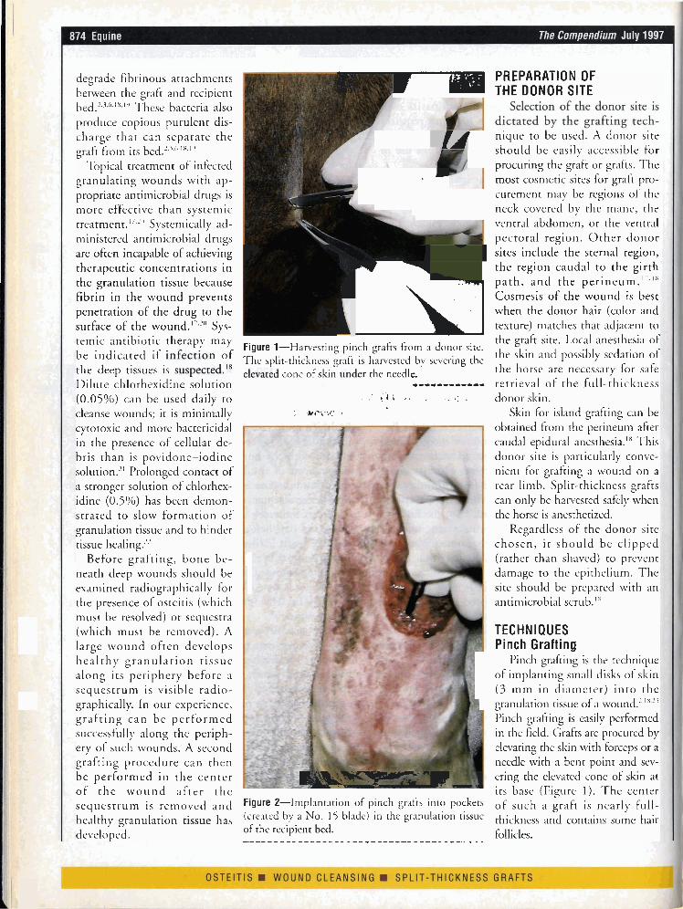
surface of the wound.".'" Sys- texture) matches that adjacent to teniic antibiotic . therapy - nlay Figure 1-Harvesting pinch 3.. gmfts from a donor site. the graft site. Local anesthesia of
be ind ica t ed i f o t -1-he split-thickness graft is harvested by ,evering the the skin and possibly sedation of the deep tissues is cone ofshn [he needle the horse are necessary for safe Dilute chlorhexidine solution ------------ retrieval o f the f i~ll- thickness (0.05%) can be used daily to . ' :'! ; , . . . . : donor skin.
; , U,..,'l''.<.' +
. cleanse wounds; it is niinimally Skin for island grafting can be cytotoxic and more bactericidal obtained from the perineun~ after in the presence of cellular de- caudal epidural anes t I~es ia .~This
bris than is povidone-iodine donor site is particularly conve- solution." Prolonged contact of nienr for grafting a wound on a a stronger solution of chlorhex- rear limb. Split-thickness grafts idine (0.5%) has been denion- can only be harvested safely when strated to slow formation of the horse is anesthetized. granulation tissue and to hinder Regardless of the donor sire
tissue healing." chosen, i t shou ld be cl ipped Before graft ing, b o n e bc- (rather than shaved) to prevent
neath deep wou~ids should be darnage to the epithelium. The
examined radiographically for site should be prepared with an the presence of osteitis (which antimicrobial scrub.'" must be resolved) or sequestra (which n ~ u s t be removed). A TECHNIQUES large wound often develops Pinch Grafting hea l thy g r a n u l a t i o n t i ssue Pinch grafting is the technique along its periphery before a of implanting small disks of skin seqilestrum is visible radio- (3 m m in d iameter ) i n to the graphically. In our experience, granulation tissue of a wound.' '"' gra f t ing can be p e r f o r m e d Pinch grafiing is easily performed successfi~lly along the periph- in rhc field. Grafts are procured by ery of such wounds. A second elevating the skin \vith forceps or a grafting procedure can then needle with a bent point and sev- be performed in the center ering the elevated conc of skin at o f t h e w o u n d a f t e r t h e its base (Figure 1 ) . 'The center seq l lcs r rum i s removed a n d Figure 2-Iniplantarion of pinch grafts into pockets of such a graft i s ,,Carly full- healthy granulation has (crcatcd hv a No. 15 blade) in [he gran~llarion tissue thickness and contains sornc hair
developed. of the recipient bed. ------------------------------------- follicles.
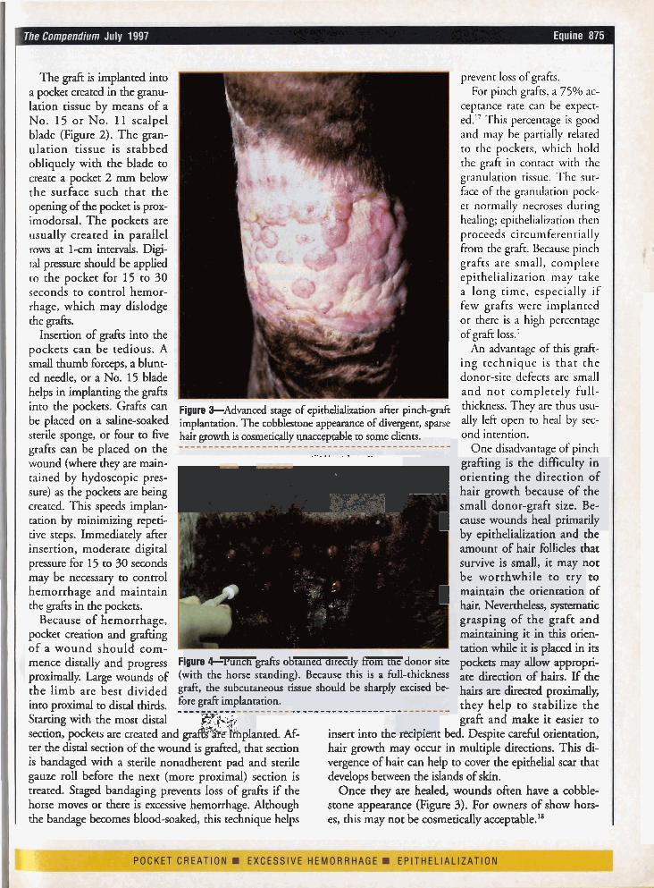
The graft is implanted into I a pocket created in the granu-
lation tissue by means of a
i No. 15 or No. 1 1 scalpel 1 blade (Figure 2). The gran-
ulation tissue is stabbed
1 obliquely with the blade to
i create a pocket 2 mm below I the surface such that the
opening of the pocket is prox- imodorsal. The pockets are usually created in parallel rows at I-cm intervals. Digi- tal pressure should be applied to the pocket for 15 to 30 seconds to control hemor- rhage, which may dislodge the grafts.
Insertion of grafts into the pockets can be tedious. A small thumb forceps, a blunt- ed needle, or a No. 15 blade helps in implanting the grafts into the pockets. Grafts can be placed on a saline-soaked sterile sponge, or four to five grafts can be placed on the
Figure *Advanced stage of epithelializaaon afcer pinch-grafi implantation. The cobblestone appearance of divergent, sparse hair tztowth is cosmetically unacceptable to same clients.
prevent loss of grafts. For pinch grafts, a 75% ac-
ceptance rate can be expect- ed." This percentage is good and may be partially related to the pockets, which hold the graft in contact with the granulation tissue. The sur- face of the granulation pock- et normally necroses during healing; epithelialization then proceeds circumferentially from the graft. Because pinch grafts are small, complete epithelialization may take a long time, especially if few grafts were implanted
I or there is a high percentage of graft loss.'
An advantage of this graft- ing technique is that the donor-site defects are small and not completely full-
. thickness. They are thus usu- : ally left open to heal by sec-
ond intention. One disadvantage of pinch .-..,. .. . -. .
wound (where ;hey are main- r - r grafting is the difficulty in tained by hydoscopic pres- orienting the direction of sure) as the pockets are being hair growth because of the created. This speeds implan- small donor-graft size. Be- tation by minimizing repeti- I cause woundsheal primarily tive steps. Immediately after by epithelialization and the insertion, moderate digital amount of hair follicles that pressure for 15 to 30 seconds survive is small, it may not may be necessary to control be worthwhile t o try to hemorrhage and maintain I maintain the orientation of the grafts in the pockets. hair. Nevertheless, systematic
Because of hemorrhage, grasping of the graft and pocket creation and grafting maintaining it in this orien- of a wound should com- tation while it is placed in its mence distally and progress Fioure 4-Yunch grafts obmned directly from the donor site pockets may allow appropri- proximally. Large wounds of (with the horse standing). Because this is a full-thickness ate direction of hairs. If the the limb are best divided graft, the subcutaneous tissue should be sharply excised be- hairs are direaed p r o ~ ~ P
into proximal to distal thirds. for:!:* f ?Iantation. .------------------ they help to stabilize the Starting with the most distal gY.3. & graft and make it easier to
2;. section, pockets are created and grA are ~ p l a n t e d . Af- insert into tne recipient bed. Despite carem orientation, ter the distal section of the wound is grafied, that section hair growth may occur in multiple directions. This di- is bandaged with a sterile nonadherent pad and sterile vergence of hair can help to cover the epithelial scar that gauze roll before the next (more proximal) section is develops benveen the islands ofskin. treated. Staged bandaging prevents loss of grafts if the Once they are healed, wounds often have a cobble- horse moves or there is excessive hemorrhage. Although stone appearance (Figure 3). For owners of show hors- the bandage becomes blood-soaked, this technique helps es, this may not be cosmetically acceptable."'

Punch Grafting Punch !grafting is similar to
pinch grafting and can be readily performed in the field with the horse standing. Us- ing a skin biopsy punch, circular plugs of skin are removed directlv from the anesthetized donor site (Fig- ure 4) or punched from a piece of skin that has been removed from the donor site
- I for the punch biopsies.' We
use 8-mm biopsy punches for harvesting and 6-mm punch-
(Figure 5). During harvest- ing from the ventral pectoral
es to create the recipient sites (Figure 6). Biopsy holes in the granulation tissue are placed at 5- to 15-mm inter- vals. Cotton-tipped applica- tors can be placed in a hole to aid in identifying the hole in the granulation tissue, which rapidly becomes ob- scured by hemorrhageVFig- ure7). "
region, grafts are best Figure 5-An elliptic piece of skin removed from the chest to A second approach t o ,,laced o n a saline-soaked create punch o r tunnel grafts.
. . . . . . . . . . . . . . . . . . . . . . ---------------.----- controlling hemorrhage after sponge.'"ecause p ~ l n c h placement of punch sites is !grafis are full-thickness, sub- to bandage the recipient site cutaneous tissue and fascia must be removed with a for several hours. The grafts can be applied directly from blade before the grafts are implanted."' It is easier to re- the donor site or can be stored in a refrigerator on move subcutaneous tissue from a section of skin before saline-soaked gauzes during this ~ e r i o d . Because some punches are harvested because removal from individual hair follicles are present, it may be beneficial to orient plugs is tedious. the g a f t so that hair g o w t h is in a proper direction. If
A biopsy punch, iisually 2 mm smaller than that used the wound is large, staged bandaging during the im- to obtain the donor sgrafi, is ~ised to create recipient sites plantation process (as described for pinch grafting) may
prevent loss of grafts by pre-
Figure 6-Creation of the recipient sires for donor punch grafts using a 6-mm skin biopsy punch. - - - - - - - - *I~oI)*.-*mc)aL---
. .
Figure 7-Sterile applicators placed in the re- cipient-site pouches to facilitate hemostasis and identification of the site (which is ob- scured by blood) in rhe gran~~lat ion tissue.
eluding excessive hemor - - rhage or movement of the graft.
The donor graft sites can be closed with sutures or sta- ples or can be left open to heal by second intention. If an elliptic piece of skin is re- moved to obtain the punch sgrafts, the donor site should be sutured." If punches are removed directly, the donor sites heal with multiple stel- late scars." As with pinch grafting, the recipient site may develop an uncosmetic cobblestone appearance. Nev- ertheless, hair coverage is more prominent with this technique.'-'~"'"tudies have reported grafi survival as high as 95% and epirhelialization of the entire wound within 47 days.l.7.1%.l&l(,
Tunnel Grafting Tunnel grafting requires

urn Julv 1997
harvesting of full-thickness 0, split-thickness strips of skin that are 2 to 5 mm wide and slightly longer than the WOUnd3.4.6,17.18,27 (Figure 8). Full-thickness skin is often used in field situations be- cause split-thickness skin grafcs are difficult to obtain without special equipment (e.g., free-hand knives or motorized dermatomes) and !general anesthesia." To im- prove acceptance of the graft, subcutaneous fascia and fat should be sharply dissected from full-thickness donor skin." Ofren, the easi- est method of obtaining tun- nel grafts is to remove a sheet of skin from the donor site. Strips of skin are then easily cut from the stretched and stabilized sheet'' (Figure 5). To minimize scarring, the donor site is sutured by pri- mary clo~ure.'~
The grafts are implanted in tunnels created in the granulation tissue, which has been allowed to develop 4 to 8 mm above skin level4 These tunnels are created via a cutting needle, a flattened Kirschner wire with a trocar point, a straight teat blade, or a malleable alligator for- ceps.3.4.~4,27.2s plfl all igator for-
ceps or a small tendon for- ceps is then used to grasp and pull the skin strip into the bed4 (Figure 9). To pre- vent vascular compromise of the granulation tissue, tun- nel grafts should be placed no less than 1 cm apart.4
In one popular method used to implant tunnel grafcs, adhesive tape (slightly longer than the graft) is placed on the haired side of tl
The graft and tape the eye of a 10- to 12-cm,
cutting needle.4 The nee-
1 dle and graft are inserted through the granulation tis- sue 5 rnm below the sur- face. The haired side should be facing outward, and the hair should be oriented in the proper dire~t ion.~
Because the needle is of- ten smaller than the wound length, multiple small strips of grahs are used or a longer graft is placed over the width of the wound by a
Figure 8-Creation of a tunnel graft from skin along the neck. two- or three-step passage . . . . . . . . . . . . . . . . . . . . . . . . . . . . . . . . . . . . . . . . . . . . . . of the needle.'' The needle
reenters the point of exit for the second and third bite. Tunnel grafts inserted into a wound on a limb should be placed circumferentially be- cause of the increased diffi- culty in placing the grafts in the proximodistal direction.
When tape is used to fa- cilitate implantation of the graft, the graft may become dislodged, especially if the tape is removed immediate- ly after insertion or later with excision of overlying granulation t i s s ~ e . ~ * ' ~ At- taching the tape to the graft can be tedious and can in- crease surgical time.28 Such attachment is unnecessary if an alligator forceps is used."
In some cases, tunnels can be created with the horse standing; however, because slight movement may de- stroy the tunnel, short-term general anesthesia may be required.ls In our opinion, general anesthesia facilitates
Figure %--Creation of a tunnel in the recipient granulation placement of grafts.
tissue and implantation of a tunnel graft using a tendon for- minimize
ceps to pull the grafted tissue through the wound bed. time, a full-thickness sheet -------------------we.).)e--------------------- of tissue can be harvested
while the horse is standing. 1e ga f c to minimize graft The sheet is cut into strips, stored on sponges soaked are then threaded through with physiologic saline, and then placed into the granu- half-curved or straight lation bed after the horse is anesthetized. The ends of
the graft are then sutured, rained from the recipient stapled, or glued to the edge site and trimmed to fit the of the wound for additional wound. Alternatively, a tem- security." Graft survival is plate of the wound can be enhanced by exposing the made from sterilized paper tunnel grafts away from the and used to trace the recipi- overlying granulation tissue ent bed to ensure that the 6 to 10 days after implanta- !graft is the correct size." ti~n.~"ecause most failures Once excised, full-thick- can be at tr ibuted to acci- ness grafts undergo primary dental removal of the graft shrinkage due to recoil of o r fai lure t o expose t h e elastic fibers in the deep graf t , t h e su rgeon w h o dermal layers." To allow for placed the gaf ts should re- primary shrinkage, the graft move the overlying granula- should be cut slightly larger tion tissue.I8 than the template.30,3' If the
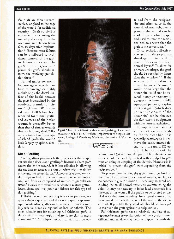
Tunnel grafts are useful section of d o n o r skin re- for coverage of sites that are quired to cover the wound hard to bandage or highly would be so large that the mobile (e.g., the dorsal sur- donor site could not be su- face of the hock) because I tured, it may be necessary to the graft is restrained by the transport the horse to a fully overlying granulation tis-
I equipped practice; a split-
sue2," (Figure 10). Survi- thickness graft (which does val rates of 80% have been not require closure of the reported for tunnel grafts, donor site) can be obtained and cosmesis of the healed via dermatome equipment wound is generally better with the horse anesthetized. than that of similar wounds To improve acceptance of that are left ungrafted.zx B ~ - Figure 10-Epithelialization after tunnel grafting of a wound. a fl,ll-thickness sheet graft
cause a tunnel graft is a type (Courtesy of Dr. D. G. Wilson, Department of Surgical Sci- by the recipient bed, it is ences, College of Veterinary Medicine, University of Wiscon-
of island graft, the wound generally necessary to (1) re- sin)
heals largely by epithelializa- ,,,,, -. - . - . - .. _ . . . _ move the subcutaneous tis- R k k T tion. s.;:: =.,, ..-... t sue from the graft, (2) es-
tablish hemostasis o f the Sheet Grafting wound, and (3) stabilize the graft. The subcutaneous
Sheet grafting produces better cosmesis at the recipi- tissue should be carefully excised with a scalpel to pre- ent site than does island grafting.18 Because a sheet graft vent crushing or scraping of the dermis. Hemostasis is covers the entire wound, it is less effective in allowing critical to prevent the graft from separating from the the exudate to escape; this can interfere with the ability recipient bed.3 of the graft to revascularize.'%cceptance is good only if To prevent contraction, the graft should be fixed to the recipient bed is uncontaminated, at an immobile the edge of the wound by means of sutures, staples, or site, and fresh or composed of immature granulation ~~anoac ry la t e glue." Caution is necessary to avoid oc- tissue.' Horses with wounds that contain mature granu- cluding the small dermal vessels by overstretching the lation tissue are thus poor candidates for this type of skin.32 It may be necessary to inject local anesthetic into skin grafting.' the edge of the wound if the graft is to be sutured or sta-
Full-thickness sheet grafting is easy to perform, re- pled with the horse standing. Interrupted sutures may quires slight expertise, and does not require expensive be required to attach the center of the graft to the recipi- equipment. Most grafts can be obtained from a stand- ent bed. If possible, the grafted site should be bandaged ing, sedated horse via regional or local anesthesia. The to maintain the graft against the recipient bed. most suitable area for obtaining full-thickness grafts is Full-thickness gafts have a relatively low rate of ac- the cranial pectoral region, where loose skin is most ceptance because revascularization of these grafts is more abundant.1',2%n elliptic section of skin can be ob- difficult and exudate may become trapped beneath the