epsin n-terminal homology domains perform an essential ... · essential function regulating cdc42...
TRANSCRIPT

Epsin N-terminal homology domains perform anessential function regulating Cdc42 throughbinding Cdc42 GTPase-activating proteinsRuben C. Aguilar*, Silvia A. Longhi†, Jonathan D. Shaw, Lan-Yu Yeh, Sean Kim, Arne Schon, Ernesto Freire, Ariel Hsu,William K. McCormick, Hadiya A. Watson‡, and Beverly Wendland§
Department of Biology, Johns Hopkins University, 3400 North Charles Street, Baltimore, MD 21218
Edited by Pietro V. De Camilli, Yale University School of Medicine, New Haven, CT, and approved January 17, 2006 (received for review December 6, 2005)
Epsins are endocytic proteins with a structured epsin N-terminalhomology (ENTH) domain that binds phosphoinositides and apoorly structured C-terminal region that interacts with ubiquitinand endocytic machinery, including clathrin and endocytic scaf-folding proteins. Yeast has two redundant genes encoding epsins,ENT1 and ENT2; deleting both genes is lethal. We demonstrate thatthe ENTH domain is both necessary and sufficient for viability ofent1�ent2� cells. Mutational analysis of the ENTH domain re-vealed a surface patch that is essential for viability and that bindsguanine nucleotide triphosphatase-activating proteins for Cdc42, acritical regulator of cell polarity in all eukaryotes. Furthermore, theepsins contribute to regulation of specific Cdc42 signaling path-ways in yeast cells. These data support a model in which the epsinsfunction as spatial and temporal coordinators of endocytosis andcell polarity.
actin � endocytosis � polarity
Endocytosis is an essential mechanism for internalizing extracel-lular material and controlling the composition of the plasma
membrane; this is critical for cellular homeostasis, including down-regulation of signaling receptors and recycling of transmembraneproteins such as v-SNAREs that reside transiently at the plasmamembrane (1). Many cytosolic proteins that contribute to themechanisms and regulation of endocytosis have been identified, butassigning precise functions to each protein has been more chal-lenging (2, 3). Some of these proteins may also participate inmultiple steps or pathways (4, 5), either related to or independentfrom endocytosis, further complicating the elucidation of theirfunction(s). Additionally, roles for the actin cytoskeleton in regu-lating or effecting specific stages of endocytosis are another activearea of investigation (2). One goal is to identify multifunctionalproteins that coordinate these various cellular processes.
The epsin proteins are proposed to function as endocyticclathrin adaptors for ubiquitinated cargo (6, 7). They are foundin all eukaryotes and have an N-terminal phosphatidylinositol-4,5-bisphosphate [PtdIns(4,5)P2]-binding epsin N-terminal ho-mology (ENTH) domain, two ubiquitin interaction motifs, andseveral peptide ligands that bind components of the endocyticmachinery (7). In addition to putative adaptor roles, it has beenshown previously that mammalian epsin binds RalBP1�RLIP76,a GTPase-activating protein (GAP) for Cdc42 and Rac1 (8).RalBP1 has been implicated in endocytosis, because it binds theplasma membrane clathrin adaptor AP-2 (8). The Cdc42 andRac GTPases are key regulators of the actin cytoskeleton (9),thus suggesting that this complex links signaling, endocytosis,and actin cytoskeleton regulation.
The budding yeast Saccharomyces cerevisiae has two epsins, Ent1and Ent2; deleting either alone leads to no detectable phenotype,but a double deletion is lethal (10). Here we show that the ENTHdomain of yeast epsin is necessary and sufficient for viability ofent1�ent2� cells (��). The essential function requires a patch ofresidues, conserved only in the ENTH domains of epsin proteins,
that interacts with Cdc42 GAPs. Our results indicate that thisinteraction regulates levels of activated Cdc42 in vivo, and that an[epsin�GAP] complex plays a critical physiological role togetherwith specific Cdc42 effectors. Furthermore, Cdc42 regulation viaepsins is likely to be conserved in higher eukaryotes. Based on thesedata, we propose a model in which the epsins link the endocytic andcell polarity machineries to coordinate membrane and cytoskeletalpathways in response to polarized signals.
ResultsThe ENTH Domain Is Sufficient for Viability. To determine whichregions of epsin contribute to its essential role, we used ��containing a wild-type ENT1 or ENT2 plasmid and a marker forcounterselection on a toxic compound; this allows for shuffling ofmutated epsin plasmids with a different selection marker andevicting the original one. Cells will not grow on plates containingthe toxic compound [5-fluoroorotic acid or 5-fluoroanthranilicacid�alcohol (5-FAA)] if the new plasmid encodes a protein thatcannot fulfill the essential function of the wild-type epsin; this assayis used throughout. The ENTH domain of either Ent1 or Ent2, withor without an N-terminal hemagglutinin epitope tag and expressedfrom either endogenous or methionine (Met)-regulated MET25promoters, was both necessary and sufficient for �� viability (Fig.1A and data not shown). The ENTH domain of the related proteinsEnt3 or Ent4 did not complement �� (Fig. 1A and data not shown).Thus, the epsin ENTH domain is required for cell viability.
To identify candidate regions of epsin ENTH domains thatcould mediate essential functions, we compared the sequences ofS. cerevisiae Ent1�2 ENTH domains with the homologousdomains from Drosophila melanogaster (liquid facets) and Xe-nopus laevis (MP90) epsin, which also complement �� (ref. 11and our unpublished results). We found three residues (Y100,T104, and E137) that were both conserved exclusively in epsinENTH domains and also different in all other nonessentialENTH and AP180 N-terminal homology domain-containingproteins encoded by the yeast genome (Ent3, Ent4, Ent5,
Conflict of interest statement: No conflicts declared.
This paper was submitted directly (Track II) to the PNAS office.
Abbreviations: Met, methionine; ENTH, epsin N-terminal homology; CRIB, Cdc42�Racinteractive binding; EBS, ENTH-binding site; RnENTH, rat epsin1 ENTH domain; ��,ent1�ent2� cells; GAP, GTPase-activating protein; 5-FAA, 5-fluoroanthranilic acid; UIM,ubiquitin-interaction motif; NPF, Asn-Pro-Phe tripeptides; CBM, clathrin-binding motif.
See Commentary on page 3953.
*Present address: Department of Biological Sciences, Purdue University, West Lafayette,IN 47907-2054.
†Present address: Instituto de Investigaciones en Ingenieria Genetica y Biologia Molecular(INGEBI-CONICET) and Facultad de Ciencias Exactas y Naturales, Universidad de BuenosAires, 1428 Buenos Aires, Argentina.
‡Present address: Cell Biology and Metabolism Branch, National Institute of Child Healthand Human Development, National Institutes of Health, Bethesda, MD 20892.
§To whom correspondence should be addressed. E-mail: [email protected].
© 2006 by The National Academy of Sciences of the USA
4116–4121 � PNAS � March 14, 2006 � vol. 103 � no. 11 www.pnas.org�cgi�doi�10.1073�pnas.0510513103

Yap1801, Yap1802, and Sla2). These three candidate essentialresidues were predicted to be on the surface of a model for thefolded domain (Fig. 1B); moreover, T104 had been foundpreviously as a critical residue in epsin function (10).
To test their functional importance, we individually mutatedthese three residues to a corresponding residue found in a non-complementing ENTH�AP180 N-terminal homology domain(Y100R, T104D, and E137G) and asked whether the mutantENTH domains could fulfill the essential domain function.ENTH1Y100R and ENTH1T104D domains did not complement ��when expressed from the endogenous promoter (Fig. 1C and Fig.7A, which is published as supporting information on the PNAS website). Western blotting confirmed that wild-type or mutant ENTHdomain proteins was stable (data not shown). Purified recombinantwild-type and mutant ENTH domains had nearly identical struc-tures, based on fluorescence emission of two tryptophan residues(W70 and W94; Fig. 8A, which is published as supporting infor-mation on the PNAS web site). Differential scanning calorimetryshowed that the ENTH1 domains were properly folded, theirdenaturation being centered at 52.5°C (data not shown). ENTHdomain lipid-binding activity was also preserved in theENTH1Y100R domain; it bound IP6, which mimics the head groupof phosphatidylinositol-4,5-bisphosphate (12), with a binding affin-ity and enthalpy identical to the ENTH1WT domain (Kd � 39 � 6
mM, �H � �2.2 � 0.9 kcal�mol; Fig. 8B). Thus, the inviabilityassociated with the mutation of Y100 or T104 is unlikely to becaused by gross misfolding of the mutant domains. These resultsalso suggest that the essential function of the ENTH domainmediated by the Y100�T104 patch is independent of lipid binding.ENTH domains with lipid-binding mutations partially comple-mented ��, further suggesting independent functions for lipidbinding and the Y100�T104 patch (Fig. 8C).
ENTH1Y100R Phenotypes. Although ENTH1Y100R could not comple-ment �� when expressed from the endogenous promoter, cells withhigher-level expression from the regulated MET25 promoter wereviable, although they grew more slowly than ENTH1WT cells (Fig.1C). In contrast, even high-level expression of ENTH1T104D couldnot support �� viability (Fig. 7B). This suggests that ENTH1Y100R
acts as a hypomorphic allele. Thus, we used plasmids encodingENTH1WT or ENTH1Y100R domains expressed from the MET25promoter to study in vivo the effect of mutating the Y100�T104essential patch. Cells with these plasmids grown in 0–0.02 mM Methad the highest protein levels; those grown in higher Met concen-trations (0.2–2 mM) had lower protein levels (Fig. 9, which ispublished as supporting information on the PNAS web site).ENTH1Y100R-expressing cells grown in 1 mM Met (low proteinlevels) exhibited dramatically slow growth, inviability at 37°C, andgreatly enlarged, fragile, and often broken cells (Fig. 2 A and B).
Sites of cell wall synthesis, secretion, and endocytosis in yeastcorrelate with the polarized locations of F-actin-containing struc-tures (13, 14). Actin cables deliver newly synthesized secretory
Fig. 1. Epsin ENTH domain essential residues. (A) The ENTH domain of Ent1is necessary and sufficient for cell viability. Schematic of Ent1 constructs; theENTH domain, ubiquitin-interaction motifs (UIMs), Asn-Pro-Phe tripeptides(NPF), and clathrin-binding motif (CBM) are indicated. �� with an ENT2 TRP1plasmid and a second URA3 plasmid, empty or encoding the proteins ordomains indicated (Right), were grown on plates lacking uracil and trypto-phan (Left) or containing 5-FAA to evict the ENT2 TRP1 plasmid (Center) at30°C for 3 days. The presence of an N-terminal hemagglutinin tag, plasmidcopy number, and promoter used are also indicated. (B) ENTH domain-specificresidues, Y100, T104, and E137, are solvent-exposed in a 3D model. Residuesthat are conserved in ENTH domains, different in nonepsin ENTH�AP180N-terminal homology domains, and exposed to the solvent in a 3D model areshown [Y100 (green), T104 (red), and E137 (magenta)]. Lipid-binding residues(blue) and the N- and C-termini are indicated. (C) ENTH1Y100R domain is ahypomorph. �� cells containing an ENT2 TRP1 plasmid and a second high copyor single copy URA3 plasmid encoding ENTH1WT or ENTH1Y100R from endog-enous or MET25 promoter were assayed as in A.
Fig. 2. ENTH1Y100R phenotypes. (A) ENTH1Y100R cells are larger and morefragile than ENTH1WT cells. �� cells expressing ENTH1WT or ENTH1Y100R do-mains from the MET25 promoter were grown in selective liquid media � 1 mMMet and visualized by differential interference contrast microscopy. (Scalebar, 10 �m.) (B) ENTH1Y100R cells are sensitive to high temperature andhyposmolarity. Serial dilutions of �� cells expressing ENTH1WT or ENTH1Y100R
domains from the MET25 promoter were grown on YPD or 0.5xYPD plates at30°C or 37°C for 3 days. (C) ENTH1Y100R cells have a depolarized actin cytoskel-eton. �� cells expressing ENTH1WT or ENTH1Y100R domains from the MET25promoter were grown in liquid media plus 1 mM Met. Fixed cells were labeledwith rhodamine-phalloidin and DAPI and visualized by confocal microscopy.Arrowheads highlight small buds where actin patches should concentrate.(Scale bar, 5 �m.) (D) ENTHY100R cells show no defect on endocytosis ofGFP-Ste3. ENTH1WT (Left) or ENTH1Y100R (Right) cells expressing a Ste3-GFPfusion protein were grown in liquid media plus 1 mM Met and visualized byconfocal microscopy.
Aguilar et al. PNAS � March 14, 2006 � vol. 103 � no. 11 � 4117
CELL
BIO
LOG
YSE
ECO
MM
ENTA
RY

material to sites of growth (e.g., buds and necks) (15), whereascortical patches are dynamic structures in the process of endocytosis(16). Unlike the normal actin organization observed in ENTH1WT
cells, cortical patches were not polarized in the buds and few, if any,actin cables were visible in ENTH1Y100R cells (Fig. 2C). Thus,mutation of the essential patch of the ENTH domain altered cellgrowth, morphology, and polarity. These data also demonstratethat a wild-type ENTH domain is the minimal functional unit of theepsin molecule required to maintain proper actin organization�polarity (Fig. 2C). Interestingly, under the conditions of our exper-iments, the Ste3 mating pheromone receptor is endocytosed nor-mally in both ENTH1WT and ENTH1Y100R cells (Fig. 2D; seeDiscussion).
ENTH Domains Bind Cdc42 GAPs. Because the lipid-binding activity ofthe ENTH domain appeared to be dispensable for the functionsmediated by the Y100�T104 patch, we hypothesized that thisessential region might bind proteins. A yeast two-hybrid screen withan ENTH1WT bait versus a yeast cDNA prey library identifiedfragments from all three known Cdc42 GAP proteins, one each forRga1 and Bem3 and two for Rga2. Prey plasmids encoding eachGAP interacted with baits for both ENTH1�2WT, as indicated byactivation of the reporter genes ADE2, HIS3, and lacZ (Fig. 3A Topand data not shown). The interactions were confirmed to be bothdirect and specific by in vitro recombinant protein-binding assays.GST-Rga1�2 fusion proteins immobilized on glutathione-agarosebeads were incubated with purified His6
�-ENTH1�2; bound His6�-
ENTH domains were detected by Western blotting with anti-His6�
antibody (Fig. 3B Upper). Endogenous Ent1 from yeast lysates wasalso pulled down by GST-Rga2 immobilized on glutathione beads
(data not shown). Isothermal titration calorimetry experimentsgave an approximate affinity for the ENTH1–Rga2 interaction asa Kd of �0.6 mM (data not shown; see Supporting Text, which ispublished as supporting information on the PNAS web site). TheENTH1–Rga1 interaction appears to be considerably weaker.
Directed two-hybrid experiments showed a reduced interactionwhen ENTH1Y100R bait was paired with the Cdc42 GAPs (Fig. 3AMiddle). More severe impairment of the ENTH1–GAP interactionwas seen with ENTH1T104D or ENTH1Y100R, T104D as bait (Fig. 3ABottom). A role for the essential patch in the interaction wasconfirmed by in vitro binding assays showing reduced His-6-ENTH1Y100R–Rga2 interactions (Fig. 3B Lower). Together, ourdata indicate that the ENTH domain interacts with Cdc42 GAPs,and that this interaction depends upon the essential patch contain-ing the Y100 and T104 residues. To complement these data, theminimal region of Rga2 that binds to ENTH1WT was mapped byusing GST pull-down assays with Rga2 truncations (Fig. 3C). TheENTH-binding site (EBS) corresponded to Rga2604–786. Interest-ingly, Rga2710–721 shows sequence homology with the more diver-gent Cdc42 GAP, Bem3 (Bem3425–433). Preliminary data suggestthat this homology region is involved in, but not sufficient for,ENTH domain recognition (data not shown).
In Vivo Requirement for ENTH Domain–Cdc42 GAP Interaction. Toaddress the role of Cdc42 GAP binding to the ENTH domain,we used several strategies predicted to influence [ENTH�GAP]complex formation in vivo.Impaired ENTH–Cdc42 GAP interaction leads to decreased viability and cellpolarity defects. We affected the equilibrium of [ENTH�GAP]complex formation by manipulating each interaction partner (im-pairing the ENTH-binding affinity or decreasing the GAP dosage)or by adding a Cdc42 GAP competitor. First, mutations in theENTH domain that affect its interaction with Cdc42 GAPs, yieldinglow levels of [ENTH�GAP] complex, led to impaired cell viabilityand cell polarity defects (Figs. 1C and 7A). Second, �� with lowerlevels of intracellular GAPs were obtained by deleting RGA1. rga1�in �� cells expressing ENTH1WT were temperature-sensitive, andthe phenotypes were enhanced with rga1� in ���ENTH1Y100R
cells (Fig. 4A).We also used competitive inhibition of the ENTH–GAP inter-
action by overexpressing the Rga2 EBS. High levels of EBSexpression in endogenous ENTH1WT cells yielded morphology andactin disorganization similar to ENTH1Y100R cells (Fig. 4B). Thesedata indicate a dominant negative consequence of EBS expressionconsistent with competition for the endogenous interaction andsuggest that the ENTH–Rga2 interaction is important for normalcell physiology.Enhanced ENTH1Y100R–Cdc42 GAP interaction alleviates ENTH1Y100R phe-notypes. An in vivo role for the ENTH1–Cdc42 GAP interactionpredicts that promoting the formation of [ENTH1Y100R�GAP]complexes by overexpressing either binding partner (ENTH1Y100R
or GAP) should suppress the ENTH1Y100R phenotypes. Fig. 1Cshows that increased levels of ENTH1Y100R improve ENTH1Y100R
cell phenotypes. Conversely, overexpressing either Rga1 or Rga2improved growth on hyposmotic media and at 37°C (Fig. 4C).Overexpression of Bem3 only mildly rescued ENTH1Y100R pheno-types (Table 1, which is published as supporting information on thePNAS web site; data not shown). In contrast, Bem2, a Rho GAPfor both Rho1 (17) and Cdc42 (18), did not suppress ENTH1Y100R
phenotypes when overexpressed (Table 1).
Cdc42�GTP Levels Depend on the ENTH Domain. Our data indicate thatimpairing the ENTH–GAP interaction leads to cell viability andpolarity defects, whereas promoting the formation of the[ENTH1Y100R�GAP] complex partially suppresses theENTH1Y100R cell phenotypes. Because the newly identified ENTHinteraction partners are Cdc42 regulators, we asked whether Cdc42activation might be affected by the ENTH1Y100R mutation by
Fig. 3. The ENTH domain binds Cdc42 GTPase activating proteins. (A) ENTHdomains interact with Cdc42 GAPs. Yeast two-hybrid cells with plasmidsencoding GAL4 DNA-binding domain fusions to ENTH1WT, ENTH1Y100R,ENTH1T104D, ENTH1Y100R, T104D, ENTH2WT, or mouse p53 (mp53), and GAL4activation domain fusions to Rga1, Rga2, Bem3, or SV40 T-large antigen(T-LAg) were grown on plates lacking histidine and adenine and containing 10mM 3-amino-triazole. (B) In vitro interaction between ENTH domains andRga1�2. GST, GST-Rga1, and GST-Rga2 were immobilized on glutathione-agarose beads and incubated with purified His-6-ENTH1, His-6-ENTH2, orHis-6-ENTH1Y100R. Bound His-6-ENTH was detected by Western blot with anti-His-6 antibody. One percent of the input is shown. (C) Mapping the ENTHdomain-binding site on Rga2. (Upper) GST-Rga2 fragments were used inHis-6-ENTH1 pull-down experiments as in B. (Lower) Rga2 contains two LIM(Lin11, Isl-1, and Mec-3) domains, a coiled-coil (CC) region, and a RhoGAPdomain. The dotted bar indicates the Rga2 minimal fragment from thetwo-hybrid screen. Solid bars represent the GST-Rga2 truncations used to mapthe EBS.
4118 � www.pnas.org�cgi�doi�10.1073�pnas.0510513103 Aguilar et al.
measuring the levels of activated GFP-Cdc42 (Cdc42�GTP) inlysates from cells expressing ENTH1WT or ENTH1Y100R. TheCdc42�Rac interactive binding (CRIB) domain of the p21-activated kinase 1 binds specifically to the GTP-bound form ofCdc42 (19). A GST-CRIB fusion protein immobilized on glutathi-one beads was incubated with yeast lysates; bound GFP-Cdc42�GTP was visualized by Western blotting with anti-GFPantibody. Total Cdc42 present in the lysates was measured byWestern blotting with anti-GFP (not shown) or by loading withnonhydrolyzable GTP�S and pulling down with CRIB beads asdescribed above, as a measure of the total pool that can be activated(Fig. 4D). Amounts of GFP-Cdc42�GTP vs. GFP-Cdc42�GTP�Swere quantified by densitometry after Western blotting. We founda 62% reduction in Cdc42�GTP in ENTH1Y100R cells relative towild-type or ENTH1WT cells (Fig. 4D), indicating a dependence ofCdc42 regulation on ENTH1 function. Similar results were ob-tained for endogenous Cdc42 (data not shown). Attempts todemonstrate in vitro a direct regulation of the Cdc42 GAP activityby ENTH domain binding were unsuccessful. This suggests thatfull-length GAP, other proteins, posttranslational modifications, orlocalization-dependent events are required for the epsin-dependenteffects observed in vivo. Thus, our data indicate that, at least in vivo,ENTH1Y100R mutants with reduced interaction with Cdc42 GAPsexhibit enhanced inactivation (or deficient activation) of Cdc42-regulated pathways.
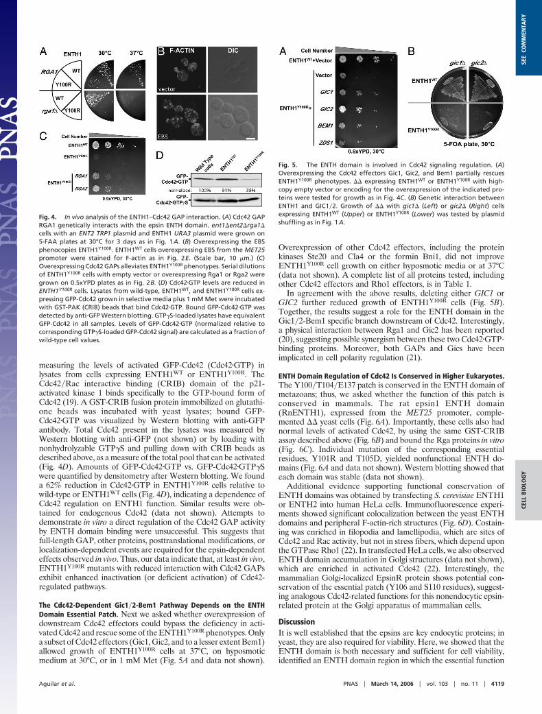
The Cdc42-Dependent Gic1�2-Bem1 Pathway Depends on the ENTHDomain Essential Patch. Next we asked whether overexpression ofdownstream Cdc42 effectors could bypass the deficiency in acti-vated Cdc42 and rescue some of the ENTH1Y100R phenotypes. Onlya subset of Cdc42 effectors (Gic1, Gic2, and to a lesser extent Bem1)allowed growth of ENTH1Y100R cells at 37°C, on hyposmoticmedium at 30°C, or in 1 mM Met (Fig. 5A and data not shown).
Overexpression of other Cdc42 effectors, including the proteinkinases Ste20 and Cla4 or the formin Bni1, did not improveENTH1Y100R cell growth on either hyposmotic media or at 37°C(data not shown). A complete list of all proteins tested, includingother Cdc42 effectors and Rho1 effectors, is in Table 1.
In agreement with the above results, deleting either GIC1 orGIC2 further reduced growth of ENTH1Y100R cells (Fig. 5B).Together, the results suggest a role for the ENTH domain in theGic1�2-Bem1 specific branch downstream of Cdc42. Interestingly,a physical interaction between Rga1 and Gic2 has been reported(20), suggesting possible synergism between these two Cdc42�GTP-binding proteins. Moreover, both GAPs and Gics have beenimplicated in cell polarity regulation (21).
ENTH Domain Regulation of Cdc42 Is Conserved in Higher Eukaryotes.The Y100�T104�E137 patch is conserved in the ENTH domain ofmetazoans; thus, we asked whether the function of this patch isconserved in mammals. The rat epsin1 ENTH domain(RnENTH1), expressed from the MET25 promoter, comple-mented �� yeast cells (Fig. 6A). Importantly, these cells also hadnormal levels of activated Cdc42, by using the same GST-CRIBassay described above (Fig. 6B) and bound the Rga proteins in vitro(Fig. 6C). Individual mutation of the corresponding essentialresidues, Y101R and T105D, yielded nonfunctional ENTH do-mains (Fig. 6A and data not shown). Western blotting showed thateach domain was stable (data not shown).
Additional evidence supporting functional conservation ofENTH domains was obtained by transfecting S. cerevisiae ENTH1or ENTH2 into human HeLa cells. Immunofluorescence experi-ments showed significant colocalization between the yeast ENTHdomains and peripheral F-actin-rich structures (Fig. 6D). Costain-ing was enriched in filopodia and lamellipodia, which are sites ofCdc42 and Rac activity, but not in stress fibers, which depend uponthe GTPase Rho1 (22). In transfected HeLa cells, we also observedENTH domain accumulation in Golgi structures (data not shown),which are enriched in activated Cdc42 (22). Interestingly, themammalian Golgi-localized EpsinR protein shows potential con-servation of the essential patch (Y106 and S110 residues), suggest-ing analogous Cdc42-related functions for this nonendocytic epsin-related protein at the Golgi apparatus of mammalian cells.
DiscussionIt is well established that the epsins are key endocytic proteins; inyeast, they are also required for viability. Here, we showed that theENTH domain is both necessary and sufficient for cell viability,identified an ENTH domain region in which the essential function
Fig. 4. In vivo analysis of the ENTH1–Cdc42 GAP interaction. (A) Cdc42 GAPRGA1 genetically interacts with the epsin ENTH domain. ent1�ent2�rga1�cells with an ENT2 TRP1 plasmid and ENTH1 URA3 plasmid were grown on5-FAA plates at 30°C for 3 days as in Fig. 1A. (B) Overexpressing the EBSphenocopies ENTH1Y100R. ENTH1WT cells overexpressing EBS from the MET25promoter were stained for F-actin as in Fig. 2E. (Scale bar, 10 �m.) (C)Overexpressing Cdc42 GAPs alleviates ENTH1Y100R phenotypes. Serial dilutionsof ENTH1Y100R cells with empty vector or overexpressing Rga1 or Rga2 weregrown on 0.5xYPD plates as in Fig. 2B. (D) Cdc42�GTP levels are reduced inENTH1Y100R cells. Lysates from wild-type, ENTH1WT, and ENTH1Y100R cells ex-pressing GFP-Cdc42 grown in selective media plus 1 mM Met were incubatedwith GST-PAK (CRIB) beads that bind Cdc42�GTP. Bound GFP-Cdc42�GTP wasdetected by anti-GFP Western blotting. GTP�S-loaded lysates have equivalentGFP-Cdc42 in all samples. Levels of GFP-Cdc42�GTP (normalized relative tocorresponding GTP�S-loaded GFP-Cdc42 signal) are calculated as a fraction ofwild-type cell values.
Fig. 5. The ENTH domain is involved in Cdc42 signaling regulation. (A)Overexpressing the Cdc42 effectors Gic1, Gic2, and Bem1 partially rescuesENTH1Y100R phenotypes. �� expressing ENTH1WT or ENTH1Y100R with high-copy empty vector or encoding for the overexpression of the indicated pro-teins were tested for growth as in Fig. 4C. (B) Genetic interaction betweenENTH1 and GIC1�2. Growth of �� with gic1� (Left) or gic2� (Right) cellsexpressing ENTH1WT (Upper) or ENTH1Y100R (Lower) was tested by plasmidshuffling as in Fig. 1A.
Aguilar et al. PNAS � March 14, 2006 � vol. 103 � no. 11 � 4119
CELL
BIO
LOG
YSE
ECO
MM
ENTA
RY

resides, and showed its interaction with Cdc42 GAPs. Impairing thisinteraction in vivo (by mutating the ENTH domain, using dominantnegative approaches with GAP fragments, or deleting GAP genes),led to reduced cell viability and defects in polarity and actinorganization. ENTH domain mutations in this region are associ-ated with decreased levels of Cdc42�GTP, a biochemical deficiencythat explains some phenotypes of the epsin mutant cells.
There are many independent observations supporting this rolefor the epsins in the Cdc42 pathway. (i) ENTH1Y100R cells havedecreased levels of Cdc42�GTP in vivo. (ii) The ENTH domaininteracts with the three known Cdc42 GAPs, all at least in part viathe Y100�T104 patch. (iii) Y100�T104 patch mutants of the ENTHdomain exhibit genetic interactions with the Cdc42 GAPs and asubset of Cdc42 effectors (Gic1 and Gic2). (iv) Cells with deletionof both GIC1 and GIC2 Cdc42 effectors phenocopy ENTH1Y100R
cells. (v) Cdc42 GAPs and a subset of Cdc42 effectors (Bem1, Gic1,and Gic2), but not GAPs or effectors for other Rho proteins, werehigh-copy suppressors of the ENTH1Y100R phenotypes. (vi) Theepsins localize to sites of polarized growth and to the bud neck ofyeast, whereas in mammalian cells, the ENTH domain localizes tofilopodia and lamellipodia (and not to Rho-dependent actin stressfibers), all sites of activated Cdc42 accumulation. (vii) Other
authors’ and our unpublished data show that the yeast epsins bindCdc24, the Cdc42 guanine nucleotide exchange factor.
The Essential Epsin Function May Be in a Cdc42-Gic1�Gic2�Bem1Pathway. Gene deletion and overexpression studies indicated that,despite the pleiotropic nature of Cdc42 signaling, the epsins arespecifically involved in a Gic1�Gic2�Bem1-dependent Cdc42 path-way (23, 24). Genetic interactions between the ENTH1Y100R mu-tant and deletion of either GIC1 or GIC2 were more severe thanthose produced by GAP deletion, indicating a fundamental linkbetween the epsins and these Cdc42 effectors. Thatgic1�gic2�bem1� triple deletion is lethal (23) further suggests anessential role of these effectors may be linked to an essential ENTHfunction on the Cdc42 pathway. Because Bem1 recruits the Cdc42guanine nucleotide exchange factor to sites of polarized growth(25), its impaired function would also contribute to deficient Cdc42activation.
It is also significant that a gic1�gic2� double knockout strainexhibits phenotypes very similar to ENTH1Y100R cell phenotypes,including enlarged fragile cells with actin cytoskeleton depolariza-tion. The absence of actin cables in ENTH1Y100R cells is compatiblewith deficient function of the yeast formin Bni1, which in turndepends on Gic1�2 activation (26).
The Cdc42 effectors Gic1�2 have also been proposed to act asscaffold proteins that stabilize the interactions of activated Cdc42with yet other effectors (26). Interestingly, a similar effector com-plex-stabilizing role has been proposed for the Cdc42 GAPs (27).We postulate an effector-stabilizing role for Cdc42 GAPs that ismediated by a protein complex containing epsin, Cdc42 GAP,Cdc42�GTP, and the effectors Gic1�2-Bem1. Besides further sup-porting the hypothesis that the epsins are involved in Cdc42signaling, this pathway selectivity suggests the interesting possibilitythat the epsins could specify activation of one particular branchamong multiple Cdc42 pathways. Although we do not favor thisalternative possibility, it could be that all Cdc42 pathways areequally affected in ENTH domain cells, but only some of these, suchas the Gic1�2-Bem1 pathway, are required for viability.
Cdc42 Regulation Function Is Independent of the Endocytic Role of theEpsins. Our results suggest independent functions for the epsins inendocytosis and cell polarity, because the endocytic determinants(UIMs, NPF tripeptides, CBMs) are dispensable for the epsinessential function (Fig. 1A), and ENTH1Y100R cells lack endocyticdefects (Fig. 2D). We are investigating the lack of an endocyticdefect in ENTH1Y100R cells, which may be explained by ourpreliminary evidence that the endocytic function of the epsins canbe fulfilled by other proteins (L. Maldonado-Baez and B.W.,unpublished work). Thus, we propose that dual functions in endo-cytosis and polarity allow epsins to coordinate these two processes;for example, endocytosis and recycling of Cdc42 and other proteinsare part of a positive-feedback loop for establishment and main-tenance of polarity (28–30).
Evolutionary Conservation of Epsin Regulation of Cdc42 Pathways.We began our study by analyzing the sequences of the yeastENTH�AP180 N-terminal homology domain-containing proteinsand later found our conclusions to be valid in other species.Residues Y100 and T104 are conserved among the epsins fromevolutionarily distant species and among epsin isoforms (includingthe tolerated Y100F substitution; Fig. 7).
We showed genetic and physical evidence that the rat ENTHdomain functions like the yeast ENTH domain in nearly everyrespect (Fig. 6). Interestingly, an interaction between mammalianepsin and a Cdc42 GAP has been previously reported, although itsphysiological role was unclear (8). We also found that the yeastENTH domain, expressed in human cells, colocalizes with F-actinstructures rich in activated Cdc42 and not with Rho-dependentactin stress fibers. Thus, the essential function of the epsin ENTH
Fig. 6. Epsin ENTH domain essential patch in higher eukaryotes. (A) RatENTH1 domain complements ��. �� with MET25 promoter plasmids encodingyeast ENTH1 and ENTH3 or RnENTH1WT and RnENTH1Y101R were grown on5-FAA plates as in Fig. 1A. (B) RnENTH1 sustains normal levels of activatedCdc42. �� cells expressing RnENTH1 or yeast ENTH1 and GFP-Cdc42; levels ofGFP-Cdc42�GTP were determined as in Fig. 4D. (C) RnENTH1 interacts with theCdc42 GAPs. Binding of RnENTH1 domain to Cdc42 GAPs was assayed bytwo-hybrid (Upper) and by in vitro pull down (Lower) as in Fig. 3 A and B. (D)Yeast ENTH1 and ENTH2 domains localize to lamellipodia and filopodia inHeLa cells. HeLa cells transiently transfected with yeast hemagglutinin (HA)-ENTH1 (i–ix) were fixed, permeabilized, and incubated with mouse anti-HAantibody, Alexa 448-conjugated secondary antibody, and rhodamine-phalloidin. Representative cells showing ENTH domain localization (i, iv, andvii), actin structures (ii, v, and viii), and merge (iii, vi, and ix) are shown. (Scalebars, 10 �m.)
4120 � www.pnas.org�cgi�doi�10.1073�pnas.0510513103 Aguilar et al.

domain in Cdc42 regulation and actin polarity may be conserved inmammalian cells.
Other ENTH Domain Interaction Partners. The triple rga1�rga2-�bem3� Cdc42 GAP deletion is viable, in contrast to epsindeletion, suggesting the existence of unidentified ENTH domain-interaction partners contributing to the epsin essential function. Webelieve that a main function of the [epsin�GAP] complex is toactivate the essential Cdc42 effectors Gic1�2 and Bem1(gic1�gic2�bem1� cells are inviable). Thus, the unidentified ENTHdomain-binding proteins need not be Cdc42 GAPs but could befactors that contribute in other ways to signaling downstream ofCdc42�GTP. Candidates for such factors were also found in ourtwo-hybrid screen, Fir1 and Nis1. Like the Cdc42 GAPs, both arelocalized at the bud neck (31). Nis1 also interacts with the budneck-localized septins (32); the septins in turn interact with Gic1�2and are also regulated by the Cdc42 GAPs (20, 33).
A Model for Epsin Regulation of Cdc42. Our working model proposesthat, by binding to sites of nascent endocytosis, the epsins stabilizeor reinforce sites of Cdc42 activation to establish a spatial-temporallink between the endocytic and the cell polarity machineries. Atthese sites, the epsins will form complexes with the Cdc42 GAPs,Cdc42�GTP, and perhaps certain effectors (Bem1, Gic1, and Gic2).Although in vivo there might be an ENTH domain-dependentinhibition of the GAP activity, we think that the key function of theENTH-GAP interaction is to allow proper effector selection andactivation to trigger the appropriate signal transduction pathway.We speculate that other proteins (not epsin) selectively activatenon-Gic1�2-Bem1-dependent Cdc42 pathways.
Epsin�Cdc42 GAP complexes may also promote the cyclingof Cdc42 through iterative rounds of GTP hydrolysis. It hasbeen proposed that assembly of septins requires this type ofiterative Cdc42 activation (34). The ENTH1Y100R cells showseptin mislocalization and cytokinesis defects (unpublishedresults), as predicted for cells with impaired Bem1 activation,and therefore defective Cdc42 guanine nucleotide exchangefactor recruitment (35).
Motile chemotactic cells have enhanced endocytosis toward theleading edge, where secretion and polarized growth are directed(36). These processes also occur in metastatic cells as they migrate(37). We propose that the endocytic epsin protein links sites ofendocytosis and polarized cell growth and, by interacting withCdc42 regulators, epsin contributes to the stabilization of theposition of cellular responses to internal or external signals.
Materials and MethodsMaterials were purchased from Fisher Scientific or Sigma.
Yeast Culture Conditions and Transformation Procedures. Yeaststrains were grown in standard yeast extract–peptone–dextrose(YPD) or synthetic medium with dextrose and amino acids requiredfor plasmid maintenance at 30°C or 37°C for 3–4 days. 0.5xYPDplates are equivalent to YPD plates but with half the concentrationof each component.
For liquid culture assays, �105 cells were inoculated in 10 ml ofselective media, incubated at 30°C for 48 h in the presence orabsence different concentrations of Met, and the OD at 600 nmmeasured. Yeast were transformed by the Li-Acetate method in theClontech yeast handbook.
Plasmid Shuffle. �� expressing ENT1 or ENT2 from a plasmidcontaining an auxotrophic marker (TRP1 or URA3) were used forcounterselection on 5-FAA or 5-fluoroorotic acid (5-FOA), re-spectively. Cells cotransformed with mutant URA3 and wild-typeTRP1 plasmids were grown on 5-FAA plates (to evict the TRP1plasmid) at 30°C for 3 days. Alternatively, cells cotransformed withmutant TRP1 and wild-type URA3 plasmids were grown on 5-FOAplates (to evict the URA3 plasmid) at 30°C for 3 days.
Plasmids, strains, and antibodies used and detailed experi-mental procedures are in Supporting Text. Detailed experimentalprocedures and plasmids, strains, and antibodies used can befound in Tables 2, 3, and 4, respectively, which are published assupporting information on the PNAS web site.
We thank Erfei Bi (University of Pennsylvania, Philadelphia), Alan Hall(University College London, London), George Sprague (University ofOregon, Eugene), Daniel Lew (Duke University, Durham, NC), ElmarSchiebel (Zentrum fur Molekulare Biologie, Heidelberg), Doug Johnson(University of Vermont, Burlington), Michael Hall (University of Basel,Basel), Yoshikazu Ohya (University of Tokyo, Tokyo), Maria Molina(Universidad Complutense de Madrid, Madrid), Kazuma Tanaka (Hok-kaido University, Sapporo, Japan), Yoshimi Takai (Osaka University,Osaka), Keith Kozminski (University of Virginia, Charlottesville), Clar-ence Chan (University of Texas, Austin), Brian Kay (University ofIllinois, Chicago), Jean Francois (Institut National des Sciences Appli-quees, Toulouse, France), Dave Katzmann (Mayo Clinic, Rochester,NY), Michael Edidin (Johns Hopkins University, Baltimore), LudwigBrand (Johns Hopkins University, Baltimore), and Sarah Boyle (JohnsHopkins University, Baltimore) for strains, plasmids, and other reagents.We also thank Erfei Bi for helpful discussions and David Katzmann andmembers of the Wendland laboratory for constructive comments on themanuscript. We are grateful to Catherine Sciambi and Keisha Meeks forexcellent technical assistance, Michael Edidin and Sarah Boyle for tissueculture facilities, and Ludwig Brand for help with tryptophan fluores-cence. This work was supported by grants from the National Institutesof Health (National Institute of General Medical Sciences Grant R01-60979), the Human Frontiers Scientific Program (to B.W.), and theNational Science Foundation (Grant 0131241, to E.F.); by a HowardHughes Medical Institute predoctoral fellowship (to J.D.S.); and by aFord Foundation Thesis Fellowship (to H.A.W.).
1. Sorkin, A. & Von Zastrow, M. (2002) Nat. Rev. Mol. Cell Biol. 3, 600–614.2. Engqvist-Goldstein, A. E. & Drubin, D. G. (2003) Annu. Rev. Cell Dev. Biol. 19, 287–332.3. Conner, S. D. & Schmid, S. L. (2003) Nature 422, 37–44.4. McLauchlan, H., Newell, J., Morrice, N., Osborne, A., West, M. & Smythe, E. (1998) Curr.
Biol. 8, 34–45.5. Sever, S. (2002) Curr. Opin. Cell Biol. 14, 463–467.6. Chen, H., Fre, S., Slepnev, V. I., Capua, M. R., Takei, K., Butler, M. H., Di Fiore, P. P. &
De Camilli, P. (1998) Nature 394, 793–797.7. Wendland, B. (2002) Nat. Rev. Mol. Cell Biol. 3, 971–977.8. Rosse, C., L’Hoste, S., Offner, N., Picard, A. & Camonis, J. (2003) J. Biol. Chem. 278,
30597–30604.9. Ridley, A. J. (2001) Traffic 2, 303–310.
10. Wendland, B., Steece, K. E. & Emr, S. D. (1999) EMBO J. 18, 4383–4393.11. Overstreet, E., Chen, X., Wendland, B. & Fischer, J. A. (2003) Curr. Biol. 13, 854–860.12. Ford, M. G., Mills, I. G., Peter, B. J., Vallis, Y., Praefcke, G. J., Evans, P. R. & McMahon,
H. T. (2002) Nature 419, 361–366.13. Sloat, B. F., Adams, A. & Pringle, J. R. (1981) J. Cell Biol. 89, 395–405.14. Adams, A. E. & Pringle, J. R. (1984) J. Cell Biol. 98, 934–945.15. Schott, D., Huffaker, T. & Bretscher, A. (2002) Curr. Opin. Microbiol 5, 564–574.16. Kaksonen, M., Sun, Y. & Drubin, D. G. (2003) Cell 115, 475–487.17. Wang, T. & Bretscher, A. (1995) Mol. Biol. Cell 6, 1011–1024.18. Marquitz, A. R., Harrison, J. C., Bose, I., Zyla, T. R., McMillan, J. N. & Lew, D. J. (2002)
EMBO J. 21, 4012–4025.19. Burbelo, P. D., Drechsel, D. & Hall, A. (1995) J. Biol. Chem. 270, 29071–29074.
20. Drees, B. L., Sundin, B., Brazeau, E., Caviston, J. P., Chen, G. C., Guo, W., Kozminski, K. G.,Lau, M. W., Moskow, J. J., Tong, A., et al. (2001) J. Cell Biol. 154, 549–571.
21. Brown, J. L., Jaquenoud, M., Gulli, M. P., Chant, J. & Peter, M. (1997) Genes Dev. 11, 2972–2982.22. Fukata, M., Nakagawa, M. & Kaibuchi, K. (2003) Curr. Opin. Cell Biol. 15, 590–597.23. Kawasaki, R., Fujimura-Kamada, K., Toi, H., Kato, H. & Tanaka, K. (2003) Genes Cells 8,
235–250.24. Bi, E., Chiavetta, J. B., Chen, H., Chen, G. C., Chan, C. S. & Pringle, J. R. (2000) Mol. Biol.
Cell 11, 773–793.25. Gulli, M. P., Jaquenoud, M., Shimada, Y., Niederhauser, G., Wiget, P. & Peter, M. (2000)
Mol. Cell 6, 1155–1167.26. Jaquenoud, M. & Peter, M. (2000) Mol. Cell. Biol. 20, 6244–6258.27. Smith, G. R., Givan, S. A., Cullen, P. & Sprague, G. F., Jr. (2002) Eukaryot. Cell 1, 469–480.28. Irazoqui, J. E., Howell, A. S., Theesfeld, C. L. & Lew, D. J. (2005) Mol. Biol. Cell 16, 1296–1304.29. Valdez-Taubas, J. & Pelham, H. R. (2003) Curr. Biol. 13, 1636–1640.30. Wedlich-Soldner, R., Altschuler, S., Wu, L. & Li, R. (2003) Science 299, 1231–1235.31. Huh, W. K., Falvo, J. V., Gerke, L. C., Carroll, A. S., Howson, R. W., Weissman, J. S. &
O’Shea, E. K. (2003) Nature 425, 686–691.32. Iwase, M. & Toh-e, A. (2001) Genes Genet. Syst. 76, 335–343.33. Caviston, J. P., Longtine, M., Pringle, J. R. & Bi, E. (2003) Mol. Biol. Cell 14, 4051–4066.34. Gladfelter, A. S., Bose, I., Zyla, T. R., Bardes, E. S. & Lew, D. J. (2002) J. Cell Biol. 156, 315–326.35. Bose, I., Irazoqui, J. E., Moskow, J. J., Bardes, E. S., Zyla, T. R. & Lew, D. J. (2001) J. Biol.
Chem. 276, 7176–7186.36. Rappoport, J. Z. & Simon, S. M. (2003) J. Cell Sci. 116, 847–855.37. Schmitz, A. A., Govek, E. E., Bottner, B. & Van Aelst, L. (2000) Exp. Cell Res. 261, 1–12.
Aguilar et al. PNAS � March 14, 2006 � vol. 103 � no. 11 � 4121
CELL
BIO
LOG
YSE
ECO
MM
ENTA
RY