epithelial tissue histology - pogil 1 epithelial tissue histology. 27 7. name the specific type of...
TRANSCRIPT

25
Epithelial Tissue Histology
Model 1: Structure of Epithelial Tissue
QUESTIONS: 1. Describe the morphology (shape) of the following epithelial tissue cells.
a) Cuboidal:
b) Columnar:
c) Squamous:
WWW.POGIL.ORG
Copyright © 2014
26
2. Write a grammatically correct sentence to describe the difference between simple and stratified epithelial tissue:
3. Reviewing your answers to questions #1 and #2, what two characteristics do you think are used to classify epithelial tissue?
4. Based on your examination of Model 1, which epithelial tissues do not conform to the naming classification rules you described in question #3? (The manager should have two different team members state a tissue and describe its structure).
5. Compare pseudostratified columnar epithelium and simple columnar epithelium and answer the following questions:
a) How are these two tissues the same?
b) What differentiates pseudostratified columnar epithelium from simple columnar?
6. The bottom (basal) layer of stratified epithelial tissue may have different cell shapes from the top (apical) layer. Which layer determines the shape classification of the tissue? Provide evidence to support your answer.
Model 1 Epithelial Tissue Histology

27
7. Name the specific type of epithelium described by the following: a tissue that consists of multiple layers of cells, in which the top layer is composed of columnar cells.
8. Before answering as a group, each person should individually complete this question by themselves. Once everyone is done, complete Question #9 as a group. a. Label the apical surface on each epithelial tissue photomicrograph. b. Label the basal surface on each epithelial tissue photomicrograph. c. Draw a bracket to indicate the location of the epithelial tissue. d. Name the specific epithelial type under both tissue photomicrographs.
Images courtesy of The University of Kansas
Tissue = ________________________ Tissue = _________________________
9. Discuss and compare your individual answers to the above question with your group. (The manager should ensure that all group members share their answers). Develop a group consensus and write the best answer below:
10. The majority of dust is composed of human skin cells. What does this indicate about the rate of mitosis for epithelial tissue?
Model 1 Epithelial Tissue Histology

28
11. Epithelial cancers are the most common types of cancer. Answer the following questions together as a group: a) Briefly explain what cancer is.
b) What do you think makes skin so susceptible to uncontrolled cellular growth?
Model 1 Epithelial Tissue Histology
29
PTH, Osteoporosis, and Calcium Homeostasis
ScenarioYour cousin, Suzanne, an outside hitter for the University of Kentucky volleyball, fell during practice. She was transported to the emergency room because she was in extreme pain and couldn’t support her weight. Upon examination, the doctor explains that Suzanne has suffered a hip fracture. Because Suzanne is young, it is important to determine the underlying cause of the fracture. A number of tests have been ordered.
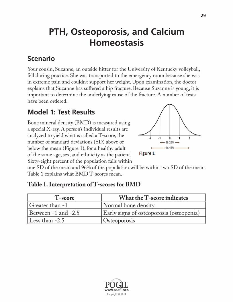
Model 1: Test ResultsBone mineral density (BMD) is measured using a special X-ray. A person’s individual results are analyzed to yield what is called a T-score, the number of standard deviations (SD) above or below the mean (Figure 1), for a healthy adult of the same age, sex, and ethnicity as the patient. Sixty-eight percent of the population falls within one SD of the mean and 96% of the population will be within two SD of the mean. Table 1 explains what BMD T-scores mean.
Table 1. Interpretation of T-scores for BMD
T-score What the T-score indicatesGreater than -1 Normal bone densityBetween -1 and -2.5 Early signs of osteoporosis (osteopenia)Less than -2.5 Osteoporosis
WWW.POGIL.ORG
Copyright © 2014

30
Table 2. Blood Test Results
*serum values were taken from the Normal Laboratory Values MKSAP®14 (http://www.acpinternist.org/weekly/archives/2008/7/1/mksap.pdf ) and Clinician’s Ultimate Reference (http://www.globalrph.com/index.htm)
QUESTIONS: Answer all questions with complete sentences
1. With your group members, discuss T-scores. Can negative T-scores be considered normal? Why or why not? (Hint: use information from both the table and the text to answer this question)
2. Suzanne receives a T-score of -3.6 in her bone mineral density (BMD) test results. What does this score indicate to her doctor?
3. Which items in Suzanne’s blood test are: a) Within the normal range? _______________________________________ b) Above the normal range? _______________________________________ c) Below the normal range? _______________________________________
4. Without using outside resources, what does your group suppose is the relationship between parathyroid levels and calcium levels in the blood?
Normal Serum Values for Females
Suzanne’s Serum Values
Parathyroid hormone
10-65 pg/ml [Conventional units] 10-65 ng/L [SI units] 128 pg/ml
Calcium 9.0-10.5 mg/dL [Conventional units] 2.2-2.6 mmol/L [SI units] 11.7 mg/dL
Calcitonin < 6.4 pg/ml [Conventional units] < 6.4ng/L [SI units] 2.8 pg/ml
Model 1 PTH, Osteoporosis, and Calcium Homeostasis

31
Model 2: Histology of Bone Tissue
The doctor diagnoses Suzanne with osteoporosis.
QUESTIONS: Answer all questions with complete sentences
5. Describe at least two differences between the photos of a typical bone (figure 2A) and Suzanne’s bones (figure 2B).
6. Using the information here (and not outside resources), write a definition for osteoporosis:
7. Which of the following explanations do you think is most accurate based on the information? Why? A. Suzanne fell which caused her hip to break. B. Suzanne’s hip broke, causing her to fall.
Model 2 PTH, Osteoporosis, and Calcium Homeostasis
32
Model 3: Hormones and Calcium Level RegulationSuzanne’s blood test indicates a condition called hyperparathyroidism, which means her parathyroid gland is producing an excess of parathyroid hormone (PTH). The doctor explained how this condition relates to Suzanne’s broken hip and osteoporosis.
“Calcium is necessary for many functions in the body, one being hardening of bone tissues which also serve as the body’s storehouse of that mineral. Calcium is also important in cardiac function and signaling within the nervous system. The primary hormone utilized to regulate the levels of calcium circulating in our blood is parathyroid hormone, also called PTH. There is a group of small glands, called the parathyroid glands, on the back side of the thyroid gland in your throat. The parathyroid gland senses calcium levels and adjusts its secretion rate of PTH. PTH indirectly stimulates osteoclasts, the bone pruners, in order to increase blood calcium.”
QUESTIONS: 8. Under what conditions is parathyroid hormone released?
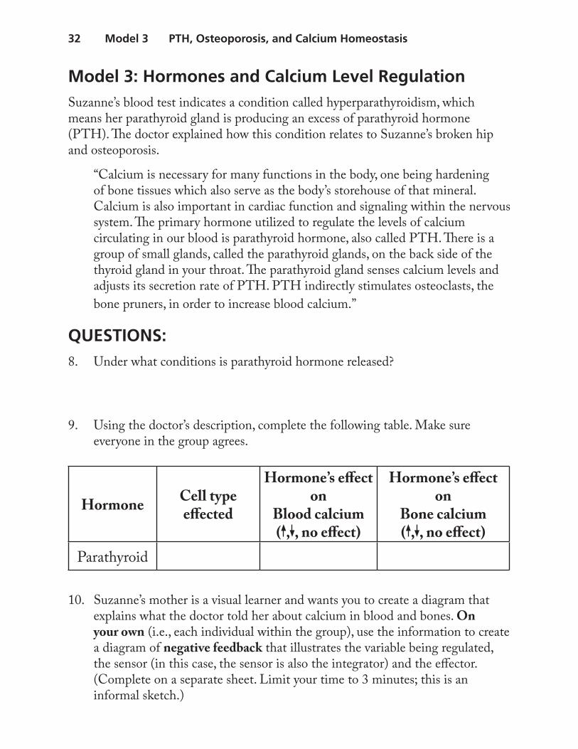
9. Using the doctor’s description, complete the following table. Make sure everyone in the group agrees.
10. Suzanne’s mother is a visual learner and wants you to create a diagram that explains what the doctor told her about calcium in blood and bones. On your own (i.e., each individual within the group), use the information to create a diagram of negative feedback that illustrates the variable being regulated, the sensor (in this case, the sensor is also the integrator) and the effector. (Complete on a separate sheet. Limit your time to 3 minutes; this is an informal sketch.)
Hormone Cell type effected
Hormone’s effect on
Blood calcium( , , no effect)
Hormone’s effect on
Bone calcium( , , no effect)
Parathyroid
Model 3 PTH, Osteoporosis, and Calcium Homeostasis

33
11. Back in your group, compare diagrams. As a group, create a new diagram of the calcium homeostasis feedback system based on the strengths of the individual diagrams. (Complete on a separate sheet. Limit your time to 5 minutes discussion and no more than 5 minutes drawing.)
12. Recall that Suzanne’s blood test indicates a condition called hyperparathyroidism. With input from all group members, explain how this condition contributes to osteoporosis. (use complete sentences)
13. Without using outside resources, create a list of factors, excluding hyper- parathyroidism, which might result in osteoporosis. Designate which factors are within a person’s control and which are not. (Make sure your group comes up with at least three in each column)
14. Now review your answers in the table above using reliable medical websites. List the websites you consult. Star the factors in your list above that you find on the websites, and add any additional factors you find to the list.
Factors within a Person’s Control
Factors not within a Person’s Control
Model 3 PTH, Osteoporosis, and Calcium Homeostasis