epidemiological studies of streptococcus … here an image sónia das neves nicolau nunes leitão...
TRANSCRIPT

Insert here an image
Sónia das Neves Nicolau Nunes Leitão
Epidemiological studies of Streptococcus pneumoniae
carriage in the post-vaccination era among two risk groups: children and the elderly.
Insert here an image with rounded corners
Dissertation presented to obtain the Ph.D. degree
in Biology/Molecular BiologyInstituto de Tecnologia Química e Biológica | Universidade Nova de Lisboa
Oeiras,November, 2012
Sónia das Neves Nicolau Nunes Leitão
Dissertation presented to obtain the Ph.D. degree
in Biology/ Molecular Biology
Epidemiological studies of Streptococcus pneumoniae
carriage in the post-vaccination era among two risk groups: children and the elderly.
in Biology/ Molecular BiologyInstituto de Tecnologia Química e Biológica | Universidade Nova de Lisboa
Oeiras, 14 November, 2012

iii

iv
Financial support from Fundação para a Ciência e a Tecnologia, Portugal
through grant SFRH/BD/40706/2007 awarded to Sónia Nunes.
First edition, September 2012
Second edition, November 2012
Cover design by Gonçalo Nunes
© Sónia Nunes
ISBN: 978-989-20-3211-5

v
Supervisors:
Raquel Sá-Leão
Hermínia de Lencastre
Examiners:
Ron Dagan
Carmen Muñoz-Almagro
Mário Ramirez
Isabel Couto
Dissertation presented on November 14, 2012 to obtain a Ph.D. degree in Biology/Molecular Biology by Instituto de Tecnologia Química e Biológica, Universidade Nova de Lisboa.

vi

vii
ACKNOWLEDGMENTS First of all, I would like to thank my supervisor, Dr. Raquel Sá-Leão, Head of the
Laboratory of Molecular Microbiology of Human Pathogens at Instituto de
Tecnologia Química e Biológica, for her support and supervision during my
PhD. I am intensely grateful for encouraging me to pursue PhD. She is not only
a supervisor, she is also a friend. Thank you for the confidence, guidance and
critical sense.
I would like to thank Dr Hermínia de Lencastre, my co-supervisor, for the
opportunity to start this PhD in the Laboratory of Molecular Genetics. Thank you
for trusting me enough and making me believe in myself demystifying the bad
“wolf” for me - the English language to “finally” start my PhD. Thank you for
critical sense and helpful commentaries.
I would like to thank my PhD thesis committee, Dr. Josefina Liñares and Dr. Ron
Dagan, for their availability, critical sense and interesting discussions. I learned
a lot with them.
All my friends and colleagues at the Laboratory of Molecular
Genetics/Laboratory of Molecular Microbiology of Human Pathogens at the
Instituto de Tecnologia Química e Biológica for their encouragement, sometimes
help in the bench, critical sense and for providing a good work environment.
To Hospital Pediátrico de Coimbra, mainly to Dr. Fernanda Rodrigues, for the
collaboration in a study that resulted in a publication.
To the National Institute for Public Health and the Environment (RIVM) in
Bilthoven, The Netherlands. Thanks to Dr. Leo Schouls and Karin Elberse for
the collaboration that resulted in a publication.
viii
I would like to thank Dr. Ilda Santos-Sanches my first supervisor in research
training.
I would like to thank also old colleagues and friends that shared with me my first
adventure in research, Rosario Mato, Marta Aires de Sousa, Susana Gardete,
Carla Alves, and Natacha Sousa.
À Sra. D. Manuela Nogueira, Sra. D. Maria Cândida e Sra. D. Isilda pela sua
amizade e ajuda desde o primeiro instante.
I would like to thank the Instituto de Tecnologia Química e Biológica that
accepted me as PhD student and in particularly the Laboratory of Molecular
Genetics and more recently the Laboratory of Molecular Microbiology of Human
Pathogens, which provided excellent conditions without which it would have
been impossible to perform this work.
It is also important to mention the financial support of Fundação para a Ciência
e Tecnologia (SFRH/BD/40706/2007, my PhD grant, and PEst-
OE/EQB/LA0004/2011 to Associate Laboratory of Oeiras).
Aos meus amigos, que por várias vezes me apoiaram, tanto ficando com os
meus filhos quando tinha que me deslocar a congressos ou necessitava de ficar
a trabalhar até mais tarde, como dando força para continuar.
Aos meus pais e ao meu irmão, que sempre me apoiaram.
Aos meus filhos e ao meu marido…

ix
ABSTRACT
Streptococcus pneumoniae is a global cause of disease including pneumonia,
otitis media, conjunctivitis, sepsis, and bacterial meningitis. These infections are
not essential to the transmission or long-term survival of the bacterium; indeed,
S. pneumoniae depends on asymptomatic colonization of the human
nasopharynx for its dissemination to additional hosts. Considering this,
colonization studies are a good way to monitor changes in the pneumococcal
epidemiology that may result from the use of antibiotics and vaccines. The
molecular characterization of pneumococci is crucial to assess these changes
which highlight the need for the development and validation of easier and faster
methods of molecular typing.
Since 1996 our group has been monitoring the pneumococcal population
colonizing children attending day care centers. However, for several years these
studies have been confined to the Lisbon area. In this PhD we have addressed
this situation by including other regions of Portugal in our study. In addition, we
have started to study pneumococcal colonization in the elderly, the other age
group where the incidence of pneumococcal infections is high.
This thesis summarizes five studies conducted during this PhD. The first four
studies were focused on the pneumococcal epidemiology among the two age
groups where the rates of pneumococcal disease are highest: children up to six
years old and adults older than 60 years. The fifth and last study describes the
evaluation and validation of a new genotyping strategy for pneumococci.
The first study was a retrospective study aimed to describe the epidemiology of
the recently discovered serotype 6C colonizing healthy children attending day
care centers between 1996 and 2007. In that study, using PCR serotyping and
the Quellung reaction, pulsed-field gel electrophoresis (PFGE), multilocus
x
sequence typing (MLST), and antibiotyping, we observed that this serotype had
been circulating in Portugal since at least 1996, it is genetically diverse, and
often antibiotic resistant.
The second study was conducted in Coimbra and aimed to determine the
prevalence of pneumococcal carriage and to characterize the pneumococcal
strains colonizing children attending day care centers in this region in the era of
pneumococcal vaccines. Between January and February 2007, nasopharyngeal
swabs were obtained from 507 children (76.7% had received at least one dose
of PCV7) and 61.3% were pneumococcal carriers. All 311 pneumococcal
samples were antibiotyped and serotyped and the pneumococcal strains that
were resistant to at least one antibiotic were also typed by PFGE. We have
found in Coimbra similar rates of colonization and antimicrobial resistance
patterns and similar genotypes to those previously described in the Lisbon area.
The aim of the third study was to compare the patterns of colonization among
young children who attended day care centers and lived in two different areas:
an urban area, Oeiras, and Montemor-o-Novo, a rural area in the Southern part
of Portugal. In this study 1088 and 756 nasopharyngeal swabs from day care
center attendees from urban and rural area, respectively, were collected.
Pneumococcal strains were characterized by antibiotype and serotype. Similar
rates of colonization (c.a. 61%) were found; however, in the urban area there
were higher rates of antimicrobial resistance (32.4% vs 21.6%, p<0.001) and
higher rates of antimicrobial consumption one month before sampling (16.7% vs
11.6%, p=0.004). Multivariable logistic regression analysis was performed to
identify the factors associated with differences between the two regions.
Antibiotic consumption during last month, being colonized with serotype 19A or
non-typeable strains, and attending day care in Oeiras and being colonized with
serotype 19A, were risk factors for being colonized by pneumococcal resistant
strains. This study highlighted that in the era of widespread use of
xi
pneumococcal conjugate vaccines in Portugal, antibiotic consumption remains a
main driving force for the maintenance of antimicrobial resistant pneumococci in
the community.
To gain insights into the pneumococcal carriage patterns among the elderly, the
fourth study was performed in two distinct areas: Oeiras and Montemor-o-Novo.
In this study, 1,298 nasopharyngeal samples were collected. All pneumococci
were antibiotyped, serotyped and characterized by MLST. Association between
pneumococcal carriage, socio-demographic and clinical factors was evaluated
using a logistic regression. The rates of colonization in this age group were low
(2.2%), but there were high variability of colonizing serotypes and genetic
backgrounds. Out of the 30 pneumococcal isolated, sixteen showed
antimicrobial resistance. Smoking was a risk factor for pneumococcal
colonization and living in the rural area seemed to increase the rate of
pneumococcal colonization in this population. This study provided an important
baseline to monitor the impact of pneumococcal vaccines on the patterns of
colonization among the elderly.
In the last part of this thesis, we described and validated a newly developed
multiple-locus variable tandem repeat analysis (MLVA) method to easily and
rapidly genotype pneumococcus. This method was compared to the current gold
standards methods, MLST and PFGE. The three typing methods showed
Simpson´s diversity indices of 98.5% or higher. The Wallace coefficient of MLVA
and MLST was 0.874, meaning that if two strains had the same MLVA type they
had an 88% chance of having the same MLST type. For some isolates
belonging to a single MLST clonal complex, despite displaying different
serotypes, MLVA was more discriminatory, generating groups according to
serotype or serogroup. This study showed that MLVA is a promising genotyping
method for S. pneumoniae.

xii
Altogether, these studies have contributed to improve our knowledge on
pneumococcal colonization in Portugal in two age groups, children and the
elderly.
The main conclusions of this thesis were:
i) the recently described serotype 6C is frequently carried by healthy young
children in Portugal, is genetically diverse, and has been circulating in
our country at least since 1996;
ii) the patterns of pneumococcal colonization among healthy children living
in Coimbra are similar to those living in the Lisbon area;
iii) antibiotic consumption remains a main cause for the maintenance of
antimicrobial resistance, in the era of widespread use of pneumococcal
conjugate vaccines;
iv) the rates of pneumococcal colonization in the elderly are low and the
serotype and genotype diversity are high;
v) MLVA is a promising genotyping method to characterize pneumococci.
xiii
RESUMO
Streptococcus pneumoniae, ou pneumococos, é responsável por várias
doenças em todo o mundo nas quais se incluem as infeções do trato
respiratório, do ouvido médio, conjuntivite, meningite e sepsis. No entanto, é a
elevada prevalência na nasogaringe em portadores assintomáticos que
contribui para a sua disseminação na população. Assim, os estudos de
colonização por pneumococos são importantes pois permitem monitorizar a
flora da nasofaringe em vários grupos etários, e estudar a influência da
utilização de vacinas e do uso de antibióticos. Igualmente importante é a
possibilidade de otimização das vacinas de acordo com as características da
população alvo. A caracterização molecular de pneumococos é crucial para a
avaliação dessas alterações, o que alerta para a necessidade do
desenvolvimento e validação de métodos de tipagem molecular fáceis e
rápidos.
Desde 1996 que o nosso grupo tem vindo a estudar a população pneumocócica
em crianças saudáveis que frequentam creches e jardins de infância. No
entanto, esses estudos estiveram desde sempre confinados à área da grande
Lisboa. Com o intuito de obter uma amostra populacional mais abrangente e
variada, esta tese inclui estudos de outras regiões de Portugal não só em
crianças mas também na população idosa, uma vez que este último constitui
também um grupo etário de risco, com elevada incidência de doença
pneumocócica.
Nesta tese são apresentados cinco estudos que foram realizados durante este
doutoramento. Os primeiros quatro estudos focaram-se na epidemiologia de
pneumococos, tendo sido envolvidas nos primeiros três, crianças até aos 6
anos de idade e no quarto estudo, adultos com mais de 60 anos. O quinto e
xiv
último estudo descreve e faz-se a validação de uma nova estratégia de tipagem
molecular para pneumococos.
O primeiro trabalho é um estudo retrospectivo que descreve a epidemiologia de
um serotipo recentemente descoberto, 6C. Este estudo utilizou amostras que
foram recolhidas da nasofaringe de crianças saudáveis que frequentavam
creches e jardins de infância entre 1996 e 2007. Nesse estudo, utilizou-se a
serotipagem por PCR complementada com a reacção de Quellung,
electroforese em campo pulsado (PFGE), “multilocus sequence typing” (MLST)
e antibiograma, para caracterizar os isolados. Concluiu-se que este serotipo já
circulava em Portugal pelo menos desde 1996, que é geneticamente diverso, e
que frequentemente apresenta resistência a antibióticos.
O segundo estudo foi realizado em Coimbra e teve como objectivos determinar
a prevalência de portadores de pneumococos e caracterizar as estirpes de
pneumococos que colonizam crianças que frequentam creches e jardins de
infância nesta região, na era das vacinas pneumocócicas. Entre Janeiro e
Fevereiro de 2007 foram pesquisadas 507 crianças (76.7% tinham recebido
pelo menos uma dose de vacina pneumocócica conjugada 7-valente) das quais
61.3% eram portadores de pneumococos. Todos os 311 pneumococos isolados
foram serotipados e foi realizado o antibiograma. Às estirpes de pneumococcus
que apresentavam resistência a pelo menos um agente antimicrobiano foi
também realizada a tipagem por PFGE. Em Coimbra as taxas de colonização,
os padrões de resistência a antibióticos e os genotipos encontrados foram
semelhantes aos descritos previamente na área metropolitana de Lisboa.
O objectivo do terceiro estudo foi comparar os padrões de colonização em
crianças que vivem e frequentam creches e jardins de infância em duas áreas
distintas: uma área urbana, Oeiras, e uma área rural na região centro-sul de
Portugal, Montemor-o-Novo. Neste estudo foram recolhidas 1088 e 756
zaragatoas da nasofaringe de crianças da área urbana e da área rural,

xv
respectivamente. Os pneumococos foram caracterizados por serotipagem e
antibiograma. As taxas de colonização encontradas foram semelhantes nas
duas regiões (c.a. 61%). No entanto, na área urbana o nível de resistência a
agentes antimicrobianos foi mais elevado (32.4% vs 21.6%, p<0.001), o mesmo
acontecendo relativamente às taxas de consumo de antibióticos um mês antes
da amostragem (16.7% vs 11.6%, p=0.004). Foi realizada uma análise
estatística multivariada para identificar os fatores associados às diferenças
encontradas nas duas regiões. O consumo de antibióticos durante o mês
antecedente à recolha da amostra, estar colonizado com o serotipo 19A ou com
uma estirpe não-tipável, e frequentar creche ou jardim de infância em Oeiras
foram considerados fatores de risco para a colonização por pneumococos
resistentes a antibióticos. Este estudo alerta para o facto do consumo de
antibióticos continuar a ser a principal causa para a manutenção dos níveis de
pneumococos resistentes a antibióticos encontrados na comunidade apesar da
grande utilização de vacinas pneumocócicas conjugadas na comunidade infantil
em Portugal.
O quarto estudo foi realizado com o objetivo de obter informação relativa aos
padrões de colonização de pneumococos na população idosa. Neste estudo,
1298 amostras da nasofaringe foram recolhidas em duas áreas distintas: Oeiras
e Montemor-o-Novo. Todos os pneumococos foram serotipados e
caracterizados por MLST e foi também realizado o antibiograma. A associação
entre portadores de pneumococos, fatores socio-demográficos e fatores
clínicos foi avaliada usando uma regressão logística. As taxas de colonização
neste grupo etário foram baixas (2.2%), mas encontrou-se uma grande
variedade de serotipos e genotipos. Dos 30 pneumococos isolados, 16
mostraram resistência a agentes antimicrobianos. Foram identificados como
fatores de risco para a colonização por pneumococos tabagismo e habitar em
área rural. Este estudo possibilitou a criação de uma importante linha de
xvi
referência para monitorizar o impacto das vacinas pneumocócicas nos padrões
de colonização nos idosos.
Na última parte desta tese foi descrita e validada uma nova estratégia de
“multiple-locus variable tandem repeat analysis” (MLVA) aplicada a
pneumococos que permite de uma forma fácil e rápida, fazer uma análise
genotípica deste microrganismo. Este método foi comparado com os métodos
mais utilizados atualmente MLST e PFGE. Os três métodos mostraram índices
de diversidade de Simpson de 98.5% ou superiores. O coeficiente de Wallace
para MLVA e MLST foi de 0.874, o que significa que se duas estirpes tiverem o
mesmo tipo de MLVA tem 88% de probabilidade de terem o mesmo tipo de
MLST. O MLVA foi mais discriminativo para alguns isolados do mesmo
complexo clonal (por MLST), gerando grupos de acordo com o serotipo ou
serogrupo. Este estudo mostrou que o MLVA é um método promissor para
genotipar S. pneumoniae.
Em suma, os trabalhos apresentados contribuíram para melhorar o nosso
conhecimento em relação à colonização por pneumococos em Portugal em
duas faixas etárias, crianças e idosos. As principais conclusões desta tese
foram:
i) o serotipo 6C, recentemente descrito, é frequentemente encontrado em
crianças saudáveis em Portugal, é geneticamente diverso e tem estado
em circulação no nosso país pelo menos desde 1996;
ii) os padrões de colonização por pneumococos em crianças saudáveis
que vivem em Coimbra são semelhantes aos encontrados nas crianças
que vivem na área de Lisboa;
iii) o consumo de antibióticos continua a ser a principal causa da
manutenção da resistência antimicrobiana na era da grande utilização
de vacinas pneumocócicas conjugadas;
xvii
iv) as taxas de colonização por pneumococos nos idosos são baixas mas a
diversidade de serotipos e de genotipos é elevada;
v) o MLVA é um método de genotipagem promissor para caracterizar
pneumococos.

xviii
THESIS OUTLINE
The studies presented in this thesis provide an overview of pneumococcal
colonization in Portugal in two age groups: children and the elderly.
Chapter I provides an outline of Streptococcus pneumoniae as a bacterium that
frequently colonizes the nasopharynx but can be also responsible for serious
diseases. Topics such as historical highlights, epidemiology, identification,
typing methods and the effect of pneumococcal vaccines are addressed.
Chapter II describes the temporal trends and molecular epidemiology of the
recently described serotype 6C of Streptococcus pneumoniae. The study
analyses the evolution of this serotype from 1996 to 2007, by antibiotyping,
serotyping and also using PFGE and MLST.
Chapter III describes the pneumococcal population colonizing the nasopharynx
of children attending day care centers in Coimbra, a city in the central region of
Portugal.
Chapter IV compares the patterns of pneumococcal colonization of young
children attending day care centers in two distinct areas of Portugal: one rural,
Montemor-o-Novo, and other urban, Oeiras.
Chapter V is a study of pneumococcal colonization in the elderly in Portugal
that was also performed in an urban area, Oeiras, and a rural area, Montemor-
o-Novo.
Chapter VI describes and validates a novel genotyping strategy applied to
pneumococci: multiple-locus variable tandem repeat analysis (MLVA).
xix
Chapter VII presents general conclusions and futures perspectives.
Chapters II to VI are reproductions of the publications indicated below. They can be read independently.
Chapter II
Nunes S.*, C. Valente*, R. Sá-Leão, and H. de Lencastre. 2009.
Temporal trends and molecular epidemiology of recently described
serotype 6C of Streptococcus pneumoniae. J Clin Microbiol. 47:472-4.
*These authors contributed equally.
Chapter III
Rodrigues F.*, S. Nunes *, R. Sá-Leão, G. Gonçalves, L. Lemos, H.
de Lencastre. 2009. Streptococcus pneumoniae nasopharyngeal
carriage in children attending day-care centers in the central region of
Portugal, in the era of 7-valent pneumococcal conjugate vaccine. Microb
Drug Resist. 15:269-77. *These authors contributed equally.
Chapter IV
Nunes S., C. Valente, A. S. Simões, A. C. Paulo, A. Brito-Avô, H. de
Lencastre and R. Sá-Leão. 2012. Antibiotic consumption remains a
main driving force of antimicrobial resistance in the era of pneumococcal
conjugate vaccines. In preparation.
Chapter V
Nunes S., S. Almeida, A. C. Paulo, I. Valadares, S. Martins, F. Breia,
A. Brito-Avô, A. Morais, H. de Lencastre, and R. Sá-Leão. 2012. Low
pneumococcal carriage and high serotype diversity among elderly living
in Portugal. In preparation.

xx
Chapter VI
Elberse KE*, S. Nunes *, R. Sá-Leão, H.G. van der Heide, and L.M.
Schouls. 2011. Multiple-locus variable number tandem repeat analysis
for Streptococcus pneumoniae: comparison with PFGE and MLST. PLoS
One. 6:19668. *These authors contributed equally.
xxi
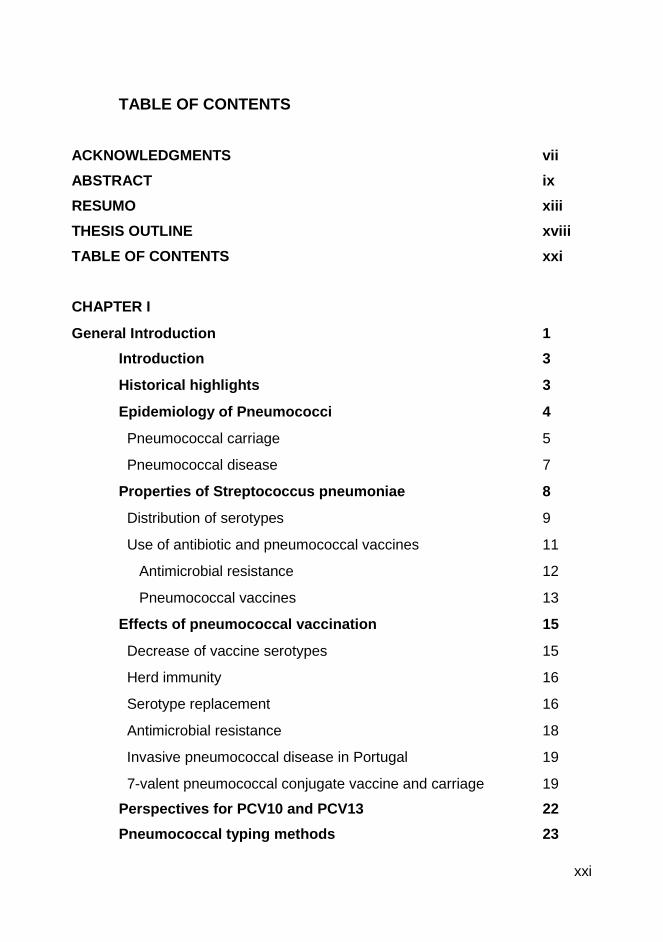
TABLE OF CONTENTS ACKNOWLEDGMENTS vii
ABSTRACT ix
RESUMO xiii
THESIS OUTLINE xviii
TABLE OF CONTENTS xxi
CHAPTER I
General Introduction 1
Introduction 3
Historical highlights 3
Epidemiology of Pneumococci 4
Pneumococcal carriage 5
Pneumococcal disease 7
Properties of Streptococcus pneumoniae 8
Distribution of serotypes 9
Use of antibiotic and pneumococcal vaccines 11
Antimicrobial resistance 12
Pneumococcal vaccines 13
Effects of pneumococcal vaccination 15
Decrease of vaccine serotypes 15
Herd immunity 16
Serotype replacement 16
Antimicrobial resistance 18
Invasive pneumococcal disease in Portugal 19
7-valent pneumococcal conjugate vaccine and carriage 19
Perspectives for PCV10 and PCV13 22
Pneumococcal typing methods 23

xxii
Antibiotyping 24
Pneumococcal serotyping 24
PFGE and MLST 26
MLVA 27
Whole genome sequencing 28
Aim of the work 29
References 30
CHAPTER II
Temporal trends and molecular epidemiology of the recently described serotype 6C of Streptococcus pneumoniae 53
Abstract 55
Introduction 55
Materials and Methods 56
Results and discussion 58
Conclusion 62
Acknowledgments 62
References 63
CHAPTER III
Streptococcus pneumoniae nasopharyngeal carriage in children attending day-care centers in the central region of Portugal, in the era of 7-valent pneumococcal conjugate vaccine 67
Abstract 69
Introduction 70
Materials and Methods 71
Results 75
Discussion 85
Acknowledgments 87

xxiii
References 88
CHAPTER IV
Antibiotic consumption remains a main driving force of antimicrobial resistance in the era of pneumococcal conjugate vaccines 95
Abstract 97
Introduction 97
Materials and Methods 98
Results 101
Discussion 107
Acknowledgments 109
References 109
CHAPTER V
Low pneumococcal carriage and high serotype diversity among elderly living in Portugal 115
Abstract 117
Introduction 118
Materials and Methods 119
Results 121
Discussion 126
Acknowledgments 127
Funding 128
References 128
CHAPTER VI
Multiple-locus variable number tandem repeat analysis for Streptococcus pneumoniae: comparison with PFGE and MLST 133

xxiv
Abstract 135
Introduction 136
Materials and Methods 139
Results 142
Discussion 150
References 153
Supplementary tables 160
CHAPTER VII
Conclusions and future perspectives 169

1
General Introduction

2
3
INTRODUCTION
Streptococcus pneumoniae, or pneumococcus, is a Gram-positive bacterium
responsible for high rates of mortality and morbidity worldwide. It is a leading
cause of disease particularly among young children, the elderly, and the immuno-
compromised of all ages. Although it colonizes asymptomatically the nasopharynx,
S. pneumoniae can cause otitis media, sinusitis, pneumonia and more severe
diseases such as bacteremia and meningitis. Pneumococcal disease remains a
great concern worldwide despite great advances aiming to decrease it, such as
the use of antibiotic therapy and prevention through vaccination.
Since colonization precedes disease, epidemiological studies monitoring the
nasopharynx flora are valuable tools to anticipate the emergence of serotypes that
may became responsible for disease in the future.
HISTORICAL HIGHLIGHTS
In 1875 Edwin Klebs was the first to recognize Streptococcus pneumoniae in lung
tissue and in infected sputum (reviewed in (54)), but only six years later this
bacterium was isolated by two researchers working in France and the USA. In
January 1881, Louis Pasteur in France reported for the first time the isolation of S.
pneumoniae, after finding bacteria in the saliva of a youngster with rabies. George
Miller Sternberg was in New Orleans studying malaria fever when he sampled his
own saliva (as a control for his experiments) and injected it subcutaneously in a
rabbit, which quickly died. This observation was in September 1880, but did not
publish his report until April 1881(reviewed in (54)).
More than 90 years went by before this bacterium was named as Streptococcus
pneumoniae. Pasteur called his isolate the “microbe septicémique du saliva” and
Gen
eral
intr
oduc
tion

4
Sternberg called his “Micrococcus pasteuri”. In 1883 Mátray applied the term
“pneumoniekokken” and Albert Fraenkel in 1886, after the first complete
description of the bacterium, gave us the familiar name “pneumokokkus”. In the
same year, Anton Weichselbaum suggested the name Diplococcus pneumoniae,
which was used until the reclassification, on the basis of its growth in chains in
liquid medium, as Streptococcus pneumoniae, in 1974 (reviewed in (54)).
After its isolation and identification, S. pneumoniae was the model organism used
in important discoveries, including Gram staining in 1884 and the putative use of
polysaccharide antigens as vaccines (reviewed in (54)). The identification of DNA
as the source of genetic information was done using pneumococcus. Griffith in
1928 showed that avirulent strains could be transformed into virulent strains.
Avery, MacLeod and McCarty demonstrated in 1944 that DNA was responsible for
the transformation and thus the carrier of genetic information (reviewed in (54)).
The therapeutic efficacy of penicillin, the role of bacterial capsule in resistance to
phagocytosis (reviewed in (54)), and the first quorum sensing factor (149) were
also described for the first time in pneumococci.
EPIDEMIOLOGY OF PNEUMOCOCCI
Diseases caused by the pneumococcus constitute a major global public health
problem. According to the World Health Organization (WHO), in 2005, about 1.6
million deaths were caused by this agent annually, from which 0.7-1 million were
among children under five years. In developing countries, pneumococcal disease
is common in children under two years; in the elderly population the burden of
disease is largely unknown. HIV infection and other conditions associated with
immunodeficiency greatly increase the likelihood of contracting pneumococcal
disease. Of the deaths occurring in HIV-negative children, over 60% occur in ten
African and Asian countries (109) (Figure 1).

5
In the USA and Europe S. pneumoniae is the most common cause of pneumonia
in adults. The annual incidence of invasive pneumococcal disease (IPD) in these
regions ranges from 10 to 100 cases per 100,000 (WHO,
www.who.int/ith/disease/pneumococcal).
Figure 1. – Rates of Streptococcus pneumoniae death (per 100,000 children under age 5).
Data from WHO August, 2009.
Pneumococcal carriage
The pneumococcus is a normal component of the nasopharyngeal mucous
membrane microflora. Acquisition is the first stage towards carriage and possible
infection with pneumococcus. Carriage is usually asymptomatic. Transmission
occurs by airborne droplets or direct contact with respiratory secretions (102).
Recently, it has been suggested that environmental surfaces may also serve as
sources of pneumococcal infection as pneumococci were found to be able to
survive long-term desiccation (157).
Gen
eral
intr
oduc
tion

6
Children are the major carriers and colonization occurs soon after birth (6). The
peak in incidence is more or less at three years of age (15) and then declines with
increasing age, until 10 years of age, remaining low in adulthood (15). Few studies
have studied nasopharyngeal colonization in the elderly, but the carriage rates are
typically low, ranging between 2 and 5% (2, 15, 68, 121).
Since it is in young children where the highest frequency of pneumococcal
colonization and the highest crowding index are found, this group is considered
the most important vector for horizontal dissemination of pneumococci in the
community (88). Consequently, part of the strategy to prevent pneumococcal
disease focuses on prevention of nasopharyngeal colonization.
In Portugal, data of pneumococcal carriage in healthy children attending day care
centers, have been published by our group since 1996 and rates have been
generally close to 60-65% (42, 43, 95, 104, 125, 128, 137). Concerning
colonization in the elderly in Portugal, no studies had been conducted until
recently. This topic was addressed for the first time in the pioneer study described
in Chapter V of this thesis.
Pneumococcal carriage is not a permanent state. In fact, once established in the
nasopharynx, pneumococci are cleared within weeks to months; subsequently the
nasopharynx can be recolonized by another strain. The duration of carriage is
variable (5 to 290 days) depending on the serotype and the age of the carrier (53,
141, 143). Risk factors for colonization among others are young age (lower than 6
yrs), having young siblings, crowding (e.g. attending day-care centers, prisons,
military training campus, sports with contact, retirement homes), season, low
socioeconomic status, other respiratory infections such as influenza,
immunosuppression such as HIV-infection, exposure to cigarette smoke, and
asthma (1, 7, 23, 51, 53, 80).
7
Most studies on pneumococcal colonization studies are based on the
characterization of a single isolate of each individual. However, more than one
pneumococcal serotype or clone can coexist in the nasopharynx of an individual
(50, 53, 108, 129, 151). For this reason, studies on co-colonization are very
important to identify changes at the individual level, such as de novo acquisition,
clearance and unmasking of pneumococcal strains (which can include serotypes
or clones) (37, 90), particularly in the era of multivalent pneumococcal conjugate
vaccines.
Pneumococcal disease
S. pneumoniae may spread from the nasopharynx into the respiratory tract or the
bloodstream to cause infections. These infections can be divided in two groups:
invasive (isolated from sterile body sites) and non-invasive (isolated from non-
sterile body sites). Otitis media and sinusitis are non-invasive infections. The
nasopharynx is connected with the middle ear cavity via the Eustachian tube,
which is a pressure regulator and prevents the entrance of the bacteria in the
middle ear cavity. However, if the Eustachian tube is blocked, the bacteria may be
trapped in the middle ear and cause otitis media. Sinusitis is caused when the
bacteria spread locally into the sinuses and cause an accumulation of fluids in the
sinuses due to an obstruction. The pneumococcus can also be responsible for
more severe infections such as pneumonia, bacteremia and meningitis. From the
nasopharynx, the bacteria may spread into the lungs or directly to the
bloodstream. If bacteria spread into the lungs the immune system reacts and the
accumulation of fluids and bacteria in the alveoli decreases oxygen transport and
causes pneumonia (74). In adults, pneumonia without bacteremia is the most
common non-invasive pneumococcal infection, however if the pneumococcus
spreads into bloodstream, the infection becomes invasive. Pneumonia with
bacteremia and meningitis are the most frequently recognized invasive disease.
Pneumococcal meningitis occurs when the bacteria spread directly to bloodstream
into the meninges (membranes that surround and protect the brain and spinal
Gen
eral
intr
oduc
tion

8
cord). The meninges are filled with a liquid called cerebrospinal fluid where the
bacteria can multiply freely, causing inflammation and swelling of the meninges
and the brain tissue. Other invasive diseases are sepsis, peritonitis and arthritis.
Risk factors for pneumococcal infection are numerous and include, among many
others, extremes of age (under 5 years of age and over 60 years old),
immunosuppression, presence of underlying medical conditions, defects in host
immune responses, low socioeconomic status, malnutrition, cigarette smoking and
alcoholism (29, 91, 106, 107).
In Portugal, the report of pneumococcal infectious disease is not mandatory and,
to my knowledge, no study has been carried out to calculate the incidence of
invasive pneumococcal disease (IPD) over time. However, epidemiological studies
on pneumococcal infections using data from several hospitals in different areas of
Portugal have been conducted (3, 5, 63, 134, 135).
PROPERTIES OF STREPTOCOCCUS PNEUMONIAE
Streptococcus pneumoniae is a lancet-shaped Gram-positive bacterium, whose
diameter ranges between 0.5 and 1.25 µm for an individual cell. Pneumococci are
frequently seen as pairs of cocci (diplococci), but they can also be in single cell
and in short chains. It is a fastidious organism, growing best in media containing
blood and in an enriched atmosphere with 5% CO2. On blood agar, colonies
characteristically produce alpha hemolysis, form an inhibition halo ≥14 mm around
a 5 mg optochin disc, and are soluble in bile salts (e.g. deoxycholate). Addition of
a few drops of 10% deoxycholate at 37°C, lyses the entire culture in minutes. The
majority of pneumococci have a polysaccharide capsule and the assignment of a
capsule type through serotyping is also used for identification of this bacterium.
However, some pneumococci do not have a capsule and others give an atypical

9
result in one or more of the assays described above (4, 32, 105, 111, 117, 127,
138).
The capsule is the main virulence factor of the pneumococcus and, with the
exception of serotype 3, is covalently linked to the peptidoglycan of the cell wall
(146). The capsule is 200 to 400nm thick (140), protects against
opsonophagocytosis, and plays an important role in the interaction between the
bacteria and the epithelium in colonization of the upper respiratory tract (103, 158).
In addition, the pneumococcus may shift from transparent-phase (capsule less
dense and thicker cell wall) to opaque-phase and vice-versa. This allows for
attachment to the epithelial cells in carriage. The pneumococci in opaque-phase
have denser capsules, providing better protection against opsonization and killing
in invasive disease (159).
Based on the reaction of the capsular polysaccharide with polyclonal factor sera
and more recently with the use of monoclonal antibodies, 94 serotypes are
recognized up to now (16, 21, 22, 61, 115).
The first “novel” serotype recognized using monoclonal antibodies was 6C (115).
As it was previously classified as 6A by the Quellung reaction, the real prevalence
of this serotype and its characteristics were not known. In order to obtain insights
on its epidemiology in colonization in Portugal, a retrospective study was
performed and is described in this thesis in Chapter II.
Distribution of serotypes
Although 94 distinct pneumococcal serotypes, varying in capsular polysaccharide
structure, have been described, not all seem to have the same capacity to cause
disease (18, 60, 126, 136). Some serotypes can be carried in the nasopharynx
asymptomatically and cause disease in a small proportion of infected individuals
only. Others are rarely identified in the nasopharynx but are frequently associated
with disease. Serotypes 1 and 5 are examples of serotypes that rarely are
Gen
eral
intr
oduc
tion
10
detected in colonization, but are responsible for a high proportion of invasive
disease and are frequently associated with outbreaks (8, 13, 38, 96, 123). Before
the introduction of pneumococcal conjugate vaccines 10 to 12 serogroups were
responsible for the majority of pneumococcal invasive disease worldwide,
including 1, 3, 4, 6A, 6B, 7F, 8, 9V, 14, 18C, 19F, and 23F (62, 112).
The prevalence of pneumococcal serotypes/serogroups causing IPD depends on
several factors, such as geographic location, disease manifestation, and age of
the host (15, 18, 19, 60). In young children the serogroups/serotypes 6, 14, 18, 19
and 23F are predominant due to the lower immunogenicity of these serotypes
(20).
Several authors have been looking at the distribution of serotypes in colonization
and disease to estimate the invasive potential (18, 19, 55, 131, 141). According to
Brueggeman et al. (18) and Sleeman et al. (141) serotypes 1, 4, 5, 7F, 9V, 14,
18C, and 19A are classified as highly invasive serotypes. In a study reported by
Greenberg et al. (55) the authors estimated the disease potential in pediatric
community-acquired alveolar pneumonia. In this study the nasopharyngeal
samples were also taken from healthy children during disease and the serotype-
specific odds ratios were calculated. They observed that serotypes 1, 5, 7F, 9V,
14, 19A, and 22F have a higher disease potential than serotypes 6A, 6B, 23A, and
35B in childhood pneumonia. In Israel another study was conducted to assess the
invasive disease potential of pneumococcal serotypes causing IPD, acute otitis
media and acute conjunctivitis in children. Odds ratios for each disease were
calculated and a significant positive association with IPD was found for serotypes
1, 5 and 12F. Also, a significant positive association was found in otitis media for
serotypes 1, 3, 5, 12F, 19A and 19F, and in acute conjunctivitis, for serotype 3 and
NT strains. On the other hand, a significant negative association with IPD was
found in NT strains and in acute otitis media in serotypes 6A, 6B, 15A and NT
strains (136).

11
In Portugal, a study was published (126) where the invasive disease potential of
serotypes and clones circulating in Portugal before the introduction of PCV7 was
calculated. In this study serotypes 1, 3, 4, 5, 7F, 8, 9N, 9L, 12B, 14, 18C and 20
were found to have a high propensity to cause invasive disease, whilst serotypes
6A, 6B, 11A, 15B/C, 16F, 19F, 23F, 34, 35F, and 37 were associated with
carriage. Additionally, significant differences in invasiveness were found between
clones that shared the same serotype, namely among serotypes 3, 6A, 6B, 11A,
14, 19A, 19F, 22F, 23F, 34 and NT, which highlights the importance of the genetic
background when analyzing the invasive disease potential of certain serotypes.
The distribution of serotypes is also affected by temporal changes, inherent of
secular trends of specific serotypes, and this has been reported in several studies
(57, 59, 71, 120).
Serotype distribution can be altered by antibiotic consumption and vaccination
patterns. Following the introduction of the first pneumococcal conjugate vaccine,
the increase of non-vaccine serotypes has been reported in several countries, in
disease and also in colonization (more details are presented in the next topic).
Use of antibiotics and pneumococcal vaccines
Nowadays, the main tool to control pneumococcal disease, besides antibiotic
therapy, is vaccination. Efforts to develop effective pneumococcal vaccines began
in 1911, when the first clinical trial on the effectiveness of a whole-cell
pneumococcal vaccine was conducted by Wright et al. in South-Africa (reviewed in
(93)). Several decades were needed before the effectiveness of pneumococcal
vaccines was demonstrated. Only at the end of World War II in 1945 (92), the first
capsular polysaccharide vaccine trials were undertaken.
Gen
eral
intr
oduc
tion
12
Antimicrobial resistance
Before penicillin became available for medical treatment in the early 1940s, no
cure for pneumonia existed and many people died with pneumococcal infection.
The introduction and use of antimicrobial drugs changed this scenario and led to
the withdrawn from the market of a pneumococcal vaccine that was licensed and
in usage. However, it did not take long before it became clear that antimicrobials
per se had not eliminated pneumococcal disease.
In 1964, Austrian and Gold reported that one in four patients admitted with
pneumococcal bacteremia died even with antimicrobial therapy (12). In 1967, the
first pneumococcal isolate with intermediate resistance to penicillin was described
by Hansman et al. (58) in Australia. Within a decade, similar strains were found in
South Africa, Europe and North America (77). The first multiresistant
pneumococcal isolates were reported by Jacobs et al. in 1978 (70) and since then
we have been observing a widespread of multidrug resistance has been observed
(33, 97).
The rates of antimicrobial resistance differ from country to country and depend on
several factors as antibiotic consumption habits, use of pneumococcal vaccines,
and geographic areas. The last report from the European Antimicrobial Resistance
Surveillance Network (EARS-Net Database) from 2010 indicated that, in Europe,
pneumococcal resistance to macrolides ranges between 4%, in Sweden and UK,
and around 30% in Spain and France. Concerning the prevalence of penicillin non-
susceptible pneumococci, Belgium showed the lower rate (0.4%) and, in contrast,
Spain and France appear in the top of the ranking, with values of 27.6% and
29.8%, respectively (45). In the USA, in 2008 CDC reported that 10.7% of the
pneumococcal isolates in invasive disease showed decreased susceptibility to
penicillin. Resistance to macrolides increased in all parts of the USA since 2000
and in 2006 the rate was 30% (72). In Asian countries in non-meningeal isolates
recovered from 2008-2009, 5.3% of the isolates showed a MIC≥4 µg/mL to
13
penicillin. Resistance to erythromycin was very prevalent, 72.7%, and multidrug
resistance was observed in 59.3% of the isolates (76) (reviewed in (144)).
In Portugal, among invasive disease isolates in adults, the latest data (2006-2008)
showed rates of penicillin non-susceptibility around 17% and the resistance to
macrolides was 18% (63). In children (including children until 17 years) during the
same period, around 19% of the isolates expressed non-susceptibility to penicillin
and resistance to erythromycin was found in 22.9% of the isolates. Additionally
11.6% of the isolates showed simultaneously resistance to both antimicrobial
agents. In young healthy children data from 2006/2007 showed rates of
macrolides resistance of 25.3% and 26.6% of the strains showed decreased
susceptibility to penicillin (137).
Pneumococcal vaccines
In 1977, Austrian et al. reinforced the interest of pneumococcal prevention and a
pneumococcal polysaccharide vaccine including 14 serotypes was licensed (11).
This vaccine was extended to 23 serotypes and licensed in 1983 (Pneumovax23,
Merck&Co, PPV23).
In children, responses induced by polysaccharide vaccines are low (44).
Conjugation of the polysaccharides with a carrier protein induced better antibody
responses in infants due to the T-cell dependent immune pathway. In 2000, the
first pneumococcal conjugate vaccine was licensed (Prevnar™; Wyeth-Lederle,
PCV7). It included the seven serotypes most common in pneumococcal disease in
children under 2 years old in the United States (4, 6B, 9V, 14, 18C, 19F and 23F).
In PCV7 the polysaccharide of each serotype is individually conjugated to the
protein carrier, CRM197. In Portugal, this vaccine was neither introduced in the
National Vaccination Plan nor subsidized by the state. However, the majority of
pediatricians recommended this vaccine what explained high intake of the vaccine;
the most frequently used was three doses at 2, 4, 6 months and a booster after 12
Gen
eral
intr
oduc
tion
14
months. Data obtained from Pfizer, Portugal, and our data from colonization
studies in the Lisbon area, until 2007 (137), around 70% of the children under six
years old were vaccinated suggested that at least one vaccine dose.
In 2009, a new pneumococcal conjugate vaccine was introduced in the market
(Synflorix®, GlaxoSmithKline Inc., PCV10). It included the same serotypes in
PCV7 plus three additional serotypes (1, 5, 7F; serotypes 18C and 19F are
conjugated to tetanus and diphtheria toxoids, respectively, and the remaining 8
serotypes are conjugated to the non-typeable Haemophilus influenzae protein D).
In 2010, PCV7 was replaced by PCV13 (Prevenar®; Pfizer). This vaccine included
serotypes 1, 3, 4, 5, 6A, 6B, 7F, 9V, 14, 18C, 19A, 19F and 23F. These vaccines
have not been introduced in the National Vaccination Plan, and are not subsidized
by the state in Portugal.
According to the Global Alliance for Vaccines and Immunisation (GAVI), in July of
2012, 18 countries in the developing world started the introduction of
pneumococcal conjugate vaccine and 36 GAVI eligible countries have been
approved for GAVI support to introduce pneumococcal conjugate vaccine into the
national immunization programmes. GAVI and partners hope that by 2015, 90
million children can be immunized with pneumococcal vaccines and the
pneumococcal vaccine rollout has been achieved in 58 countries
(http://www.gavialliance.org).
Recent data from Active Bacterial Core Surveillance (ABCs) in the USA indicated
a decreased in the incidence of IPD among children <5 years from approximately
99 cases per 100,000 during 1998-1999 to 21 cases per 100,000 in 2008 (28).
Data from the last report of ABCs from 2012 (25), indicated 12,8 cases of
pneumococcal infection per 100,000, with higher rates in children under one year
of age and adults over 65. Considering the rates of death by this bacterium, in total
15
1,24 cases per 100,000 were reported, with higher rates in adults aged more than
50 years.
Considering the high serotype diversity within S. pneumoniae, the high cost of
these vaccines, and the fact that serotype coverage varies between geographic
regions, there is the need of developing vaccines that confer broader coverage,
preferentially independent of the capsular polysaccharide and with a lower cost.
Currently a 15-valent pneumococcal conjugate vaccine (PCV13 serotypes plus
serotypes 22F and 33F) is in evaluation in an infant-rhesus monkey model (139)
and a new approach based on whole-cell is also in development (100).
A vaccine which goal was protection against pneumococcal nasopharyngeal
colonization was also designed. This vaccine was based in specific antigens that
activated CD4+ T cells to secrete the cytokine interleukin-17A that mediate
resistance to mucosal colonization. Recently, was reported that this vaccine was
tested in a mouse model and showed protection against pneumococcal infection
(99, 101).
EFFECTS OF PNEUMOCOCCAL VACCINATION
Decrease of vaccine serotypes
In the USA the introduction of PCV7 in the vaccination schedule began shortly
after the vaccine was licensed, resulting in a decrease of 77% of IPD in vaccinated
children aged up to 5 years old by 2005 (26). Two years later, Pilishvili et al.
analyzed IPD cases, identified between January of 1998 and December of 2007,
in eight participating ABCs in the USA. They reported a decline in IPD incidence of
76% in children under 5 years old, with a decline around 100% of PCV7 types
(118).
Gen
eral
intr
oduc
tion

16
In Europe, Isaacman et al. observed the impact of PCV7 use on the incidence of
IPD in children from eight countries. The results were variable across studies, with
the decrease of IPD incidences ranging from 28% to 68%, depending on the
country, vaccine uptake and the type of disease manifestation (69).
Herd immunity
The extensive vaccination with PCV7 has also resulted in an indirect protection of
the non-vaccinated population in all age groups, an effect known as herd immunity
(26). This was shown for example in a surveillance study conducted in eight areas
of the USA with adults older than 50 years old, from which Lexau et al. concluded
that the use of conjugate vaccine in children also benefited the adults included in
the study, with a decline of 28% in IPD in this age group (89). Herd immunity was
also reported in newborns, which are too young to receive the vaccine, as
described by Poehling et al. (119). Also in other countries such as England and
Wales (98) and Australia (73), herd immunity was detected in the population.
Serotype replacement
Another effect of pneumococcal vaccination was serotype replacement that results
from the ability of non-PCV7 serotype strains to fill the niche left vacant by the
PCV7 serotypes. In several countries, although there was a reduction in IPD cases
caused by PCV7 serotypes, cases associated to non-PCV7 serotypes increased
post-PCV7 introduction (3, 5, 63, 64, 69, 86, 118).
The increase of IPD associated to non-PCV7, especially to serotype 19A, was first
documented by a surveillance study conducted in Massachusetts during 2001 to
2007 (64). Pilishvili and co-authors also reported that in the USA, between 1998
and 2007, in ABCs surveillance, the incidence of IPD caused by serotype 19A and
also other non-PCV7 types increased in all age groups. Serotype 19A increased
from 0.8 to 2.7 cases per 100,000 and other non-PCV7 types increased from 6.1
to 7.9 cases per 100,000 (118). However, this increase remains low comparing
17
with the substantial decrease of PCV7 types. In this study the increase in IPD
caused by non-PCV7 types was more pronounced in meningitis and invasive
pneumonia, whereas in primary bacteremia there was no change (118).
Isaacman and co-authors showed also serotype replacement in Europe in a
review that describes the trends of IPD in European children between 1990 and
2008. Before the use of PCV7 the serotypes most common recovered in IPD were
6B, 14, 19F and 23F, although with some variation among countries. After the
widespread use of PCV, the most common IPD serotypes in Europe were 1, 19A,
3, 6A and 7F (69).
Recently, a study from Alberta, Canada between 1998 and 2010 encompassing
PCV7 introduction in the routine vaccination plan, reported that PCV7 types were
eradicated from IPD in children under two years old and almost eliminated in all
other age groups (86). On the other hand, they observed an increase in non-PCV7
serotype IPD, mainly 19A in children and serotypes 5 and 19A in adults (86). The
authors justify this increase in serotype 5 incidence with the occurrence of an
outbreak in 2007 (154). Nevertheless, after exclusion of epidemiological data from
the year 2007, the increase in the IPD cases associated with serotype 5 was still
significant, compared to the pre-PCV7 era (0.03 per 100,000 versus 0.51 per
100,000; p<0.001) (86).
However, decrease in IPD incidence and serotype replacement were not observed
in particular populations. As an example, among the White Mountain Apache in
Arizona, despite the use of PCV7, high rates of IPD continue to be seen (83). One
explanation could be the low rates of coverage of this vaccine, only 56.2%, due to
the fact that the serotypes that were more prevalent in that population are not
included in PCV7 (83). Interestingly, in this population the rate of 19A has been
stable or has decreased after vaccination.
Gen
eral
intr
oduc
tion
18
These observations highlight that replacement of pneumococcal serotypes may be
related to other factors apart from the introduction of a vaccine. For example, in
Israel and South Korea, an increase of 19A in IPD was noted before the
introduction of PCV7 in the market (30, 36). Also, in the study referred to above, in
Canada, the authors mention that a significant increase of serotype 19A in all ages
was noticed before the widespread use of PCV7 (86).
The use of PCV7 had also an effect in otitis media, and a global review has been
recently published by Rodgers et al. (122). In the USA after the introduction of
PCV7 it has been observed a decrease in the number of PCV7 cases and an
increase in the number of infections produced by non-PCV7 serotypes, such as
19A, 3, 6A (122). Another important aspect reported in some studies from
countries where PCVs were introduced was the decrease in the number of
medical visits mainly due to otitis and also the number of antibiotic prescriptions
(14, 114, 161).
In a study conducted in Israel, with children attending day care centers, children
vaccinated with PCV9 needed significantly fewer days of antibiotic uptake for
treating respiratory illnesses and otitis than the control group, vaccinated with
meningococcal group C vaccine (48).
Antimicrobial resistance
Antimicrobial resistance rates also suffered some alterations with the introduction
of PCV7, since the serotypes included in PCV7 tended to have high rates of
resistance (39). But, as described before, the decreased of PCV7 serotypes varied
according to the geographic location and vaccine uptake (39, 64, 82).
A good example of how PCV7 could be used to help decreasing antimicrobial
resistance rates happened in France. In this country, a campaign for the
rationalization of antimicrobial prescription paralleled vaccination of young children
with PCV7. Although serotype replacement was observed in carriage and in
19
disease, the rates of IPD in children less than two years decreased as well as the
rates of pneumococcal antimicrobial resistance (130, 155).
Invasive pneumococcal disease in Portugal
In Portugal, serotype replacement in pediatric IPD was also reported (3, 5).
However, this replacement was not as pronounced as in the USA, where PCV7
was introduced in the national vaccination program unlike what happens in
Portugal. Another important aspect was the low coverage of PCV7 in IPD in
Portugal, mainly due to the high prevalence of serotype 1 compared to the USA.
The fact that PCV7 is not included in the national vaccination plan and the low
coverage of this vaccine in IPD in Portugal affect also herd immunity: despite a
marked reduction in the proportion of IPD in adults caused by PCV7 serotypes,
they still accounted for 18% of the cases between 2006 and 2008 (63). In the
same study a decline of serotypes 4, 8 and 14, and an increase of serotypes 1, 7F
and 19A were observed in adult IPD in the post-PCV7 period. Similar with adults,
in pediatric IPD, serotypes 1, 19A, 7F, 14 and 3 were the serotypes more
prevalent in 2006-2008 (3).
Concerning antimicrobial resistance, it remained stable after a small decrease of
penicillin non-susceptibility in IPD in children under five years old in the first post-
PCV7 years (5). Penicillin non-susceptibility was observed in 18.7% of the isolates
and resistance to erythromycin was found in 22.9% of the isolates, from which
18% were NVT (3). On the other hand, in IPD in adults, serotypes 19A and 14
accounted for the majority of erythromycin and penicillin non-susceptible isolates
in 2008. Although penicillin non-susceptibility remained stable (17%), resistance to
erythromycin has been increasing in the post-PCV7 years (63).
7-valent pneumococcal vaccine and carriage
The first report to show that conjugate pneumococcal vaccines could decrease
pneumococcal carriage and pneumococcal antibiotic resistance was published in
Gen
eral
intr
oduc
tion
20
1996. In that study carried out by Dagan et al. the effect of a seven valent
pneumococcal conjugate vaccine (which included the serotypes: 4, 6B, 9V, 14,
18C, 19F and 23F conjugated to the outer-membrane protein complex of Neisseria
meningitis group B) was evaluated in children 12-18 months of age (40). The
authors observed a decrease of the serotypes included in the vaccine among the
vaccinated while no changes occurred in the control group. One year after
vaccination, carriage of antibiotic resistant vaccine-types was still lower in
vaccinated children than in the control group. After that, a second study conducted
by the same group was published and both studies showed clearly that conjugate
vaccine could reduce the carriage and the resistance of pneumococcal serotypes
included in conjugate vaccines (41).
Later, Obaro et al., studied the effect of a pentavalent pneumococcal vaccine
(including serotypes 6B, 14, 18C, 19F and 23F conjugated to CRM197) in infants in
Gambia and observed the maintenance of high pneumococcal carriage of
serotypes included in vaccine in control group and a decreased of these serotypes
in vaccinees. However, an increased of serotypes not included in the vaccine was
observed in vaccinated children and this effect was not observed in the control
group (110). This phenomenon of replacement was also observed in a study
conducted in toddlers attending day-care centers in Southern Israel with 9-valent
pneumococcal vaccine conjugate with CRM197 (35).
After introduction of PCV7, multiple studies worldwide have demonstrated a
decrease in nasopharyngeal carriage of PCV7 types and an increased of non-
PCV7 types (39, 49, 66, 116, 125, 137, 156). As observed in IPD the introduction
of PCV7 interfered with pneumococcal antimicrobial resistance rates, but also
varied with geographic location, the levels antibiotic consumption and vaccine
uptake. A pediatric cross-sectional study conducted in Massachusetts, USA (65)
showed that the vaccine serotypes virtually disappeared in young children, with a
rapid replacement of non-PCV7 types, which were frequently penicillin non
susceptible, mainly 19A and 35B. Pelton et al. (116) reported that in two Boston
21
communities serotypes 19F and 6B were the PCV7 types most frequently isolated
post-PCV7 vaccination, whereas serotypes 4, 9V, 14 and 18C were rarely
recovered. In Norway, after vaccine introduction, the pneumococcal carriage
remained high, around 80%, but a decrease in PCV7 serotypes was observed
(156). In Calgary, Canada a study conducted in seven community health centers
where routinely PCV7 vaccination began in 2002, a decrease of PCV7 serotypes
and an increased of non-PCV7 was also observed. In this study the pneumococcal
carriage rate was low, 20%, and the largest increased of non-PCV7 type was
noticed in serotypes 6A, 15C, and 11A (75).
Serotype replacement has also been observed in pneumococcal colonization in
Portugal. The first evidence was in a controlled study conducted before the
introduction of PCV7 in the market, which included 236 vaccinated children and
354 control children that attended the same day care center (49). Additionally, a
reduction on pneumococcal resistance PCV7 serotypes was replaced by an
increase of resistance in non-PCV7 serotypes (49). Surveillance studies
conducted after the availability of PCV7 in Portugal also described a marked
serotype replacement effect in the population (125). Despite serotype
replacement, maintenance of the antimicrobial resistance rates was observed, due
to expansion of pre-existent non-PCV7 resistant clones (125, 137). In 2006, five
years after the introduction of PCV7 in the Portuguese market, non-vaccine types
1, 6C, 7F, 15A, 16F, 21, 23A, 29, and non-typeable strains (NT) increased
significantly. A non-significant increase of serotype 19A was also noticed (125). In
2006 and 2007, the major serotypes were 6A, 6C, 14, 15A, 19A, 19F, 23F and
non-typeable strains. A significant decrease in clonal diversity was observed for
serotypes 14 and 19F, whereas a significant increase in clonal diversity was
observed for serotype 6C and non-typeable strains. For serotypes 6A and 19A no
significant changes in clonal diversity occurred with introduction of PCV7 (137).
Gen
eral
intr
oduc
tion

22
All the above mentioned studies were conducted in the Lisbon area; in order to
evaluate if this data would be representative of the country, surveillance initiatives
were expanded to other areas of Portugal and the results are in Chapters III and
IV of this thesis.
Recently, a study published by Valente et al. suggested a novel potential benefit of
conjugate vaccines. In the study the effect of PCV7 on pneumococcal co-
colonization was evaluated. Lower co-colonization rates were observed among
fully vaccinated children when compared with unvaccinated children. Since a
decrease of co-colonization could translate in lower opportunities for horizontal
gene transfer, this effect may function as a “brake” for capsular switch or
acquisition of resistant genes (151).
PERSPECTIVES FOR PCV10 AND PCV13
The introduction of PCV10 and PCV13 is expected to have an impact on
pneumococcal disease and carriage. In 2010, a study conducted in the USA
predicted the effectiveness of PCV13 from observed PCV7 data. According to their
model PCV13 would prevent 106,000 invasive pneumococcal disease cases and
2.9 million pneumonia cases over a 10-year period (124). Recently, another study
examined public-health and economic impacts of PCV pediatric national
immunization programs in Germany, Greece and The Netherlands and estimated
that PCV13 would eliminate 31.7%, 46.4%, and 33.8% of IPD in Germany,
Greece, and The Netherlands, respectively. PCV13 was found to be cost-effective
or cost saving when compared to PCV7 and PCV10 (147).
The use of the polysaccharide vaccine (PPV23) remains controversial, as
described by Huss et al. in a recent meta-analysis (67). In this meta-analysis 22
trials were included. The authors analyzed several databases, all reports of
reviews and meta-analysis for clinical trials that compared the use of PPV with a
23
control. The vaccine efficacy on clinical outcomes and the quality of the
methodology used in the trials were evaluated. For the authors in all trials
analyzed there was little evidence of protection of this vaccine against the elderly
or in adults with chronic disease. This vaccine seems not to be effective for
prevention of pneumonia, even in countries where it is currently recommended.
Recently, PCV13 has been approved for prevention of pneumonia and invasive
disease caused by PCV13 serotypes among adults aged 50 years and older (27).
However, the efficacy of this vaccine in this age group has not been established
yet. A trial is in progress in The Netherlands involving 85,000 persons aged 65
years and older (56). Nevertheless, the full impact of PCV13 in children and in
adults is not known yet.
PNEUMOCOCCAL TYPING METHODS
In the era of pneumococcal vaccines, with several and rapid changes in the
pneumococcal population is important and essential to have good and adequate
typing methods.
The choice of a typing method will depend upon the needs, skill level, and the
resources of the laboratory. An optimal typing method should show high
typeability, adequate stability, high reproducibility and high discriminative power.
Additionally, ease of use and interpretation, quick, and low cost are also
convenient criteria.
Typing pneumococcus has been useful for understanding the evolution of the
species, in epidemiological studies and also in outbreak cases (153).
Characteristics expressed by the microorganism permit to classify them by
phenotyping methods. Methods that involve direct DNA-based analysis of
chromosomal or extra-chromossomal genetic determinants are considering
Gen
eral
intr
oduc
tion

24
genotyping methods. In general, genotyping methods have higher discriminative
power and higher typeability than phenotyping methods and constitute the best
approach for bacterial comparison.
Antibiotyping
Antibiotyping consists of an antimicrobial susceptibility testing where the isolates
are tested by diffusion or dilution methods against a panel of antimicrobial agents.
The results are interpreted according to international guidelines. This technique is
easy to perform, gives rapid results, it is cheap and readily available in the routine
microbiology laboratories. However, the major disadvantage is the poor
discriminative power for typing proposes. Antibiotic resistance patterns are, to
some extent, influenced by the local environment and the antibiotic pressure (81).
Pneumococcal serotyping
This method consists of a reaction of the capsule polysaccharides with polyclonal
factor sera (145) and recently, also with the use of monoclonal antibodies (21).
Classical serotyping is performed using the Quellung or Neufeld reaction (10).
With this assay the swelling of the capsule of the pneumococcus is observed using
a contrast phase microscope after mixing the bacteria with serotype specific
antiserum. The assay includes one chessboard sequential testing of antisera of a
pool that gives a serogroup, and then a specific factor serum that gives the
serotype. The bacteria will frequently also agglutinate. The subjectivity in
interpretation, the high cost of antisera, the need for a complete set of control
strains, and technical expertise requirements associated with these serologic
methods have resulted in development of PCR based serotyping systems (17).
Several other methods for pneumococcal serotyping have been described, such
as coagglutination, enzyme–linked immunosorbent assay and
countrimmunoelectrophoresis (9, 85, 160). However, the sensitivity, specificity and
the fact that they are very laborious do not make these methods a good alternative

25
to Quellung reaction. Latex agglutination tests also have been developed since at
least 1988 (84). However, only in 2004 Slotved et al. (142) developed a simple,
rapid latex agglutination test (Pneumotest-Latex), which is used nowadays in
several laboratories.
More recently, a PCR scheme using a sequential series of multiplex PCRs to
assess serotypes or related sets of serotypes has been developed by the CDC
(113) (www.cdc.org). The use of this approach to assess the serotype is
convenient because of the relatively low cost compared to the classic method and
the time consumed.
Recently, Elberse et al., described a new genotyping method, capsular sequence
typing (CST), based on the determination of the partial sequence of the capsular
wzh gene, using a single PCR reaction with multiple primer sets (46). Another
promising methodology published for serotyping pneumococci is based on three
multiplex PCR combined with fragment analysis and automated fluorescent
capillary electrophoresis (FAF-mPCR) which detects a great number of
serotypes/serogroups. Although it does not differentiate some groups of serotypes,
this automatic method allows the analysis of 30 samples in a few hours and at low
cost (133).
Additionally, some isolates do not react with any commercially available antiserum.
These atypical isolates are called non-typeable S. pneumoniae (NTPn). These
isolates are difficult to identify as their differentiation from closely related species
such as Streptococcus pseudopneumoniae and other streptococcus of the mitis
group is not always straightforward. Recently, Simões et al. (138) developed a low
cost and easy assay to detect NTPn. The strategy is based on a multiplex PCR
targeting lytA, cpsA, aliB-like ORF2 and 16S rDNA genes, plus a RFLP assay to
differentiate typical from atypical lytA.
Gen
eral
intr
oduc
tion
26
PFGE and MLST
Currently, the gold standard genotyping methods for pneumococci are pulsed-field
gel electrophoresis (PFGE) (132) and multilocus sequence typing (MLST) (47).
Pulsed-field gel electrophoresis (PFGE) was developed in 1984 by Schwartz and
Cantor (132) and is based on the digestion of the total chromosomal DNA with a
restriction endonuclease that cleaves the DNA infrequently. The macro-restriction
fragments are separated by gel electrophoresis according to molecular weight in
an apparatus which voltage is periodically switched among three directions. The
different orientation of the electric pulses during electrophoresis allows the
separation of large DNA fragments. Normally the total chromosomal DNA of S.
pneumoniae is digested with SmaI and 10 to 19 bands of 20 to 300 kb are
obtained (87). This technique is inexpensive, shows a good typeability,
reproducibility, and a good discriminative power. However, it is time consuming
and standardization of the method and good quality of the gels are pre-requisites
to compare results between laboratories. Despite established interpretation criteria
by visual comparison (148) and the standardization of the method (31) the
comparison of results remains sometimes difficult. As an alternative to visual
classification, automatic methods that use band-based similarity coefficient to
classify by type/subtype the microbial isolates analyzed by PFGE have been
developed. Carriço et al. demonstrated a good correspondence between visual
and automatic classification for S. pneumoniae (24).
MLST for pneumococci is a nucleotide sequence based approach that includes
PCR amplification and sequencing of ~450-bp internal fragments of seven
housekeeping genes: aroE - shikimate dehydrogenase, gdh - glucose-6-
phosphate dehydrogenase, gki – glucose kinase, recP - transketolase, spi – signal
peptidase, xpt – xanthine phosphoribosyltransferase, and ddl – D-alanine-D-
alanine ligase (47). The sequence of each allele is compared to all know alleles
available online in an international database (www.mlst.net), and each allele is

27
assigned with a number. The combination of the seven numbers defines the allelic
profile or sequence type (ST). The major advantage of this technique is the
unambiguous ability to compare the results obtained in different laboratories, using
the online database. However, this method is time consuming if need to analyze a
big collection and expensive for laboratories without a sequencer. MLST has a
good discriminative power and is a good tool for local and global epidemiology.
MLVA
Multiple-Locus Variable number tandem repeat Analysis (MLVA) is based in
repetitive DNA localized in multiple loci of all bacterial genome. The number of
repeat sequences and the sizes of these units, named BOX elements, may vary
for different isolates of the same species. These BOX elements were identified in
the genome of the pneumococcus in 1992 (94). In the genome of prototype strains
R6 and TIGR4 115 and 127 BOX elements, have been found, respectively. The
function and origin of BOX elements are unknown; however, it is thought that they
may be involved in regulating the expression of virulence-associated genes when
they are located in the promoter region of genes (78, 94). Van Belkum and
colleagues were the first to show the usefulness of these repeats for genotyping S.
pneumoniae using PCR-BOX. However the banding profiles were difficult to
interpret and often lacked reproducibility (152). The first MLVA scheme based on
BOX typing applied to pneumococcus was developed by Koeck et al. in 2005 (79).
In this scheme 16 BOX loci are analyzed after amplification in a single PCR
reaction and the product are visualized by agarose gel electrophoresis. This
technique has high congruence with MLST and shows high discriminative power,
providing a good alternative to MLST. Although it allows for the comparison of data
between laboratories using a website (www.mlva.eu), this scheme and the use of
agarose gel electrophoresis makes this method very fastidious. A new scheme
and easier protocol was needed and is described in chapter VI of this thesis.
Gen
eral
intr
oduc
tion

28
Whole genome sequencing
The sequencing of whole genomes (WGS) represents a great advance for the
study of bacterial populations because current approaches such as MLST are
based on the analysis of only seven housekeeping gene fragments amounting
only less than 0,2% of the total length of the genome. Horizontal gene transfer
(HGT) is a fundamental process in bacterial genome evolution where a great
portion of DNA can be incorporated from another species genome. Since S.
pneumoniae is naturally transformable these events occur frequently. Small-scale
mutations as substitutions, deletions or insertions could also occur. With this
technique all these events can be identified by comparing the sequences with a
reference genome. These differences in the core genome allow the construction of
phylogenetic trees to show the evolutionary relationships among the isolates of the
same species. WGS can also be useful in epidemiological studies, namely to
follow the spread of a clone which isolates seem to be identical in several
countries. One example of this was demonstrated with the genome sequencing of
several isolates of the international multiresistant Spain23F-1 clone, which showed
that the genome sequence of each isolate differs slightly and these differences
represent an evolution of the specie, which was impossible to evaluate without
WGS (34).
In addition WGS has the capacity to simultaneously elucidate serotype (if the
capsule is expressed), sequence type and details of genetic information, which
provide a good approach to monitor the impact of pneumococcal vaccines.
Recently, Golubchick et al. reported that using whole-genome resequencing
allowed them to identify five independent occurrences of vaccine escape through
capsular switches between serotype 4, which is included in PCV7, by serotype
19A that is not. By using WGS the authors were able to search for putative donor
and recipient genomes and demonstrated the independent origin of each
recombinant lineage. Their results also reinforced the idea that during a single

29
episode of recombination multiple and large additional DNA fragments can be
imported (52).
Although very promising the price of WGS remains high and the bioinformatic tools
for data analysis need further development for routine use by hospitals and by
scientific community in general like as mention by Török et al. (150).
AIM OF THE WORK The aims of this thesis were to gain insights into: (i) the epidemiology of the
recently discovered serotype 6C, (ii) the pneumococcal colonization patterns in
different regions of Portugal among healthy children and (iii) the pneumococcal
colonization patterns in the elderly. Additionally, the need for the development and
validation of easier and faster methods of molecular typing was also addressed.
Gen
eral
intr
oduc
tion

30
REFERENCES
1. Abdullahi, O., A. Karani, C. C. Tigoi, D. Mugo, S. Kungu, E. Wanjiru, J.
Jomo, R. Musyimi, M. Lipsitch, and J. A. Scott. 2012. The prevalence
and risk factors for pneumococcal colonization of the nasopharynx among
children in Kilifi district, Kenya. PLoS One 7:e30787.
2. Abdullahi, O., J. Nyiro, P. Lewa, M. Slack, and J. A. Scott. 2008. The
descriptive epidemiology of Streptococcus pneumoniae and Haemophilus
influenzae nasopharyngeal carriage in children and adults in Kilifi district,
Kenya. Pediatr Infect Dis J 27:59-64.
3. Aguiar, S. I., M. J. Brito, J. Gonçalo-Marques, J. Melo-Cristino, and M.
Ramirez. 2010. Serotypes 1, 7F and 19A became the leading causes of
pediatric invasive pneumococcal infections in Portugal after 7 years of
heptavalent conjugate vaccine use. Vaccine 28:5167-5173.
4. Aguiar, S. I., M. J. Frias, L. Santos, J. Melo-Cristino, and M. Ramirez.
2006. Emergence of optochin resistance among Streptococcus
pneumoniae in Portugal. Microb Drug Resist 12:239-245.
5. Aguiar, S. I., I. Serrano, F. R. Pinto, J. Melo-Cristino, and M. Ramirez.
2008. Changes in Streptococcus pneumoniae serotypes causing invasive
disease with non-universal vaccination coverage of the seven-valent
conjugate vaccine. Clin Microbiol Infect 14:835-843.
6. Aniansson, G., B. Alm, B. Andersson, P. Larsson, O. Nylen, H.
Peterson, P. Rigner, M. Svanborg, and C. Svanborg. 1992.
Nasopharyngeal colonization during the first year of life. J Infect Dis 165
Suppl 1: S38-42.
7. Anthony, L., A. Meehan, B. Amos, G. Mtove, J. Mjema, R. Malahiyo, J.
K. Yin, S. Oftadeh, G. L. Gilbert, D. Shingadia, H. Reyburn, J. Deen, P.
C. Richmond, and R. Booy. 2012. Nasopharyngeal carriage of

31
Streptococcus pneumoniae: prevalence and risk factors in HIV-positive
children in Tanzania. Int J Infect Dis.
8. Antonio, M., I. Hakeem, T. Awine, O. Secka, K. Sankareh, D.
Nsekpong, G. Lahai, A. Akisanya, U. Egere, G. Enwere, S. M. Zaman,
P. C. Hill, T. Corrah, F. Cutts, B. M. Greenwood, and R. A. Adegbola.
2008. Seasonality and outbreak of a predominant Streptococcus
pneumoniae serotype 1 clone from The Gambia: expansion of ST217
hypervirulent clonal complex in West Africa. BMC Microbiol 8:198.
9. Artman, M., M. Weiner, and G. Frankl. 1980.
Counterimmunoelectrophoresis for early detection and rapid identification
of Haemophilus influenzae type b and Streptococcus pneumoniae in blood
cultures. J Clin Microbiol 12:614-616.
10. Austrian, R. 1976. The quellung reaction, a neglected microbiologic
technique. Mt Sinai J Med 43:699-709.
11. Austrian, R., R. M. Douglas, G. Schiffman, A. M. Coetzee, H. J.
Koornhof, S. Hayden-Smith, and R. D. Reid. 1976. Prevention of
pneumococcal pneumonia by vaccination. Trans Assoc Am Physicians
89:184-194.
12. Austrian, R., and J. Gold. 1964. Pneumococcal bacteremia with especial
reference to bacteremic pneumococcal pneumonia. Ann Intern Med
60:759-776.
13. Balicer, R. D., S. Zarka, H. Levine, E. Klement, T. Sela, N. Porat, N.
Ash, and R. Dagan. 2010. Control of Streptococcus pneumoniae serotype
5 epidemic of severe pneumonia among young army recruits by mass
antibiotic treatment and vaccination. Vaccine 28:5591-5596.
14. Black, S., H. Shinefield, B. Fireman, E. Lewis, P. Ray, J. R. Hansen, L.
Elvin, K. M. Ensor, J. Hackell, G. Siber, F. Malinoski, D. Madore, I.
Chang, R. Kohberger, W. Watson, R. Austrian, and K. Edwards. 2000.
Efficacy, safety and immunogenicity of heptavalent pneumococcal
Gen
eral
intr
oduc
tion

32
conjugate vaccine in children. Northern California Kaiser Permanente
Vaccine Study Center Group. Pediatr Infect Dis J 19:187-195.
15. Bogaert, D., R. De Groot, and P. W. Hermans. 2004. Streptococcus
pneumoniae colonisation: the key to pneumococcal disease. Lancet Infect
Dis 4:144-154.
16. Bratcher, P. E., K. H. Kim, J. H. Kang, J. Y. Hong, and M. H. Nahm.
2010. Identification of natural pneumococcal isolates expressing serotype
6D by genetic, biochemical and serological characterization. Microbiology
156:555-560.
17. Brito, D. A., M. Ramirez, and H. de Lencastre. 2003. Serotyping
Streptococcus pneumoniae by multiplex PCR. J Clin Microbiol 41:2378-
2384.
18. Brueggemann, A. B., D. T. Griffiths, E. Meats, T. Peto, D. W. Crook,
and B. G. Spratt. 2003. Clonal relationships between invasive and
carriage Streptococcus pneumoniae and serotype- and clone-specific
differences in invasive disease potential. J Infect Dis 187:1424-1432.
19. Brueggemann, A. B., T. E. Peto, D. W. Crook, J. C. Butler, K. G.
Kristinsson, and B. G. Spratt. 2004. Temporal and geographic stability of
the serogroup-specific invasive disease potential of Streptococcus
pneumoniae in children. J Infect Dis 190:1203-1211.
20. Butler, J. C., R. F. Breiman, H. B. Lipman, J. Hofmann, and R. R.
Facklam. 1995. Serotype distribution of Streptococcus pneumoniae
infections among preschool children in the United States, 1978-1994:
implications for development of a conjugate vaccine. J Infect Dis 171:885-
889.
21. Calix, J. J., and M. H. Nahm. 2010. A new pneumococcal serotype, 11E,
has a variably inactivated wcjE gene. J Infect Dis 202:29-38.
22. Calix, J. J., R. J. Porambo, A. M. Brady, T. R. Larson, J. Yother, C.
Abeygunwardana, and M. H. Nahm. 2012. Biochemical, genetic and
serological characterization of two capsule subtypes among Streptococcus
33
pneumoniae serotype 20 strains: discovery of a new pneumococcal
serotype. J Biol Chem 287:27885-27894.
23. Cardozo, D. M., C. M. Nascimento-Carvalho, A. L. Andrade, A. M.
Silvany-Neto, C. H. Daltro, M. A. Brandao, A. P. Brandao, and M. C.
Brandileone. 2008. Prevalence and risk factors for nasopharyngeal
carriage of Streptococcus pneumoniae among adolescents. J Med
Microbiol 57:185-189.
24. Carriço, J. A., F. R. Pinto, C. Simas, S. Nunes, N. G. Sousa, N. Frazão,
H. de Lencastre, and J. S. Almeida. 2005. Assessment of band-based
similarity coefficients for automatic type and subtype classification of
microbial isolates analyzed by pulsed-field gel electrophoresis. J Clin
Microbiol 43:5483-5490.
25. CDC. 2012. Active Bacterial Core surveillance (ABCs) report, emerging
infections program network, Streptococcus pneumoniae, 2010.
26. CDC. 2005. Direct and indirect effects of routine vaccination of children
with 7-valent pneumococcal conjugate vaccine on incidence of invasive
pneumococcal disease-United States, 1998-2003. MMWR Morb Mortal
Wkly Rep 54:893-897.
27. CDC. 2012. Licensure of 13-valent pneumococcal conjugate vaccine for
adults aged 50 years and older. MMWR Morb Mortal Wkly Rep 61:394-
395.
28. CDC. 2010. Prevention of pneumococcal disease among infants and
children-use of 13-valent pneumococcal conjugate vacine and 23-valent
pneumococcal polyssaccharide vaccine: Recommendations of Adisory
Committee on Imunization Practices (ACIP). MMWR Morb Mortal Wkly
Rep 59 1-18.
29. Chi, R. C., L. A. Jackson, and K. M. Neuzil. 2006. Characteristics and
outcomes of older adults with community-acquired pneumococcal
bacteremia. J Am Geriatr Soc 54:115-120.
Gen
eral
intr
oduc
tion

34
30. Choi, E. H., S. H. Kim, B. W. Eun, S. J. Kim, N. H. Kim, J. Lee, and H. J.
Lee. 2008. Streptococcus pneumoniae serotype 19A in children, South
Korea. Emerg Infect Dis 14:275-281.
31. Chung, M., H. de Lencastre, P. Matthews, A. Tomasz, I. Adamsson, M.
Aires de Sousa, T. Camou, C. Cocuzza, A. Corso, I. Couto, A.
Dominguez, M. Gniadkowski, R. Goering, A. Gomes, K. Kikuchi, A.
Marchese, R. Mato, O. Melter, D. Oliveira, R. Palacio, R. Sá-Leão, I.
Santos Sanches, J. H. Song, P. T. Tassios, and P. Villari. 2000.
Molecular typing of methicillin-resistant Staphylococcus aureus by pulsed-
field gel electrophoresis: comparison of results obtained in a
multilaboratory effort using identical protocols and MRSA strains. Microb
Drug Resist 6:189-198.
32. Cortes, P. R., A. G. Orio, M. Regueira, G. E. Pinas, and J. Echenique.
2008. Characterization of in vitro-generated and clinical optochin-resistant
strains of Streptococcus pneumoniae isolated from Argentina. J Clin
Microbiol 46:1930-1934.
33. Crook, D. W., and B. G. Spratt. 1998. Multiple antibiotic resistance in
Streptococcus pneumoniae. Br Med Bull 54:595-610.
34. Croucher, N. J., S. R. Harris, C. Fraser, M. A. Quail, J. Burton, M. van
der Linden, L. McGee, A. von Gottberg, J. H. Song, K. S. Ko, B.
Pichon, S. Baker, C. M. Parry, L. M. Lambertsen, D. Shahinas, D. R.
Pillai, T. J. Mitchell, G. Dougan, A. Tomasz, K. P. Klugman, J. Parkhill,
W. P. Hanage, and S. D. Bentley. 2011. Rapid pneumococcal evolution in
response to clinical interventions. Science 331:430-434.
35. Dagan, R., D. Fraser, N. Givon, and P. Yagupsky. 1999. Carriage of
resistant pneumococci by children in southern Israel and impact of
conjugate vaccines on carriage. Clin Microbiol Infect 5 Suppl 4: S29-S37.
36. Dagan, R., N. Givon-Lavi, E. Leibovitz, D. Greenberg, and N. Porat.
2009. Introduction and proliferation of multidrug-resistant Streptococcus
35
pneumoniae serotype 19A clones that cause acute otitis media in an
unvaccinated population. J Infect Dis 199:776-785.
37. Dagan, R., N. Givon-Lavi, O. Zamir, and D. Fraser. 2003. Effect of a
nonavalent conjugate vaccine on carriage of antibiotic-resistant
Streptococcus pneumoniae in day-care centers. Pediatr Infect Dis J
22:532-540.
38. Dagan, R., S. Gradstein, I. Belmaker, N. Porat, Y. Siton, G. Weber, J.
Janco, and P. Yagupsky. 2000. An outbreak of Streptococcus
pneumoniae serotype 1 in a closed community in southern Israel. Clin
Infect Dis 30:319-321.
39. Dagan, R., and K. P. Klugman. 2008. Impact of conjugate pneumococcal
vaccines on antibiotic resistance. Lancet Infect Dis 8:785-795.
40. Dagan, R., R. Melamed, M. Muallem, L. Piglansky, D. Greenberg, O.
Abramson, P. M. Mendelman, N. Bohidar, and P. Yagupsky. 1996.
Reduction of nasopharyngeal carriage of pneumococci during the second
year of life by a heptavalent conjugate pneumococcal vaccine. J Infect Dis
174:1271-1278.
41. Dagan, R., M. Muallem, R. Melamed, O. Leroy, and P. Yagupsky. 1997.
Reduction of pneumococcal nasopharyngeal carriage in early infancy after
immunization with tetravalent pneumococcal vaccines conjugated to either
tetanus toxoid or diphtheria toxoid. Pediatr Infect Dis J 16:1060-1064.
42. de Lencastre, H., K. G. Kristinsson, A. Brito-Avô, I. S. Sanches, R. Sá-
Leão, J. Saldanha, E. Sigvaldadottir, S. Karlsson, D. Oliveira, R. Mato,
M. Aires de Sousa, and A. Tomasz. 1999. Carriage of respiratory tract
pathogens and molecular epidemiology of Streptococcus pneumoniae
colonization in healthy children attending day care centers in Lisbon,
Portugal. Microb Drug Resist 5:19-29.
43. de Lencastre, H., I. Santos Sanches, A. Brito-Avô, R. Sá-Leão, J.
Saldanha, K. G. Kristinsson, and A. Tomasz. 1999. Carriage and
antibiotic resistance of respiratory pathogens and molecular epidemiology
Gen
eral
intr
oduc
tion
36
of antibiotic-resistant Streptococcus pneumoniae colonizing children in
day-care centers in Lisbon: the Portuguese day-care center initiative. Clin
Microbiol Infect 5:S55-S63.
44. Douglas, R. M., J. C. Paton, S. J. Duncan, and D. J. Hansman. 1983.
Antibody response to pneumococcal vaccination in children younger than
five years of age. J Infect Dis 148:131-137.
45. EARS. 2010. Annual report of the European Antimicrobial Resistance
Surveillance Network.
46. Elberse, K. E., I. van de Pol, S. Witteveen, H. G. van der Heide, C. S.
Schot, A. van Dijk, A. van der Ende, and L. M. Schouls. 2011.
Population structure of invasive Streptococcus pneumoniae in The
Netherlands in the pre-vaccination era assessed by MLVA and capsular
sequence typing. PLoS One 6:e20390.
47. Enright, M. C., and B. G. Spratt. 1998. A multilocus sequence typing
scheme for Streptococcus pneumoniae: identification of clones associated
with serious invasive disease. Microbiology 144:3049-3060.
48. Fireman, B., S. B. Black, H. R. Shinefield, J. Lee, E. Lewis, and P. Ray.
2003. Impact of the pneumococcal conjugate vaccine on otitis media.
Pediatr Infect Dis J 22:10-16.
49. Frazão, N., A. Brito-Avô, C. Simas, J. Saldanha, R. Mato, S. Nunes, N.
G. Sousa, J. A. Carriço, J. S. Almeida, I. Santos-Sanches, and H. de
Lencastre. 2005. Effect of the seven-valent conjugate pneumococcal
vaccine on carriage and drug resistance of Streptococcus pneumoniae in
healthy children attending day-care centers in Lisbon. Pediatr Infect Dis J
24:243-252.
50. Frazão, N., R. Sá-Leão, and H. de Lencastre. 2010. Impact of a single
dose of the 7-valent pneumococcal conjugate vaccine on colonization.
Vaccine 28:3445-3452.

37
51. Ghaffar, F., I. R. Friedland, and G. H. McCracken, Jr. 1999. Dynamics of
nasopharyngeal colonization by Streptococcus pneumoniae. Pediatr Infect
Dis J 18:638-646.
52. Golubchik, T., A. B. Brueggemann, T. Street, R. E. Gertz, Jr., C. C.
Spencer, T. Ho, E. Giannoulatou, R. Link-Gelles, R. M. Harding, B.
Beall, T. E. Peto, M. R. Moore, P. Donnelly, D. W. Crook, and R.
Bowden. 2012. Pneumococcal genome sequencing tracks a vaccine
escape variant formed through a multi-fragment recombination event. Nat
Genet 44:352-355.
53. Gray, B. M., G. M. Converse 3rd, and H. C. Dillon Jr. 1980.
Epidemiologic studies of Streptococcus pneumoniae in infants: acquisition,
carriage, and infection during the first 24 months of life. J Infect Dis
142:923-933.
54. Gray, B. M., and D. M. Musher. 2008. The history of pneumococcal
disease, p. 3-18, Pneumococcal vaccines: the impact of conjugate vaccine.
American Society for Microbiology, Washington, D. C.
55. Greenberg, D., N. Givon-Lavi, N. Newman, J. Bar-Ziv, and R. Dagan.
2011. Nasopharyngeal carriage of individual Streptococcus pneumoniae
serotypes during pediatric pneumonia as a means to estimate serotype
disease potential. Pediatr Infect Dis J 30:227-233.
56. Hak, E., D. E. Grobbee, E. A. Sanders, T. J. Verheij, M. Bolkenbaas, S.
M. Huijts, W. C. Gruber, S. Tansey, A. McDonough, B. Thoma, S.
Patterson, A. J. van Alphen, and M. J. Bonten. 2008. Rationale and
design of CAPITA: a RCT of 13-valent conjugated pneumococcal vaccine
efficacy among older adults. Neth J Med 66:378-383.
57. Hanquet, G., E. Kissling, A. Fenoll, R. George, A. Lepoutre, T. Lernout,
D. Tarrago, E. Varon, and J. Verhaegen. 2010. Pneumococcal serotypes
in children in 4 European countries. Emerg Infect Dis 16:1428-1439.
58. Hansman, D. a. M., Bullen. 1967. A resistant pneumococcus. Lancet
ii: 264-265.
Gen
eral
intr
oduc
tion

38
59. Harboe, Z. B., T. L. Benfield, P. Valentiner-Branth, T. Hjuler, L.
Lambertsen, M. Kaltoft, K. Krogfelt, H. C. Slotved, J. J. Christensen,
and H. B. Konradsen. 2010. Temporal trends in invasive pneumococcal
disease and pneumococcal serotypes over 7 decades. Clin Infect Dis
50:329-337.
60. Hausdorff, W. P., J. Bryant, P. R. Paradiso, and G. R. Siber. 2000.
Which pneumococcal serogroups cause the most invasive disease:
implications for conjugate vaccine formulation and use, part I. Clin Infect
Dis 30:100-121.
61. Henrichsen, J. 1995. Six newly recognized types of Streptococcus
pneumoniae. J Clin Microbiol 33:2759-2762.
62. Henriques, B., M. Kalin, A. Ortqvist, B. Olsson Liljequist, M. Almela, T.
J. Marrie, M. A. Mufson, A. Torres, M. A. Woodhead, S. B. Svenson,
and G. Kallenius. 2000. Molecular epidemiology of Streptococcus
pneumoniae causing invasive disease in 5 countries. J Infect Dis 182:833-
839.
63. Horácio, A. N., J. Diamantino-Miranda, S. I. Aguiar, M. Ramirez, and J.
Melo-Cristino. 2012. Serotype changes in adult invasive pneumococcal
infections in Portugal did not reduce the high fraction of potentially vaccine
preventable infections. Vaccine 30:218-224.
64. Hsu, K. K., K. M. Shea, A. E. Stevenson, and S. I. Pelton. 2010.
Changing serotypes causing childhood invasive pneumococcal disease:
Massachusetts, 2001-2007. Pediatr Infect Dis J 29:289-293.
65. Huang, S. S., V. L. Hinrichsen, A. E. Stevenson, S. L. Rifas-Shiman, K.
Kleinman, S. I. Pelton, M. Lipsitch, W. P. Hanage, G. M. Lee, and J. A.
Finkelstein. 2009. Continued impact of pneumococcal conjugate vaccine
on carriage in young children. Pediatrics 124:e1-11.
66. Huang, S. S., R. Platt, S. L. Rifas-Shiman, S. I. Pelton, D. Goldmann,
and J. A. Finkelstein. 2005. Post-PCV7 changes in colonizing
39
pneumococcal serotypes in 16 Massachusetts communities, 2001 and
2004. Pediatrics 116:e408-413.
67. Huss, A., P. Scott, A. E. Stuck, C. Trotter, and M. Egger. 2009. Efficacy
of pneumococcal vaccination in adults: a meta-analysis. CMAJ 180:48-58.
68. Hussain, M., A. Melegaro, R. G. Pebody, R. George, W. J. Edmunds, R.
Talukdar, S. A. Martin, A. Efstratiou, and E. Miller. 2005. A longitudinal
household study of Streptococcus pneumoniae nasopharyngeal carriage in
a UK setting. Epidemiol Infect 133:891-898.
69. Isaacman, D. J., E. D. McIntosh, and R. R. Reinert. 2010. Burden of
invasive pneumococcal disease and serotype distribution among
Streptococcus pneumoniae isolates in young children in Europe: impact of
the 7-valent pneumococcal conjugate vaccine and considerations for future
conjugate vaccines. Int J Infect Dis 14:e197-209.
70. Jacobs, M. R., H. J. Koornhof, R. M. Robins-Browne, C. M. Stevenson,
Z. A. Vermaak, I. Freiman, G. B. Miller, M. A. Witcomb, M. Isaacson, J.
I. Ward, and R. Austrian. 1978. Emergence of multiply resistant
pneumococci. N Engl J Med 299:735-740.
71. Jefferies, J. M., A. J. Smith, G. F. Edwards, J. McMenamin, T. J.
Mitchell, and S. C. Clarke. 2010. Temporal analysis of invasive
pneumococcal clones from Scotland illustrates fluctuations in diversity of
serotype and genotype in the absence of pneumococcal conjugate
vaccine. J Clin Microbiol 48:87-96.
72. Jenkins, S. G., and D. J. Farrell. 2009. Increase in pneumococcus
macrolide resistance, United States. Emerg Infect Dis 15:1260-1264.
73. Johnson, D. R., K. D'Onise, R. A. Holland, J. C. Raupach, and A. P.
Koehler. 2012. Pneumococcal disease in South Australia: vaccine success
but no time for complacency. Vaccine 30:2206-2211.
74. Kadioglu, A., J. N. Weiser, J. C. Paton, and P. W. Andrew. 2008. The
role of Streptococcus pneumoniae virulence factors in host respiratory
colonization and disease. Nat Rev Microbiol 6:288-301.
Gen
eral
intr
oduc
tion

40
75. Kellner, J. D., D. Scheifele, O. G. Vanderkooi, J. Macdonald, D. L.
Church, and G. J. Tyrrell. 2008. Effects of routine infant vaccination with
the 7-valent pneumococcal conjugate vaccine on nasopharyngeal
colonization with Streptococcus pneumoniae in children in Calgary,
Canada. Pediatr Infect Dis J 27:526-532.
76. Kim, S. H., J. H. Song, D. R. Chung, V. Thamlikitkul, Y. Yang, H. Wang,
M. Lu, T. M. So, P. R. Hsueh, R. M. Yasin, C. C. Carlos, H. V. Pham, M.
K. Lalitha, N. Shimono, J. Perera, A. M. Shibl, J. Y. Baek, C. I. Kang, K.
S. Ko, and K. R. Peck. 2012. Changing trends in antimicrobial resistance
and serotypes of Streptococcus pneumoniae isolates in Asian countries: an
Asian Network for Surveillance of Resistant Pathogens (ANSORP) study.
Antimicrob Agents Chemother 56:1418-1426.
77. Klugman, K. P. 1990. Pneumococcal resistance to antibiotics. Clin
Microbiol Rev 3:171-196.
78. Knutsen, E., O. Johnsborg, Y. Quentin, J. P. Claverys, and L. S.
Havarstein. 2006. BOX elements modulate gene expression in
Streptococcus pneumoniae: impact on the fine-tuning of competence
development. J Bacteriol 188:8307-8312.
79. Koeck, J. L., B. M. Njanpop-Lafourcade, S. Cade, E. Varon, L. Sangare,
S. Valjevac, G. Vergnaud, and C. Pourcel. 2005. Evaluation and
selection of tandem repeat loci for Streptococcus pneumoniae MLVA strain
typing. BMC Microbiol 5:66.
80. Kristinsson, K. G. 1997. Effect of antimicrobial use and other risk factors
on antimicrobial resistance in pneumococci. Microb Drug Resist 3:117-123.
81. Kummerer, K. 2004. Resistance in the environment. J Antimicrob
Chemother 54:311-320.
82. Kyaw, M. H., R. Lynfield, W. Schaffner, A. S. Craig, J. Hadler, A.
Reingold, A. R. Thomas, L. H. Harrison, N. M. Bennett, M. M. Farley, R.
R. Facklam, J. H. Jorgensen, J. Besser, E. R. Zell, A. Schuchat, and C.
G. Whitney. 2006. Effect of introduction of the pneumococcal conjugate

41
vaccine on drug-resistant Streptococcus pneumoniae. N Engl J Med
354:1455-1463.
83. Lacapa, R., S. J. Bliss, F. Larzelere-Hinton, K. J. Eagle, D. J. McGinty,
A. J. Parkinson, M. Santosham, M. J. Craig, and K. L. O'Brien. 2008.
Changing epidemiology of invasive pneumococcal disease among White
Mountain Apache persons in the era of the pneumococcal conjugate
vaccine. Clin Infect Dis 47:476-484.
84. Lafong, A. C., and E. Crothers. 1988. Simple latex agglutination method
for typing pneumococci. J Clin Pathol 41:230-231.
85. Lalitha, M. K., R. Pai, T. J. John, K. Thomas, M. V. Jesudason, K. N.
Brahmadathan, G. Sridharan, and M. C. Steinhoff. 1996. Serotyping of
Streptococcus pneumoniae by agglutination assays: a cost-effective
technique for developing countries. Bull World Health Organ 74:387-390.
86. Leal, J., O. G. Vanderkooi, D. L. Church, J. Macdonald, G. J. Tyrrell,
and J. D. Kellner. 2012. Eradication of invasive pneumococcal disease
due to the seven-valent pneumococcal conjugate vaccine serotypes in
Calgary, Alberta. Pediatr Infect Dis J 31:e169-e175.
87. Lefevre, J. C., G. Faucon, A. M. Sicard, and A. M. Gasc. 1993. DNA
fingerprinting of Streptococcus pneumoniae strains by pulsed-field gel
electrophoresis. J Clin Microbiol 31:2724-2728.
88. Leiberman, A., R. Dagan, E. Leibovitz, P. Yagupsky, and D. M. Fliss.
1999. The bacteriology of the nasopharynx in childhood. Int J Pediatr
Otorhinolaryngol 49 Suppl 1: S151-153.
89. Lexau, C. A., R. Lynfield, R. Danila, T. Pilishvili, R. Facklam, M. M.
Farley, L. H. Harrison, W. Schaffner, A. Reingold, N. M. Bennett, J.
Hadler, P. R. Cieslak, and C. G. Whitney. 2005. Changing epidemiology
of invasive pneumococcal disease among older adults in the era of
pediatric pneumococcal conjugate vaccine. JAMA 294:2043-2051.
Gen
eral
intr
oduc
tion

42
90. Lipsitch, M. 1999. Bacterial vaccines and serotype replacement: lessons
from Haemophilus influenzae and prospects for Streptococcus
pneumoniae. Emerg Infect Dis 5:336-345.
91. Lipsky, B. A., E. J. Boyko, T. S. Inui, and T. D. Koepsell. 1986. Risk
factors for acquiring pneumococcal infections. Arch Intern Med 146:2179-
2185.
92. Macleod, C. M., R. G. Hodges, M. Heidelberger, and W. G. Bernhard.
1945. Prevention of pneumococcal pneumonia by immunization with
specific capsular polysaccharides. J Exp Med 82:445-465.
93. Mäkelä, P. H. 2008. History of pneumococcal immunization, p. 19-29,
Pneumococcal vaccines: the impact of conjugate vaccine. American
Society of Microbiology, Washington, D. C.
94. Martin, B., O. Humbert, M. Camara, E. Guenzi, J. Walker, T. Mitchell, P.
Andrew, M. Prudhomme, G. Alloing, R. Hakenbeck, and et al. 1992. A
highly conserved repeated DNA element located in the chromosome of
Streptococcus pneumoniae. Nucleic Acids Res 20:3479-3483.
95. Mato, R., I. S. Sanches, C. Simas, S. Nunes, J. A. Carriço, N. G. Sousa,
N. Frazão, J. Saldanha, A. Brito-Avô, J. S. Almeida, and H. de
Lencastre. 2005. Natural history of drug-resistant clones of Streptococcus
pneumoniae colonizing healthy children in Portugal. Microb Drug Resist
11:309-322.
96. Mehiri-Zghal, E., J. W. Decousser, W. Mahjoubi, L. Essalah, N. El
Marzouk, A. Ghariani, P. Allouch, and N. L. Slim-Saidi. 2010. Molecular
epidemiology of a Streptococcus pneumoniae serotype 1 outbreak in a
Tunisian jail. Diagn Microbiol Infect Dis 66:225-227.
97. Mera, R. M., L. A. Miller, J. J. Daniels, J. G. Weil, and A. R. White. 2005.
Increasing prevalence of multidrug-resistant Streptococcus pneumoniae in
the United States over a 10-year period: Alexander Project. Diagn
Microbiol Infect Dis 51:195-200.

43
98. Miller, E., N. J. Andrews, P. A. Waight, M. P. Slack, and R. C. George.
2011. Herd immunity and serotype replacement 4 years after seven-valent
pneumococcal conjugate vaccination in England and Wales: an
observational cohort study. Lancet Infect Dis 11:760-768.
99. Moffitt, K. L., T. M. Gierahn, Y. J. Lu, P. Gouveia, M. Alderson, J. B.
Flechtner, D. E. Higgins, and R. Malley. 2011. T(H)17-based vaccine
design for prevention of Streptococcus pneumoniae colonization. Cell Host
Microbe 9:158-165.
100. Moffitt, K. L., and R. Malley. 2011. Next generation pneumococcal
vaccines. Curr Opin Immunol 23:407-413.
101. Moffitt, K. L., P. Yadav, D. M. Weinberger, P. W. Anderson, and R.
Malley. 2012. Broad antibody and T cell reactivity induced by a
pneumococcal whole-cell vaccine. Vaccine 30:4316-4322.
102. Musher, D. M. 2003. How contagious are common respiratory tract
infections? N Engl J Med 348:1256-1266.
103. Nelson, A. L., A. M. Roche, J. M. Gould, K. Chim, A. J. Ratner, and J.
N. Weiser. 2007. Capsule enhances pneumococcal colonization by limiting
mucus-mediated clearance. Infect Immun 75:83-90.
104. Nunes, S., R. Sá-Leão, J. Carriço, C. R. Alves, R. Mato, A. Brito-Avô, J.
Saldanha, J. S. Almeida, I. S. Sanches, and H. de Lencastre. 2005.
Trends in drug resistance, serotypes, and molecular types of
Streptococcus pneumoniae colonizing preschool-age children attending
day care centers in Lisbon, Portugal: a summary of 4 years of annual
surveillance. J Clin Microbiol 43:1285-1293.
105. Nunes, S., R. Sá-Leão, and H. de Lencastre. 2008. Optochin resistance
among Streptococcus pneumoniae strains colonizing healthy children in
Portugal. J Clin Microbiol 46:321-324.
106. Nuorti, J. P., J. C. Butler, M. M. Farley, L. H. Harrison, A. McGeer, M. S.
Kolczak, and R. F. Breiman. 2000. Cigarette smoking and invasive
Gen
eral
intr
oduc
tion
44
pneumococcal disease. Active Bacterial Core Surveillance Team. N Engl J
Med 342:681-689.
107. Nuorti, J. P., J. C. Butler, L. Gelling, J. L. Kool, A. L. Reingold, and D.
J. Vugia. 2000. Epidemiologic relation between HIV and invasive
pneumococcal disease in San Francisco County, California. Ann Intern
Med 132:182-190.
108. O'Brien, K. L., E. V. Millar, E. R. Zell, M. Bronsdon, R. Weatherholtz, R.
Reid, J. Becenti, S. Kvamme, C. G. Whitney, and M. Santosham. 2007.
Effect of pneumococcal conjugate vaccine on nasopharyngeal colonization
among immunized and unimmunized children in a community-randomized
trial. J Infect Dis 196:1211-1220.
109. O'Brien, K. L., L. J. Wolfson, J. P. Watt, E. Henkle, M. Deloria-Knoll, N.
McCall, E. Lee, K. Mulholland, O. S. Levine, and T. Cherian. 2009.
Burden of disease caused by Streptococcus pneumoniae in children
younger than 5 years: global estimates. Lancet 374:893-902.
110. Obaro, S. K., R. A. Adegbola, W. A. Banya, and B. M. Greenwood.
1996. Carriage of pneumococci after pneumococcal vaccination. Lancet
348:271-272.
111. Obregon, V., P. Garcia, E. Garcia, A. Fenoll, R. Lopez, and J. L. Garcia.
2002. Molecular peculiarities of the lytA gene isolated from clinical
pneumococcal strains that are bile insoluble. J Clin Microbiol 40:2545-
2554.
112. Ortqvist, A., J. Hedlund, and M. Kalin. 2005. Streptococcus pneumoniae:
epidemiology, risk factors, and clinical features. Semin Respir Crit Care
Med 26:563-574.
113. Pai, R., R. E. Gertz, and B. Beall. 2006. Sequential multiplex PCR
approach for determining capsular serotypes of Streptococcus pneumoniae
isolates. J Clin Microbiol 44:124-131.
45
114. Palmu, A. A., J. Verho, J. Jokinen, P. Karma, and T. M. Kilpi. 2004. The
seven-valent pneumococcal conjugate vaccine reduces tympanostomy
tube placement in children. Pediatr Infect Dis J 23:732-738.
115. Park, I. H., D. G. Pritchard, R. Cartee, A. Brandao, M. C. Brandileone,
and M. H. Nahm. 2007. Discovery of a new capsular serotype (6C) within
serogroup 6 of Streptococcus pneumoniae. J Clin Microbiol 45:1225-1233.
116. Pelton, S. I., A. M. Loughlin, and C. D. Marchant. 2004. Seven valent
pneumococcal conjugate vaccine immunization in two Boston
communities: changes in serotypes and antimicrobial susceptibility among
Streptococcus pneumoniae isolates. Pediatr Infect Dis J 23:1015-1022.
117. Phillips, G., R. Barker, and O. Brogan. 1988. Optochin-resistant
Streptococcus pneumoniae. Lancet 2:281.
118. Pilishvili, T., C. Lexau, M. M. Farley, J. Hadler, L. H. Harrison, N. M.
Bennett, A. Reingold, A. Thomas, W. Schaffner, A. S. Craig, P. J.
Smith, B. W. Beall, C. G. Whitney, and M. R. Moore. 2010. Sustained
reductions in invasive pneumococcal disease in the era of conjugate
vaccine. J Infect Dis 201:32-41.
119. Poehling, K. A., T. R. Talbot, M. R. Griffin, A. S. Craig, C. G. Whitney,
E. Zell, C. A. Lexau, A. R. Thomas, L. H. Harrison, A. L. Reingold, J. L.
Hadler, M. M. Farley, B. J. Anderson, and W. Schaffner. 2006. Invasive
pneumococcal disease among infants before and after introduction of
pneumococcal conjugate vaccine. JAMA 295:1668-1674.
120. Porat, N., U. Amit, N. Givon-Lavi, E. Leibovitz, and R. Dagan. 2010.
Increasing importance of multidrug-resistant serotype 6A Streptococcus
pneumoniae clones in acute otitis media in southern Israel. Pediatr Infect
Dis J 29:126-130.
121. Regev-Yochay, G., M. Raz, R. Dagan, N. Porat, B. Shainberg, E. Pinco,
N. Keller, and E. Rubinstein. 2004. Nasopharyngeal carriage of
Streptococcus pneumoniae by adults and children in community and family
settings. Clin Infect Dis 38:632-639.
Gen
eral
intr
oduc
tion
46
122. Rodgers, G. L., A. Arguedas, R. Cohen, and R. Dagan. 2009. Global
serotype distribution among Streptococcus pneumoniae isolates causing
otitis media in children: potential implications for pneumococcal conjugate
vaccines. Vaccine 27:3802-3810.
123. Romney, M. G., M. W. Hull, R. Gustafson, J. Sandhu, S. Champagne,
T. Wong, A. Nematallah, S. Forsting, and P. Daly. 2008. Large
community outbreak of Streptococcus pneumoniae serotype 5 invasive
infection in an impoverished, urban population. Clin Infect Dis 47:768-774.
124. Rubin, J. L., L. J. McGarry, D. R. Strutton, K. P. Klugman, S. I. Pelton,
K. E. Gilmore, and M. C. Weinstein. 2010. Public health and economic
impact of the 13-valent pneumococcal conjugate vaccine (PCV13) in the
United States. Vaccine 28:7634-7643.
125. Sá-Leão, R., S. Nunes, A. Brito-Avô, N. Frazão, A. S. Simões, M. I.
Crisóstomo, A. C. Paulo, J. Saldanha, I. Santos-Sanches, and H. de
Lencastre. 2009. Changes in pneumococcal serotypes and antibiotypes
carried by vaccinated and unvaccinated day-care centre attendees in
Portugal, a country with widespread use of the seven-valent pneumococcal
conjugate vaccine. Clin Microbiol Infect 15:1002-1007.
126. Sá-Leão, R., F. Pinto, S. Aguiar, S. Nunes, J. A. Carriço, N. Frazão, N.
Gonçalves-Sousa, J. Melo-Cristino, H. de Lencastre, and M. Ramirez.
2011. Analysis of invasiveness of pneumococcal serotypes and clones
circulating in Portugal before widespread use of conjugate vaccines
reveals heterogeneous behavior of clones expressing the same serotype. J
Clin Microbiol 49:1369-1375.
127. Sá-Leão, R., A. S. Simoes, S. Nunes, N. G. Sousa, N. Frazão, and H. de
Lencastre. 2006. Identification, prevalence and population structure of
non-typable Streptococcus pneumoniae in carriage samples isolated from
preschoolers attending day-care centres. Microbiology 152:367-376.
128. Sá-Leão, R., A. Tomasz, I. S. Sanches, S. Nunes, C. R. Alves, A. Brito-
Avô, J. Saldanha, K. G. Kristinsson, and H. de Lencastre. 2000.
47
Genetic diversity and clonal patterns among antibiotic-susceptible and -
resistant Streptococcus pneumoniae colonizing children: day care centers
as autonomous epidemiological units. J Clin Microbiol 38:4137-4144.
129. Sá-Leão, R., A. Tomasz, I. Santos Sanches, and H. de Lencastre. 2002.
Pilot study of the genetic diversity of the pneumococcal nasopharyngeal
flora among children attending day care centers. J Clin Microbiol 40:3577-
3585.
130. Sabuncu, E., J. David, C. Bernede-Bauduin, S. Pepin, M. Leroy, P. Y.
Boelle, L. Watier, and D. Guillemot. 2009. Significant reduction of
antibiotic use in the community after a nationwide campaign in France,
2002-2007. PLoS Med 6:e1000084.
131. Sandgren, A., K. Sjostrom, B. Olsson-Liljequist, B. Christensson, A.
Samuelsson, G. Kronvall, and B. Henriques Normark. 2004. Effect of
clonal and serotype-specific properties on the invasive capacity of
Streptococcus pneumoniae. J Infect Dis 189:785-796.
132. Schwartz, D. C., and C. R. Cantor. 1984. Separation of yeast
chromosome-sized DNAs by pulsed field gradient gel electrophoresis. Cell
37:67-75.
133. Selva, L., E. Del Amo, P. Brotons, and C. Muñoz-Almagro. 2012. Rapid
and easy identification of capsular serotypes of Streptococcus pneumoniae
using fragment analysis by automated fluorescent capillary electrophoresis.
J Clin Microbiol.
134. Serrano, I., J. Melo-Cristino, J. A. Carriço, and M. Ramirez. 2005.
Characterization of the genetic lineages responsible for pneumococcal
invasive disease in Portugal. J Clin Microbiol 43:1706-1715.
135. Serrano, I., M. Ramirez, and J. Melo-Cristino. 2004. Invasive
Streptococcus pneumoniae from Portugal: implications for vaccination and
antimicrobial therapy. Clin Microbiol Infect 10:652-656.
136. Shouval, D. S., D. Greenberg, N. Givon-Lavi, N. Porat, and R. Dagan.
2006. Site-specific disease potential of individual Streptococcus
Gen
eral
intr
oduc
tion
48
pneumoniae serotypes in pediatric invasive disease, acute otitis media and
acute conjunctivitis. Pediatr Infect Dis J 25:602-607.
137. Simões, A. S., L. Pereira, S. Nunes, A. Brito-Avô, H. de Lencastre, and
R. Sá-Leão. 2011. Clonal evolution leading to maintenance of antibiotic
resistance rates among colonizing Pneumococci in the PCV7 era in
Portugal. J Clin Microbiol 49:2810-2817.
138. Simões, A. S., C. Valente, H. de Lencastre, and R. Sá-Leão. 2011.
Rapid identification of noncapsulated Streptococcus pneumoniae in
nasopharyngeal samples allowing detection of co-colonization and
reevaluation of prevalence. Diagn Microbiol Infect Dis 71:208-216.
139. Skinner, J. M., L. Indrawati, J. Cannon, J. Blue, M. Winters, J. Macnair,
N. Pujar, W. Manger, Y. Zhang, J. Antonello, J. Shiver, M. Caulfield,
and J. H. Heinrichs. 2011. Pre-clinical evaluation of a 15-valent
pneumococcal conjugate vaccine (PCV15-CRM197) in an infant-rhesus
monkey immunogenicity model. Vaccine 29:8870-8876.
140. Skov Sorensen, U. B., J. Blom, A. Birch-Andersen, and J. Henrichsen.
1988. Ultrastructural localization of capsules, cell wall polysaccharide, cell
wall proteins, and F antigen in pneumococci. Infect Immun 56:1890-1896.
141. Sleeman, K. L., D. Griffiths, F. Shackley, L. Diggle, S. Gupta, M. C.
Maiden, E. R. Moxon, D. W. Crook, and T. E. Peto. 2006. Capsular
serotype-specific attack rates and duration of carriage of Streptococcus
pneumoniae in a population of children. J Infect Dis 194:682-688.
142. Slotved, H. C., M. Kaltoft, I. C. Skovsted, M. B. Kerrn, and F. Espersen.
2004. Simple, rapid latex agglutination test for serotyping of pneumococci
(Pneumotest-Latex). J Clin Microbiol 42:2518-2522.
143. Smith, T., D. Lehmann, J. Montgomery, M. Gratten, I. D. Riley, and M.
P. Alpers. 1993. Acquisition and invasiveness of different serotypes of
Streptococcus pneumoniae in young children. Epidemiol Infect 111:27-39.
49
144. Song, J. H., R. Dagan, K. P. Klugman, and B. Fritzell. 2012. The
relationship between pneumococcal serotypes and antibiotic resistance.
Vaccine 30:2728-2737.
145. Sorensen, U. B. 1993. Typing of pneumococci by using 12 pooled
antisera. J Clin Microbiol 31:2097-2100.
146. Sorensen, U. B., J. Henrichsen, H. C. Chen, and S. C. Szu. 1990.
Covalent linkage between the capsular polysaccharide and the cell wall
peptidoglycan of Streptococcus pneumoniae revealed by immunochemical
methods. Microb Pathog 8:325-334.
147. Strutton, D. R., R. A. Farkouh, S. R. Earnshaw, S. Hwang, U. Theidel,
S. Kontodimas, R. Klok, and S. Papanicolaou. 2012. Cost-effectiveness
of 13-valent pneumococcal conjugate vaccine: Germany, Greece, and The
Netherlands. J Infect 64:54-67.
148. Tenover, F. C., R. D. Arbeit, R. V. Goering, P. A. Mickelsen, B. E.
Murray, D. H. Persing, and B. Swaminathan. 1995. Interpreting
chromosomal DNA restriction patterns produced by pulsed-field gel
electrophoresis: criteria for bacterial strain typing. J Clin Microbiol 33:2233-
2239.
149. Tomasz, A. 1965. Control of the competent state in pneumococcus by a
hormone-like cell product: an example for a new type of regulatory
mechanism in bacteria. Nature 208:155-159.
150. Török, M. E., and S. J. Peacock. 2012. Rapid whole-genome sequencing
of bacterial pathogens in the clinical microbiology laboratory--pipe dream or
reality? J Antimicrob Chemother 67:2307-2308.
151. Valente, C., J. Hinds, F. Pinto, S. D. Brugger, K. Gould, K. Muhlemann,
H. de Lencastre, and R. Sá-Leão. 2012. Decrease in pneumococcal co-
colonization following vaccination with the seven-valent pneumococcal
conjugate vaccine. PLoS One 7:e30235.
Gen
eral
intr
oduc
tion

50
152. van Belkum, A., M. Sluijuter, R. de Groot, H. Verbrugh, and P. W.
Hermans. 1996. Novel BOX repeat PCR assay for high-resolution typing of
Streptococcus pneumoniae strains. J Clin Microbiol 34:1176-1179.
153. van Belkum, A., P. T. Tassios, L. Dijkshoorn, S. Haeggman, B.
Cookson, N. K. Fry, V. Fussing, J. Green, E. Feil, P. Gerner-Smidt, S.
Brisse, and M. Struelens. 2007. Guidelines for the validation and
application of typing methods for use in bacterial epidemiology. Clin
Microbiol Infect 13 Suppl 3: 1-46.
154. Vanderkooi, O. G., D. L. Church, J. MacDonald, F. Zucol, and J. D.
Kellner. 2011. Community-based outbreaks in vulnerable populations of
invasive infections caused by Streptococcus pneumoniae serotypes 5 and
8 in Calgary, Canada. PLoS One 6:e28547.
155. Varon, E. 2012. Epidemiology of Streptococcus pneumoniae. Med Mal
Infect 42:361-365.
156. Vestrheim, D. F., E. A. Hoiby, I. S. Aaberge, and D. A. Caugant. 2010.
Impact of a pneumococcal conjugate vaccination program on carriage
among children in Norway. Clin Vaccine Immunol 17:325-334.
157. Walsh, R. L., and A. Camilli. 2011. Streptococcus pneumoniae is
desiccation tolerant and infectious upon rehydration. MBio 2:e00092-
00011.
158. Weinberger, D. M., K. Trzcinski, Y. J. Lu, D. Bogaert, A. Brandes, J.
Galagan, P. W. Anderson, R. Malley, and M. Lipsitch. 2009.
Pneumococcal capsular polysaccharide structure predicts serotype
prevalence. PLoS Pathog 5:e1000476.
159. Weiser, J. N., R. Austrian, P. K. Sreenivasan, and H. R. Masure. 1994.
Phase variation in pneumococcal opacity: relationship between colonial
morphology and nasopharyngeal colonization. Infect Immun 62:2582-2589.
160. Wernette, C. M., C. E. Frasch, D. Madore, G. Carlone, D. Goldblatt, B.
Plikaytis, W. Benjamin, S. A. Quataert, S. Hildreth, D. J. Sikkema, H.
Kayhty, I. Jonsdottir, and M. H. Nahm. 2003. Enzyme-linked
51
immunosorbent assay for quantitation of human antibodies to
pneumococcal polysaccharides. Clin Diagn Lab Immunol 10:514-519.
161. Zhou, F., A. Shefer, Y. Kong, and J. P. Nuorti. 2008. Trends in acute
otitis media-related health care utilization by privately insured young
children in the United States, 1997-2004. Pediatrics 121:253-260.
Gen
eral
intr
oduc
tion

52
53
Temporal trends and molecular epidemiology
of the recently described serotype 6C of
Streptococcus pneumoniae
Published in: Nunes S., C. Valente, R. Sá-Leão, and H. de Lencastre . 2009. J
Clin Microbiol. 47:472-4.

54
The experimental work was done by S. Nunes and C. Valente.

55
Abstract
We studied the epidemiology of the recently described serotype 6C of
Streptococcus pneumoniae among a collection of carriage isolates recovered
between 1996 and 2007 in Portugal. Of 4,064 isolates, 106 (2.6%) were of
serotype 6C of which 17.9% were multidrug resistant. The strains were genetically
diverse.
Introduction
The polysaccharide capsule of Streptococcus pneumoniae is considered the most
important virulence factor in this species. It is antigenically diverse and its
identification by serology (Quellung reaction) has been used for decades as a
primary criterion for classification of pneumococci (18). Up to now 91 serotypes
have been described being 6C the most recent one (14). The latter is
indistinguishable from 6A by the Quellung reaction although it contains a galactose
molecule instead of a glucose in the capsular repeating oligosaccharide unit due to
the presence of a different glycosyl transferase (wciN) gene (14). Serotypes 6A
and 6C, together with serotype 6B constitute the serogroup 6 which has related
capsular structures (1, 10). A putative serotype 6D that would result from the
introduction of wciN6C in the 6B operon has been proposed although it remains
unidentified (7).
The 7-valent pneumococcal conjugate vaccine (PCV7, targeting serotypes 4, 6B,
9V, 14, 18C, 19F and 23F) was introduced in the USA in 2000 and became
commercially available in several European countries during 2001. PCV7 confers
protection, within serogroup 6, against serotype 6B. The most recent available
data suggest it provides cross-protection to disease caused by serotype 6A but not
serotype 6C (12).
Epi
dem
iolo
gy o
f ser
otyp
e 6C
56
Due to its recent identification, data on the epidemiology of serotype 6C are still
very scarce (13). A recent study from South Africa found that serotype 6C strains
had a higher propensity to cause meningitis than serotypes 6A or 6B (5). A study
from the CDC in the United States reported that in 2006 the rate of invasive
disease caused by serotype 6C was significantly higher than in 1999 but lower for
serotype 6A (3). Another very recent publication from the USA also documented
the increasing prevalence of this serotype after 2001 (8).
In this study, we describe the epidemiology of serotype 6C strains colonizing
healthy preschool Portuguese children in studies conducted between 1996 and
2007.
Materials and Methods
We screened 4,064 S. pneumoniae strains isolated from a total of 6,559
nasopharyngeal samples obtained from children attending day-care centers (DCC)
in Lisbon and Oeiras areas, Portugal, in studies conducted in nine different years,
between 1996 and 2007: 1996-1999, 2001-2003, and 2006-2007. All samples
were collected between January and March of each year. Identification of
pneumococci was done as previously described (17). The serotypes of drug-
resistant isolates (n=1,659) have been reported (9, 11, 16, 17).
Antimicrobial susceptibility testing was performed using the Kirby-Bauer technique,
according to the Clinical and Laboratory Standards Institute (4) recommendations
and definitions (4) for chloramphenicol, erythromycin, clindamycin, tetracycline,
and sulfamethoxazole-trimethoprim (SXT). MICs of penicillin and ceftriaxone were
determined with E-test (AB Biodisk, Solna, Sweden) according to the
manufacturer’s recommendations and interpreted according to CLSI guidelines
(4). Multidrug resistance was defined as resistance to three or more antibiotics
tested.

57
To identify strains of serogroup 6, all isolates lacking serotype assignment were
screened by PCR for the presence of a specific region of wzy gene as described
(2). Specifically, two primer pairs were used, 6Bwzy-f (5’-CGA CGT AAC AAA
GAA CTA GGT GCT GAA AC-3’) and 6Bwzy-r (5’- AAG TAT ATA ACC CTG TAA
AAC TCT GAC-3’), generating a product of 200 bp for all serogroup 6 strains, and
cpsA-f (5’-GGT GTT CTC TAT CCT TGT CAG CTC TGT GTC GCT C-3’) and
cpsA-r (5’-GTG TGA ATG GTC GAA TCA ACT CTA TAA ATG CC-3’), generating
a product of 657 bp. The highly conserved gene cpsA exists in all but two capsular
loci and was used as internal control (2). To assign serotypes 6A, 6B, and 6C (and
the putative 6D) all serogroup 6 isolates were (i) typed by the Quellung reaction
(18) and (ii) screened by PCR for a region of wciN, using previously described
primers (5’-TACCATGCAGGGTGGAATGT-3’ and 5’-CCATCCTTCGAGTATTGC-
3’) that result in a product size of 2.0 kb for serotypes 6A and 6B and of 1.8 kb for
serotype 6C (13). The PCR reactions were done in a 10 µl volume with 1x GoTaq®
Flexi Buffer (Promega, Madison, USA), 2.5 mM of MgCl2, 0.08 mM of dNTPs, 1.0
µM of each primer, and 0.1 Uµl-1 of GoTaq® Flexi DNA Polymerase (Promega,
Madison, USA). DNA was isolated from freshly grown bacterial cultures, picked
with a sterile tip, and briefly immersed in the PCR reaction mix. Thermocycling
was performed in My Cycler thermal cycler (Bio-Rad Laboratories, Hercules,
California) with the following conditions: 94ºC for 4 min; 35 cycles of 94ºC for 30 s,
60ºC for 45 s and 72ºC for 45 s (for wzy) or 2 min (for wciN); and a final extension
step at 72ºC for 5 min. PCR analysis was done by electrophoresis on 1% or 2%
Seakem LE agarose gels in 1x TAE buffer. Gels were stained in a 0.1 µg ml-1
ethidium bromide solution.
Preparation of chromosomal DNA, restriction with SmaI endonuclease, PFGE, and
analysis of patterns with Bionumerics software (version 5.10, Applied Maths, Gent,
Belgium) were done as previously described (15, 17). Multilocus sequence typing
(MLST) was performed as previously described (6) on selected strains of each
Epi
dem
iolo
gy o
f ser
otyp
e 6C

58
PFGE cluster, choosing within each group, at least one representative of each
year.
Results and discussion
Out of 4,064 pneumococci strains recovered from colonization samples in studies
conducted between 1996 and 2007, 809 (19.9%) were of serogroup 6: 7.3% were
of serotype 6A, 9.6% were of serotype 6B, and 2.6% were of serotype 6C.
Temporal fluctuations were observed for all serotypes (Table 1). Serotype 6B was
dominant among serogroup 6 isolates from 1996 until 2002, when it started to
decline, being almost absent by 2006. For serotype 6A a sharp decrease was
observed in 1998 and no significant fluctuations were observed with the availability
of PCV7 (since June 2001). Serotype 6C ranged from 0.2% to 5.8% reaching the
highest value in 2007. There were no strains of the hypothetic serotype 6D.
Of the 106 serotype 6C strains identified, 35.8% were resistant to at least one of
the antibiotics tested. The proportion of penicillin-resistant isolates according to
parenteral meningeal and non-meningeal criteria was 30.2% and 0%, respectively,
as 29.2% of the 106 isolates had a MIC of 0.12µg ml-1 and a single isolate (1%)
had a MIC of 0.25 µg ml-1 (4). Resistance levels were 21.7% for erythromycin,
18.9% for clindamycin, 18.9% for tetracycline and 1.9% for SXT. Multidrug
resistance was detected in 17.9% of the isolates and was observed in 2006 and
2007 only.
PFGE fingerprint clustered the 106 serotype 6C strains into eleven groups
(Table 2). By MLST, the 27 strains representing all PFGE groups yielded 11
sequence types STs that, using eBURST (20) and the complete database of S.
pneumoniae (available at http://spneumoniae.mlst.net/), fell into four clonal
complexes based on a minimum similarity of five identical loci (numbered by ST of
the predicted founder as CC138, CC395, CC386, and CC3034) and two singletons
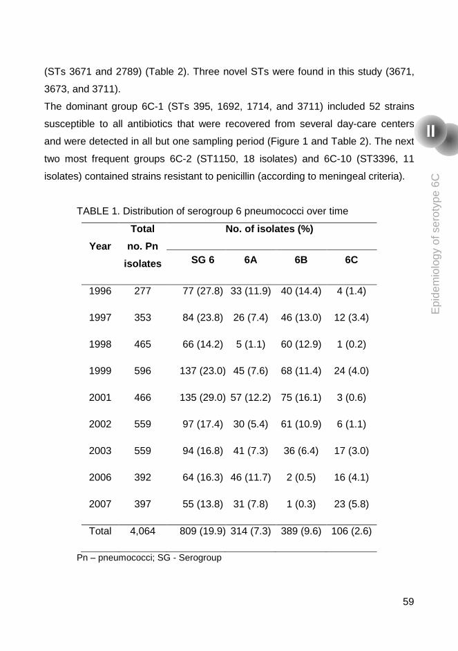
59
(STs 3671 and 2789) (Table 2). Three novel STs were found in this study (3671,
3673, and 3711).
The dominant group 6C-1 (STs 395, 1692, 1714, and 3711) included 52 strains
susceptible to all antibiotics that were recovered from several day-care centers
and were detected in all but one sampling period (Figure 1 and Table 2). The next
two most frequent groups 6C-2 (ST1150, 18 isolates) and 6C-10 (ST3396, 11
isolates) contained strains resistant to penicillin (according to meningeal criteria).
TABLE 1. Distribution of serogroup 6 pneumococci over time
Year
Total
no. Pn
isolates
No. of isolates (%)
SG 6 6A 6B 6C
1996 277 77 (27.8) 33 (11.9) 40 (14.4) 4 (1.4)
1997 353 84 (23.8) 26 (7.4) 46 (13.0) 12 (3.4)
1998 465 66 (14.2) 5 (1.1) 60 (12.9) 1 (0.2)
1999 596 137 (23.0) 45 (7.6) 68 (11.4) 24 (4.0)
2001 466 135 (29.0) 57 (12.2) 75 (16.1) 3 (0.6)
2002 559 97 (17.4) 30 (5.4) 61 (10.9) 6 (1.1)
2003 559 94 (16.8) 41 (7.3) 36 (6.4) 17 (3.0)
2006 392 64 (16.3) 46 (11.7) 2 (0.5) 16 (4.1)
2007 397 55 (13.8) 31 (7.8) 1 (0.3) 23 (5.8)
Total 4,064 809 (19.9) 314 (7.3) 389 (9.6) 106 (2.6)
Pn – pneumococci; SG - Serogroup
Epi
dem
iolo
gy o
f ser
otyp
e 6C

60
TABLE 2. Characteristics of serotype 6C strains
PFGE group
(no. of isolates)
ST
(no. of isolates)a CC
Resistance pattern
(no. of isolates)
No. of
DCC
6C-1 (52) ST395 (5) 395 Susceptible 12
ST1714 (1)
ST1692 (1)
ST3711 (1)
6C-2 (18) ST1150 (4) 138 P (11) 3
Susceptible (7)
6C-3 (1) ST1150 (1) 138 P 1
6C-4 (4) ST3673 (2) 138 Ery 2
6C-5 (1) ST2789 (1) Singleton Susceptible 1
6C-6 (2) ST3671 (1) Singleton Susceptible 1
6C-7 (6) ST1150 (1) 138 P (3) 3
ST2689 (2) Susceptible (3)
6C-8 (2) ST2185 (1) 3034 Ery, Clin, Tet, SXT 1
6C-9 (3) ST395 (2) 395 Susceptible 1
6C-10 (11) ST3396 (1) 386 P, Ery, Clin, Tet (8) 1
Ery, Clin, Tet (3)
6C-11 (6) ST3396 (1) 386 P, Ery, Clin, Tet 1
P, penicillin G MIC of 0.12 µg ml-1 with the exception of a single isolate that had an MIC of
0.25 µg ml-1; Ery, erythromycin; Clin, clindamycin; Tet, tetracycline; SXT, trimethoprim-
sulfamethoxazole; No. of DCC, number of day-care centers where PFGE group was found. aNumber of representative strains tested.
The dominant group 6C-1 (STs 395, 1692, 1714, and 3711) included 52 strains
susceptible to all antibiotics that were recovered from several day-care centers
and were detected in all but one sampling period (Figure 1 and Table 2). The next
two most frequent groups 6C-2 (ST1150, 18 isolates) and 6C-10 (ST3396, 11
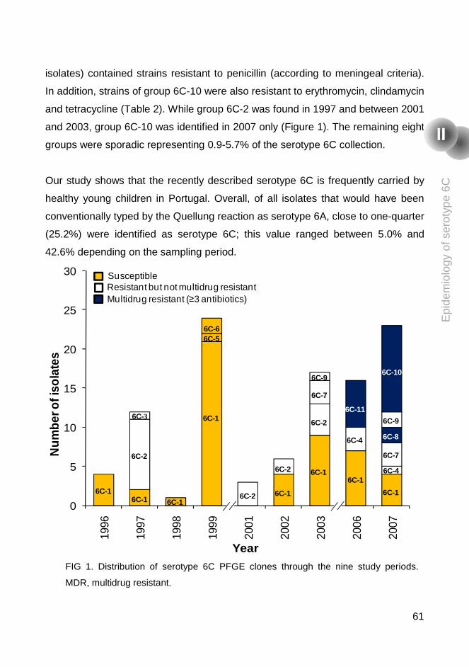
61
isolates) contained strains resistant to penicillin (according to meningeal criteria).
In addition, strains of group 6C-10 were also resistant to erythromycin, clindamycin
and tetracycline (Table 2). While group 6C-2 was found in 1997 and between 2001
and 2003, group 6C-10 was identified in 2007 only (Figure 1). The remaining eight
groups were sporadic representing 0.9-5.7% of the serotype 6C collection.
Our study shows that the recently described serotype 6C is frequently carried by
healthy young children in Portugal. Overall, of all isolates that would have been
conventionally typed by the Quellung reaction as serotype 6A, close to one-quarter
(25.2%) were identified as serotype 6C; this value ranged between 5.0% and
42.6% depending on the sampling period.
FIG 1. Distribution of serotype 6C PFGE clones through the nine study periods.
MDR, multidrug resistant.
0
5
10
15
20
25
30
1996
1997
1998
1999
2001
2002
2003
2006
2007
Num
ber o
f iso
late
s
Year
SusceptibleResistant but not multidrug resistantMultidrug resistant (≥3 antibiotics)
6C-16C-1
6C-2
6C-3
6C-1
6C-1
6C-1
6C-16C-1
6C-16C-2
6C-2
6C-2
6C-4
6C-4
6C-56C-6
6C-7
6C-7
6C-9
6C-9
6C-11
6C-10
6C-8E
pide
mio
logy
of s
erot
ype
6C
62
Serotype 6C strains circulating in Portugal are genetically diverse. Similar
observations were recently reported in a USA study (8) and, of interest, there were
no common STs among serotype 6C isolates from both studies. This observation
implies that, if the introduction of wciN6C in the capsular operon of 6A occurred only
once as suggested by Park et al., it must have occurred a sufficiently long time
ago to yield such lineage diversity (13). In our collection, one fully antibiotic-
susceptible lineage (6C-1, STs 395, 1692, 1714, and 3711) persisted over one
decade and accounted for close to half of all serotype 6C isolates suggesting it is
well adapted even when considering the natural selection driven by antibiotic
pressure. Still, strains of serotype 6C circulating in Portugal are often drug-
resistant and, in particular, multidrug resistance has been detected in 2006 and
2007. These data contrast with recent observations from South Africa where most
invasive strains of this serotype were fully susceptible or resistant to macrolides
only, but are comparable to resistance rates reported in a recent study from the
USA (8).
Conclusion
In summary, serotype 6C circulates in Portugal at least since 1996. It is genetically
diverse and often antibiotic resistant. Continued surveillance of this serotype is
important since it is not targeted by PCV7 and is not known if PCV13 (which
targets not only 6B but also 6A) will confer cross-protection to it. Additional data
from other countries and estimates of its invasive disease potential will help us
understand the epidemiology of this serotype.
Acknowledgments
This work was supported by projects PREVIS (LSHM-CT-2003-503413), and
GRACE (LSHM-CT-2005-518226) from the European Commission and
PTDC/SAU-ESA/65048/2006 from Fundação para a Ciência e Tecnologia (FCT),
Portugal. SN was supported by grant SFRH/BD/40706/2007 from FCT.

63
We acknowledge the use of the pneumococcal MLST database which is located at
the Imperial College of London and is funded by the Wellcome Trust.
References
1. Bentley, S. D., D. M. Aanensen, A. Mavroidi, D. Saunders, E.
Rabbinowitsch, M. Collins, K. Donohoe, D. Harris, L. Murphy, M. A. Quail,
G. Samuel, I. C. Skovsted, M. S. Kaltoft, B. Barrell, P. R. Reeves, J.
Parkhill, and B. G. Spratt. 2006. Genetic analysis of the capsular biosynthetic
locus from all 90 pneumococcal serotypes. PLoS Genet 2:e31.
2. Brito, D. A., M. Ramirez, and H. de Lencastre. 2003. Serotyping
Streptococcus pneumoniae by multiplex PCR. J Clin Microbiol 41:2378-84.
3. Carvalho, M. G., C. Henderson, A. Trujillo, H. H. Joshi, I. H. Park, S.
Hollingshead, C. Whitney, M. Nahm, B. Beall, and TABC ABCs Team.
2008. Emergence of genetically diverse invasive pneumococcal serotype 6C,
abstr. S01-O1, p. 35. Abstr. 6th International Symposium on Pneumococci and
Pneumococcal Diseases, Reykjavik, Iceland.
4. CLSI.Clinical and Laboratory Standards Institute 2008. Performance
Standards for Antimicrobial Susceptibility Testing; Eighteenth Informational
Supplement. Vol.28 No.1. Approved standard M2-A9. Clinical and Laboratory
Standards Institute. Wayne, Pa.
5. du Plessis, M., A. von Gottberg, S. A. Madhi, O. Hattingh, L. de Gouveia,
and K. P. Klugman. 2008. Serotype 6C is associated with penicillin-
susceptible meningeal infections in human immunodeficiency virus (HIV)-
infected adults among invasive pneumococcal isolates previously identified as
serotype 6A in South Africa. Int J Antimicrob Agents 32 (Suppl 1): S66-70.
6. Enright, M. C., and B. G. Spratt. 1998. A multilocus sequence typing scheme
for Streptococcus pneumoniae: identification of clones associated with serious
invasive disease. Microbiology 144:3049-60.
7. Hermans, P. W., M. Blommaart, I. H. Park, M. H. Nahm, and D. Bogaert.
2008. Low prevalence of recently discovered pneumococcal serotype 6C
Epi
dem
iolo
gy o
f ser
otyp
e 6C

64
isolates among healthy Dutch children in the pre-vaccination era. Vaccine
26:449-50.
8. Jacobs, M. R., S. Bajaksouzian, R. A. Bonomo, C. E. Good, A. R. Windau,
A. M. Hujer, C. Massire, R. Melton, L. B. Blyn, D. J. Ecker, and R.
Sampath. 2008. Occurrence, distribution and origins of serotype 6C
Streptococcus pneumoniae, a recently recognized serotype. J Clin Microbiol
47:64-72.
9. Mato, R., I. S. Sanches, C. Simas, S. Nunes, J. A. Carriço, N. G. Sousa, N.
Frazão, J. Saldanha, A. Brito-Avô, J. S. Almeida, and H. de Lencastre.
2005. Natural history of drug-resistant clones of Streptococcus pneumoniae
colonizing healthy children in Portugal. Microb Drug Resist 11:309-22.
10. Mavroidi, A., D. Godoy, D. M. Aanensen, D. A. Robinson, S. K.
Hollingshead, and B. G. Spratt. 2004. Evolutionary genetics of the capsular
locus of serogroup 6 pneumococci. J Bacteriol 186:8181-92.
11. Nunes, S., R. Sá-Leão, J. Carriço, C. R. Alves, R. Mato, A. Brito-Avô, J.
Saldanha, J. S. Almeida, I. S. Sanches, and H. de Lencastre. 2005. Trends
in drug resistance, serotypes, and molecular types of Streptococcus
pneumoniae colonizing preschool-age children attending day care centers in
Lisbon, Portugal: a summary of 4 years of annual surveillance. J Clin Microbiol
43:1285-93.
12. Park, I. H., M. R. Moore, J. J. Treanor, S. Pelton, T. Pilishvili, B. Beall, M.
Shelly, G. Gallagher, B. Mahon, and M. H. Nahm. 2008. Reduction in
serotype 6A invasive pneumococcal disease after accounting for effect of
serotype 6C, abstr. P1-006, p. 88. Abstr. 6th International Symposium on
Pneumococci and Pneumococcal Diseases, Reykjavik, Iceland.
13. Park, I. H., S. Park, S. K. Hollingshead, and M. H. Nahm. 2007. Genetic
basis for the new pneumococcal serotype, 6C. Infect Immun 75:4482-9.
14. Park, I. H., D. G. Pritchard, R. Cartee, A. Brandao, M. C. Brandileone, and
M. H. Nahm. 2007. Discovery of a new capsular serotype (6C) within
serogroup 6 of Streptococcus pneumoniae. J Clin Microbiol 45:1225-33.

65
15. Sá-Leão, R., A. S. Simões, S. Nunes, N. G. Sousa, N. Frazão, and H. de
Lencastre. 2006. Identification, prevalence and population structure of non-
typable Streptococcus pneumoniae in carriage samples isolated from
preschoolers attending day-care centres. Microbiology 152:367-76.
16. Sá-Leão, R., A. Tomasz, I. S. Sanches, A. Brito-Avô, S. E. Vilhelmsson, K.
G. Kristinsson, and H. de Lencastre. 2000. Carriage of internationally spread
clones of Streptococcus pneumoniae with unusual drug resistance patterns in
children attending day care centers in Lisbon, Portugal. J Infect Dis 182:1153-
60.
17. Sá-Leão, R., A. Tomasz, I. S. Sanches, S. Nunes, C. R. Alves, A. Brito-
Avô, J. Saldanha, K. G. Kristinsson, and H. de Lencastre. 2000. Genetic
diversity and clonal patterns among antibiotic-susceptible and -resistant
Streptococcus pneumoniae colonizing children: day care centers as
autonomous epidemiological units. J Clin Microbiol 38:4137-44.
18. Sorensen, U. B. 1993. Typing of pneumococci by using 12 pooled antisera. J
Clin Microbiol 31:2097-100.
Epi
dem
iolo
gy o
f ser
otyp
e 6C

66

67
Streptococcus pneumoniae nasopharyngeal
carriage in children attending day-care centers in
the central region of Portugal, in the era of 7-valent
pneumococcal conjugate vaccine.
Published in: Rodrigues F*, S. Nunes*, R. Sá-Leão, G. Gonçalves, L. Lemos,
H. de Lencastre . 2009. Microb Drug Resist. 15:269-77. *These authors
contributed equally.

68
S. Nunes was responsible for the characterization of pneumococcal isolates.

69
ABSTRACT The seven-valent pneumococcal conjugate vaccine (PCV7) became available in
Portugal in 2001. Although not included in the national immunization program,
vaccination coverage is high (over 60%). We studied for the first time the rates of
nasopharyngeal (NP) carriage of pneumococci, antibiotic resistance patterns and
serotypes among children attending day care centers (DCCs) in Coimbra, a city in
the Central Region of Portugal. Between January and February 2007, a cross-
sectional study was conducted among children aged 6 months to 6 years old
attending eight DCC. NP swabs were obtained from 507 children: 76.7% had
received at least one dose of PCV7 and 64.3% were age-appropriately vaccinated.
The global pneumococcal carriage rate was 61.3%. Colonization proportions
varied with age and number of children attending each DCC. Serotyping revealed
that 20.7% of the pneumococci were vaccine types (VT), 70.8% were non-vaccine
types (NVT), and 8.5% were non-typeable. Serotype 19F was the second most
frequent serotype being detected in 10.5% of the samples. While global NP
carriage was not associated with vaccination status, NVT were predominant
among vaccinated children, who had significantly lower prevalence of VT. Of all
isolates, 15.7% had penicillin MICs that ranged between 0.12 and 2 µg/ml. The
proportion of resistant strains was significantly higher among VT and unvaccinated
children. In conclusion, the rates of vaccination and prevalence of pneumococcal
NP were high. Rates of antimicrobial resistance were similar to those found in
studies conducted in Oeiras and Lisbon. This study is a platform for future
surveillance activities.
Pne
umoc
occa
l col
oniz
atio
n in
Coi
mbr
a
70
INTRODUCTION
Pneumococcus is a leading cause of invasive and noninvasive disease among
children worldwide. The importance of pneumococcal infections is highlighted by
an increasing prevalence of drug-resistant strains (28, 34, 45).
A 7-valent pneumococcal conjugate vaccine (PCV7) was licensed in the USA in
2000. Pneumococcal conjugate vaccines (PCVs) have been shown to be highly
efficacious against vaccine-type (VT) serotypes invasive pneumococcal disease
(IPD). They are also efficacious against chest radiograph–confirmed pneumonia
and otitis media (OM) (2, 6, 9, 12, 24, 31). Increase in NVT pneumococcal disease
has been observed among some age groups in invasive-disease (16, 21) and in
OM efficacy trials (12, 23).
Because NP carriage is a precondition for developing disease, and it is this
condition, rather than pneumococcal disease, that results in person to person
pneumococcal transmission, it is essential to thoroughly understand the impact of
PCV on NP colonization. Conjugate vaccines are thought to reduce VT
pneumococcal carriage by preventing new acquisitions, rather than by terminating
existing carriage episodes (8), although the mechanism is incompletely
understood. Reductions in VT pneumococcal carriage may prevent disease
among unvaccinated individuals by decreasing the transmission of these
serotypes in the community (7, 33, 44). Numerous studies have shown that
conjugate vaccines protect against VT carriage in the months immediately after
vaccination (27, 28, 32) and more recently Millar et al. showed that community-
wide PCV7 vaccination in infancy reduces the prevalence of VT carriage and
increases the prevalence of NVT carriage (serotype replacement) through at least
3 years of age (28). The indirect protective effects against VTs (herd immunity)
and the indirect and direct replacement effects result in large part from the impact
of PCV on NP colonization (30).
71
Day-care centers (DCCs), for preschool children are unique settings for the spread
of pneumococcal carriage. DCC attendance has been found to be an important
risk factor for development of IPD in children and infants (25, 43). The role of
DCCs in the transmission of S. pneumoniae to the community is also well
documented (19).
PCV7 became available in Portugal in 2001 but has not been included in the
national vaccination program. Thus, vaccines are given after medical prescription
and paid by the parents and not reimbursed by the National Health Service. Two
studies reported vaccination coverage values (10, 35) among children within the
age group attending DCC, but samples were not representative of the whole
population. Portugal has high antibiotic consumption (17) and penicillin non-
susceptible S. pneumoniae are frequently isolated (1, 37). Previous colonization
studies in Portuguese DCCs, were conducted in the areas of Lisbon and Oeiras
(26, 29, 36).
This study intended to estimate the prevalence of NP carriage of pneumococci,
identifying serotype distribution and antibiotic resistance patterns among children
attending DCCs, in the era of widespread use of PCV7, in Coimbra, a city in the
central region of Portugal, 200 Km away from Lisbon.
METHODS
Study population
A convenience sample of four urban and four suburban DCCs was selected. The
target population consisted of 660 children, aged 6 months to 6 years old,
attending eight DCCs in Coimbra, a city in the central region of Portugal.
Pne
umoc
occa
l col
oniz
atio
n in
Coi
mbr
a
72
Study design and approval
A cross-sectional study was performed in January and February of 2007. The
study was approved by the Ethical Committee of Hospital Pediátrico de Coimbra.
Parents or guardians provided written informed consent for their child to
participate.
Questionnaire
One week before sample collection, the following information was obtained from a
questionnaire filled in by the parents or guardians of participating children: age of
the child, number of children in the household who were ≤6 years of age, presence
of any underlying illness, presence of a cigarette smoker at home, use of
antibiotics in the preceding month, maternal educational level and dates of all
PCV7 doses received. In the few cases where the number of doses was reported
without vaccination dates, the parents were contacted either by one of the
investigators by phone or by the nurses during the sample collection visit.
Vaccination status
Vaccination with PCV7 was considered appropriate if it was done according to one
of the following schemes: a 4-dose series if starting vaccination at 2 months of
age; a 3-dose series if starting vaccination at 7-11 months of age; a 2-dose series
if starting vaccination at 12-24 months of age and 1 dose if starting vaccination at
≥ 24 months, as recommended by the vaccine manufacturer. A child was defined
as age-appropriately vaccinated if at the time of sampling had received all
recommended doses, according to the age at initiation of vaccination. Those with
fewer doses than recommended by age at initiation of vaccination were classified
as partially vaccinated. Doses of PCV7 received during the four weeks that
preceded sampling were not considered for the vaccination status classification.
73
Sample collection and isolation of pneumococci
Nasopharyngeal samples were obtained by trained nurses with a small flexible
swab (BBL Culture Swab; Becton-Dickinson, Sparks, MD) inserted through the
nostril until resistance was encountered. The swab was then removed and
inoculated into the transport medium (Stuart medium) and transported at ambient
temperature to the Laboratory of Microbiology at Centro Hospitalar de Coimbra
and was stored at 4°C for a maximum of 12 hours before samples were plated.
Specimens were streaked onto tryptic soy 5% sheep blood agar plates (Becton
Dickinson) and incubated overnight at 37°C in 5% CO2. Phenotypic characteristics
(morphology and α-hemolysis) were used for the presumptive identification of
pneumococci. Pneumococcal identification was confirmed by optochin
susceptibility and bile solubility assays (29). When suspected pneumococcal
colonies with more than one morphology were observed, each type was purified
for further testing. Isolates were frozen and stored at -70ºC.
Antimicrobial susceptibility testing
Susceptibility of all isolates was tested by the disk diffusion method in accordance
with the guidelines of the Clinical and Laboratory Standards Institute (CLSI) (5),
against erythromycin, clindamycin, tetracycline, chloramphenicol,
sulfamethoxazole-trimethoprim (SXT), levofloxacin, and vancomycin (Becton-
Dickinson). Minimum inhibitory concentrations (MICs) of penicillin and cefotaxime
were determined by E-test (AB Biodisk, Solna, Sweden) according to the
manufacturer’s recommendations. Interpretation of penicillin and cefotaxime MICs
followed CLSI criteria (5).
Serotyping
Pneumococci were serotyped by the Quellung reaction using commercially
available antisera (Statens Serum Institut, Copenhagen, Denmark) (41). Serotypes
were grouped into one of three categories: (1) vaccine type (VT; i.e., types 4, 6B,
Pne
umoc
occa
l col
oniz
atio
n in
Coi
mbr
a

74
9V, 14, 18C, 19F, and 23F); (2) non-vaccine type (NVT) and (3) non-typeable
(NT).
Pulsed-field gel electrophoresis (PFGE)
Pneumococci strains that were resistant to at least one of the antibiotics tested,
were also typed by PFGE. All strains with penicillin MICs ≥0.12 µg/ml were
analyzed. Preparation of chromosomal DNA, restriction with SmaI endonuclease,
and PFGE were done as previously described by Sá-Leão et al. (39). PFGE
patterns were analyzed with Bionumerics software (version 5.10, Applied Maths,
Gent, Belgium). Patterns were clustered by UPGMA, and a dendrogram was
generated from a similarity matrix calculated using the Dice similarity coefficient
with an optimization of 1.0% and a tolerance of 1.5%. PFGE clusters were defined
as isolates with a similarity of 80% or higher on the dendrogram (38).
Statistical analysis
Univariate analysis was done using Epi Info 2000 (11). Chi-square tests with Yates
correction were used. Multivariate analysis, with carriage status of S. pneumoniae
as the dependent variable, was done using a logistic regression model fitted by
Stata (42). Population diversity was measured by calculating the Simpson´s Index
of Diversity (SID) with 95% confidence intervals (CI) for drug-resistant NVT and VT
strains. Congruence between PFGE clones and DCCs was evaluated with the
Adjusted Rand (AR) coefficients (4).
75
RESULTS
General characteristics of the study group
A NP swab was obtained in 507 (77%) of the 660 children attending the eight
DCC. Between 19 and 113 children were tested at each DCC. General
characteristics of the study group at the time of sampling are described in Table 1.
The mean age of participants was 40.6 months (range 6.1 to 80.9 months) and
262 (51.7%) were male; 148 (29%) had siblings ≤ 6 years of age and none had
more than two siblings in that age group. Using maternal educational level as a
proxy for socio-economic level, it was observed that more than 74.2% of the
mothers had more than the compulsory schooling level in Portugal (nine years).
The use of an antibiotic within one month prior to study enrollment was reported
for 126 (24.9%) children; 29.9% lived with one or more cigarette smoker.
Vaccination status
Three hundred and eighty nine children (76.7%) had received one to four doses of
PCV7; 64.5% (327/507) were classified as age-appropriately vaccinated and
12.3% participants had not completed the schedule recommended by the
manufacturer and were classified as partially vaccinated (Table 2). A higher
proportion of age-appropriately vaccinated children was observed among children
aged 6-12 months, decreasing thereafter to a minimum at the age group older
than 60 months (Figure 1).
Pne
umoc
occa
l col
oniz
atio
n in
Coi
mbr
a

76
Fig. 1. Vaccination status according to the age group
Carriage
The global carriage rate was 61.3% (311/507). Pneumococcal colonization
proportions varied with age (p<0.0001); among children below one year of age,
57.9% were colonized, and the peak (76.2%) was observed among children 12–24
months; from this age group on the proportion of colonized children decreased
gradually to the older age groups (p<0.000001 in Chi-square test for linear trend),
reaching the minimum level of 47.0% among the older age group (Table 1 and
Figure 2).
0%
20%
40%
60%
80%
100%
6 to 12 >12 to 24 >24 to 36 >36 to 48 >48 to 60 >60
Age-group in months
Age appropriately vaccinated Partially vaccinated Unvaccinated
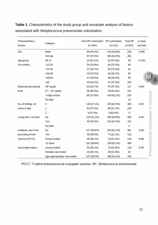
77
Table 1. Characteristics of the study group and univariate analysis of factors
associated with Streptococcus pneumoniae colonization
PCV7, 7-valent pneumococcal conjugate vaccine; SP, Streptococcus pneumoniae
Characteristics /
factorsCategory
Non-SP colonization
(n=196 )
SP colonization
(n=311)
Total SP
(n=507)
p-value
(χ2 test)
Sex Male
Female
99 (40.4%)
97 (37.0%)
146 (59.6%)
165 (62.9%)
245
262
0.490
Age group
(in months)
06-12
>12-24
>24-36
>36-48
>48-60
>60
16 (42.1%)
20 (23.8%)
27 (29.7%)
33 (33.3%)
47 (49.5%)
53 (53.0%)
22 (57.9%)
64 (76.2%)
64 (70.3%)
66 (66.7%)
48 (50.5%)
47 (47.0%)
38
84
91
99
95
100
<0.001
Maternal educational
level
≤9th grade
9th – 12th grade
> High school
No data
50 (42.7%)
58 (38.6%)
85 (37.8%)
67 (57.3%)
93 (61.6%)
140 (62.2%)
117
151
225
14
0.656
No. of siblings ≤6
years of age
0
1
2
139 (37.1%)
54 (37.9%)
3 (37.5%)
220 (62.9%)
86 (61.4%)
5 (62.5%)
359
140
8
0.997
Living with a smoker No
Yes
No data
144 (41.1%)
49 (32.5%)
206 (58.9%)
102 (67.5%)
350
151
6
0.083
Antibiotic use in the
preceding month
No
Yes
147 (38.6%)
49 (38.8%)
234 (61.4%)
77 (61.1%)
381
126
0.965
Vaccine (PCV7) Unvaccinated
≥1 dose
45 (38.1%)
151 (38.8%)
73 (61.9%)
238 (61.2%)
118
389
0.980
Vaccination status Unvaccinated
Partially vaccinated
Age-appropriately vaccinated
45 (38.1%)
24 (38.1%)
127 (39.0%)
73 (61.9%)
39 (61.9%)
199 (61.0%)
118
63
326
0.983P
neum
ococ
cal c
olon
izat
ion
in C
oim
bra
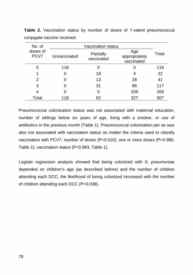
78
Table 2. Vaccination status by number of doses of 7-valent pneumococcal
conjugate vaccine received
No. of doses of
PCV7
Vaccination status
Total Unvaccinated Partially
vaccinated
Age appropriately vaccinated
0 1 2 3 4
118 0 0 0 0
0 18 13 31 0
0 4
28 86 209
118 22 41
117 209
Total 118 62 327 507
Pneumococcal colonization status was not associated with maternal education,
number of siblings below six years of age, living with a smoker, or use of
antibiotics in the previous month (Table 1). Pneumococcal colonization per se was
also not associated with vaccination status no matter the criteria used to classify
vaccination with PCV7: number of doses (P=0.510); one or more doses (P=0.980,
Table 1); vaccination status (P=0.983, Table 1).
Logistic regression analysis showed that being colonized with S. pneumoniae
depended on children’s age (as described before) and the number of children
attending each DCC; the likelihood of being colonized increased with the number
of children attending each DCC (P=0.038).

79
Fig. 2. S. pneumoniae colonization according to age group
Serotype distribution
The serotype distribution is shown in Table 3. Of the 315 isolates (311 carriers of
which four had two strains of pneumococci), ten were lost. Thus, the information
on specific serotype was available for 305 isolates. Among these, 63 (20.7%) were
VT, 216 (70.8%) NVT and the remaining 26 (8.5%) were NT. Serotypes 19F, 6B
and 14 were the most prevalent among VTs. Several NVT (23 types) were isolated
being the most prevalent (in decreasing order) 23A, 16F, 23B, 19A, 21, 17F, 35F,
11A, and 6A (Table 3).
0%
20%
40%
60%
80%
100%
>6-12 >12-24 >24-36 >36-48 >48-60 >60
Age-group in months
Vaccine types Non-vaccine types Non-typeable Non-colonized
Pne
umoc
occa
l col
oniz
atio
n in
Coi
mbr
a
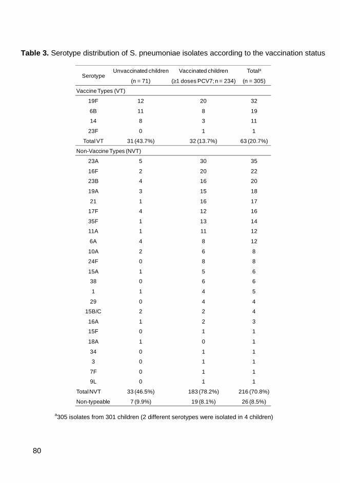
80
Table 3. Serotype distribution of S. pneumoniae isolates according to the vaccination status
a305 isolates from 301 children (2 different serotypes were isolated in 4 children)
SerotypeUnvaccinated children
(n = 71)
Vaccinated children
(≥1 doses PCV7; n = 234)
Totala
(n = 305)
VaccineTypes (VT)
19F
6B
14
23F
12
11
8
0
20
8
3
1
32
19
11
1
Total VT 31 (43.7%) 32 (13.7%) 63 (20.7%)
Non-Vaccine Types (NVT)
23A
16F
23B
19A
21
17F
35F
11A
6A
10A
24F
15A
38
1
29
15B/C
16A
15F
18A
34
3
7F
9L
5
2
4
3
1
4
1
1
4
2
0
1
0
1
0
2
1
0
1
0
0
0
0
30
20
16
15
16
12
13
11
8
6
8
5
6
4
4
2
2
1
0
1
1
1
1
35
22
20
18
17
16
14
12
12
8
8
6
6
5
4
4
3
1
1
1
1
1
1
Total NVT 33 (46.5%) 183 (78.2%) 216 (70.8%)
Non-typeable 7 (9.9%) 19 (8.1%) 26 (8.5%)

81
Although colonization per se was not associated with vaccination status, the
distribution of serotypes among vaccinated and unvaccinated children was
different (Table 3). While prevalence of NVT (46.5%) and VT (43.7%) was similar
among unvaccinated, NVT were clearly predominant (78.2%) among children with
≥1 doses of PCV7 (P<0.0001).The proportions of VT and NVT were also
associated with age group (P<0.0001) as the proportion of NVT which increased
to maximum levels among age groups “>12-24” and “>24-36” months of age and
then decreased gradually to the minimum among the eldest (Figure 2).
Antibiotic resistance
The antibiotic resistance profile of serotyped pneumococci is show on Table 4. Of
all isolates, 14.4% strains had a penicillin MIC ranging from 0.12 to 1 µg/ml and
1.3% had a MIC of 2 µg/ml; 7.2% of the strains had a cefotaxime MIC of 1 µg/ml
and 0.7 had a MIC of 2 µg/ml (Table 4). Other rates of resistance were 20.5% to
erythromycin, 17.4% to clindamycin, 15.0% to tetracycline, 10.2% to SXT, and
1.3% to chloramphenicol. All pneumococci were susceptible to levofloxacin and
vancomycin. For any studied antibiotic on Table 4, the proportions of resistant
strains were significantly higher among VT (p <0.0001).
Significantly higher proportions of decreased susceptibility to penicillin, and
resistance to tetracycline were observed among strains from unvaccinated, when
compared to vaccinated children (Table 4). Nevertheless, when the confounding
effect by VT strain was controlled through stratification of vaccination status, the
apparent association between vaccination status and resistance to antibiotics was
no longer observed.
Pne
umoc
occa
l col
oniz
atio
n in
Coi
mbr
a

82
Table 4. Resistant Streptococcus pneumoniae according to serotype and vaccination status
*≥1doses of Prevenar; Oral penicillin V: PenicillinI 0.12≤MIC<1 µg/mL, PenicillinR MIC≥2 µg/mL; Penicillin parenteral meningitis: PenicillinR MIC≥0.12 µg/mL; Penicillin parenteral nonmeningitis: PenicillinI MIC=4 µg/mL; Cefotaxime (meningitis): CefotaximeI MIC=1.0µg/mL, CefotaximeR MIC≥2.0µg/mL; Cefotaxime (nonmeningitis): CefotaximeI MIC=2.0µg/mL; SXT, sulfamethoxazole-trimethoprim; n.d., not done (small number of isolates); MIC, minimum inhibitory concentration.
Of note, serotype 19F, a VT, was the second most frequently isolated serotype
(being 23A the most frequent one). Six out of 32 children with 19F were <2 years
of age, 22/32 (69%) had received at least one dose of PCV7 and 18/32 (56%)
were age-appropriately vaccinated. Of 32 serotype 19F isolates, 46.9% had
decreased susceptibility to penicillin (intermediate and high-level penicillin
resistance were found in 15.7% and 31.2% of the strains, respectively). The rates
of resistance to other antimicrobial agents were: 40.6% to cefotaxime, 90.6% to
erythromycin, clindamycin and tetracycline. All were susceptible to
chloramphenicol, levofloxacin and vancomycin.
Among NVT, penicillin non-susceptibility and resistance to macrolides was more
frequent among serotypes 19A, 15A and 6A. For serotype 19A (n=18), penicillin
non-susceptibility was 33%, resistance to macrolides and to clindamycin was
55.6% and to cefotaxime was 27.8%.
Antibiotic
All isolates n=305no. (%)
Type Vaccination statusNon-VTn=216no. (%)
Non-typeablen=26
no. (%)
VTn=63
no. (%) p-value
Unvaccinatedn= 71
no. (%)
Vaccinated*n = 234no. (%) p-value
Oral penicillin VPenicillinI
PenicillinR44 (14.4)
4 (1.3)17 (7.9)
03 (11.5)
024 (38.1)
4 (6.3)<0.0001
n.d.16 (22.5)
2 (2.8)28 (12.0)
2 (0.9)0.026n.d.
Penicillin parenteral (meningitis) 48 (15.7) 17 (7.9) 3 (11.5) 28 (44.4) <0.0001 18 (25.4) 30 (12.8) 0.011Penicillin parenteral (nonmeningitis) 0 0 0 0 - 0 0 -Cefotaxime (meningitis)
CefotaximeI
CefotaximeR22 (7.2)2 (0.7)
5 (2.3)0
1 (3.8)0
16 (25.4)2 (3.2)
<0.0001n.d.
10 (14.1)2 (2.8)
12 (5.1)0
0.011n.d.
CefotaximeI (nonmeningitis) 2 (0.7) 0 0 2 (3.2) n.d. 2 (2.8) 0 n.d.SXT 31 (10.2) 12 (5.5) 5 (19.2) 14 (22.2) <0.0001 13 (18.3) 18 (7.7) 0.035Chloramphenicol 4 (1.3) 4 (1.9) 0 0 n.d. 0 4 (1.7) n.d.Erythromycin 48 (20.5) 25 (11.6) 5 (19.2) 37 (58.7) <0.0001 19 (26.8) 50 (21.3) 0.341Clindamycin 40 (17.4) 20 (9.3) 2 (7.7) 35 (55.6) <0.0001 17 (23.6) 41 (17.4) 0.227Tetracycline 35 (15.0) 15 (6.9) 3 (11.5) 35 (55.6) <0.0001 18 (25.4) 36 (15.3) 0.054

83
Clonal analysis of drug-resistant strains
DNA fingerprinting through PFGE was performed for all 86 drug-resistant isolates
which included 13 serotypes and 7 NT strains (Table 5). Twenty-four clones were
identified, six associated with specific VT, 15 associated with specific NVT, and
three associated with both VT and NVT. Among the latter, two clones had isolates
of serotypes 19A and 19F and one had isolates expressing serotypes 14, 15A,
19A, and 19F. Fifteen clones were represented by unique isolates and the
remaining were represented by two to 31 isolates. The largest clone, 19F/19A-1,
included 26 of the 30 drug resistant serotype 19F isolates and five of the 13 drug-
resistant 19A isolates. This clone was isolated from 6 day-care centers.
Clonal diversity was higher among drug resistant NVT strains than among drug-
resistant VT strains: SID for NVT was 0.92 (95% CI, 0.89-0.96) compared to 0.68
(95% CI, 0.54-0.82) for VT. There was a low association between drug-resistant
clones and individual DCCs as the AR was only 0.17 for NVT strains and 0.13 for
VT strains.
Pne
umoc
occa
l col
oniz
atio
n in
Coi
mbr
a

84
Table 5. Clonal distribution of all antibiotic-resistant strains
Ery – erythromycin; Da – clindamycin; Tet – tetracycline; Chl – chloramphenicol; SXT - sulfamethoxazole-trimethoprim; PenicillinI 0.12≤MIC<1 µg/mL; PenicillinR MIC≥2 µg/mL; aPFGE was done for antimicrobial-resistant isolates; bPFGE clones were named by using a combination of serotype(s) found within the clone followed by an arbitrary sequential number.
SerotypeNo. of isolates with
PFGE doneaPFGE clonesb Resistance profile
Day-care centers
(no. of isolates)
Vaccine types
6B 4/19 6B-1 (1)
6B-2 (3)
IP, Ery, Da, Tet
IP, SXT
A (1)
C (3)
14 11/11 14-1 (3)
14-2 (3)
15A/14/19F/19A-1 (5)
P (1), IP, SXT
Ery (2); Tet, SXT
IP, Tet (4), Ery, Da, SXT (4)
A (2); H (1)
E (1); F (2)
B (3); C (1); F (1)
19F 30/32 19F/19A-1 (26)
15A/14/19F/19A-1 (2)
19F/19A-2 (1)
19F-3 (1)
IP(1), P(2),Tet, Ery, Da
IP, Tet, Ery, Da
IP, Tet, Ery, Da, SXT
P, SXT
A (9); B (10); C (3); D (2); E (2)
B (2)
H (1)
F (1)
23F 1/1 23F-1 (1) IP C (1)
Nonvaccine types
6A 4/12 6A-1 (1)
6A-2 (3)
IP, Tet, Ery, Da
IP, Tet, Ery, Da
H (1)
H (3)
11A 5/12 11A-1 (3)
11A-2 (2)
IP (1), Ery, Da (2), ISXT (1)
Ery, SXT
A (1); B (2)
A (2)
15A 5/6 15A/14/19F/19A-1 (5) IP, Ery, Da A (3); C (1); D (1)
15B/C 1/4 15B/C-1 (1) Ery, SXT A (1)
17F 1/16 17F-1 (1) Tet C (1)
18A 1/1 18A-1 (1) IP B (1)
19A 13/18 19F/19A-1 (5)
15A/14/19F/19A-1 (1)
19F/19A-2 (5)
19A-3 (2)
Ery, Da, Tet, Chl
P, Tet, Ery, Da
IP, Tet, Ery, Da, ISXT
ISXT, RSXT
B (2); G (3)
A (1)
A (4); H (1)
B (1); C (1)
21 1/17 21-1 (1) SXT G (1)
29 1/4 29-1 (1) Ery E (1)
NT 7/26 NT-1 (3)
NT-2 (1)
NT-3 (1)
NT-4 (1)
NT-5 (1)
Tet (2), Ery, Da (2), SXT(1)
P
IP, SXT
Ery, Da
IP, Tet, Ery, SXT
D (1); H (2)
C (1)
C (1)
C (1)
C (1)
85
DISCUSSION
This is the first study of NP pneumococcal carriage undertaken among healthy
children attending DCC in the central region of Portugal. Although similar studies
have been conducted for over a decade in the Oeiras and Lisbon area, the
epidemiology of colonization in other regions of the country has not been studied.
Thus it is unclear whether features consistently found in Oeiras and Lisbon are
representative of the country.
Several parallels can be drawn between the findings of this study and those
conducted in Oeiras (i) the high prevalence (61.7%) of S. pneumoniae NP carriage
was similar to that reported in Oeiras (14, 29, 36), but above those observed in
other countries after the introduction of PCV7, such as Greece (18) and Canada
(22); (ii) relatively high rates of drug resistance were found, with 29% of the
isolates being resistant to at least one antibiotic, a finding comparable to what has
been found in Oeiras (26, 29, 40); (iii) in this study we found very high PCV7
uptake among children attending DCC in Coimbra (76.7% had received at least
one dose), consistent with reports from Oeiras (37).
Of interest, colonization proportions were found to vary with the number of children
attending each DCC and with age but not with vaccination status or other studied
factors. The association between S. pneumoniae colonization and the number of
children in each DCC found in our study is consistent with the finding that DCC
play a key role in the pneumococcal carriage rate of its attendees and even on the
levels of carriage in the community where they are located (19). The association
between colonization and age group has also been reported (3). In our study,
colonization was not associated with level of the mother’s education, number of
siblings below six years of age, living with a smoker or previous use of antibiotics,
factors which have been associated with increased carriage of pneumococci (13,
22).
Pne
umoc
occa
l col
oniz
atio
n in
Coi
mbr
a

86
Consistent with other studies NP carriage was not associated with PCV7
vaccination status. However, while prevalence of NVT and VT was similar among
unvaccinated, NVT were clearly predominant among vaccinated children
(P<0.0001), that had significantly lower prevalence of VT. Although we have no
pre-vaccination data to compare with, our findings are consistent with those
reported by others (14, 15, 18, 20, 27, 28, 32), with reduction of VT strains and
increase in NVT (serotype replacement) among vaccinated children.
In our study, VT serotypes 4, 9V and 18C were not identified and the most
prevalent VT serotype were 6B, 14 and 19F, which accounted for approximately
20% of the isolates. Of note, despite widespread use of PCV7, 19F was the
second most frequently isolated serotype. Grivea et al. (18) on their observational
study about the impact of PCV7 in DCCs in Central Greece reported a substantial
reduction of colonization for VT 6B, 14 and 23F; serotype 19F decreased but the
decline did not reach a statistically significant level. Of interest, a recent study
aimed to estimate the vaccine efficacy against acquisition of pneumococcal
carriage using previously published trial data found no statistically significant
efficacy of PCV7 against vaccine serotypes 19F and 14 (35a). In the present
study, 9.1% of the pneumococci isolates were NT. In other studies, including the
ones conducted in Oeiras, comparable proportions were found (18, 38).
In this study the clonal diversity among drug-resistant NVT strains was significantly
higher than among drug-resistant VT strains a finding that warrants continuous
surveillance as NVT are increasingly abundant. Three clones were identified that
expressed more than one serotype. The same clones expressing these various
serotypes were detected in a previous study conducted in Oeiras between 2001-
2003 an observation that suggests that capsular switch events that led to the
circulation of these isolates sharing a common molecular type but expressing
different capsular types occurred at least a few years ago (26).
87
In conclusion, rates of vaccination and NP carriage are high in DCCs in the central
region of Portugal and most children are colonized with NVT. While prevalence of
NVT and VT was similar among unvaccinated, NVT were clearly predominant
among vaccinated children. Although we have no pre-vaccination data from this
region of Portugal to compare with, our findings are consistent with those reported
by others showing reduction of colonization by VT after vaccination and
replacement by NVT. The proportion of resistant pneumococci was significantly
higher among unvaccinated children and among VT. Relatively high rates of drug
resistance were found. This study is a platform for future surveillance activities.
Ongoing surveillance is critical, because changes in the resistance patterns
among NVT can occur with time.
ACKNOWLEDGEMENTS
We gratefully acknowledge children and parents, for their participation, nurses and
staff of the DCCs for their dedicated work on this study. We also acknowledge the
staff of the Microbiology Laboratory of Centro Hospitalar de Coimbra for isolation
of pneumococci from the nasopharyngeal samples. The study was partially
supported by an unrestricted, investigator-initiated grant from Wyeth Portugal,
Network of Excellence GRACE (PL518226) from the European Comission and
Project PTDC/SAU-ESA/65048/2006 from Fundação para a Ciência e Tecnologia
(FCT), Portugal. Sónia Nunes was supported by grant SFRH/BD/40706/2007 from
FCT.
Pne
umoc
occa
l col
oniz
atio
n in
Coi
mbr
a

88
REFERENCES
1. Aguiar, S. I., I. Serrano, F. R. Pinto, J. Melo-Cristino, and M. Ramirez.
2008. Changes in Streptococcus pneumoniae serotypes causing invasive
disease with non-universal vaccination coverage of the seven-valent
conjugate vaccine. Clin Microbiol Infect 14:835-43.
2. Black, S., H. Shinefield, B. Fireman, E. Lewis, P. Ray, J. R. Hansen, L.
Elvin, K. M. Ensor, J. Hackell, G. Siber, F. Malinoski, D. Madore, I.
Chang, R. Kohberger, W. Watson, R. Austrian, and K. Edwards. 2000.
Efficacy, safety and immunogenicity of heptavalent pneumococcal
conjugate vaccine in children. Northern California Kaiser Permanente
Vaccine Study Center Group. Pediatr Infect Dis J 19:187-95.
3. Bogaert, D., R. De Groot, and P. W. Hermans. 2004. Streptococcus
pneumoniae colonisation: the key to pneumococcal disease. Lancet Infect
Dis 4:144-54.
4. Carriço, J. A., C. Silva-Costa, J. Melo-Cristino, F. R. Pinto, H. de
Lencastre, J. S. Almeida, and M. Ramirez. 2006. Illustration of a
common framework for relating multiple typing methods by application to
macrolide-resistant Streptococcus pyogenes. J Clin Microbiol 44:2524-32.
5. Clinical and Laboratory Standards Institute (CLSI). 2008. Performance
Standards for Antimicrobial Susceptibility Testing; Eighteenth Informational
Supplement. Vol.28 No.1. Approved standard M2-A9. Clinical and
Laboratory Standards Institute. Wayne, Pa.
6. Cutts, F. T., S. M. Zaman, G. Enwere, S. Jaffar, O. S. Levine, J. B.
Okoko, C. Oluwalana, A. Vaughan, S. K. Obaro, A. Leach, K. P.
McAdam, E. Biney, M. Saaka, U. Onwuchekwa, F. Yallop, N. F. Pierce,
B. M. Greenwood, and R. A. Adegbola. 2005. Efficacy of nine-valent
pneumococcal conjugate vaccine against pneumonia and invasive
89
pneumococcal disease in The Gambia: randomised, double-blind, placebo-
controlled trial. Lancet 365:1139-46.
7. Dagan, R., N. Givon-Lavi, D. Fraser, M. Lipsitch, G. R. Siber, and R.
Kohberger. 2005. Serum serotype-specific pneumococcal anticapsular
immunoglobulin g concentrations after immunization with a 9-valent
conjugate pneumococcal vaccine correlate with nasopharyngeal
acquisition of pneumococcus. J Infect Dis 192:367-76.
8. Dagan, R., R. Melamed, M. Muallem, L. Piglansky, D. Greenberg, O.
Abramson, P. M. Mendelman, N. Bohidar, and P. Yagupsky. 1996.
Reduction of nasopharyngeal carriage of pneumococci during the second
year of life by a heptavalent conjugate pneumococcal vaccine. J Infect Dis
174:1271-8.
9. Dagan, R., M. Sikuler-Cohen, O. Zamir, J. Janco, N. Givon-Lavi, and D.
Fraser. 2001. Effect of a conjugate pneumococcal vaccine on the
occurrence of respiratory infections and antibiotic use in day-care center
attendees. Pediatr Infect Dis J 20:951-8.
10. De Queirós, L., L. Castro, M. C. Ferreira, and G. Goncalves. 2004.
[Acceptance of the new conjugate vaccines. Meningococcal and
pneumococcal vaccines, in the cohort born in 1999, in the North Region of
Portugal]. Acta Med Port 17:49-53.
11. Dean, J., J. Dean, and D. Coulombier. 2000. Epi Info 2000, p. A word
processing database and statistics program for epidemiology on IBM
compatible computers for Windows 95, 98, NT and 2000., 1.0.5 ed.
Centers for Disease Control and Prevention, Atlanta, Georgia.
12. Eskola, J., T. Kilpi, A. Palmu, J. Jokinen, J. Haapakoski, E. Herva, A.
Takala, H. Kayhty, P. Karma, R. Kohberger, G. Siber, and P. H. Makela.
2001. Efficacy of a pneumococcal conjugate vaccine against acute otitis
media. N Engl J Med 344:403-9.
13. Finkelstein, J. A., S. S. Huang, J. Daniel, S. L. Rifas-Shiman, K.
Kleinman, D. Goldmann, S. I. Pelton, A. DeMaria, and R. Platt. 2003.
Pne
umoc
occa
l col
oniz
atio
n in
Coi
mbr
a
90
Antibiotic-resistant Streptococcus pneumoniae in the heptavalent
pneumococcal conjugate vaccine era: predictors of carriage in a
multicommunity sample. Pediatrics 112:862-9.
14. Frazão, N., A. Brito-Avô, C. Simas, J. Saldanha, R. Mato, S. Nunes, N.
G. Sousa, J. A. Carriço, J. S. Almeida, I. Santos-Sanches, and H. de
Lencastre. 2005. Effect of the seven-valent conjugate pneumococcal
vaccine on carriage and drug resistance of Streptococcus pneumoniae in
healthy children attending day-care centers in Lisbon. Pediatr Infect Dis J
24:243-52.
15. Ghaffar, F., T. Barton, J. Lozano, L. S. Muniz, P. Hicks, V. Gan, N.
Ahmad, and G. H. McCracken, Jr. 2004. Effect of the 7-valent
pneumococcal conjugate vaccine on nasopharyngeal colonization by
Streptococcus pneumoniae in the first 2 years of life. Clin Infect Dis
39:930-8.
16. Gonzalez, B. E., K. G. Hulten, S. L. Kaplan, and E. O. Mason, Jr. 2004.
Clonality of Streptococcus pneumoniae serotype 1 isolates from pediatric
patients in the United States. J Clin Microbiol 42:2810-2.
17. Goossens, H., M. Ferech, R. Vander Stichele, and M. Elseviers. 2005.
Outpatient antibiotic use in Europe and association with resistance: a
cross-national database study. Lancet 365:579-87.
18. Grivea, I. N., M. Panagiotou, A. G. Tsantouli, and G. A.
Syrogiannopoulos. 2008. Impact of heptavalent pneumococcal conjugate
vaccine on nasopharyngeal carriage of penicillin-resistant Streptococcus
pneumoniae among day-care center attendees in central Greece. Pediatr
Infect Dis J 27:519-25.
19. Huang, S. S., J. A. Finkelstein, and M. Lipsitch. 2005. Modeling
community- and individual-level effects of child-care center attendance on
pneumococcal carriage. Clin Infect Dis 40:1215-22.
20. Huang, S. S., R. Platt, S. L. Rifas-Shiman, S. I. Pelton, D. Goldmann,
and J. A. Finkelstein. 2005. Post-PCV7 changes in colonizing
91
pneumococcal serotypes in 16 Massachusetts communities, 2001 and
2004. Pediatrics 116:e408-13.
21. Kaplan, S. L., E. O. Mason, Jr., E. R. Wald, G. E. Schutze, J. S. Bradley,
T. Q. Tan, J. A. Hoffman, L. B. Givner, R. Yogev, and W. J. Barson.
2004. Decrease of invasive pneumococcal infections in children among 8
children's hospitals in the United States after the introduction of the 7-
valent pneumococcal conjugate vaccine. Pediatrics 113:443-9.
22. Kellner, J. D., D. Scheifele, O. G. Vanderkooi, J. Macdonald, D. L.
Church, and G. J. Tyrrell. 2008. Effects of routine infant vaccination with
the 7-valent pneumococcal conjugate vaccine on nasopharyngeal
colonization with Streptococcus pneumoniae in children in Calgary,
Canada. Pediatr Infect Dis J 27:526-32.
23. Kilpi, T., H. Ahman, J. Jokinen, K. S. Lankinen, A. Palmu, H.
Savolainen, M. Gronholm, M. Leinonen, T. Hovi, J. Eskola, H. Kayhty,
N. Bohidar, J. C. Sadoff, and P. H. Makela. 2003. Protective efficacy of a
second pneumococcal conjugate vaccine against pneumococcal acute
otitis media in infants and children: randomized, controlled trial of a 7-
valent pneumococcal polysaccharide-meningococcal outer membrane
protein complex conjugate vaccine in 1666 children. Clin Infect Dis
37:1155-64.
24. Klugman, K. P., S. A. Madhi, R. E. Huebner, R. Kohberger, N. Mbelle,
and N. Pierce. 2003. A trial of a 9-valent pneumococcal conjugate vaccine
in children with and those without HIV infection. N Engl J Med 349:1341-8.
25. Levine, O. S., M. Farley, L. H. Harrison, L. Lefkowitz, A. McGeer, and
B. Schwartz. 1999. Risk factors for invasive pneumococcal disease in
children: a population-based case-control study in North America.
Pediatrics 103:E28.
26. Mato, R., I. S. Sanches, C. Simas, S. Nunes, J. A. Carriço, N. G. Sousa,
N. Frazão, J. Saldanha, A. Brito-Avô, J. S. Almeida, and H. D.
Lencastre. 2005. Natural history of drug-resistant clones of Streptococcus
Pne
umoc
occa
l col
oniz
atio
n in
Coi
mbr
a

92
pneumoniae colonizing healthy children in Portugal. Microb Drug Resist
11:309-22.
27. Mbelle, N., R. E. Huebner, A. D. Wasas, A. Kimura, I. Chang, and K. P.
Klugman. 1999. Immunogenicity and impact on nasopharyngeal carriage
of a nonavalent pneumococcal conjugate vaccine. J Infect Dis 180:1171-6.
28. Millar, E. V., K. L. O'Brien, J. P. Watt, M. A. Bronsdon, J. Dallas, C. G.
Whitney, R. Reid, and M. Santosham. 2006. Effect of community-wide
conjugate pneumococcal vaccine use in infancy on nasopharyngeal
carriage through 3 years of age: a cross-sectional study in a high-risk
population. Clin Infect Dis 43:8-15.
29. Nunes, S., R. Sá-Leão, J. Carriço, C. R. Alves, R. Mato, A. Brito-Avô, J.
Saldanha, J. S. Almeida, I. S. Sanches, and H. de Lencastre. 2005.
Trends in drug resistance, serotypes, and molecular types of
Streptococcus pneumoniae colonizing preschool-age children attending
day care centers in Lisbon, Portugal: a summary of 4 years of annual
surveillance. J Clin Microbiol 43:1285-93.
30. O'Brien, K. L., E. V. Millar, E. R. Zell, M. Bronsdon, R. Weatherholtz, R.
Reid, J. Becenti, S. Kvamme, C. G. Whitney, and M. Santosham. 2007.
Effect of pneumococcal conjugate vaccine on nasopharyngeal colonization
among immunized and unimmunized children in a community-randomized
trial. J Infect Dis 196:1211-20.
31. O'Brien, K. L., L. H. Moulton, R. Reid, R. Weatherholtz, J. Oski, L.
Brown, G. Kumar, A. Parkinson, D. Hu, J. Hackell, I. Chang, R.
Kohberger, G. Siber, and M. Santosham. 2003. Efficacy and safety of
seven-valent conjugate pneumococcal vaccine in American Indian children:
group randomised trial. Lancet 362:355-61.
32. Pelton, S. I., A. M. Loughlin, and C. D. Marchant. 2004. Seven valent
pneumococcal conjugate vaccine immunization in two Boston
communities: changes in serotypes and antimicrobial susceptibility among
Streptococcus pneumoniae isolates. Pediatr Infect Dis J 23:1015-22.

93
33. Poehling, K. A., T. R. Talbot, M. R. Griffin, A. S. Craig, C. G. Whitney,
E. Zell, C. A. Lexau, A. R. Thomas, L. H. Harrison, A. L. Reingold, J. L.
Hadler, M. M. Farley, B. J. Anderson, and W. Schaffner. 2006. Invasive
pneumococcal disease among infants before and after introduction of
pneumococcal conjugate vaccine. Jama 295:1668-74.
34. Poole, M. D. 1995. Otitis media complications and treatment failures:
implications of pneumococcal resistance. Pediatr Infect Dis J 14:S23-6.
35. Queirós L, Vieira M, and F. A. 2008. Cobertura pela vacina pnemocócica
conjugada heptavalente nas coortes de nascimento de 2001 a 2005 na
região Norte [ARS web site]. August, 2007. Available at:
http://portal.arsnorte.min-saude.pt/ Accessed May 19, 2008.
35a Rinta-Kokko, H., R. Dagan, N. Givon-Lavi, and K. Auranen. 2009.
Estimation of vaccine efficacy against acquisition of pneumococcal
carriage. Vaccine. 27:3831-7.
36. Sá-Leão, R., S. Nunes, A. Brito-Avô, C. R. Alves, J. A. Carriço, J.
Saldanha, J. S. Almeida, I. Santos-Sanches, and H. de Lencastre.
2008. High rates of transmission of and colonization by Streptococcus
pneumoniae and Haemophilus influenzae within a day care center
revealed in a longitudinal study. J Clin Microbiol 46:225-34.
37. Sá-Leão, R., S. Nunes, A. Brito-Avô, N. Frazão, A. S. Simões, M. I.
Crisóstomo, A. C. S. Paulo, J. Saldanha, I. Santos-Sanches, and H. de
Lencastre. 2009. Changes in pneumococccal serotypes and antibiotypes
carried by vaccinated and unvaccinated day-care centre attendees in
Portugal, a country with widespread use of the seven-valent pneumococcal
conjugate vaccine. Clin Microbiol Infect In press.
38. Sá-Leão, R., A. S. Simões, S. Nunes, N. G. Sousa, N. Frazão, and H. de
Lencastre. 2006. Identification, prevalence and population structure of
non-typable Streptococcus pneumoniae in carriage samples isolated from
preschoolers attending day-care centres. Microbiology 152:367-76.
Pne
umoc
occa
l col
oniz
atio
n in
Coi
mbr
a
94
39. Sá-Leão, R., A. Tomasz, I. S. Sanches, A. Brito-Avô, S. E. Vilhelmsson,
K. G. Kristinsson, and H. de Lencastre. 2000. Carriage of internationally
spread clones of Streptococcus pneumoniae with unusual drug resistance
patterns in children attending day care centers in Lisbon, Portugal. J Infect
Dis 182:1153-60.
40. Sá-Leão, R., A. Tomasz, I. S. Sanches, S. Nunes, C. R. Alves, A. Brito-
Avô, J. Saldanha, K. G. Kristinsson, and H. de Lencastre. 2000.
Genetic diversity and clonal patterns among antibiotic-susceptible and -
resistant Streptococcus pneumoniae colonizing children: day care centers
as autonomous epidemiological units. J Clin Microbiol 38:4137-44.
41. Sorensen, U. B. 1993. Typing of pneumococci by using 12 pooled
antisera. J Clin Microbiol 31:2097-100.
42. StataCorp. 2001. Stata Statistical Software, p. College Station, TX, 7.0 ed.
Stata Corporation.
43. Takala, A. K., J. Jero, E. Kela, P. R. Ronnberg, E. Koskenniemi, and J.
Eskola. 1995. Risk factors for primary invasive pneumococcal disease
among children in Finland. Jama 273:859-64.
44. Whitney, C. G., M. M. Farley, J. Hadler, L. H. Harrison, N. M. Bennett,
R. Lynfield, A. Reingold, P. R. Cieslak, T. Pilishvili, D. Jackson, R. R.
Facklam, J. H. Jorgensen, and A. Schuchat. 2003. Decline in invasive
pneumococcal disease after the introduction of protein-polysaccharide
conjugate vaccine. N Engl J Med 348:1737-46.
45. Whitney, C. G., M. M. Farley, J. Hadler, L. H. Harrison, C. Lexau, A.
Reingold, L. Lefkowitz, P. R. Cieslak, M. Cetron, E. R. Zell, J. H.
Jorgensen, and A. Schuchat. 2000. Increasing prevalence of multidrug-
resistant Streptococcus pneumoniae in the United States. N Engl J Med
343:1917-24.
95
Antibiotic consumption remains a main
driving force of antimicrobial resistance in
the era of pneumococcal conjugate vaccines
In preparation by S. Nunes, C. Valente, A. S. Simões, A. C. Paulo, A. Brito-
Avô, H. de Lencastre and R. Sá-Leão

96
The characterization of pneumococci was responsibility of S. Nunes. The
statistical analysis was performed by A.C. Paulo.
97
Abstract
We compared colonization patterns between two areas: Montemor-o-Novo, a
rural area, and Oeiras, an urban area. Between January-March of 2009 and
2010, 1,088 and 756 nasopharyngeal samples were obtained from children
attending day care centers in the urban and in the rural area, respectively.
Socio-demographic and clinical data were also obtained. Pneumococci were
isolated, serotyped and antibiotyped. We used multiple logistic regression to
evaluate association between carriage, socio-demographic and clinical
variables. Carriage (c.a. 61%) and PCV7 use (complete schedule, c.a. 70%)
were comparable in both areas. In the urban area there were higher rates of
antimicrobial resistance (32.4% vs 21.6%, p<0.001) and higher rates of
antimicrobial consumption in the month preceding sampling (16.6% vs 11.6%,
p=0.004). Antimicrobial consumption during the last month (OR=2.58, 95%CI:
1.61-4.17), being colonized with serotype 19A (OR=3.52, 95%CI: 1.79-6.91) or
a NT strain (OR=11.33, 95%CI: 5.36-23.96) and attending day care in the urban
area and simultaneously being colonized with 19A (OR=2.49, 95%CI: 1.07-5.81)
were the variables that better explained the differences in antibiotic resistance
between the two areas. This study shows that despite the widespread use of
PCV in Portugal, antibiotic consumption remains a driving force for maintenance
of antimicrobial resistant pneumococci in the community.
Introduction
Streptococcus pneumoniae is one of the major pathogens implicated in pediatric
infections that can range from acute otitis media to more severe conditions such
as bacteremia and meningitis. S. pneumoniae is also a frequent colonizer of the
human nasopharynx. Since nasopharyngeal carriage is particularly common in
Ant
imic
robi
al r
esis
tanc
e an
d
pneu
moc
occa
l con
juga
te v
acci
nes
98
pre-school children (1) and colonization precedes disease (8) it is very important
to monitor the epidemiology of pneumococci in this age group.
Our group carried out pneumococcal colonization studies since 1996 in day care
centers (DCC) in the Lisbon area (2, 6, 11, 13, 20). Recently, we described the
impact of the 7-pneumococcal conjugate vaccine (PCV7) in pneumococcal
colonization in this particular population (18, 21) and also in Coimbra, a city in
the central region of the country (17).
The study presented here was conducted in a rural area, in the southern part of
Portugal, Montemor-o-Novo. The results were compared to the ones obtained in
Oeiras (a suburb of Lisbon).
Methods
Study population
Nasopharyngeal samples were collected from DCC attendees up to 6 years old
living in Oeiras or Montemor-o-Novo. Oeiras is a village and a municipality in
western Lisbon metropolitan area, with 168,475 inhabitants. It is one of the most
populous municipalities in Portugal, with a density of 3,687/km2 distributed
through ten civil parishes in a total area of 45.7km2. Montemor-o-Novo is a city
and municipality in the southern region and is one of the largest municipalities of
Portugal with a total area of 1232.10km2, but with a low population density of
about 15/km2. The majority of the population (12,000 of 18,540 inhabitants) lives
in the city that includes 2 out of 10 civil parishes. In Oeiras we invited 9 DCC to
participate in the study, while in Montemor-o-Novo all DCC were invited (n=16)
in order to have a comparable number of children in the two regions. The
number of attendees in each DCC varied between 25 and 174 in Oeiras and
between 7 and 104 in Montemor-o-Novo. At time of this study, PCV7 and
99
PCV10 were not included in the National Vaccination Program neither subsided
by the state but were available in the Portuguese market since June 2001 and
April 2009, respectively. PCV13 replaced PCV7 in January of 2010.
Questionnaire and pneumococcal isolation
In the winter months of January - March of 2009 and 2010 one nasopharyngeal
swab was taken from each child, whose parents or guardians had signed
informed consent, as described in our previous studies (19). Additionally,
information about age, gender, antibiotic consumption, recent disease, and
pneumococcal and flu vaccinations was obtained through questionnaires.
Pneumococcal identification was done by routine procedures (20), based on
colony morphology on blood agar plates, optochin susceptibility and bile
solubility. All strains were stored at -80ºC. In both regions the same specialized
nurses did sampling; pneumococcal isolation was performed in our laboratory.
Interpretation of vaccination status
The classification “complete”, “complete to age” and “incomplete” was done as
previously described by Sá-Leão (18). Vaccination was considered “complete”
if, at the time of sampling, it was done according to one of the following
schemes: 4 doses, if starting vaccination at 2 months of age; 3 doses, if starting
vaccination at 7-11 months of age; 2 doses, if starting vaccination at 12-24
months of age and 1 dose if starting vaccination at ≥ 24 months. Vaccination
was considered “complete to age” if at the time of sampling the child had
received all recommended doses, according to the age at initiation of
vaccination. Vaccination was considered “incomplete” if at time of sampling the
child had not received all recommended doses according to the age at initiation
of vaccination.
Ant
imic
robi
al r
esis
tanc
e an
d
pneu
moc
occa
l con
juga
te v
acci
nes
100
Antimicrobial susceptibility testing
Antimicrobial susceptibility testing was performed using the Kirby-Bauer
technique, according to the Clinical and Laboratory Standards Institute (CLSI)
recommendations and definitions (4). The antimicrobial agents tested were
chloramphenicol, erythromycin, clindamycin, tetracycline, and
sulfamethoxazole-trimethoprim (SXT). Antibiotic disks were purchased from
Oxoid (Hampshire, England). MICs for penicillin, ceftriaxone and amoxicillin
were performed by E-test (AB Biodisk, Solna, Sweden) according to the
manufacturer’s recommendations. In the interpretation of decreased penicillin
susceptibility, isolates were divided into intermediately resistant (0.1 µg/ml ≤
MIC ˂ 1.5 µg/ml) and resistant (MIC ≥ 1.5 µg/ml), according to CLSI guidelines
for oral penicillin V (4).
Capsular typing
Serotyping was carried out for all pneumococcal isolates by a combination of
multiplex PCRs using the primers previously described by Pai et al. (14) or by
CDC (www.cdc.gov). The isolates that could not be typed by this method were
serotyped by Quellung reaction using specific antisera (Statens Serum Institute,
Copenhagen, Denmark) (23). Non-typeable strains (NT) were defined by the
absence of cpsA gene and a negative reaction with Omniserum (Statens Serum
Institute, Copenhagen, Denmark).
Statistical analysis
Differences between Montemor-o-Novo and Oeiras were tested using a two
tailed t-test or a two tailed Wilcoxon test to compare means and the Fisher exact
test to compare percentages (25). A p-value of 0.05 was used as a threshold to
decide which variables were significantly associated with differences between
the urban (Oeiras) and the rural (Montemor-o-Novo) regions. A modified
Simpson’s index of diversity was used to analyze serotype diversity (3). For the
multiple regression logistic analysis a backward stepwise regression was used

101
to choose the most parsimonious model, that is, the one with the lowest Akaike
information criterion (AIC) and the lowest number of independent variables. The
model was further inspected for multicollinearity and the goodness of fit was
checked (7). For each variable odds ratio (OR) were estimated as well as the
corresponding confidence interval at 95%. A variable whose OR confidence
interval did not contain 1 was considered statistically significant. Accordingly, an
OR lower than 1 was considered a protective factor and an OR higher than 1
was considered a risk factor.
Results
Population characterization
A total of 1,848 children were enrolled in this study, 1,092 from Oeiras and 756
from Montemor-o-Novo (Table 1). About 53% of these children were enrolled
during 2009 and 48% were female. The mean number of children in each DCC
in the two regions was significantly different (p-value <0.001). The mean age of
the children enrolled was lower in Oeiras (3.6 vs 3.2, p<0.001).
During this study 79.5% of the children had taken at least one dose of
pneumococcal conjugate vaccine, however only 1.1% had taken PCV10 and
none had taken the PCV13. A high percentage of children in Montemor-o-Novo
had taken at least one dose of pneumococcal vaccine (83.2% vs 76.4%) (Table
1). There was, nonetheless, a difference in the vaccination status in the two
regions. The proportion of unvaccinated children in Oeiras (23.1%) was
significantly higher (p=0.001) than in Montemor-o-Novo (16.7%). On the other
hand, the number of incomplete doses was higher among children in Montemor-
o-Novo (Table 1). No difference between the regions was noticed regarding the
vaccination against seasonal influenza and H1N1 (Table 1).
Ant
imic
robi
al r
esis
tanc
e an
d
pneu
moc
occa
l con
juga
te v
acci
nes

102
Table 1 – Demographic data
Bold, statistically significant; NA, not available; athe majority of children took only PCV7.
Nine children (5 in Oeiras and 4 in Montemor-o-Novo) took also one dose of PCV10 and
4 children in each region took only PCV10; bin this group children with 4 doses of
vaccine are included; conly asked in 2010; donly available in 2010
The consumption of antibiotics among children in the two regions was not
statistically different at the time of sampling, but it was significantly higher in
Oeiras during the preceding month and this difference was even more clear with
recurrent use of antibiotics (three or more courses in the last 6 months) (Table
1). Amoxicillin and the association between amoxicillin and clavulanic acid were
the antibiotics more frequently used in these populations and otitis and throat
infections were the major causes for antibiotherapy (data not shown).
Total Urban area Rural area p-value
Population (< 7 years) 1848 1092 756
Average number of children 94.4±50.7 36.1±30.7 <0.001
Mean age (years) 3.2±1.5 3.6±1.5 <0.001
Gender (female) 958 48.3% (525/1088) 47.7% (361/756) 0.84
Pneumococcal vaccinea N=1069 N=720
unvaccinated 367 23.1% (247) 16.7% (120) <0.001
at least one dose 1422 76.9% (822) 83.2% (600) 0.001
4 doses 986 52.1% (557) 59.6% (429) 0.002
complete to ageb 1227 67.7% (724) 69.9% (503) 0.35
incomplete 195 9.2% (98) 13.5% (97) 0.004
NA 59 23 36
Flu vaccinec 45 7.2% (15/422) 4.6% (30/331) 0.164
H1N1d 102 10.2% (49/481) 14.1% (53/376) 0.109
Antibiotic consumption
at sampling 62 3.8% (41/1065) 2.8% (21/743) 0.293
during last month 255 16.6% (172/1034) 11.6% (83/713) 0.004
6 months before (≥3 courses) 546 41.3% (406/983) 19.6% (140/713) <0.001
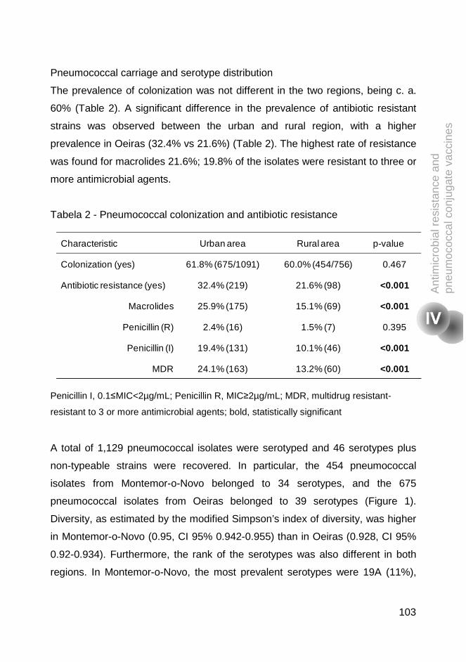
103
Pneumococcal carriage and serotype distribution
The prevalence of colonization was not different in the two regions, being c. a.
60% (Table 2). A significant difference in the prevalence of antibiotic resistant
strains was observed between the urban and rural region, with a higher
prevalence in Oeiras (32.4% vs 21.6%) (Table 2). The highest rate of resistance
was found for macrolides 21.6%; 19.8% of the isolates were resistant to three or
more antimicrobial agents.
Tabela 2 - Pneumococcal colonization and antibiotic resistance
Penicillin I, 0.1≤MIC<2µg/mL; Penicillin R, MIC≥2µg/mL; MDR, multidrug resistant-
resistant to 3 or more antimicrobial agents; bold, statistically significant
A total of 1,129 pneumococcal isolates were serotyped and 46 serotypes plus
non-typeable strains were recovered. In particular, the 454 pneumococcal
isolates from Montemor-o-Novo belonged to 34 serotypes, and the 675
pneumococcal isolates from Oeiras belonged to 39 serotypes (Figure 1).
Diversity, as estimated by the modified Simpson’s index of diversity, was higher
in Montemor-o-Novo (0.95, CI 95% 0.942-0.955) than in Oeiras (0.928, CI 95%
0.92-0.934). Furthermore, the rank of the serotypes was also different in both
regions. In Montemor-o-Novo, the most prevalent serotypes were 19A (11%),
Characteristic Urban area Rural area p-value
Colonization (yes) 61.8% (675/1091) 60.0% (454/756) 0.467
Antibiotic resistance (yes) 32.4% (219) 21.6% (98) <0.001
Macrolides 25.9% (175) 15.1% (69) <0.001
Penicillin (R) 2.4% (16) 1.5% (7) 0.395
Penicillin (I) 19.4% (131) 10.1% (46) <0.001
MDR 24.1% (163) 13.2% (60) <0.001
Ant
imic
robi
al r
esis
tanc
e an
d
pneu
moc
occa
l con
juga
te v
acci
nes

104
11A (8.8%) and NT (8.6%) whereas in Oeiras the most prevalent serotypes
were 19A (13.9%), 6C (12.4%) and 3 (9.9%) (Figure 1). Potential coverage of
the PCV10 and PCV13 were 9% and 35%, respectively, in Oeiras. In Montemor-
o-Novo corresponding figures were 9% and 27%.
Figure 1 – Serotype and antibiotype distribution in Montemor-o-Novo (A) and in Oeiras
(B).
Antibiotic resistance and serotypes
Figure 1 shows the distribution of the major serotypes in the two regions. In the
majority of cases resistance to β-lactam and macrolides was associated with the
PCV7
PCV10
PCV13
PCV7
PCV10
PCV13
Susceptible to all antibiotics
Erythromycin resistant (REry)
Penicillin non-susceptible (NSPG)
NSPG + REry
Other resistance patterns
A
B
105
same serotypes in both regions (19A, 15A and NT strains), with the exception of
serotype 6C, that was associated with this type of resistance in Oeiras, only.
Multiple logistic regression: pneumococcal colonization
Based on the differences found in the previous exploratory analysis we adjusted
a model to explain pneumococcal colonization considering the number of
children in each DCC, pneumococcal vaccination status, geographic region, and
antibiotic consumption as independent variables. The final model included the
variables in which we observed a significant difference in the univariate
analysis: region, age, antibiotic consumption during the last month and the
recurrent use of antibiotics in the last six months. Additionally, we tested for one
interaction term between children age and antibiotic consumption during the last
month. Only age and antibiotic consumption during the last month were
statistically associated with pneumococcal colonization (Table 3). Children that
took antibiotic during the last month had 73% (OR=0.27, 95%CI: 0.13-0.52) less
probability of being colonized than children that did not. Being older decreased
the odds to be colonized with pneumococci, in average the decreased was
about 1.8% in prevalence for each year of age.
Multiple logistic regression: pneumococcal resistant strains
Concerning the proportion of resistant isolates and taking into account the
results obtained after data exploration we adjusted a multiple logistic model to
explain the proportion of resistant isolates colonizing children, considering the
region, antibiotic consumption, class of antibiotic, age, vaccine status and the
pneumococcal serotypes in which the observed number of resistant isolates
was significantly different from the one estimated - 19A and NT. We also tested
two interactions terms region and serotype, and serotype and antibiotic
consumption during last month. The final model included region, serotype,
antibiotic consumption during the last month and an interaction term between
region and serotype. In the model, only serotype, antibiotic consumption during
Ant
imic
robi
al r
esis
tanc
e an
d
pneu
moc
occa
l con
juga
te v
acci
nes

106
the last month and the interaction term between region and serotypes explained
the differences in the prevalence of resistance between the two regions (Table
4). Antibiotic consumption during the last month increased the odds to be
colonized by resistant pneumococcal strains in 2.5 fold (OR=2.58, 95%CI: 1.61-
4.17). Colonization by serotype 19A and NT strains increased the odds to be
colonized by resistant pneumococcal strain in 3.5 (OR=3.52, 95%CI: 1.79-6.91)
and 11.3 (OR=11.33, 95%CI: 5.36-23.96), respectively. The odds of a child to
be colonized with a resistant strain of 19A serotype was 2.5 higher (OR=2.49,
95%CI: 1.07-5.81) in children attending DCC in Oeiras compare to Montemor-o-
Novo.
Table 3 - Odds ratio and confidence intervals estimated from logistic model to
explain pneumococcal colonization
Bold, statistically significant
VARIABLEunadjusted
OR
adjusted
ORCI 95% p-value
Region
Montemor-o-Novo
Oeiras
1
1.02
1
1.18 0.95 – 1.45 0.131
Antibiotic consumption:
during last month (yes)
6 months before sampling (yes)
0.54
0.89
0.27
0.81
0.13 – 0.52
0.64 – 1.01
<0.001
0.066
Age 0.95 0.92 0.86 – 0.99 0.045
Age x antibiotic consumption
during the last month0.99 1.01 0.83 – 1.23 0.900
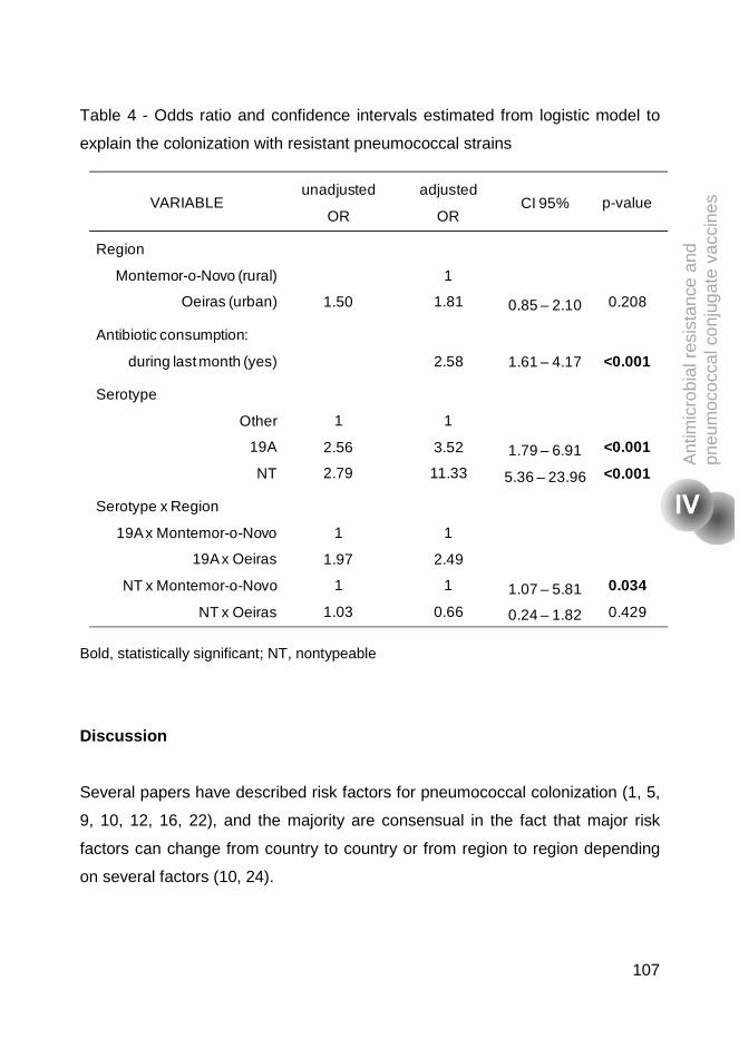
107
Table 4 - Odds ratio and confidence intervals estimated from logistic model to
explain the colonization with resistant pneumococcal strains
Bold, statistically significant; NT, nontypeable
Discussion
Several papers have described risk factors for pneumococcal colonization (1, 5,
9, 10, 12, 16, 22), and the majority are consensual in the fact that major risk
factors can change from country to country or from region to region depending
on several factors (10, 24).
VARIABLE unadjusted
OR
adjusted
OR CI 95% p-value
Region
Montemor-o-Novo (rural)
Oeiras (urban) 1.50
1
1.81 0.85 – 2.10 0.208
Antibiotic consumption:
during last month (yes) 2.58 1.61 – 4.17 <0.001
Serotype
Other
19A
NT
1
2.56
2.79
1
3.52
11.33
1.79 – 6.91
5.36 – 23.96
<0.001
<0.001
Serotype x Region
19A x Montemor-o-Novo
19A x Oeiras
NT x Montemor-o-Novo
NT x Oeiras
1
1.97
1
1.03
1
2.49
1
0.66
1.07 – 5.81
0.24 – 1.82
0.034
0.429
Ant
imic
robi
al r
esis
tanc
e an
d
pneu
moc
occa
l con
juga
te v
acci
nes
108
In this study we compared the prevalence of pneumococcal colonization and the
prevalence of antibiotic resistant pneumococcal strains in urban and rural area
of Portugal.
We observed that although the pneumococcal carriage was similar, the serotype
diversity and the prevalence of resistant strains were different. We tried to
investigate the variables that could account for this difference. We accessed for
differences among the demographic data in the two areas and we observed that
the mean age of children from Montemor-o-Novo was significantly higher. Also,
on average, the number of children per DCC unit was higher in the urban area.
Antibiotic consumption during the last month and the recurrent use of antibiotics
were also significantly higher in Oeiras. The proportion of unvaccinated children
in Oeiras was also significantly higher; however the proportion of children with
incomplete vaccination was higher in Montemor-o-Novo. However, all these
variables were not significant in the multiple logistic regression models that were
constructed to explain pneumococcal colonization in general and carriage of
resistant strains, in particular.
The colonization model showed that only previous antibiotic consumption and
being older were protective factors for pneumococcal colonization. In a previous
study conducted in Italy, where the colonization rates were compared between a
rural and urban area, the authors mention that living in a rural area significantly
decreased the odds of carrying nasopharyngeal respiratory pathogens (15).
This difference in the conclusions reached by the two studies could be attributed
to the differences between regions.
When we performed the multivariable analysis to explain the difference in the
resistance rates in the two regions, we observed that (i) antibiotic consumption
during the last month, (ii) colonization with pneumococcal serotype 19A or a NT
109
strain, and (iii) attending DCC in Oeiras and being colonized by a resistant 19A
strain increased the odds of being colonized by pneumococcal resistant strains.
The results obtained in this study led us to conclude that the most important
factor influencing carriage of resistant pneumococci is antibiotic consumption.
These results alert, that in the era of widespread use of PCVs antibiotic
consumption remains a main driving force for maintenance of pneumococcal
resistant strains in the community. Judicious use of antibiotics remains a priority
in public health.
Acknowledgments
The authors are grateful to the Directors and staff of the day-care centers and to
the parents and children that collaborated in the study. We thank the excellent
skills of the pediatric nurse Anabela Gonçalves who collected the
nasopharyngeal samples. This work was supported by PTDC/SAU-
ESA/65048/2006, PTDC/BIA-BEC/098289/2008, and PEst-
OE/EQB/LA0004/2011, from Fundação para a Ciência e Tecnologia, Portugal.
SN was supported by SFRH/BD/40706/2007, CV was supported by
SFRH/BD/70058/2010, and ASS was supported by SFRH/BD/27325/2006.
References
1. Bogaert, D., R. De Groot, and P. W. Hermans. 2004. Streptococcus
pneumoniae colonisation: the key to pneumococcal disease. Lancet
Infect Dis 4:144-154.
2. Carriço, J. A., F. R. Pinto, C. Simas, S. Nunes, N. G. Sousa, N.
Frazão, H. de Lencastre, and J. S. Almeida. 2005. Assessment of
band-based similarity coefficients for automatic type and subtype
Ant
imic
robi
al r
esis
tanc
e an
d
pneu
moc
occa
l con
juga
te v
acci
nes

110
classification of microbial isolates analyzed by pulsed-field gel
electrophoresis. J Clin Microbiol 43:5483-5490.
3. Carriço, J. A., C. Silva-Costa, J. Melo-Cristino, F. R. Pinto, H. de
Lencastre, J. S. Almeida, and M. Ramirez. 2006. Illustration of a
common framework for relating multiple typing methods by application to
macrolide-resistant Streptococcus pyogenes. J Clin Microbiol 44:2524-
2532.
4. CLSI. 2009. Performance standards for antimicrobial susceptibility
testing; approved standard-tenth edition M02-A10. CLSI, Wayne, PA,
USA.
5. Cohen, R., C. Levy, E. Bonnet, S. Grondin, V. Desvignes, A.
Lecuyer, B. Fritzell, and E. Varon. 2010. Dynamic of pneumococcal
nasopharyngeal carriage in children with acute otitis media following
PCV7 introduction in France. Vaccine 28:6114-6121.
6. de Lencastre, H., I. Santos Sanches, A. Brito-Avô, R. Sá-Leão, J.
Saldanha, K. G. Kristinsson, and A. Tomasz. 1999. Carriage and
antibiotic resistance of respiratory pathogens and molecular
epidemiology of antibiotic-resistant Streptococcus pneumoniae
colonizing children in day-care centers in Lisbon: the Portuguese day-
care center initiative. Clin Microbiol Infect 5:S55-S63.
7. Dobson, A. J. a. B., Adrian G. 2008. An Introduction to Generalized
Linear models, Thirth ed. Chapman and Hall/CRC Press, Boca Raton,
FL.
8. Faden, H., L. Duffy, R. Wasielewski, J. Wolf, D. Krystofik, and Y.
Tung. 1997. Relationship between nasopharyngeal colonization and the
development of otitis media in children. Tonawanda/Williamsville
Pediatrics. J Infect Dis 175:1440-1445.
9. Katsarolis, I., G. Poulakou, A. Analitis, I. Matthaiopoulou, E.
Roilides, C. Antachopoulos, D. A. Kafetzis, G. L. Daikos, R. Vorou,
C. Koubaniou, I. Pneumatikos, G. Samonis, V. Syriopoulou, H.
111
Giamarellou, and K. Kanellakopoulou. 2009. Risk factors for
nasopharyngeal carriage of drug-resistant Streptococcus pneumoniae:
data from a nation-wide surveillance study in Greece. BMC Infect Dis
9:120.
10. Lynch, J. P., 3rd, and G. G. Zhanel. 2010. Streptococcus pneumoniae:
epidemiology and risk factors, evolution of antimicrobial resistance, and
impact of vaccines. Curr Opin Pulm Med 16:217-225.
11. Mato, R., I. S. Sanches, C. Simas, S. Nunes, J. A. Carriço, N. G.
Sousa, N. Frazão, J. Saldanha, A. Brito-Avô, J. S. Almeida, and H.
de Lencastre. 2005. Natural history of drug-resistant clones of
Streptococcus pneumoniae colonizing healthy children in Portugal.
Microb Drug Resist 11:309-322.
12. Millar, E. V., K. L. O'Brien, E. R. Zell, M. A. Bronsdon, R. Reid, and
M. Santosham. 2009. Nasopharyngeal carriage of Streptococcus
pneumoniae in Navajo and White Mountain Apache children before the
introduction of pneumococcal conjugate vaccine. Pediatr Infect Dis J
28:711-716.
13. Nunes, S., C. Valente, R. Sá-Leão, and H. de Lencastre. 2009.
Temporal trends and molecular epidemiology of recently described
serotype 6C of Streptococcus pneumoniae. J Clin Microbiol 47:472-474.
14. Pai, R., R. E. Gertz, and B. Beall. 2006. Sequential multiplex PCR
approach for determining capsular serotypes of Streptococcus
pneumoniae isolates. J Clin Microbiol 44:124-131.
15. Principi, N., P. Marchisio, G. C. Schito, and S. Mannelli. 1999. Risk
factors for carriage of respiratory pathogens in the nasopharynx of
healthy children. Ascanius Project Collaborative Group. Pediatr Infect
Dis J 18:517-523.
16. Regev-Yochay, G., M. Raz, B. Shainberg, R. Dagan, M. Varon, M.
Dushenat, and E. Rubinstein. 2003. Independent risk factors for
Ant
imic
robi
al r
esis
tanc
e an
d
pneu
moc
occa
l con
juga
te v
acci
nes
112
carriage of penicillin-non-susceptible Streptococcus pneumoniae. Scand
J Infect Dis 35:219-222.
17. Rodrigues, F., S. Nunes, R. Sá-Leão, G. Gonçalves, L. Lemos, and
H. de Lencastre. 2009. Streptococcus pneumoniae nasopharyngeal
carriage in children attending day-care centers in the central region of
Portugal, in the era of 7-valent pneumococcal conjugate vaccine. Microb
Drug Resist 15:269-277.
18. Sá-Leão, R., S. Nunes, A. Brito-Avô, N. Frazão, A. S. Simões, M. I.
Crisóstomo, A. C. Paulo, J. Saldanha, I. Santos-Sanches, and H. de
Lencastre. 2009. Changes in pneumococcal serotypes and antibiotypes
carried by vaccinated and unvaccinated day-care centre attendees in
Portugal, a country with widespread use of the seven-valent
pneumococcal conjugate vaccine. Clin Microbiol Infect 15:1002-1007.
19. Sá-Leão, R., A. Tomasz, I. S. Sanches, A. Brito-Avô, S. E.
Vilhelmsson, K. G. Kristinsson, and H. de Lencastre. 2000. Carriage
of internationally spread clones of Streptococcus pneumoniae with
unusual drug resistance patterns in children attending day care centers
in Lisbon, Portugal. J Infect Dis 182:1153-1160.
20. Sá-Leão, R., A. Tomasz, I. S. Sanches, S. Nunes, C. R. Alves, A.
Brito-Avô, J. Saldanha, K. G. Kristinsson, and H. de Lencastre.
2000. Genetic diversity and clonal patterns among antibiotic-susceptible
and -resistant Streptococcus pneumoniae colonizing children: day care
centers as autonomous epidemiological units. J Clin Microbiol 38:4137-
4144.
21. Simões, A. S., L. Pereira, S. Nunes, A. Brito-Avô, H. de Lencastre,
and R. Sá-Leão. 2011. Clonal evolution leading to maintenance of
antibiotic resistance rates among colonizing pneumococci in the PCV7
era in Portugal. J Clin Microbiol 49:2810-2817.
22. Somech, I., R. Dagan, N. Givon-Lavi, N. Porat, S. Raiz, A. Leiberman,
M. Puterman, N. Peled, D. Greenberg, and E. Leibovitz. 2011.

113
Distribution, dynamics and antibiotic resistance patterns of
Streptococcus pneumoniae serotypes causing acute otitis media in
children in southern Israel during the 10 year-period before the
introduction of the 7-valent pneumococcal conjugate vaccine. Vaccine
29:4202-4209.
23. Sorensen, U. B. 1993. Typing of pneumococci by using 12 pooled
antisera. J Clin Microbiol 31:2097-2100.
24. Tatochenko, V. K., L. K. Katosova, M. A. Ulanova, A. P. Baturo, A. M.
Fedorov, L. N. Padiukov, V. I. Shilko, V. G. Ishutinova, S. N.
Boiarskii, V. K. Kozlov, and et al. 1994. The periodic and regional
characteristics of the pneumococcal serotype spectrum in children with
respiratory diseases and in healthy carriers. Zh Mikrobiol Epidemiol
Immunobiol:3-10.
25. Zar, J. H. 1999. Bioestatistical Analysis, 4th ed. Prentice Hall INC, New
Jersey.
Ant
imic
robi
al r
esis
tanc
e an
d
pneu
moc
occa
l con
juga
te v
acci
nes

114

115
Low pneumococcal carriage and high serotype
diversity among elderly living in Portugal.
In preparation by S. Nunes, S. Almeida, A. C. Paulo, I. Valadares, S. Martins, F.
Breia, A. Brito-Avô, A. Morais, H. de Lencastre, and R. Sá-Leão

116
The experimental work was carried out by S. Nunes with help of S. Almeida. The
statistical analysis was performed by A. C. Paulo.

117
ABSTRACT Background: While numerous studies have looked at colonization among young
children, much less is known among the elderly, the other age group for which the
incidence of pneumococcal disease is high. This study, pioneer in Portugal, aimed
to evaluate pneumococcal carriage in adults aged over 60 years.
Methods: Between April and November of 2010, nasopharyngeal and
oropharyngeal swabs of adults (>60 years), living in Oeiras (n= 651), an urban
area, or in Montemor-o-Novo (n=647), a rural area, were obtained. Pneumococci
were identified by standard procedures and by PCR detection of genes cpsA and
lytA. Isolates were characterized by antibiotype, serotype and MLST. Associations
between pneumococcal carriage, socio-demographic and clinical factors were
evaluated using a multiple logistic regression.
Results: A total of 1,298 adults were enrolled. Only 4.1% were smokers. Fifty-nine
percent of the participants were vaccinated with the seasonal influenza vaccine
and only 4.3% with the 23-valent pneumococcal polysaccharide vaccine. Twenty-
nine (2.2%) adults carried bona fide capsulated pneumococci. There were more
adults carrying pneumococci in the rural area (3.4%) than in the urban area (1.1%)
(p=0.004). One adult carried two different pneumococcal strains in the
nasopharynx. The 30 strains expressed 18 capsular types and 22 MLST types.
Sixteen isolates showed antimicrobial resistance. Smoking increased seven-fold
the odds for carriage (OR=7.4, CI 95%: 2.56-21.27) whereas living in an urban
area decreased the odds to colonization by 72% (OR=0.28, CI 95%: 0.11-0.68).
Conclusion: The prevalence of pneumococcal carriage in the elderly is low and
serotype and genotype diversity is high. Smoking is an important risk factor for
colonization whereas living in Oeiras seems to be a protective factor.
Pne
umoc
occa
l col
oniz
atio
n in
adu
lts
118
INTRODUCTION
Streptococcus pneumoniae or pneumococcus remains a leading cause of
infectious diseases worldwide. The incidence of invasive pneumococcal disease is
age-dependent being highest at the extremes of age. While the burden of disease
is substantial, pneumococcus is a commensal that colonizes asymptomatically the
nasopharynx of humans. Carriage is important as it is a fundamental step for
infection and for transmission (reviewed in (3)). While, several pneumococcal
carriage studies have been conducted among young children much less is known
about the carrier state among the elderly, the other age group where the incidence
of invasive pneumococcal disease is highest.
One of the main virulence factors of pneumococcus is the capsule and until now,
more than 90 serotypes have been described (4, 5, 13, 25). Vaccination proved to
be the best way to decrease infections and in 1983 a pneumococcal
polysaccharide vaccine, Pneumovax23, was licensed. This polysaccharide
vaccine was designed preferentially for adults and for children older than 2 years
old, but its efficacy remains controversial (8, 22). In 2000 another pneumococcal
vaccine, the seven-valent conjugate vaccine (PCV7) targeting the seven serotypes
that frequently caused invasive disease in children in USA, was introduced in the
market. The efficacy of this vaccine on the decline of invasive pneumococcal
infection (IPD) caused by serotypes covered by PCV7 was reported in several
countries (2, 9, 11, 14-16). In the USA, the effect of PCV7 was also observed in all
populations, with a concomitant reduced incidence of IPD in adults (26, 28).
Recently, we published a study that described the changes that occurred in
colonization among Portuguese children when c.a. 60% of them had received at
least one dose of PCV7 (29). In that study we observed a decline of PCV
serotypes, but carriage and antimicrobial resistance was maintained. In 2010, a
new pneumococcal conjugate vaccine (PCV13) appeared in the market, potentially
covering for six additional serotypes: 1, 3, 5, 7F, 6A, and 19A. The effect of this

*Health care centers are part of the National Health Service and provide primary care, with consultations are on general practice and routines; they serve the entire Portuguese population.
119
vaccine in IPD and in pneumococcal colonization has been studied for the past
two years. In February of 2012 this vaccine was also licensed for adults.
In the current study, conducted in 2010, we aimed to gain insights on
pneumococcal colonization patterns among elderly and establish a baseline before
the introduction of the conjugate vaccine in adults with more than 50 years of age.
METHODS
Study design. Between April and November of 2010, adults older than 60 years
of age, living in Oeiras, an urban area, or in Montemor-o-Novo, a rural area of
Portugal, were invited to participate in the study. At the time of the study the 10-
valent and 13-valent pneumococcal conjugate vaccines were commercially
available in the country although they were not part of the national immunization
plan nor were subsidized by the state. Still the commercial use of the vaccine was
high with an estimated national coverage of PCV13 in infants in 2010 of 58%
(Pfizer, personal communication). The municipality of Oeiras has a population of
over 160,000 inhabitants of which 15% are over 65 years of age, and a population
density of 3,537/km2. By contrast, the municipality of Montemor-o-Novo has a
population of 18,500 inhabitants of which 26% are over 65 years of age, and a
population density of 13/km2. In both areas individuals were enrolled in the local
health care center* and, in addition, in Montemor-o-Novo retirement homes were
also visited. Samples were obtained by specialized laboratory technicians (in
Oeiras) or nurses (in Montemor-o-Novo). The study was registered and approved
at each health care center. Study participants provided informed written consent
prior to enrollment. For each adult one nasopharyngeal sample and one
oropharyngeal sample were obtained. Information about socio-demographic data
and medical history were obtained through a questionnaire. All samples and
information were coded upon collection and processed anonymously afterwards.
Pne
umoc
occa
l col
oniz
atio
n in
adu
lts

120
Sampling and isolation. Nasopharyngeal and oropharyngeal samples were
collected as previously described (30). Swabs were streaked on gentamycin blood
agar and suspected pneumococcal colonies were isolated and identified following
routine standard procedures based on optochin susceptibility and bile solubility
(19, 30). Additionally, isolates showing one or more atypical results in the latter
assays but displaying a characteristic pneumococcal colony morphology were
further characterized by the identification of lytA pneumococcal-specific RFLP
signatures as described by Lull et al. (17). The swabs and the pure cultures were
frozen and stored at -80ºC in STGG (20).
Capsular typing. Isolates were capsular typed by multiplex PCR using primers
previously described (23) and www.cdc.gov and/or by the Quellung reaction using
commercially available pneumococcal antisera (Statens Serum Institute,
Copenhagen, Denmark) (31).
Antimicrobial susceptibility testing. Antimicrobial susceptibility testing was
performed using the Kirby-Bauer technique, according to the CLSI
recommendations and definitions (7). The antimicrobial agents tested were
chloramphenicol, erythromycin, clindamycin, tetracycline, and sulfamethoxazole-
trimethoprim (SXT). Antibiotic disks were purchased from Oxoid (Hampshire,
England). Interpretation of results followed the CLSI guidelines (7). Isolates were
also screened for MICs to penicillin and ciprofloxacin with the E-test (AB Biodisk,
Solna, Sweden) according to the manufacturer’s recommendations. In the
interpretation of decreased penicillin susceptibility epidemiological breakpoints
were used: isolates were considered intermediately resistant if the MIC was ≥0.1
µg/ml and ˂1.5 µg/ml and were considered resistant if the MIC was ≥1.5 µg/ml.
For ciprofloxacin, strains with MIC≥2 µg/ml were considered resistant.

121
Multilocus sequence typing (MLST). MLST was done essentially as previously
described (10) using primers with universal M13 tails. Sequencing was conducted
at Macrogen, Inc. (Seoul, Korea).
Statistical analysis. Socio-demographic and clinical variables were described
using the mean and standard deviation or percentages as appropriate. Differences
for each variable, between Oeiras and Montemor-o-Novo, were tested using the
Student's t-test or the Mann-Whitney U-test for continuous variables and the
Fisher's exact test to compare proportions (32). Univariate and multiple logistic
regression analysis were used to assess the goodness of fit of the socio-
demographic and clinical variables to the dependent variable, to be colonized or
not and, to calculate non- and adjusted odds ratios (OR). A conservative p-value
<0.1 was used to select the variables in the univariate model to be included in the
multiple logistic model. For all statistical tests, differences were considered to be
statistically significant if the p-value < 0.05 or when the adjusted 95% CI odds ratio
did not include 1.0. Statistical analyses were carried out using R.
RESULTS
Characteristics of the study population. A total of 651 adults over 60 yrs of age
living in the urban area of Oeiras and 647 living in the rural area of Montemor-o-
Novo participated in the study. Women were slightly overrepresented in both
populations. The two populations differed significantly in several socio-
demographic and medical characteristics (detailed in Table 1). On average,
participants from the rural area were older and had less years of formal education.
Although most participants were retirees, the proportion of those still working was
significantly lower in the rural area. The household size was comparable in both
areas but participants from the rural area had significantly less contact with young
children. On the other hand, in the rural area, participants engaged more
Pne
umoc
occa
l col
oniz
atio
n in
adu
lts
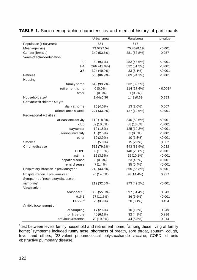
122
TABLE 1. Socio-demographic characteristics and medical history of participants
atest between levels family household and retirement home; bamong those living at family home; csymptoms included runny nose, shortness of breath, sore throat, sputum, cough, fever and others; d23-valent pneumococcal polysaccharide vaccine; COPD, chronic obstructive pulmonary disease.
Urban area Rural area p-value
Population (> 60 years) 651 647Mean age (yrs) 73.07±7.54 75.45±8.19 <0.001Gender (female) 349 (53.6%) 381 (58.8%) 0.057Years of school education
0 59 (9.1%) 282 (43.6%) <0.0011-4≥ 5
266 (41.0%)324 (49.9%)
332 (51.3%)33 (5.1%)
<0.001<0.001
Retirees 566 (86.9%) 609 (94.1%) <0.001Housing
family home 649 (99.7%) 532 (82.2%)retirement home 0 (0.0%) 114 (17.6%) <0.001a
other 2 (0.3%) 1 (0.2%)Household sizeb 1.44±0.36 1.43±0.39 0.553Contact with children ≤ 6 yrs
daily at home 26 (4.0%) 13 (2.0%) 0.007at least once a week 221 (33.9%) 127 (19.6%) <0.001
Recreational activitiesat least one activity 119 (18.3%) 340 (52.6%) <0.001
club 69 (10.6%) 88 (13.6%) <0.001day center 12 (1.8%) 125 (19.3%) <0.001
senior university 16 (2.5%) 3 (0.5%) <0.001other 19 (2.9%) 10 (1.5%) <0.001
Smoker 38 (5.9%) 15 (2.3%) 0.002Chronic disease 515 (79.1%) 543 (83.9%) 0.032
COPD 59 (11.6%) 140 (25.8%) <0.001asthma 18 (3.5%) 55 (10.1%) <0.001
hepatic disease 3 (0.6%) 23 (4.2%) <0.001renal disease 7 (1.4%) 35 (6.4%) <0.001
Respiratory infection in previous year 219 (33.6%) 365 (56.3%) <0.001
Hospitalization in previous year 95 (14.6%) 93(14.4%) 0.937Symptoms of respiratory disease at samplingc 212 (32.6%) 273 (42.2%) <0.001Vaccination
seasonal flu 363 (55.8%) 397 (61.4%) 0.043H1N1 77 (11.8%) 36 (5.6%) <0.001
PPV23d 26 (3.9%) 20 (3.1%) 0.454Antibiotic consumption
at sampling 17 (2.6%) 10 (1.5%) 0.249month before 40 (6.1%) 32 (4.9%) 0.396
previous 3 months 70 (10.8%) 44 (6.8%) 0.014
123
frequently in regular recreational activities. Although smoking was not common in
these populations, the proportion was higher in the urban area (Table 1) with no
differences (among smokers from the two regions) in the number of smoking years
and cigarettes per day (data not shown).
Regarding medical history, participants from the rural area were globally less
healthy. A high prevalence of chronic diseases was reported in both populations
being higher in the rural area. When individual diseases were analyzed, chronic
obstructive pulmonary disease (COPD), asthma, hepatic and renal diseases
remained significantly higher in the rural area. The proportion of participants
reporting a respiratory infection in the previous year, and current respiratory
infection symptoms was also higher in the rural area.
Regarding vaccination over half of the participants had received the seasonal flu
vaccine but significantly less had been vaccinated against H1N1. A tiny proportion
(<4%) had received PPV23. Antibiotic consumption was low in both regions
although higher in the urban area. Urinary infections were reported as the main
reason for taking antibiotics.
Pneumococcal carriage. Of the 1,298 adults, 29 (2.2%) were pneumococcal
carriers. One adult carried two different isolates. To explore which factors
contributed to pneumococcal carriage, univariate and multiple logistic regression
analyses were performed. There were significantly more carriers in the rural area
than in the urban area (3.4% vs 1.1%, respectively, p=0.004). Carriage was also
more frequent among smokers (9.4% vs 1.9%, p=0.005), and those with COPD
(4.5% vs 1.8%, p=0.032). Other variables did not yield statistical significant
differences.
The best multiple logistic regression model included the region, smoking and
having COPD. In the final model, elderly living in the urban area had a lower risk of
carrying pneumococci, while smokers had an increased risk (Table 2).
Pne
umoc
occa
l col
oniz
atio
n in
adu
lts

124
TABLE 2. Socio-demographic characteristics and medical history of participants
COPD, chronic obstructive pulmonary disease.
Characteristics of pneumococcal isolates. Among the 30 pneumococcal strains
a high diversity of serotypes and genotypes was detected as 18 serotypes and 22
sequence types were identified (Table 3). Overall, 23% and 43% of the isolates
belonged to serotypes included in PCV13 and PPV23, respectively. By MLST, two
novel sequence types were identified (STs 6987 and 6988 associated with
serotypes 18A and 23B, respectively). No major lineages were detected and the
largest group of strains sharing an ST contained three isolates only. Close to half
of the isolates (46.7%) were resistant to at least one antimicrobial agent; only two
isolates were multidrug resistant (i.e. resistant to three or more classes of
antimicrobials) (Table 3).
Variable Unadjusted ORAdjusted OR
(95% CI)p-value
Region
rural 1
urban 0.31 0.28 (0.11 – 0.68) 0.004
Smoking (yes) 5.29 7.38 (2.56 – 21.27) 0.001
COPD (yes) 2.55 2.00 (0.88 – 4.54) 0.111

125
TABLE 3. Characterization of the pneumococcal strains
IPG, intermediately resistant to penicillin; Ery, erythromycin; Clin, clindamycin; Tet,
tetracycline; SXT sulfamethoxazole-trimethoprim; Cip, ciprofloxacin
Serotype MLST Resistance pattern Origin No. of isolates
3 180 susceptible rural 2
5 1223 Cip rural 1
6A 1714 Tet, Cip urban 1
6A 460 susceptible urban 1
9L 66 Cip rural 1
11A 62 SXT urban 1
11A 62 Cip urban 1
11A 408 Cip rural 2
15A 63 IPG, Tet urban 1
15B 3097 IPG, Ery, Clin, Tet rural 1
16A 30 susceptible urban 1
18A 6987 susceptible urban 1
19A 276 IPG, Ery, Clin, Tet, Cip rural 1
19A 1201 susceptible rural 1
21 1877 Cip rural 1
22F 433 susceptible rural 3
23A 42 Cip rural 1
23B 6988 susceptible rural 1
23B 439 susceptible rural 2
31 1766 susceptible rural 1
35F 1635 Ery rural 1
35F 1368 susceptible rural 1
36 1635 Ery, Cip rural 1
37 447 Cip rural 2
Pne
umoc
occa
l col
oniz
atio
n in
adu
lts

126
DISCUSSION
This is the first study conducted in Portugal aiming to evaluate pneumococcal
carriage in the elderly population. Previous studies from other countries such as
Belgium, Israel, and Kenya, described low rates of colonization (typically up to 5%)
among adults (1, 12, 27). In our study, the pneumococcal carriage rates were also
low, being higher in the rural area when compared to the urban area (3.4% vs
1.1%, p=0.004).
Although several differences were noticed among socio-demographic
characteristics and medical history of the two populations studied, statistical
analysis identified only two factors that were associated with pneumococcal
carriage: living in the urban area and being a smoker. While living in Oeiras
seemed to be a protective effect, decreasing the risk of colonization by 3.5 fold,
smoking increased the risk of being colonized by seven-fold. Smoking has been
previously associated with pneumococcal carriage (18). Why living in the urban
area reduced the risk of carriage is not self-evident. One may hypothesize that
other important variables to explain this observation were not studied.
Alternatively, it is conceivable that due to the small number of carriers identified in
the samples resulted in low statistical power to identify additional variables as
significant.
Characterization of pneumococcal isolates revealed a high serotype and genotype
diversity. Only one serotype (11A) was common to both areas; however, the
genotypes and antibiotypes associated with it were distinct.
This study has some limitations. Although the size of the population enrolled was
high (c.a. 650 adults enrolled in each area) the low number of pneumococcal
isolates identified hindered the statistical analysis. Secondly, we used culture-
based only methods to detect pneumococci, which may have resulted in a lower
127
carriage rate estimate. Although we used a selective medium to prevent
overgrowth of undesirable bacteria, and easily identify pneumococci, we may have
been unable to detect low-density carriers. In fact, Ogami et al. reported that real-
time PCR increased significantly detection of carriage in healthy children when
compared to culture alone (21). The same is plausible for detection of carriage in
adults (24). More recently, Chien et al. reported that at carriage density of ≤105
CFU/ml detection of pneumococci by culture was significantly less sensitive than
PCR (6). These limitations are being further explored in ongoing initiatives carried
by this group. Our study has also some strength. It generated for the first time data
on pneumococcal colonization patterns in the elderly in Portugal. In addition,
pneumococcal isolates were characterized in detail and a wealth of
epidemiological data was obtained.
In conclusion, this study showed that pneumococcal colonization in the elderly in
Portugal is low (2.2%). Colonizing isolates are very diverse in terms of genotype
and serotype. Smoking increases the risk of being colonized with pneumococci by
7-fold while living in an urban area seems to be a protective factor for colonization.
This study will contribute to establish a baseline that may be used in future studies
to monitor how novel pneumococcal vaccines impact on colonization in adults.
ACKNOWLEDGMENTS
The authors are grateful to the participants that collaborated in the study. We
thank Ana Paula Ribeiro from Câmara Municipal de Montemor-o-Novo for her
extraordinary support in the implementation of fieldwork in Montemor-o-Novo. We
thank the excellent skills of nurses Anabela Gonçalves and Pedro Fonseca.
Pne
umoc
occa
l col
oniz
atio
n in
adu
lts

128
FUNDING
SN was supported by grant SFRH/BD/40706/2007 from Fundação para a Ciência
e Tecnologia (FCT), Portugal. This work was partially supported by an unrestricted
research grant from Pfizer and grant PEst-OE/EQB/LA0004/2011 (to the Oeiras
Associated Laboratory) from FCT. The funders had no role in study design, data
collection and analysis, preparation of the manuscript or decision to submit the
manuscript for publication.
REFERENCES
1. Abdullahi, O., J. Nyiro, P. Lewa, M. Slack, and J. A. Scott. 2008. The
descriptive epidemiology of Streptococcus pneumoniae and Haemophilus
influenzae nasopharyngeal carriage in children and adults in Kilifi district,
Kenya. Pediatr Infect Dis J 27:59-64.
2. Aljunid, S., G. Abuduxike, Z. Ahmed, S. Sulong, A. M. Nur, and A. Goh.
2011. Impact of routine PCV7 (Prevenar) vaccination of infants on the
clinical and economic burden of pneumococcal disease in Malaysia. BMC
Infect Dis 11:248.
3. Bogaert, D., R. De Groot, and P. W. Hermans. 2004. Streptococcus
pneumoniae colonisation: the key to pneumococcal disease. Lancet Infect
Dis 4:144-154.
4. Bratcher, P. E., K. H. Kim, J. H. Kang, J. Y. Hong, and M. H. Nahm.
2010. Identification of natural pneumococcal isolates expressing serotype
6D by genetic, biochemical and serological characterization. Microbiology
156:555-560.
5. Calix, J. J., and M. H. Nahm. 2010. A new pneumococcal serotype, 11E,
has a variably inactivated wcjE gene. J Infect Dis 202:29-38.
129
6. Chien, Y. W., J. E. Vidal, C. G. Grijalva, C. Bozio, K. M. Edwards, J. V.
Williams, M. R. Griffin, H. Verastegui, S. M. Hartinger, A. I. Gil, C. F.
Lanata, and K. P. Klugman. 2012. Density interactions between
Streptococcus pneumoniae, Haemophilus influenzae and Staphylococcus
aureus in the nasopharynx of young Peruvian children. Pediatr Infect Dis J.
7. CLSI. 2009. Performance standards for antimicrobial susceptibility testing;
approved standard-tenth edition M02-A10. CLSI, Wayne, PA, USA.
8. Conaty, S., L. Watson, J. Dinnes, and N. Waugh. 2004. The
effectiveness of pneumococcal polysaccharide vaccines in adults: a
systematic review of observational studies and comparison with results
from randomised controlled trials. Vaccine 22:3214-3224.
9. Dagan, R. 2009. Impact of pneumococcal conjugate vaccine on infections
caused by antibiotic-resistant Streptococcus pneumoniae. Clin Microbiol
Infect 15 Suppl 3: 16-20.
10. Enright, M. C., and B. G. Spratt. 1998. A multilocus sequence typing
scheme for Streptococcus pneumoniae: identification of clones associated
with serious invasive disease. Microbiology 144:3049-3060.
11. Espinosa-de Los Monteros, L. E., F. Aguilar-Ituarte, R. N. Jimenez-
Juarez, R. S. Rodriguez-Suarez, and D. Gomez-Barreto. 2010.
Streptococcus pneumoniae serotype replacement in nasopharyngeal
colonization in children vaccinated with PCV7 in Mexico. Salud Publica
Mex 52:4-13.
12. Flamaing, J., W. E. Peetermans, J. Vandeven, and J. Verhaegen. 2010.
Pneumococcal colonization in older persons in a nonoutbreak setting. J Am
Geriatr Soc 58:396-398.
13. Henrichsen, J. 1995. Six newly recognized types of Streptococcus
pneumoniae. J Clin Microbiol 33:2759-2762.
14. Hsu, K. K., K. M. Shea, A. E. Stevenson, and S. I. Pelton. 2010.
Changing serotypes causing childhood invasive pneumococcal disease:
Massachusetts, 2001-2007. Pediatr Infect Dis J 29:289-293.
Pne
umoc
occa
l col
oniz
atio
n in
adu
lts

130
15. Isaacman, D. J., E. D. McIntosh, and R. R. Reinert. 2010. Burden of
invasive pneumococcal disease and serotype distribution among
Streptococcus pneumoniae isolates in young children in Europe: impact of
the 7-valent pneumococcal conjugate vaccine and considerations for future
conjugate vaccines. Int J Infect Dis 14:e197-209.
16. Lacapa, R., S. J. Bliss, F. Larzelere-Hinton, K. J. Eagle, D. J. McGinty,
A. J. Parkinson, M. Santosham, M. J. Craig, and K. L. O'Brien. 2008.
Changing epidemiology of invasive pneumococcal disease among White
Mountain Apache persons in the era of the pneumococcal conjugate
vaccine. Clin Infect Dis 47:476-484.
17. Llull, D., R. Lopez, and E. Garcia. 2006. Characteristic signatures of the
lytA gene provide a basis for rapid and reliable diagnosis of Streptococcus
pneumoniae infections. J Clin Microbiol 44:1250-1256.
18. Mackenzie, G. A., A. J. Leach, J. R. Carapetis, J. Fisher, and P. S.
Morris. 2010. Epidemiology of nasopharyngeal carriage of respiratory
bacterial pathogens in children and adults: cross-sectional surveys in a
population with high rates of pneumococcal disease. BMC Infect Dis
10:304.
19. Murray, P. R. 1979. Modification of the bile solubility test for rapid
identification of Streptococcus pneumoniae. J Clin Microbiol 9:290-291.
20. O'Brien, K. L., M. A. Bronsdon, R. Dagan, P. Yagupsky, J. Janco, J.
Elliott, C. G. Whitney, Y. H. Yang, L. G. Robinson, B. Schwartz, and G.
M. Carlone. 2001. Evaluation of a medium (STGG) for transport and
optimal recovery of Streptococcus pneumoniae from nasopharyngeal
secretions collected during field studies. J Clin Microbiol 39:1021-1024.
21. Ogami, M., M. Hotomi, A. Togawa, and N. Yamanaka. 2010. A
comparison of conventional and molecular microbiology in detecting
differences in pneumococcal colonization in healthy children and children
with upper respiratory illness. Eur J Pediatr 169:1221-1225.
131
22. Ortqvist, A., I. Henckaerts, J. Hedlund, and J. Poolman. 2007. Non-
response to specific serotypes likely cause for failure to 23-valent
pneumococcal polysaccharide vaccine in the elderly. Vaccine 25:2445-
2450.
23. Pai, R., R. E. Gertz, and B. Beall. 2006. Sequential multiplex PCR
approach for determining capsular serotypes of Streptococcus pneumoniae
isolates. J Clin Microbiol 44:124-131.
24. Palmu, A. A., T. Kaijalainen, A. Saukkoriipi, M. Leinonen, and T. M.
Kilpi. 2012. Nasopharyngeal carriage of Streptococcus pneumoniae and
pneumococcal urine antigen test in healthy elderly subjects. Scand J Infect
Dis 44:433-438.
25. Park, I. H., D. G. Pritchard, R. Cartee, A. Brandao, M. C. Brandileone,
and M. H. Nahm. 2007. Discovery of a new capsular serotype (6C) within
serogroup 6 of Streptococcus pneumoniae. J Clin Microbiol 45:1225-1233.
26. Pilishvili, T., C. Lexau, M. M. Farley, J. Hadler, L. H. Harrison, N. M.
Bennett, A. Reingold, A. Thomas, W. Schaffner, A. S. Craig, P. J.
Smith, B. W. Beall, C. G. Whitney, and M. R. Moore. 2010. Sustained
reductions in invasive pneumococcal disease in the era of conjugate
vaccine. J Infect Dis 201:32-41.
27. Regev-Yochay, G., M. Raz, R. Dagan, N. Porat, B. Shainberg, E. Pinco,
N. Keller, and E. Rubinstein. 2004. Nasopharyngeal carriage of
Streptococcus pneumoniae by adults and children in community and family
settings. Clin Infect Dis 38:632-639.
28. Rosen, J. B., A. R. Thomas, C. A. Lexau, A. Reingold, J. L. Hadler, L.
H. Harrison, N. M. Bennett, W. Schaffner, M. M. Farley, B. W. Beall,
and M. R. Moore. 2011. Geographic variation in invasive pneumococcal
disease following pneumococcal conjugate vaccine introduction in the
United States. Clin Infect Dis 53:137-143.
29. Sá-Leão, R., S. Nunes, A. Brito-Avô, N. Frazão, A. S. Simões, M. I.
Crisóstomo, A. C. Paulo, J. Saldanha, I. Santos-Sanches, and H. de
Pne
umoc
occa
l col
oniz
atio
n in
adu
lts

132
Lencastre. 2009. Changes in pneumococcal serotypes and antibiotypes
carried by vaccinated and unvaccinated day-care centre attendees in
Portugal, a country with widespread use of the seven-valent pneumococcal
conjugate vaccine. Clin Microbiol Infect 15:1002-1007.
30. Sá-Leão, R., A. Tomasz, I. S. Sanches, S. Nunes, C. R. Alves, A. Brito-
Avô, J. Saldanha, K. G. Kristinsson, and H. de Lencastre. 2000.
Genetic diversity and clonal patterns among antibiotic-susceptible and -
resistant Streptococcus pneumoniae colonizing children: day care centers
as autonomous epidemiological units. J Clin Microbiol 38:4137-4144.
31. Sorensen, U. B. 1993. Typing of pneumococci by using 12 pooled
antisera. J Clin Microbiol 31:2097-2100.
32. Zar, J. H. 1999. Bioestatistical Analysis, 4th ed. Prentice Hall INC, New
Jersey.

133
Multiple-Locus Variable number tandem repeat
Analysis for Streptococcus pneumoniae :
comparison with PFGE and MLST
Published in: Elberse KE*, S. Nunes*, R. Sá-Leão, H.G. van der Heide, and
L.M. Schouls . 2011. PLoS One. 6:19668. *These authors contributed equally.

134
S. Nunes was responsible for MLVA.

135
Abstract
In the era of pneumococcal conjugate vaccines, surveillance of pneumococcal
disease and carriage remains of utmost importance as important changes may
occur in the population. To monitor these alterations reliable genotyping
methods are required for large-scale applications. We introduced a high
throughput multiple-locus variable number tandem repeat analysis (MLVA) and
compared this method with pulsed-field gel electrophoresis (PFGE) and
multilocus sequence typing (MLST).
The MLVA described here is based on 8 BOX loci that are amplified in two
multiplex PCRs. The labeled PCR products are sized on an automated DNA
sequencer to accurately determine the number of tandem repeats. The
composite of the number of repeats of the BOX loci makes up a numerical
profile that is used for identification and clustering. In this study, MLVA was
performed on 263 carriage isolates that were previously characterized by MLST
and PFGE.
MLVA, MLST and PFGE (cut-off of 80%) yielded 164, 120, and 87 types,
respectively. The three typing methods had Simpson’s diversity indices of
98.5% or higher. Congruence between MLST and MLVA was high. The Wallace
of MLVA to MLST was 0.874, meaning that if two strains had the same MLVA
type they had 88% chance of having the same MLST type. Furthermore, the
Wallace of MLVA to clonal complex of MLST was even higher: 99.5%. For some
isolates belonging to a single MLST clonal complex although displaying different
serotypes, MLVA was more discriminatory generating groups according to
serotype or serogroup.
Overall, MLVA is a promising genotyping method that is easy to perform and a
relatively cheap alternative to PFGE and MLST. In the companion paper
published simultaneously in this issue we applied the MLVA to assess the
pneumococcal population structure of isolates causing invasive disease in the
Netherlands before the introduction of the 7-valent conjugate vaccine.
MLV
A fo
r S
trep
toco
ccus
pne
umon
iae

136
Introduction
Streptococcus pneumoniae is an important human pathogen causing diseases
like otitis, pneumonia, sepsis, and meningitis. The major virulence factor of the
pneumococcus is the capsule (1, 17). Currently, over 90 different capsules have
been identified (3, 5, 16, 31). Such high antigenic diversity has long been
recognized and serotyping using Quellung reaction has been used for
pneumococcal typing for several decades. In recent years, as the DNA
sequences of the capsular biosynthetic loci became available (2, 31), alternative
strategies for capsular typing based on genotyping methods have been
developed (4, 22, 30) (Elberse et al. companion paper, PLoS One, this issue).
For the past two decades, a number of genotyping methods aimed to assess
the genetic diversity of pneumococcal isolates have been used. Currently,
pulsed-field gel electrophoresis (PFGE) (15, 23) and multilocus sequence typing
(MLST) (10) are the gold standards for genotyping of pneumococci.
In PFGE, total DNA is digested with a rare cutter endonuclease (such as SmaI
or ApaI) that yields a limited number of fragments of high molecular weight. The
fragments are separated by a variant of gel electrophoresis in which the
orientations of the electric field change periodically, enabling megabase size
DNA fragments to be effectively separated by size (39). The DNA banding
patterns are then compared between isolates and clonal relationships are
inferred (23). Although the interpretation criteria may vary depending on the size
of the collection and on the goal of the research being conducted, there are
general criteria, both visual and computer-assisted that seem to work well (6,
42). Advantages of PFGE are that it has good typeability (the percentage of
isolates that can be assigned a type), reproducibility, and resolving power (26,
42). In addition, the costs for materials and equipment are relatively low and
handling of the equipment is easy. However, it is laborious and time consuming

137
and may yield ambiguous results if not performed by a well trained technician.
PFGE is quite useful for local epidemiology and it has also been used for global
epidemiology once standardized (27, 34). Portability between laboratories is not
straightforward, but seems to be attainable provided protocol harmonization is
achieved (42).
Multilocus sequence typing (MLST) is a DNA sequence-based method that
relies on PCR amplification and sequencing of internal fragments of 7
housekeeping genes (10, 24). For allele assignment, each sequence is
compared to all known alleles which are available at an online database
(www.mlst.net). Different sequences (even single nucleotide difference) are
assigned different allelic numbers. The 7 assigned allele numbers form an allelic
profile or sequence type (ST). MLST is expensive if in-house sequencing
facilities are not available and, therefore, many laboratories cannot afford to use
it routinely. However, it has the advantages of being reproducible,
unambiguous, portable allowing intra-laboratory comparisons and suited to
create international databases. For S. pneumoniae, MLST has a good resolving
power being useful for local and global epidemiology. Furthermore, in contrast
to PFGE, MLST does not always require a culture and can sometimes be
directly performed on samples containing bacterial DNA such as cerebrospinal
fluid (9).
In 1992 conserved repeated sequences, named BOX elements, were identified
in the genome of the pneumococcus (25). The sequenced genomes of R6 and
TIGR4 contain 115 and 127 BOX elements, respectively. BOX elements consist
of 3 different subunits, BoxA, BoxB and BoxC. BoxB is the tandem repetitive
unit of 45 base pairs flanked by BoxA and BoxC, although elements missing
BoxA or missing BoxC have been described (20, 25). The function and origin of
BOX elements are unknown; however, it is thought that they may be involved in
regulating the expression of virulence-associated genes when they are located
MLV
A fo
r S
trep
toco
ccus
pne
umon
iae

138
in the promoter regions of genes (20, 25). In the years after the discovery of
these elements a number of genotyping methods were introduced based on the
variability of these elements. A BOX-PCR described by van Belkum and
colleagues showed the usefulness of these repeats for genotyping (44). The
primer of this PCR is based on the BoxA sequence and yields PCR products of
regions present between BOX loci that are close to each other and in opposite
direction. However, the method has the disadvantage of creating banding
profiles, which are difficult to interpret and often lack reproducibility. A multiple-
locus variable number tandem repeat analysis (MLVA) scheme based on BOX
typing was introduced by Koeck et al. in 2005 (21). It analyzes 16 BOX loci that
are PCR-amplified in single PCR reactions and products are analyzed by
agarose gel electrophoresis. A website (www.mlva.eu) providing a database in
which profiles can be compared has been created. At the time of writing this
manuscript the MLVA profiles of 1147 isolates had been deposited in the
database. The many loci that are analyzed and the choice of agarose gel
electrophoresis for sizing makes this method somewhat laborious. This MLVA
scheme was successfully applied and compared with MLST and was shown to
yield high congruence with MLST, but was more discriminatory than MLST (32,
35).
In this paper we describe a newly developed MLVA typing scheme for
pneumococci based on 8 BOX elements. The 8 BOX loci are amplified in 2
multiplex PCRs and the fluorescently labeled PCR products are sized on an
automated sequencer yielding high throughput and unambiguous typing results.
The generated MLVA results can be compared internationally using the newly
available MLVA website www.MLVA.net. On the website the MLVA profiles can
be imported to assess the MLVA type (MT). In the current study we assess the
validity of this method by comparing MLVA with two well established genotyping
methods: PFGE and MLST. In the companion paper published simultaneously
in PLoS One, we applied this MLVA scheme to assess the population structure

139
of pneumococci causing invasive disease in the Netherlands before the
introduction of the 7-valent pneumococcal conjugate vaccine.
Materials and methods
Isolate collection.
In this study 263 pneumococcal isolates were used for the comparison of MLVA
with PFGE and MLST. The isolates were obtained between 1996 and 2007 from
the nasopharynx of children attending several day-care centers in the area of
Lisbon, Portugal. The isolates were stored in glycerol broth at -80ºC and were
characterized by antibiogram, serotyping, PFGE, and MLST under the scope of
previous studies (28, 33, 34). Approval for the study was obtained from the
Ministry of Education and from the directors of each day-care center. Written
informed consent was obtained from parents or guardians of each child before
sampling. All information was stored in an in-house developed online database.
Overall, the collection comprised 41 serotypes, 87 PFGE types and 120 STs.
Serotyping, PFGE and MLST.
These techniques were performed as previously described. Briefly, serotyping
was performed by the Quellung reaction (41) and/or multiplex PCR (8, 30). For
PFGE, total DNA was extracted, restricted with SmaI, and DNA fragments were
resolved by PFGE (34). MLST was performed using primers and conditions
described previously (10). For allele assignment the S. pneumoniae MLST
database available at www.mlst.net was interrogated. Sequence traces of novel
alleles were submitted to the curator for new allele and ST assignment.
Multiple-Locus Variable number tandem repeat Analysis (MLVA).
In the genome sequence of isolate R6 (GenBank number NC_003098), one of
the publicly available genomes, 115 BOX tandem repeats were found. Thirteen
MLV
A fo
r S
trep
toco
ccus
pne
umon
iae

140
randomly chosen BOX loci scattered throughout the R6 genome to be used in
the MLVA scheme were tested on a panel of 84 isolates. Eight BOX loci were
chosen and primers were designed targeting the flanking regions of the BoxA
and BoxC elements (Figure 1, Table 1).
Fig. 1. Schematic representation of pneumococcus R6 genome indicating the
location of the 8 BOX loci that are used in the MLVA scheme.
One primer of each primer set was fluorescently labeled with FAM, NED, VIC or
PET. For BOX loci 4 and 6 respectively 3 and 4 primers were used to primer set
was fluorescently labeled with FAM, NED, VIC or PET. For BOX loci 4 and 6
respectively 3 and 4 primers were used to enhance amplification within all
isolates. The 8 loci were amplified in 2 separate multiplex PCRs, creating
mixtures each with 4 different fluorescent labels enabling analysis of 4 BOX loci
in a single fragment analysis reaction (Table 1). Amplification of the loci was
done in Applied Biosystems 9700 PCR machines. The 25 µl PCR reaction
mixtures consisted of Qiagen multiplex PCR mix, 10 µM of each primer and 2 µl
of 1:10 lysates diluted in sterile water. Lysates were prepared by suspending a
loop full of pneumococci in 500 µl TE (10 mM Tris.HCl and 1 mM EDTA, pH 8)
followed by heating for 10 minutes by 95ºC. Amplification was performed using
the following PCR program: 15 min 95ºC, 25 cycles of 30 sec 95ºC, 1 min 54ºC
and 1 min 72ºC followed by a 30 min incubating at 68ºC to ensure complete

141
addition of the extra 3’adenosin by the terminal transferase activity of the Taq
polymerase. Of the PCR product mixtures, 2 µl aliquots, diluted 1:200 in water,
were mixed with 10 µl of 1:200 diluted Genescan 1200 LIZ-marker (Applied
Biosystems, Foster City, U.S.A.). The product was heated for 5 minutes at 95ºC
for denaturation and sized on the AB 3730 DNA sequencer using the Fragment
Analysis module.
Determination of the number of repeats in each locus was done using the
GeneMarker software (Softgenetics, State College, Pennsylvania, USA). For
this purpose, fragment analysis files with the .fsa extension were imported into
the software and the number of repeats in each locus was derived from the
data. All alleles that yielded PCR product sizes that had not been found before,
were analyzed by sequencing using unlabeled versions of the MLVA primers.
This included PCR products with aberrant sizes representing loci in which
deletions or insertions had occurred in the region flanking the BOX repeats.
Thereafter, the loci were assigned the number of BoxB repeats present. For
each isolate an allelic profile was generated consisting in a string of 8 integers
reflecting the number of repeats in the 8 BOX loci. The allelic profile was
assigned an arbitrary sequential MLVA type (MT). The correspondence between
MT and allelic profile was deposited at the MLVA database located at
www.mlva.net to guarantee that the same nomenclature was maintained for all
isolates characterized by this technique regardless of its origin and study.
Data Analysis.
Analysis of PFGE patterns, MLST and MLVA numerical profiles, was performed
using Bionumerics version 6.1 (Applied Maths, Sint-Martens-Latem, Belgium).
PFGE patterns were clustered by UPGMA. A dendrogram was generated from a
similarity matrix calculated using the Dice similarity coefficient with an
optimization of 1.0% and a tolerance of 1.5%. PFGE clusters were defined as
isolates with a similarity of 80% or higher on the dendrogram (13, 33).
MLV
A fo
r S
trep
toco
ccus
pne
umon
iae

142
Clustering of MLST and MLVA types were displayed in minimum spanning
trees. In this graphic representation, the circles in the tree represent the various
types. The size of the circle is proportional to the number of isolates with the
same type. Lines linking two types in the tree denote single locus difference
between those types. For assignment of MLST or MLVA clonal complex, the
entire MLST database (available at www.mlst.net, last accessed on October 20,
2010) or the entire in-house MLVA database (available at www.mlva.net, last
accessed on October 20, 2010) were interrogated. MLVA complex assignment
was based on a maximum distance of one locus between related types. The
minimum number of MLVA types in a MLVA complex was set to 3 with a
minimum of 9 entries per MLVA complex, resulting in on average 3 isolates
within an MLVA type. These settings resulted in MLVA complexes that were
strongly correlated to serotype.
Statistical analysis.
The discriminatory ability of the three typing systems - PFGE, MLST and MLVA
- was measured using the Simpson’s index of diversity (SID) and 95%
confidence intervals were calculated as proposed before (14, 40). The
congruence between the typing methods was calculated using the Wallace
coefficient (7). All calculations were done using the freely available online tool
Comparing Partitions located at www.comparingpartitions.info (7).
Results
Multiple-locus variable number tandem repeat analysis (MLVA)
A combination of highly diverse and less diverse BOX loci were chosen aiming
to have a good balance between loci that would evolve faster than others. The
13 randomly chosen BOX loci were tested on a panel of 84 isolates and 8 BOX
loci were chosen for the MLVA scheme. The overlap in BOX loci of our MLVA

143
scheme with the MLVA scheme previously described by Koeck et al. (21) is
provided in Table 1. In particular, BOX loci 1, 4, 12 and 13 were rather diverse
while BOX locus 11 had less diversity. If BOX loci could not be amplified they
were assigned allele number 99. To exclude the possibility that BOX loci could
change by laboratory storage and repeated subculture, 4 pneumococcal isolates
were subcultured for 29 sequential days. MLVA was performed on samples from
days 1, 15, and 29 and no changes in the number of repeats in the 8 BOX loci
were observed indicating that the composition of BOX loci remained stable
under laboratory conditions (data not shown). In addition, 84 isolates were typed
at least twice at different occasions by independent technicians and the same
MLVA types (MT) were obtained indicating that the MLVA scheme is
reproducible.
Discriminatory power of MLVA
The discriminatory power of MLVA was calculated by using the Simpson’s Index
of Diversity (SID) applied to the test population. The 263 isolates were resolved
in 164 types that contained between 1 and 14 isolates. The SID for MLVA was
0.993, a very high value comparable to the ones obtained for PFGE (0.985) and
MLST (0.987) (Table 2). In the Supplemental Table the MLVA, MLST and PFGE
data is provided for all isolates that are included in this study (Table S1). The
companion paper (Plos One, this issue) highlights its wide application to over
1000 isolates, including 444 MLVA types. Overall, the current MLVA database
consists of 2973 isolates, 960 MLVA types and 43 MLVA complexes. The SID
for MLVA based on the entire database was 0.986 (95% CI, 0.985-0.988)
(www.MLVA.net, last accessed on October 20, 2010).
Comparison of MLVA with serotyping, PFGE, and MLST
To assess the congruence between typing methods the Wallace index was
calculated (Table 3). This coefficient indicates the probability that a pair of
isolates which is assigned to the same type by one typing method is also typed
MLV
A fo
r S
trep
toco
ccus
pne
umon
iae

144
as identical by the other method. A very good directional correlation between
MLVA and MLST results was found: the probability of two isolates having the
same MLVA type (MT) also sharing the same MLST type (ST) was 87.4%. This
value was even higher for predicting the MLST clonal complex: 99.5%.
Corresponding Wallace indexes between MLVA and other typing methods were:
82.2% for serotyping, and 46.5% for PFGE. By
isolates sharing the same ST also shared the same MT was 43.3% and sharing
the same MLVA complex was 99.2%. Lower values were obtained for PFGE
and serotyping. These differences can be explained by the higher discriminatory
power of MLVA compared to the other typing methods.
Table 1. Oligonucleotide primer sequences used in
1 Primer is fluorescently labeled. F: FAM; N: NED; V: VIC; P: PET.2 Additional primer because of polymorphism in primer sites
as identical by the other method. A very good directional correlation between
MLVA and MLST results was found: the probability of two isolates having the
MLVA type (MT) also sharing the same MLST type (ST) was 87.4%. This
value was even higher for predicting the MLST clonal complex: 99.5%.
Corresponding Wallace indexes between MLVA and other typing methods were:
82.2% for serotyping, and 46.5% for PFGE. By contrast, the chance that two
isolates sharing the same ST also shared the same MT was 43.3% and sharing
the same MLVA complex was 99.2%. Lower values were obtained for PFGE
and serotyping. These differences can be explained by the higher discriminatory
ower of MLVA compared to the other typing methods.
Oligonucleotide primer sequences used in S. pneumoniae MLVA scheme.
Primer is fluorescently labeled. F: FAM; N: NED; V: VIC; P: PET.
Additional primer because of polymorphism in primer sites

145
Relatedness of isolates with similar types when assessed by MLVA or
MLST
Although there was considerable concordance between MLST and MLVA, some
of the isolates that were grouped by MLST could be further distinguished by
MLVA. In Figure 1A a minimum spanning tree is depicted based on the MLST
results for the 263 isolates of the test collection. Fifty-six clonal complexes (CC),
and 13 singletons were detected and the 7 largest CCs were chosen for further
comparisons and are highlighted in color. To visualize the relationship between
MLST and MLVA, the same color legend was applied to the minimum spanning
tree based on MLVA (Fig. 1B). To create this minimum spanning tree, the
entries in the entire MLVA database (n=2973) were used. Subsequently, a
subnetwork was generated displaying the branches and complexes of the
complete tree but only the nodes representing the isolates used in this study.
Table 2. Simpson’s Index of Diversity of the different typing methods
Typing method SID1 [95% CI] No. of types
Serotyping 0.937 [0.926-0.948] 41
PFGE 0.985 [0.983-0.988] 87
MLST 0.987 [0.981-0.992] 120
MLST_CC2 0.963 [0.955-0.971] 56
MLVA 0.993 [0.991-0.996] 164
MLVA_MC3 0.935 [0.918-0.953] 25 1 SID, Simpson’s Index of Diversity; 95% CI, 95% confidence interval 2 CC, clonal complex 3 MC, MLVA complex
MLST CC28 included isolates with serotype 6A and 6C which were also closely
related when characterized by MLVA. Similar results were obtained for MLST
CC1, CC7 and CC32. CC16 included isolates of serotypes 6A, 6C, and 10A;
isolates of serotype 10A were clearly separated by MLVA from the serotype 6A
MLV
A fo
r S
trep
toco
ccus
pne
umon
iae

146
and 6C isolates. CC2 consisted of serotype 6A, 6B, 6C, 15B and 23F isolates.
The serotype 6A, 6B and 6C isolates were separated in 2 distinct groups when
they were characterized by MLVA. The 15B and 23F isolates were clearly
distinct and yielded completely different MTs. This was an example were MLVA
distinguished isolates with different serotypes that were grouped within a single
CC. The same could be observed for MLST CC4 that comprised serotypes 23A,
23B and 23F that were dispersed into 2 groups using the MLVA.
In this test collection there were 9 instances where isolates were characterized
by MLST as two different types, whereas by MLVA these isolates yielded the
same type. These isolates were all single locus variants, except for a single
isolate.
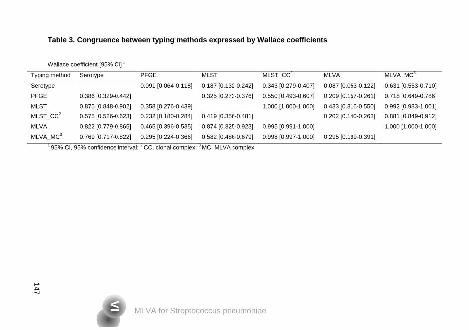
147
Table 3. Congruence between typing methods expressed by Wallace coefficients
Wallace coefficient [95% CI] 1
Typing method Serotype PFGE MLST MLST_CC2 MLVA MLVA_MC3
Serotype 0.091 [0.064-0.118] 0.187 [0.132-0.242] 0.343 [0.279-0.407] 0.087 [0.053-0.122] 0.631 [0.553-0.710]
PFGE 0.386 [0.329-0.442] 0.325 [0.273-0.376] 0.550 [0.493-0.607] 0.209 [0.157-0.261] 0.718 [0.649-0.786]
MLST 0.875 [0.848-0.902] 0.358 [0.276-0.439] 1.000 [1.000-1.000] 0.433 [0.316-0.550] 0.992 [0.983-1.001]
MLST_CC2 0.575 [0.526-0.623] 0.232 [0.180-0.284] 0.419 [0.356-0.481] 0.202 [0.140-0.263] 0.881 [0.849-0.912]
MLVA 0.822 [0.779-0.865] 0.465 [0.396-0.535] 0.874 [0.825-0.923] 0.995 [0.991-1.000] 1.000 [1.000-1.000]
MLVA_MC3 0.769 [0.717-0.822] 0.295 [0.224-0.366] 0.582 [0.486-0.679] 0.998 [0.997-1.000] 0.295 [0.199-0.391] 1 95% CI, 95% confidence interval; 2 CC, clonal complex; 3 MC, MLVA complex
147
MLVA for Streptococcus pneumoniae
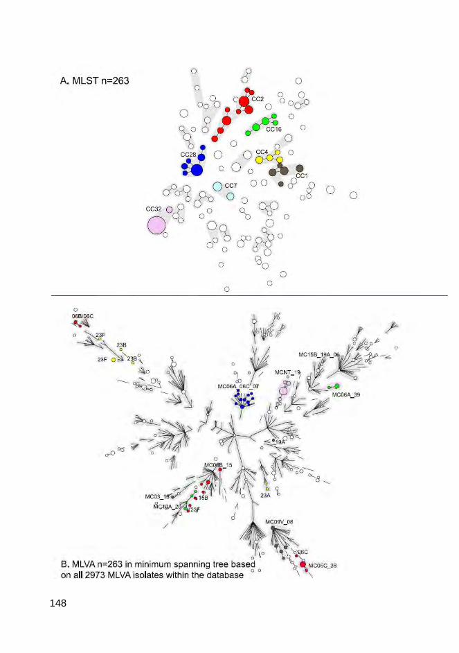
148

149
Fig. 2 (left page). High congruence between MLVA and MLST. Minimum
spanning tree of the results obtained by MLST (A) and MLVA (B) for the 263
isolates of the test collection. Each circle indicates a genotype. The size of the
circle is proportional to the number of isolates with the same type. Lines linking
two types denote a single locus difference between those types. In Figure 1A
the MLST complexes are indicated by halos. Seven MLST complexes are
discussed in the manuscript and these are colored and named according to the
nomenclature of S. pneumoniae MLST database (www.mlst.net, last accessed
on October 20, 2010). The white nodes represent the isolates within this test
collection that are not discussed. The same colors were applied to the minimum
spanning tree based on MLVA in Figure 1B. This minimum spanning tree is
based on the entire MLVA database (last accessed on October 20, 2010), and
only the branches are made visible. The MLVA types of the 263 isolates used in
this study were depicted as circles. The halos indicate the MLVA complexes.
MLV
A fo
r S
trep
toco
ccus
pne
umon
iae
150
Discussion
In recent years, typing schemes based on MLVA have been designed and
implemented for a number of micro-organisms of public health importance,
including Bacillus anthracis, Staphylococcus aureus, Enteroccus faecium,
Haemoplhilus influenza, Bordetella pertussis and many others (11, 12, 18, 19,
29, 36-38, 43). MLVA-based typing strategies have several appealing
characteristics that match the convenience criteria desirable for typing methods
(45). For example, once implemented MLVA requires minimal technical
expertise and typing results can be obtained in a few hours providing a
sequencing facility is readily available. The equipment needed is the same as
for MLST, i.e., a thermocycler and a DNA sequencer, equipment that many
laboratories performing typing have already access to. It is relatively low cost
(cheaper than MLST) and the data are amenable to automatic analysis by
computer software (as in MLST). Furthermore, the results are unambiguous
numerical profiles, portable, and thus suitable for intra-laboratory comparisons.
In addition, PFGE can only be performed on bacterial cultures while MLST can
be performed on clinical samples containing bacterial DNA such as
cerebrospinal fluid (9). The MLVA has the same potential as MLST, although
this has not been described in detail yet.
In this study we assessed the validity of a MLVA strategy developed for
Streptococcus pneumoniae using as a test collection over 263 pneumococcal
isolates previously characterized by the three gold-standard pneumococcal
typing methods: serotyping, PFGE, and MLST. The scheme explored
polymorphisms scattered throughout the genome of R6 focusing in eight BOX
loci for size estimation. The amplification of the 8 loci was combined in two
multiplex PCR reactions. Fragment sizing was performed automatically on a
DNA sequencer, resulting in a fast, easy and accurate interpretation of results.
151
The MLVA for the pneumococcus was first described by Koeck et al. (21). In
that MLVA scheme, 16 BOX loci were used and analyzed by agarose gel
electrophoresis. Remarkably, 7 of the 8 BOX loci used in our MLVA scheme
overlap with loci included in the previously published MLVA. We started the
selection of the loci for our MLVA already in 2005, before the publication of the
MLVA by Koeck et al. and thus the selection was performed independently.
Therefore, the overlap is coincidental, but suggests the proper choice of a
combination of more and less diverse loci in both MLVA schemes was made.
The main advantage of our MLVA is the high throughput that is facilitated by the
use of two multiplex assays instead of 16 separate reactions as done by Koeck
et al. and the use of capillary electrophoresis for sizing. Furthermore, the
electronic data management that we propose makes it easier to analyze the
data than images of bands obtained by agarose gel electrophoresis. We
restricted our MLVA to 8 VNTR loci to limit the amount of work required to type
an isolate. Our results show that this scheme yields a high resolution. The
resolution will probably be even higher if more loci were used like in Koeck’s
MLVA scheme. However, this will depend on the questions that need to be
answered. For our studies MLVA should be utilized to determine the genetic
background of isolates and relate this to changes in other properties such as
capsular composition. In that case MLVA based on 8 loci seems to suffice. Of
note, both MLVA schemes showed a high congruence with MLST (32, 35).
Although the test collection included only carriage isolates, we do not consider it
a limitation to the validation study for the following reasons: (i) it is a very
diverse collection that includes epidemiologically unrelated, as well as closely-
related isolates; (ii) it is well known that there are substantial overlaps among
isolates being carried and causing disease at a given time and geographic
location although the relative frequency in each group may vary significantly
(34); (iii) this MLVA scheme has in the mean time been successfully applied to a
collection of over 1000 invasive disease isolates from all over the Netherlands
MLV
A fo
r S
trep
toco
ccus
pne
umon
iae

152
(Elberse et al. companion manuscript, PLoS One, this issue). In addition, in a
manuscript in preparation we will use the MLVA to assess temporal changes
and vaccine related changes in the genetic background of the pneumococcus.
We found that the MLVA scheme described here met all the performance
criteria needed for a good typing method (45): it was stable, typed 100% of the
isolates, was reproducible and had a high discriminatory power. Of interest, the
congruence between MLVA and MLST was very good suggesting it can be
routinely used as an alternative technique to MLST. Furthermore, MLVA was
able to discriminate between putative capsular variants sharing the same MLST
in a number of occasions suggesting an enhanced epidemiological usefulness.
To make this MLVA scheme easily available to the typing community, an
electronic pneumococcal MLVA database located at www.MLVA.net has been
created. This application contains the detailed protocol and materials needed for
implementation of the MLVA scheme, contact information to obtain control
samples, an application for MLVA assignment, and a database for international
deposition of MLVA types.
In summary, the MLVA scheme for pneumococci proposed in this study has the
convenience and performance criteria needed for a good typing method. It has
the advantages of being portable as MLST but is faster and has lower costs.
The high congruence between MLVA and MLST suggests it can be applied as a
general methodology and, if needed, MLST may be performed on selected
isolates from clusters sharing related MT. In the era of pneumococcal conjugate
vaccines, close surveillance of pneumococcal populations is of crucial
importance; this MLVA scheme represents a high-throughput typing technique
which combines easiness, speed, low cost, and portability and therefore can be
a rather useful tool to achieve such goal.

153
References
1. Austrian, R. 1981. Some observations on the pneumococcus and on
the current status of pneumococcal disease and its prevention. Rev
Infect Dis 3 Suppl: S1-17.
2. Bentley, S. D., D. M. Aanensen, A. Mavroidi, D. Saunders, E.
Rabbinowitsch, M. Collins, K. Donohoe, D. Harris, L. Murphy, M. A.
Quail, G. Samuel, I. C. Skovsted, M. S. Kaltoft, B. Barrell, P. R.
Reeves, J. Parkhill, and B. G. Spratt. 2006. Genetic analysis of the
capsular biosynthetic locus from all 90 pneumococcal serotypes. PLoS
Genet 2:e31.
3. Bratcher, P. E., K. H. Kim, J. H. Kang, J. Y. Hong, and M. H. Nahm.
2010. Identification of natural pneumococcal isolates expressing
serotype 6D by genetic, biochemical and serological characterization.
Microbiology 156:555-560.
4. Brito, D. A., M. Ramirez, and H. de Lencastre. 2003. Serotyping
Streptococcus pneumoniae by multiplex PCR. J Clin Microbiol 41:2378-
2384.
5. Calix, J. J., and M. H. Nahm. 2010. A new pneumococcal serotype,
11E, has a variably inactivated wcjE gene. J Infect Dis 202:29-38.
6. Carriço, J. A., F. R. Pinto, C. Simas, S. Nunes, N. G. Sousa, N.
Frazão, H. de Lencastre, and J. S. Almeida. 2005. Assessment of
band-based similarity coefficients for automatic type and subtype
classification of microbial isolates analyzed by pulsed-field gel
electrophoresis. J Clin Microbiol 43:5483-5490.
7. Carriço, J. A., C. Silva-Costa, J. Melo-Cristino, F. R. Pinto, H. de
Lencastre, J. S. Almeida, and M. Ramirez. 2006. Illustration of a
common framework for relating multiple typing methods by application to
macrolide-resistant Streptococcus pyogenes. J Clin Microbiol 44:2524-
2532.
MLV
A fo
r S
trep
toco
ccus
pne
umon
iae
154
8. Dias, C. A., L. M. Teixeira, G. Carvalho Mda, and B. Beall. 2007.
Sequential multiplex PCR for determining capsular serotypes of
pneumococci recovered from Brazilian children. J Med Microbiol
56:1185-1188.
9. Enright, M. C., K. Knox, D. Griffiths, D. W. Crook, and B. G. Spratt.
2000. Molecular typing of bacteria directly from cerebrospinal fluid. Eur J
Clin Microbiol Infect Dis 19:627-630.
10. Enright, M. C., and B. G. Spratt. 1998. A multilocus sequence typing
scheme for Streptococcus pneumoniae: identification of clones
associated with serious invasive disease. Microbiology 144:3049-3060.
11. Farlow, J., D. Postic, K. L. Smith, Z. Jay, G. Baranton, and P. Keim.
2002. Strain typing of Borrelia burgdorferi, Borrelia afzelii, and Borrelia
garinii by using multiple-locus variable-number tandem repeat analysis. J
Clin Microbiol 40:4612-4618.
12. Farlow, J., K. L. Smith, J. Wong, M. Abrams, M. Lytle, and P. Keim.
2001. Francisella tularensis strain typing using multiple-locus, variable-
number tandem repeat analysis. J Clin Microbiol 39:3186-3192.
13. Gertz, R. E., Jr., M. C. McEllistrem, D. J. Boxrud, Z. Li, V. Sakota, T.
A. Thompson, R. R. Facklam, J. M. Besser, L. H. Harrison, C. G.
Whitney, and B. Beall. 2003. Clonal distribution of invasive
pneumococcal isolates from children and selected adults in the United
States prior to 7-valent conjugate vaccine introduction. J Clin Microbiol
41:4194-4216.
14. Grundmann, H., S. Hori, and G. Tanner. 2001. Determining confidence
intervals when measuring genetic diversity and the discriminatory
abilities of typing methods for microorganisms. J Clin Microbiol 39:4190-
4192.
15. Hall, L. M., R. A. Whiley, B. Duke, R. C. George, and A. Efstratiou.
1996. Genetic relatedness within and between serotypes of
Streptococcus pneumoniae from the United Kingdom: analysis of
155
multilocus enzyme electrophoresis, pulsed-field gel electrophoresis, and
antimicrobial resistance patterns. J Clin Microbiol 34:853-859.
16. Henrichsen, J. 1995. Six newly recognized types of Streptococcus
pneumoniae. J Clin Microbiol 33:2759-2762.
17. Kadioglu, A., J. N. Weiser, J. C. Paton, and P. W. Andrew. 2008. The
role of Streptococcus pneumoniae virulence factors in host respiratory
colonization and disease. Nat Rev Microbiol 6:288-301.
18. Keim, P., L. B. Price, A. M. Klevytska, K. L. Smith, J. M. Schupp, R.
Okinaka, P. J. Jackson, and M. E. Hugh-Jones. 2000. Multiple-locus
variable-number tandem repeat analysis reveals genetic relationships
within Bacillus anthracis. J Bacteriol 182:2928-2936.
19. Klevytska, A. M., L. B. Price, J. M. Schupp, P. L. Worsham, J. Wong,
and P. Keim. 2001. Identification and characterization of variable-
number tandem repeats in the Yersinia pestis genome. J Clin Microbiol
39:3179-3185.
20. Knutsen, E., O. Johnsborg, Y. Quentin, J. P. Claverys, and L. S.
Havarstein. 2006. BOX elements modulate gene expression in
Streptococcus pneumoniae: impact on the fine-tuning of competence
development. J Bacteriol 188:8307-8312.
21. Koeck, J. L., B. M. Njanpop-Lafourcade, S. Cade, E. Varon, L.
Sangare, S. Valjevac, G. Vergnaud, and C. Pourcel. 2005. Evaluation
and selection of tandem repeat loci for Streptococcus pneumoniae
MLVA strain typing. BMC Microbiol 5:66.
22. Kong, F., W. Wang, J. Tao, L. Wang, Q. Wang, A. Sabananthan, and
G. L. Gilbert. 2005. A molecular-capsular-type prediction system for 90
Streptococcus pneumoniae serotypes using partial cpsA-cpsB
sequencing and wzy- or wzx-specific PCR. J Med Microbiol 54:351-356.
23. Lefevre, J. C., G. Faucon, A. M. Sicard, and A. M. Gasc. 1993. DNA
fingerprinting of Streptococcus pneumoniae strains by pulsed-field gel
electrophoresis. J Clin Microbiol 31:2724-2728.
MLV
A fo
r S
trep
toco
ccus
pne
umon
iae

156
24. Maiden, M. C., J. A. Bygraves, E. Feil, G. Morelli, J. E. Russell, R.
Urwin, Q. Zhang, J. Zhou, K. Zurth, D. A. Caugant, I. M. Feavers, M.
Achtman, and B. G. Spratt. 1998. Multilocus sequence typing: a
portable approach to the identification of clones within populations of
pathogenic microorganisms. Proc Natl Acad Sci U S A 95:3140-3145.
25. Martin, B., O. Humbert, M. Camara, E. Guenzi, J. Walker, T. Mitchell,
P. Andrew, M. Prudhomme, G. Alloing, R. Hakenbeck, and et al.
1992. A highly conserved repeated DNA element located in the
chromosome of Streptococcus pneumoniae. Nucleic Acids Res 20:3479-
3483.
26. McGee, L., L. McDougal, J. Zhou, B. G. Spratt, F. C. Tenover, R.
George, R. Hakenbeck, W. Hryniewicz, J. C. Lefevre, A. Tomasz, and
K. P. Klugman. 2001. Nomenclature of major antimicrobial-resistant
clones of Streptococcus pneumoniae defined by the pneumococcal
molecular epidemiology network. J Clin Microbiol 39:2565-2571.
27. McGee, L., H. Wang, A. Wasas, R. Huebner, M. Chen, and K. P.
Klugman. 2001. Prevalence of serotypes and molecular epidemiology of
Streptococcus pneumoniae strains isolated from children in Beijing,
China: identification of two novel multiply-resistant clones. Microb Drug
Resist 7:55-63.
28. Nunes, S., R. Sá-Leão, J. Carriço, C. R. Alves, R. Mato, A. Brito-Avô,
J. Saldanha, J. S. Almeida, I. S. Sanches, and H. de Lencastre. 2005.
Trends in drug resistance, serotypes, and molecular types of
Streptococcus pneumoniae colonizing preschool-age children attending
day care centers in Lisbon, Portugal: a summary of 4 years of annual
surveillance. J Clin Microbiol 43:1285-1293.
29. Overduin, P., L. Schouls, P. Roholl, A. van der Zanden, N.
Mahmmod, A. Herrewegh, and D. van Soolingen. 2004. Use of
multilocus variable-number tandem-repeat analysis for typing

157
Mycobacterium avium subsp. paratuberculosis. J Clin Microbiol 42:5022-
5028.
30. Pai, R., R. E. Gertz, and B. Beall. 2006. Sequential multiplex PCR
approach for determining capsular serotypes of Streptococcus
pneumoniae isolates. J Clin Microbiol 44:124-131.
31. Park, I. H., S. Park, S. K. Hollingshead, and M. H. Nahm. 2007.
Genetic basis for the new pneumococcal serotype, 6C. Infect Immun
75:4482-4489.
32. Pichon, B., L. Moyce, C. Sheppard, M. Slack, D. Turbitt, R. Pebody,
D. A. Spencer, J. Edwards, D. Krahe, and R. George. 2010. Molecular
typing of pneumococci for investigation of linked cases of invasive
pneumococcal disease. J Clin Microbiol 48:1926-1928.
33. Sá-Leão, R., A. S. Simoes, S. Nunes, N. G. Sousa, N. Frazão, and H.
de Lencastre. 2006. Identification, prevalence and population structure
of non-typable Streptococcus pneumoniae in carriage samples isolated
from preschoolers attending day-care centres. Microbiology 152:367-
376.
34. Sá-Leão, R., A. Tomasz, I. S. Sanches, A. Brito-Avô, S. E.
Vilhelmsson, K. G. Kristinsson, and H. de Lencastre. 2000. Carriage
of internationally spread clones of Streptococcus pneumoniae with
unusual drug resistance patterns in children attending day care centers
in Lisbon, Portugal. J Infect Dis 182:1153-1160.
35. Sadowy, E., A. Kuch, M. Gniadkowski, and W. Hryniewicz. 2010.
Expansion and evolution of the Streptococcus pneumoniae Spain9V-
ST156 clonal complex in Poland. Antimicrob Agents Chemother
54:1720-1727.
36. Schouls, L. M., E. C. Spalburg, M. van Luit, X. W. Huijsdens, G. N.
Pluister, M. G. van Santen-Verheuvel, H. G. van der Heide, H.
Grundmann, M. E. Heck, and A. J. de Neeling. 2009. Multiple-locus
variable number tandem repeat analysis of Staphylococcus aureus:
MLV
A fo
r S
trep
toco
ccus
pne
umon
iae

158
comparison with pulsed-field gel electrophoresis and spa-typing. PLoS
One 4:e5082.
37. Schouls, L. M., A. van der Ende, I. van de Pol, C. Schot, L.
Spanjaard, P. Vauterin, D. Wilderbeek, and S. Witteveen. 2005.
Increase in genetic diversity of Haemophilus influenzae serotype b (Hib)
strains after introduction of Hib vaccination in The Netherlands. J Clin
Microbiol 43:2741-2749.
38. Schouls, L. M., H. G. van der Heide, L. Vauterin, P. Vauterin, and F.
R. Mooi. 2004. Multiple-locus variable-number tandem repeat analysis of
Dutch Bordetella pertussis strains reveals rapid genetic changes with
clonal expansion during the late 1990s. J Bacteriol 186:5496-5505.
39. Schwartz, D. C., and C. R. Cantor. 1984. Separation of yeast
chromosome-sized DNAs by pulsed field gradient gel electrophoresis.
Cell 37:67-75.
40. Simpson, E. H. 1949. Measurement of diversity. Nature:688.
41. Sorensen, U. B. 1993. Typing of pneumococci by using 12 pooled
antisera. J Clin Microbiol 31:2097-2100.
42. Tenover, F. C., R. D. Arbeit, R. V. Goering, P. A. Mickelsen, B. E.
Murray, D. H. Persing, and B. Swaminathan. 1995. Interpreting
chromosomal DNA restriction patterns produced by pulsed-field gel
electrophoresis: criteria for bacterial strain typing. J Clin Microbiol
33:2233-2239.
43. Top, J., L. M. Schouls, M. J. Bonten, and R. J. Willems. 2004.
Multiple-locus variable-number tandem repeat analysis, a novel typing
scheme to study the genetic relatedness and epidemiology of
Enterococcus faecium isolates. J Clin Microbiol 42:4503-4511.
44. van Belkum, A., M. Sluijuter, R. de Groot, H. Verbrugh, and P. W.
Hermans. 1996. Novel BOX repeat PCR assay for high-resolution typing
of Streptococcus pneumoniae strains. J Clin Microbiol 34:1176-1179.
159
45. van Belkum, A., P. T. Tassios, L. Dijkshoorn, S. Haeggman, B.
Cookson, N. K. Fry, V. Fussing, J. Green, E. Feil, P. Gerner-Smidt, S.
Brisse, and M. Struelens. 2007. Guidelines for the validation and
application of typing methods for use in bacterial epidemiology. Clin
Microbiol Infect 13 Suppl 3: 1-46.
MLV
A fo
r S
trep
toco
ccus
pne
umon
iae

160
Supplementary tables
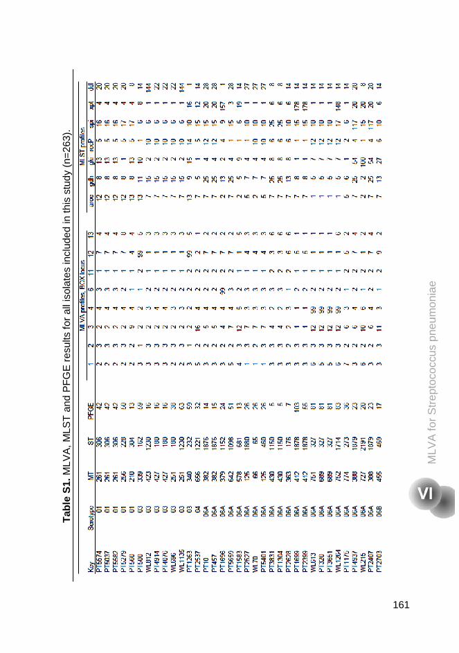
Tab
le S
1. M
LVA
, MLS
T a
nd P
FG
E r
esul
ts fo
r al
l iso
late
s in
clud
ed in
this
stu
dy (
n=26
3).
MLV
A fo
r S
trep
toco
ccus
pne
umon
iae
161
MLV
A, M
LST
and
PF
GE
res
ults
for
all i
sola
tes
incl
uded
in th
is s
tudy
(n=
263)
.
Str
epto
cocc
us p
neum
onia
e
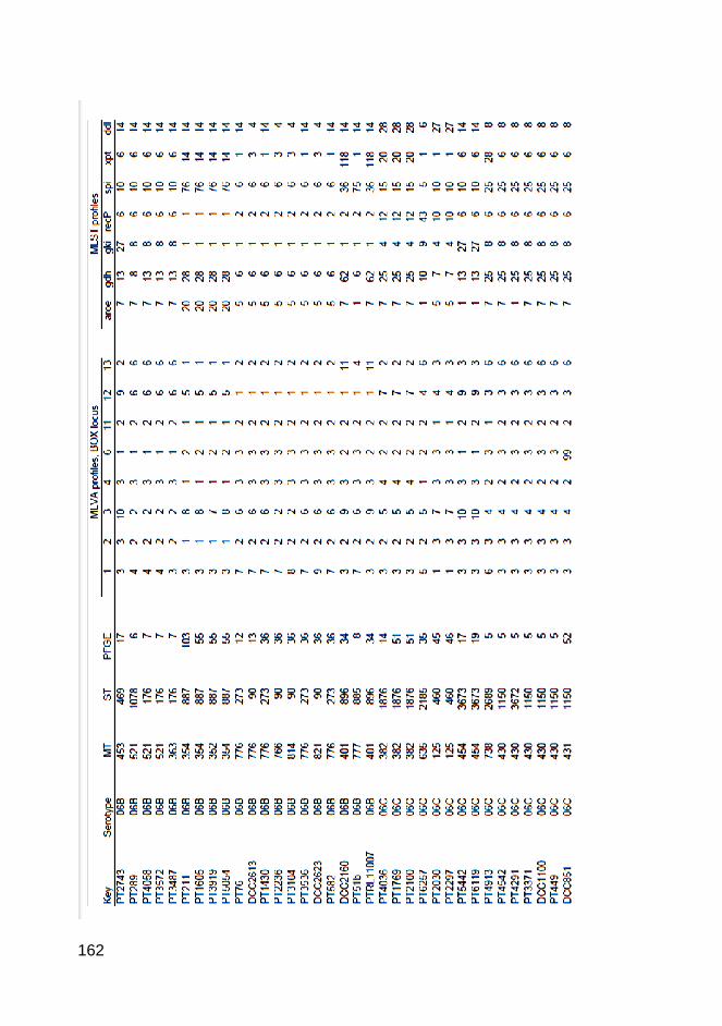
162
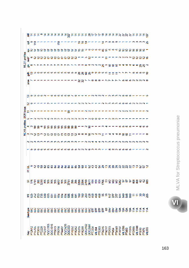
163
MLV
A fo
r S
trep
toco
ccus
pne
umon
iae
pneu
mon
iae

164
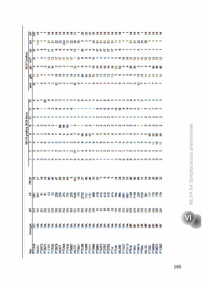
165
MLV
A fo
r S
trep
toco
ccus
pne
umon
iae
Str
epto
cocc
us p
neum
onia
e
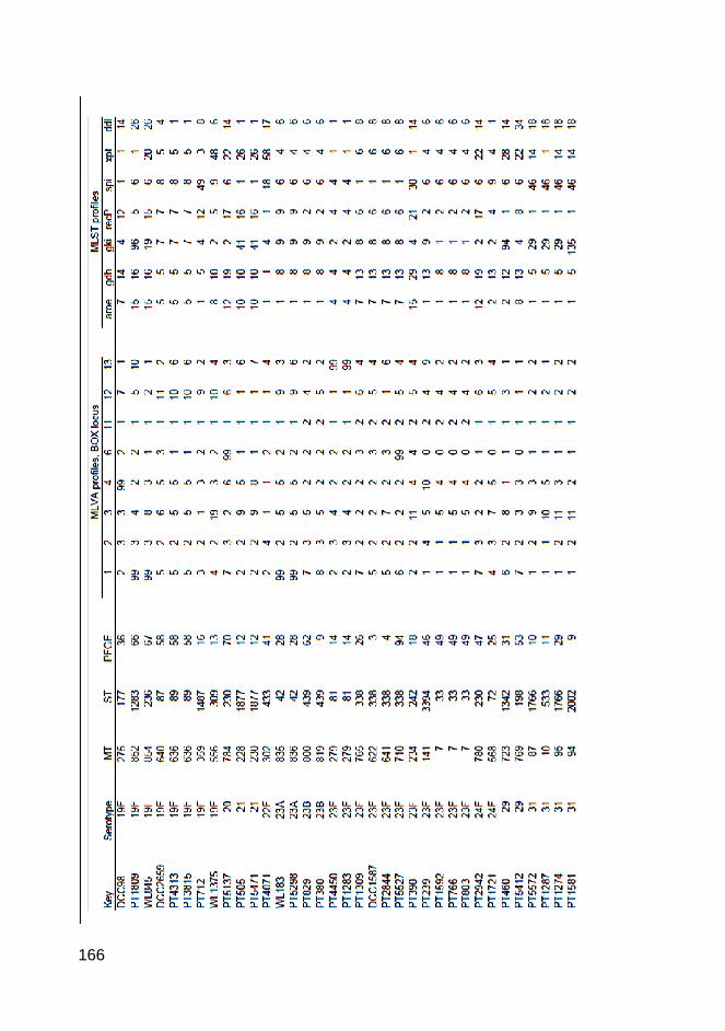
166
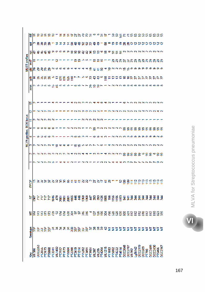
167
MLV
A fo
r S
trep
toco
ccus
pne
umon
iae
Str
epto
cocc
us p
neum
onia
e

168

169
Conclusions and future perspectives
170
Concluding Remarks
Since 1996, our research group has been monitoring the pneumococcal
population that colonizes children attending day care centers in the Lisbon area,
Portugal. Our studies have been providing valuable information on the
pneumococcal population, the role of DCC as epidemiological units and the
importance of the nasopharynx of children as a reservoir for dissemination into
the community of epidemic clones frequently associated with disease.
The introduction of the seven-valent pneumococcal conjugate vaccine (PCV7) in
2001 in Portugal led to a massive shift in pneumococcal nasopharyngeal flora in
children attending day care centers in the Lisbon area (22, 23). In those studies
it was observed that, despite the dramatic decrease of PCV7 serotypes among
carriers, the pneumococcal carriage and the antimicrobial resistance rates
remained essentially unchanged in the years following the introduction of PCV7
in Portugal.
To explore to which extent this scenario was mimicked in other regions of
Portugal we performed the studies described in Chapters III and IV in two other
regions: Coimbra, a city in the central region, and Montemor-o-Novo, a rural
area in the south.
In the study conducted in Coimbra, described in Chapter III, nasopharyngeal
swabs were taken from children attending day care centers. Around 65% of the
children were age-appropriately vaccinated with PCV7 and similar rates of
pneumococcal colonization (c.a. 60%), antimicrobial resistance patterns, and
genotypes were found when compared to previous studies conducted in the
Lisbon area.
In the study, described in Chapter IV, we compared the patterns of
pneumococcal colonization among young children attending day care centers in

171
the rural area of Montemor-o-Novo with those of young children attending day-
care in the urban area of Lisbon. In this study around 70% of the children, in
both areas, were age-appropriately vaccinated. Although similar rates of
colonization were found in the two areas (c.a. 60%), the rates of antimicrobial
consumption and pneumococcal antimicrobial resistance were significantly
higher in the urban area.
A multivariable logistic regression helped us to identify the factors which
contributed for these differences between the two areas and we found that
antibiotic consumption in the month preceding sampling, attending day care in
the urban area, and being colonized with serotype 19A and non-typeable strains
increased the probability of being colonized with pneumococcal antimicrobial
resistant strains.
With these studies we were able to conclude that: (i) although the
pneumococcal vaccines are not introduced in the Nacional Vaccination Plan of
Portugal, at least in these three regions, at the time of these studies, the use of
PCV7 was high and comparable, (ii) the rates of pneumococcal colonization
were similar in these regions, and (iii) antibiotic consumption remains the main
cause for maintenance of antimicrobial resistance among circulating
pneumococci.
Pneumococcal colonization is required for the occurrence of pneumococcal
disease and also for transmission. For this reason it is essential to study
colonization in the two age groups where the rates of pneumococcal infection
are higher: children under six years old and adults with more than 60 years of
age. To gain insights of pneumococcal carriage patterns in adults living in
Portugal, a study was started in 2010 and the first results are described in
Chapter V. Low rates of colonization (2.2%) were found and these are in
agreement with those reported in other countries (1, 10, 16, 20). Taking into
Con
clus
ions
and
futu
re p
ersp
ectiv
es
172
account the low number of pneumococcal carriers, we were not able to reach
robust conclusions in this study regarding factors associated with carriage in the
elderly. However, smoking increased in seven fold the risk of being colonized by
pneumococci and living in a rural area seems to increase the risk of
pneumococcal colonization.
In 2007, Park and co-authors described a new pneumococcal serotype, 6C (17),
that was previously indistinguishable from serotype 6A by the Quellung reaction.
More recently, other serotypes have been discovered: 6D (3), 11E (previously
indistinguishable from serotype 11A) (4), and serogroup 20 was divided in 20A
and 20B (5). All these discoveries and the exact determination of the serotypes
are particularly important, in the era of conjugate pneumococcal vaccines, to
better understand vaccine impact and control for the possibility of cross-reaction
with non-vaccine types. Serotype 6C aroused a special interest since PCV7
targets serotype 6B, and it is presumed to induce cross-protection against
serotype 6A, but not against 6C (17).
Additionally at the time of the study described in Chapter II, few studies about
the epidemiology of this serotype had been published. Nevertheless, one study
from South Africa had reported a higher propensity of this serotype to cause
meningitis, in HIV+ adults, than serotype 6A or 6B strains (9). A study from CDC
in the USA reported a significant increased of the rate of invasive disease
caused by serotype 6C between 1999 and 2006 (7). Another study from Ohio
also reported increase of serotype 6C after 2001 (14).
In the retrospective study described in Chapter II, a collection of strains of
serogroup 6, recovered from young healthy children, between 1996 and 2007
were serotyped by PCR using the specific primers for serotype 6C described by
Park et al. (17). Additionally, the serotype 6C strains were characterized by
PFGE and MLST. It was observed that this serotype had been circulating in
173
Portugal at least since 1996. This serotype was genetically diverse, and often
antibiotic resistant.
Several studies about serotype 6C have been published in more recent years,
from different geographic areas. Some have documented an increase of this
serotype in carriage and in disease after PCV7 vaccination (11, 13, 21, 25). In
other studies, conducted before the introduction of PCV7, as in Brazil and Israel,
serotype 6C has also been found in circulation in colonization and disease (6,
18). PCV13 can potentially impact on this serotype, since this vaccine includes
serotype 6A and may provide some cross-protection against serotype 6C (8).
With the introduction of pneumococcal vaccines, surveillance of pneumococcal
disease and carriage remains of extreme importance since changes are
expected to occur in the pneumococcal population. To monitor these alterations
reliable genotyping methods are required for large-scale applications. In
Chapter VI a new scheme of multiple-locus variable number tandem repeat
analysis (MLVA) for Streptococcus pneumoniae is proposed. This technique
was compared with the two gold standard methodologies for pneumococcal
typing, MLST and PFGE. Since the MLVA methodology proposed is a high-
throughput typing technique that can be easily done, in short time, and with low
cost (as long as there is access to a sequencer), and has the advantages of
portability and access to a web-based database (as MLST), MLVA can be a
rather useful tool to achieve such goal.
Overall, the studies performed in this thesis have contributed to a better
knowledge on pneumococcal colonization in Portugal in two age groups,
children and the elderly. In summary the main conclusions were:
i) the recently described serotype 6C is frequently carried by healthy young
children in Portugal, is genetically diverse, and has been circulating in
our country at least since 1996;
Con
clus
ions
and
futu
re p
ersp
ectiv
es
174
ii) the patterns of pneumococcal colonization among healthy children living
in Coimbra are similar to those living in the Lisbon area;
iii) antibiotic consumption remains a main cause for the maintenance of
antimicrobial resistance, in the era of widespread use of pneumococcal
conjugate vaccines;
iv) the rates of pneumococcal colonization in the elderly are low and the
serotype and genotype diversity are high;
v) MLVA is a promising genotyping method to characterize pneumococci.
Future perspectives
Further studies would be important in the context of the work presented in this
thesis and should involve:
I) characterization of serotype 6C after 2007, mainly observing the
possible cross-protective effect of PCV13 in this serotype;
II) continuous surveillance on the impact of novel pneumococcal
conjugate vaccines (PCV10 and PCV13), not only in pneumococcal
serotypes but also in the levels of antimicrobial resistance among
pneumococcal colonizing healthy children;
III) continuous surveillance studies on colonization in the elderly in order
to increase the data that could help us to take more precise
conclusions, mainly in risk factors associated with pneumococcal
carriage, the diversity of serotypes and genotypes. In this latter study
the use of real time PCR would be a good tool in order to estimate
the real rates of pneumococcal colonization;
IV) widespread use of MLVA by other laboratories in other countries in
order to use this tool linked to its online database to rapidly, and
cost-effectively characterize pneumococci by being able to easily
make inter-laboratory comparison.
175
Additionally, it would be important to design a good follow-up study that could
closely measure the variations of the nasopharyngeal flora over time. It would
also be interesting to understand the relationships of the entire microbiome in
the nasopharynx and verity if “virus colonization” exists as it has been proposed
by some authors (19, 24, 26, 27). This notion could be important to complement
the idea of a recent paper published by Launes et al. (15), which mentions that
viruses may facilitate the invasiveness of less invasive pneumococcal
serotypes.
Other important aspect, which was mentioned in the introduction, are the rates
of pneumococcal vaccine coverage and its impact in on pneumococcal
epidemiology. In Portugal pneumococcal vaccines are not part of the National
Vaccination plan but in 2010, about 70% of our population in pre-school age
was age-appropriately vaccinated. Taking into account the serious economic
situation of the country, probably the vaccination rate will decrease and this
might change the epidemiology of pneumococci in Portugal. I wonder what will
happen. Can the PCV7 serotypes become more prevalent again? Which
consequences might this have on the levels of antimicrobial resistance? And in
disease, what will happen? The latest reports on IPD in Portugal indicate that
serotypes 1, 7F and 19A are the most prevalent in IPD in our country (2, 12).
Considering that these serotypes are included in PCV13, which is currently on
the market, a decrease in the vaccination uptake might counteract the
decreasing tendency in the proportion of IPD in Portugal.
Finally, the studies described are an added value to our knowledge on
pneumococcal epidemiology in the nasopharynx, in the era of pneumococcal
conjugate vaccines. We described for the first time the pneumococcal
colonization patterns in adults aged over 60 years, in Portugal. This can
constitute a baseline to monitor the changes that might occur with the
introduction of PCV13 for adults. Moreover, the study of serotype 6C was one of
Con
clus
ions
and
futu
re p
ersp
ectiv
es
176
the first to be published after its discovery, reporting important information
regarding this serotype. As a final point, in this thesis we validated the
application of a promising genotyping method to pneumococci, which, due to its
characteristics, might be a good candidate as a gold-standard genotyping
method.
From my point of view, epidemiological surveillance studies in carriage as well
as in disease will continue to be helpful to show more pieces of the enigmatic
pneumococcus that “everyday” lifts the veil and lets us learn a little more about
itself.
References
1. Abdullahi, O., A. Karani, C. C. Tigoi, D. Mugo, S. Kungu, E. Wanjiru,
J. Jomo, R. Musyimi, M. Lipsitch, and J. A. Scott. 2012. The
prevalence and risk factors for pneumococcal colonization of the
nasopharynx among children in Kilifi district, Kenya. PLoS One
7:e30787.
2. Aguiar, S. I., M. J. Brito, J. Gonçalo-Marques, J. Melo-Cristino, and
M. Ramirez. 2010. Serotypes 1, 7F and 19A became the leading causes
of pediatric invasive pneumococcal infections in Portugal after 7 years of
heptavalent conjugate vaccine use. Vaccine 28:5167-5173.
3. Bratcher, P. E., K. H. Kim, J. H. Kang, J. Y. Hong, and M. H. Nahm.
2010. Identification of natural pneumococcal isolates expressing
serotype 6D by genetic, biochemical and serological characterization.
Microbiology 156:555-560.
4. Calix, J. J., and M. H. Nahm. 2010. A new pneumococcal serotype,
11E, has a variably inactivated wcjE gene. J Infect Dis 202:29-38.
177
5. Calix, J. J., R. J. Porambo, A. M. Brady, T. R. Larson, J. Yother, C.
Abeygunwardana, and M. H. Nahm. 2012. Biochemical, genetic and
serological characterization of two capsule subtypes among
Streptococcus pneumoniae serotype 20 strains: discovery of a new
pneumococcal serotype. J Biol Chem 287:27885-27894.
6. Campos, L. C., G. Carvalho Mda, B. W. Beall, S. M. Cordeiro, D.
Takahashi, M. G. Reis, A. I. Ko, and J. N. Reis. 2009. Prevalence of
Streptococcus pneumoniae serotype 6C among invasive and carriage
isolates in metropolitan Salvador, Brazil, from 1996 to 2007. Diagn
Microbiol Infect Dis 65:112-115.
7. Carvalho, M. G., C. Henderson, A. Trujillo, H. H. Joshi, I. H. Park, S.
Hollingshead, C. Whitney, M. Nahm, B. Beall, and a. T. A. Team.
2008. Emergence of genetically diverse invasive pneumococcal serotype
6C. 6th International Symposium on Pneumococci and Pneumococcal
Diseases S01-O1 35.
8. Cooper, D., X. Yu, M. Sidhu, M. H. Nahm, P. Fernsten, and K. U.
Jansen. 2011. The 13-valent pneumococcal conjugate vaccine (PCV13)
elicits cross-functional opsonophagocytic killing responses in humans to
Streptococcus pneumoniae serotypes 6C and 7A. Vaccine 29:7207-
7211.
9. du Plessis, M., A. von Gottberg, S. A. Madhi, O. Hattingh, L. de
Gouveia, and K. P. Klugman. 2008. Serotype 6C is associated with
penicillin-susceptible meningeal infections in human immunodeficiency
virus (HIV)-infected adults among invasive pneumococcal isolates
previously identified as serotype 6A in South Africa. Int J Antimicrob
Agents 32 Suppl 1: S66-70.
10. Flamaing, J., W. E. Peetermans, J. Vandeven, and J. Verhaegen.
2010. Pneumococcal colonization in older persons in a nonoutbreak
setting. J Am Geriatr Soc 58:396-398. Con
clus
ions
and
futu
re p
ersp
ectiv
es
178
11. Green, M. C., E. O. Mason, S. L. Kaplan, L. B. Lamberth, S. H.
Stovall, L. B. Givner, J. S. Bradley, T. Q. Tan, W. J. Barson, J. A.
Hoffman, P. L. Lin, and K. G. Hulten. 2011. Increase in prevalence of
Streptococcus pneumoniae serotype 6C at eight children's hospitals in
the United States from 1993 to 2009. J Clin Microbiol 49:2097-2101.
12. Horácio, A. N., J. Diamantino-Miranda, S. I. Aguiar, M. Ramirez, and
J. Melo-Cristino. 2012. Serotype changes in adult invasive
pneumococcal infections in Portugal did not reduce the high fraction of
potentially vaccine preventable infections. Vaccine 30:218-224.
13. Jacobs, M. R., S. Bajaksouzian, R. A. Bonomo, C. E. Good, A. R.
Windau, A. M. Hujer, C. Massire, R. Melton, L. B. Blyn, D. J. Ecker,
and R. Sampath. 2009. Occurrence, distribution, and origins of
Streptococcus pneumoniae Serotype 6C, a recently recognized
serotype. J Clin Microbiol 47:64-72.
14. Jacobs, M. R., C. E. Good, S. Bajaksouzian, and A. R. Windau. 2008.
Emergence of Streptococcus pneumoniae serotypes 19A, 6C, and 22F
and serogroup 15 in Cleveland, Ohio, in relation to introduction of the
protein-conjugated pneumococcal vaccine. Clin Infect Dis 47:1388-1395.
15. Launes, C., M. F. de-Sevilla, L. Selva, J. J. Garcia-Garcia, R.
Pallares, and C. Munoz-Almagro. 2012. Viral coinfection in children
less than five years old with invasive pneumococcal disease. Pediatr
Infect Dis J 31:650-653.
16. Palmu, A. A., T. Kaijalainen, A. Saukkoriipi, M. Leinonen, and T. M.
Kilpi. 2012. Nasopharyngeal carriage of Streptococcus pneumoniae and
pneumococcal urine antigen test in healthy elderly subjects. Scand J
Infect Dis 44:433-438.
17. Park, I. H., D. G. Pritchard, R. Cartee, A. Brandao, M. C. Brandileone,
and M. H. Nahm. 2007. Discovery of a new capsular serotype (6C)
within serogroup 6 of Streptococcus pneumoniae. J Clin Microbiol
45:1225-1233.
179
18. Porat, N., I. H. Park, M. H. Nahm, and R. Dagan. 2010. Differential
circulation of Streptococcus pneumoniae serotype 6C clones in two
Israeli pediatric populations. J Clin Microbiol 48:4649-4651.
19. Prill, M. M., M. K. Iwane, K. M. Edwards, J. V. Williams, G. A.
Weinberg, M. A. Staat, M. J. Willby, H. K. Talbot, C. B. Hall, P. G.
Szilagyi, M. R. Griffin, A. T. Curns, and D. D. Erdman. 2012. Human
coronavirus in young children hospitalized for acute respiratory illness
and asymptomatic controls. Pediatr Infect Dis J 31:235-240.
20. Regev-Yochay, G., M. Raz, R. Dagan, N. Porat, B. Shainberg, E.
Pinco, N. Keller, and E. Rubinstein. 2004. Nasopharyngeal carriage of
Streptococcus pneumoniae by adults and children in community and
family settings. Clin Infect Dis 38:632-639.
21. Rolo, D., A. Fenoll, C. Ardanuy, L. Calatayud, M. Cubero, A. G. de la
Campa, and J. Linares. 2011. Trends of invasive serotype 6C
pneumococci in Spain: emergence of a new lineage. J Antimicrob
Chemother 66:1712-1718.
22. Sá-Leão, R., S. Nunes, A. Brito-Avô, N. Frazão, A. S. Simões, M. I.
Crisóstomo, A. C. Paulo, J. Saldanha, I. Santos-Sanches, and H. de
Lencastre. 2009. Changes in pneumococcal serotypes and antibiotypes
carried by vaccinated and unvaccinated day-care centre attendees in
Portugal, a country with widespread use of the seven-valent
pneumococcal conjugate vaccine. Clin Microbiol Infect 15:1002-1007.
23. Simões, A. S., L. Pereira, S. Nunes, A. Brito-Avô, H. de Lencastre,
and R. Sá-Leão. 2011. Clonal evolution leading to maintenance of
antibiotic resistance rates among colonizing pneumococci in the PCV7
era in Portugal. J Clin Microbiol 49:2810-2817.
24. Singleton, R. J., L. R. Bulkow, K. Miernyk, C. DeByle, L. Pruitt, K. B.
Hummel, D. Bruden, J. A. Englund, L. J. Anderson, L. Lucher, R. C.
Holman, and T. W. Hennessy. 2010. Viral respiratory infections in Con
clus
ions
and
futu
re p
ersp
ectiv
es
180
hospitalized and community control children in Alaska. J Med Virol
82:1282-1290.
25. Tocheva, A. S., J. M. Jefferies, M. Christodoulides, S. N. Faust, and
S. C. Clarke. 2010. Increase in serotype 6C pneumococcal carriage,
United Kingdom. Emerg Infect Dis 16:154-155.
26. van Benten, I., L. Koopman, B. Niesters, W. Hop, B. van Middelkoop,
L. de Waal, K. van Drunen, A. Osterhaus, H. Neijens, and W.
Fokkens. 2003. Predominance of rhinovirus in the nose of symptomatic
and asymptomatic infants. Pediatr Allergy Immunol 14:363-370.
27. Wiertsema, S. P., G. R. Chidlow, L. A. Kirkham, K. J. Corscadden, E.
N. Mowe, S. Vijayasekaran, H. L. Coates, G. B. Harnett, and P. C.
Richmond. 2011. High detection rates of nucleic acids of a wide range
of respiratory viruses in the nasopharynx and the middle ear of children
with a history of recurrent acute otitis media. J Med Virol 83:2008-2017.

