eosinophilic otitis media treated with anti-ige monoclonal ... · intractable otitis media is a...
TRANSCRIPT

J Int Adv Otol 2018; 14(1): 144-7 • DOI: 10.5152/iao.2018.4517
Case Report
INTRODUCTIONIntractable otitis media is a challenging problem for otologists. Some cases of intractable otitis media are caused by eosinophilic otitis media (EOM), which is associated with bronchial asthma and is related to eosinophilic infiltration in the middle ear. Patients suffer from high-viscosity secretions and granulation in the middle ear, and they usually complain of hearing loss and ear fullness [1]. EOM is often accompanied by bronchial asthma, allergic rhinitis, chronic sinusitis, and nasal polyps; [1, 2] it also shows resistance to conventional antibiotic medications. A ventilation tube is not effective, and tympanoplasty and mastoidectomy are also usually not effective. Subtotal petrosectomy is needed for complete removal of the pathology [3]. However, patients have to close the external auditory canal and may have permanent conductive hearing loss. EOM responds well to systemic or topical steroids.[4] However, it recurs readily and may show steroid dependence. There are few cases and controversies in using bone-implantable hearing aids in EOM as it usually shows progressive hearing loss [5]. Recently, it was reported that anti-IgE monoclonal antibody (omalizumab) therapy could maintain bone conduction stably over an extended period [6]. Here, we report a case with stable hearing following treatment with anti-IgE monoclonal antibodies (omalizumab) and a bone-implantable hearing aid for rehabilitation.
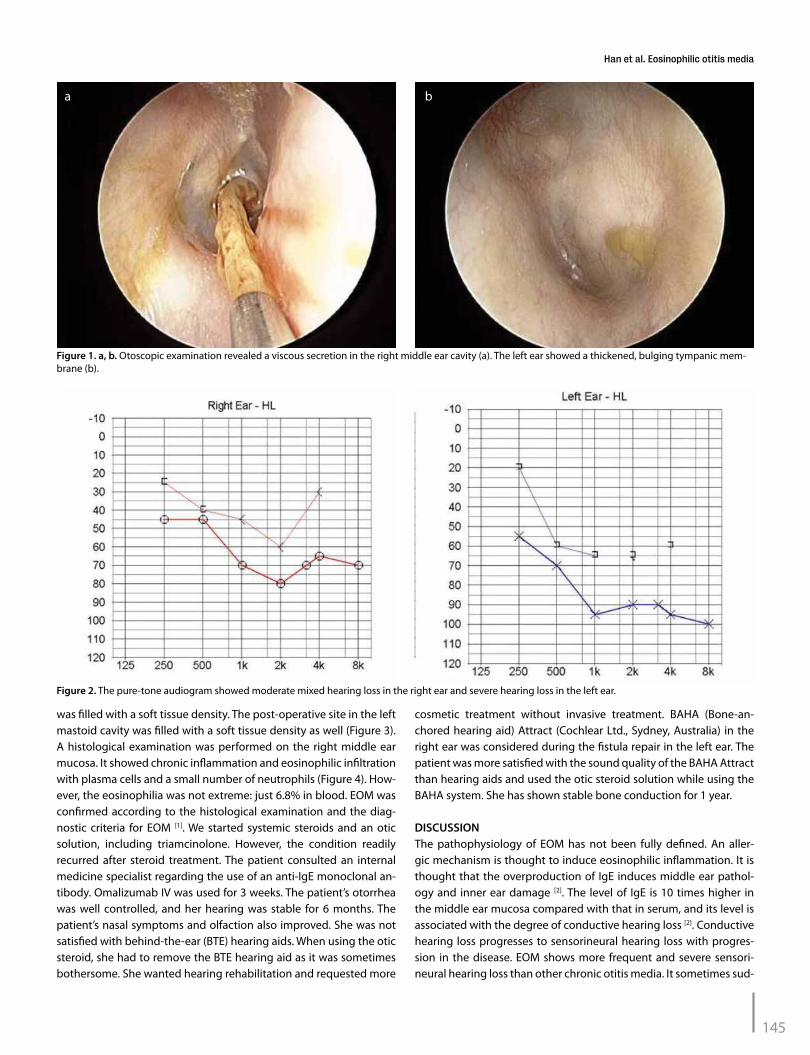
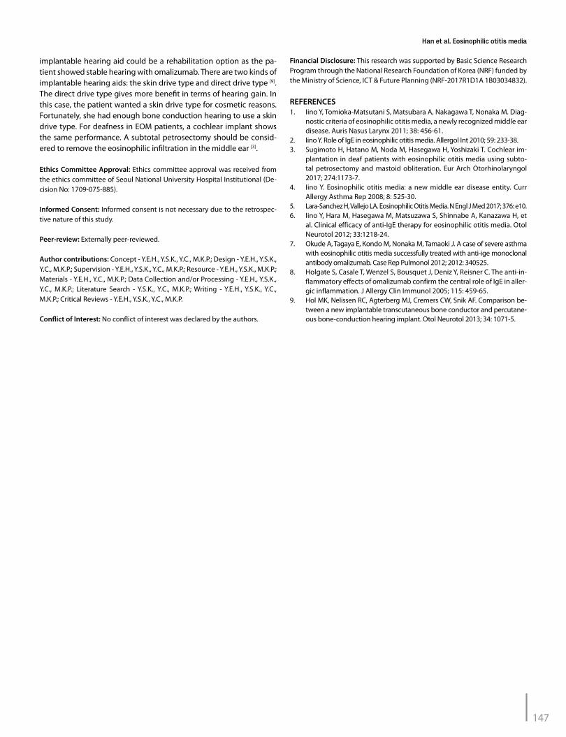
CASE PRESENTATIONA 57-year-old female had presented for 7 years with old bilateral otorrhea. She had two mastoidectomies 7 and 12 years earlier in her left ear. The patient’s otorrhea responded to steroids but recurred immediately after steroid treatment. She had bronchial asth-ma for 17 years with medication. Aspirin and NSAIDs (Nonsteroidal anti-inflammatory drugs) aggravated her asthma. In addition, she unde rwent a nasal polypectomy twice, but polyp recurrence was observed. On otoscopic examination, the right tympanic membrane showed a moderate perforation with a thick, yellowish discharge. The left tympanic membrane looked pale and was thickened and bulging (Figure 1). There was a skin fistula behind the left ear. On nasal inspection, many polyps and polypoid changes in the turbinates in both nasal cavities were observed. Her hearing was progressively distorted after otorrhea aggravation. The pure-tone average at 500, 1000, 2000, and 4000 Hz was 65 dB in air and 43.8 in bone conduction in the right ear and 86.25 dB in air in the left ear. Bone conduction in the left ear was scaled out. Also, a disyllabic word recognition score of 50% was obtained at 56 dB in the right ear and 84 dB in the left ear. The monosyllabic word recognition score at 96 dB was 76% in the right ear and 48% at 110 dB in the left ear (Figure 2). The patient had used a hearing aid for 5 years, but she used it rarely due to recurrent otor-rhea. It sometimes seemed to aggravate the otorrhea. On a temporal bone computed tomography scan, the right mastoid cavity
Eosinophilic Otitis Media Treated with Anti-IgE Monoclonal Antibodies and A Bone Conduction Implant
Eosinophilic otitis media (EOM) are intractable otitis media characterized by highly viscous secretions containing eosinophils in the middle ear. They are resistant to conventional medication and surgery. This condition occurs primarily in patients with bronchial asthma or allergic rhinitis and is often complicated by rhinosinusitis. Systemic and topical steroid therapies are effective treatments. However, long-term steroid therapy is often limited by a high risk of serious adverse effects. The use of topical steroids and otorrhea are bothersome when wearing hearing aids. Here, we report a case of intractable otitis media due to EOM. Otorrhea was controlled with topical steroids. Bone conduction hearing was stable for an extended period with anti-IgE monoclonal antibodies (omalizumab). An implantable bone conduction hearing aid was used for rehabilitation of conductive hearing loss.
KEYWORDS: Otitis media, surgery, treatment, hearing loss
Young Eun Han , Yong Seok Kang , Younhoon Cho , Moo Kyun Park Department of Otolaryngology-Head and Neck Surgery, Seoul National University School of Medicine, Seoul, Republic of Korea
Corresponding Address: Moo Kyun Park E-mail: [email protected]
Submitted: 06.09.2018 • Accepted: 14.02.2018 ©Copyright 2018 by The European Academy of Otology and Neurotology and The Politzer Society - Available online at www.advancedotology.org
ORCID IDs of the authors: Y.E.H. 0000-0001-5732-4303; Y.S.K. 0000-0002-8554-8731; Y.C. 0000-0002-9542-3916; M.K.P. 0000-0002-8635-797X
Cite this article as: Han YE, Kang YS, Cho Y, Park MK. Eosinophilic Otitis Media Treated with Anti-IgE Monoclonal Antibodies and A Bone Conduction Implant. J Int Adv Otol 2018; 14(1): 144-7.
144
was filled with a soft tissue density. The post-operative site in the left mastoid cavity was filled with a soft tissue density as well (Figure 3). A histological examination was performed on the right middle ear mucosa. It showed chronic inflammation and eosinophilic infiltration with plasma cells and a small number of neutrophils (Figure 4). How-ever, the eosinophilia was not extreme: just 6.8% in blood. EOM was confirmed according to the histological examination and the diag-nostic criteria for EOM [1]. We started systemic steroids and an otic solution, including triamcinolone. However, the condition readily recurred after steroid treatment. The patient consulted an internal medicine specialist regarding the use of an anti-IgE monoclonal an-tibody. Omalizumab IV was used for 3 weeks. The patient’s otorrhea was well controlled, and her hearing was stable for 6 months. The patient’s nasal symptoms and olfaction also improved. She was not satisfied with behind-the-ear (BTE) hearing aids. When using the otic steroid, she had to remove the BTE hearing aid as it was sometimes bothersome. She wanted hearing rehabilitation and requested more
cosmetic treatment without invasive treatment. BAHA (Bone-an-chored hearing aid) Attract (Cochlear Ltd., Sydney, Australia) in the right ear was considered during the fistula repair in the left ear. The patient was more satisfied with the sound quality of the BAHA Attract than hearing aids and used the otic steroid solution while using the BAHA system. She has shown stable bone conduction for 1 year.
DISCUSSIONThe pathophysiology of EOM has not been fully defined. An aller-gic mechanism is thought to induce eosinophilic inflammation. It is thought that the overproduction of IgE induces middle ear pathol-ogy and inner ear damage [2]. The level of IgE is 10 times higher in the middle ear mucosa compared with that in serum, and its level is associated with the degree of conductive hearing loss [2]. Conductive hearing loss progresses to sensorineural hearing loss with progres-sion in the disease. EOM shows more frequent and severe sensori-neural hearing loss than other chronic otitis media. It sometimes sud-
Figure 1. a, b. Otoscopic examination revealed a viscous secretion in the right middle ear cavity (a). The left ear showed a thickened, bulging tympanic mem-brane (b).
a b
Figure 2. The pure-tone audiogram showed moderate mixed hearing loss in the right ear and severe hearing loss in the left ear.
145
Han et al. Eosinophilic otitis media

denly progresses to deafness [4]. In the late period, patients usually show bilateral hearing loss with poor bone conduction. Yukiko et al [1]. suggested diagnostic criteria for EOM in 2011. Otitis media with ef-fusion or chronic otitis media with eosinophil-dominant effusion is a major criterion. Diagnosis requires a major criterion with at least two additional minor criteria. The minor criteria include a highly viscous middle ear effusion, resistance to conventional treatments for otitis media, an association with bronchial asthma, and an association with nasal polyposis. Our patient had all of these symptoms and thus was a definitive case.
It has been reported that EOM shows a good response to topical and systemic steroids. Oral leukotrienes also show some effect. However, long-term usage of steroids can induce several severe complications.
Recently, the use of an anti-IgE monoclonal antibody, omalizumab, was introduced for the control of EOM [6, 7]. Omalizumab binds to IgE and inactivates immune cells such as mast cells and basophils, and it prevents the secretion of histamine and leukotrienes. Furthermore, it inactivates IgE receptors on the cell surface [8].
In sensorineural hearing loss caused by EOM, hearing aids should be considered first. However, hearing aids sometimes aggravate otor-rhea by blocking aeration. The use of an otic solution can also be annoying. To overcome these problems, implantable hearing aids can be considered [5]. To date, implantable hearing aids have rarely been considered for EOM as it often shows progressive hearing loss. However, bone conduction hearing can be maintained over an ex-tended period with omalizumab [6]. In our case, we thought that an
Figure 3. a, b. Temporal computed tomography showed soft tissue densities in the right middle ear and antrum (a). The left ear showed soft tissue densities at the mastoidectomy site. There was also a large post-auricular fistula.
a b
Figure 4. a, b. Histological examination showed chronic inflammation with plasmacytic and eosinophilic infiltration (A: hematoxylin and eosin [H&E], ×100; B: H&E, ×400).
a b
146
J Int Adv Otol 2018; 14(1): 144-7
implantable hearing aid could be a rehabilitation option as the pa-tient showed stable hearing with omalizumab. There are two kinds of implantable hearing aids: the skin drive type and direct drive type [9]. The direct drive type gives more benefit in terms of hearing gain. In this case, the patient wanted a skin drive type for cosmetic reasons. Fortunately, she had enough bone conduction hearing to use a skin drive type. For deafness in EOM patients, a cochlear implant shows the same performance. A subtotal petrosectomy should be consid-ered to remove the eosinophilic infiltration in the middle ear [3].
Ethics Committee Approval: Ethics committee approval was received from the ethics committee of Seoul National University Hospital Institutional (De-cision No: 1709-075-885).
Informed Consent: Informed consent is not necessary due to the retrospec-tive nature of this study.
Peer-review: Externally peer-reviewed.
Author contributions: Concept - Y.E.H., Y.S.K., Y.C., M.K.P.; Design - Y.E.H., Y.S.K., Y.C., M.K.P.; Supervision - Y.E.H., Y.S.K., Y.C., M.K.P.; Resource - Y.E.H., Y.S.K., M.K.P.; Materials - Y.E.H., Y.C., M.K.P.; Data Collection and/or Processing - Y.E.H., Y.S.K., Y.C., M.K.P.; Literature Search - Y.S.K., Y.C., M.K.P.; Writing - Y.E.H., Y.S.K., Y.C., M.K.P.; Critical Reviews - Y.E.H., Y.S.K., Y.C., M.K.P.
Conflict of Interest: No conflict of interest was declared by the authors.
Financial Disclosure: This research was supported by Basic Science Research Program through the National Research Foundation of Korea (NRF) funded by the Ministry of Science, ICT & Future Planning (NRF-2017R1D1A 1B03034832).
REFERENCES1. Iino Y, Tomioka-Matsutani S, Matsubara A, Nakagawa T, Nonaka M. Diag-
nostic criteria of eosinophilic otitis media, a newly recognized middle ear disease. Auris Nasus Larynx 2011; 38: 456-61.
2. Iino Y. Role of IgE in eosinophilic otitis media. Allergol Int 2010; 59: 233-38. 3. Sugimoto H, Hatano M, Noda M, Hasegawa H, Yoshizaki T. Cochlear im-
plantation in deaf patients with eosinophilic otitis media using subto-tal petrosectomy and mastoid obliteration. Eur Arch Otorhinolaryngol 2017; 274:1173-7.
4. Iino Y. Eosinophilic otitis media: a new middle ear disease entity. Curr Allergy Asthma Rep 2008; 8: 525-30.
5. Lara-Sanchez H, Vallejo LA. Eosinophilic Otitis Media. N Engl J Med 2017; 376: e10.6. Iino Y, Hara M, Hasegawa M, Matsuzawa S, Shinnabe A, Kanazawa H, et
al. Clinical efficacy of anti-IgE therapy for eosinophilic otitis media. Otol Neurotol 2012; 33:1218-24.
7. Okude A, Tagaya E, Kondo M, Nonaka M, Tamaoki J. A case of severe asthma with eosinophilic otitis media successfully treated with anti-ige monoclonal antibody omalizumab. Case Rep Pulmonol 2012; 2012: 340525.
8. Holgate S, Casale T, Wenzel S, Bousquet J, Deniz Y, Reisner C. The anti-in-flammatory effects of omalizumab confirm the central role of IgE in aller-gic inflammation. J Allergy Clin Immunol 2005; 115: 459-65.
9. Hol MK, Nelissen RC, Agterberg MJ, Cremers CW, Snik AF. Comparison be-tween a new implantable transcutaneous bone conductor and percutane-ous bone-conduction hearing implant. Otol Neurotol 2013; 34: 1071-5.
147
Han et al. Eosinophilic otitis media