enzymology: histone h4 promotes prothrombin...
TRANSCRIPT

D. Vogt and Enrico Di CeraSergio Barranco-Medina, Nicola Pozzi, Austin AutoactivationHistone H4 Promotes ProthrombinEnzymology:
doi: 10.1074/jbc.M113.509786 originally published online October 30, 20132013, 288:35749-35757.J. Biol. Chem.
10.1074/jbc.M113.509786Access the most updated version of this article at doi:
.JBC Affinity SitesFind articles, minireviews, Reflections and Classics on similar topics on the
Alerts:
When a correction for this article is posted•
When this article is cited•
to choose from all of JBC's e-mail alertsClick here
http://www.jbc.org/content/288/50/35749.full.html#ref-list-1
This article cites 43 references, 13 of which can be accessed free at
at SLU
Health Sciences C
enter Library on D
ecember 17, 2013
http://ww
w.jbc.org/
Dow
nloaded from
at SLU
Health Sciences C
enter Library on D
ecember 17, 2013
http://ww
w.jbc.org/
Dow
nloaded from

Histone H4 Promotes Prothrombin Autoactivation*
Received for publication, August 12, 2013, and in revised form, October 28, 2013 Published, JBC Papers in Press, October 30, 2013, DOI 10.1074/jbc.M113.509786
Sergio Barranco-Medina1, Nicola Pozzi1, Austin D. Vogt, and Enrico Di Cera2
From the Edward A. Doisy Department of Biochemistry and Molecular Biology, Saint Louis University School of Medicine,St. Louis, Missouri 63104
Background: The molecular origin of the prothrombotic properties of histones is unknown.Results: Histone H4 interacts with prothrombin and promotes autoactivation.Conclusion: The prothrombotic properties of histone H4 are due to direct interaction with prothrombin.Significance: Prothrombin autoactivation has pathophysiological relevance.
Recent studies havedocumented the ability of prothrombin tospontaneously convert to the mature protease thrombin whenArg-320 becomes exposed to solvent for proteolytic attack uponmutation of residues in the activation domain. Whether pro-thrombin autoactivation occurs in the wild-type under condi-tions relevant to physiology remains unknown. Here, we reportthat binding of histone H4 to prothrombin under physiologicalconditions generates thrombin by autoactivation. The effect isabrogated by mutation of the catalytic Ser-525 and requires thepresence of the Gla domain. Fluorescence titrations documentdirect binding of histone H4 to prothrombin with an affinity inthe low nM range. Stopped flow data and luminescence reso-nance energy transfer measurements indicate that the bindingmechanism obeys conformational selection. Among the twoconformations of prothrombin, collapsed and fully extended,histoneH4 binds selectively to the collapsed form and induces atransition toward a new conformation where the distancebetween Ser-101 in kringle-1 and Ser-210 in kringle-2 increasesby 13 A. These findings confirm the molecular plasticity of pro-thrombin emerged from recent structural studies and suggestthat different conformations of the inter-kringle linker domaindetermine the functional behavior of prothrombin. The resultsalso broaden our mechanistic understanding of the prothrom-botic phenotype observed during cellular damage due to therelease of histones in the blood stream. Prothrombin autoacti-vation induced by histoneH4 emerges as amechanismof patho-physiological relevance through which thrombin is generatedindependently of activation of the coagulation cascade.
Prothrombin, or coagulation factor II, is a 579-residue-longvitamin K-dependent zymogen that circulates in the blood at aconcentration of 0.1 mg/ml (1). The protein has a multidomainstructure comprising fragment 1 containing the Gla domainand kringle-1, fragment 2 containing kringle-2, and the prote-ase domain containing the A chain and the catalytic B chain. Inthe penultimate step of the coagulation cascade, prothrombin isproteolytically converted to the active protease thrombin by the
prothrombinase complex composed of the protease factor Xaand the cofactor Va assembled on the surface of platelets in thepresence of Ca2� (2). The conversion involves cleavage at tworesidues: Arg-271 located in a linker region connecting krin-gle-2 to theA chain andArg-320 in the activation domain of theA chain. Cleavage at Arg-271 sheds fragment 1 and fragment 2and generates the inactive precursor prethrombin-2. The alter-native cleavage at Arg-320 separates the A and B chains thatremain connected through a disulfide bond and yields theactive intermediate meizothrombin. Under physiological con-ditions on the surface of platelets, activation of prothrombinproceeds via the prethrombin-2 intermediate (3, 4). The recentcrystal structure of prothrombin supports this preferred path-way of activation based on the different solvent accessibility ofthe two sites of cleavage and the distinct electrostatic propertiesof the epitope recognizing factor Xa in prothrombin and pre-thrombin-2 (5). Of particular importance is the observationthat Arg-320 in the activation domain is not accessible to pro-teolysis in solution. Burial of Arg-320 acts as a safety mecha-nism to prevent prothrombin autoactivation (6) and directsprothrombinase to cleave at Arg-271 first to generate thrombinvia the inactive prethrombin-2 intermediate.The recently discovered property of prothrombin and other
inactive thrombin precursors prethrombin-1 and prethrom-bin-2 to autoactivate has mechanistic significance. Mutationsin the activation domain of prethrombin-2, prethrombin-1, andprothrombin that expose Arg-320 to solvent elicit a spontane-ous conversion of the zymogen to the mature protease. Theconversion typically unfolds over several hours, but in somecases, it is extremely rapid and completes during purification(6). The conversion is initiated by the zymogen itself by virtue ofthe pre-existing equilibrium between inactive (E*) and active(E) forms (7) and is then propagated and amplified by themature enzyme. Whether autoactivation takes place in thewild-type protein under the influence of specific cofactors, asobserved in other zymogens (8, 9), remains an unresolved issueof potential physiological significance because it would afford amechanism of thrombin generation that bypasses activation ofthe coagulation cascade. Cofactor-induced autoactivation rep-resents an alternative strategy for thrombin generation com-pared with that used by Staphylococcus aureus, an importanthuman pathogen implicated in sepsis and endocarditis, whosecofactor staphylocoagulase activates prothrombin by insertingits N-terminal peptide into the activation pocket to allosteri-
* This work was supported in part by National Institutes of Health ResearchGrants HL49413, HL73813, and HL112303.
1 Both authors contributed equally to this work.2 To whom correspondence should be addressed: Dept. of Biochemistry and
Molecular Biology, Saint Louis University School of Medicine, St. Louis, MO63104. Tel.: 314-977-9201; Fax: 314-977-9206; E-mail: [email protected].
THE JOURNAL OF BIOLOGICAL CHEMISTRY VOL. 288, NO. 50, pp. 35749 –35757, December 13, 2013© 2013 by The American Society for Biochemistry and Molecular Biology, Inc. Published in the U.S.A.
DECEMBER 13, 2013 • VOLUME 288 • NUMBER 50 JOURNAL OF BIOLOGICAL CHEMISTRY 35749
at SLU
Health Sciences C
enter Library on D
ecember 17, 2013
http://ww
w.jbc.org/
Dow
nloaded from

cally induce a functional catalytic triad without cleaving at Arg-320 (10). We therefore screened a number of molecules rele-vant to blood pathophysiology for the ability to specificallypromote prothrombin autoactivation.During cell necrosis, a broad range of modulators such as
polyamines, RNA, and histones are released into the extracel-lularmedia where they interact withmany players of inflamma-tion, sepsis, and thrombosis. Direct administration of histonestomice elicits lethal prothrombotic responses, thereby suggest-ing a direct link with clotting factors (11). Histones promoteactivation of factor VII activating protease in vivo (8) and havebeen suggested to interact with fibrinogen (12, 13), prothrom-bin (12), thrombomodulin, and protein C (14). Histones havebeen postulated to enhance thrombin generation indirectly byreducing the thrombomodulin-dependent activation of proteinC anticoagulant pathway (14) or directly through plateletdependent mechanisms involving the Toll-like receptors TLR2and TLR4 (15). Here, we show that histone H4 promotes pro-thrombin autoactivation by interacting directly with the zymo-gen, and we elucidate the underlying mechanism. The resultsbroaden our understanding of the prothrombotic role of his-toneH4 andoffer the first example of a physiological cofactor ofprothrombin autoactivation.
MATERIALS AND METHODS
Numbering—All prothrombin residues in this study arenumbered sequentially based on the sequence of the zymogen.The corresponding number according to chymotrypsinogenfor residues in the protease domain (A and B chains) is noted byparentheses, e.g. the catalytic Ser is identified as Ser-525(Ser-195).Materials—Recombinant full-length prothrombin (residues
1–579) wild-type and mutants S525A (S195A) and S101C/S210C, Gla domainless prothrombin (residues 45–579) andprethrombin-1 were cloned into the pNUT vector usingrestriction sites HindIII and NdeI, transfected into baby ham-ster kidney cells by lipofection and selected by methotrexate.Prethrombin-2 wild-type and mutant S195A were cloned intothe pET-21-b vector using the same restriction enzymes andexpressed in Escherichia coli as described elsewhere (16). Thenucleotide sequence of the constructs was confirmed by DNAsequencing. Addition of vitamin K to the full-length prothrom-bin media ensured correct �-carboxylation of the Gla domain.Purification of the recombinant proteins was carried out byaffinity chromatography, ion exchange chromatography, andsize exclusion chromatography, as described previously (16–20). Homogeneity and chemical identity of final preparationswere verified by SDS-PAGE and by RP-HPLC mass spectrom-etry analysis, giving a purity of�98%. Recombinant histone H3and histone H4 were purchased from New England Biolabs.Histones were thawed and buffer exchanged immediatelybefore use. Long-chain synthetic polyphosphate was differen-tially solubilized as described previously (21). Polymer lengthsranged from �50 to 1500 phosphates, with a modal length of�650 phosphates. Polyphosphate concentration was expressedin terms of the concentration of phosphate monomers (mono-mer formula, NaPO3). Spermidine was purchased from Sigma-Aldrich. Protein concentrations were determined by reading at
280 nm with molar extinction coefficients � � 111,280 M�1
cm�1 for recombinant prothrombin (molecular mass, 72,000kDa) and � � 5,120 M�1 cm�1 for histone H4 (molecular mass,11237 kDa).Autoactivation of Prothrombin—Prothrombin at physiologi-
cal concentration (0.1mg/ml) was incubated at 37 °Cwith sper-midine (40 �M), RNA (10 �g/ml), DNA (10 �g/ml), polyphos-phate (10 �M), and histones H3 and H4 (2 �M) in buffercontaining 150 mM NaCl, 5 mM CaCl2, 20 mM Tris, pH 7.4(TBS-Ca2�). Prothrombin autoactivation was followed for upto 150 h. The reaction leading to generation of mature enzymeand depletion of zymogenwas quenched at different times with15�l of NuPAGE LDS buffer containing �-mercaptoethanol asreducing agent. Samples were processed by SDS-PAGE. Gelswere stained with Coomassie Brilliant Blue R-250. Identity ofthe bands was confirmed by N-terminal sequencing. The samesamples were tested for activity using the thrombin specificchromogenic substrate H-D-Phe-Pro-Arg-p-nitroanilide (FPR).The release of p-nitroanline was followed at 405 nm, and theamount of active enzymewas quantifiedwith a standard referencecurve.Fluorescence Titration Studies—The prothrombin mutant
S101C/S210C was labeled with Alexa Fluor 647 after selectivereduction of the engineered cysteines as described elsewhere(5). The labeled protein was separated from the unreacted dyeand aggregates by size exclusion chromatography. Incorpora-tion of the probe was confirmed by reading at 650 nm whereAlexa Fluor 647 absorbs with an extinction coefficient � �239,000 M�1 cm�1. The derivatization yield at the two sites wasestimated by the ratio of readings at 280/650 nm and found tobe 91%. The labeled mutant was tested in the prothrombinasecomplex assay and returned kinetic parameters comparablewith the recombinant wild-type and plasma prothrombin (datanot shown). Titration measurements were performed in a300-�l cuvette at a protein concentration of 5, 20, or 200 nM inthe presence of 150 mM NaCl, 5 mM CaCl2, 0.1% PEG 8000, 20mM Tris, pH 7.4, at 25 °C. The protein was excited at 650 nm(slits 5/10, exposure time of 0.02 s), and the emission spectrumwas recorded between 660 and 750 nm.No photobleachingwasobserved under these conditions. Measurements were per-fomed on a Fluoromax-4 spectrofluorimeter (Horiba Scientific,Edison, NJ) and analyzed with Origin (version 8.1, OriginLab,Northampton, MA) using Equation 1,
F � F0 � �Fmax� (Eq. 1)
where F is the fluorescence reading, F0 is the value of F in theabsence of histoneH4,�Fmax is the total change in fluorescenceupon saturation, and
� �Kd,app � Npt � ht � ��Kd,app � Npt � ht�
2 � 4Nptht
2Npt(Eq. 2)
Equation 2 shows the fractional saturation defined in terms ofthe apparent equilibriumdissociation constant,Kd,app, the totalconcentrations of prothrombin, pt, and histone H4, ht, and thehistone H4:prothrombin stoichiometry N. Histone H4 bindingcurves obtained at different values of ptwere analyzed simulta-neously to derive the independent parameters Kd,app and N.
Histone H4 Interaction with Prothrombin
35750 JOURNAL OF BIOLOGICAL CHEMISTRY VOLUME 288 • NUMBER 50 • DECEMBER 13, 2013
at SLU
Health Sciences C
enter Library on D
ecember 17, 2013
http://ww
w.jbc.org/
Dow
nloaded from
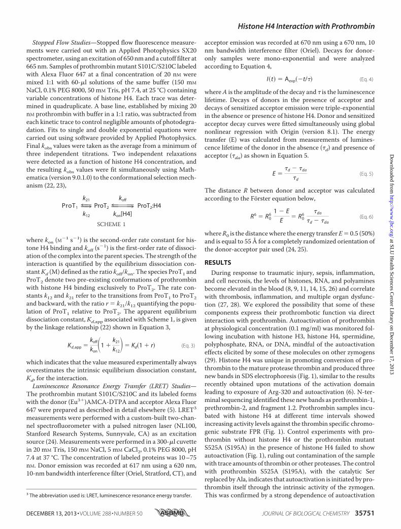
Stopped Flow Studies—Stopped flow fluorescence measure-ments were carried out with an Applied Photophysics SX20spectrometer, using an excitation of 650 nmand a cutoff filter at665 nm. Samples of prothrombinmutant S101C/S210C labeledwith Alexa Fluor 647 at a final concentration of 20 nM weremixed 1:1 with 60-�l solutions of the same buffer (150 mM
NaCl, 0.1% PEG 8000, 50 mM Tris, pH 7.4, at 25 °C) containingvariable concentrations of histone H4. Each trace was deter-mined in quadruplicate. A base line, established by mixing 20nM prothrombin with buffer in a 1:1 ratio, was subtracted fromeach kinetic trace to control negligible amounts of photodegra-dation. Fits to single and double exponential equations werecarried out using software provided by Applied Photophysics.Final kobs values were taken as the average from a minimum ofthree independent titrations. Two independent relaxationswere detected as a function of histone H4 concentration, andthe resulting kobs values were fit simultaneously using Math-ematica (version 9.0.1.0) to the conformational selectionmech-anism (22, 23),
ProT1 9|=k21
k12
ProT2 9|=koff
kon[H4]ProT2:H4
SCHEME 1
where kon (M�1 s�1) is the second-order rate constant for his-tone H4 binding and koff (s�1) is the first-order rate of dissoci-ation of the complex into the parent species. The strength of theinteraction is quantified by the equilibrium dissociation con-stantKd (M) defined as the ratio koff/kon. The species ProT1 andProT2 denote two pre-existing conformations of prothrombinwith histone H4 binding exclusively to ProT2. The rate con-stants k12 and k21 refer to the transitions from ProT1 to ProT2and backward, with the ratio r � k21/k12 quantifying the popu-lation of ProT1 relative to ProT2. The apparent equilibriumdissociation constant, Kd,app associated with Scheme 1, is givenby the linkage relationship (22) shown in Equation 3,
Kd,app �koff
kon�1 �
k21
k12� � Kd�1 � r� (Eq. 3)
which indicates that the value measured experimentally alwaysoverestimates the intrinsic equilibrium dissociation constant,Kd, for the interaction.Luminescence Resonance Energy Transfer (LRET) Studies—
The prothrombin mutant S101C/S210C and its labeled formswith the donor (Eu3�)AMCA-DTPA and acceptor Alexa Fluor647 were prepared as described in detail elsewhere (5). LRET3
measurements were performed with a custom-built two-chan-nel spectrofluorometer with a pulsed nitrogen laser (NL100,Stanford Research Systems, Sunnyvale, CA) as an excitationsource (24). Measurements were performed in a 300-�l cuvettein 20 mM Tris, 150 mM NaCl, 5 mM CaCl2, 0.1% PEG 8000, pH7.4 at 37 °C. The concentration of labeled proteins was 10–75nM. Donor emission was recorded at 617 nm using a 620 nm,10-nm bandwidth interference filter (Oriel, Stratford, CT), and
acceptor emission was recorded at 670 nm using a 670 nm, 10nm bandwidth interference filter (Oriel). Decays for donor-only samples were mono-exponential and were analyzedaccording to Equation 4,
I�t� � Aexp��t/� (Eq. 4)
whereA is the amplitude of the decay and is the luminescencelifetime. Decays of donors in the presence of acceptor anddecays of sensitized acceptor emission were triple-exponentialin the absence or presence of histone H4. Donor and sensitizedacceptor decay curves were fitted simultaneously using globalnonlinear regression with Origin (version 8.1). The energytransfer (E) was calculated from measurements of lumines-cence lifetime of the donor in the absence (d) and presence ofacceptor (da) as shown in Equation 5.
E �d � da
d(Eq. 5)
The distance R between donor and acceptor was calculatedaccording to the Forster equation below,
R6 � R06
1 � E
E� R0
6da
d � da(Eq. 6)
whereR0 is the distancewhere the energy transferE� 0.5 (50%)and is equal to 55 Å for a completely randomized orientation ofthe donor-acceptor pair used (24, 25).
RESULTS
During response to traumatic injury, sepsis, inflammation,and cell necrosis, the levels of histones, RNA, and polyaminesbecome elevated in the blood (8, 9, 11, 14, 15, 26) and correlatewith thrombosis, inflammation, and multiple organ dysfunc-tion (27, 28). We explored the possibility that some of thesecomponents express their prothrombotic function via directinteraction with prothrombin. Autoactivation of prothrombinat physiological concentration (0.1 mg/ml) was monitored fol-lowing incubation with histone H3, histone H4, spermidine,polyphosphate, RNA, or DNA, mindful of the autoactivationeffects elicited by some of these molecules on other zymogens(29). Histone H4 was unique in promoting conversion of pro-thrombin to themature protease thrombin and produced threenew bands in SDS electrophoresis (Fig. 1), similar to the resultsrecently obtained upon mutations of the activation domainleading to exposure of Arg-320 and autoactivation (6). N-ter-minal sequencing identified these newbands as prethrombin-1,prethrombin-2, and fragment 1.2. Prothrombin samples incu-bated with histone H4 at different time intervals showedincreasing activity levels against the thrombin specific chromo-genic substrate FPR (Fig. 1). Control experiments with pro-thrombin without histone H4 or the prothrombin mutantS525A (S195A) in the presence of histone H4 failed to showautoactivation (Fig. 1), ruling out contamination of the samplewith trace amounts of thrombin or other proteases. The controlwith prothrombin S525A (S195A), with the catalytic Serreplaced byAla, indicates that autoactivation is initiated by pro-thrombin itself through the intrinsic activity of the zymogen.This was confirmed by a strong dependence of autoactivation3 The abbreviation used is: LRET, luminescence resonance energy transfer.
Histone H4 Interaction with Prothrombin
DECEMBER 13, 2013 • VOLUME 288 • NUMBER 50 JOURNAL OF BIOLOGICAL CHEMISTRY 35751
at SLU
Health Sciences C
enter Library on D
ecember 17, 2013
http://ww
w.jbc.org/
Dow
nloaded from

on the prothrombin concentration, as expected for a reactioninitiated by the zymogen itself as a catalyst (data not shown).Histone H3 also promoted autoactivation but over a time scalethree times longer than that observedwith histoneH4 (data notshown), in agreement with the observation that the procoagu-lant effect of histones is due mainly to histone H4 (14).Previous studies have shown a requirement for the Gla
domain in the binding of histones to vitamin K-dependentproteins (12, 14). Accordingly, autoactivation of prothrom-bin by histone H4 required the presence of the Gla domainand was not observed with Gla domainless prothrombin, orthe thrombin precursors prethrombin-1 and prethrombin-2that lack fragment 1 or both fragments 1 and 2 (Fig. 1),respectively.The disappearance of the band corresponding to histone H4
in the gel during prothrombin autoactivation was due to pro-teolytic cleavage by the mature enzyme thrombin because nodegradation was observed with prothrombin S525A (S195A).Activated protein C is currently the only enzyme known totarget histones in the blood and able to counter their prothrom-botic effects and cytotoxicity (11). The uniqueness of this effectshould be reconsidered in view of the data reported in Fig. 1.The ability of thrombin to degrade histone H4 explains why
prothrombin autoactivation does not proceed to completion(Fig. 1).The evidence that histone H4 interacts with prothrombin is
compelling but indirect. Histones are positively charged andmay interact non-specifically with the surface of prothrombinthat is mostly negatively charged, as recently revealed by thex-ray crystal structure (5). Evidence of a direct interaction ofhistone H4 with prothrombin comes from fluorescence titra-tions using the prothrombin mutant S101C/S210C engineeredrecently for quantitative LRET measurements (5). The mutantlabeled with the dye Alexa Fluor 647 at both Cys residues wasused first for direct fluorescence titrations (Fig. 2). Upon inter-action with histone H4, the fluorescence of prothrombindecreases 30% and produces binding curves with an apparentequilibrium dissociation constant Kd,app � 9 1 nM and a stoi-chiometry N � 1.2 0.1 indicating binding to a single site.Binding of histone H4 is reversed completely by addition ofheparin that is known to interact strongly with histones (30).Information on the mechanism of interaction of histone H4
with prothrombin was obtained from rapid kinetics measure-ments. Binding of histone H4 to the prothrombin mutantS101C/S210C labeled with Alexa Fluor 647 induces a signifi-cant fluorescence change consistent with a double exponential
FIGURE 1. Histone H4 promotes autoactivation of prothrombin under physiological conditions. A, prothrombin (0.1 mg/ml) was incubated with histoneH4 (2 �M) in 150 mM NaCl, 5 mM CaCl2, 20 mM Tris, pH 7.4, at 37 °C, and the reaction was followed by SDS-PAGE under reducing conditions as a function of timeas indicated (h). The generation of thrombin was confirmed by cleavage of prothrombin (ProT) at Arg-155 and Arg-284 to generate the intermediatesprethrombin-1 (Pre-1, 50 kDa, N-terminal sequence detected SEGSS) and prethrombin-2 (Pre-2, 38 kDa, N-terminal sequence detected TFGSG), respectively, asrevealed by N-terminal sequencing. A third lower band was also identified by N-terminal sequencing and assigned to fragment 1.2 (F1.2, 34 kDa, N-terminalsequence detected ANTFL). Autoactivation requires histone H4, the catalytic Ser-525 (S195) and the Gla domain, because it is not observed in Gla domainlessprothrombin (GD-ProT), prethrombin-1 or prethrombin-2. Cleavage of histone H4 by thrombin explains why autoactivation does not reach completion evenafter 84 h. B, schematic representation of prothrombin and its auto-proteolytic products upon incubation with histone H4. Cleavage at Arg-155 generatesprethrombin-1 (Pre-1) and fragment 1 (F1). Cleavage at Arg-284 produces prethrombin-2 (Pre-2) and fragment 1.2 (F1.2), and additional cleavage of prethrom-bin-2 at Arg-320 generates the A and B chains of the mature enzyme thrombin. Direct cleavage of Arg-320 generates the B chain and fragment 1.2.A (F1.2.A)of meizothrombin, where fragment 1.2 remains attached to the A chain. C, formation of active enzyme confirmed by spectrophotometric analysis. Time pointaliquots of the prothrombin autoactivation experiments were tested for activity toward the thrombin specific chromogenic substrate FPR (filled circles). Initialvelocities were transformed in concentrations by using a thrombin standard curve. No activity was detected in the absence of histone H4 (open circles).
Histone H4 Interaction with Prothrombin
35752 JOURNAL OF BIOLOGICAL CHEMISTRY VOLUME 288 • NUMBER 50 • DECEMBER 13, 2013
at SLU
Health Sciences C
enter Library on D
ecember 17, 2013
http://ww
w.jbc.org/
Dow
nloaded from

relaxation to equilibrium (Fig. 3). Two relaxations accessibleto experimental measurements show distinct dependence onthe histone H4 concentration (Fig. 3): one is fast andincreases linearly at high concentrations, and the otheris slow and increases hyperbolically. The concentrationdependence of the two relaxations is consistent with a bind-ing mechanism obeying conformational selection (22, 23) asdescribed in Scheme 1: prothrombin undergoes a pre-exist-ing equilibrium between two conformations, ProT1 andProT2, of which only ProT2 specifically interacts with his-tone H4. The scenario agrees well with the recent crystalstructure of prothrombin documenting conformational flex-ibility of the zymogen due to extreme mobility of the linkerdomain connecting kringle-1 and kringle-2 (5). The data inFig. 3 enable derivation of important parameters for the bindinginteraction and the linked conformational transitions (22, 23).The slope of the straight line in the fast relaxation gives thesecond order rate constant of association kon � 5.5 0.5 �M�1
s�1 and the asymptotic value of the slow relaxation gives thevalue of k12 � 0.6 0.2 s�1 for the conversion of ProT1 toProT2. A global fit of the two relaxations to Equation 6 of Ref. 22yields the remaining parameters in Scheme 1: the rate constantfor the reverse ProT2 to ProT1 transition is k21 � 0.5 0.1 s�1,and the rate of dissociation of the complex into the parent spe-cies ProT2 and histone H4 is koff � 0.04 0.01 s�1. Thesevalues give a Kd,app � 13 1 nM (see Equation 3), which is veryclose to the value of Kd,app � 9 1 nM derived from fluores-cence titrations under identical solution conditions (Fig. 2).The two conformations of prothrombin, ProT1 and ProT2,detected by stopped flow are populated as k21/(k12� k21)� 45%and k12/(k12 � k21) � 55%, respectively. Notably, this distribu-tion is similar to that involving the collapsed and fully extendedconformations of prothrombin identified recently by LRETmeasurements (5). However, stopped flow data are unable toaddress whether ProT1 and ProT2 in Scheme 1 have any con-nection with the fully extended and collapsed conformations ofprothrombin.
To further explore the mechanistic implications of theresults from rapid kinetics data, the interaction of histoneH4 with prothrombin was investigated by LRET measure-ments of the mutant S101C/S210C labeled with the donor(Eu3�)AMCA-DTPA and acceptor Alexa Fluor 647 as dyes.The recent crystal structure of prothrombin along withLRET measurements have revealed that kringle-1 movessimilar to a dumbbell relative to the rest of the zymogen dueto the extreme flexibility of the linker connecting the twokringles (5). As a result, prothrombin assumes two alternativeconformations in solution: a fully extended conformation witha population of 55% where Ser-101 in kringle-1 and Ser-210 in
FIGURE 2. Histone H4 binding to prothrombin. A, prothrombin mutant S101C/S210C (20 nM) labeled with Alexa Fluor 647 at both Cys residues was excitedat 650 nm in the absence (solid line) or presence (dotted line) of 0.5 �M histone H4. Upon complex formation, the fluorescence intensity decreases 30%. Additionof 3.5 �M unfractioned heparin (gray solid line) neutralizes the effect of histone H4 and restores the original spectrum of prothrombin. B, histone H4 binds toprothrombin to a single site (N � 1.2 0.2) with an apparent equilibrium dissociation constant Kd,app � 9 1 nM. Data refer to three different prothrombinconcentrations (5 nM, open circles; 20 nM, gray circles; 200 nM, black circles) and are plotted as intrinsic fluorescence F normalized by F0 to facilitate comparison.Data were analyzed simultaneously according to Equations 1 and 2 in the text with eight independent parameters and best fit values: Kd,app � 9 1 nM, N �1.2 0.2 and (open circles), F0 � 2.1 0.1 MV (1 MV � 106 volts); �Fmax � �0.86 0.02 MV (gray circles); F0 � 6.6 0.1 MV, �Fmax � �2.6 0.1 MV; (black circles)F0 � 12 1 MV, �Fmax � �4.8 0.1. Experimental conditions were as follows: 150 mM NaCl, 5 mM CaCl2, 0.1% PEG8000, 20 mM Tris, pH 7.4, at 25 °C.
FIGURE 3. Kinetic mechanism of histone H4 binding to prothrombin. A,kinetic traces in the 0 –10-s time scale of histone H4 binding to the prothrom-bin mutant S101C/S210C labeled with Alexa Fluor 647 at both Cys residues.Shown are the traces obtained by mixing 20 nM prothrombin with 100 nM
(red), 200 nM (cyan), and 300 nM (magenta) histone H4. Solid lines were drawnaccording to a double exponential fit. Experimental conditions were as fol-lows: 150 mM NaCl, 0.1% PEG 8000, 50 mM Tris, pH 7.4, at 25 °C. The inset givesthe distribution of residuals for double exponential fits to the kinetic trace forthe mixing of prothrombin with 100 nM histone H4. Double exponential fitsrectify deviations from the experimental data present in the single exponen-tial fits. B, plots of the two independent relaxations, fast (red) and slow (cyan),derived from the double exponential fits as a function of histone H4 concen-tration. Solid lines were drawn according to Scheme 1 in the text depicting aconformational selection mechanism, using the expressions in Equation 6 ofRef. 22 with best fit parameter values: k12 � 0.6 0.2 s�1, k21 � 0.5 0.1 s�1,kon � 5.5 0.5 �M
�1s�1, and koff � 0.04 0.01 s�1. The resulting value of theapparent equilibrium dissociation constant, Kd,app, for this mechanism wasderived from Equation 3 in the text as 13 1 nM, in agreement with the valuederived from fluorescence titrations under identical solution conditions (seeFig. 2).
Histone H4 Interaction with Prothrombin
DECEMBER 13, 2013 • VOLUME 288 • NUMBER 50 JOURNAL OF BIOLOGICAL CHEMISTRY 35753
at SLU
Health Sciences C
enter Library on D
ecember 17, 2013
http://ww
w.jbc.org/
Dow
nloaded from

kringle-2 are separated by a distance of 54 2 Å, and a col-lapsed conformationwith a population of 45%where Ser-101 inkringle-1 and Ser-210 in kringle-2 are separated by a distance of34 Å or are at least 20 Å closer than in the fully extendedconformation (5). LRET measurements carried out at physio-logical temperature in the absence or presence of a saturatingconcentration (0.9 �M) of histone H4 show a drastic effect onthe distribution of prothrombin conformers. Three independ-ent relaxations are detected experimentally in each case (Fig. 4).In the absence of histone H4, the slowest lifetime of 603 �srefers to the decay of the donor-only control, the short lifetimeof 28 3 �s with 45% population has a distance between Ser-101 in kringle-1 and Ser-210 in kringle-2 of34Å, and the longlifetime of 272 8 �s with 55% population has a distance of54 2 Å. These results are practically identical to thosereported recently (5). Under saturating concentrations of his-tone H4 the lifetime of the collapsed conformation remainsunchanged, but a new lifetime of 170 5�s corresponding to adistance between the probes of 47 2 Å replaces the one per-
taining to the fully extended conformation. Binding of histoneH4 occurs exclusively to the collapsed conformation, followedby an induced fit transition into a new more extended formwhere the distance between the probes in kringle-1 and krin-gle-2 increases by 13 Å. LRET measurements enable assign-ment of ProT1 in Scheme 1 as the fully extended conformationof prothrombin and identify ProT2 with the collapsed confor-mation. The relative distributions of these two conformationsare close to 1:1 based on stopped flow andLRETmeasurements.Upon binding to ProT2, histone H4 causes prothrombin toassume a new conformation (Fig. 4). The overall mechanism ofhistone H4 recognition calls for a mixed kinetic schemewhere conformational selection is followed by an induced fitstep (Fig. 4) that is spectroscopically silent. These findingsprovide evidence that histone H4 interacts specifically withprothrombin by selecting the collapsed conformation andpromoting autoactivation via an induced fit mechanism thatoptimizes the conformation of the initial prothrombin-his-tone H4 complex.
FIGURE 4. LRET measurements of histone H4 binding to prothrombin. Semilog plot of lifetime data for the LRET donor-acceptor pair conjugated to residues101 in kringle-1 and 210 in kringle-2 of the full-length prothrombin mutant S101C/S210C (150 nM) in the absence (A) or presence (B) of 0.9 �M histone H4. Thedonor quenching (red dots) and acceptor sensitization (gray dots) curves of prothrombin both fit to a triple-exponential decay (green line) and two populations,collapsed and fully extended (5). Decomposition of the triple exponential fit into its individual components (dotted lines) reveals a short relaxation with alifetime 1 � 28 3 �s associated with the collapsed conformation and a longer relaxation with 2 � 272 8 �s associated with the fully extendedconformation. The third, slowest relaxation with 3 � 603 9 �s corresponds to the donor only control. Binding of histone H4 to prothrombin does not alterthe short relaxation but selectively perturbs the longer relaxation that is replaced by a shorter decay with a lifetime of 170 5 �s. The amplitudes A1 for 1 andA2 for 2, which define the distribution between the two conformations, also change in favor of the newly generated species bound to histone H4. In theabsence of histone H4, the ratio between collapsed and fully extended conformations is 0.85, consistent with the 1.2 ratio measured by stopped-flow for theProT2:ProT1 distribution (see Fig. 3). In the presence of saturating concentrations of histone H4, the fully extended conformation disappears and the collapsedconformation partially converts to the new conformation to a final 0.35 ratio (collapsed:new). Best fit parameters for the triple-exponential decay curves are asfollows: A, A1 � 13,400 200, 1 � 28 3 �s; A2 � 950 20, 2 � 272 8 �s; A3 � 367, 3 � 603 9 �s; B, A1 � 22,900 200, 1 � 28 3 �s; A2 � 6950 70, 2 � 170 5 �s; A3 � 650 8, 3 � 603 9 �s. Experimental conditions were as follows: 150 mM NaCl, 5 mM CaCl2, 0.1% PEG 8000, 20 mM Tris, pH 7.4, at37 °C. C, proposed mechanism of histone H4 binding to prothrombin based on rapid kinetics and LRET measurements. When free in solution, prothrom-bin exists in at least two conformations where the distance between Ser-101 in kringle-1 and Ser-210 in kringle-2 is either partially collapsed (34 Å,corresponding to ProT2 in Scheme 1) or fully extended (54 Å, corresponding to ProT1 in Scheme 1), as reported recently (5). Histone H4 selectively bindsto the collapsed conformation of prothrombin and then induces a conformational change that increases the distance between the two reporters (yellowand cyan stars) to 47 Å. The donor-acceptor distances were derived from the Foster equation (Equation 6 in the text). The induced fit step in the kineticmechanism detected by LRET measurements is not detected in the stopped flow measurements (see Fig. 3) because the associated relaxation isspectroscopically silent.
Histone H4 Interaction with Prothrombin
35754 JOURNAL OF BIOLOGICAL CHEMISTRY VOLUME 288 • NUMBER 50 • DECEMBER 13, 2013
at SLU
Health Sciences C
enter Library on D
ecember 17, 2013
http://ww
w.jbc.org/
Dow
nloaded from

DISCUSSION
Recent studies have documented the ability of thrombin pre-cursors prothrombin, prethrombin-1 and prethrombin-2 toautoactivate (6, 16). Autoactivation is started by the zymogenitself because it is specifically abrogated by replacement of thecatalytic Ser-525 (Ser-195) with Ala. The reaction is then prop-agated by thrombin and rapidly amplified (6). Autoactivation isfound in other zymogens such as proprotein convertases furinand kexin type 9 (31–33), plasma hyaluronan-binding protein(26), recombinant factor VII (34), and the membrane-boundmatriptases (35, 36). The pre-existing equilibriumof the trypsinfold between the E* (inactive) and E (active) forms (7, 37)explains autoactivation in terms of the small intrinsic activity ofthe zymogen (6). However, in the case of prothrombin, thequestion remains as to the physiological relevance of autoacti-vation given that Arg-320 (Arg-15) is not accessible to proteo-lytic attack in the wild-type (5). The results reported in thisstudy demonstrate that autoactivation of prothrombin can betriggered in the wild-type by histone H4 over a time scale of�8h. The effect is slow but physiologically relevant because thehigh concentration (0.1 mg/ml) of prothrombin in the bloodprovides a reservoir for continuous thrombin generation byautoactivation. It is also possible that autoactivation in vivounravels under amuch shorter time scale because of conditionsfavoring sequestration of prothrombin by histone H4 or thepresence of other cofactors yet to be identified. Other zymo-gens autoactivate upon interaction with external cofactors.Hyaluronan-binding protein (factor VII activating protease)autoactivates upon binding of histones (8), negatively chargedmolecules such as heparin and RNA (9, 29, 38–40), and sper-midine (41). Coagulation factors XI and XII autoactivate uponbinding of RNA (9). Histone H4 specifically requires the Gladomain to promote autoactivation of prothrombin and likelyinduces exposure of Arg-320 (Arg-15) to solvent via a longrange allosteric effect that propagates from the Gla domain tothe activation domain situated nearly 80 Å away (5). The effectis mechanistically relevant and draws attention to the presenceof long range communication within the prothrombin mole-cule mediated by the extreme flexibility of its structure (5).Fluorescence and LRETmeasurements show that histoneH4
binds selectively to the collapsed conformation of the zymogenwith an affinity in the low nM range and induces a new confor-mation where the distance between Ser-101 in kringle-1 andSer-210 in kringle-2 increases from 34 to 47 Å. The new con-formation is distinct from the fully extended formof prothrom-bin where the same distance is 54 Å (5). Histone H4 binds toprothrombin according to a kinetic mechanism that includesconformational selection and induced fit (Fig. 4), a highly ver-satile scheme whose properties span the entire landscapeallowed for independent relaxations (23). The conformationalselection component of the kinetic mechanism is supported bystopped flowmeasurements where histoneH4 binds selectivelyto one of the two possible pre-existing conformations of pro-thrombin. The result is mechanistically important because itreinforces the notion that trypsin-like zymogens exist in multi-ple conformations as probed by binding to the active site (16) orauxiliary domains such as the Gla domain reported here.
Whether a linkage exists in prothrombin between the E*-Eequilibrium affecting the active site region (7, 37, 42) and theequilibrium between the collapsed and fully extended confor-mations made possible by the flexibility of the linker betweenkringle-1 and kringle-2 (5) remains to be established. The pos-sibility is intriguing as it suggests a long range communicationbetween the catalytic domain of the zymogen and its auxiliarydomains, thereby offering a much needed structural perspec-tive on the mechanism of prothrombin activation.Because histone H4 promotes autoactivation, we specu-
late that the new conformation produced by induced fit fromthe original collapsed form exposes Arg-320 (Arg-15) to sol-vent for proteolytic attack and stabilizes the active site in theE form. In this context, it becomes of interest to ponder whatconformation of prothrombin is stabilized upon interactionwith the prothrombinase complex and whether the selectiondepends on the environment in which prothrombin activa-tion takes place. Based on the results reported here, the fullyextended conformation of prothrombin is not prone to auto-activation and may be the one stabilized by assembly of theprothrombinase complex on the surface of platelets, therebymaking cleavage at Arg-271 preferred over cleavage at Arg-320 under conditions relevant to physiology (3, 4). On theother hand, conditions where factor Xa and cofactor Vainduce conformational transitions in prothrombin similar tothose induced by histone H4 may direct cleavage at Arg-320and promote thrombin generation along the meizothrombinpathway, where cleavage at Arg-320 precedes cleavage atArg-271 (1). In this scenario, selection between the two sitesof cleavage at Arg-271 or Arg-320 would depend on theirsolvent accessibility and on the conformation of the linkerdomain between kringle-1 and kringle-2.The induced fit component of the kinetic mechanism of his-
tone H4 recognition also deserves attention. Stopped flow datado not detect such component, but that may be due to a lack ofspectroscopic signal associated with it. Alternatively, theinduced fit component may be a simplification of a more gen-eral mechanism where the new conformation stabilized by his-tone H4 binding to the collapsed form actually pre-exists insolution but in minuscule amounts. A mechanism where thepre-existing equilibrium includes such conformation presentonly as 1% of the total population would fit adequately theLRET data in Fig. 4 and the stopped flow data in Fig. 3, therebyobviating the need for induced fit. This argument is in line withthe recent conclusion that conformational selection is alwayssufficient and induced fit is never necessary as a mechanism ofligand binding (23). Conclusive assessment of the role ofinduced fit in prothrombin functionwill requiremeasurementsof the underlying conformational transitions at the single mol-ecule level.The results reported here broaden our understanding of the
prothrombotic activity of histones in the context of the wellestablished link between inflammation and thrombosis (28).Infusion of intravenous histones into mice causes sepsis,thrombosis, and death (11). The concentration of histones inthe blood reaches 20�Munder pathological conditions (43) andis sufficient to saturate prothrombin and promote autoactiva-tion leading to thrombin generation within few hours. Pro-
Histone H4 Interaction with Prothrombin
DECEMBER 13, 2013 • VOLUME 288 • NUMBER 50 JOURNAL OF BIOLOGICAL CHEMISTRY 35755
at SLU
Health Sciences C
enter Library on D
ecember 17, 2013
http://ww
w.jbc.org/
Dow
nloaded from

longed exposure to histone H4 is equivalent to a slow infusionof small amounts of thrombin in the circulation, which eventu-ally culminates in prothrombotic and proinflammatory effects(28). Prothrombin autoactivation induced by endogenouscofactors such as histone H4 or other mediators of inflamma-tion yet to be identified demonstrates that thrombin can begenerated by amechanism that bypasses activation of the bloodcoagulation cascade.
REFERENCES1. Butenas, S., van’t Veer, C., and Mann, K. G. (1999) “Normal” thrombin
generation. Blood 94, 2169–21782. Rosing, J., Tans, G., Govers-Riemslag, J. W., Zwaal, R. F., and Hemker,
H. C. (1980) The role of phospholipids and factor Va in the prothrombi-nase complex. J. Biol. Chem. 255, 274–283
3. Wood, J. P., Silveira, J. R., Maille, N. M., Haynes, L. M., and Tracy, P. B.(2011) Prothrombin activation on the activated platelet surface optimizesexpression of procoagulant activity. Blood 117, 1710–1718
4. Haynes, L. M., Bouchard, B. A., Tracy, P. B., and Mann, K. G. (2012)Prothrombin activation by platelet-associated prothrombinase proceedsthrough the prethrombin-2 pathway via a concerted mechanism. J. Biol.Chem. 287, 38647–38655
5. Pozzi, N., Chen, Z., Gohara, D. W., Niu, W., Heyduk, T., and Di Cera, E.(2013) Crystal structure of prothrombin reveals conformational flexibilityand mechanism of activation. J. Biol. Chem. 288, 22734–22744
6. Pozzi, N., Chen, Z., Zapata, F., Niu, W., Barranco-Medina, S., Pelc, L. A.,and Di Cera, E. (2013) Autoactivation of thrombin precursors. J. Biol.Chem. 288, 11601–11610
7. Pozzi, N., Vogt, A. D., Gohara, D. W., and Di Cera, E. (2012) Conforma-tional selection in trypsin-like proteases. Curr. Opin. Struct. Biol. 22,421–431
8. Yamamichi, S., Fujiwara, Y., Kikuchi, T., Nishitani,M.,Matsushita, Y., andHasumi, K. (2011) Extracellular histone induces plasma hyaluronan-bind-ing protein (factor VII activating protease) activation in vivo. Biochem.Biophys. Res. Commun. 409, 483–488
9. Kannemeier, C., Shibamiya, A., Nakazawa, F., Trusheim, H., Ruppert, C.,Markart, P., Song, Y., Tzima, E., Kennerknecht, E., Niepmann, M., vonBruehl, M. L., Sedding, D., Massberg, S., Gunther, A., Engelmann, B., andPreissner, K. T. (2007) Extracellular RNA constitutes a natural procoagu-lant cofactor in blood coagulation. Proc. Natl. Acad. Sci. U.S.A. 104,6388–6393
10. Friedrich, R., Panizzi, P., Fuentes-Prior, P., Richter, K., Verhamme, I., An-derson, P. J., Kawabata, S., Huber, R., Bode, W., and Bock, P. E. (2003)Staphylocoagulase is a prototype for the mechanism of cofactor-inducedzymogen activation. Nature 425, 535–539
11. Xu, J., Zhang, X., Pelayo, R., Monestier, M., Ammollo, C. T., Semeraro, F.,Taylor, F. B., Esmon, N. L., Lupu, F., and Esmon, C. T. (2009) Extracellularhistones are major mediators of death in sepsis.Nat. Med. 15, 1318–1321
12. Pemberton, A. D., Brown, J. K., and Inglis, N. F. (2010) Proteomic identi-fication of interactions between histones and plasma proteins: implica-tions for cytoprotection. Proteomics 10, 1484–1493
13. Gonias, S. L., Pasqua, J. J., Greenberg, C., and Pizzo, S. V. (1985) Precipi-tation of fibrinogen, fibrinogen degradation products and fibrinmonomerby histone H3. Thromb. Res. 39, 97–116
14. Ammollo, C. T., Semeraro, F., Xu, J., Esmon,N. L., and Esmon,C. T. (2011)Extracellular histones increase plasma thrombin generation by impairingthrombomodulin-dependent protein C activation. J. Thromb. Haemost. 9,1795–1803
15. Semeraro, F., Ammollo, C. T., Morrissey, J. H., Dale, G. L., Friese, P.,Esmon, N. L., and Esmon, C. T. (2011) Extracellular histones promotethrombin generation through platelet-dependent mechanisms: involve-ment of platelet TLR2 and TLR4. Blood 118, 1952–1961
16. Pozzi, N., Chen, Z., Zapata, F., Pelc, L. A., Barranco-Medina, S., and DiCera, E. (2011) Crystal structures of prethrombin-2 reveal alternative con-formations under identical solution conditions and the mechanism ofzymogen activation. Biochemistry 50, 10195–10202
17. Chen, Z., Pelc, L. A., and Di Cera, E. (2010) Crystal structure of prethrom-bin-1. Proc. Natl. Acad. Sci. U.S.A. 107, 19278–19283
18. Papaconstantinou, M. E., Gandhi, P. S., Chen, Z., Bah, A., and Di Cera, E.(2008) Na(�) binding to meizothrombin desF1. Cell Mol. Life Sci. 65,3688–3697
19. Guinto, E. R., Vindigni, A., Ayala, Y.M., Dang,Q.D., andDiCera, E. (1995)Identification of residues linked to the slow3fast transition of thrombin.Proc. Natl. Acad. Sci. U.S.A. 92, 11185–11189
20. Pozzi, N., Barranco-Medina, S., Chen, Z., and Di Cera, E. (2012) Exposureof R169 controls protein C activation and autoactivation. Blood 120,664–670
21. Smith, S. A., Choi, S. H., Davis-Harrison, R., Huyck, J., Boettcher, J., Rien-stra, C. M., Reinstra, C. M., and Morrissey, J. H. (2010) Polyphosphateexerts differential effects on blood clotting, depending on polymer size.Blood 116, 4353–4359
22. Vogt, A. D., and Di Cera, E. (2012) Conformational selection or inducedfit? A critical appraisal of the kinetic mechanism. Biochemistry 51,5894–5902
23. Vogt, A. D., andDi Cera, E. (2013) Conformational selection is a dominantmechanism of ligand binding. Biochemistry 52, 5723–5729
24. Heyduk, E., and Heyduk, T. (1997) Thiol-reactive, luminescent Europiumchelates: luminescence probes for resonance energy transfer distancemeasurements in biomolecules. Anal. Biochem. 248, 216–227
25. Selvin, P. R., and Hearst, J. E. (1994) Luminescence energy transfer using aterbium chelate: improvements on fluorescence energy transfer. Proc.Natl. Acad. Sci. U.S.A. 91, 10024–10028
26. Yamamoto, E., Kitano, Y., and Hasumi, K. (2011) Elucidation of crucialstructures for a catechol-based inhibitor of plasma hyaluronan-bindingprotein (factor VII activating protease) autoactivation. Biosci. Biotechnol.Biochem. 75, 2070–2072
27. Semeraro, N., Ammollo, C. T., Semeraro, F., and Colucci, M. (2012) Sep-sis, thrombosis and organ dysfunction. Thromb. Res. 129, 290–295
28. Esmon, C. T. (2013) Molecular circuits in thrombosis and inflammation.Thromb. Haemost. 109, 416–420
29. Gansler, J., Jaax, M., Leiting, S., Appel, B., Greinacher, A., Fischer, S., andPreissner, K. T. (2012) Structural requirements for the procoagulant ac-tivity of nucleic acids. PLoS One 7, e50399
30. Pal, P. K., Starr, T., and Gertler, M.M. (1983) Neutralization of heparin byhistone and its subfractions. Thromb. Res. 31, 69–79
31. Gawlik, K., Shiryaev, S. A., Zhu, W., Motamedchaboki, K., Desjardins, R.,Day, R., Remacle, A. G., Stec, B., and Strongin, A. Y. (2009) Autocatalyticactivation of the furin zymogen requires removal of the emerging en-zyme’s N-terminus from the active site. PLoS One 4, e5031
32. Artenstein, A.W., andOpal, S.M. (2011) Proprotein convertases in healthand disease. N. Engl. J. Med. 365, 2507–2518
33. Piper, D. E., Jackson, S., Liu, Q., Romanow, W. G., Shetterly, S., Thibault,S. T., Shan, B., andWalker, N. P. (2007) The crystal structure of PCSK9: aregulator of plasma LDL-cholesterol. Structure 15, 545–552
34. Sichler, K., Banner, D.W., D’Arcy, A., Hopfner, K. P., Huber, R., Bode,W.,Kresse, G. B., Kopetzki, E., and Brandstetter, H. (2002) Crystal structuresof uninhibited factor VIIa link its cofactor and substrate-assisted activa-tion to specific interactions. J. Mol. Biol. 322, 591–603
35. Whitcomb, D. C., Gorry, M. C., Preston, R. A., Furey, W., Sossenheimer,M. J., Ulrich, C. D., Martin, S. P., Gates, L. K., Jr., Amann, S. T., Toskes,P. P., Liddle, R., McGrath, K., Uomo, G., Post, J. C., and Ehrlich, G. D.(1996) Hereditary pancreatitis is caused by a mutation in the cationictrypsinogen gene. Nat. Genet. 14, 141–145
36. Stirnberg, M., Maurer, E., Horstmeyer, A., Kolp, S., Frank, S., Bald, T.,Arenz, K., Janzer, A., Prager, K.,Wunderlich, P.,Walter, J., andGutschow,M. (2010) Proteolytic processing of the serine proteasematriptase-2: iden-tification of the cleavage sites required for its autocatalytic release fromthe cell surface. Biochem. J. 430, 87–95
37. Gohara, D. W., and Di Cera, E. (2011) Allostery in trypsin-like proteasessuggests new therapeutic strategies. Trends Biotechnol. 29, 577–585
38. Etscheid,M.,Hunfeld,A.,Konig,H., Seitz,R., andDodt, J. (2000)ActivationofproPHBSP, the zymogen of a plasma hyaluronan binding serine protease, byan intermolecular autocatalytic mechanism. Biol. Chem. 381, 1223–1231
39. Choi-Miura, N. H., Saito, K., Takahashi, K., Yoda, M., and Tomita, M.
Histone H4 Interaction with Prothrombin
35756 JOURNAL OF BIOLOGICAL CHEMISTRY VOLUME 288 • NUMBER 50 • DECEMBER 13, 2013
at SLU
Health Sciences C
enter Library on D
ecember 17, 2013
http://ww
w.jbc.org/
Dow
nloaded from

(2001) Regulation mechanism of the serine protease activity of plasmahyaluronan binding protein. Biol. Pharm. Bull. 24, 221–225
40. Nakazawa, F., Kannemeier, C., Shibamiya, A., Song, Y., Tzima, E., Schubert,U., Koyama, T., Niepmann, M., Trusheim, H., Engelmann, B., and Preissner,K.T. (2005)ExtracellularRNAis anatural cofactor for the (auto-)activationoffactor VII-activating protease (FSAP). Biochem. J. 385, 831–838
41. Yamamichi, S., Nishitani, M., Nishimura, N., Matsushita, Y., and Hasumi,K. (2010) Polyamine-promoted autoactivation of plasma hyaluronan-
binding protein. J. Thromb. Haemost. 8, 559–56642. Niu, W., Chen, Z., Gandhi, P. S., Vogt, A. D., Pozzi, N., Pelc, L. A., Zapata,
F., andDiCera, E. (2011)Crystallographic and kinetic evidence of allosteryin a trypsin-like protease. Biochemistry 50, 6301–6307
43. Abrams, S. T., Zhang, N., Manson, J., Liu, T., Dart, C., Baluwa, F., Wang,S. S., Brohi, K., Kipar, A., Yu, W., Wang, G., and Toh, C. H. (2013) Circu-lating histones are mediators of trauma-associated lung injury. Am. J. Re-spir. Crit. Care Med. 187, 160–169
Histone H4 Interaction with Prothrombin
DECEMBER 13, 2013 • VOLUME 288 • NUMBER 50 JOURNAL OF BIOLOGICAL CHEMISTRY 35757
at SLU
Health Sciences C
enter Library on D
ecember 17, 2013
http://ww
w.jbc.org/
Dow
nloaded from