enzymatic synthesis of dimeric glycomimetic ligands of nk cell activation receptors
TRANSCRIPT
Carbohydrate Research 346 (2011) 1599–1609
Contents lists available at ScienceDirect
Carbohydrate Research
journal homepage: www.elsevier .com/locate /carres
Enzymatic synthesis of dimeric glycomimetic ligands of NK cellactivation receptors
Anna Drozdová a,b, Pavla Bojarová a,b, Karel Krenek a,b, Lenka Weignerová a, Birgit Henßen c, Lothar Elling c,Helle Christensen d, Henrik H. Jensen d, Helena Pelantová a, Marek Kuzma a, Karel Bezouška a,b,Monika Krupová a,b, David Adámek a,b, Kristyna Slámová a, Vladimír Kren a,⇑a Institute of Microbiology, Academy of Sciences of the Czech Republic, Vídenská 1083, CZ-14220 Praha 4, Czech Republicb Department of Biochemistry, Faculty of Science, Charles University in Prague, Hlavova 8, CZ-12840 Praha 2, Czech Republicc Laboratory for Biomaterials, Helmholtz Institute for Biomedical Engineering, RWTH Aachen University, Pauwelsstr. 20, D-52074 Aachen, Germanyd Department of Chemistry, Aarhus University, Langelandsgade 140, DK-8000 Aarhus C, Denmark
a r t i c l e i n f o a b s t r a c t
Article history:Received 1 February 2011Received in revised form 20 April 2011Accepted 27 April 2011Available online 3 May 2011
Keywords:b-N-AcetylhexosaminidaseGalactosyltransferaseEnzymatic glycosylationNatural killer cellCD69Triazole
0008-6215/$ - see front matter � 2011 Elsevier Ltd. Adoi:10.1016/j.carres.2011.04.043
Abbreviations: Ins2P, inositolbisphosphate; Ins3Pnatural killer; PBMC, peripheral blood mononuclearGlcNAc-tBoc, 1-(b-D-2-acetamido-2-deoxyglucopyranbonylamino)ethyl]-thiourea; LacdiNAc, N-acetyl-acetyl-D-glucosamine; PBS, phosphate buffered saline8.1 mM Na2HPO4.2H2O, 1.8 mM KH2PO4 pH 7.4); UDgalactose; UDP-GalNAc, uridine 5‘-diphospho-N-acetNAc, p-nitrophenyl N-acetyl-b-D-glucosaminide; pNPacetyl-b-D-galactosaminide.⇑ Corresponding author. Tel.: +420 296 442 510; fa
E-mail address: [email protected] (V. Kren).
This work reveals new structural relationships in the complex process of the interaction between acti-vation receptors of natural killer cells (rat NKR-P1, human CD69) and novel bivalent carbohydrate gly-comimetics. The length, glycosylation pattern and linker structure of receptor ligands were examinedwith respect to their ability to precipitate the receptor protein from solution, which simulates thein vivo process of receptor aggregation during NK cell activation. It was found that di-LacdiNAc tria-zole compounds show optimal performance, reaching up to 100% precipitation of the present proteinreceptors, and achieving high immunostimulatory activities without any tendency to trigger activa-tion-induced apoptosis. In the synthesis of the compounds tested, two enzymatic approaches wereapplied. Whereas a b-N-acetylhexosaminidase could only glycosylate one of the two acceptor sitesavailable with yields below 10%, the Y284L mutant of human placental b1,4-galactosyltransferase-1worked as a perfect synthetic tool, accomplishing even quantitative glycosylation at both acceptorsites and with absolute regioselectivity for the C-4 position. This work insinuates new directionsfor further ligand structure optimisation and demonstrates the strong synthetic potential of themutant human placental b1,4-galactosyltransferase-1 in the synthesis of multivalent glycomimeticsand glycomaterials.
� 2011 Elsevier Ltd. All rights reserved.
1. Introduction This study is primarily aimed at the preparation of divalent Lacd-
Glycosylation processes set up platforms for numerous in vivomechanisms. Two enzyme classes, glycosidases (EC 3.2.1.-) andglycosyltransferases (EC 2.4.-.-), are conveniently applied in thepreparative synthesis of carbohydrate structures, targeted, interalia, for fighting malignant diseases in the form of immunothera-peutics. Both of these enzyme classes can transfer glycosyl moie-ties from activated sugar donors (nitrophenyl glycosides or sugarnucleotides, respectively) to various acceptors.1–3
ll rights reserved.
, inositoltrisphosphate; NK,cells; PI, propidium iodide;osyl)-3-[2-(tert-butyloxycar-
b-D-galactosaminyl-(1?4)-N-(137 mM NaCl, 2.7 mM KCl,
P-Gal, uridine 5‘-diphospho-ylgalactosamine; pNP-b-Glc--b-GalNAc, p-nitrophenyl N-
x: +420 296 442 509.
iNAc ligands for the NK cell activation receptors and, secondly, atcomparing the performance of two different enzymatic approachesfor glycosylation of multivalent (divalent) acceptors. GlcNAc dimerscontaining various types of spacers and glycosidic linkages, i. e., thio-urea and triazoline linkers, were employed. Dimeric LacdiNAc gly-comimetics represent a new generation of agonists of activationreceptors of natural killer (NK) cells, primarily NKR-P1 and CD69.4
The pioneer compound of this type, 1,2-bis[N0-(2-acetamido-2-deoxy-b-D-galactopyranosyl-(1?4)-2-acetamido-2-deoxy-b-D-glu-copyranosyl)-thioureido]-ethane, could efficiently precipitate bothsoluble and cellular forms of CD69, and thus trigger leucocyte activa-tion and cytokine secretion.5,6 Since NK cells are a unique populationof lymphocytes able to eliminate malignant, virally infected, ordamaged cells,7,8 this class of compounds may show a new way inexperimental tumour therapy.9,10
The two enzymes used in the synthetic pathway are theb-N-acetylhexosaminidase from Aspergillus oryzae CCF 1066 (EC3.2.1.52) and the Y284L mutant of human placental b1,4-galacto-syltransferase-1 (EC 2.4.1.38).
1600 A. Drozdová et al. / Carbohydrate Research 346 (2011) 1599–1609
The synthetic potential of b-N-acetylhexosaminidases has beendemonstrated many times with a wide array of more or less pecu-liar glycosyl donors and acceptors.11,12 In the transglycosylationsynthetic mode, it is a key factor to optimise the reactant concen-trations and the reaction time in order to eliminate the formationof unwanted products such as autocatalytic products and otherregioisomers or eventual hydrolysis of products. The b-N-acetyl-hexosaminidase from A. oryzae has previously been shown to pre-fer the formation of b(1?4)-bound products,13–15 which is animportant factor in the proposed synthesis.
b1,4-Galactosyltransferase-1 (b4GalT-1) is a membrane-bound,Mn2+-dependent glycoprotein. In vivo, it produces N-acetyllactos-amine or lactose using UDP-Gal as a galactosyl donor and b-D-Glc-NAc or b-D-Glc as acceptors, respectively.16,17 It disposes over abroad substrate specificity, as abundantly demonstrated previ-ously.18–21 Despite this fact, its ability to utilise N-acetylhexosaminenucleotides (UDP-GalNAc) as glycosyl donors is negligible.22 This isbecause an extra hydrogen bond is formed between the active-siteTyr and the substrate N-acetamido group, which hinders the transferof GalNAc.23 In bovine milk b4GalT-1, the mutation of Tyr289 to Leuincreased the ratio of GalNAc/Gal transferase activity to quasi 1:1.23
We have recently demonstrated the preparation of optimised fusionconstructs of human placental b4GalT-124,25 and of its Y284L mu-tant4 and their use in galactosylation reactions. In contrast to the bo-vine mutant b4GalT-1,23 the mutant of the human enzyme usesexclusively UDP-GalNAc as a glycosyl donor and not UDP-Gal – thus,its specificity became much more pronounced through the mutationof the analogous Tyr residue, compared to the bovine mutant.
In this article, we compare the synthetic potential of both enzy-matic glycosylations and we demonstrate the synthesis of threenovel dimeric LacdiNAc structures based on thiourea- and tria-zole-linkers.
2. Results and discussion
2.1. Synthesis of glycosyl acceptors 3, 7, 8
All three acceptors 3, 7 and 8 were designed in order to enablesufficient flexibility of the divalent linker for bridging two bindingsites on the NK cell activation receptors and, at the same time, tobe biocompatible and resistant to in vivo cleavage by glycosidases.The choice of the triazole-linkers was also supported by our previousstudy on 2-acetamido-2-deoxy-b-D-glucopyranosyl-1H-1,2,3-tria-zole demonstrating that the attached triazole moiety promotesone to two order better binding compared with free GlcNAc.26
Acceptor 3 contains an N-glycosidic thiourea bond and was preparedfrom the established GlcNAc isothiocyanate (1)27 by reaction with
HN
OOAc
AcOAcO
3a : R = Ac3 : R = H
1 2
+
MeOH/MeONacat.
a
NHAcNCS
OOR
RORO NHAc
HN
HN NH
S S
OAcHN
OROR
OR10
H2N NH2
10
A
ROR
B
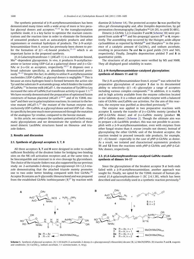
Scheme 1. Synthesis of glycosyl acceptors. (A) 1,10-bis[N-(2-acetamido-2-deoxy-b-D-gluand conditions: (b) Cu(OAc)2, sodium ascorbate, 1:1 acetone/water, rt, Ar(g).
diamine 2 (Scheme 1A). The protected acceptor 3a was purified bysilica gel chromatography and, after Zemplén deprotection, by gelpermeation chromatography (Sephadex™ LH-20), to yield 3 (48%).
Dimeric b-GlcNAc 1,2,3-triazoles 7 and 8 (Scheme 1B) were pre-pared from azide 428,29 and bis-propargyl spacers 539 or 6, respec-tively. The assembling step occurred by the Huisgen azide-alkynecycloaddition as reported by Meldal30 and Sharpless31 in the pres-ence of a catalytic amount of Cu(OAc)2 and sodium ascorbate,resulting in peracetates 7a and 8a in good yields (51% and 56%,respectively). Finally, Zemplén deprotection yielded 7 and 8 inquantitative yields.
The structures of all acceptors were verified by MS and NMR.They all displayed good solubility in water.
2.2. b-N-Acetylhexosaminidase-catalysed glycosylation:synthesis of dimers 11 and 12
The b-N-acetylhexosaminidase from A. oryzae32 was selected forpreparative glycosylation of acceptor 3 due to its documentedability to selectively—b(1?4)—glycosylate a range of acceptorsincluding various complex compounds.33 In addition, it is readilyand in high activity available from the enzyme collection locatedin our laboratory. It is a robust and stable enzyme with a balancedratio of GlcNAc-ase/GalNAc-ase activities. For the aim of this reac-tion, the enzyme was purified as described previously.34
The enzyme was applied in two preparative reactions withacceptor 3, namely the transfer of b-D-GlcNAc moiety (product 9,pNP-b-GlcNAc donor) and of b-D-GalNAc moiety (product 10,pNP-b-GalNAc donor) (Scheme 2). Though the ultimate aim wasto prepare a di-LacdiNAc product, this was not possible to accom-plish with a b-N-acetylhexosaminidase, even with enzymes fromother fungal strains than A. oryzae (results not shown). Instead ofglycosylating the other GlcNAc unit of the bivalent acceptor, thereaction tended to proceed towards side products, for example,b(1?6)-bound - especially in the case of pNP-b-GlcNAc as donor.As a result, we isolated and characterised asymmetric products11 and 12 from the reactions with pNP-b-GlcNAc and pNP-b-Gal-NAc donors, respectively.
2.3. b1,4-Galactosyltransferase-catalysed GalNAc-transfer:synthesis of dimers 14–17
Since the glycosylation of the bivalent acceptor 3 at both endsfailed with a b-N-acetylhexosaminidase, another approach wassought for. Finally, we opted for the Y284L mutant of human pla-cental b1,4-galactosyltransferase-1 (EC 2.4.1.38), which has beendescribed and successfully used in a synthetic reaction previously.4
OOAc
AcOAcO
4
OOn
5: n = 16: n = 2
NHAc+
b
OOR
OR
ORAcHN
O
O
ORN
N
N
OO
N
N
N
7a: R = Ac, n = 17: R = H, n = 18a: R = Ac, n = 28: R = H, n = 2
MeOH/MeONacat.
MeOH/MeONacat.
n
NHAc
N3
copyranosyl)-thioureido]-decane (3), (a) MeCN/DIPEA. (B) triazoles 7 and 8, reagents

3
O
NO2
+
β-N-AcetylhexosaminidaseA. oryzae CCF 1066
OR2
HO
OH
NHAc
R1
9: R1 = OH, R2 = H10: R1 = H, R2 = OH
11: R1 = OH, R2 = H, 7 %12: R1 = H, R2 = OH, 6 %
HN
OOH
HOHO NHAc
HN
HN NH
S S
OAcHN
OHOH
OH10
HN
OOH
OHO NHAc
HN
HN NH
S S
OAcHN
OHOH
OH10
OOHR2
HO NHAc
R1
Scheme 2. Synthesis of compounds 11 and 12.
A. Drozdová et al. / Carbohydrate Research 346 (2011) 1599–1609 1601
It can selectively transfer the b-D-GalNAc unit and its purification isstraightforward thanks to the presence of a His-tag. The need of arelatively expensive UDP-GalNAc donor can be overcome byemploying UDP-GlcNAc in combination with the UDP-GlcNAc-4’-epimerase from Campylobacter jejuni in one pot.35 Monitoring ofthe reaction course and optimisation of reaction conditions wereperformed by HPLC (Multokrom 100-5 C18 column, CS-Chroma-tographie Service GmbH, Cologne, DE). The glycosylation of accep-tor 3 (Scheme 3) was very efficient. Apart from a small amount ofthe product 12 (yield 9%) and the starting material 3 (5%) themajority of the desired di-LacdiNAc product 14 (86%) originatedin the reaction on an analytical scale (Fig. 1). The kinetic character-isation of the mutant b4GalT-1 (Y284L) revealed Km and Vmax
values of 0.213 mM and 137 mU/mg, respectively, for compound3, and 0.395 mM and 134 mU/mg, respectively, for UDP-GalNAc.The di-LacdiNAc product 14 was synthesised on preparative scalein 2 days and isolated in a single-step purification by gel chroma-
Scheme 3. Synthesis of co
tography (Sephadex™ LH-20 column) with 80% overall yield(Scheme 3).
Encouraged by this result, we decided to glycosylate two otherGlcNAc dimers with an alternative linker structure, i. e. triazole-linked compounds 7, 8. In both cases, the reactions led to therespective di-LacdiNAc products in good yields (16 in 28% yield,17 in 47% yield). During the glycosylation of acceptor 7, themono-LacdiNAc product was also obtained (15, 25% yield) (Scheme4).
2.4. Binding and precipitation studies with NKR-P1 and CD69activation receptors of NK cells
All prepared divalent compounds (acceptor 3 with products 11,12 and 14, acceptor 7 with products 15 and 16, and acceptor 8 withproduct 17) were subjected to binding and precipitation studieswith two fluorescently-labelled activation receptors of NK cells,
mpounds 12 and 14.

Figure 1. Time course of the conversion of acceptor 3 (N), intermediate 12 (�), and the final product 14 (j) generation.
Scheme 4. Synthesis of compounds 15–17.
Table 1Affinity of carbohydrate ligands to two NK cell activation receptors, rat NKR-P1A, andhuman CD69, expressed in the logarithmic scale (�log IC50 ± SD). The values expressthe ability of the studied compounds to inhibit binding between the respectiveprotein and a standard high affinity ligand GlcNAc23BSA
Compound NKR-P1A CD69
GlcNAc 5.4 ± 0.1 3.5 ± 0.13 9.3 ± 0.5 5.9 ± 0.111 9.3 ± 0.4 5.8 ± 0.112 9.5 ± 0.6 7.3 ± 1.514 8.3 ± 0.5 5.8 ± 0.17 5.9 ± 0.1 4.0 ± 0.115 6.3 ± 0.5 5.3 ± 1.616 7.4 ± 0.6 3.8 ± 0.18 7.2 ± 0.5 3.9 ± 0.117 7.9 ± 0.1 6.0 ± 0.2
1602 A. Drozdová et al. / Carbohydrate Research 346 (2011) 1599–1609
rat NKR-P1 and human CD69. The assays were essentially per-formed as described previously,4 with D-mannose as a negative(non-inhibitory) control carbohydrate and GlcNAc as a positivecontrol, providing IC50 values of 4.0 � 10�6 M (NKR-P1A) and3.2 � 10�4 M (CD69). The results for NKR-P1 and CD69 receptorsare summarised in Table 1 as �log IC50. It is apparent that both tri-azole compounds are weaker ligands than the thioureido com-pound 3 (generally in the range of 2–3 orders of magnitude). Theextension of the ligand with one or two GalNAc units did notessentially bring a notable increase in binding; the differencewas mostly within one order of magnitude. Only in the case ofthe CD69 receptor, some more pronounced increase could betraced (cf. 8 and 17). The acquired results are quite comparablewith those found for similar structures we described previously.4
We also performed precipitation studies (see Fig. 2 and Supple-mentary data, Fig. S1) with the above set of compounds (Schemes2–4 and Table 1). Contrary to the simple binding assays, precipita-tion assays are not competitive, and the amount of the formed
precipitate reflects the ligand’s ability to bind more receptormolecules at the same time, causing their cross-linking and finally
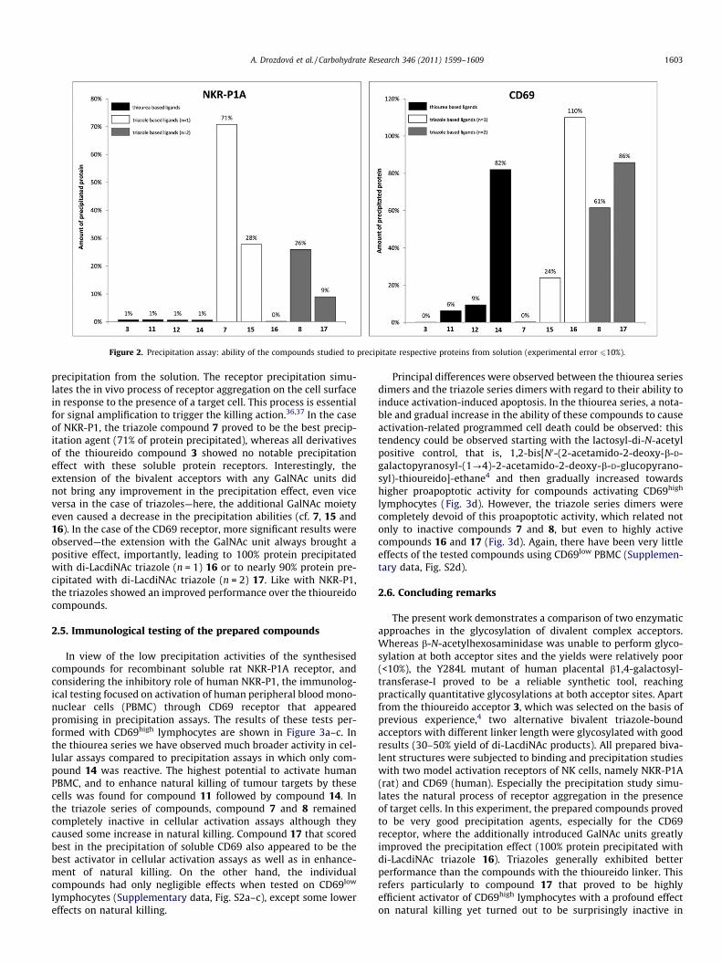
Figure 2. Precipitation assay: ability of the compounds studied to precipitate respective proteins from solution (experimental error 610%).
A. Drozdová et al. / Carbohydrate Research 346 (2011) 1599–1609 1603
precipitation from the solution. The receptor precipitation simu-lates the in vivo process of receptor aggregation on the cell surfacein response to the presence of a target cell. This process is essentialfor signal amplification to trigger the killing action.36,37 In the caseof NKR-P1, the triazole compound 7 proved to be the best precip-itation agent (71% of protein precipitated), whereas all derivativesof the thioureido compound 3 showed no notable precipitationeffect with these soluble protein receptors. Interestingly, theextension of the bivalent acceptors with any GalNAc units didnot bring any improvement in the precipitation effect, even viceversa in the case of triazoles—here, the additional GalNAc moietyeven caused a decrease in the precipitation abilities (cf. 7, 15 and16). In the case of the CD69 receptor, more significant results wereobserved—the extension with the GalNAc unit always brought apositive effect, importantly, leading to 100% protein precipitatedwith di-LacdiNAc triazole (n = 1) 16 or to nearly 90% protein pre-cipitated with di-LacdiNAc triazole (n = 2) 17. Like with NKR-P1,the triazoles showed an improved performance over the thioureidocompounds.
2.5. Immunological testing of the prepared compounds
In view of the low precipitation activities of the synthesisedcompounds for recombinant soluble rat NKR-P1A receptor, andconsidering the inhibitory role of human NKR-P1, the immunolog-ical testing focused on activation of human peripheral blood mono-nuclear cells (PBMC) through CD69 receptor that appearedpromising in precipitation assays. The results of these tests per-formed with CD69high lymphocytes are shown in Figure 3a–c. Inthe thiourea series we have observed much broader activity in cel-lular assays compared to precipitation assays in which only com-pound 14 was reactive. The highest potential to activate humanPBMC, and to enhance natural killing of tumour targets by thesecells was found for compound 11 followed by compound 14. Inthe triazole series of compounds, compound 7 and 8 remainedcompletely inactive in cellular activation assays although theycaused some increase in natural killing. Compound 17 that scoredbest in the precipitation of soluble CD69 also appeared to be thebest activator in cellular activation assays as well as in enhance-ment of natural killing. On the other hand, the individualcompounds had only negligible effects when tested on CD69low
lymphocytes (Supplementary data, Fig. S2a–c), except some lowereffects on natural killing.
Principal differences were observed between the thiourea seriesdimers and the triazole series dimers with regard to their ability toinduce activation-induced apoptosis. In the thiourea series, a nota-ble and gradual increase in the ability of these compounds to causeactivation-related programmed cell death could be observed: thistendency could be observed starting with the lactosyl-di-N-acetylpositive control, that is, 1,2-bis[N0-(2-acetamido-2-deoxy-b-D-galactopyranosyl-(1?4)-2-acetamido-2-deoxy-b-D-glucopyrano-syl)-thioureido]-ethane4 and then gradually increased towardshigher proapoptotic activity for compounds activating CD69high
lymphocytes (Fig. 3d). However, the triazole series dimers werecompletely devoid of this proapoptotic activity, which related notonly to inactive compounds 7 and 8, but even to highly activecompounds 16 and 17 (Fig. 3d). Again, there have been very littleeffects of the tested compounds using CD69low PBMC (Supplemen-tary data, Fig. S2d).
2.6. Concluding remarks
The present work demonstrates a comparison of two enzymaticapproaches in the glycosylation of divalent complex acceptors.Whereas b-N-acetylhexosaminidase was unable to perform glyco-sylation at both acceptor sites and the yields were relatively poor(<10%), the Y284L mutant of human placental b1,4-galactosyl-transferase-I proved to be a reliable synthetic tool, reachingpractically quantitative glycosylations at both acceptor sites. Apartfrom the thioureido acceptor 3, which was selected on the basis ofprevious experience,4 two alternative bivalent triazole-boundacceptors with different linker length were glycosylated with goodresults (30–50% yield of di-LacdiNAc products). All prepared biva-lent structures were subjected to binding and precipitation studieswith two model activation receptors of NK cells, namely NKR-P1A(rat) and CD69 (human). Especially the precipitation study simu-lates the natural process of receptor aggregation in the presenceof target cells. In this experiment, the prepared compounds provedto be very good precipitation agents, especially for the CD69receptor, where the additionally introduced GalNAc units greatlyimproved the precipitation effect (100% protein precipitated withdi-LacdiNAc triazole 16). Triazoles generally exhibited betterperformance than the compounds with the thioureido linker. Thisrefers particularly to compound 17 that proved to be highlyefficient activator of CD69high lymphocytes with a profound effecton natural killing yet turned out to be surprisingly inactive in
d
PBS diL 3 11 12 14 7 8 16 17% a
popt
otic
cel
ls (
Ann
exin
V+/ P
I- )0
10
20
30
40
50
60
PBS diL 3 11 12 14 7 8 16 17
Inos
itol p
hosp
hate
s [d
pm]
0
1000
2000
3000
4000
5000
6000
Ins2PIns3P
a b
PBS diL 3 11 12 14 7 8 16 17
Intr
acel
lula
r ca
lciu
m [n
M]
0
200
400
600
800
1000
c
PBS diL 3 11 12 14 7 8 16 17Rel
ativ
e ly
tic e
ffici
ency
[x10
-3]
0
2
4
6
8
10
12
14
16
18
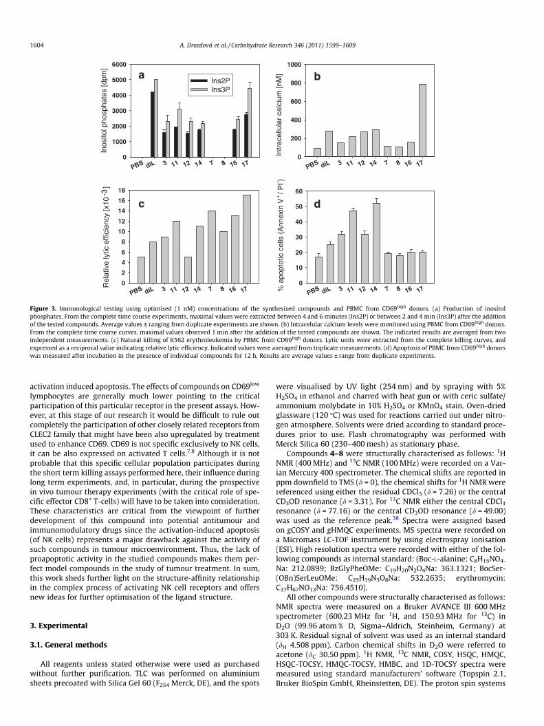
Figure 3. Immunological testing using optimised (1 nM) concentrations of the synthesised compounds and PBMC from CD69high donors. (a) Production of inositolphosphates. From the complete time course experiments, maximal values were extracted between 4 and 6 minutes (Ins2P) or between 2 and 4 min (Ins3P) after the additionof the tested compounds. Average values ± ranging from duplicate experiments are shown. (b) Intracelular calcium levels were monitored using PBMC from CD69high donors.From the complete time course curves, maximal values observed 1 min after the addition of the tested compounds are shown. The indicated results are averaged from twoindependent measurements. (c) Natural killing of K562 erythroleukemia by PBMC from CD69high donors. Lytic units were extracted from the complete killing curves, andexpressed as a reciprocal value indicating relative lytic efficiency. Indicated values were averaged from triplicate measurements. (d) Apoptosis of PBMC from CD69high donorswas measured after incubation in the presence of individual compounds for 12 h. Results are average values ± range from duplicate experiments.
1604 A. Drozdová et al. / Carbohydrate Research 346 (2011) 1599–1609
activation induced apoptosis. The effects of compounds on CD69low
lymphocytes are generally much lower pointing to the criticalparticipation of this particular receptor in the present assays. How-ever, at this stage of our research it would be difficult to rule outcompletely the participation of other closely related receptors fromCLEC2 family that might have been also upregulated by treatmentused to enhance CD69. CD69 is not specific exclusively to NK cells,it can be also expressed on activated T cells.7,8 Although it is notprobable that this specific cellular population participates duringthe short term killing assays performed here, their influence duringlong term experiments, and, in particular, during the prospectivein vivo tumour therapy experiments (with the critical role of spe-cific effector CD8+ T-cells) will have to be taken into consideration.These characteristics are critical from the viewpoint of furtherdevelopment of this compound into potential antitumour andimmunomodulatory drugs since the activation-induced apoptosis(of NK cells) represents a major drawback against the activity ofsuch compounds in tumour microenvironment. Thus, the lack ofproapoptotic activity in the studied compounds makes them per-fect model compounds in the study of tumour treatment. In sum,this work sheds further light on the structure-affinity relationshipin the complex process of activating NK cell receptors and offersnew ideas for further optimisation of the ligand structure.
3. Experimental
3.1. General methods
All reagents unless stated otherwise were used as purchasedwithout further purification. TLC was performed on aluminiumsheets precoated with Silica Gel 60 (F254 Merck, DE), and the spots
were visualised by UV light (254 nm) and by spraying with 5%H2SO4 in ethanol and charred with heat gun or with ceric sulfate/ammonium molybdate in 10% H2SO4 or KMnO4 stain. Oven-driedglassware (120 �C) was used for reactions carried out under nitro-gen atmosphere. Solvents were dried according to standard proce-dures prior to use. Flash chromatography was performed withMerck Silica 60 (230–400 mesh) as stationary phase.
Compounds 4–8 were structurally characterised as follows: 1HNMR (400 MHz) and 13C NMR (100 MHz) were recorded on a Var-ian Mercury 400 spectrometer. The chemical shifts are reported inppm downfield to TMS (d = 0), the chemical shifts for 1H NMR werereferenced using either the residual CDCl3 (d = 7.26) or the centralCD3OD resonance (d = 3.31). For 13C NMR either the central CDCl3
resonance (d = 77.16) or the central CD3OD resonance (d = 49.00)was used as the reference peak.38 Spectra were assigned basedon gCOSY and gHMQC experiments. MS spectra were recorded ona Micromass LC-TOF instrument by using electrospray ionisation(ESI). High resolution spectra were recorded with either of the fol-lowing compounds as internal standard: (Boc-L-alanine: C8H15NO4-
Na: 212.0899; BzGlyPheOMe: C19H20N2O4Na: 363.1321; BocSer-(OBn)SerLeuOMe: C25H39N3O8Na: 532.2635; erythromycin:C37H67NO13Na: 756.4510).
All other compounds were structurally characterised as follows:NMR spectra were measured on a Bruker AVANCE III 600 MHzspectrometer (600.23 MHz for 1H, and 150.93 MHz for 13C) inD2O (99.96 atom % D, Sigma–Aldrich, Steinheim, Germany) at303 K. Residual signal of solvent was used as an internal standard(dH 4.508 ppm). Carbon chemical shifts in D2O were referred toacetone (dC 30.50 ppm). 1H NMR, 13C NMR, COSY, HSQC, HMQC,HSQC-TOCSY, HMQC-TOCSY, HMBC, and 1D-TOCSY spectra weremeasured using standard manufacturers’ software (Topspin 2.1,Bruker BioSpin GmbH, Rheinstetten, DE). The proton spin systems

A. Drozdová et al. / Carbohydrate Research 346 (2011) 1599–1609 1605
of each sugar unit were assigned by COSY, TOCSY, HSQC-TOCSYand 1D-TOCSY. The assignment was transferred to carbons byHSQC. The position of substitution of sugar moiety was con-firmed by long-range heteronuclear correlations in the HMBCspectrum and configuration at C-1 was deduced from couplingconstants JH-1,H-2 observed in the 1H NMR spectra. Several signalsboth in the 1H and 13C NMR spectra in the substances containingthiourea moiety were doubled due to their conformationalbehaviour.
Chemical shifts are given in d-scale [ppm], and coupling con-stants in Hz. Digital resolution enabled us to report chemical shiftsof protons to three and coupling constants to one and carbonchemical shifts to two decimal places. Some hydrogen chemicalshifts were read out from HSQC or HMQC and are reported totwo decimal places.
Mass spectra were measured on a MALDI-TOF/TOF ultraFLEX IIImass spectrometer (Bruker-Daltonics, Bremen, DE). Positive spec-tra were calibrated externally using the monoisotopic [M+H]+ ionsof PepMixII calibrant or using average mass of ProtMixI calibrant(Bruker-Daltonics, Bremen, DE). A 50 mg/mL solution of 2,5-dihy-drobenzoic acid in 50% MeCN/0.1% TFA was used as a MALDI ma-trix. The sample (0.4 lL) was dissolved in 50% MeOH and it wasallowed to dry at ambient temperature and over-laid with the ma-trix solution (0.3 lL) on the target. The MALDI-TOF positive spectrawere collected in reflectron mode.
3.2. Chemical synthesis of dimeric acceptors 3, 7, 8
3.2.1. 1,10-Bis[N0-(2-acetamido-2-deoxy-b-D-glucopyranosyl)-thioureido]-decane (3)
2-Acetamido-3,4,6-tri-O-acetyl-2-deoxy-b-D-glucopyranosyl iso-thiocyanate (1; 400 mg, 1.03 mmol)27 and 1,10-diaminodecane(2; 90 mg, 0.52 mmol) were dissolved in dry acetonitrile (10 mL)with DIPEA (0.5 mL). After 24 h at 22 �C, the reaction mixturewas purified by column chromatography on silica gel (chloro-form/MeOH, 30:1). The dry peracetate 3a was suspended in drymethanol (4 mL), sodium methoxide (2 M solution in methanol,0.5 mL) was added, and the mixture was stirred until the disap-pearance of the starting material as indicated by TLC (acetoni-trile/H2O, 5:1). The reaction mixture was neutralised with Dowex50 W-X2 (H+ form), filtered, concentrated, purified by gel chroma-tography on Sephadex™ LH-20 (Sigma, USA) in MeOH as a mobilephase, 12 mL/min), evaporated to dryness, dissolved in water andlyophilised to obtain 3 (121 mg, 48%) as white solid. For NMR data,see Tables S1 and S2 (Supplementary data).
3.2.2. 1,4-Bis(propynyloxy)butane (6)To the solution of 1,4-butanediol (557 mg, 6.18 mmol, 1 equiv)
in dry THF (10 mL) NaH (55–65% in mineral oil, 928 mg,30.9 mmol, 5 equiv) was added at 0 �C. The mixture was stirredat 0 �C under N2 atmosphere for 15 min. Propargyl bromide(80 wt. % in toluene, 1.7 mL, 15.5 mmol, 2.5 equiv) was added.The resulting mixture was stirred overnight at room temperature.The mixture was diluted with CH2Cl2 followed by addition ofwater. The aqueous phase was extracted three times with CH2Cl2,the combined organic layers were dried over anhydrous MgSO4,filtered and concentrated in vacuo. The crude product was purifiedby flash column chromatography (silica gel, EtOAc/pentane 1:20)yielding 698 mg (4.20 mmol, 68%) of 6 as bright yellow oil.Rf: 0.24 (EtOAc/pentane 1:20). 1H NMR (400 MHz, CDCl3) dH 4.08(d, 4H, J 2.4 Hz, H-3), 3.53–3.50 (m, 4H, H-4), 2.39 (t, 2H, J 2.4 Hz,H-1), 1.67–1.64 (m, 4H, H-5). 13C NMR (100 MHz, CDCl3) dC 80.2(C-2), 74.4 (C-1), 67.0 (C-4), 58.2 (C-3), 26.4 (C-5). HRMS(ES): Calculated for C10H14O2Na m/z 189.0891. Found m/z189.0891.
3.2.3. 1,6-Bis[1-(2-acetamido-2-deoxy-3,4,6-tri-O-acetyl-b-D-glucopyranosyl)-1,2,3-triazole-4-yl]-2,5-dioxa-hexane (7a)
1,2-Bis(propynyloxy)ethane 539 (359 mg, 2.60 mmol, 1 equiv)was dissolved in 50 mL of argon-degassed mixture acetone/water1:1 (resulting in 0.05 M concentration of 5). To this solutionCu(OAc)2 (94 mg, 0.518 mmol, 0.2 equiv), sodium ascorbate(206 mg, 1.04 mmol, 0.4 equiv) and finally GlcNAc azide 4(2.13 g, 5.72 mmol, 2.2 equiv) were added. The reaction mixturewas stirred under argon atmosphere at room temperature over-night. Acetone was removed from the mixture by rotatory evapo-rator. CHCl3 and 5% aqueous ammonia were added to theresidue. The aqueous phase was extracted three times with CHCl3,the combined organic phases were washed with brine, dried overanhydrous MgSO4, filtered and concentrated in vacuo. The crudeproduct was purified by flash column chromatography (gradienteluent system: 1:3 acetone/EtOAc, then acetone) yielding 1.18 g(1.34 mmol, 51%) of 7a as white powder. Mp (uncorr.): 102.2–104.4 �C. Rf: 0.29 (acetone/EtOAc, 1:4). 1H NMR (400 MHz, CDCl3)dH 8.00 (s, 2H, H-7), 7.18 (d, 2H, JNH,2 9.1 Hz, NH), 6.15 (d, 2H, J1,2
9.8 Hz, H-1), 5.53 (t, 2H, J2,3 = J3,4 9.6 Hz, H-3), 5.31 (t, 2H,J3,4 = J4,5 9.6 Hz, H-4), 4.66–4.56 (m, 6H, H-2, H-8), 4.31 (dd, 2H,J5,6a 4.8 Hz, J6a,6b 12.5 Hz, H-6a), 4.16 (dd, 2H, J5,6b 1.9 Hz, J6a,6b
12.5 Hz, H-6b), 4.13–4.09 (m, 2H, H-5), 3.68–3.61 (m, 4H, H-9),2.05 (s, 18H, OCOCH3), 1.70 (s, 6H, NHCOCH3). 13C NMR(100 MHz, CDCl3) dC 171.3, 171.0, 170.7, 169.7 (C@O), 145.5 (tria-zole-C), 122.5 (C-7), 85.9 (C1), 75.0 (C5), 72.7 (C3), 69.7 (C9),68.5 (C4), 64.4 (C8), 62.1 (C6), 53.8 (C2), 22.9 (NHCOCH3), 21.0,20.9, 20.8 (OCOCH3). HRMS (ES): Calculated for C36H50N8O18Nam/z 905.3141. Found m/z 905.3144.
3.2.4. 1,8-Bis[1-(2-acetamido-2-deoxy-3,4,6-tri-O-acetyl-b-D-glucopyranosyl)-1,2,3-triazole-4-yl]-2,7-dioxa-octane (8a)
Compound 8a was prepared by the same procedure as 7a using1,4-bis(propynyloxy)butane 6 (310 mg, 1.86 mmol). The crudeproduct was purified by flash column chromatography (gradienteluent system: EtOAc; then acetone/EtOAc, 1:3; then acetone)yielding 956 mg (1.05 mmol, 56 %) of 8a as white powder. Mp (un-corr.): 220 �C (decomp.). Rf: 0.43 (1:4 acetone/EtOAc). 1H NMR(400 MHz, CDCl3) dH 7.94 (s, 2H, H7), 7.05 (d, 2H, JNH,2 9.2 Hz,NH), 6.16 (d, 2H, J1,2 9.9 Hz, H-1), 5.52 (t, 2H, J2,3 = J3,4 9.8 Hz, H-3), 5.27 (t, 2H, J3,4 = J4,5 9.8 Hz, H-4), 4.68–4.61 (m, 2H, H-2), 4.56(s, 4H, H-8), 4.30 (dd, 2H, J5,6a 4.7 Hz, J6a,6b 12.5 Hz, H-6a), 4.15(dd, 2H, J5,6b 2.0 Hz, J6a,6b 12.5 Hz, H-6b), 4.09 (ddd, 2H, J5,6b
2.0 Hz , J5,6a 4.7 Hz, J4,5 9.8 Hz, H-5), 3.49–3.43 (m, 4H, H-9), 2.06(s, 6H, OCOCH3), 2.04 (s, 6H, OCOCH3), 2.03 (s, 6H, OCOCH3), 1.73(s, 6H, NHCOCH3), 1.59–1.56 (m, 4H, H10). 13C NMR (100 MHz,CDCl3) dC 171.2, 171.1, 170.9, 169.6 (C@O), 145.8 (triazole-C),122.2 (C7), 85.9 (C1), 75.1 (C5), 72.7 (C3), 70.4 (C9), 68.4 (C4),64.0 (C8), 62.0 (C6), 53.7 (C2), 26.5 (C10), 22.9 (NHCOCH3), 20.9,20.8, 20.7 (OCOCH3). HRMS (ES): calcd for C38H54N8O18Na m/z933.3454. Found m/z 933.3446.
3.2.5. 1,6-Bis[1-(2-acetamido-2-deoxy-b-D-glucopyranosyl)-1,2,3-triazole-4-yl]-2,5-dioxa-hexane (7)
A catalytic amount of freshly prepared CH3ONa in CH3OH wasadded to a stirred solution of 7a (312 mg, 0.353 mmol) in dryCH3OH (4.5 mL). The reaction mixture was stirred at room temper-ature until TLC analysis indicated reaction completion. The reac-tion mixture was then concentrated under reduced pressure toafford 223 mg (0.353 mmol, 100%) of 7 as a white solid. For furtherwork with enzymes a gel filtration purification step was necessary(Biogel P2, 100 � 2 cm, H2O 10 mL/h). Mp (uncorr.): 125 �C (de-comp.). Rf: 0.48 (CH3OH/acetone, 2:3). 1H NMR (400 MHz, CD3OD)dH 8.18 (s, 2H, H7), 5.81 (d, 2H, J1,2 9.8 Hz, H1), 4.62 (s, 2H, H8), 4.25(t, 2H, J 10.0 Hz, H2), 3.90 (dd, 2H, J6a,5 1.5 Hz, J6a,6b 12.3 Hz, H6a),3.78–3.68 (m, 4H, H3, H6b), 3.64 (s, 4H, H9), 3.59–3.54 (m, 4H,

1606 A. Drozdová et al. / Carbohydrate Research 346 (2011) 1599–1609
H4, H5), 1.77 (s, 3H, NHCOCH3). 13C NMR (100 MHz, CD3OD) dC
173.5 (C@O), 146.0 (triazole-C), 124.0 (C7), 88.2 (C1), 81.3 (C5),75.8 (C3), 71.4 (C9), 70.6 (C4), 64.9 (C8), 62.4 (C6), 56.9 (C2),22.6 (NHCOCH3). HRMS (ES): Calculated for C24H38N8O12Na m/z653.2507. Found m/z 653.2511.
3.2.6. 1,8-Bis[1-(2-acetamido-2-deoxy-b-D-glucopyranosyl)-1,2,3-triazole-4-yl]-2,7-dioxa-octane (8)
Starting from 8a (908 mg, 0.997 mmol) following the same pro-cedure as for 7, reaction yielded 656 mg (0.997 mmol, 100%) of 8 aswhite solid. For further work with enzymes a gel filtration purifica-tion step was necessary (Biogel P2, 100 � 2 cm, H2O 10 mL/h) to re-move low molecular impurities inhibiting enzyme activity. Mp(uncorr.): 83.3–85.6 �C. Rf: 0.54 (CH3OH/acetone, 2:3). 1H NMR(400 MHz, CD3OD) dH 8.15 (s, 2H, H7), 5.82 (d, 2H, J1,2 9.9 Hz,H1), 4.56 (s, 4H, H8), 4.25 (t, 2H, J 9.9 Hz, H2), 3.90 (br d, 2H,J6a,6b 12.0 Hz, H6a), 3.78–3.69 (m, 4H, H3, H6b), 3.64–3.54 (m,4H, H4, H5), 3.54–3.44 (m, 4H, H9), 1.78 (s, 6H, NHCOCH3), 1.64–1.60 (m, 4H, H10). 13C NMR (100 MHz, CD3OD) dC 173.4 (C@O),146.2 (triazole-C), 123.8 (C7), 88.1 (C1), 81.3 (C5), 75.8 (C3), 71.4(C9), 71.2 (C4), 64.5 (C8), 62.4 (C6), 56.8 (C2), 27.4 (C10), 22.7(NHCOCH3). HRMS (ES): Calculated for C26H42N8O12Na m/z681.2820. Found m/z 681.2819.
3.3. b-N-Acetylhexosaminidase-catalysed glycosylation
3.3.1. b-N-Acetylhexosaminidase from A. oryzae CCF 1066The b-N-acetylhexosaminase (EC 3.2.1.52) from a filamentous
fungus A. oryzae CCF 1066 (Culture Collection of Fungi (CCF),Department of Botany, Charles University in Prague, CZ) was pro-duced in mineral medium consisting of [g/L] yeast extract (0.5),KH2PO4 (3), NH4H2PO4 (5), (NH4)2SO4 (2), GlcNAc (5), NaCl (15),pH 6.0. Flasks (1 L) with medium (200 mL) were inoculated withthe suspension of spores in 0.1% Tween 80 and cultivated on a ro-tary shaker at 28 �C for 6 days. Then, the medium was filtered offand the supernatant was fractionally precipitated by (NH4)2SO4
(0–80%). For purification, the precipitate was dissolved in 10 mMcitrate–phosphate buffer, pH 3.5 (10 mM Na2HPO4/10 mMC6H8O7), and dialysed against this buffer at 4 �C overnight. Cat-ion-exchange chromatography was performed on Fractogel EMDSO3
� (Merck, DE) equilibrated with 10 mM citrate-phosphate buf-fer, pH 3.5 at 22 �C. Bound proteins were eluted with a linear gra-dient of 0–1 M NaCl in the same buffer at a flow rate of 2 mL/min.b-N-Acetylhexosaminidase activity was determined by standardassay40 and positive fractions were pooled and concentrated. Thefinal purification yield was 5%.
3.3.2. Transglycosylation reactions on analytical scaleAnalytical reactions for preparing compound 11 (total volume
100 lL) contained acceptor 3 (9–30 mg, 13–43 lmol) and pNP-b-GlcNAc (9; 2.5–5 mg, 7–15 lmol) in 50 mM citrate-phosphate buf-fer pH 5.0 (50 mM Na2HPO4/50 mM C6H8O7). Analytical reactionsfor preparing compound 12 (total volume 200 lL) contained accep-tor 3 (5–15 mg, 7–28 lmol) and pNP-b-GalNAc (10; 5–20 mg, 15–58 lmol) in 50 mM citrate-phosphate buffer pH 5.0.
The reactions were started by the addition of the b-N-acetyl-hexosaminidase from A. oryzae CCF 1066 (0.2–5 U) and were incu-bated at 35 �C with shaking for 24 h. The reaction course wasmonitored by TLC (acetonitrile/H2O, 5:1).
3.3.3. 1-[N0-(2-Acetamido-2-deoxy-b-D-glucopyranosyl)-thioure-ido]-10-[N0-(2-acetamido-2-deoxy-b-D-glucopyranosyl-(1?4)-2-acetamido-2-deoxy-b-D-glucopyranosyl)-thioureido]-decane (11)
The reaction mixture containing acceptor 3 (120 mg, 170 lmol),pNP-b-GlcNAc (9, 30 mg, 87 lmol), and the purified b-N-acetylhex-
osaminidase from A. oryzae CCF 1066 (0.5 U) in 50 mM citrate-phosphate buffer pH 5.0 (600 lL) was incubated under shakingat 35 �C. After 4.5 h the reaction was stopped by adding methanol(1200 lL) and loaded onto Sephadex™ LH-20 column(1850 � 25 mm, mobile phase MeOH, flow rate 10 mL/h). The finalpurification (separation of the b(1?6) by-product) was performedby preparative HPLC on Chromolith SemiPrep RP-18e column(100 � 10 mm, Merck, mobile phase MeOH/H2O 1:1, flow rate2.5 mL/min). Compound 11 was obtained as a white solid (11 mg,12 lmol, 7%). MS (MALDI-TOF) calculated for C36H65N7O15S2Na,923.086, found m/z 922.407. For NMR data, see Tables S1 and S2(Supplementary data).
3.3.4. 1-[N0-(2-Acetamido-2-deoxy-b-D-glucopyranosyl)-thioure-ido]-10-[N0-(2-acetamido-2-deoxy-b-D-glucopyranosyl-(1?4)-2-acetamido-2-deoxy-b-D-galactopyranosyl)-thioureido]-decane(12)
The reaction mixture containing acceptor 3 (45 mg, 64 lmol),pNP-b-GalNAc, (10, 60 mg, 175 lmol), and the purified b-N-acetyl-hexosaminidase from A. oryzae CCF 1066 (0.5 U) in 50 mM citrate-phosphate buffer pH 5.0 (600 lL) was incubated under shaking at35 �C. After 4.5 h the reaction was stopped by adding methanol(1200 lL) and loaded onto Sephadex™ LH-20 column (1850 �25 mm, mobile phase MeOH, flow rate 10 mL/h). The final purifica-tion was performed by preparative HPLC on MAG 5 100 C18column (25 � 250 mm, Labio a.s., Prague, CZ) in a mobile phaseof 40% methanol in H2O, flow rate 2.5 mL/min. Compound 12(12 mg, 13 lmol, 6%) was obtained as white solid. MS (MALDI-TOF) calculated for C36H65N7O15S2Na, 923.086, found m/z922.510; for NMR data, see Tables S1 and S2 (Supplementary data).
3.4. Synthesis by mutant b1,4-galactosyltransferase-1
3.4.1. Mutant b1,4-galactosyltransferase-1 (His6-Tag-propep-tide-catb4GalT1 Y284L)
The construction, expression, and purification of the Y284L mu-tant of human placental b1,4-galactosyltransferase-1 (EC 2.4.1.38;His6-Tag-propeptide-catb4GalT-1 construct) was performed asdescribed previously.4
Escherichia coli BL21(DE3) cells (11.3 g) were suspended in lysisbuffer (18.3 mL; 50 mM NaH2PO4/ 300 mM NaCl/ 10 mM imidaz-ole, pH 7.5), and benzonase nuclease (10 lL; 25 U/lL) was added.The mixture was sonicated on ice (3 � 20 s with 60 s breaks,sonotrode MS 72, 52% pulsion). The supernatant obtained aftercentrifugation (20 min, 19,000 rpm, 4 �C) was purified by immobi-lised metal affinity chromatography on Ni-NTA column (90 �16 mm), pre-equilibrated with lysis buffer. The column waswashed with washing buffer (50 mM NaH2PO4/300 mM NaCl/20 mM imidazole, pH 7.5; linear flow rate 1.5 mL/min), and thenwith elution buffer (50 mM NaH2PO4/300 mM NaCl/200 mM imid-azole, pH 7.5; linear flow rate 1.5 mL/min). Fractions with the high-est galactosyltransferase activity were pooled and concentrated.Addition of 20% (v/v) glycerol stabilised the final enzyme solution,which had more than 40 mU/mL activity.
3.4.2. His6-Tag-propeptide-catb4GalT1 Y284L activity assaysThe spectrophotometric assay was described previously.4 The
transferase activity was determined by HPLC as follows. The reac-tion mixture (total volume 100 lL) consisting of 100 mM MOPS/25 mM KCl, pH 6.8; GlcNAc-tBoc (1 mM), UDP-GalNAc (1.2 mM),alkaline phosphatase (10 U/mL), MnCl2 (2 mM), and 10 lL ofenzyme sample was incubated at 30 �C with shaking. Two aliquots(20, 40 min) were taken, the reactions were stopped by boiling(5 min) and after centrifugation (10 min, 10,000 rpm), the superna-tant was loaded onto Multokrom 100-5 C18 column (250 � 4 mm;mobile phase was a 36 min linear gradient of 11–42.2% v/v CH3CN
A. Drozdová et al. / Carbohydrate Research 346 (2011) 1599–1609 1607
in H2O, 0.5 mL/min, UV detection at 205 nm and 254 nm). Enzymeactivity was calculated as a ratio of peak areas depending on timeof the reaction, A [U/mL] = [1 mM � (Area LacDiNAc-tBoc/(AreaLacDiNAc-tBoc + Area GlcNAc-tBoc)) � 0.1/(incubation time � 0.01)].
The kinetic constants of compound 3 were determined at a con-stant concentration of UDP-GalNAc donor (5 mM) with 0–6 mM of3. The kinetic constants of the donor substrate were measured at aconstant concentration of acceptor 3 (5 mM) with 0–20 mM UDP-GalNAc. Donor and acceptor substrates were mixed in 100 mMMOPS-KOH buffer (100 lL final volume), pH 6.8, containing2 mM MnCl2, 25 mM KCl, alkaline phosphatase (10 mU), and theenzyme sample (10 lL). The mixture was incubated at 30 �C. Ali-quots of the reaction mixtures were taken at set time intervalswhen substrate conversion was linear over time, and subsequentlystopped by heating at 95 �C for 5 min. The product formation wasanalyzed by HPLC as described above using 240 nm for detection ofcompound 3, and products 12 and 14. Kinetic constants were ob-tained by nonlinear regression of the data points using Sigma Plot10 (SPSS GmbH Software, München, DE) following the Michaelis-Menten kinetic.
3.4.3. GalNAc transfer on analytical scaleThe reaction mixture (total volume 100 lL) containing UDP-
GlcNAc donor (13; 0.3–0.7 mg, 0.5–1 lmol) and acceptor 3 (0.2–0.4 mg, 0.3–0.6 lmol) 7, or 8 (both 0.3 mg, 0.5 lmol), 100 mMMOPS/ 25 mM KCl, pH 6.8, 2 mM MnCl2, alkaline phosphatase(10 U/mL), UDP-GlcNAc-40-epimerase from Campylobacter jejuni(1 U/mL, prepared as described previously)35,41 and the Y284L mu-tant of His6-propeptide-catb4GalT-1 (10 -20 mU/mL) was incu-bated at 30 �C with shaking for 72 h. Reactions were stopped byboiling (5 min) and after centrifugation (10 min, 10,000 rpm) theywere analysed by TLC and HPLC. The TLC mobile phase was CH3CN/H2O/HCOOH, 4:1:0.1 for 3 or CH2Cl2/MeOH/EtOH/H2O/HCOOH,5:4:1:1:0.1 for 7 and 8. HPLC was performed on Multokrom 100-5 C18 column (250 � 4 mm). Mobile phase was a 36-min lineargradient of 11–42.2% v/v CH3CN in H2O for 3 or of 5–26% CH3CNin H2O for 7 and 8, flow rate 0.5 mL/min, UV detection at 240 nmfor 3 or 230 nm for 7 and 8.
The conversion of acceptor 3 and formation of products 12 and14 was followed over time and quantified by HPLC as describedabove. The reaction mixture (100 lL) consisted of 5 mM compound3, 12 mM UDP-GalNAc, 100 mM MOPS-KOH buffer, pH 6.8 with25 mM KCl, 2 mM MnCl2, alkaline phospatase (10 mU), and the en-zyme sample (1.39 mU). The mixture was incubated at 30 �C. Ali-quots of the reaction mixture were taken at indicated timeintervals and stopped by heating at 95 �C for 5 min.
3.4.4. 1-[N0-2-Acetamido-2-deoxy-b-D-galactopyranosyl-(1?4)-2-acetamido-2-deoxy-b-D-glucopyranosyl)-thioureido]-10-[N0-(2-acetamido-2-deoxy-b-D-glucopyranosyl-(1?4)-2-acetamido-2-deoxy-b-D-galactopyranosyl)-thioureido]-decane (14)
Acceptor 3 (24 mg, 34 lmol) and UDP-GlcNAc, (13; 54 mg,90 lmol) were dissolved in 100 mM MOPS/25 mM KCl, pH 6.8 and2 mM MnCl2. Alkaline phosphatase (70 U), UDP-GlcNAc-4’-epimer-ase (7 U) and the Y284L mutant of His6-propeptide-catb4GalT-1(175 mU) were added (total volume 7 mL). The reaction mixturewas incubated at 30 �C with shaking. The reaction progress wasmonitored by HPLC and TLC. After 48 h, the reaction was stoppedby boiling (5 min), centrifuged (10 min, 10,000 rpm), the superna-tant was evaporated to dryness, dissolved in 80% MeOH in H2O(2 mL) and loaded onto Sephadex™ LH-20 column (800 � 25 mm,mobile phase 80% MeOH in H2O, flow rate 9 mL/h). Compound 14was eluted pure and after lyophilisation it was obtained as whitesolid (32 mg, 29 lmol) in 80% yield. For NMR data, see Tables S1,S2 (Supplementary data). MS (MALDI-TOF) calculated forC44H78N8O20S2Na, 1125.437, found m/z 1125.462.
3.4.5. 1-[1-(2-Acetamido-2-deoxy-b-D-galactopyranosyl-(1?4)-2-acetamido-2-deoxy-b-D-glucopyranosyl)-1,2,3-triazole-4-yl]-2,5-dioxa-6-[1-(2-acetamido-2-deoxy-b-D-glucopyranosyl)-1,2,-3-triazole-4-yl]-hexane (15) and 1,6-bis-[1-(2-acetamido-2-de-oxy-b-D-galactopyranosyl-(1?4)-2-acetamido-2-deoxy-b-D-glu-copyranosyl)-1,2,3-triazole-4-yl]-2,5-dioxa-hexane (16)
Acceptor 7 (19 mg, 30 lmol) and UDP-GlcNAc (13; 47 mg,77 lmol) were dissolved in 100 mM MOPS/ 25 mM KCl, pH 6.8and 2 mM MnCl2. Alkaline phosphatase (60 U), UDP-GlcNAc-40-epimerase (6 U) and the Y284L mutant of His6-propeptide-catb4-GalT1 (120 mU) were added (total volume 6 mL). The reaction mix-ture was incubated at 30 �C with shaking. The reaction progresswas monitored by HPLC and TLC. After 72 h, the reaction wasstopped by boiling (5 min), centrifuged (10 min, 10,000 rpm) andthe supernatant was evaporated to dryness, dissolved in 80%MeOH in H2O (2 mL) and loaded onto Sephadex™ LH-20 column(800 � 25 mm, mobile phase 80% MeOH in H2O, flow rate 9 mL/h). Two compounds, 15 and 16, were eluted pure. After lyophilisa-tion, compound 15 (6.5 mg, 8 lmol) was obtained as white solid in26% yield, compound 16 as white solid (9 mg, 8.6 lmol) in 28%yield. For NMR data, see Tables S1, S2 (Supplementary data). MS(MALDI-TOF) calculated for C32H51N9O17Na (15), 856.330, foundm/z 855.517; calculated for C40H64N10O22Na (16), 1059.409, foundm/z 1059.462.
3.4.6. 1,8-Bis-[1-(2-acetamido-2-deoxy-b-D-galactopyranosyl-(1?4)-2-acetamido-2-deoxy-b-D-glucopyranosyl)-1,2,3-triaz-ole-4-yl]-2,7-dioxa-octane (17)
Acceptor 8 (24 mg, 36 lmol) and UDP-GlcNAc (13; 56 mg,92 lmol) were dissolved in 100 mM MOPS/25 mM KCl, pH 6.8and 2 mM MnCl2. Alkaline phosphatase (72 U), UDP-GlcNAc-40-epimerase (7.2 U) and the Y284L mutant of His6-propeptide-catb4-GalT-1 (144 mU) were added (total volume 7.2 mL). The reactionmixture was incubated at 30 �C with shaking. The reaction pro-gress was monitored by HPLC and TLC. After 74 h, the reactionwas stopped by boiling (5 min), centrifuged (10 min, 10,000 rpm)and the supernatant was evaporated to dryness, dissolved in 80%MeOH in H2O (2 mL) and loaded onto Sephadex™ LH-20 column(800 � 25 mm, mobile phase 80% MeOH in H2O, flow rate 8 mL/h). After lyophilisation, compound 17 was obtained as white solid(19 mg, 18 lmol) in 47% yield. For NMR data, see Tables S1, S2(Supplementary data). MS (MALDI-TOF) calculated for C42H68N-
10O22Na (17), 1087.440, found m/z 1087.525.
3.5. Competitive inhibition assay
Inhibition assays were performed as described previously42,43
with the following modification: the soluble NKR-P1 and CD69protein receptors were labelled with fluorescent labels (fluoresceinand rhodamine, respectively). The concentration of bound proteinreceptors in the microtiter wells was determined by fluorescencemeasurement (kex/kem 496/519 nm and kex/kem 546/577 nm,respectively) using a Safire 2 spectrophotometer (Tecan, AT). Theresults are given as a negative logarithm of the ligand concentra-tion required to cause 50% inhibition of the receptor’s binding tothe standard high-affinity ligand GlcNAc23BSA (�log IC50). Proteinswere labelled by the covalent attachment of fluorescent labels,using N-hydroxysuccinimide fluorescein and N-hydroxy-succinimide rhodamine (both from Pierce Biotechnology, USA)for rat NKR-P1A and human CD69 receptors, respectively.
The 96-well round-bottomed plate was coated with Glc-NAc23BSA ligand, blocked with 2% BSA and after incubation at4 �C for 2 h, the plate was washed three times with PBS (PhosphateBuffered Saline: 137 mM NaCl, 2.7 mM KCl, 8.1 mM Na2HPO4,1.8 mM KH2PO4, pH 7.4). The labelled proteins and serial dilutionsof the inhibitor (GlcNAc, 3, 7, 8, 11, 12, 14, 15, 16, 17) were put in

1608 A. Drozdová et al. / Carbohydrate Research 346 (2011) 1599–1609
each well, incubated at 4 �C for 1 h, then the plate was washedthree times with PBS and incubated at 4 �C overnight with 0.1 Msodium acetate buffer supplemented with 0.1% octyl b-glucosideand 0.1% Triton X-100. Then, the solution was transferred to 96-well flat-bottomed UV transparent plates and the results were ob-tained by fluorescence measurement. Complete inhibition curveswere constructed and the IC50 values were calculated from at leasttwo independent experiments.
3.6. Precipitation assay
Each ligand was dissolved in water at concentrations of 200, 60,20, 6, and 2 nM. The fluorescein-labelled NKR-P1A or rhodamine-labelled CD69 in 2 � PBS (20 nM, 50 lL)37,43 was added to eachsample (50 lL) in 96-well microtiter plates. Mixtures were incu-bated at 4 �C for 30 min, and then 20% (v/v) solution of PEG 8000in PBS was added (100 lL). The mixture was left to precipitatefor 1 h at 4 �C. After centrifugation (10 min, 4 �C, 1800 rpm), thesupernatant was carefully removed, and 10% (v/v) solution ofPEG 8000 in PBS (100 lL) was added. This procedure was repeatedtwo more times to wash the precipitate. After additional centrifu-gation and supernatant removal, the precipitates were incubatedovernight in acid/ detergent dissociation buffer (100 lL), trans-ferred to UV Star plates, and fluorescein/ rhodamine fluorescencewas measured under standard conditions. Each compound wasmeasured in duplicate. The maximum experimental error is 10%.
3.7. Immunological tests
Peripheral blood mononuclear cells were obtained from stan-dard blood fraction enriched in leucocytes (buffy coats from the lo-cal Blood Transfusion service), after dilution with RPMI1640medium, and centrifugation over Ficoll-Paque. Cells were incu-bated overnight in complete RPMI1640 in plastic cell culturedishes to allow the adherent cells to attach. Collected non-adher-ent fraction of PBMC (N-PBMC) contained mostly lymphocytes (T,B, and NK cells). Lymphocytes from donors expressing less than5% of CD69 were designated as CD69low. Lymphocytes from donorswith more than 20% CD69 positive cells were further activated byincubation at a density of 2 � 106 cells/mL in complete RPMI1640medium for 4 h with PMA (50 ng/mL) and ionomycin (500 ng/mL).This procedure increased the surface expression of CD69 to 75–85%, as analyzed by flow cytometry using monoclonal antibodyagainst CD69 labelled with phycoerythrin. Such lymphocytes weredesignated as CD69high. Individual compounds were dissolved inPBS, and tested at 10�9 M final concentrations found to be optimalin the initial dose dependence assays using both CD69low andCD69high lymphocytes. Cellular activation assays and test of naturalkilling were performed essentially as described previously.6 Forapoptosis assays cells were resuspended at 2 � 106/mL in completeRPMI1640 medium, aliquoted (50 lL) into duplicate wells inround-bottomed 96-well plates, and the tested compounds wereadded in 50 lL to provide the final (10�9 M) tested concentrations.Individual tested compounds were added 12 and 6 h before theestimation of the percentage of apoptotic cells using Annexin V-FITC/Hoechst 33258 staining and flow cytometry. Percentage ofapoptotic cells (Annexin V+/Hoechst 33258�) observed in the pres-ence of PBS only or in the presence of 5 � 10�6 M arsenite was usedas a negative and as a positive control, respectively.
Acknowledgements
Financial support from the Czech Science Foundation (grants203/09/P024 to P.B.; 305/09/H008, and 303/09/0477 to K.B.), theresearch concepts of the Institute of Microbiology AV0Z50200510and of the Charles University in Prague MSM21620808, Carlsberg
research Foundation, Faculty of Sciences and OChem GraduateSchool, Aarhus University, Denmark (H.C. and H.H.J.), EU ESF pro-ject COST CM 0701 (V.K. & L.E.), and Deutsche Forschungsgemeins-chaft (grant DFG EL 135/10-1 to L.E.) is acknowledged.
Supplementary data
Supplementary data associated with this article can be found, inthe online version, at doi:10.1016/j.carres.2011.04.043.
References
1. Bojarová, P.; Kren, V. Chimia 2011, 65, 65–70.2. Bojarová, P.; Kren, V. Trends Biotechnol. 2009, 27, 199–209.3. Roseman, S. J. Biol. Chem. 2001, 276, 41527–41542.4. Bojarová, P.; Krenek, K.; Wetjen, K.; Adamiak, K.; Pelantová, H.; Bezouška, K.;
Elling, L.; Kren, V. Glycobiology 2009, 19, 509–517.5. Kavan, D.; Kubícková, M.; Bíly, J.; Vanek, O.; Hofbauerová, K.; Mrázek, H.;
Rozbesky, D.; Bojarová, P.; Kren, V.; Zídek, L.; Sklenár, V.; Bezouška, K.Biochemistry 2010, 49, 4060–4067.
6. Kovalová, A.; Ledvina, M.; Šaman, D.; Zyka, D.; Kubícková, M.; Zídek, L.; Sklenár,V.; Pompach, P.; Kavan, D.; Bíly, J.; Vanek, O.; Kubínková, Z.; Vancurová, M.;Antolíková, M.; Lejsková, Z.; Mrázek, H.; Rozbesky, D.; Hofbauerová, K.; Kren,V.; Bezouška, K. J. Med. Chem. 2010, 53, 4050–4065.
7. Vivier, E.; Tomasello, E.; Baratin, M.; Walter, T.; Ugolini, S. Nat. Immunol. 2008,9, 503–551.
8. Di Santo, J. P. Nat. Immunol. 2008, 9, 473–475.9. Pospíšil, M.; Vannucci, L.; Fišerová, A.; Krausová, K.; Horváth, O.; Kren, V.;
Mosca, F.; Sadalapure, K.; Lindhorst, T. K.; Bezouška, K. Adv. Exp. Biol. Med. 2001,495, 343–348.
10. Bezouška, K.; Sklenár, J.; Dvoráková, J.; Havlícek, V.; Pospíšil, M.; Thiem, J.;Kren, V. Biochem. Biophys. Res. Commun. 1997, 238, 149–153.
11. Fialová, P.; Namdjou, D. J.; Ettrich, R.; Prikrylová, V.; Rauvolfová, J.; Krenek, K.;Kuzma, M.; Elling, L.; Bezouška, K.; Kren, V. Adv. Synth. Catal. 2005, 347, 997–1006.
12. Rajnochová, E.; Dvoráková, J.; Hunková, Z.; Kren, V. Biotechnol. Lett. 1997, 19,869–872.
13. Weignerová, L.; Vavrušková, P.; Pišvejcová, A.; Thiem, J.; Kren, V. Carbohydr.Res. 2003, 338, 1003–1008.
14. Fialová, P.; Carmona, A. T.; Robina, I.; Ettrich, R.; Sedmera, P.; Prikrylová, V.;Hušáková, L.; Kren, V. Tetrahedron Lett. 2005, 46, 8715–8718.
15. Hušáková, L.; Riva, S.; Casali, M.; Nicotra, S.; Kuzma, M.; Hunková, Z.; Kren, V.Carbohydr. Res. 2001, 331, 143–148.
16. Ramakrishnan, B.; Quasba, P. K. J. Mol. Biol. 2001, 310, 205–218.17. Pišvejcová, A.; Rossi, C.; Hušáková, L.; Kren, V.; Riva, S.; Monti, D. J. Mol. Catal B:
Enzym. 2006, 39, 98–104.18. Brockhausen, I.; Benn, M.; Bhat, S.; Marone, S.; Riley, J. G.; Montoya-Peleaz, P.;
Vlahakis, J. Z.; Paulsen, H.; Schutzbach, J. S.; Szarek, W. A. Glycoconjugate J.2006, 23, 525–541.
19. Nishida, Y.; Tamakoshi, H.; Kitagawa, Y.; Kobayashi, K.; Thiem, J. Angew. Chem.,Int. Ed. 2000, 39, 2000–2003.
20. Qian, X.; Sujino, K.; Palcic, M. M. Enzymatic Glycosylations with Non-naturalDonors and Acceptors. In Carbohydrates in Chemistry and Biology—Part IChemistry of Saccharides; Ernst, B., Hart, G. W., Sinaÿ, P., Eds.; EnzymaticSynthesis of Glycosides and Carbohydrate-Receptor Interaction; Wiley-VCH:Weinheim (DE), 2000; Vol. 2, pp 685–703.
21. Zervosen, A.; Elling, L. J. Am. Chem. Soc. 1996, 118, 1836–1840.22. Palcic, M. M.; Hindsgaul, O. Glycobiology 1991, 1, 205–209.23. Ramakrishnan, B.; Qasba, P. K. J. Biol. Chem. 2002, 277, 20833–20839.24. Sauerzapfe, B.; Namdjou, D. J.; Schumacher, T.; Linden, N.; Krenek, K.; Kren, V.;
Elling, L. J. Mol. Catal. B: Enzym. 2008, 50, 128–140.25. Sauerzapfe, B.; Krenek, K.; Schmiedel, J.; Wakarchuk, W. W.; Pelantová, H.;
Kren, V.; Elling, L. Glycoconjugate J. 2009, 26, 141–159.26. Slámová, K.; Marhol, P.; Bezouška, K.; Lindkvist, L.; Hansen, S. G.; Kren, V.;
Jensen, H. H. Bioorg. Med. Chem. Lett. 2010, 20, 4263–4265.27. Camarasa, M. J.; Fernandezresa, P.; Garcialopez, M. T.; Delasheras, F. G.;
Mendezcastrillon, P. P.; Felix, A. S. Synthesis 1984, 6, 509–510.28. Tropper, F. D.; Andersson, F. O.; Braun, S.; Roy, R. Synthesis 1992, 618–620.29. Macmillian, D.; Daines, A. M.; Bayrhuber, M.; Flitsch, S. L. Org. Lett. 2002, 4,
1467–1470.30. Tornøe, C. W.; Christensen, C.; Meldal, M. J. Org. Chem. 2002, 67, 3057–
3064.31. Rostovtsev, V. V.; Green, L. G.; Fokin, V. V.; Sharpless, K. B. Angew. Chem., Int. Ed.
2002, 41, 2596–2599.32. Hunková, Z.; Kren, V.; Šcigelová, M.; Weignerová, L.; Scheel, O.; Thiem, J.
Biotechnol. Lett. 1996, 18, 725–730.33. Slámová, K.; Bojarová, P.; Petrásková, L.; Kren, V. Biotech. Adv. 2010, 28, 682–
693.34. Plíhal, O.; Sklenár, J.; Hofbauerová, K.; Novák, P.; Man, P.; Pompach, P.; Ryšlavá,
H.; Charvátová-Pišvejcová, A.; Kren, V.; Bezouška, K. Biochemistry 2007, 46,2719–2734.
A. Drozdová et al. / Carbohydrate Research 346 (2011) 1599–1609 1609
35. Namdjou, D.-J.; Sauerzapfe, B.; Schmiedel, J.; Dräger, G.; Bernatchez, S.;Wakarchuk, W. W.; Elling, L. Adv. Synth. Catal. 2007, 349, 314–318.
36. Chambers, W. H.; Vujanovic, N. L.; DeLeo, A. B.; Olszowy, M. W.; Herberman, R.B.; Hiserodt, J. C. J. Exp. Med. 1989, 169, 1373–1389.
37. Bezouška, K.; Yuen, C. T.; OBrien, J.; Childs, R. A.; Chai, W.; Lawson, A. M.; Drbal,K.; Fišerová, A.; Pospíšil, M.; Feizi, T. Nature 1994, 372, 150–157.
38. Gottlieb, H. E.; Kotlyar, V.; Nudelman, A. J. Org. Chem. 1997, 62, 7512–7515.39. Chwalek, M.; Auzély, R.; Fort, S. Org. Biomol. Chem. 2009, 7, 1680–1688.40. Slámová, K.; Gazák, R.; Bojarová, P.; Kulik, N.; Ettrich, R.; Pelantová, H.;
Sedmera, P.; Kren, V. Glycobiology 2010, 20, 1002–1009.
41. Bojarová, P.; Krenek, K.; Kuzma, M.; Petrásková, L.; Bezouška, K.; Namdjou, D.-J.; Elling, L.; Kren, V. J. Mol. Catal. B: Enzym. 2008, 50, 69–73.
42. Pavlícek, J.; Sopko, B.; Kopecky, E. R.; Baumruk, V.; Man, P.; Havlícek, V.;Vrbacky, M.; Martínková, L.; Kren, V.; Pospíšil, M.; Bezouška, K. Biochemistry2003, 42, 9295–9306.
43. Vanek, O.; Nálezková, M.; Kavan, D.; Borovicková, I.; Pompach, P.; Novák, P.;Kumar, V.; Vannucci, L.; Hudecek, J.; Hofbauerová, K.; Kopecky, V.; Brynda, J.;Kolenko, P.; Dohnálek, J.; Kaderávek, P.; Chmelík, J.; Gorcík, L.; Zídek, L.;Sklenár, V.; Bezouška, K. FEBS J. 2008, 275, 5589–5606.