enterovirus 71 disrupts interferon signaling by reducing the
TRANSCRIPT

Enterovirus 71 Disrupts Interferon Signaling by Reducing the Level ofInterferon Receptor 1
Jing Lu,a Lina Yi,a Jin Zhao,a,d Jun Yu,b Ying Chen,a Marie C. Lin,c Hsiang-Fu Kung,a and Ming-Liang Hea
Stanley Ho Center for Emerging Infectious Diseases and Li Ka Shing Institute of Health Sciences, Faculty of Medicine, The Chinese University of Hong Kong, Hong Kong,Chinaa; Department of Medicine and Therapeutics, Faculty of Medicine, The Chinese University of Hong Kong, Hong Kong, Chinab; Department of Surgery, Faculty ofMedicine, The Chinese University of Hong Kong, Hong Kong, Chinac; and Shenzhen Center for Disease Prevention and Control, Nanshan District, Shenzhen, Chinad
The recent outbreak of enterovirus 71 (EV71) infected millions of children and caused over 1,000 deaths. To date, neither an ef-fective vaccine nor antiviral treatment is available for EV71 infection. Interferons (IFNs) have been successfully applied to treatpatients with hepatitis B and C viral infections for decades but have failed to treat EV71 infections. Here, we provide the evidencethat EV71 antagonizes type I IFN signaling by reducing the level of interferon receptor 1 (IFNAR1). We show that the host cellscould sense EV71 infection and stimulate IFN-� production. However, the induction of downstream IFN-stimulated genes isinhibited by EV71. Also, only a slight interferon response and antiviral effects could be detected in cells treated with recombi-nant type I IFNs after EV71 infection. Further studies reveal that EV71 blocks the IFN-mediated phosphorylation of STAT1,STAT2, Jak1, and Tyk2 by reducing IFNAR1. Finally, we identified the 2A protease encoded by EV71 as an antagonist of IFNs andshow that the protease activity is required for reducing IFNAR1 levels. Taken together, our study for the first time uncovers amechanism used by EV71 to antagonize type I IFN signaling and provides new targets for future antiviral strategies.
Enterovirus 71 (EV71) is a typical positive-strand RNA viruswhich belongs to the Picornaviridae family (44). The genome
of EV71 is approximately 7.5 kb in length and contains a singleopen reading frame encoding a polyprotein precursor. This poly-protein is cleaved by two viral proteases, 2Apro and 3Cpro, to formfour structural proteins (VP1, VP2, VP3, and VP4) and sevennonstructural proteins (2A, 2B, 2C, 3A, 3B, 3C, and 3D) (39).EV71 infection was a major cause of outbreaks of hand, foot, andmouth disease (HFMD) in infants and young children (19, 22). Asa typical neurotropic virus, EV71 has a propensity to cause neu-rological disease during acute infection and may lead to perma-nent paralysis and even death (1, 42, 45). The outbreaks and se-verity of EV71 infection have been frequently reported worldwide(60), and recently EV71 has become a major threat to public healthin China (56). The Chinese government reported that there were�1,770,000 cases of HFMD and herpangina with over 900 deaths in2010 (http://www.moh.gov.cn/publicfiles/business/htmlfiles/zwgkzt/pyq/list.htm). However, to date, no effective vaccine or therapy canbe applied to prevent or treat EV71 infection.
Type I interferon (IFN), as the first line of host immune response,is critical in mediating antiviral defense. The host recognizes viralinvasion and activates the type I IFN response through the recogni-tion of pathogen-associated molecular patterns (PAMPs) by pattern-recognition receptors (PRRs) (61). Binding with the correspondingreceptors, IFN receptor 1 (IFNAR1) and IFNAR2 (11, 50), the se-creted IFNs activate the Janus kinases Jak1 and Tyk2 and then phos-phorylate signal transducers and activators of transcription STAT1and STAT2 (23). The phosphorylated STAT1 and STAT2 form het-erodimers and bind with IFN regulatory factor 9 to form the tran-scription factor ISGF3 (IFN-stimulated gene factor 3) (20). ISGF3translocates to the nucleus and binds a promoter region called theIFN-stimulated response element (ISRE) (31). This interaction initi-ates activation of hundreds of IFN-stimulated genes (ISGs) that col-lectively alter the cellular or viral processes and modulate the immuneresponse toward establishing an antiviral state (15, 16). Because of thepowerful antiviral activities, IFNs have been used clinically to control
and treat viral infections for decades (10, 12, 51). However, conven-tional IFNs showed limited effects in EV71-infected patients for un-known reasons. Also, the type I IFNs protected mice only when theywere administered before EV71 challenge, while little effect could beachieved when IFNs were administered after viral infection (40). Inclinical settings, IFNs showed only slight anti-EV71 effects. In cases inwhich neurological complications occurred in EV71-infected chil-dren, IFNs displayed minor, even unobservable, effects in patients.We have recently observed and reported that the conventional type IIFNs can control EV71 infection and replication only at high concen-trations (59). These results suggest that EV71 may develop mecha-nisms to circumvent the type I IFN response.
In this study, we provided the evidence that EV71 efficientlyrepressed the type I IFN signaling pathway by targeting a subunitof the IFN receptor, IFNAR1. To our knowledge, this was also thefirst study showing that a picornavirus could evade the host IFNresponse by blocking IFN signaling. By suppressing IFNAR1 lev-els, EV71 reduced the IFN-inducible phosphorylation of Jak1,Tyk2, and STAT proteins, thereby inhibiting the activation ofISGs, and established its productive infection. The 2Apro was dem-onstrated to be the viral protein that reduces IFNAR1 levels andinterferes with type I IFN signaling.
MATERIALS AND METHODSCells and viruses. Rhabdomyosarcoma (RD), HEK293T, and HeLa cells(ATCC) were maintained in Dulbecco’s modified Eagle’s medium(DMEM) containing 10% fetal bovine serum (FBS) with 100 U/ml peni-cillin and 100 �g/ml streptomycin. EV71 (SHZH98 strain; GenBank ac-
Received 31 October 2011 Accepted 7 January 2012
Published ahead of print 18 January 2012
Address correspondence to Ming-Liang He, [email protected].
Copyright © 2012, American Society for Microbiology. All Rights Reserved.
doi:10.1128/JVI.06687-11
0022-538X/12/$12.00 Journal of Virology p. 3767–3776 jvi.asm.org 3767
Dow
nloa
ded
from
http
s://j
ourn
als.
asm
.org
/jour
nal/j
vi o
n 19
Nov
embe
r 20
21 b
y 18
8.16
3.97
.22.
cession number AF302996.1) was obtained from the Shenzhen Center forDisease Control and Prevention, Shenzhen, China. To prepare virusstocks, viruses were propagated on 90% confluent monolayer cells inDMEM with 2% FBS as described previously (59).
Plasmids and site-directed mutation. All viral proteins expressingvectors were generated with the pcDNA4/HisMax B (Invitrogen) back-bone. Primer sequences for the constructions are available upon request.Each cDNA fragment was directionally cloned into the NotI and XbaI sitesof the vector. Mutation of Cys110 to Ala110 of 2Apro (yielding 2AC110A) wascarried out by site-directed mutagenesis with a one-step mutagenesis kit(Invitrogen).
Cell infection, transfection, and stimulation. EV71 infection wasperformed as previously described (59). Briefly, cells were washed twicewith PBS and infected with EV71 at the multiplicity of infection (MOI)indicated in the figure legends. After adsorption for 1 h, the inoculum wasremoved, and cells were washed twice to remove the unattached viruses;thereafter the culture medium was added. EV71-conditioned mediumwas collected when RD cells were infected with EV71 at an MOI of 10 for9 h. IFN-�-specific antibody (ab6979; Abcam) was used for neutralizationat 2.7 �g/ml. HEK293T cells were cotransfected with pAAV-EGFP (whereEGFP is enhanced green fluorescent protein) and a plasmid expressingviral proteins (ratio, 1:5) by using Lipofectamine 2000 (Invitrogen) asdescribed previously (43). The transfection efficiency was measured un-der a fluorescence microscope. The concentration of human IFN-�2b orIFN-� (PBL) was used for IFN treatment at the time points indicated inthe figure legends. Proteins and RNA were extracted and applied for West-ern blotting and quantitative real-time PCR analysis, respectively.
Western blotting. Total cellular proteins were prepared using radio-immunoprecipitation assay (RIPA) buffer (50 mM Tris-HCl, pH 7.5, 150mM NaCl, 1 mM EDTA, 1% Triton X-100, 0.1% SDS, 1� Roche proteaseinhibitor cocktail) with occasional vortexing. Lysates were then collectedby centrifugation at 14,000 rpm for 10 min at 4°C, and protein concen-trations were determined by Bradford assay (Bio-Rad). The same amountof total protein for each sample was loaded and separated by 8% to 12%SDS-PAGE and transferred onto polyvinylidene difluoride (PVDF) mem-branes (Amersham Biosciences). Membranes were blocked with 5% skimmilk in TBST (20 mM Tris-HCl, pH 7.4, 150 mM NaCl, 0.1% Tween 20)for 1 h and incubated with specific antibodies. The unphosphorylatedforms of STAT1 and STAT2 were detected by using anti-STAT1 and anti-STAT2 antibodies (sc-346 and sc-476, respectively; Santa Cruz Biotech-nology). Antibodies 9171 and 4441 (Cell Signaling Technology) were usedto detect the phosphorylated forms of STAT1 and STAT2, respectively.The phosphorylated forms of Jak1 and Tyk2 were detected by using anti-phospho-Jak1 and anti-phospho-Tyk2 antibodies (3331 and 9321; CellSignaling Technology). The viral structural protein VP1 and IFNAR1were detected with specific antibodies against VP1 (PAB7631-D01P; Ab-nova) and IFNAR1 (sc-7391; Santa Cruz Biotechnology). Target proteinswere detected with corresponding secondary antibodies (Santa Cruz Bio-technology) and finally visualized by color development with a chemilu-minescence detection system (Amersham Biosciences). Each immuno-blot assay was carried out at least three times.
qRT-PCR. The total RNA was reverse transcribed into cDNA by us-ing a reverse transcription system (Promega). Quantitative reversetranscription-PCR (qRT-PCR) was carried out by using an ABI 7500Real-Time PCR system with Power SYBR green Master Mix (AppliedBiosystems). The PCR was set up under the following thermal cyclingconditions: 95°C for 10 min, followed by 45 cycles of 95°C for 15 s and60°C for 1 min. Fluorescence signals were collected by the machine duringthe extension phase of each PCR cycle. The threshold cycle (CT) value wasnormalized to that of glyceraldehyde-3-phosphate dehydrogenase(GAPDH), EGFP, and each viral protein (43). The qRT-PCR was per-formed by using the following primer pairs: for human IFN-�, GACCAACAAGTGTCTCCTCCAAA and GAACTGCTGCAGCTGCTTAATC;ISG15, ATGGGCTGGGACCTGACG and GCCAATCTTCTGGGTGATCTG; ISG56, TCCCCTAAGGCAGGCTGTC and GACATGTTGGCTAG
AGCTTCTTC; MxA, GCTTGCTTTCACAGATGTTTCG and AAGGGATGTGGCTGGAGATG; OAS1, TCCACCTGCTTCACAGAACTACAand TGGGCTGTGTTGAAATGTGTTT; GAPDH, GATTCCACCCATGGCAAATTCCA and TGGTGATGGGATTTCCATTGATGA. The EV71RNA viral loads were quantified by using the specific forward primerRT-VP1F (GCAGCCCAAAAGAACTTCAC) and reverse primer RT-VP1R (ATTTCAGCAGCTTGGAGTGC). All samples were run in tripli-cate, and the experiment was repeated three times. The relative mRNAlevel of each target gene was expressed as fold change relative to the valueof corresponding control (set as 1).
Statistical analysis. Results were expressed as mean � standard devi-ation (SD). All statistical analyses were carried out with SPSS, version 14.0,software (SPSS Inc.). A two-tailed Student t test was applied for two-group comparisons. A P value of �0.05 was considered statistically signif-icant.
RESULTSEV71 infection induced IFN-� transcription but inhibited ISGactivation. To assess the effect of EV71 infection on the type I IFNresponse, the mRNA level of IFN-� was investigated at differenttime points postinfection (p.i.). RD cells were infected with EV71at an MOI of 1 or 10 or mock infected as described in Materialsand Methods. Cellular RNA was isolated at 0, 3, 6, 9, 12, and 24 hp.i., and the mRNA level of IFN-� was quantified by qRT-PCR.When cells were infected with EV71 at an MOI of 10, few could becollected at 24 h p.i. because of cytopathic effects (CPE) and celldeath. As indicated in Fig. 1A and B, the mRNA level of IFN-�increased following the infection. At 6 h, the induction of IFN-�was observed, and the mRNA level of IFN-� was much higher incells infected with virus at a higher MOI. In the case of cells in-fected at an MOI of 1, the mRNA level of IFN-� was continuouslyincreased following viral infection and reached a level 25-foldhigher than that of mock-infected cells at 24 h. When the cells wereinfected at an MOI of 10, the induction of IFN-� was faster, andthe mRNA level of IFN-� peaked at 9 h p.i. These results showedthat IFN-� was actually induced in the process of EV71 replica-tion. To check if the stimulation of IFN-� production by EV71 iscell type specific, we also checked the mRNA levels of IFN-� inHeLa cells, a cell line derived from a different organ (cervix), andEV71-induced IFN-� production was also observed (data notshown).
To further confirm the functional IFN-� protein induced byEV71, the secretion of IFN-� in EV71-conditioned medium wastested. As shown in Fig. 1C, the expression level of the MxA andISG56 genes was significantly upregulated by the conditioned me-dium from EV71-infected cells but not from the mock-infectedcells. More importantly, the activation was completely blocked byIFN-�-specific neutralizing antibody, indicating that EV71 in-duced functional IFN-� production as an innate immune re-sponse against EV71 infection.
The produced type I IFN interferes with viral infection andmodulates the host immune response by activating the transcrip-tion of different ISGs. To dissect the IFN response during EV71infection, we checked the downstream antiviral effectors, includ-ing ISG15, ISG56, MxA, and OAS1. To our surprise, the mRNAlevels of ISGs were unchanged and even decreased following EV71infection compared with levels in the mock-infected cells (Fig. 1Dand E). Previous studies have shown that the expression of IFN-induced MxA can efficiently inhibit the replication of enterovi-ruses, including coxsackievirus B4 (CVB4), coxsackievirus B1(CVB1), and echovirus 9 (8, 34). However, the MxA mRNA level
Lu et al.
3768 jvi.asm.org Journal of Virology
Dow
nloa
ded
from
http
s://j
ourn
als.
asm
.org
/jour
nal/j
vi o
n 19
Nov
embe
r 20
21 b
y 18
8.16
3.97
.22.

in EV71-infected cells was significantly repressed in the late phaseof viral infection compared with the level in mock-infected cells(Fig. 1D and E).
EV71 antagonized the antiviral effect of IFN treatment. Aninteresting question was that whether EV71 also antagonizes theantiviral effects of exogenous IFNs. We have previously demon-strated that IFN-� displayed the same anti-EV71 activities as IFN-�2b (58). As IFN-�2b is more frequently used to treat patientswith certain viral infections, IFN-�2b was first chosen to addressthis question. RD cells were infected with EV71 at an MOI of 1 andtreated with IFN-�2b at a concentration of 100 or 1,000 U/ml at 6h before infection, at the time of infection, and at 1 h, 3 h, and 6 hp.i. If EV71 could antagonize exogenous interferon, pretreatmentwould show the best antiviral effects, while the treatments at thetime of or after infection would display weaker and weaker anti-viral effects as the accumulated viruses or viral components wouldblock or weaken the antiviral effects of the exogenous IFNs. Asshown in Fig. 2A, the uninfected cells were healthy and flat (framea), while the cells became sick and appeared round (frame b) at 20h p.i. The cells were well protected from CPE when pretreated with100 U/ml of IFN-�2b 6 h before infection (frame c), indicatingthat strong IFN-signaling-mediated antiviral effects had beenachieved at this low concentration. However, little protection wasobserved when the treatment was carried out at the time of infec-tion and postinfection at this low concentration (frames d to g).With a high dose of IFN-�2b treatment (1,000 U/ml), the cellswere well protected from CPE when cells had been treated 6 hbefore infection or upon infection (frames h and i), while theprotection became weaker and weaker when the cells were treatedat 1, 3, and 6 h p.i. (frames j, k, and l, respectively). These resultswere further confirmed by measuring the intracellular viral RNAcopies 20 h after infection (Fig. 2B). Compared with the mock-
treated control, the cellular viral load was reduced by about 40%and 15% when cells were pretreated with 100 U/ml of IFN-�2b 6h before infection and upon infection, respectively. However, sim-ilar viral RNA levels were observed only in the cells treated with ahigh concentration of IFN-�2b (1,000 U/ml) at 1 h, 3 h, and 6 hpostinfection. In the case of treatment with 1,000 U/ml, the viralload was significantly reduced by approximately 90%, 60%, and35% when cells were treated at 6 h before infection, upon infec-tion, and at 1 h postinfection. Surprisingly, no significant reduc-tion of viral load was observed even if the cells were treated withthis high concentration of IFN-�2b at 3 h and 6 h p.i. Similarresults were also obtained from IFN-� treatments (data notshown).
We further checked the activation of ISGs in EV71-infectedcells after IFN treatment. The RD cells were mock or EV71 in-fected for 9 h, at which time viral replication and viral proteinexpression reached the peak (41). Thereafter, cells were stimulatedwith IFN-�2b (100 U/ml) for another 2 h. Then the cells werecollected, and total cellular RNA was isolated to measure the rel-ative mRNA levels of ISGs. As shown in Fig. 2C, IFN stimulationinduced robust expression of all checked ISGs in the mock-infected cells, as expected, but not in the EV71-infected cells.Compared to the mock-infected cells, ISG activation in the EV71-infected cells was dramatically suppressed after IFN-� treatment.
Taken together, these results revealed that EV71 for its survivaland replication developed a defense mechanism that could notonly eliminate the innate interferon immune response but alsoweaken the effect of exogenous IFN treatment.
EV71 inhibited the IFN-signaling-mediated phosphoryla-tion of STAT1 and STAT2. Antagonizing IFN signaling is a pos-sible mechanism to explain the observed defect in ISG activationabove. It is well known that IFN-�/� binds to its cognate receptors
FIG 1 EV71 infection activated type I IFN production but inhibited ISG activation. RD cells were mock infected or infected with EV71 at an MOI of 1 (A andD) or 10 (B and E). The expression levels of IFN-� (A and B) and ISGs (D and E) were measured by qRT-PCR. (C) RD cells were cultured in DMEM with 2%FBS for 24 h, and then the culture medium was replaced with the EV71-conditioned medium. After incubation for 2 h, the cells were harvested to check themRNA levels of ISGs. IFN-�-specific antibody was used for the neutralization. Con, treated with control medium; VM, treated with EV71-conditioned medium;VM�Ab, treated with EV71-conditioned medium and IFN-� antibody. Data are shown as mean � SD of three independent experiments; each was done intriplicate. *, P � 0.05.
EV71 Disrupts Interferon Signaling
April 2012 Volume 86 Number 7 jvi.asm.org 3769
Dow
nloa
ded
from
http
s://j
ourn
als.
asm
.org
/jour
nal/j
vi o
n 19
Nov
embe
r 20
21 b
y 18
8.16
3.97
.22.
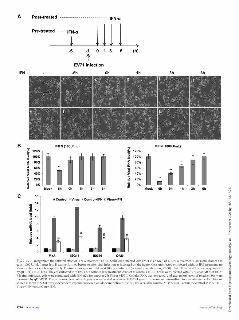
FIG 2 EV71 antagonized the antiviral effect of IFN-� treatment. (A) RD cells were infected with EV71 at an MOI of 1. IFN-� treatment (100 U/ml, frames c tog; or 1,000 U/ml, frames h to l) was performed before or after viral infection as indicated on the figure. Cells uninfected or infected without IFN treatment areshown in frames a or b, respectively. Photomicrographs were taken at 20 h postinfection (original magnification, �100). (B) Cellular viral loads were quantifiedby qRT-PCR at 20 h p.i. The cells infected with EV71 but without IFN treatment were set as controls. (C) RD cells were infected with EV71 at an MOI of 10. At9 h after infection, cells were stimulated with IFN-�2b for another 2 h (Virus�IFN). Cellular RNA was extracted, and expression levels of relative ISGs weremeasured by qRT-PCR. The expression level of each gene was calculated relative to GAPDH gene expression and normalized to mock-treated cells. Data areshown as mean � SD of three independent experiments; each was done in triplicate. *, P � 0.05, versus the control; **, P � 0.001, versus the control; #, P � 0.001,Virus�IFN versus Con�IFN.
3770 jvi.asm.org Journal of Virology
Dow
nloa
ded
from
http
s://j
ourn
als.
asm
.org
/jour
nal/j
vi o
n 19
Nov
embe
r 20
21 b
y 18
8.16
3.97
.22.
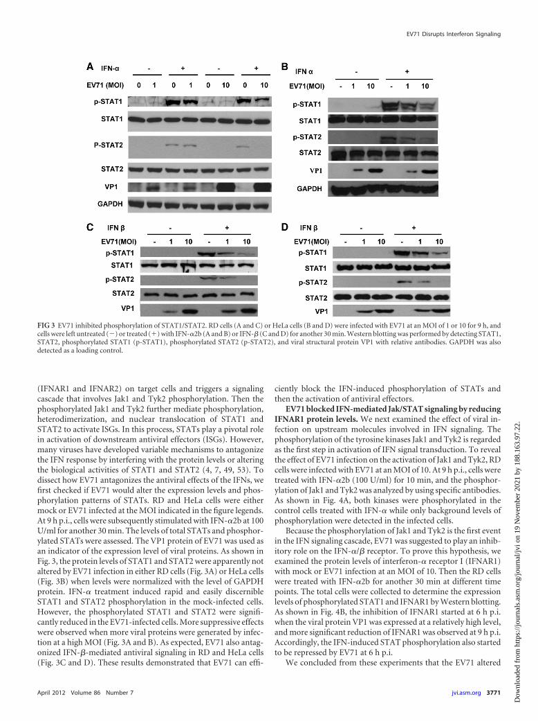
(IFNAR1 and IFNAR2) on target cells and triggers a signalingcascade that involves Jak1 and Tyk2 phosphorylation. Then thephosphorylated Jak1 and Tyk2 further mediate phosphorylation,heterodimerization, and nuclear translocation of STAT1 andSTAT2 to activate ISGs. In this process, STATs play a pivotal rolein activation of downstream antiviral effectors (ISGs). However,many viruses have developed variable mechanisms to antagonizethe IFN response by interfering with the protein levels or alteringthe biological activities of STAT1 and STAT2 (4, 7, 49, 53). Todissect how EV71 antagonizes the antiviral effects of the IFNs, wefirst checked if EV71 would alter the expression levels and phos-phorylation patterns of STATs. RD and HeLa cells were eithermock or EV71 infected at the MOI indicated in the figure legends.At 9 h p.i., cells were subsequently stimulated with IFN-�2b at 100U/ml for another 30 min. The levels of total STATs and phosphor-ylated STATs were assessed. The VP1 protein of EV71 was used asan indicator of the expression level of viral proteins. As shown inFig. 3, the protein levels of STAT1 and STAT2 were apparently notaltered by EV71 infection in either RD cells (Fig. 3A) or HeLa cells(Fig. 3B) when levels were normalized with the level of GAPDHprotein. IFN-� treatment induced rapid and easily discernibleSTAT1 and STAT2 phosphorylation in the mock-infected cells.However, the phosphorylated STAT1 and STAT2 were signifi-cantly reduced in the EV71-infected cells. More suppressive effectswere observed when more viral proteins were generated by infec-tion at a high MOI (Fig. 3A and B). As expected, EV71 also antag-onized IFN-�-mediated antiviral signaling in RD and HeLa cells(Fig. 3C and D). These results demonstrated that EV71 can effi-
ciently block the IFN-induced phosphorylation of STATs andthen the activation of antiviral effectors.
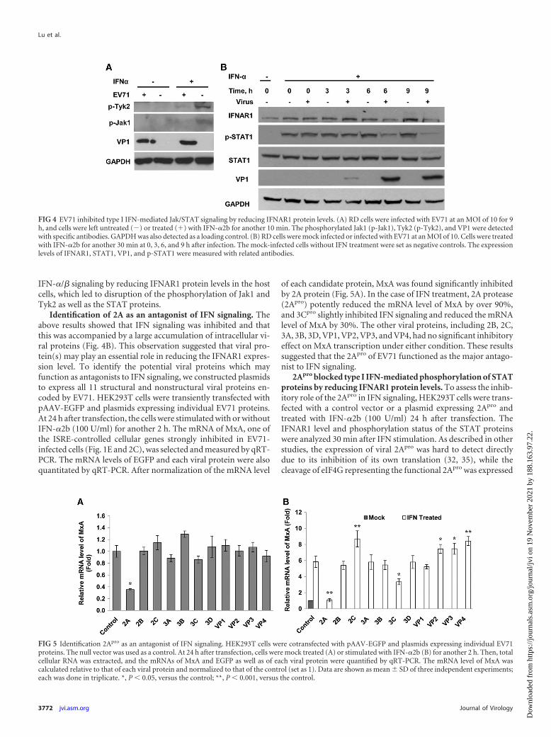
EV71 blocked IFN-mediated Jak/STAT signaling by reducingIFNAR1 protein levels. We next examined the effect of viral in-fection on upstream molecules involved in IFN signaling. Thephosphorylation of the tyrosine kinases Jak1 and Tyk2 is regardedas the first step in activation of IFN signal transduction. To revealthe effect of EV71 infection on the activation of Jak1 and Tyk2, RDcells were infected with EV71 at an MOI of 10. At 9 h p.i., cells weretreated with IFN-�2b (100 U/ml) for 10 min, and the phosphor-ylation of Jak1 and Tyk2 was analyzed by using specific antibodies.As shown in Fig. 4A, both kinases were phosphorylated in thecontrol cells treated with IFN-� while only background levels ofphosphorylation were detected in the infected cells.
Because the phosphorylation of Jak1 and Tyk2 is the first eventin the IFN signaling cascade, EV71 was suggested to play an inhib-itory role on the IFN-�/� receptor. To prove this hypothesis, weexamined the protein levels of interferon-� receptor I (IFNAR1)with mock or EV71 infection at an MOI of 10. Then the RD cellswere treated with IFN-�2b for another 30 min at different timepoints. The total cells were collected to determine the expressionlevels of phosphorylated STAT1 and IFNAR1 by Western blotting.As shown in Fig. 4B, the inhibition of IFNAR1 started at 6 h p.i.when the viral protein VP1 was expressed at a relatively high level,and more significant reduction of IFNAR1 was observed at 9 h p.i.Accordingly, the IFN-induced STAT phosphorylation also startedto be repressed by EV71 at 6 h p.i.
We concluded from these experiments that the EV71 altered
FIG 3 EV71 inhibited phosphorylation of STAT1/STAT2. RD cells (A and C) or HeLa cells (B and D) were infected with EV71 at an MOI of 1 or 10 for 9 h, andcells were left untreated (�) or treated (�) with IFN-�2b (A and B) or IFN-� (C and D) for another 30 min. Western blotting was performed by detecting STAT1,STAT2, phosphorylated STAT1 (p-STAT1), phosphorylated STAT2 (p-STAT2), and viral structural protein VP1 with relative antibodies. GAPDH was alsodetected as a loading control.
EV71 Disrupts Interferon Signaling
April 2012 Volume 86 Number 7 jvi.asm.org 3771
Dow
nloa
ded
from
http
s://j
ourn
als.
asm
.org
/jour
nal/j
vi o
n 19
Nov
embe
r 20
21 b
y 18
8.16
3.97
.22.
IFN-�/� signaling by reducing IFNAR1 protein levels in the hostcells, which led to disruption of the phosphorylation of Jak1 andTyk2 as well as the STAT proteins.
Identification of 2A as an antagonist of IFN signaling. Theabove results showed that IFN signaling was inhibited and thatthis was accompanied by a large accumulation of intracellular vi-ral proteins (Fig. 4B). This observation suggested that viral pro-tein(s) may play an essential role in reducing the IFNAR1 expres-sion level. To identify the potential viral proteins which mayfunction as antagonists to IFN signaling, we constructed plasmidsto express all 11 structural and nonstructural viral proteins en-coded by EV71. HEK293T cells were transiently transfected withpAAV-EGFP and plasmids expressing individual EV71 proteins.At 24 h after transfection, the cells were stimulated with or withoutIFN-�2b (100 U/ml) for another 2 h. The mRNA of MxA, one ofthe ISRE-controlled cellular genes strongly inhibited in EV71-infected cells (Fig. 1E and 2C), was selected and measured by qRT-PCR. The mRNA levels of EGFP and each viral protein were alsoquantitated by qRT-PCR. After normalization of the mRNA level
of each candidate protein, MxA was found significantly inhibitedby 2A protein (Fig. 5A). In the case of IFN treatment, 2A protease(2Apro) potently reduced the mRNA level of MxA by over 90%,and 3Cpro slightly inhibited IFN signaling and reduced the mRNAlevel of MxA by 30%. The other viral proteins, including 2B, 2C,3A, 3B, 3D, VP1, VP2, VP3, and VP4, had no significant inhibitoryeffect on MxA transcription under either condition. These resultssuggested that the 2Apro of EV71 functioned as the major antago-nist to IFN signaling.
2Apro blocked type I IFN-mediated phosphorylation of STATproteins by reducing IFNAR1 protein levels. To assess the inhib-itory role of the 2Apro in IFN signaling, HEK293T cells were trans-fected with a control vector or a plasmid expressing 2Apro andtreated with IFN-�2b (100 U/ml) 24 h after transfection. TheIFNAR1 level and phosphorylation status of the STAT proteinswere analyzed 30 min after IFN stimulation. As described in otherstudies, the expression of viral 2Apro was hard to detect directlydue to its inhibition of its own translation (32, 35), while thecleavage of eIF4G representing the functional 2Apro was expressed
FIG 4 EV71 inhibited type I IFN-mediated Jak/STAT signaling by reducing IFNAR1 protein levels. (A) RD cells were infected with EV71 at an MOI of 10 for 9h, and cells were left untreated (�) or treated (�) with IFN-�2b for another 10 min. The phosphorylated Jak1 (p-Jak1), Tyk2 (p-Tyk2), and VP1 were detectedwith specific antibodies. GAPDH was also detected as a loading control. (B) RD cells were mock infected or infected with EV71 at an MOI of 10. Cells were treatedwith IFN-�2b for another 30 min at 0, 3, 6, and 9 h after infection. The mock-infected cells without IFN treatment were set as negative controls. The expressionlevels of IFNAR1, STAT1, VP1, and p-STAT1 were measured with related antibodies.
FIG 5 Identification 2Apro as an antagonist of IFN signaling. HEK293T cells were cotransfected with pAAV-EGFP and plasmids expressing individual EV71proteins. The null vector was used as a control. At 24 h after transfection, cells were mock treated (A) or stimulated with IFN-�2b (B) for another 2 h. Then, totalcellular RNA was extracted, and the mRNAs of MxA and EGFP as well as of each viral protein were quantified by qRT-PCR. The mRNA level of MxA wascalculated relative to that of each viral protein and normalized to that of the control (set as 1). Data are shown as mean � SD of three independent experiments;each was done in triplicate. *, P � 0.05, versus the control; **, P � 0.001, versus the control.
Lu et al.
3772 jvi.asm.org Journal of Virology
Dow
nloa
ded
from
http
s://j
ourn
als.
asm
.org
/jour
nal/j
vi o
n 19
Nov
embe
r 20
21 b
y 18
8.16
3.97
.22.
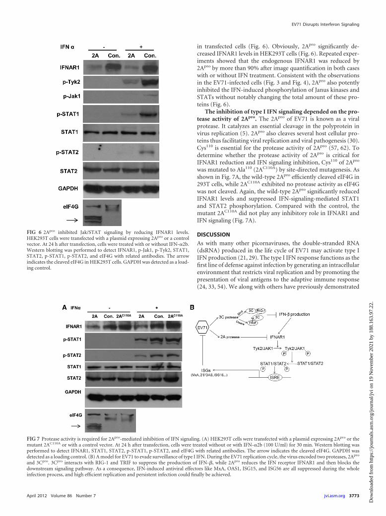
in transfected cells (Fig. 6). Obviously, 2Apro significantly de-creased IFNAR1 levels in HEK293T cells (Fig. 6). Repeated exper-iments showed that the endogenous IFNAR1 was reduced by2Apro by more than 90% after image quantification in both caseswith or without IFN treatment. Consistent with the observationsin the EV71-infected cells (Fig. 3 and Fig. 4), 2Apro also potentlyinhibited the IFN-induced phosphorylation of Janus kinases andSTATs without notably changing the total amount of these pro-teins (Fig. 6).
The inhibition of type I IFN signaling depended on the pro-tease activity of 2Apro. The 2Apro of EV71 is known as a viralprotease. It catalyzes an essential cleavage in the polyprotein invirus replication (5). 2Apro also cleaves several host cellular pro-teins thus facilitating viral replication and viral pathogenesis (30).Cys110 is essential for the protease activity of 2Apro (57, 62). Todetermine whether the protease activity of 2Apro is critical forIFNAR1 reduction and IFN signaling inhibition, Cys110 of 2Apro
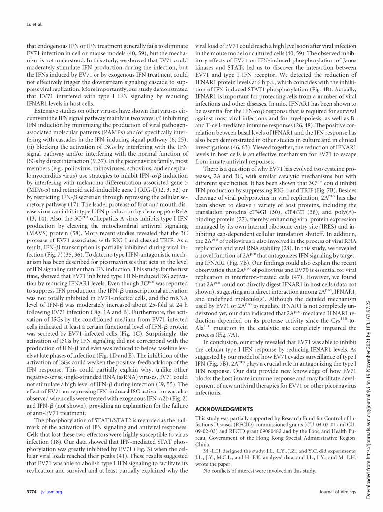
was mutated to Ala110 (2AC110A) by site-directed mutagenesis. Asshown in Fig. 7A, the wild-type 2Apro efficiently cleaved eIF4G in293T cells, while 2AC110A exhibited no protease activity as eIF4Gwas not cleaved. Again, the wild-type 2Apro significantly reducedIFNAR1 levels and suppressed IFN-signaling-mediated STAT1and STAT2 phosphorylation. Compared with the control, themutant 2AC110A did not play any inhibitory role in IFNAR1 andIFN signaling (Fig. 7A).
DISCUSSION
As with many other picornaviruses, the double-stranded RNA(dsRNA) produced in the life cycle of EV71 may activate type IIFN production (21, 29). The type I IFN response functions as thefirst line of defense against infection by generating an intracellularenvironment that restricts viral replication and by promoting thepresentation of viral antigens to the adaptive immune response(24, 33, 54). We along with others have previously demonstrated
FIG 6 2Apro inhibited Jak/STAT signaling by reducing IFNAR1 levels.HEK293T cells were transfected with a plasmid expressing 2Apro or a controlvector. At 24 h after transfection, cells were treated with or without IFN-�2b.Western blotting was performed to detect IFNAR1, p-Jak1, p-Tyk2, STAT1,STAT2, p-STAT1, p-STAT2, and eIF4G with related antibodies. The arrowindicates the cleaved eIF4G in HEK293T cells. GAPDH was detected as a load-ing control.
FIG 7 Protease activity is required for 2Apro-mediated inhibition of IFN signaling. (A) HEK293T cells were transfected with a plasmid expressing 2Apro or themutant 2AC110A or with a control vector. At 24 h after transfection, cells were treated without or with IFN-�2b (100 U/ml) for 30 min. Western blotting wasperformed to detect IFNAR1, STAT1, STAT2, p-STAT1, p-STAT2, and eIF4G with related antibodies. The arrow indicates the cleaved eIF4G. GAPDH wasdetected as a loading control. (B) A model for EV71 to evade surveillance of type I IFN. During the EV71 replication cycle, the virus encoded two proteases, 2Apro
and 3Cpro. 3Cpro interacts with RIG-1 and TRIF to suppress the production of IFN-�, while 2Apro reduces the IFN receptor IFNAR1 and then blocks thedownstream signaling pathway. As a consequence, IFN-induced antiviral effectors like MxA, OAS1, ISG15, and ISG56 are all suppressed during the wholeinfection process, and high efficient replication and persistent infection could finally be achieved.
EV71 Disrupts Interferon Signaling
April 2012 Volume 86 Number 7 jvi.asm.org 3773
Dow
nloa
ded
from
http
s://j
ourn
als.
asm
.org
/jour
nal/j
vi o
n 19
Nov
embe
r 20
21 b
y 18
8.16
3.97
.22.
that endogenous IFN or IFN treatment generally fails to eliminateEV71 infection in cell or mouse models (40, 59), but the mecha-nism is not understood. In this study, we showed that EV71 couldmoderately stimulate IFN production during the infection, butthe IFNs induced by EV71 or by exogenous IFN treatment couldnot effectively trigger the downstream signaling cascade to sup-press viral replication. More importantly, our study demonstratedthat EV71 interfered with type I IFN signaling by reducingIFNAR1 levels in host cells.
Extensive studies on other viruses have shown that viruses cir-cumvent the IFN signal pathway mainly in two ways: (i) inhibitingIFN induction by minimizing the production of viral pathogen-associated molecular patterns (PAMPs) and/or specifically inter-fering with cascades in the IFN-inducing signal pathway (6, 25);(ii) blocking the activation of ISGs by interfering with the IFNsignal pathway and/or interfering with the normal function ofISGs by direct interaction (9, 37). In the picornavirus family, mostmembers (e.g., poliovirus, rhinoviruses, echovirus, and encepha-lomyocarditis virus) use strategies to inhibit IFN-�/� inductionby interfering with melanoma differentiation-associated gene 5(MDA-5) and retinoid acid-inducible gene I (RIG-I) (2, 3, 52) orby restricting IFN-� secretion through repressing the cellular se-cretory pathway (17). The leader protease of foot and mouth dis-ease virus can inhibit type I IFN production by cleaving p65-RelA(13, 14). Also, the 3Cpro of hepatitis A virus inhibits type I IFNproduction by cleaving the mitochondrial antiviral signaling(MAVS) protein (58). More recent studies revealed that the 3Cprotease of EV71 associated with RIG-I and cleaved TRIF. As aresult, IFN-� transcription is partially inhibited during viral in-fection (Fig. 7) (35, 36). To date, no type I IFN-antagonistic mech-anism has been described for picornaviruses that acts on the levelof IFN signaling rather than IFN induction. This study, for the firsttime, showed that EV71 inhibited type I IFN-induced ISG activa-tion by reducing IFNAR1 levels. Even though 3Cpro was reportedto suppress IFN production, the IFN-� transcriptional activationwas not totally inhibited in EV71-infected cells, and the mRNAlevel of IFN-� was moderately increased about 25-fold at 24 hfollowing EV71 infection (Fig. 1A and B). Furthermore, the acti-vation of ISGs by the conditioned medium from EV71-infectedcells indicated at least a certain functional level of IFN-� proteinwas secreted by EV71-infected cells (Fig. 1C). Surprisingly, theactivation of ISGs by IFN signaling did not correspond with theproduction of IFN-� and even was reduced to below baseline lev-els at late phases of infection (Fig. 1D and E). The inhibition of theactivation of ISGs could weaken the positive-feedback loop of theIFN response. This could partially explain why, unlike othernegative-sense single-stranded RNA (ssRNA) viruses, EV71 couldnot stimulate a high level of IFN-� during infection (29, 55). Theeffect of EV71 on repressing IFN-induced ISG activation was alsoobserved when cells were treated with exogenous IFN-�2b (Fig. 2)and IFN-� (not shown), providing an explanation for the failureof anti-EV71 treatment.
The phosphorylation of STAT1/STAT2 is regarded as the hall-mark of the activation of IFN signaling and antiviral responses.Cells that lost these two effectors were highly susceptible to virusinfection (18). Our data showed that IFN-mediated STAT phos-phorylation was greatly inhibited by EV71 (Fig. 3) when the cel-lular viral loads reached their peaks (41). These results suggestedthat EV71 was able to abolish type I IFN signaling to facilitate itsreplication and survival and at least partially explained why the
viral load of EV71 could reach a high level soon after viral infectionin the mouse model or cultured cells (40, 59). The observed inhib-itory effects of EV71 on IFN-induced phosphorylation of Januskinases and STATs led us to discover the interaction betweenEV71 and type I IFN receptor. We detected the reduction ofIFNAR1 protein levels at 6 h p.i., which coincides with the inhibi-tion of IFN-induced STAT1 phosphorylation (Fig. 4B). Actually,IFNAR1 is important for protecting cells from a number of viralinfections and other diseases. In mice IFNAR1 has been shown tobe essential for the IFN-�/� response that is required for survivalagainst most viral infections and for myelopoiesis, as well as B-and T-cell-mediated immune responses (26, 48). The positive cor-relation between basal levels of IFNAR1 and the IFN response hasalso been demonstrated in other studies in culture and in clinicalinvestigations (46, 63). Viewed together, the reduction of IFNAR1levels in host cells is an effective mechanism for EV71 to escapefrom innate antiviral responses.
There is a question of why EV71 has evolved two cysteine pro-teases, 2A and 3C, with similar catalytic mechanisms but withdifferent specificities. It has been shown that 3Cpro could inhibitIFN production by suppressing RIG-1 and TRIF (Fig. 7B). Besidescleavage of viral polyproteins in viral replication, 2Apro has alsobeen shown to cleave a variety of host proteins, including thetranslation proteins eIF4GI (30), eIF4GII (38), and poly(A)-binding protein (27), thereby enhancing viral protein expressionmanaged by its own internal ribosome entry site (IRES) and in-hibiting cap-dependent cellular translation shutoff. In addition,the 2Apro of poliovirus is also involved in the process of viral RNAreplication and viral RNA stability (28). In this study, we revealeda novel function of 2Apro that antagonizes IFN signaling by target-ing IFNAR1 (Fig. 7B). Our findings could also explain the recentobservation that 2Apro of poliovirus and EV70 is essential for viralreplication in interferon-treated cells (47). However, we foundthat 2Apro could not directly digest IFNAR1 in host cells (data notshown), suggesting an indirect interaction among 2Apro, IFNAR1,and undefined molecule(s). Although the detailed mechanismused by EV71 or 2Apro to regulate IFNAR1 is not completely un-derstood yet, our data indicated that 2Apro-mediated IFNAR1 re-duction depended on its protease activity since the Cys110-to-Ala110 mutation in the catalytic site completely impaired theprocess (Fig. 7A).
In conclusion, our study revealed that EV71 was able to inhibitthe cellular type I IFN response by reducing IFNAR1 levels. Assuggested by our model of how EV71 evades surveillance of type IIFN (Fig. 7B), 2Apro plays a crucial role in antagonizing the type IIFN response. Our data provide new knowledge of how EV71blocks the host innate immune response and may facilitate devel-opment of new antiviral therapies for EV71 or other picornavirusinfections.
ACKNOWLEDGMENTS
This study was partially supported by Research Fund for Control of In-fectious Diseases (RFCID)-commissioned grants (CU-09-02-01 and CU-09-02-03) and RFCID grant 09080482 and by the Food and Health Bu-reau, Government of the Hong Kong Special Administrative Region,China.
M.-L.H. designed the study; J.L., L.Y., J.Z., and Y.C. did experiments;J.L., J.Y., M.C.L., and H.-F.K. analyzed data; and J.L., L.Y., and M.-L.H.wrote the paper.
No conflicts of interest were involved in this study.
Lu et al.
3774 jvi.asm.org Journal of Virology
Dow
nloa
ded
from
http
s://j
ourn
als.
asm
.org
/jour
nal/j
vi o
n 19
Nov
embe
r 20
21 b
y 18
8.16
3.97
.22.
REFERENCES1. AbuBakar S, et al. 1999. Identification of enterovirus 71 isolates from an
outbreak of hand, foot and mouth disease (HFMD) with fatal cases ofencephalomyelitis in Malaysia. Virus Res. 61:1–9.
2. Barral PM, et al. 2007. MDA-5 is cleaved in poliovirus-infected cells. J.Virol. 81:3677–3684.
3. Barral PM, Sarkar D, Fisher PB, Racaniello VR. 2009. RIG-I is cleavedduring picornavirus infection. Virology 391:171–176.
4. Brzozka K, Finke S, Conzelmann KK. 2006. Inhibition of interferonsignaling by rabies virus phosphoprotein P: activation-dependent bindingof STAT1 and STAT2. J. Virol. 80:2675–2683.
5. Buenz EJ, Howe CL. 2006. Picornaviruses and cell death. Trends Micro-biol. 14:28 –36.
6. Cassady KA. 2005. Human cytomegalovirus TRS1 and IRS1 gene prod-ucts block the double-stranded-RNA-activated host protein shutoff re-sponse induced by herpes simplex virus type 1 infection. J. Virol. 79:8707–8715.
7. Chee AV, Roizman B. 2004. Herpes simplex virus 1 gene products oc-clude the interferon signaling pathway at multiple sites. J. Virol. 78:4185–4196.
8. Chehadeh W, Abdulkareem HA. 2010. Difference in susceptibility toMxA protein between a coxsackievirus B1 isolate and prototype, impact ofserial cell culture passage. J. Med. Virol. 82:424 – 432.
9. Christen V, et al. 2007. Inhibition of alpha interferon signaling by hepa-titis B virus. J. Virol. 81:159 –165.
10. Chu CJ, Lee SD. 2008. Hepatitis B virus/hepatitis C virus coinfection:epidemiology, clinical features, viral interactions and treatment. J. Gastro-enterol. Hepatol. 23:512–520.
11. Cleary CM, Donnelly RJ, Soh J, Mariano TM, Pestka S. 1994. Knockoutand reconstitution of a functional human type I interferon receptor com-plex. J. Biol. Chem. 269:18747–18749.
12. Craxi A, Antonucci G, Camma C. 2006. Treatment options in HBV. J.Hepatol. 44:S77–S83.
13. de Los Santos T, de Avila Botton S, Weiblen R, Grubman MJ. 2006. Theleader proteinase of foot-and-mouth disease virus inhibits the inductionof beta interferon mRNA and blocks the host innate immune response. J.Virol. 80:1906 –1914.
14. de Los Santos T, Diaz-San Segundo F, Grubman MJ. 2007. Degradationof nuclear factor kappa B during foot-and-mouth disease virus infection.J. Virol. 81:12803–12815.
15. Der SD, Zhou A, Williams BR, Silverman RH. 1998. Identification ofgenes differentially regulated by interferon alpha, beta, or gamma usingoligonucleotide arrays. Proc. Natl. Acad. Sci. U. S. A. 95:15623–15628.
16. de Veer MJ, et al. 2001. Functional classification of interferon-stimulatedgenes identified using microarrays. J. Leukoc. Biol. 69:912–920.
17. Dodd DA, Giddings TH, Jr, Kirkegaard K. 2001. Poliovirus 3A proteinlimits interleukin-6 (IL-6), IL-8, and beta interferon secretion during viralinfection. J. Virol. 75:8158 – 8165.
18. Dupuis S, et al. 2003. Impaired response to interferon-alpha/beta andlethal viral disease in human STAT1 deficiency. Nat. Genet. 33:388 –391.
19. Fowlkes AL, et al. 2008. Enterovirus-associated encephalitis in the Cali-fornia encephalitis project, 1998 –2005. J. Infect. Dis. 198:1685–1691.
20. Fu XY, Kessler DS, Veals SA, Levy DE, and Darnell JE, Jr. 1990. ISGF3,the transcriptional activator induced by interferon alpha, consists of mul-tiple interacting polypeptide chains. Proc. Natl. Acad. Sci. U. S. A. 87:8555– 8559.
21. Gitlin L, et al. 2006. Essential role of mda-5 in type I IFN responses topolyriboinosinic:polyribocytidylic acid and encephalomyocarditis picor-navirus. Proc. Natl. Acad. Sci. U. S. A. 103:8459 – 8464.
22. Hamaguchi T, et al. 2008. Acute encephalitis caused by intrafamilialtransmission of enterovirus 71 in adult. Emerg. Infect. Dis. 14:828 – 830.
23. Heim MH. 1999. The Jak-STAT pathway: cytokine signalling from thereceptor to the nucleus. J. Recept. Signal Transduct. Res. 19:75–120.
24. Honda K, et al. 2003. Selective contribution of IFN-alpha/beta signalingto the maturation of dendritic cells induced by double-stranded RNA orviral infection. Proc. Natl. Acad. Sci. U. S. A. 100:10872–10877.
25. Hornung V, et al. 2006. 5=-Triphosphate RNA is the ligand for RIG-I.Science 314:994 –997.
26. Hwang SY, et al. 1995. A null mutation in the gene encoding a type Iinterferon receptor component eliminates antiproliferative and antiviral
responses to interferons alpha and beta and alters macrophage responses.Proc. Natl. Acad. Sci. U. S. A. 92:11284 –11288.
27. Joachims M, Van Breugel PC, Lloyd RE. 1999. Cleavage of poly(A)-binding protein by enterovirus proteases concurrent with inhibition oftranslation in vitro. J. Virol. 73:718 –727.
28. Jurgens CK, et al. 2006. 2Apro is a multifunctional protein that regulatesthe stability, translation and replication of poliovirus RNA. Virology 345:346 –357.
29. Kato H, et al. 2006. Differential roles of MDA5 and RIG-I helicases in therecognition of RNA viruses. Nature 441:101–105.
30. Kempf BJ, Barton DJ. 2008. Poliovirus 2APro increases viral mRNA andpolysome stability coordinately in time with cleavage of eIF4G. J. Virol.82:5847–5859.
31. Kessler DS, Levy DE, and Darnell JE, Jr. 1988. Two interferon-inducednuclear factors bind a single promoter element in interferon-stimulatedgenes. Proc. Natl. Acad. Sci. U. S. A. 85:8521– 8525.
32. Kuo RL, Kung SH, Hsu YY, Liu WT. 2002. Infection with enterovirus 71or expression of its 2A protease induces apoptotic cell death. J. Gen. Virol.83:1367–1376.
33. Le Bon A, Tough DF. 2002. Links between innate and adaptive immunityvia type I interferon. Curr. Opin. Immunol. 14:432– 436.
34. Lee SH, Vidal SM. 2002. Functional diversity of Mx proteins: variationson a theme of host resistance to infection. Genome Res. 12:527–530.
35. Lei X, et al. 2010. The 3C protein of enterovirus 71 inhibits retinoidacid-inducible gene I-mediated interferon regulatory factor 3 activationand type I interferon responses. J. Virol. 84:8051– 8061.
36. Lei X, et al. 2011. Cleavage of the adaptor protein TRIF by enterovirus 713C inhibits antiviral responses mediated by Toll-like receptor 3. J. Virol.85:8811– 8818.
37. Li Q, Means R, Lang S, Jung JU. 2007. Downregulation of gammainterferon receptor 1 by Kaposi’s sarcoma-associated herpesvirus K3 andK5. J. Virol. 81:2117–2127.
38. Liebig HD, Seipelt J, Vassilieva E, Gradi A, Kuechler E. 2002. A ther-mosensitive mutant of HRV2 2A proteinase: evidence for direct cleavageof eIF4GI and eIF4GII. FEBS Lett. 523:53–57.
39. Lin JY, et al. 2009. Viral and host proteins involved in picornavirus lifecycle. J. Biomed. Sci. 16:103.
40. Liu ML, et al. 2005. Type I interferons protect mice against enterovirus 71infection. J. Gen. Virol. 86:3263–3269.
41. Lu J, et al. Viral kinetics of Enterovirus 71 in human abdomyosarcomacells. World J. Gastroenterol. 17:4135– 4142.
42. Lum LC, et al. 1998. Fatal enterovirus 71 encephalomyelitis. J. Pediatr.133:795–798.
43. Ma Y, et al. 2009. Glucose-regulated protein 78 is an intracellular antiviralfactor against hepatitis B virus. Mol. Cell Proteomics 8:2582–2594.
44. Manki A, Oda M, Seino Y. 1997. Neurologic diseases of enterovirusinfections: polioviruses, coxsackieviruses, echoviruses, and enterovirusestype 68 –72. Nippon Rinsho 55:849 – 854.
45. McMinn PC. 2002. An overview of the evolution of enterovirus 71 andits clinical and public health significance. FEMS Microbiol. Rev. 26:91–107.
46. Morita K, et al. 1998. Expression of interferon receptor genes (IFNAR1and IFNAR2 mRNA) in the liver may predict outcome after interferontherapy in patients with chronic genotype 2a or 2b hepatitis C virus infec-tion. J. Clin. Gastroenterol. 26:135–140.
47. Morrison JM, Racaniello VR. 2009. Proteinase 2Apro is essential forenterovirus replication in type I interferon-treated cells. J. Virol. 83:4412–4422.
48. Muller U, et al. 1994. Functional role of type I and type II interferons inantiviral defense. Science 264:1918 –1921.
49. Nanda SK, Baron MD. 2006. Rinderpest virus blocks type I and type IIinterferon action: role of structural and nonstructural proteins. J. Virol.80:7555–7568.
50. Novick D, Cohen B, Rubinstein M. 1994. The human interferon alpha/beta receptor: characterization and molecular cloning. Cell 77:391– 400.
51. Palumbo E. 2009. PEG-interferon in acute and chronic hepatitis C: areview. Am. J. Ther. 16:573–578.
52. Papon L, et al. 2009. The viral RNA recognition sensor RIG-I is degradedduring encephalomyocarditis virus (EMCV) infection. Virology 393:311–318.
53. Ulane CM, et al. 2005. Composition and assembly of STAT-targetingubiquitin ligase complexes: paramyxovirus V protein carboxyl terminus isan oligomerization domain. J. Virol. 79:10180 –10189.
EV71 Disrupts Interferon Signaling
April 2012 Volume 86 Number 7 jvi.asm.org 3775
Dow
nloa
ded
from
http
s://j
ourn
als.
asm
.org
/jour
nal/j
vi o
n 19
Nov
embe
r 20
21 b
y 18
8.16
3.97
.22.

54. Vollstedt S, et al. 2004. Interplay between alpha/beta and gamma inter-ferons with B, T, and natural killer cells in the defense against herpessimplex virus type 1. J. Virol. 78:3846 –3850.
55. Wang J, et al. 2010. NF-kappa B RelA subunit is crucial for early IFN-betaexpression and resistance to RNA virus replication. J. Immunol. 185:1720 –1729.
56. Wong SS, Yip CC, Lau SK, Yuen KY. 2011. Human enterovirus 71 andhand, foot and mouth disease. Epidemiol. Infect. 138:1071–1089.
57. Yang CH, et al. 2010. Enterovirus type 71 2A protease functions as atranscriptional activator in yeast. J. Biomed. Sci. 17:65.
58. Yang Y, et al. 2007. Disruption of innate immunity due to mitochondrialtargeting of a picornaviral protease precursor. Proc. Natl. Acad. Sci.U. S. A. 104:7253–7258.
59. Yi L, He Y, Chen Y, Kung HF, He ML. 2011. Potent inhibition of humanenterovirus 71 replication by type I interferon subtypes. Antivir. Ther.16:51–58.
60. Yi L, Lu J, Kung HF, He ML. 2011. The virology and developments towardcontrol of human enterovirus 71. Crit. Rev. Microbiol. 37:313–327.
61. Yoneyama M, et al. 2005. Shared and unique functions of the DExD/H-box helicases RIG-I, MDA5, and LGP2 in antiviral innate immunity. J.Immunol. 175:2851–2858.
62. Yu SF, Lloyd RE. 1992. Characterization of the roles of conserved cysteineand histidine residues in poliovirus 2A protease. Virology 186:725–735.
63. Zurney J, Howard KE, Sherry B. 2007. Basal expression levels of IFNARand Jak-STAT components are determinants of cell-type-specific differ-ences in cardiac antiviral responses. J. Virol. 81:13668 –13680.
Lu et al.
3776 jvi.asm.org Journal of Virology
Dow
nloa
ded
from
http
s://j
ourn
als.
asm
.org
/jour
nal/j
vi o
n 19
Nov
embe
r 20
21 b
y 18
8.16
3.97
.22.