enrichment of human esc-derived multipotent mesenchymal stem cells with immunosuppressive and...
TRANSCRIPT

REGENERATIVE MEDICINE
Enrichment of Human ESC-Derived Multipotent Mesenchymal Stem
Cells with Immunosuppressive and Anti-Inflammatory Properties
Capable to Protect Against Experimental Inflammatory Bowel Disease
LAURA SANCHEZ,a IVAN GUTIERREZ-ARANDA,a GERTRUDIS LIGERO,a RUTH RUBIO,a MARTIN MUNOZ-LOPEZ,a
JOSE L. GARCIA-PEREZ,aVERONICA RAMOS,
aPEDRO. J. REAL,
aCLARA BUENO,
aRENE RODRIGUEZ,
a
MARIO DELGADO,b PABLO MENENDEZa
aAndalusian Stem Cell Bank, Centro de Investigacion Biomedica, CSJA-UGR, Granada, Spain;bInstituto de Parasitologıa y Biomedicina Lopez Neyra, CSIC, Granada, Spain
Key Words. hESC • iPS cells • MSCs • SMAD-2/3 • Inflammatory bowel disease
ABSTRACT
Human ESCs provide access to the earliest stages ofhuman development and may serve as an unlimited source
of functional cells for future cell therapies. The optimiza-tion of methods directing the differentiation of human em-bryonic stem cells (hESCs) into tissue-specific precursors
becomes crucial. We report an efficient enrichment of mes-enchymal stem cells (MSCs) from hESCs through specific
inhibition of SMAD-2/3 signaling. Human ESC-derivedMSCs (hESC-MSCs) emerged as a population of fibroblas-toid cells expressing a MSC phenotype: CD731 CD901CD1051 CD441 CD1661 CD452 CD342 CD142 CD192human leucocyte antigen-DR (HLA-DR)2. After 28 days of
SMAD-2/3 inhibition, hESC cultures were enriched (>42%)in multipotent MSCs. CD731CD901 hESC-MSCs were flu-orescence activated cell sorting (FACS)-isolated and long-
term cultures were established and maintained for many
passages displaying a faster growth than somatic tissue-derived MSCs while maintaining MSC morphology and
phenotype. They displayed osteogenic, adipogenic, andchondrocytic differentiation potential and exhibitedpotent immunosuppressive and anti-inflammatory prop-
erties in vitro and in vivo, where hESC-MSCs were capa-ble of protecting against an experimental model of
inflammatory bowel disease. Interestingly, the efficientenrichment of hESCs into MSCs through inhibition ofSMAD-2/3 signaling was not reproducible with distinct
induced pluripotent stem cell lines. Our findings providemechanistic insights into the differentiation of hESCs
into immunosuppressive and anti-inflammatory multipo-tent MSCs with potential future clinical applications.STEM CELLS 2011;29:251–262
Disclosure of potential conflicts of interest is found at the end of this article.
INTRODUCTION
Human embryonic stem cells (hESCs) offer great promise forcell replacement strategies. Recent advances in somatic cellreprogramming to induced pluripotent stem (iPS) cells haveopened new avenues to generate donor-specific cells for re-generative medicine and disease modeling [1]. Human ESCsalso provide access to the earliest stages of human develop-ment and may serve as an unlimited source of functional cellsfor future cell therapies [2, 3]. However, reproducible directeddifferentiation of hESCs and iPS cells into specific cell typesposes a formidable challenge. Thus, optimization of methodsaimed at directing the differentiation of hESCs into tissue-specific precursors becomes crucial.
Mesenchymal stem cells (MSCs) may be isolated from avariety of adult somatic tissues and they may differentiateinto multiple mesodermal tissues [4–7]. Their multilineagedifferentiation potential (bone, cartilage, fat) coupled to theirimmunoprivileged properties is being exploited worldwide forboth autologous and allogeneic cell replacement strategies [8–12]. Recently, MSCs have been derived from hESC throughcoculturing with the OP9 murine bone marrow stromal cellline [13, 14]. However, there is no information available aboutthe mechanistic insights involved in the specific differentiationof hESCs into functional MSCs. Furthermore, whether hESCsand iPS cells are equally capable of generating MSCs remainto be assessed. Also, little is known about the immunologicalproperties of hESC-derived MSCs. Their immunotoleranceproperties in vitro have been partially reported [14] but no
Author contributions: L.S., I.G.-A., G.L., R. Rubio, M.M.-L., J.L.G.-P., V.R., P.J.R., C.B., and R. Rodrıguez: collection and assembly ofdata, design and performance of experiments, data analysis and interpretation; J.L.G.-P., M. D., and P.M.: data analysis andinterpretation and financial support; M.D.: conception and design, performance of experiments, data analysis, manuscript writing; P.M.:conception and design, data analysis, supervision of study, manuscript writing. L.S., I.G.-A., and G.L. contributed equally to this work.
Correspondence: Pablo Menendez, Ph.D., M.B.A., Andalusian Stem Cell Bank, Avda del Conocimiento, Granada 18100, Spain.Telephone: 34-958-894-672; Fax: 34-958-894-652; e-mail: [email protected]; or Mario Delgado, Ph.D., Instituto deParasitologıa y Biomedicina Lopez Neyra, Avda del Conocimiento, Granada 18100, Spain. Telephone: 34-958-181-665; Fax:34-958-181-632; e-mail: [email protected] Received September 14, 2010; accepted for publication November 12, 2010; first pub-lished online in STEM CELLS EXPRESS November 23, 2010. VC AlphaMed Press 1066-5099/2009/$30.00/0 doi: 10.1002/stem.569
STEM CELLS 2011;29:251–262 www.StemCells.com

information is available about their potential immunotoleranceand anti-inflammatory properties in vivo.
The transforming growth factor (TGF-b) signaling throughSMAD-2/3 downstream effectors has an increasing interest inregenerative medicine and lineage specification during humanembryonic development [15, 16]. In contrast to specific bonemorphogenic proteins (BMPs), which are known to play animportant role in directing cell fate decisions toward meso-derm and further differentiation into MSCs, the mesodermaleffects of TGF-b signaling in human embryonic developmentis controversial [15, 16]. We previously reported that specificinhibition of TGF-b signals through SMAD-2/3 results in lossof the hESC phenotype and pluripotency through induction ofhESC differentiation, while not affecting survival or self-renewal of hESCs [17]. Furthermore, TGF-b is considered apotent inhibitor of the hematopoiesis, a mesodermal tissue[18–20]. In fact, TGF-b1�/� mice display an enhancedmyelopoiesis and an increased stem/progenitor cell (HSPC)compartment. These HSPCs from TGF-b1�/� mice have acompetitive repopulation advantage in vivo [18–20]. We, there-fore, hypothesized that similar to what occurs in the hemato-poietic system, TGF-b signaling through SMAD-2/3 might bea negative regulator in the MSC generation from hESCs. We,thus, studied the role of SMAD-2/3 inhibition in the potentialenrichment of functional and multipotent MSCs from hESCsand iPS cells. We report a robust and efficient enrichment ofMSCs from hESCs through specific inhibition of the SMAD-2/3 pathway. These hESC-derived MSCs display multilineagedifferentiation potential and exhibited potent immunosuppres-sive and anti-inflammatory properties in vitro and in vivo,which are capable to protect against experimental inflammatorybowel disease. Finally, our data reveals possible mechanisticdifferences in MSC generation from hESCs versus iPS cells.
MATERIALS AND METHODS
Human ESC and iPS Cell Culture
Human ESC lines (H9, AND-1, AND-2, and SHEF-1) and iPScells (iPS-CB-CD34#2, iAND-4, iMSUH001) were maintainedundifferentiated in a feeder-free culture as previously described[21–25]. These iPS cell lines were derived from fetal humanfibroblasts using a lentiviral construct that express OCT4, SOX2,KLF4, and c-MYC. Briefly, hESCs were cultured in Matrigel(Becton Dickinson, San Jose, CA)-coated T25 flasks in conditionedmedium supplemented with 8 ng/ml of basic fibroblast growth fac-tor (bFGF) (Miltenyi, Madrid, Spain). Media was changed daily,and the cells were split weekly by dissociation with 200 U/ml ofcollagenase IV (Invitrogen, Edinburgh, Scotland). Distinct hESCand iPS cultures were treated daily with 10 ng of TGF-b1 (R&DSystems, London, U.K.), 10 lM of SB-431542 (Sigma-Aldrich, StLouise, MO), a potent and selective chemical inhibitor of ALK4,ALK5, and ALK7 receptors inducing shutdown of TGF-b signal-ing through SMAD-2/3, or dimethylsulfoxide (DMSO) as vehiclefor 28 (hESCs) or 35 (iPS cells) days. Human ESC and iPS cellcultures were visualized daily by phase-contrast microscopy.Approval from the Spanish National Embryo Ethical Committeewas obtained to work with pluripotent cells.
Flow Cytometry Characterization of hESCand iPS-Derived MSCs
Flow cytometry analysis was carried out weekly (day 7, 14,21, 28, and 35) to analyze the potential emergence ofCD73þCD90þCD34� MSCs [6, 26]. Briefly, hESC cultureswere dissociated with trypsin-EDTA and the single cell suspen-sion was stained at a concentration of 2–5 � 105 cells per millili-ter with a fluorescein isothyocianate (FITC)-conjugated anti-
CD90 and phyco-erithrin (PE)-conjugated anti-CD73 monoclonalantibodies (Becton Dickinson, San Jose, CA). Then, the cellswere washed and stained with 7-actino (7-AAD) for 15 minutesat room temperature. Live cells identified by 7-AAD exclusionwere analyzed for coexpression of CD90 and CD73 using aFACSCanto II flow cytometer equipped with the fluoresence acti-vated cell sorting (FACS) Diva analysis software (Becton Dickin-son). In parallel, the expression of the ESC-specific markers Tra-1-60-FITC (Chemicon, San Diego, CA) and Oct3/4-PE (Pharmingen,San Jose, CA) and the hematoendothelial marker CD34 (BectonDickinson) was determined by flow cytometry. Irrelevant IgG iso-type-matched antibodies were consistently used [27].
Western Blotting
The potent and specific effect of the SB-431542 chemical inhibi-tor in blocking signaling through SMAD2/3 but not via SMAD1/5/8 was confirmed as described [28]. Briefly, hESCs and iPScells were treated for 60 minutes with or without SB-431542 orTGF-b1 and cell lysates (40 lg) were prepared from equivalentcell numbers. Cells were washed and lysed using the nuclearextraction kit with protease and phosphatase inhibitors (ActiveMotif). Samples were then run on 10% SDS-polyacrylamide gel(PAGE) and transferred to polivinylidene fluoride (PVDF) mem-branes using a semidry transfer apparatus at 15 V for 1 hour.Membranes were blocked with 5% bovine serum albumin (BSA)in tris buffer saline tween-20 (TBST) and incubated with thefollowing primary antibodies: rabbit anti-phospho-SMAD2/3, rabbitanti-phospho-SMAD1/5/8 or rabbit anti-total SMAD-2/3, andrabbit anti-total SMAD-1/5/8 (all from Cell Signaling Technolo-gies, Danvers, MA 1:10,000). All secondary antibodies were conju-gated to horse radish peroxidase (HRP) and consequently detectedusing the enhanced chemiluminescence system (ECL; Pierce,Rockford, IL). The ECL signal was detected using an imaging anddetection station (Alpha-Innotech Corp, San Diego, CA).
Cell Sorting and Establishmentof hESC-MSC Cultures
Differentiating hESCs were incubated with CD73-PE and CD90-FITC and the double-positive CD73þCD90þ population waspurified with the FACSAria cell sorter (Becton Dickinson) usingthe automatic cell deposition unit. A total of 2–3 � 105 sortedcells were immediately plated back into fibronectin-coated platesto facilitate adherence. After 24 hours, nonadherent cells werewashed away and fresh prewarmed medium was added. HumanESC-derived CD73þCD90þ cells were cultured in Advance-Dulbecco’s modified Eagle’s medium (DMEM) with 10% fetalcalf serum (FCS), 1% Glutamax, and 1% penicillin/streptomycin(all from Gibco, Edinburgh, Scotland) and were allowed toexpand and reach nearly 100% confluence.
Phenotypic Characterization of hESC-MSCs
The immunophenotype of cultured hESC-MSCs was determinedby flow cytometry as previously described [29]. Briefly, hESC-MSCs were trypsinized, washed, and suspended in PBS þ 1%BSA. A total of 2 � 105 cells were incubated for 30 minutes inthe darkness with the following FITC- or PE-conjugated monoclo-nal antibodies: CD73, CD90, CD105, CD44, CD166, CD106,CD45, CD34, CD14, CD19, human leucocyte antigen-DR (HLA-DR), CD40, CD80 (Becton Dickinson), and SSEA-4 (Chemicon,Billerica, MA). Cells were then washed in PBS þ 1% BSA andanalyzed in a FACSCanto II flow cytometer.
In Vitro Differentiation of hESC-MSCs
Human ESC-MSCs were seeded at 1 � 104 cells per centimetersquare in Advance-DMEM with 10% FCS, 1% Glutamax, and1% Pen/Strep and were allowed to expand and reach nearly100% confluence. Culture medium was then replaced with spe-cific differentiation inductive medium. For adipogenic differentia-tion, cells were cultured in Adipogenic MSCs Differentiation Bul-let Kit (Lonza, Basel, Switzerland) for 2 weeks. Differentiated
252 hESC-Derived Multipotent Functional MSCs
cell cultures were stained with Oil Red (Sigma, Madrid, Spain).For osteogenic differentiation, cells were cultured in the Osteo-genic MSCs Differentiation Bullet Kit (Lonza) for 2 weeks. Dif-ferentiated cultures were stained with Alizarin Red (Sigma) [29].
Assessment of T-Lymphocyte Proliferation Responseto MSCs and Allogeneic Stimulation
Mixed lymphocyte cultures (MLCs) were performed in 96-wellround-bottom plates by stimulating 105 responder peripheralblood mononuclear cells (PBMCs) from donor A with 105 alloge-neic HLA-mismatched mitomycin C-treated stimulator PBMCsfrom donor B in 200 ll complete medium in the presence or ab-sence of different numbers of adipose-derived MSCs (Ad-MSCs),cord blood-derived MSCs (CB-MSCs), or hESC-MSCs. PBMCswere isolated from buffy coat preparations derived from the wholeblood of healthy volunteers by density sedimentation on Ficoll-Hypaque gradients (20 minutes, 600g). Cells recovered from thegradient interface were washed twice in RPMI medium and imme-diately used for culture. Human Ad-MSCs and CB-MSCs werepurchased from InbioBank (Inbiomed, San Sebastian, Spain) [26,30]. These MSCs were expanded in DMEM containing 10% fetalbovine serum (FBS), 2 mM glutamine and 1% pen/strept at 37�C,and 5% CO2.
Cells were pulsed with 5 microcurie per well [3H]-thymidinefor the last 12 hours of the culture, harvested onto membranes,and proliferation was determined by measuring [3H]-thymidineuptake in a liquid scintillation counter. After 48 hours, cytokinedeterminations for interleukin (IL)-2, IL-4, TGF-b, tumor necrosisfactor-a (TNF-a), and c-interpheron (IFN-c) in the supernatantswere determined by enzime-linked immuno sorbent assay (ELISA)using capture/biotinylated detection antibodies from BD Pharmin-gen. In similar experiments, responder PBMCs were labeled with2.5 lM carboxybluorescein diacetate succinimidyl ester (CFSE;Molecular Probes, Carlsbad, CA) prior to setting up cocultures. Af-ter culture, cells were labeled with PerCP-labeled anti-CD4 anti-body, fixed with 1% paraformaldehyde and proliferating cellswere determined by CFSE dilution in the CD4þ population on aFACScalibur cytometer (Becton Dickinson). The number of cy-cling cells was calculated as the percentage of CFSEmild/low cellsthat had divided � the total number of cells.
Anti-Inflammatory Studies
Fibroblast-like synoviocytes (FLSs) cultures were established in10% FBS/DMEM from synovial tissue obtained from two unre-lated patients with active rheumatoid arthritis (RA) at time ofknee replacement surgery. FLS cultures were conducted in com-plete medium consisting of RPMI supplemented with heat-inacti-vated human pooled serum (8%), L-glutamine (20 mM), sodiumpyruvate (1%), nonessential amino acids (1%), and pen/strept(1%) in a 5% CO2 humidified atmosphere at 37�C. A total of2 � 105 FLSs were stimulated with either lipopolysaccharide(LPS; 1 lg/ml, Sigma) or TNF-a (20 ng/ml) in the presence orabsence of 105 Ad-MSCs, CB-MSCs, or hESC-MSCs. After 24–48 hours, culture supernatants were assayed for cytokine contents(IL-6 and TNF-a) and collagenase activity. Collagenase activitywas determined using the EnzChek gelatinase/collagenase assaykit (Molecular Probes), which is a fibril degradation assay thatuses self-quenched fluorescein-conjugated type I collagen, todetermine the collagenase activity in cell-free supernatants.
Induction of Experimental In Vivo Colitis
To induce in vivo colitis, 3 mg of 2,4,6-trinitrobenzene sulfonicacid (TNBS; Sigma) in 50% ethanol (100 ll) was administeredintrarectally in 7-week-old BALB/c male mice. Control micereceived 50% ethanol alone. Animals were treated i.p. with me-dium or with 106 cells of hESC-MSCs, CB-MSCs, or Ad-MSCs12 hours after TNBS instillation. Animals were monitored for theappearance of diarrhea, body weight loss, and survival. Colonswere removed from the caecum to the anus, and colon length andweight were measured as indirect markers of inflammation.
Colons were evaluated for macroscopic damage (graded on ascale 0–10) based on criteria reflecting inflammation (i.e., hypere-mia, bowel thickening, and ulceration extent). Scores for stoolconsistency and rectal bleeding were assessed as described [31].For histopathology analysis, a colon specimen from the middlepart was fixed in 10% buffered formalin phosphate, embedded inparaffin, sectioned, and stained with H&E. Inflammation wasgraded from 0 to 4 as described [31]. Neutrophil infiltration inthe colon was monitored by measuring myeloperoxidase (MPO)activity as described [31]. The animal care committee of the Uni-versity o f Granada-CSIC approved all mice protocols.
Analysis of Biodistribution of hESC-MSCs
To trace the injected cells in vivo, hESC-MSCs were labeledwith CFSE in PBS/0.1% BSA for 10 minutes at 37�C, and afterextensive washing in DMEM medium/10% FBS, the cells wereinjected i.p. in naive and TNBS-treated animals. Two days afterinjection, single cell suspensions were isolated from spleen, mesen-teric lymph nodes, various colon segments (inflamed and nonin-flamed), stomach, intestine, liver, lung, salivary glands, skeletalmuscle, brain, and kidney following digestion with DNase I(0.1 mg/ml, Sigma) and collagenase IV (400 U/ml, Sigma) for20 minutes at 37�C in continuous shaking. After 40-lm filtration,cells were stained with PE-anti-human HLA-ABC and PerCP-anti-mouse CD11b monoclonal antibodies (Pharmingen, 5 lg/ml) at4�C for 1 hour. After extensive washing, the percentages ofCFSEþ HLA-ABCþ hESC-MSCs were determined by flow cytom-etry on a FACScalibur (Becton Dickinson) and expressed as thenumber of CFSEþ cells per 100 mg of tissue. To rule out the pos-sibility of detection of potential hESC-MSCs phagocytosed by hostmacrophages, HLA-ABCþ and/or CFSEþ cell analysis was deter-mined in the CD11b-negative cell population.
Statistical Analysis
All data are expressed as mean 6 SD of the mean. Statisticalcomparisons between experimental groups were performed witheither a paired Student’s t test or Duncan’s multiple range testafter two-way analysis of variance. Statistical significance wasdefined as a p value < .05.
RESULTS
Inhibition of SMAD-2/3 Signaling PromotesEnrichment of Human ESC-Derived MSCs
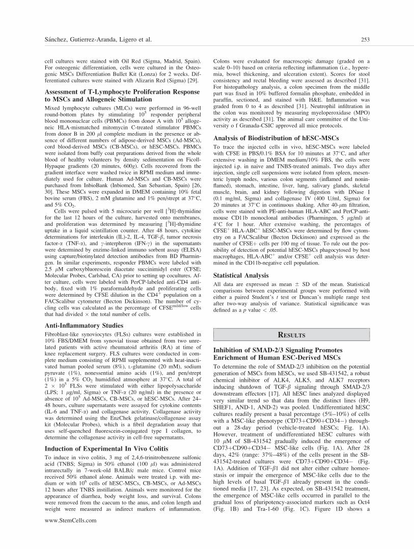
To determine the role of SMAD-2/3 inhibition on the potentialgeneration of MSCs from hESCs, we used SB-431542, a robustchemical inhibitor of ALK4, ALK5, and ALK7 receptorsinducing shutdown of TGF-b signaling through SMAD-2/3downstream effectors [17]. All hESC lines analyzed displayedvery similar trend so that data from the distinct lines (H9,SHEF1, AND-1, AND-2) was pooled. Undifferentiated hESCcultures readily present a basal percentage (5%–10%) of cellswith a MSC-like phenotype (CD73þCD90þCD34�) through-out a 28-day period (vehicle-treated hESCs; Fig. 1A).However, treatment of undifferentiated hESC cultures with10 lM of SB-431542 gradually induced the emergence ofCD73þCD90þCD34� MSC-like cells (Fig. 1A). After 28days, 42% (range: 37%–48%) of the cells present in the SB-431542-treated cultures were CD73þCD90þCD34� (Fig.1A). Addition of TGF-b1 did not alter either culture homeo-stasis or impair the emergence of MSC-like cells due to thehigh levels of basal TGF-b1 already present in the condi-tioned media [17, 23]. As expected, on SB-431542 treatment,the emergence of MSC-like cells occurred in parallel to thegradual loss of pluripotency-associated markers such as Oct4(Fig. 1B) and Tra-1-60 (Fig. 1C). Figure 1D shows a
Sanchez, Gutierrez-Aranda, Ligero et al. 253
www.StemCells.com

representative flow cytometry analysis depicting the expres-sion of CD73þCD90þ MSC-like cell population and theexpression of Oct4 and Tra-1-60 under the different culture
conditions. Morphologically, after 28 days of SB-431542treatment, hESC cultures looked fully differentiated and com-posed of fibroblastoid cells, whereas hESC cultures treated
Figure 1. Kinetics of the emergence of mesenchymal stem cells (MSCs) from SB-431542-treated human ESCs (hESCs). (A): Emergence ofMSC-like cells (CD73þ CD90þ CD34�) from four distinct SB-431542-treated hESCs over a 28-day period. Concurrent loss of the ESC-associ-ated markers Oct-4 (B) and Tra-1-60 (C) in SB-431542-treated hESCs over a 28-day period. Continuous lines represent SB-431542 treatment.Dotted lines represent TGF-b1 treatment. Dashed lines represent vehicle treatment. Data from the distinct hESC lines was pooled as very similartrend was obtained for each specific hESC line. (D): Representative flow cytometry analyses for CD73 and CD90 in hESC cultures treated withSB-431542 or TGF-b1 or DMSO. Expression of Oct4 and Tra-1-60 is also shown. An irrelevant isotype-matched antibody was always used (rightpanels). (E): Phase-contrast morphology of hESC cultures untreated (top panel) or treated with either SB-431542 (middle panel) or TGF-b1 (bot-tom panel). (F): Western blot analysis confirming dephosphorylation of SMAD-2/3 in hESCs treated with SB-431542 (left panel). Addition ofTGF-b1 to the hESC cultures does not enhance expression of SMAD-2/3. The SB-431542 inhibitor is highly specific for the SMAD2/3 signalingpathway, not altering the bone morphogenic proteins-linked SMAD-1/5/8 pathway. Abbreviations: 7-ADD, 7-actinomycin D; FITC, fluoresceinisothyocianate; PE, phyco erythrin; TGF-b1, transforming growth factor-b1.
254 hESC-Derived Multipotent Functional MSCs

with either DMSO or TGF-b1 displayed an undifferentiatedmorphology, being comprised by many round typical undiffer-entiated ESC colonies surrounded by some differentiated cells(Fig. 1E). After treatment with SB-431542, hESC cultures dis-played a dephosphorylation (inhibition) of SMAD-2/3 but notof SMAD-1/5/8, confirming the potent and specific effect ofthe SB-431542 chemical inhibitor in blocking signaling throughSMAD-2/3 but not via SMAD-1/5/8 (BMP signaling effectors;Fig. 1F). Together, these data reveal an efficient differentiationof hESCs into MSC-like cells through inhibition of the SMAD-2/3 signaling pathway.
SB-431542 ALK1/5/7 Inhibitor Fails to BlockSMAD-2/3 Signaling Pathway in hiPS CellsWhich, In Turn, Do Not Generate MSCson SB-431542 Treatment
Little information exists about how close hESC and iPS cellcultures are in terms of cellular and molecular mechanisms
regulating self-renewal versus lineage-specific differentiation[32]. We next wanted to assess whether the inhibition of theSMAD-2/3 signaling pathway by means of SB-451342 treat-ment also facilitates the enrichment of MSCs from distinctiPS cell lines. Surprisingly, in contrast to our findings inhESCs, treatment with the SB-431542 inhibitor did not exerta similar effect in the generation of MSCs from iPS cell lines.As shown in Figure 2A, SB-431542 treatment did not pro-mote the emergence of CD73þCD90þCD34� MSCs fromiPS cells, even over an extended period of up to 35 days ofSB-431542 treatment. Consistently, the expression of Oct-4(Fig. 2B) was not decreased in SB-431542-treated iPS cultures,whereas the expression levels of Tra-1-60 slightly decreasedovertime (Fig. 2C).
We, thus, wanted to analyze whether the SB-431542 in-hibitor is equally functional in iPS cells as compared withhESCs. Human iPS cells were treated with SB-451342 orDMSO as negative control and the capacity of the SB-431542inhibitor of blocking signaling through SMAD-2/3 was
Figure 2. Inhibition of the SMAD-2/3 signaling pathway does not enrich mesenchymal stem cells (MSCs) from iPS cell cultures. (A): In con-trast to human ESCs, two distinct iPS cell lines failed to differentiate into MSC-like cell on SB-431542-treatment over a 35-day period. Expres-sion of the pluripotent-associated markers Oct-4 (B) and Tra-1-60 (C) in SB-431542-treated iPS cells over a 35-day period. (D): Western blotanalysis showing that the SB-431542 treatment (used at different concentrations and time points) fails to inhibit SMAD-2/3 signaling pathway inhuman iPS cells. Abbreviations: CB, cord blood; iPS, induced pluripotent stem; SMAD, �TGF-b, transforming growth factor-b.
Sanchez, Gutierrez-Aranda, Ligero et al. 255
www.StemCells.com

determined by western blot under the same conditions usedfor hESCs. Surprisingly, in contrast to hESCs, 60 minutestreatment with 10 lM of SB-451342 was unable to dephos-phorylate SMAD-2/3. To confirm this data, we subsequentlytested different treatment conditions (15 minutes vs. 60minutes and 10 lM vs. 20 lM). SB-451342 consistently
failed to inhibit the signaling through SMAD-2/3. This datasuggests that iPS cells are resistant to SMAD-2/3 inhibitionvia this specific chemical inhibitor (Fig. 2D). Together, thisdata seems to suggest the existence of potential differences inthe way different developmental signaling pathways should bemodulated in hESCs versus iPS cells.
Figure 3. Morphological, phenotypic, and functionalcharacterization of hESC-derived MSCs. (A): Cellsorting isolation of CD73þCD90þ and CD73�CD90þ cell subsets. Purity was consistently >85%.(B): Phenotypic profile of established cultures ofhESC-derived MSCs. (C): In vitro cell growth, meas-ured as cumulative population doublings, of hESC-MSCs, CB-derived MSCs, and BM-derived MSCs.(D): Osteogenic, adipogenic, and chondrogenic differ-entiation potential of hESC-MSCs. Abbreviations:BM, bone marrow; CB, cord blood; FITC, fluoresceinisothyocyanate; hESC, human ESC; MSC, mesenchy-mal stem cell.
256 hESC-Derived Multipotent Functional MSCs

hESC-Derived MSCs Display Bona Fide MSCPhenotype, Growth Properties, and MultilineageDifferentiation Potential
To fully characterize these hESC-derived MSC-like cells, theCD73þCD90þ and CD73�CD90þ cell subsets were FACS-isolated (Fig. 3A). CD73�CD90þ cells did not establish a sta-ble culture. Indeed, sorted cells barely attached to the tissueculture plates and survived under mesenchymal culture condi-tions in suspension, eventually dying off after less than a week.In contrast, the CD73þCD90þ FACS-isolated populationrapidly attached to the tissue culture plastic allowing the rapidestablishment of long-term cultures of hESC-MSCs. ThesehESC-MSCs displayed a typical MSC phenotype: CD73þCD90þ CD105þ CD44þ CD166þ CD45� CD34� CD14�CD19� HLA-DR� (Fig. 3B). These hESC-MSCs could beeasily maintained for many passages displaying a faster growththan bone marrow-derived (BM-MSCs) but similar to CB-MSCs (Fig. 3C). More importantly, these hESC-MSCs dis-played osteogenic (alizarin red staining), adipogenic (oil redstaining), and chondrocytic (alcian blue staining) differentiationpotential, even a month after establishment of hESC-MSCs cul-tures (Fig. 3D). Together, this characterization confirms thatthe MSCs derived from hESCs display bona fide MSC pheno-type, cell growth and multipotent differentiation features.
hESC-MSCs Exert Potent Immunosuppressive andAnti-Inflammatory Properties In Vitro
The potential of hMSCs in regenerative medicine is in part dueto their immunomodulatory properties, thus, emerging as attrac-tive candidates for the treatment of immune disorders. We,therefore, investigated the ability of hESC-MSCs to inactivateT-cell responses and to inhibit inflammatory responses. Thepotential immunomodulatory activity of hESC-MSCs was com-pared with those of Ad-MSCs and CB-MSCs, which have beenpreviously characterized as potent immunosuppressor cells [6,33, 34]. The addition of hESC-MSCs to MLCs of PBMCs fromdifferent donors significantly reduced the number of total cellsin the culture and specifically decreased the number of cyclingCD4 T-cells (Fig. 4A). hESC-MSCs were very efficient inhibi-ting the proliferative response of activated T cells, showing adose-dependent effect with significant suppressive responses ata ratio as low as 1 hESC-MSC for every 10 PBMCs (Fig. 4B).As they lack class II MHC (HLA-DR, Fig. 3B) and costimula-tory molecules (CD80 and CD40; Fig. 4C), hESC-MSCs didnot stimulate the proliferation of allogeneic PBMCs (data notshown), supporting their ‘‘immuneprivilege’’ status. Moreover,hESC-MSCs significantly inhibited the production of the Th1-cytokines IL-2, IFNc, and TNFa on the allogeneic MLCs (Fig.4D). However, neither IL-4 (a Th2-cytokine) nor TGF-b1 (animmunosuppressive cytokine) was significantly affected byhESC-MSCs (Fig. 4D). Interestingly, the immunosuppressiveeffects of hESC-MSCs were only observed on allogeneic reac-tions, but not on syngeneic cultures (Fig. 4A, 4D), indicatingthe requirement of activated immune responses in such action.Moreover, the effects of hESC-MSCs were not a consequenceof induced cell mortality, as they did not affect the apoptosis/survival of PBMCs on the MLCs (data not shown). Overall, theimmunesuppressive effect of hESC-MSCs on T cells was similarthan that shown by CB-MSCs, but a bit lower than that observedfor Ad-MSCs (Fig. 4A, 4B, 4D), confirming that they are onto-genically closer to CB-MSCs than to adult adipose tissue.
We next investigated the capacity of hESC-MSCs to regulatethe inflammatory response of resident cells of the synovial mem-brane in patients with active RA. FLSs isolated from RA patientswere cultured in the presence of hESC-MSCs and assayed for the
production of inflammatory mediators and matrix-degradingenzymes (collagenase activity). hESC-MSCs treatment of RA FLScultures decreased the secretion of TNFa but not IL-6 after stimula-tion with LPS and reduced the collagenase activity after stimulationwith TNFa (Fig. 4E). These data reveal that the hESC-MSCs dis-play potent immunosuppressive and anti-inflammatory effects, akey functional immune feature of putative MSCs.
Treatment with hESC-MSCs Protects AgainstExperimental Inflammatory Bowel Disease In VivoConfirming hESC-MSCs Immunosuppressive andAnti-Inflammatory Properties In Vivo
On the basis of their in vitro immunosuppressive and anti-inflammatory properties, we next investigated the potentialtherapeutic action of hESC-MSCs in an in vivo experimentalmodel of inflammatory bowel disease induced by intrarectalinfusion of TNBS, which displays clinical, histopathological,and immunological features of human Crohn’s disease [35].In both Crohn’s disease and TNBS-induced colitis, activatedTh1 and Th17 cells promote an exaggerated macrophage andneutrophil infiltration and activation, giving rise to a prolongedsevere transmural inflamed intestinal mucosa, characterized byuncontrolled production of inflammatory cytokines and chemo-kines [35]. Inflammatory mediators such as cytokines and freeradicals, produced by infiltrating cells and resident macro-phages, play a critical role in colonic tissue destruction. Asshown in Figure 5A, TNBS-treated mice developed a severe ill-ness characterized by bloody diarrhea, rectal prolapse, pancolitisaccompanied by extensive wasting syndrome, and profoundand sustained weight loss resulting in 60% mortality. Macro-scopic examination of colons showed striking hyperemia,inflammation, and necrosis (Fig. 5B). However, mice treatedwith hESC-MSCs, similar to those treated with CB-MSCs orAd-MSCs, displayed an increased survival rate, rapidly recov-ered body weight loss, improved the wasting disease, regaineda healthy appearance, and only showed slight signs of coloninflammation, similar to control mice treated with vehicle(50% ethanol; Fig. 5A, 5B). Histological examination of thecolons showed that hESC-MSC-treatment reduced the TNBS-induced transmural inflammation, depletion of mucin-produc-ing globet and epithelial cells, disseminated fibrosis, focalloss of crypts, and infiltration of inflammatory cells (Fig. 5C).Moreover, hESC-MSC treatment significantly decreased colo-nic MPO activity, reflecting less neutrophil infiltration in thelamina propria (Fig. 5D). Together, these data confirm thetherapeutic effect in vivo of hESC-MSCs on an experimentalmodel of colitis, demonstrating for the first time that hESC-MSCs exhibit potent immunosuppressive and anti-inflamma-tory properties not only in vitro but also in vivo.
Finally, to better understand the trafficking of the infusedhESC-MSCs, we injected CFSE-labeled hESC-MSCs intocolitic mice. As previously described for Ad-MSCs [31], wedetected the inoculated hESC-MSCs in the draining lymphnodes and spleen, as well as in the inflamed colon of therecipients 2 days postinjection (Fig. 5E). Interestingly, hESC-MSCs barely homed to noninflamed colon and other regionsof the gastrointestinal tract (salivary glands, stomach, intes-tine) or to other organs/tissues (muscle, kidney, or brain).Moreover, similar to MSCs from other sources, hESC-MSCsare significantly retained in the lung and liver (Fig. 5E).
DISCUSSION
Human ESCs and iPS cells hold the promise as a potentiallyunlimited source of functional cell types for regenerativemedicine and drug screening [36]. One of the key challenges
Sanchez, Gutierrez-Aranda, Ligero et al. 257
www.StemCells.com

used to fulfill this potential is the ability to reproducibly directthe differentiation of these pluripotent stem cells to selectivecell fates in vitro and in vivo. Because of their biologicalproperties, the generation of a potentially unlimited number
of functional MSCs from hESC/iPS cells would be a greatstep forward in regenerative medicine.
MSCs from adult somatic tissues differentiate in vitro andin vivo into multiple mesodermal tissues including bone,
Figure 4. hESC-MSCs display potent immunosuppressive and anti-inflammatory effects in vitro. (A): MLCs were established by coculturingPBMCs from donor A (105 cells) and carboxybluorescein diacetate succinimidyl ester (CFSE)-labeled responder PBMCs from donor B (105
cells). Cultures of PBMCs from donor B (2 � 105 cells) were used as basal controls (syngeneic). A total of 2 � 104 hESC-MSCs, CB-MSCs,or Ad-MSCs were added to MLCs, and the total number of cells was determined after 4 days of culture (upper panel). The number of cycling(CFSEmild/low) CD4þ cells was determined by flow cytometry (lower panel). Cultures of PBMCs from donor B (2 � 105 cells) were used as basalcontrols (syngeneic). *, p value < .001 versus MLC without MSCs. (B): Dose-dependent effect of hESC-MSCs on lymphocyte proliferation. hESC-MSCs, CB-MSCs, or Ad-MSCs were added at different ratios to allogeneic MLCs (105 PBMCs from donor A and 105 PBMCs from donor B). Pro-liferation was determined by measuring [3H]-thymidine incorporation after 96 hours culture. Solid squares represent the basal proliferation of eachMSC type. PBMCs from donor A (2 � 105 cells) were used as control of basal proliferation (gray column). *, p value < .001 versus MLC withoutMSCs. (C): hESC-MSCs do not express the costimulatory molecules CD40 and CD80. (D): hESC-MSCs decrease the production of cytokines byactivated lymphocytes. hESC-MSCs, CB-MSCs, or Ad-MSCs (2 � 104) were added to allogeneic MLCs (105 PBMCs from donor A and 105 PBMCsfrom donor B) or syngeneic cultures (2 � 105 PBMCs from donor B). Cytokine contents in the supernatants were determined by ELISA after48 hours culture. *, p value < .001 versus MLC without MSCs. (E): hESC-MSCs inhibit the inflammatory response in synovial cells from patientswith rheumatoid arthritis (RA). Fibroblast-like synovial cells (2 � 105) isolated from two patients with RA were incubated with medium (unstimu-lated) or stimulated with lipopolysaccharide (1 ng/ml, for cytokine determination) or TNF-a (20 ng/ml, for collagenase activity assay) in the absenceor presence of hESC-MSCs, CB-MSCs, or Ad-MSCs (105). Culture supernatants were assayed for collagenase activity (after 24 hours) or cytokinecontents (after 48 hours). *, p value < .001 versus FLS alone. Abbreviations: Ad-MSC, adipose-derived mesenchymal stem cell; CB-MSC, cordblood-derived MSC; FLS, fibroblast-like synoviocyte; hESC, human ESC; IL, interleukin; IFNc, interpheron c; MLC, mixed lymphocyte cultures;MSC, mesenchymal stem cell; PBMC, peripheral blood mononuclear cell; TGF-b, transforming growth factor-b; TNFa, tumor necrosis factor a.
258 hESC-Derived Multipotent Functional MSCs

Figure 5. Treatment with hESC-MSCs protects against experimental colitis in vivo. (A): Colitis was induced by intracolonic administrationof TNBS. Mice (10 mice per group) were treated i.p. with 106 hESC-MSCs, CB-MSCs, or Ad-MSCs, 12 hours after TNBS injection. Controlmice received 50% ethanol (vehicle). Clinical evolution was monitored by body weight changes, colitis score, and survival. (B): Colon lengthand weight and macroscopic colonic damage score was evaluated at day 2. (C): Histopathology was determined 2 days after transplant(4–6 mice per group) Scale bar ¼ 100 lm. (D): Neutrophil infiltration was assayed by determining MPO activity in the colons isolated at day2 (4–6 mice per group). (E): CFSE-labeled hESC-MSCs were injected i.p. to colitic mice (3 mice per group) and their presence in variousorgans determined by flow cytometry 2 days after injection and expressed as the mean number of CFSEþHLA-ABCþCD11b� cells per100 mg tissue. *, p value < .001 versus TNBS-colitic mice. Abbreviations: Ad-MSC, adipose-derived mesenchymal stem cell; CB-MSC,cord blood-derived MSC; CFSE, carboxybluorescein diacetate succinimidyl ester; hESC, human ESC; MLN, mesenteric lymph node; MPO,myeloperoxidase; TNBS, 2,4,6-trinitrobenzene sulfonic acid.
Sanchez, Gutierrez-Aranda, Ligero et al. 259
www.StemCells.com

cartilage, adipose tissue, tendon, ligament, or even muscle.They also home to damaged tissues where they exert theirtherapeutic potential [31, 33]. A striking feature of the MSCsis their immunosuppressive properties in vitro. Their multili-neage differentiation potential coupled with their immunopri-vileged properties is being exploited worldwide for cellreplacement strategies and autoimmune diseases [4, 6]. De-spite clear in vivo evidence of mesenchymal specificationfrom hESCs, surprisingly little work has been performed todevelop enrichment protocols for MSCs from hESCs/iPS cells[37]. There have been reports on the derivation of specificmesenchymal cell types from hESCs. For instance, hESCs weredifferentiated into mineralizing bone [38, 39]. Improvements inosteogenic differentiation in vitro were also reported [40] andXiong et al. [41] showed adipogenic differentiation from hESCs.
However, little work has been performed in the derivationof multipotent MSCs rather than specific mesechymal deriva-tives from hESCs/iPS cells. Barberi et al. [13] and Trivediand Hematti [42] were the first to derive MSCs from hESCsthrough a xeno-coculture of hESC with the OP9 murine stro-mal cell line. Olivier et al. [43] also reported the derivation ofMSCs from hESCs through a feeder free culture system.However, the methodology employed in this study consistedin culturing MSCs from hESCs that were grown into a thickmultilayer epithelium. Unfortunately, there is no informationavailable about the mechanistic insights involved in the spe-cific differentiation of hESCs into functional MSCs. Here, wedemonstrate for the first time the involvement of the SMAD-2/3 signaling pathway inhibition in the efficient generation/enrichment of MSCs from hESCs. We previously reportedthat specific inhibition of TGF-b signals through SMAD-2/3results in loss of the hESC phenotype and pluripotencythrough induction of hESC differentiation, while not affectingsurvival or self-renewal of hESCs [17]. Furthermore, TGF-bis considered a potent inhibitor of mesodermal tissue [18–20].We, therefore, confirmed our initial hypothesis that similar towhat occurs in the hematopoietic system, TGF-b signalingthrough SMAD-2/3 is a negative regulator in the MSC gener-ation from hESCs. Human ESC-MSCs easily establishedlong-term cultures and could be maintained for many passagesdisplaying a faster growth than somatic MSCs while maintain-ing MSC morphology and phenotype. They also displayedosteogenic, adipogenic, and chondrocytic multipotent differen-tiation potential and lacked signs of oncogenic transformation[17]. An approximate quantification of our differentiationexperiments revealed that about 30% of the hESC-MSCs dif-ferentiate into adipogenic lineage and about 70% differentiateinto osteogenic or chondrogenic lineages. This data is in linewith the data reported for MSCs from many other tissuesindicating that the hESC-MSCs seem also hierarchicallyorganized and therefore, functionally heterogeneous within aphenotypically homogeneous culture [44–48].
To further determine whether these hESC-MSCs are bonafide MSCs, we analyzed for the first time their immunologicalproperties in vitro and in vivo. Their immunotolerance proper-ties have been previously suggested in vitro [14], but no in-formation is available about the potential therapeutic action ofhESC-MSCs in vivo. Here, we demonstrate how hESC-MSCsdisplayed potent immunosuppressive and anti-inflammatoryproperties in vitro and in vivo in an experimental model ofinflammatory bowel disease or colitis model [31, 33]. In vitro,they were very efficient inhibiting the proliferative responseof T cells, they lacked class II HLA and costimulatory mole-cules, and did not stimulate the proliferation of allogeneicPBMCs. Moreover, hESC-MSCs significantly inhibited theproduction of the Th1-cytokines IFNc, IL-2, and TNFa butnot the Th2-cytokine IL-4 on MLC assays. Overall, the
immunosuppressive effect of hESC-MSCs on T cells was sim-ilar to that shown by CB-MSCs, but a bit lower than thatobserved for Ad-MSCs, confirming that they are ontogenicallycloser to CB-MSCs than to adult adipose tissue. They alsoregulated the inflammatory response of resident cells of thesynovial membrane. FLS isolated from patients with activeRA cultured in the presence of hESC-MSCs produced lessinflammatory mediators and matrix-degrading enzymes.
More importantly, in vivo, using an experimental modelof inflammatory bowel disease, mice treated with hESC-MSCssimilar to those treated with CB-MSCs or Ad-MSCs, displayedan increased survival rate, rapidly recovered body weight loss,improved the wasting disease, regained a healthy appearance,and significantly reduced colon inflammation, similar to controlmice treated with vehicle, demonstrating for the first time thathESC-MSCs exhibit potent immunosuppressive and anti-inflam-matory properties not only in vitro but also in vivo. This immu-nosuppressive effect could be exerted locally on the colonicmucosa, or peripherally on lymphoid organs, as suggested by thefact that hESC-MSCs show preferential homing for inflamedtissues and secondary lymphoid organs, in addition to thesystemic filter organs (lung and liver).
Finally, whether hESCs and iPS are equally capable ofgenerating MSCs remains to be assessed and little informationexists about how close hESC and iPS cultures are in terms ofcellular and molecular mechanisms regulating self-renewal ver-sus lineage-specific differentiation [32]. Recently, differencesin the differentiation capacity between hESCs and iPS cellswere reported [49]. Accordingly, we found that in contrast tothat observed in hESCs, treatment with SB-431542 did notexert a similar effect in the generation of MSCs from distinctiPS cell lines, even over an extended period of up to 35 daysof SB-431542 treatment. Interestingly, further experimentsrevealed that the SB-451342 inhibitor consistently failed to in-hibit the signaling through SMAD-2/3 in hiPS cells, suggestingthat hiPS cells are resistant to SMAD-2/3 inhibition via thisspecific chemical inhibitor. Whether alternative chemical inhib-itors or overexpression of ectopic SMAD-6 may block thispathway in hiPS cells remains to be elucidated. Alternatively,we found that many hiPS cells reactivate the ectopic reprog-ramming factors during differentiation toward blood and neuro-ectodem (Ramos-Mejıa et al., manuscript in preparation).Whether these ectopic factors may also reactivate their expres-sion during hiPS cell differentiation toward MSCs should alsobe addressed in future studies to gain insight into whether con-stitutive expression of ectopic reprogramming factors (Oct-4,Klf-4, c-myc, and Sox-2) may be comparing signaling throughTGF-ALKs-SMAD-2/3. Finally, it is worth assessing in futurestudies that whether iPS cells generated without c-myc or usinga different cocktail of ectopic reprogramming factors (i.e., Oct-4, Nanog, Sox-2, and Lin28) are equally resistant to SMAD-2/3 inhibition. This data, although preliminary, suggests potentialdifferences in the cellular and molecular mechanisms underly-ing the generation of MSCs from hESCs versus iPS cells.These still uncharacterized differences should be kept in mindwhen attempting to differentiate hESCs or iPS cells into a cer-tain lineage. It is plausible that cellular mechanisms and molec-ular determinants involved in the stepwise differentiation maydiffer, and therefore the experimental data gained in one modelcan not be extrapolated to the other model.
CONCLUSION
We show for the first time how specific inhibition of SMAD-2/3 signaling pathway in hESCs facilitates the conversion ofhESCs into multipotent MSCs with fully immunosuppresive
260 hESC-Derived Multipotent Functional MSCs

and anti-inflammatory properties in vivo, opening up newavenues for potential future clinical applications.
ACKNOWLEDGMENTS
We are indebted to Prof. Jose Cibelli (Michigan State Univer-sity) for provision of the iPS cell line MSUH001. This work wassupported by the CSJA (0030/2006 to P.M. and 0108/2007to R.R.) and CICE (P08-CTS-3678 to P.M.), de la Junta deAndalucıa, the FIS/FEDER to P.M. (PI070026 and PI100449),C.B. (CP07/00059), and P.J.R. (CP09/0063), the MINICC toP.M. (PLE-2009-0111), and the Marie Curie IIF to V.R.-M.(PIIF-GA-2009-236430). R. Rodriguez is supported by a Fel-
lowship from the AECC. J.L.G.-P.’s group is supported byISCIII-CSJA (EMER07/056), by a Marie Curie IRG action(FP7-PEOPLE-2007-4-3-IRG), by CICE (P09-CTS-4980) fromJunta de Andalucıa, by Proyectos de Investigacion en Salud PI-002 from Junta de Andalucia, and by the FIS/FEDER (CP07/00065 and PI08171). M.D.’s group is supported by FIS/FEDER(PS09-00928) and Junta de Andalucia (P09-CTS-4723).
DISCLOSURE OF CONFLICTS
OF INTEREST
The authors reported no potential conflicts of interest.
REFERENCES
1 Menendez P, Bueno C, Wang L. Human embryonic stem cells: A jour-ney beyond cell replacement therapies. Cytotherapy 2006;8:530–541.
2 Bueno C, Garcia-Castro J, Montes R et al. Human embryonic stemcells: A potential system for modeling infant leukemia harboringMLL-AF4 fusion gen. Drug Discov Today Dis Models 2008;4:53–60.
3 Menendez P, Bueno C, Wang L et al. Human embryonic stem cells:potential tool for achieving immunotolerance? Stem Cell Rev 2005;1:151–158.
4 Dominici M, Le Blanc K, Mueller I et al. Minimal criteria for defin-ing multipotent mesenchymal stromal cells. The International Societyfor Cellular Therapy position statement. Cytotherapy 2006;8:315–327.
5 Friedenstein AJ, Chailakhyan RK, Latsinik NV et al. Stromal cells re-sponsible for transferring the microenvironment of the hemopoietictissues. Cloning in vitro and retransplantation in vivo. Transplantation1974;17:331–340.
6 Garcia-Castro J, Trigueros C, Madrenas J et al. Mesenchymal stemcells and their use as cell replacement therapy and disease modellingtool. J Cell Mol Med 2008;12:2552–2565.
7 Pittenger MF, Mackay AM, Beck SC et al. Multilineage potential ofadult human mesenchymal stem cells. Science 1999;284:143–147.
8 Aggarwal S, Pittenger MF. Human mesenchymal stem cells modulateallogeneic immune cell responses. Blood 2005;105:1815–1822.
9 Bartholomew A, Sturgeon C, Siatskas M et al. Mesenchymal stemcells suppress lymphocyte proliferation vitro and prolong skin graftsurvival in vivo. Exp Hematol 2002;30:42–48.
10 Le Blanc K, Ringden O. Immunobiology of human mesenchymalstem cells and future use in hematopoietic stem cell transplantation.Biol Blood Marrow Transplant 2005;11:321–334.
11 Menendez P, Perez-Simon JA, Mateos MV et al. Influence of the dif-ferent CD34þ and CD34- cell subsets infused on clinical outcomeafter non-myeloablative allogeneic peripheral blood transplantationfrom human leucocyte antigen-identical sibling donors. Br J Haematol2002;119:135–143.
12 Menendez P, Prosper F, Bueno C et al. Sequential analysis of CD34þand CD34- cell subsets in peripheral blood and leukapheresis productsfrom breast cancer patients mobilized with SCF plus G-CSF andcyclophosphamide. Leukemia 2001;15:430–439.
13 Barberi T, Willis LM, Socci ND et al. Derivation of multipotent mes-enchymal precursors from human embryonic stem cells. Plos Med2005;2:e161.
14 Trivedi P, Hematti P. Derivation and immunological characterizationof mesenchymal stromal cells from human embryonic stem cells. ExpHematol 2008;36:350–359.
15 Lorda-Diez CI, Montero JA, Martinez-Cue C et al. Transforminggrowth factors beta coordinate cartilage and tendon differentiation inthe developing limb mesenchyme. J Biol Chem 2009;284:29988–29996.
16 Roelen BA, Dijke P. Controlling mesenchymal stem cell differentia-tion by TGFBeta family members. J Orthop Sci 2003;8:740–748.
17 Bendall SC, Stewart MH, Menendez P et al. IGF and FGF coopera-tively establish the regulatory stem cell niche of pluripotent humancells in vitro. Nature 2007;448:1015–1021.
18 Fortunel NO, Hatzfeld A, Hatzfeld JA. Transforming growth factor-beta: Pleiotropic role in the regulation of hematopoiesis. Blood 2000;96:2022–2036.
19 Fortunel NO, Hatzfeld JA, Monier MN et al. Control of hematopoieticstem/progenitor cell fate by transforming growth factor-beta. OncolRes 2003;13:445–453.
20 Kim SJ, Letterio J. Transforming growth factor-beta signaling in nor-mal and malignant hematopoiesis. Leukemia 2003;17:1731–1737.
21 Cortes JL, Sanchez L, Ligero G et al. Mesenchymal stem cells facili-tate the derivation of human embryonic stem cells from cryopreservedpoor-quality embryos. Hum Reprod 2009;24:1844–1851.
22 Menendez P, Wang L, Chadwick K et al. Retroviral transduction ofhematopoietic cells differentiated from human embryonic stem cell-derived CD45(neg)PFV hemogenic precursors. Mol Ther 2004;10:1109–1120.
23 Montes R, Ligero G, Sanchez L et al. Feeder-free maintenance ofhESCs in mesenchymal stem cell-conditioned media: Distinct require-ments for TGF-beta and IGF-II. Cell Res 2009;19:698–709.
24 Ramos-Mejia V, Melen GJ, Sanchez L et al. Nodal/Activin signalingpredicts human pluripotent stem cell lines prone to differentiatetowards the hematopoietic lineage. Mol Ther 2010;18:2173–2181.
25 Ramos-Mejia V, Munoz-Lopez M, Garcia-Perez JL et al. iPSC linesthat do not silence the expression of the ectopic reprogramming fac-tors may display enhanced propensity to genomic instability. Cell Res2010;20:1092–1095.
26 Rodriguez R, Rubio R, Masip M et al. Loss of p53 induces tumorigene-sis in p21-deficient mesenchymal stem cells. Neoplasia 2009;11:397–407.
27 Menendez P, Redondo O, Rodriguez A et al. Comparison between alyse-and-then-wash method and a lyse-non-wash technique for theenumeration of CD34þ hematopoietic progenitor cells. Cytometry1998;34:264–271.
28 Barroso-delJesus A, Romero-Lopez C, Lucena-Aguilar G et al. Em-bryonic stem cell-specific miR302–367 cluster: Human gene structureand functional characterization of its core promoter. Mol Cell Biol2008;28:6609–6619.
29 Menendez P, Catalina P, Rodriguez R et al. Bone marrow mesenchymalstem cells from infants with MLL-AF4þ acute leukemia harbor andexpress the MLL-AF4 fusion gene. J Exp Med 2009;206:3131–3141.
30 Rubio R, Garcia-Castro J, Gutierrez-Aranda I et al. Deficiency in p53but not retinoblastoma induces the transformation of mesenchymalstem cells in vitro and initiates leiomyosarcoma in vivo. Cancer Res2010;70:4185–4194.
31 Gonzalez MA, Gonzalez-Rey E, Rico L et al. Adipose-derived mesen-chymal stem cells alleviate experimental colitis by inhibiting inflamma-tory and autoimmune responses. Gastroenterology 2009;136:978–989.
32 Kim PG, Daley GQ. Application of induced pluripotent stem cells tohematologic disease. Cytotherapy 2009;11:980–990.
33 Gonzalez-Rey E, Anderson P, Gonzalez MA et al. Human adult stemcells derived from adipose tissue protect against experimental colitisand sepsis. Gut 2009;58:929–939.
34 Yanez R, Lamana ML, Garcia-Castro J et al. Adipose tissue-derivedmesenchymal stem cells have in vivo immunosuppressive propertiesapplicable for the control of the graft-versus-host disease. Stem Cells2006;24:2582–2591.
35 Bouma G, Strober W. The immunological and genetic basis of inflam-matory bowel disease. Nat Rev Immunol 2003;3:521–533.
36 Bueno C, Catalina P, Melen GJ et al. Etoposide induces MLL rear-rangements and other chromosomal abnormalities in human embryonicstem cells. Carcinogenesis 2009;30:1628–1637.
37 Barberi T, Studer L. Mesenchymal cells. Methods Enzymol 2006;418:194–208.
38 Bielby RC, Christodoulou IS, Pryce RS et al. Time- and concentra-tion-dependent effects of dissolution products of 58S gel bioactiveglass on proliferation and differentiation of murine and human osteo-blasts. Tissue Eng 2004;10:1018–1026.
39 Sottile V, Thomson A, McWhir J. In vitro osteogenic differentiationof human ES cells. Cloning Stem Cells 2003;5:149–155.
40 Karp JM, Ferreira LS, Khademhosseini A et al. Cultivation of humanembryonic stem cells without the embryoid body step enhances osteo-genesis in vitro. Stem Cells 2006;24:835–843.
Sanchez, Gutierrez-Aranda, Ligero et al. 261
www.StemCells.com

41 Xiong C, Xie CQ, Zhang L et al. Derivation of adipocytes fromhuman embryonic stem cells. Stem Cells Dev 2005;14:671–675.
42 Trivedi P, Hematti P. Simultaneous generation of CD34þ primitivehematopoietic cells and CD73þ mesenchymal stem cells from humanembryonic stem cells cocultured with murine OP9 stromal cells. ExpHematol 2007;35:146–154.
43 Olivier EN, Rybicki AC, Bouhassira EE. Differentiation of humanembryonic stem cells into bipotent mesenchymal stem cells. StemCells 2006;24:1914–1922.
44 Bach C, Buhl S, Mueller D et al. Leukemogenic transformation byHOXA cluster genes. Blood 2010;115:2910–2918.
45 de la Fuente R, Abad JL, Garcia-Castro J et al. Dedifferentiated adultarticular chondrocytes: A population of human multipotent primitivecells. Exp Cell Res 2004;297:313–328.
46 Lee CC, Christensen JE, Yoder MC et al. Clonal analysis and hierar-chy of human bone marrow mesenchymal stem and progenitor cells.Exp Hematol 2010;38:46–54.
47 Menicanin D, Bartold PM, Zannettino AC et al. Identification of acommon gene expression signature associated with immature clonalmesenchymal cell populations derived from bone marrow and dentaltissues. Stem Cells Dev 2010;19:1501–1510.
48 Muraglia A, Cancedda R, Quarto R. Clonal mesenchymal progenitorsfrom human bone marrow differentiate in vitro according to a hier-archical model. J Cell Sci 2000;113:1161–1166.
49 Hu BY, Weick JP, Yu J et al. Neural differentiation of humaninduced pluripotent stem cells follows developmental principles butwith variable potency. Proc Natl Acad Sci USA 2010;107:4335–4340.
262 hESC-Derived Multipotent Functional MSCs