enhancing epileptic circuit targeting for optimizing
TRANSCRIPT
Enhancing Epileptic Circuit Targeting for
Optimizing Neurostimulation Therapy
Marvin A Rossi, MD, PhD
Rush Epilepsy Center
Rush University Medical Center &
Chicago, IL, U.S.A
Research Lab: www.synapticom.net
• No commercial financial disclosures or
conflicts of interest

Driving Force for Innovation
• 3 million individuals in the U.S. (pop. 300,000,000)
• 50-60 million individuals world-wide with epilepsy
• About 20% have medication-resistant epilepsy
o 500-800 thousand in the U.S.
o About 10 million individuals globally
o Over 12 billion U.S. dollars in healthcare costs
every year

• 1% of the U.S. population is diagnosed with
epilepsy (about 3 million individuals)
• About 500,000 individuals experience recurrent
seizures resistant to pharmaco-therapy
• Approximately 5000 brain surgeries for epilepsy are
performed in the U.S. every year
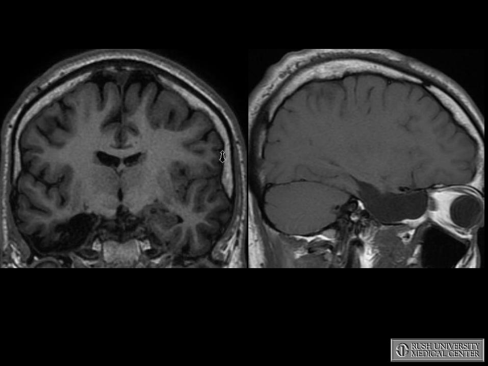
• The presurgical evaluation can be neuroimaging
intensive
Treatment:
I. Medication
II. Diet
III. Surgery
1. Resection
2. Neurostimulation Therapy
(Interfacing with Fragile Neural Networks)


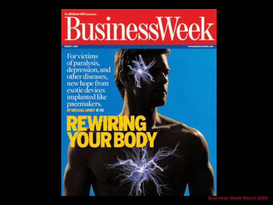
Business Week March 2005

Stimulation in the Central Nervous System
• Walker et al (1938) – cerebellum
• Cooke & Snider (1955) – cerebellum
• Chkhenkeli et al (1978) – caudate
• Cooper et al (1980) – anterior thalamus
• Velasco et al (1987) – centromedian thalamus
• Fischell et al (1988) – vagus nerve stimulation
• Vercueil et al (1998) – subthalamic nucleus
• Fisher et al (2003) – anterior thalamic nucleus
• Morrell et al (2004) – responsive cortical stimulation
Business Week March 2005
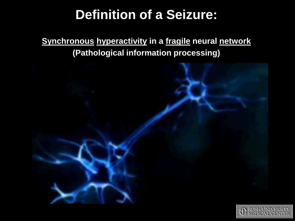
Definition of a Seizure:
Synchronous hyperactivity in a fragile neural network
(Pathological information processing)

Chronic exposure to direct neurostimulation
has been associated with distant cortical synaptic
proliferation2
Proposed Mechanisms of Neurostimulation Therapy:
Acute-onset efficacy of local neurostimulation therapy:
1. Conduction blockade
2. Synaptic inhibition
3. Synaptic depression
4. Overriding pathophysiological network activity1
1Rossi et al (2010). Predicting white matter pathways for direct neurostimulation therapy.
Epilepsy Res
2Keller et al (1992). Synaptic proliferation in the motor cortex of adult cats after thalamic
stimulation. J Neurophysiol 68:295-308.

Neurostimulation in Epilepsy:
1. Vagus Nerve Stimulation (VNS)
2. Deep Brain Stimulation (DBS)
3. Responsive (Cortical) Neurostimulation (RNS)

VNS Implant Site

Evolution of the Vagal Nerve Stimulator (VNS)

Acute-onset efficacy of local neurostimulation therapy
‘On demand’ magnetic switch triggering
Chronic exposure to direct neurostimulation therapy
Synaptic proliferation
Inhibitory (GABAergic?) upregulation
Proposed Mechanisms of Neurostimulation Therapy:

High Stimulation Low Stimulation
Henry, Bakay, Pennell, Epstein, Votaw (2004). Brain blood flow alterations induced by therapeutic vagus nerve
stimulation in partial epilepsy: II. Prolonged effects at high and low levels of stimulation. Epilepsia 45(9):1064-1070.

Neurostimulation in Epilepsy:
1. Vagus Nerve Stimulation (VNS)
2. Deep Brain Stimulation (DBS)
3. Responsive Cortical Stimulation (RNS)

stimulating electrodes
Anterior Nucleus of the Thalamus
Deep Brain Stimulator (DBS, Medtronic) Stimulation of the Anterior Nucleus of the Thalamus for Epilepsy
(SANTE) Trial
On March 12, 2010, a U.S. FDA advisory panel recommended
approval of the DBS as being safe and effective for patients
with severe and refractory partial-onset seizures with or
without generalization.
Papez Circuit
Medial Limbic Circuit
C
Fisher et al (2010). Electrical stimulation of the anterior nucleus of thalamus for treatment of refractory
epilepsy. Epilepsia 51(5):899-908.
Neurostimulation in Epilepsy:
1. Vagus Nerve Stimulation (VNS)
2. Deep Brain Stimulation (DBS)
3. Responsive (Cortical) Neurostimulation (RNS)


2004 2005 2006 2007 2008 2009 2010 2011 2012 2013 2014 2015 2016 2017
RNS® System: Clinical Studies
Feasibility Study
65 implanted
Pivotal Study
191 implanted
Long-term Treatment Study (230 enrolled) Long-term Treatment Study (230 enrolled)
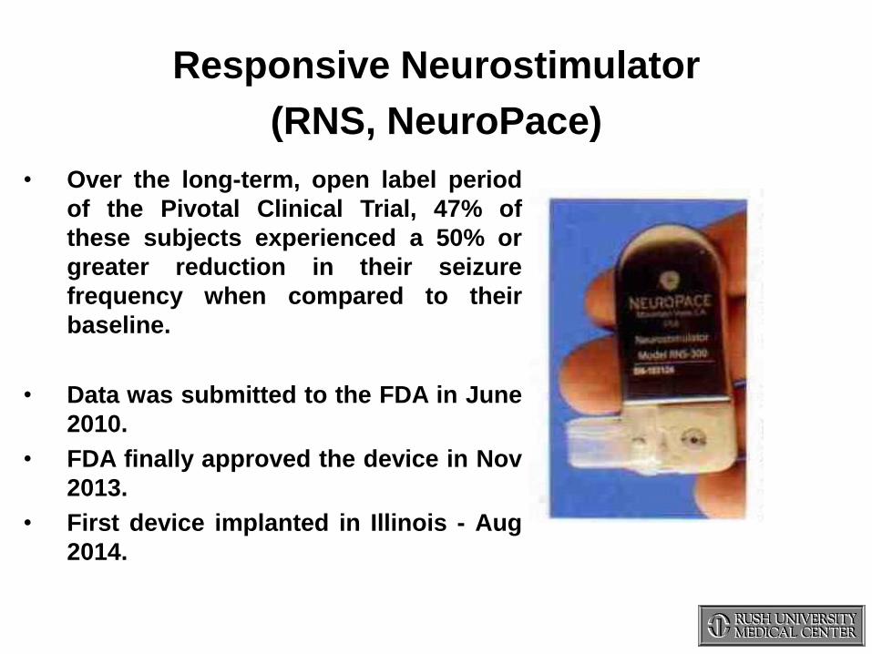
Responsive Neurostimulator
(RNS, NeuroPace)
• Over the long-term, open label period
of the Pivotal Clinical Trial, 47% of
these subjects experienced a 50% or
greater reduction in their seizure
frequency when compared to their
baseline.
• Data was submitted to the FDA in June
2010.
• FDA finally approved the device in Nov
2013.
• First device implanted in Illinois - Aug
2014.

Pivotal Study: Mean Disabling
Seizures

Responsive ‘On-Demand’ Cortical
Stimulator:
1. Detection
2. Stimulation
(A) 2.5 mA applied current
(B) 4.5 mA applied current

4
1
Electrocorticogram from
Implantable RNS Buffer:

Charge-balanced and Symmetric Waveform used in
Neurostimulation Therapy:

Acute-onset efficacy of local neurostimulation therapy
Chronic exposure to direct neurostimulation
Proposed Mechanisms of Neurostimulation Therapy:
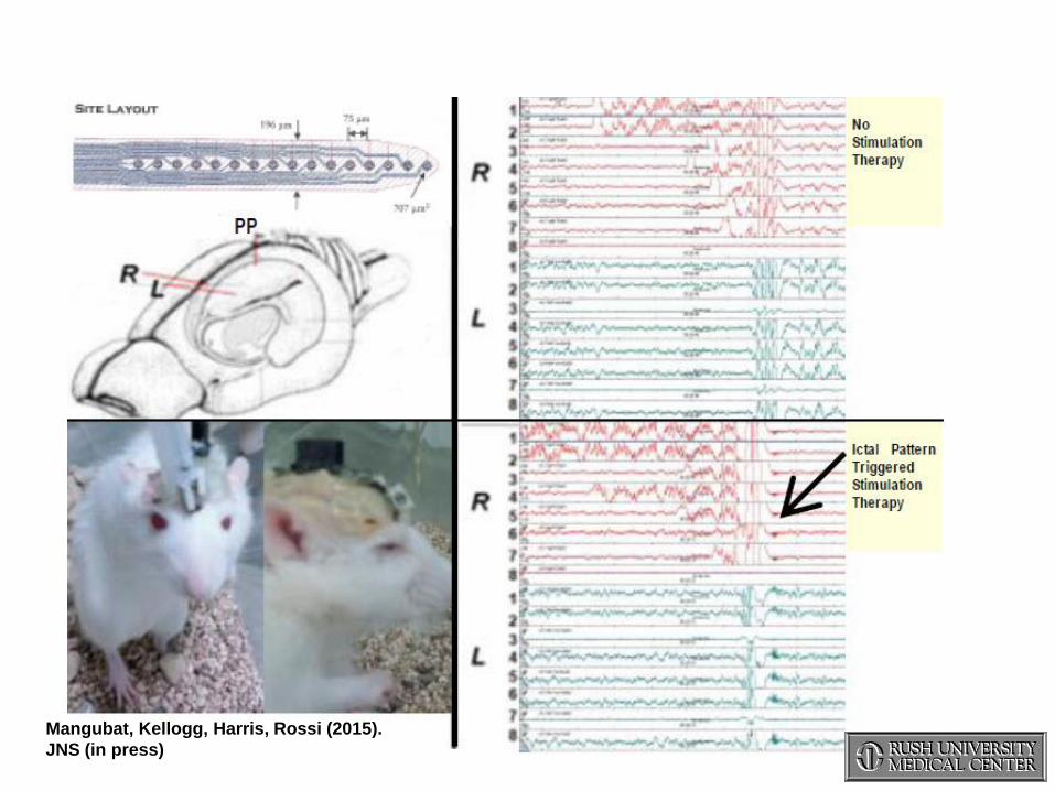
Mangubat, Kellogg, Harris, Rossi (2015).
JNS (in press)
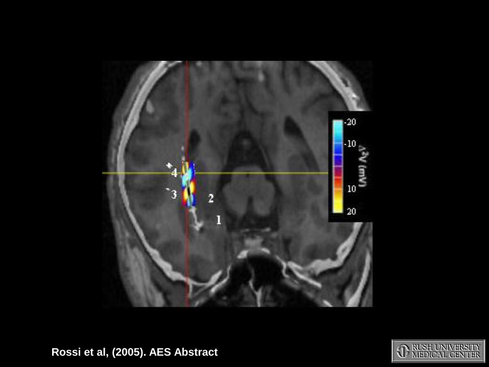
Rossi et al, (2005). AES Abstract

Stimulation of the Ictal Onset Zone:
Comparison of Grey versus White Matter
Epileptogenic Zone
Activation Field
White Matter Activation
White Matter Stimulation
Grey Matter Stimulation
Grey Matter Activation
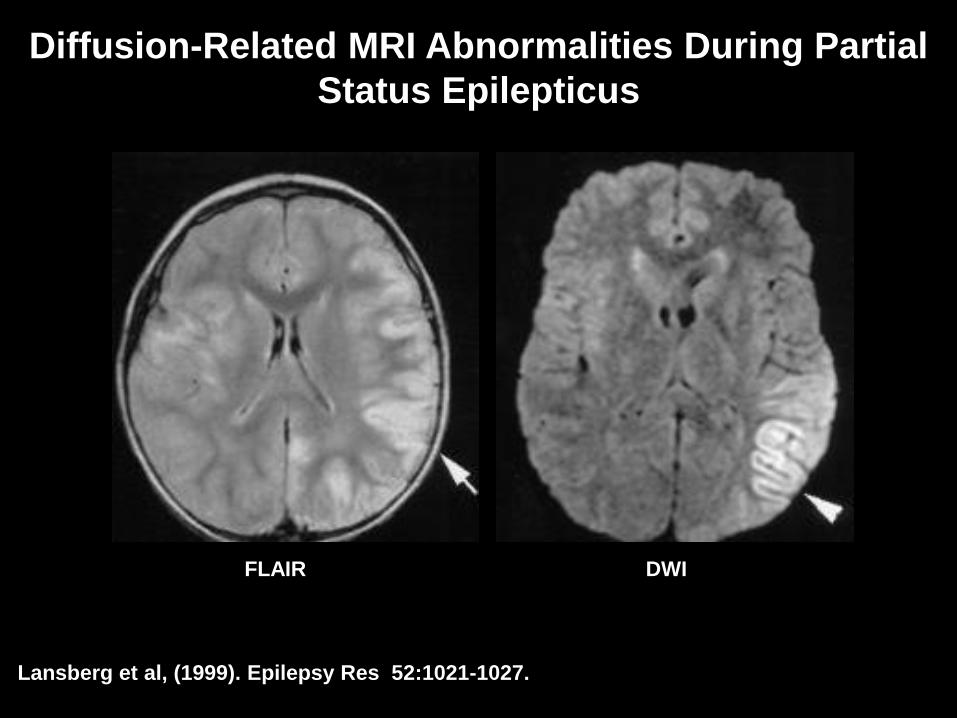
Diffusion-Related MRI Abnormalities During Partial
Status Epilepticus
Lansberg et al, (1999). Epilepsy Res 52:1021-1027.
FLAIR DWI

15 year old with localization-related epilepsy & post-
ictal hemiparesis
Prichard et al (1995); Zhong et al (1997); Diel et al 2005; Salmenpera et al (2006);
Yogarajah & Duncan, (2008)
FLAIR MR sequence acquired within 24 hrs after
brief stereotypic seizure & LUE Todd’s paresis

Diffusion Tensor Imaging:

Isotropic voxel
(FA = 0)

Anisotropic voxel - water diffusion
has a preference in directionality
(FA=1)

DTI

Focke et al (2008). NeuroImage 40: 728-737
Chronic White Matter Changes (Following Years of Focal-Onset Seizures)
R
L
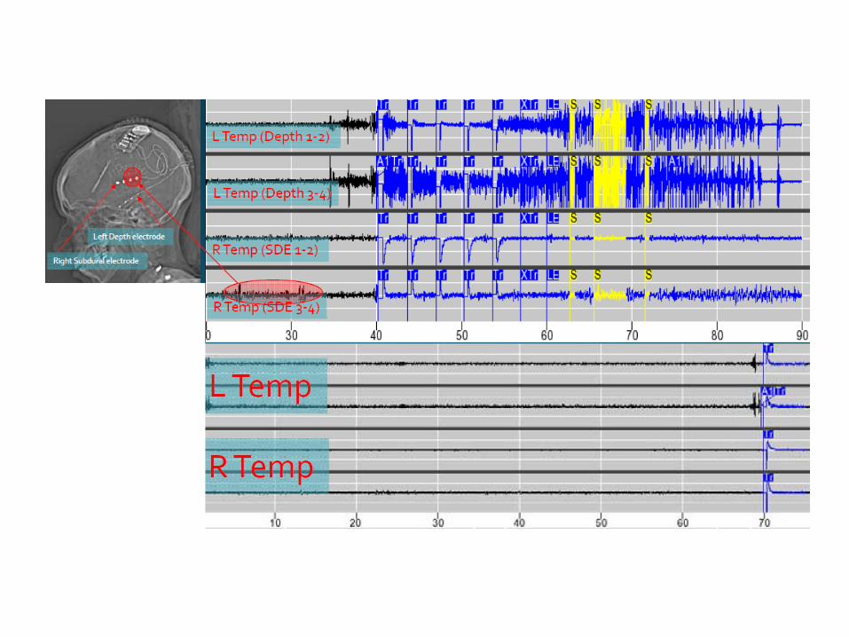
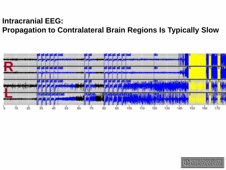
Ictal Propagation to Contralateral
Mesial Temporal Region Intracranial EEG:
Propagation to Contralateral Brain Regions Is Typically Slow

Single Photon Emission Tomography (SPECT)
1976-1984 Early tracers became available following
development of first dedicated single head
SPECT camera (Ronald Jaszczak)
~1984 Soon after, interictal SPECT scanning was
incorporated into clinical practice.
~1986 Ictal SPECT was first attempted and
compared with baseline interictal SPECT.
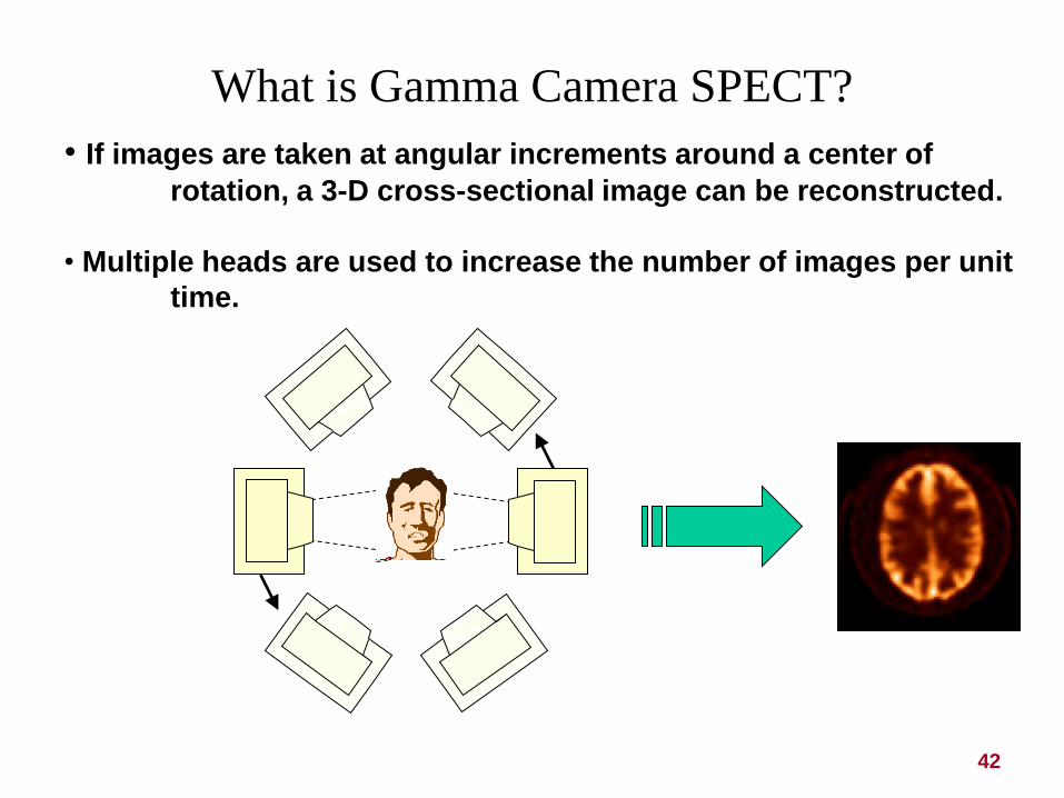
What is Gamma Camera SPECT?
42
• If images are taken at angular increments around a center of
rotation, a 3-D cross-sectional image can be reconstructed.
• Multiple heads are used to increase the number of images per unit
time.

Static tracers
99mTc-HMPAO (Ceretec)
99mTc- ECD (Neurolite)
123I-IMP (Spectamine)
123I-HIPDM
Diffusible Tracers
133Xe, 127Xe
SPECT
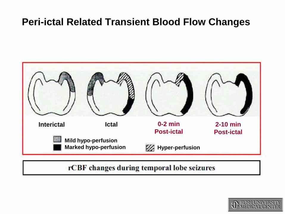
Peri-ictal Related Transient Blood Flow Changes
Mild hypo-perfusion
Marked hypo-perfusion Hyper-perfusion
Interictal Ictal 0-2 min
Post-ictal 2-10 min
Post-ictal
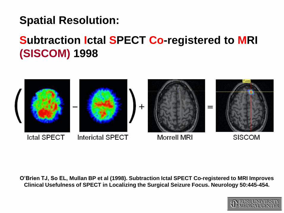
Spatial Resolution:
Subtraction Ictal SPECT Co-registered to MRI
(SISCOM) 1998
O’Brien TJ, So EL, Mullan BP et al (1998). Subtraction Ictal SPECT Co-registered to MRI Improves
Clinical Usefulness of SPECT in Localizing the Surgical Seizure Focus. Neurology 50:445-454.

Patient CH:
• CH is a 23 year old male with a 4 year history of complex partial
seizures (localization related epilepsy/lesional epilepsy) prior to RNS.)
• Initial seizure type began one day following being stung by a jellyfish
or other aquatic animal at age 16.
• Seizures were resistant to multiple antiepileptic medications.
• Seizures worsened following closed head injury from a shot put and
1 week in a comatose state.
• EEG at an outside hospital showed left temporal interictal activity.
No seizures were captured.
• Video/EEG monitoring revealed bilateral temporal lobe seizures.
• RNS implanted in June 2004.

High High Res Res 1.6mm Gapless SPGR Sequence1.6mm Gapless SPGR Sequence

Patient CH

Right Ictal Onset

Left Ictal Onset
L R L R

SISCOM: Preoperative Stereotypic Seizure Onset

Subtracted Ictal SPECT (SISCOM) + Source Modeling Right Ictal Onset

Post Implant CT

Comparison of
Two
Left Sided
Detections
Response to 2.5 mA
Seizure that generalized
Response to 4.5 mA
Did not progress

Right Sided Seizure with No Stimulation

STIM STIM
- +
RHD3 RHD4
RHD1-2
RHD3-4
LST1-2
LST3-4
RHD
LST
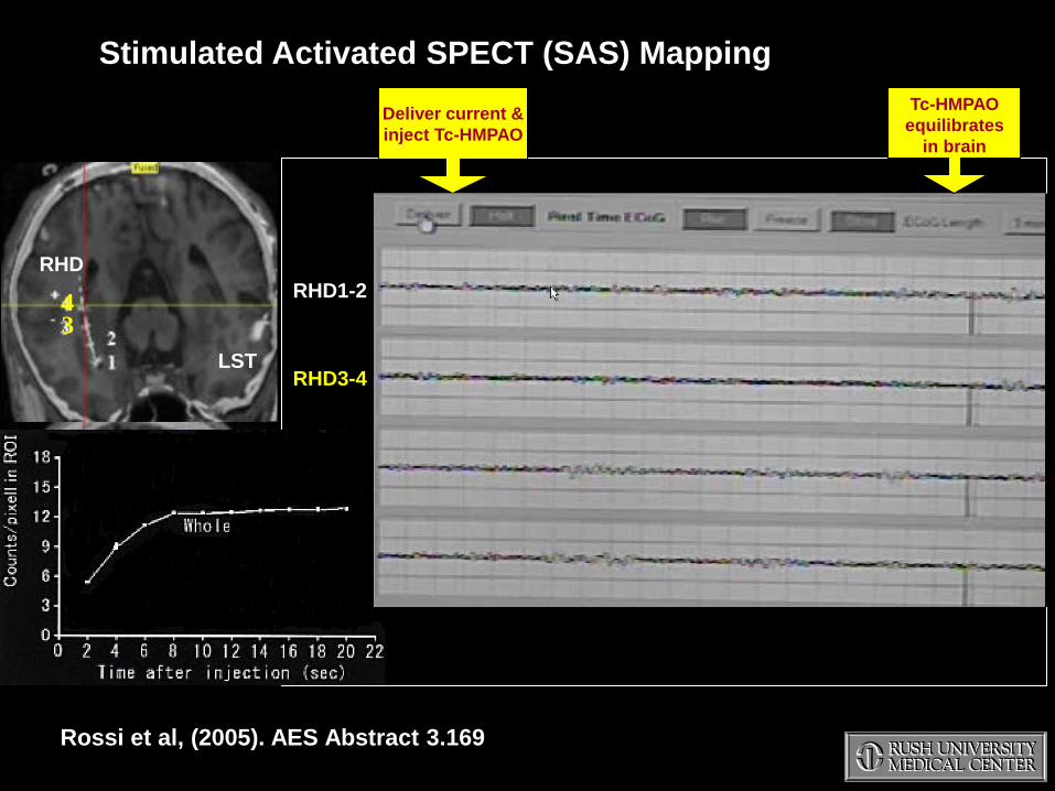
4 3
Deliver current &
inject Tc-HMPAO
Tc-HMPAO
equilibrates
in brain
Rossi et al, (2005). AES Abstract 3.169
Stimulated Activated SPECT (SAS) Mapping
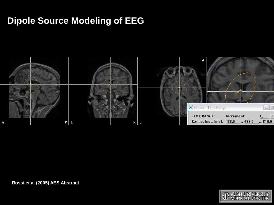
Dipole Source Modeling of EEG
Rossi et al (2005) AES Abstract

The SAS technique can demonstrate or validate changes in NEURONAL
ACTIVITY at a significant distance from a stimulated depth electrode site.
Subtracted Activated SPECT (SAS)

13 msec
30 msec
34 msec
A B
Rossi et al., (2005)

• 20 year old female diagnosed with a right temporal/thalamic grade I
astrocytoma at 13 months of age. The tumor was debulked at ages
14 months, 3 years, and 5 years.
• At ages 8-9 years of age she began experiencing vertigo/dizziness &
nausea/vomiting.
• At age 13 (2003) an outside neuropsychologist witnessed a seizure during
an neuropsychological evaluation.
• Seizure Frequency: Every 4-14 days.
Prodrome: worsening nausea and vomiting lasting up to days prior to a
‘usual’ seizure, followed by headaches, dizziness and head tremors
(titubations) within a day of her ‘usual’ seizures.
Aura: Intermittent worsening dizziness.
Ictus: staring with BUE automatisms lasting 30 sec-2 min.
Postictal period: severe nausea & vomiting, head tremors and inability to
concentrate follows these seizures for 2-3 days. Denied significant
confusion. All symptoms then resolved for 3-4 days.
• She was evaluated at: 1) Stanford, 2) Johns Hopkins, 3) Loyola
(Chicago), 4) Children’s Memorial/Northwestern (Chicago).
• Implanted with RNS at Rush in 2007.

Subtracted Post-Ictal DTI (spiDTI):

Post-Ictal Diffusion Tensor Imaging (spiDTI)
Rossi et al, (2010). Predicting White Matter Targets for Direct Stimulation Therapy. Epilepsy Research.

Pre-Implant Depth Electrode Stimulation-
Planning Model
3
3 Temporal Tip
Rossi et al, (2010). Predicting White Matter Targets for Direct Stimulation Therapy. Epilepsy Research.
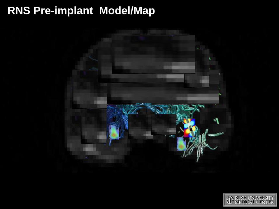
RNS Pre-implant Model/Map

Diffusion Tensor Tractography
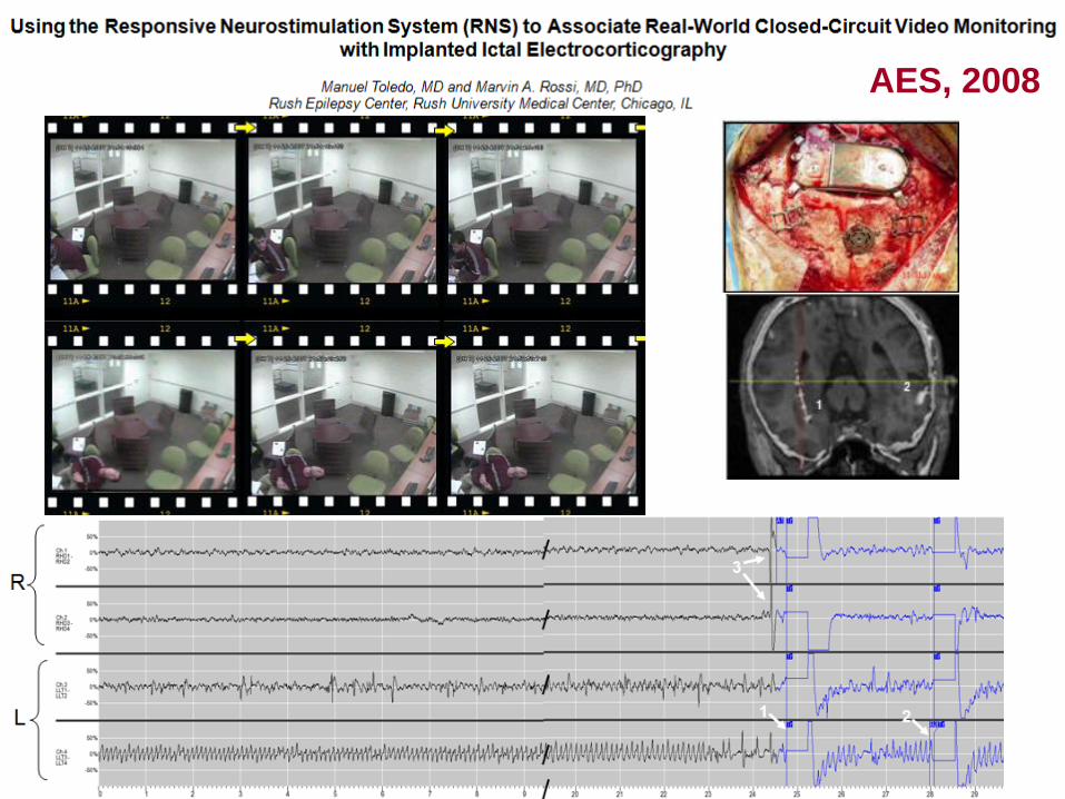
AES, 2008

Graduation:
5/9/2015
1. Next generation acute detection strategies for VNS
2. Possible reintroduction of DBS in the U.S. for
intractable focal-onset epilepsy?
3. Further explore utility of interfacing with ‘on-ramps’ for
NeuroPace RNS Therapy
4. Long-term ambulatory ECOG monitoring
5. Innovation of diagnostic neuroimaging modalities for
identifying Fragile Neural Networks:
a. SISCOM
b. spiDTI
c. SAS
SUMMARY


KEY CONTRIBUTORS
STUDENTS:
2014 Diego Garibay – ITESM Bioengineering Student
2013- Polo Cendejas Zaragoza – Bioengineer IIT Predoctoral student
2013- Tim Harris – Bioengineer (UIC Graduate)
2013- Colin Morlock – Case Western Reserve University Bioengineering Student
2011-2013 Brian Hondorp MD
2009-2010 Brian Goldberg MD
2009 Marta Wlodarzyk – Bioengineer (Illinois Institute of Technology Graduate)
2008-2009 David Sarcu MD
2008-2009 Aaron Tensharmsel MD
2008-2009 Spencer Brinker – Mechanical engineering PhD Candidate
Volodymyr Pylypyuk
Robert Dawe PhD
Glenn Stebbins PhD
SUPPORT HISTORY:
RUMC Philanthropy
Johnson & Johnson Research Grant
Mary Keane Fund
Simpleware
NeuroPace, Inc
Samsung-NeuroLogica Corp
Cyberonics, Inc