english - dental planet 7 planmeca promax utilises unique scara technology (selectively compliant...
TRANSCRIPT
ENGL
ISH

2 3
The revolutionary Planmeca ProMax X-ray unit provides a wide range of extraoral X-ray imaging modalities for the needs of modern dentistry; panoramic radiographs for imaging of the tooth arch, the jaw, the maxillary sinuses, and the temporomandibular joints, as well as dental tomographic slices and cephalometric studies.
The Planmeca ProMax platform uses robotic SCARA technology to provide utterly precise arm movements needed in digital rotational radiography for maxillofacial imaging. The three-axis SCARA robot arm moves freely without any mechanical restrictions, offering superior imaging capabilities for both existing and future radiographs.
The unique Planmeca ProMax platform makes upgrade of an existing digital Planmeca ProMax to 3D extremely easy, and new imaging programs can be added with software upgrades. The advanced design and functional concept ensure that the unit can acquire superior maxillofacial radiographs now and in years to come.
New era of dental imaging
4 5
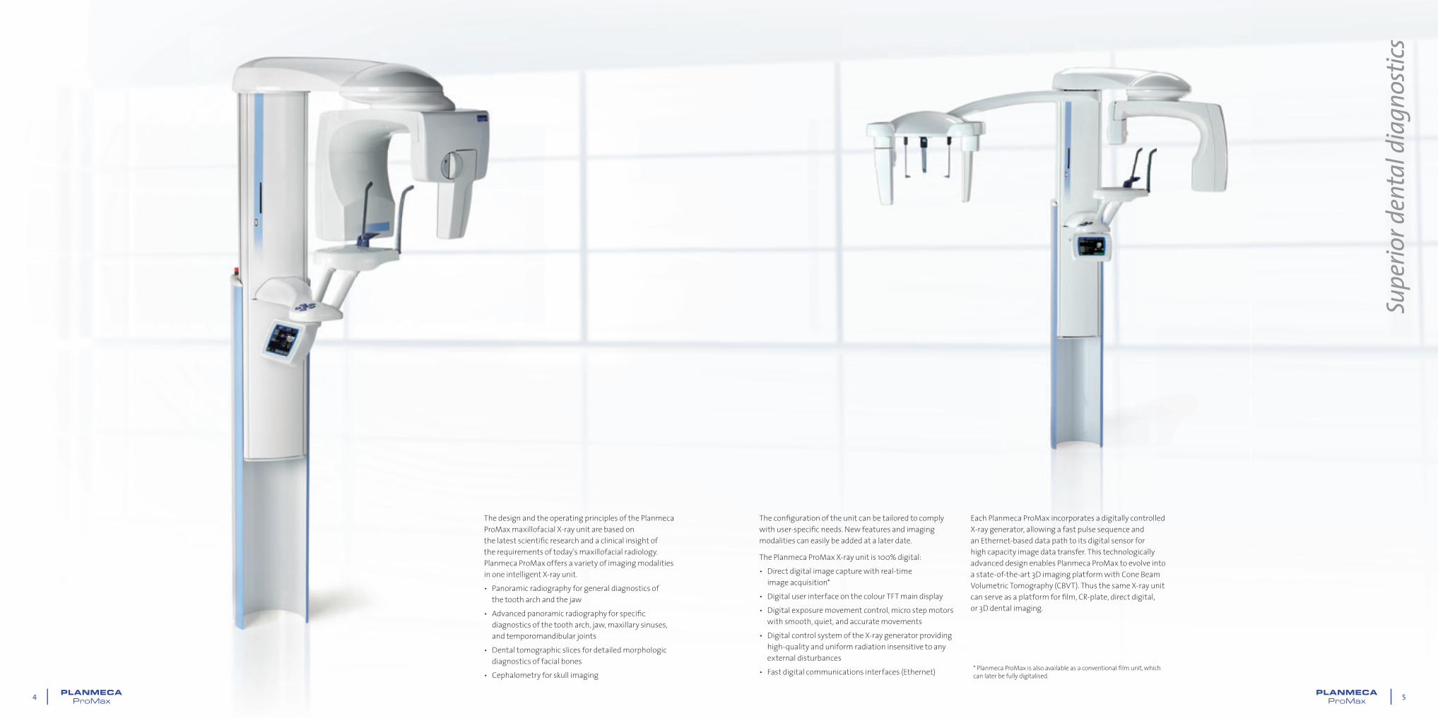
The design and the operating principles of the Planmeca ProMax maxillofacial X-ray unit are based on the latest scientifi c research and a clinical insight of the requirements of today’s maxillofacial radiology. Planmeca ProMax offers a variety of imaging modalities in one intelligent X-ray unit.• Panoramic radiography for general diagnostics of
the tooth arch and the jaw• Advanced panoramic radiography for specifi c
diagnostics of the tooth arch, jaw, maxillary sinuses, and temporomandibular joints
• Dental tomographic slices for detailed morphologic diagnostics of facial bones
• Cephalometry for skull imaging
The confi guration of the unit can be tailored to comply with user-specifi c needs. New features and imaging modalities can easily be added at a later date.
The Planmeca ProMax X-ray unit is 100% digital:• Direct digital image capture with real-time
image acquisition*• Digital user interface on the colour TFT main display• Digital exposure movement control, micro step motors
with smooth, quiet, and accurate movements• Digital control system of the X-ray generator providing
high-quality and uniform radiation insensitive to any external disturbances
• Fast digital communications interfaces (Ethernet)
Each Planmeca ProMax incorporates a digitally controlled X-ray generator, allowing a fast pulse sequence and an Ethernet-based data path to its digital sensor for high capacity image data transfer. This technologically advanced design enables Planmeca ProMax to evolve into a state-of-the-art 3D imaging platform with Cone Beam Volumetric Tomography (CBVT). Thus the same X-ray unit can serve as a platform for fi lm, CR-plate, direct digital, or 3D dental imaging.
Supe
rior d
enta
l dia
gnos
tics
* Planmeca ProMax is also available as a conventional fi lm unit, which can later be fully digitalised.
6 7
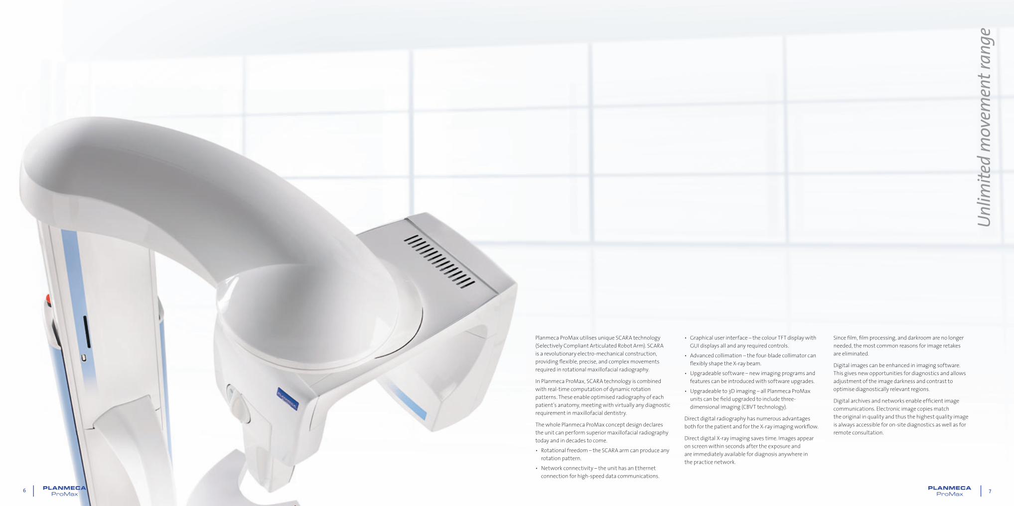
Planmeca ProMax utilises unique SCARA technology (Selectively Compliant Articulated Robot Arm). SCARA is a revolutionary electro-mechanical construction, providing fl exible, precise, and complex movements required in rotational maxillofacial radiography.
In Planmeca ProMax, SCARA technology is combined with real-time computation of dynamic rotation patterns. These enable optimised radiography of each patient’s anatomy, meeting with virtually any diagnostic requirement in maxillofacial dentistry.
The whole Planmeca ProMax concept design declares the unit can perform superior maxillofacial radiography today and in decades to come.• Rotational freedom – the SCARA arm can produce any
rotation pattern.• Network connectivity – the unit has an Ethernet
connection for high-speed data communications.
• Graphical user interface – the colour TFT display with GUI displays all and any required controls.
• Advanced collimation – the four-blade collimator can fl exibly shape the X-ray beam.
• Upgradeable software – new imaging programs and features can be introduced with software upgrades.
• Upgradeable to 3D imaging – all Planmeca ProMax units can be fi eld upgraded to include three-dimensional imaging (CBVT technology).
Direct digital radiography has numerous advantages both for the patient and for the X-ray imaging workfl ow.
Direct digital X-ray imaging saves time. Images appear on screen within seconds after the exposure and are immediately available for diagnosis anywhere in the practice network.
Since fi lm, fi lm processing, and darkroom are no longer needed, the most common reasons for image retakes are eliminated.
Digital images can be enhanced in imaging software. This gives new opportunities for diagnostics and allows adjustment of the image darkness and contrast to optimise diagnostically relevant regions.
Digital archives and networks enable effi cient image communications. Electronic image copies match the original in quality and thus the highest quality image is always accessible for on-site diagnostics as well as for remote consultation.
Unlim
ited
mov
emen
t ran
ge
8 9
Planmeca ProMax’s open positioning concept minimises errors caused by incorrect patient positioning, one of the most frequent reasons for failed radiographs. The unit features a SCARA imaging arm that can be moved completely away from the patient for positioning. This allows the operator to observe the patient freely from all directions, making patient positioning quick, precise, and easy.
A triple laser beam system ensures proper alignment, as it accurately indicates the correct anatomical positioning points.• The midsagittal plane positioning beam shows the correct sideways alignment
of the patient’s head, so that the resulting image is symmetric and undistorted in the left-right direction.
• The Frankfort horizontal plane positioning beam shows the correct forward tilt of the patient’s head, so that the dental arch lines up straight in the radiograph.
• The focal layer positioning beam indicates the focal layer’s position in the incisor region and ensures the patient is positioned fully inside the focal layer for sharp and clear images.
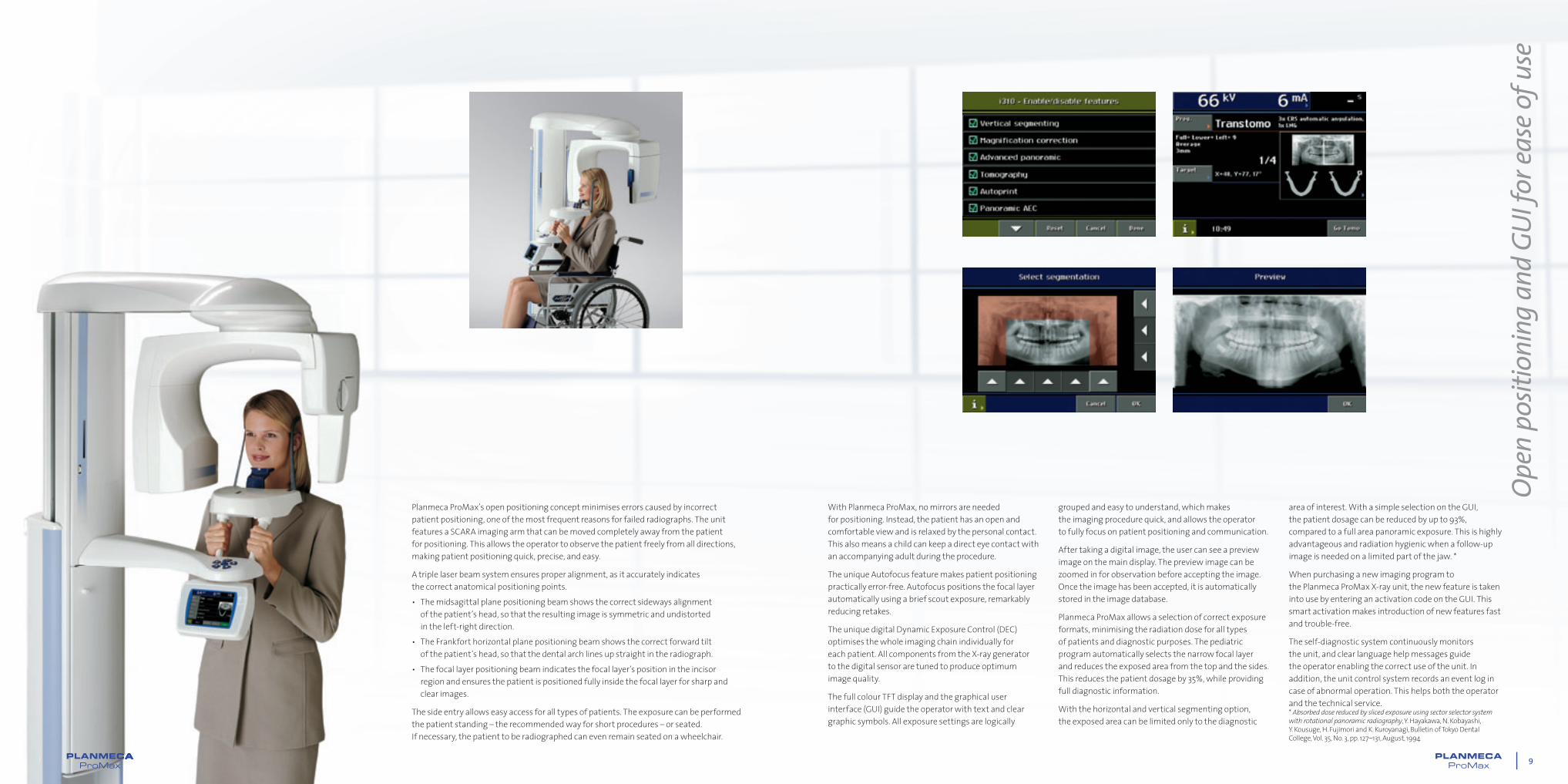
The side entry allows easy access for all types of patients. The exposure can be performed the patient standing – the recommended way for short procedures – or seated. If necessary, the patient to be radiographed can even remain seated on a wheelchair.
With Planmeca ProMax, no mirrors are needed for positioning. Instead, the patient has an open and comfortable view and is relaxed by the personal contact. This also means a child can keep a direct eye contact with an accompanying adult during the procedure.
The unique Autofocus feature makes patient positioning practically error-free. Autofocus positions the focal layer automatically using a brief scout exposure, remarkably reducing retakes.
The unique digital Dynamic Exposure Control (DEC) optimises the whole imaging chain individually for each patient. All components from the X-ray generator to the digital sensor are tuned to produce optimum image quality.
The full colour TFT display and the graphical user interface (GUI) guide the operator with text and clear graphic symbols. All exposure settings are logically
Open
pos
ition
ing
and
GUI f
or ea
se of
use
grouped and easy to understand, which makes the imaging procedure quick, and allows the operator to fully focus on patient positioning and communication.
After taking a digital image, the user can see a preview image on the main display. The preview image can be zoomed in for observation before accepting the image. Once the image has been accepted, it is automatically stored in the image database.
Planmeca ProMax allows a selection of correct exposure formats, minimising the radiation dose for all types of patients and diagnostic purposes. The pediatric program automatically selects the narrow focal layer and reduces the exposed area from the top and the sides. This reduces the patient dosage by 35%, while providing full diagnostic information.
With the horizontal and vertical segmenting option, the exposed area can be limited only to the diagnostic
area of interest. With a simple selection on the GUI, the patient dosage can be reduced by up to 93%, compared to a full area panoramic exposure. This is highly advantageous and radiation hygienic when a follow-up image is needed on a limited part of the jaw. *
When purchasing a new imaging program to the Planmeca ProMax X-ray unit, the new feature is taken into use by entering an activation code on the GUI. This smart activation makes introduction of new features fast and trouble-free.
The self-diagnostic system continuously monitors the unit, and clear language help messages guide the operator enabling the correct use of the unit. In addition, the unit control system records an event log in case of abnormal operation. This helps both the operator and the technical service.* Absorbed dose reduced by sliced exposure using sector selector system with rotational panoramic radiography, Y. Hayakawa, N. Kobayashi, Y. Kousuge, H. Fujimori and K. Kuroyanagi, Bulletin of Tokyo Dental College, Vol. 35, No. 3, pp. 127–131, August, 1994
88888

10 11
In a traditional panoramic image, the teeth interproximal contacts often overlap, which prevents diagnosis of interproximal caries. Planmeca ProMax’s optional Improved Interproximal Angulation Panoramic program produces a panoramic image where the teeth interproximal contacts are open, making the radiograph useful for caries detection.
An image taken with the optional Bitewing Panoramic program, using the improved interproximal angulation geometry, resembles an intraoral bitewing image pair. The advantage is that the image has been obtained with one simple extraoral exposure, which produces a very low radiation dosage to the patient.
The assessment of the crestal alveolar bone height is a standard study in periodontal diagnostics. The optional
Improved Orthogonality Panoramic program produces an image where the alveolar crest is clearly visible for improved diagnostics of the periodontal condition. In addition, radiographs taken with this program are highly valuable for implant planning.
The TMJ imaging programs produce lateral or posteroanterior views of open or closed temporomandibular joints. The imaging angle and position can be adjusted to correspond to the individual anatomy of each patient.
The standard Double TMJ programs produce mouth open and mouth closed images of the TMJs on the same radiograph either from lateral or from posteroanterior view.
Spec
ial p
rogr
ams f
or va
rious
diag
nosti
c nee
ds
The optional Lateral-PA TMJ program produces lateral and PA views on the same radiograph. Multi-angle TMJ programs produce radiographs with images from three different angles either from the lateral or from posteroanterior view.
In Planmeca ProMax, the PA Rotational Sinus program is a standard program. The image layer is straight and the resulting radiograph gives a clear view of the maxillary sinuses.
The optional advanced sinus programs produce lateral or posteroanterior transillumination sinus images by linear scanning. The resulting images are diagnostically similar to cephalometric projections of the sinus area.
Standard Panoramic
Stan
dard
Pedi
atric
Bitewing Panoramic PA and Lateral Non-Rotational Sinus
Lateral Double TMJ PA Double TMJ
12 13
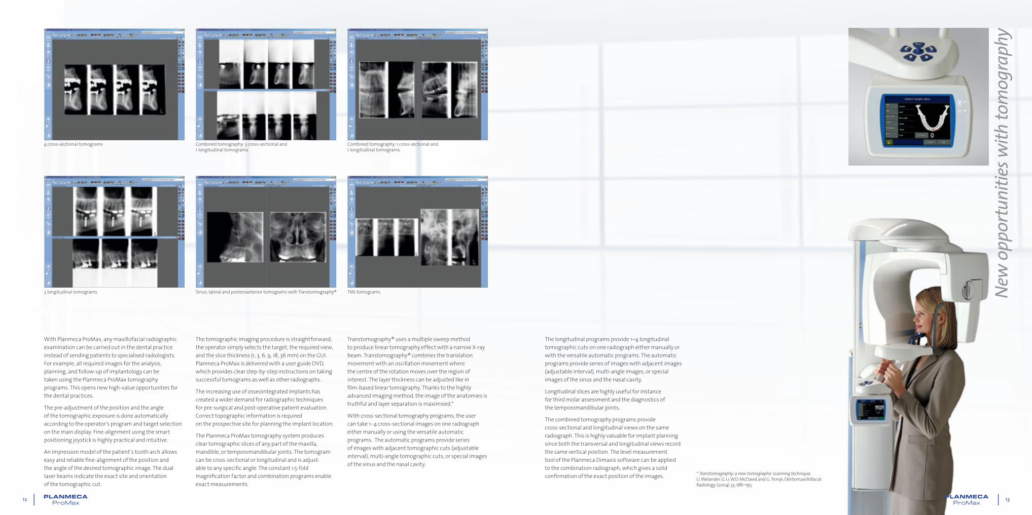
With Planmeca ProMax, any maxillofacial radiographic examination can be carried out in the dental practice instead of sending patients to specialised radiologists. For example, all required images for the analysis, planning, and follow-up of implantology can be taken using the Planmeca ProMax tomography programs. This opens new high-value opportunities for the dental practices.
The pre-adjustment of the position and the angle of the tomographic exposure is done automatically according to the operator’s program and target selection on the main display. Fine-alignment using the smart positioning joystick is highly practical and intuitive.
An impression model of the patient’s tooth arch allows easy and reliable fi ne-alignment of the position and the angle of the desired tomographic image. The dual laser beams indicate the exact site and orientation of the tomographic cut.
New
opp
ortu
nitie
s with
tom
ogra
phy
The tomographic imaging procedure is straightforward; the operator simply selects the target, the required view, and the slice thickness (1, 3, 6, 9, 18, 36 mm) on the GUI. Planmeca ProMax is delivered with a user guide DVD, which provides clear step-by-step instructions on taking successful tomograms as well as other radiographs.
The increasing use of osseointegrated implants has created a wider demand for radiographic techniques for pre-surgical and post-operative patient evaluation. Correct topographic information is required on the prospective site for planning the implant location.
The Planmeca ProMax tomography system produces clear tomographic slices of any part of the maxilla, mandible, or temporomandibular joints. The tomogram can be cross-sectional or longitudinal and is adjust-able to any specifi c angle. The constant 1.5-fold magnifi cation factor and combination programs enable exact measurements.
4 cross-sectional tomograms Combined tomography: 3 cross-sectional and 1 longitudinal tomograms
Combined tomography: 1 cross-sectional and 1 longitudinal tomograms
3 longitudinal tomograms Sinus: lateral and posteroanterior tomograms with Transtomography® TMJ tomograms
Transtomography® uses a multiple sweep method to produce linear tomography effect with a narrow X-ray beam. Transtomography® combines the translation movement with an oscillation movement where the centre of the rotation moves over the region of interest. The layer thickness can be adjusted like in fi lm-based linear tomography. Thanks to the highly advanced imaging method, the image of the anatomies is truthful and layer separation is maximised.*
With cross-sectional tomography programs, the user can take 1–4 cross-sectional images on one radiograph either manually or using the versatile automatic programs. The automatic programs provide series of images with adjacent tomographic cuts (adjustable interval), multi-angle tomographic cuts, or special images of the sinus and the nasal cavity.
The longitudinal programs provide 1–4 longitudinal tomographic cuts on one radiograph either manually or with the versatile automatic programs. The automatic programs provide series of images with adjacent images (adjustable interval), multi-angle images, or special images of the sinus and the nasal cavity.
Longitudinal slices are highly useful for instance for third molar assessment and the diagnostics of the temporomandibular joints.
The combined tomography programs provide cross-sectional and longitudinal views on the same radiograph. This is highly valuable for implant planning since both the transversal and longitudinal views record the same vertical position. The level measurement tool of the Planmeca Dimaxis software can be applied to the combination radiograph, which gives a solid confi rmation of the exact position of the images. * Transtomography: a new tomographic scanning technique,
U. Welander, G. Li, W.D. McDavid and G. Tronje, Dentomaxillofacial Radiology (2004) 33, 188–195
14 15
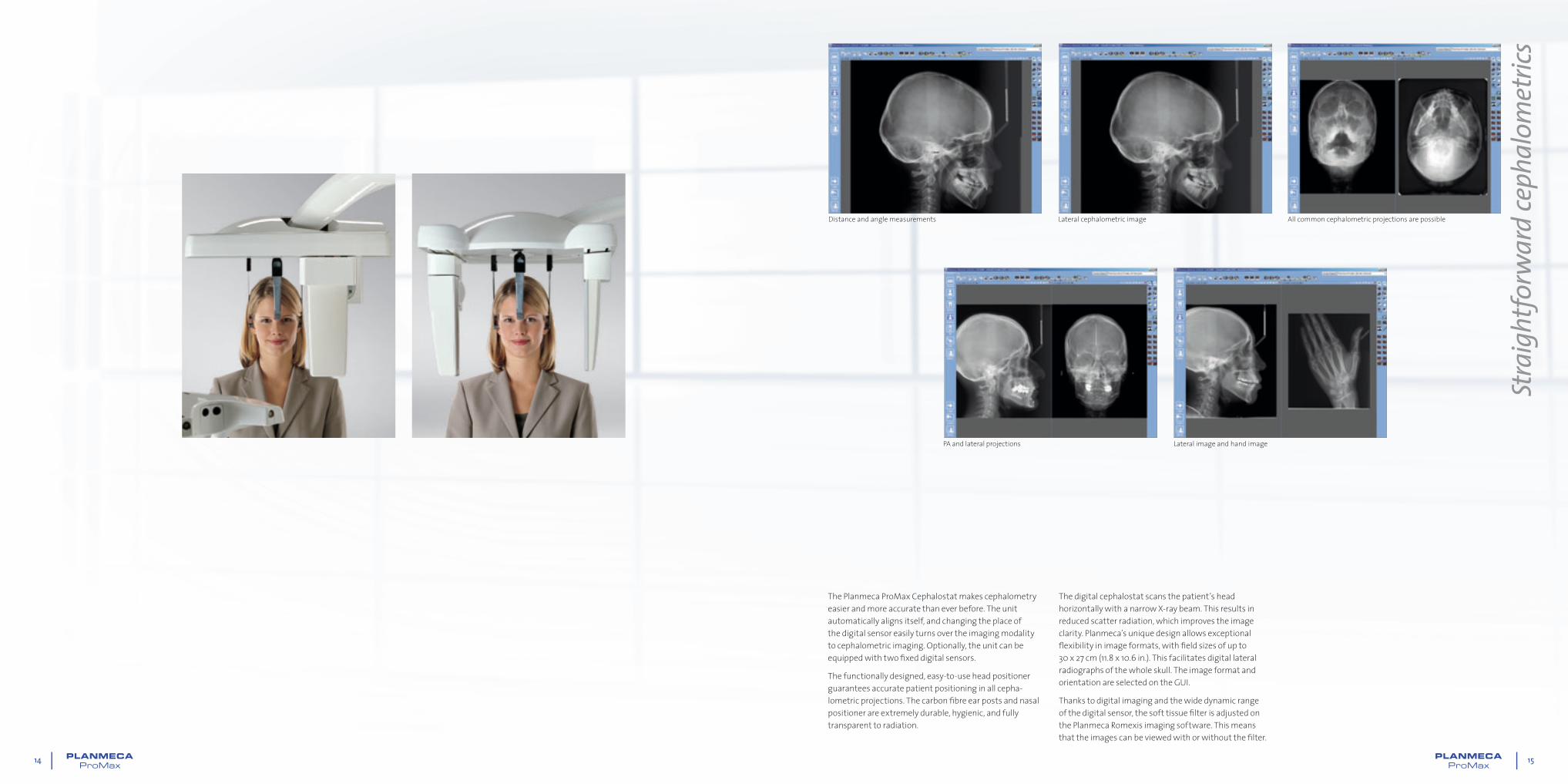
The Planmeca ProMax Cephalostat makes cephalometry easier and more accurate than ever before. The unit automatically aligns itself, and changing the place of the digital sensor easily turns over the imaging modality to cephalometric imaging. Optionally, the unit can be equipped with two fi xed digital sensors.
The functionally designed, easy-to-use head positioner guarantees accurate patient positioning in all cepha-lometric projections. The carbon fi bre ear posts and nasal positioner are extremely durable, hygienic, and fully transparent to radiation.
Stra
ight
forw
ard
ceph
alom
etric
s
Lateral cephalometric image
Lateral image and hand image
Distance and angle measurements All common cephalometric projections are possible
The digital cephalostat scans the patient’s head horizontally with a narrow X-ray beam. This results in reduced scatter radiation, which improves the image clarity. Planmeca’s unique design allows exceptional fl exibility in image formats, with fi eld sizes of up to 30 x 27 cm (11.8 x 10.6 in.). This facilitates digital lateral radiographs of the whole skull. The image format and orientation are selected on the GUI.
Thanks to digital imaging and the wide dynamic range of the digital sensor, the soft tissue fi lter is adjusted on the Planmeca Romexis imaging software. This means that the images can be viewed with or without the fi lter.
PA and lateral projections

16 17
Thanks to the technologically advanced design any Planmeca ProMax unit can be easily upgraded to 3D Cone Beam Volumetric Tomography (CBTV) imaging device by simply changing the imaging sensor and uploading software upgrades.
The 3D CBTV sensors are available in two sizes.
The larger 3D sensor captures images with volume sizes from 40 x 50 mm up to 80 x 80 mm. The 80 x 80 mm
Simpl
e upg
rade
abilit
y to
a 3D
uni
t
volume size is optimum when whole dentition, mandible, and maxilla are required in the same volume for accurate diagnosis. The 80 x 50 mm volume can be used for single views of the mandible or maxilla to decrease the radiation dose by nearly 40%. The small 40 x 50 mm volume is ideal for molar area studies and for planning of 3rd molar extractions. The volumes can also be stitched together to generate an image up to 140 mm in width or height. See examples above: TMJ study, Implant case, and Impacted canine.
Impacted canineAn impacted maxillary right canine found in the wall between sinus and nasal cavity.
TMJ studyThe condyle and the condition of the temporomandibular joint are clearly visible. Malignant fi nding can be seen inside the condyle head.
Implant caseThe lower right fi rst molar is missing. The image clearly shows there is enough bone to place an implant.
Wisdom tooth extractionThe extraction would be relatively easy.
Supernumerary toothAn extra tooth found in maxilla.
The smaller 3D s sensor offers volume sizes from 50 x 50 mm up to 50 x 80 mm. The 50 x 80 mm size is optimum for most implantology applications, whereas the 50 x 50 mm size is perfect for single tooth cases. To generate an image up to 90 mm in width, volumes can be stitched together. See examples above: Wisdom tooth extraction and Supernumerary tooth.
18 19
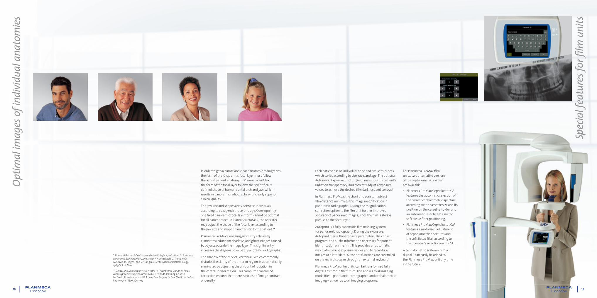
In order to get accurate and clear panoramic radiographs, the form of the X-ray unit’s focal layer must follow the actual patient anatomy. In Planmeca ProMax, the form of the focal layer follows the scientifi cally defi ned shape of human dental arch and jaw, which results in panoramic radiographs with clearly superior clinical quality.*
The jaw size and shape varies between individuals according to size, gender, race, and age. Consequently, one fi xed panoramic focal layer form cannot be optimal for all patient cases. In Planmeca ProMax, the operator may adjust the shape of the focal layer according to the jaw size and shape characteristic to the patient.**
Planmeca ProMax’s imaging geometry effi ciently eliminates redundant shadows and ghost images caused by objects outside the image layer. This signifi cantly increases the diagnostic value of panoramic radiographs.
The shadow of the cervical vertebrae, which commonly disturbs the clarity of the anterior region, is automatically eliminated by adjusting the amount of radiation in the central incisor region. This computer-controlled correction ensures that there is no loss of image contrast or density.
Optim
al im
ages
of in
divid
ual a
nato
mies
Spec
ial f
eatu
res f
or fi
lm u
nits
Each patient has an individual bone and tissue thickness, which varies according to size, race, and age. The optional Automatic Exposure Control (AEC) measures the patient’s radiation transparency, and correctly adjusts exposure values to achieve the desired fi lm darkness and contrast.
In Planmeca ProMax, the short and constant object-fi lm distance minimises the image magnifi cation in panoramic radiographs. Adding the magnifi cation correction option to the fi lm unit further improves accuracy of panoramic images, since the fi lm is always parallel to the focal layer.
Autoprint is a fully automatic fi lm marking system for panoramic radiography. During the exposure, Autoprint marks the exposure parameters, the chosen program, and all the information necessary for patient identifi cation on the fi lm. This provides an automatic way to document exposure values and to reproduce images at a later date. Autoprint functions are controlled on the main display or through an external keyboard.
Planmeca ProMax fi lm units can be transformed fully digital any time in the future. This applies to all imaging modalities – panoramic, tomographic, and cephalometric imaging – as well as to all imaging programs.
For Planmeca ProMax fi lm units, two alternative versions of the cephalometric system are available:• Planmeca ProMax Cephalostat CA
features the automatic selection of the correct cephalometric aperture according to the cassette size and its position on the cassette holder, and an automatic laser beam assisted soft tissue fi lter positioning.
• Planmeca ProMax Cephalostat CM features a motorized adjustment of cephalometric apertures and the soft tissue fi lter according to the operator’s selection on the GUI.
A cephalometric system – fi lm or digital – can easily be added to the Planmeca ProMax unit any time in the future.
* Standard Forms of Dentition and Mandible for Applications in Rotational Panoramic Radiography, U. Welander, P. Nummikoski, G. Tronje, W.D. McDavid, P.E. Legrell and R.P. Langlais, Dento-Maxillofacial Radiology, 1989, Vol. 18, May
** Dental and Mandibular Arch Widths in Three Ethnic Groups in Texas: A Radiographic Study, P. Nummikoski, T. Prihoda, R.P. Langlais, W.D. McDavid, U. Welander and G. Tronje, Oral Surgery & Oral Medicine & Oral Pathology 1988; 65:609–17
19

20 21
Dim
ensio
ns
Confi
gur
atio
ns Planmeca ProMax with fi lm-based Cephalostat Planmeca ProMax with digital CephalostatFeature Options Film Digital Required construction
ReceptorFilm ●
Digital ●
Imaging mode
Basic panoramic programs (standard):• Standard Panoramic• Standard Pediatric• Lateral Double TMJ• PA Double TMJ• PA Rotational Sinus
● ●
SCARA2 2 joint robot arm
orSCARA3
3 joint robot armVertical Segmenting (optional) ● ●
Magnification Correction Panoramic program (optional) ●SCARA2 or SCARA3
and cassette rotation
SCARA3 construction required for advanced and tomographic programs (SCARA3 is not possible to upgrade later)
● ● Adds 3rd joint
Advanced panoramic programs (optional):• Horizontal Segmenting• Improved Orthogonality Panoramic• Improved Interproximal Angulation Panoramic• Bitewing Panoramic• Lateral-PA Double TMJ• PA Multi-angle TMJ• Lateral Multi-angle TMJ • PA Non-Rotational Sinus • Lateral Non-Rotational Sinus • Midsagittal Lateral Non-Rotational Sinus
● ●
SCARA3 3 joint robot arm
Tomography (optional)
Digital tomography incl. Transtomography® ●
Wide beam linear tomography ●
Wide beam true linear tomography ●SCARA3
and cassette rotationUser interface Graphic colour TFT display ● ●
Available for both SCARA2 and SCARA3 constructions
Film markingAutoprint ●
Extra Autoprint keyboard (PS2) ●
Panoramic cassette format 15 x 30 cm ●
Cephalostat
Ceph CM ●
Ceph CA ●
Digital Ceph (Dimax)2 fixed sensors ●
1 movable sensor ●
Ceph cassette format18 x 24 cm ●
24 x 30 cm ●
8 x 10 in. ●
AECPan AEC ●
Ceph AEC ●
DECPan DEC ●
Ceph DEC ●
Additional featuresAccessory cabinet ● ●
External user interface ● ●
Column supportWall ● ●
Free standing ● ●
Physical space requirements Minimum operational space requirements Weight
Width Depth Height* Width Depth Height*
Planmeca ProMax Panoramic 96 cm
(38 in.)
125 cm
(49 in.)
153 - 243 cm
(60 - 96 in.)
150 cm
(59 in.)
163 cm
(64 in.)
243 cm
(96 in.)
113 kg
(lbs 248)
Planmeca ProMax Panoramic with Cephalostat
194 cm
(76 in.)
125 cm
(49 in.)
153 - 243 cm
(60 - 96 in.)
215 cm
(85 in.)
163 cm
(64 in.)
243 cm
(96 in.)
128 kg
(lbs 282)
*The maximum height of the unit can be adjusted for offi ces with limited ceiling space.
1293
– 2
193
(51”
– 8
6.3”
)
1250
(49
.2”)
1532
– 2
432
(60.
3” –
95.
7”)
1298
– 2
198
(51.
1” –
86.
5”)
1532
– 2
432
(60.
3” –
95.
7”)
708
(27.
9”)
698 (27.5”)150(5.9”)
1123 (44.2”) 850 (33.5”)
Ø820(32.3”)
1250
(49
.2”)
756
(29.
8”)
698 (27.5”)150(5.9”)
1128 (44.4”) 850 (33.5”)
Ø820(32.3”)
990 (39”)
Planmeca ProMax is a confi gurable product. Each unit is in di vid u al ly confi gured according to the customer's selections. When se lect ing features, please refer to the table above.
22 23
Tech
nica
l spe
cifi ca
tions
Planmeca ProMax
Generator Constant potential, resonance mode high frequency 80 - 150 kHz
X-ray tube D-054SB-P
Focal spot size 0.5 x 0.5 mm (IEC 336)
Total filtration min. 2.5 mm Al equivalent
Anode voltage 50 - 84 kV
Anode current 0.5 - 16 mA DC
Exposure time Pan 2.7 - 16 s
Ceph 0.2 - 19 s
Tomo 3 - 24 s / frame
SID Pan 500 mm (19 in.)
Ceph 163 - 170 cm (64 - 67 in.)
Magnification Pan constant 1.2
Ceph 1.08 - 1.13
CCD pixel size 33 μm
Image pixel size 66/99/132 μm selectable
CCD active surface Pan 9 x 136 mm
Ceph 9 x 270 mm
Resolution (digital) Pan max. 9 lp/mm
Ceph max. 5.7 lp/mm
Image field (digital) Pan 14 x 30 cm (5.5 x 12 in.)
Ceph 24/27 x 18/30 cm (9/10.6 x 7/11.8 in.)
File size, un compressed (digital)
Pan 4.5 - 7.7 MB
Ceph 5 - 12 MB
Line voltage 100 - 240 V, 50 or 60 Hz
Regulation Automatic, ± 10%
Line current 8 - 16 A
Colour White (RAL 9016)
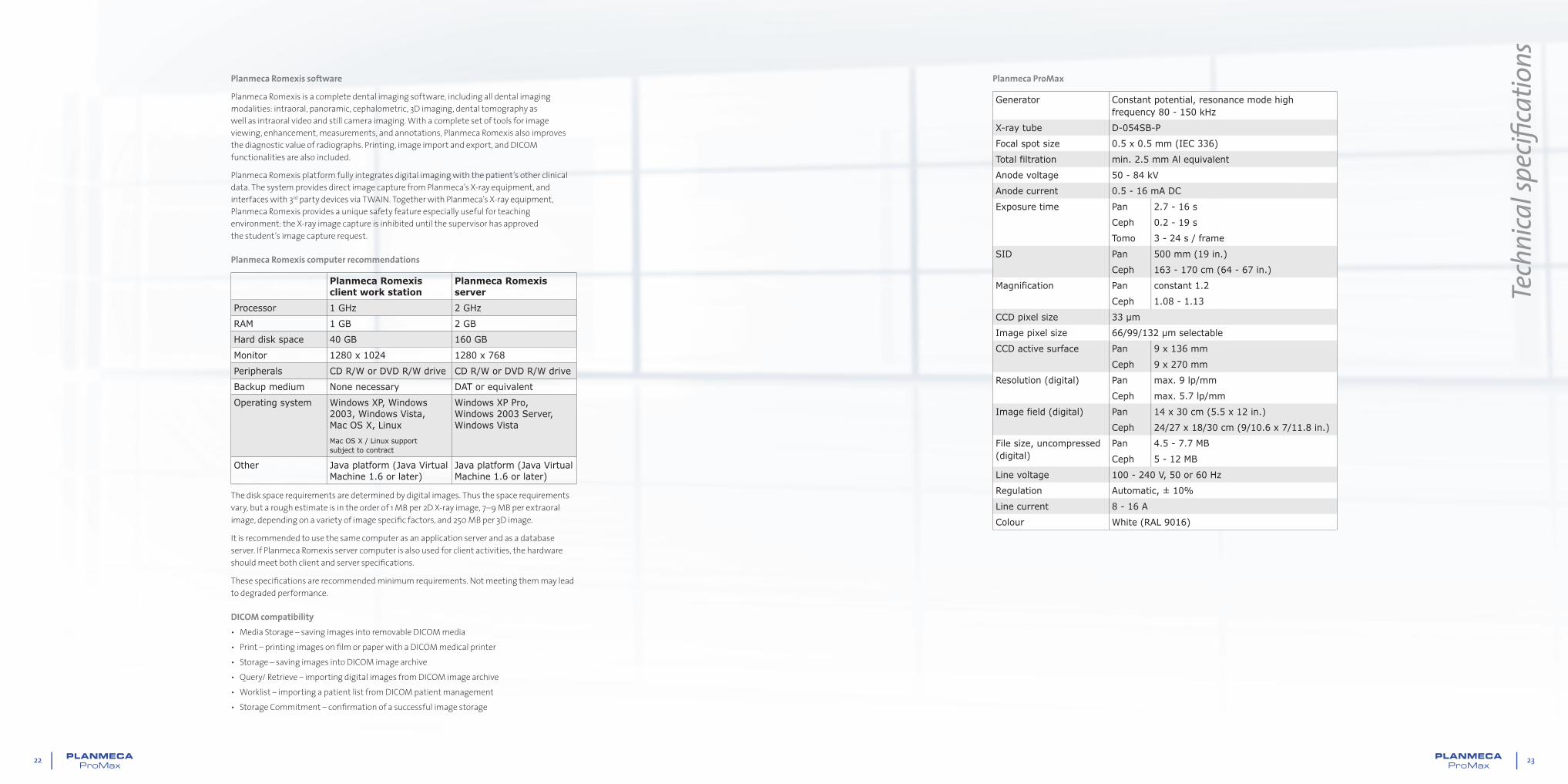
Planmeca Romexis software
Planmeca Romexis is a complete dental imaging software, including all dental imaging modalities: intraoral, panoramic, cephalometric, 3D imaging, dental tomography as well as intraoral video and still camera imaging. With a complete set of tools for image viewing, enhancement, measurements, and annotations, Planmeca Romexis also improves the diagnostic value of radiographs. Printing, image import and export, and DICOM functionalities are also included.
Planmeca Romexis platform fully integrates digital imaging with the patient’s other clinical data. The system provides direct image capture from Planmeca’s X-ray equipment, and interfaces with 3rd party devices via TWAIN. Together with Planmeca’s X-ray equipment, Planmeca Romexis provides a unique safety feature especially useful for teaching environment: the X-ray image capture is inhibited until the supervisor has approved the student’s image capture request.
Planmeca Romexis computer recommendations
Planmeca Romexis client work station
Planmeca Romexis server
Processor 1 GHz 2 GHz
RAM 1 GB 2 GB
Hard disk space 40 GB 160 GB
Monitor 1280 x 1024 1280 x 768
Peripherals CD R/W or DVD R/W drive CD R/W or DVD R/W drive
Backup medium None necessary DAT or equivalent
Operating system Windows XP, Windows 2003, Windows Vista, Mac OS X, Linux
Mac OS X / Linux support subject to contract
Windows XP Pro, Windows 2003 Server, Windows Vista
Other Java platform (Java Virtual Machine 1.6 or later)
Java platform (Java Virtual Machine 1.6 or later)
The disk space requirements are determined by digital images. Thus the space requirements vary, but a rough estimate is in the order of 1 MB per 2D X-ray image, 7–9 MB per extraoral image, depending on a variety of image specifi c factors, and 250 MB per 3D image.
It is recommended to use the same computer as an application server and as a database server. If Planmeca Romexis server computer is also used for client activities, the hardware should meet both client and server specifi cations.
These specifi cations are recommended minimum requirements. Not meeting them may lead to degraded performance.
DICOM compatibility• Media Storage – saving images into removable DICOM media • Print – printing images on fi lm or paper with a DICOM medical printer• Storage – saving images into DICOM image archive • Query/ Retrieve – importing digital images from DICOM image archive• Worklist – importing a patient list from DICOM patient management• Storage Commitment – confi rmation of a successful image storage
10016
041/0
909/e
n
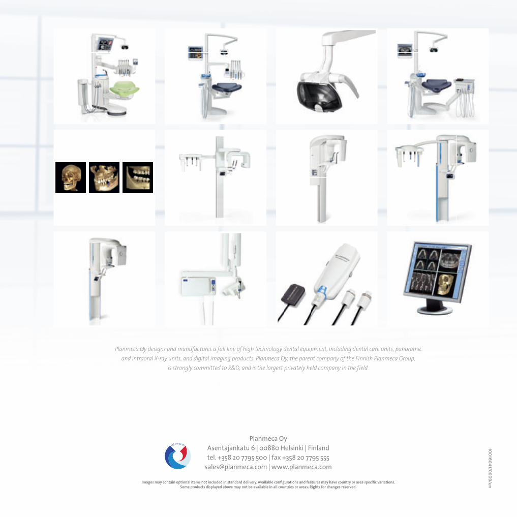
Planmeca Oy designs and manufactures a full line of high technology dental equipment, including dental care units, panoramic and intraoral X-ray units, and digital imaging products. Planmeca Oy, the parent company of the Finnish Planmeca Group,
is strongly committed to R&D, and is the largest privately held company in the field.
Planmeca OyAsentajankatu 6 | 00880 Helsinki | Finlandtel. +358 20 7795 500 | fax +358 20 7795 555
[email protected] | www.planmeca.com
Images may contain optional items not included in standard delivery. Available confi gurations and features may have country or area specifi c variations.Some products displayed above may not be available in all countries or areas. Rights for changes reserved.