engineering proteins with unique characteristics for
TRANSCRIPT

University of Kentucky University of Kentucky
UKnowledge UKnowledge
University of Kentucky Doctoral Dissertations Graduate School
2011
ENGINEERING PROTEINS WITH UNIQUE CHARACTERISTICS FOR ENGINEERING PROTEINS WITH UNIQUE CHARACTERISTICS FOR
DIAGNOSTICS AND BIOSENSORS DIAGNOSTICS AND BIOSENSORS
Smita Joel University of Kentucky, [email protected]
Right click to open a feedback form in a new tab to let us know how this document benefits you. Right click to open a feedback form in a new tab to let us know how this document benefits you.
Recommended Citation Recommended Citation Joel, Smita, "ENGINEERING PROTEINS WITH UNIQUE CHARACTERISTICS FOR DIAGNOSTICS AND BIOSENSORS" (2011). University of Kentucky Doctoral Dissertations. 180. https://uknowledge.uky.edu/gradschool_diss/180
This Dissertation is brought to you for free and open access by the Graduate School at UKnowledge. It has been accepted for inclusion in University of Kentucky Doctoral Dissertations by an authorized administrator of UKnowledge. For more information, please contact [email protected].

ABSTRACT OF DISSERTATION
Smita Joel
The Graduate School
University of Kentucky
2011

ENGINEERING PROTEINS WITH UNIQUE CHARACTERISTICS FOR MOLECULAR DIAGNOSTICS AND BIOSENSORS
ABSTRACT OF DISSERTATION
A dissertation submitted in partial fulfillment of the requirements for the degree of Doctor of Philosophy in the
College of Arts and Sciences at the University of Kentucky
By
Smita Joel
Lexington, Kentucky
Director: Sylvia Daunert, Professor of Chemistry
Lexington, KY
2011Copyright © Smita Joel 2011

ABSTRACT OF DISSERTATION
ENGINEERING PROTEINS WITH UNIQUE CHARACTERISTICS FOR MOLECULAR DIAGNOSTICS AND BIOSENSORS
Proteins possess a broad range of structural and functional properties and, therefore, can be employed in a variety of biomedical applications. While a good number of protein-based biosensing systems and biosensors for target analytes have been developed, the search for versatile, highly sensitive and selective sensors with long term stability able to provide fast detection of target analytes continues to be a challenge. To that end, we now report the design and development of modified proteins with tailored characteristics and their further utilization in the development of biosensing systems.
We take advantage of binding proteins that undergo a change in conformation upon binding to their respective target ligand analytes for the development of highly selective biosensing systems. The first class of binding proteins that was explored for this purpose was antibodies. A non-canonical site in the variable region of a monoclonal antibody was tagged with a fluorescent probe to sense the binding of analyte to its corresponding antigen-binding site. The strategy employed for designing antibody-sensing molecules is universal as it can be employed for sensing any biomolecule of interest provided that there is an available antibody against the target ligand analyte.
In a second strategy, we utilized designer glucose recognition proteins (GRPs) that were prepared by incorporation of unnatural amino acids in the glucose/galactose binding protein (GBP) of Escherichia coli and its truncated fragments. By taking advantage of the global incorporation method, we were able to fine-tune the binding affinity and thermal stability of the proteins, thus, allowing for the development of a reagentless fluorescence based fiber optic glucose biosensor capable of monitoring glucose in the hypoglycemic, normal, and hyperglycemic range, as well as in the hypothermic and hyperthermic temperature range.
Keywords: protein-based biosensing systems, antibody, glucose recognition proteins, global incorporation of unnatural amino acids, fiber optic glucose biosensor

Smita Joel
Student’s Signature
05/03/2011 Date
ENGINEERING PROTEINS WITH UNIQUE CHARACTERISTICS FOR MOLECULAR DIAGNOSTICS AND BIOSENSORS
By
Smita Joel
Sylvia Daunert
Director of Dissertation
John Anthony
Director of Graduate Studies
04/25/2011
Date

RULES FOR THE USE OF DISSERTATIONS
Unpublished dissertations submitted for the Doctor's degree and deposited in the University of Kentucky Library are as a rule open for inspection, but are to be used only with due regard to the rights of the authors. Bibliographical references may be noted, but quotations or summaries of parts may be published only with the permission of the author, and with the usual scholarly acknowledgments.
Extensive copying or publication of the dissertation in whole or in part also requires the consent of the Dean of the Graduate School of the University of Kentucky.
A library that borrows this dissertation for use by its patrons is expected to secure the signature of each user.
Name Date

DISSERTATION
Smita Joel
The Graduate School
University of Kentucky
2011

ENGINEERING PROTEINS WITH UNIQUE CHARACTERISTICS FOR DIAGNOSTICS AND BIOSENSORS
DISSERTATION
A dissertation submitted in partial fulfillment of the requirements for the degree of Doctor of Philosophy in the
College of Arts and Sciences at the University of Kentucky
By
Smita Joel
Lexington, Kentucky
Director: Sylvia Daunert, Professor of Chemistry
Lexington, KY
2011Copyright © Smita Joel 2011

iii
ACKNOWLEDGMENTS
As a graduate student I would like to acknowledge the help and support of many
people. First, I would like to thank my advisor, Dr. Sylvia Daunert, for giving me the
opportunity to work under her guidance and financial and moral support in my research
as a graduate student at University of Kentucky. Her patience and belief in my ability
provided me the opportunity to explore and utilize my scientific thoughts and skills as a
researcher. I would also like to thank Dr. Leonidas Bachas for providing support and
guidance for antibody projects. I also extend my gratitude to my committee members, Dr.
Boyd Haley, Dr. Janet Lumpp, Dr. Mark Lovell, and my external examiner Dr. Harry
LeVine, for taking the time and effort to supervise my dissertation. I acknowledge Dr.
Haley for providing his expertise and guidance for the antibody projects and encouraging
me throughout the research work.
I thank my family, specially my parents and husband for encouraging and
supporting me throughout my graduate career. Finally, I would like to thank all the lab
members, past and present, for all the help.

iv
TABLE OF CONTENTS
Acknowledgments………………………………………………………………………..iii
List of Tables……………………………………………………………………………viii
List of Figures………………………………………………………………………….....ix
CHAPTER ONE
Introduction………………………………………………………………………..1
Mutagenesis……………………………………………………………………….4
Fusion proteins…………………………………………………………………….8
Minimalist protein………………………………………………………………..12
Incorporation of unnatural amino acids……………………………………....….16
Solid phase peptide synthesis……………………………………………….……17
Site-specific incorporation of unnatural amino acids …………………………....19
Global incorporation of unnatural amino acid…………………………………...23
Binding proteins……………………………………….…………………………25
Antibody as binding protein…………………………………………..………….27
Incorporation of engineered proteins in hydrogel………..………………………34
Incorporation of biomaterials into sensing devices……….……………………..39
Fiber optic biosensors……………………………………..……………………..41
Statement of Research ………………………………………..…………………..47

v
CHAPTER TWO: UNIVERSAL STRATEGY FOR DESIGNING ANTIBODY BASED
SENSING MOLECULES BY LABELING AN INHERENT NUCLEOSIDE BINDING
SITE
Introduction………………………………………………………………………49
Experimental procedures………………………………………………………...52
Selecting the photoaffinity probe………………………………………………...52
Saturation of labeling…………………………………………………………….53
Conjugation of 2-N3ATP to Alexa Fluor 594 cadaverine………………….…….53
Determination of KD of the monoclonal antibody for 2-N3ATP-fluorophore…...54
Conjugation of the antibody to the 2-N3ATP-fluorophore……………………....54
Fluorescence studies of the interaction of the labeled antibody with IL-6……....54
Results and discussions………………………………………………………..…55
Conclusions…………………………………………………………………........62
CHAPTER THREE: BIOSENSING SYSTEM BASED ON MODIFIED ANTIBODY
FOR MONITORING BONE LOSS
Introduction………………………………………………………………………63
Experimental procedures………………………………………………………...65
Selecting the photoaffinity probe………………………………………………...65
Conjugation of 2-N3ATP to Alexa Fluor 594 cadaverine…………………….….66
Conjugation of the antibody to the 2-N3ATP-fluorophore………………………66
Calibration curve for osteonectin antibody-2-N3ATP-Alexa Fluor 594
Cadaverine……………………………………………………………………….66

vi
Association study………………………………………………………………...67
Fluorescence studies of the interaction of the labeled antibody with osteonectin.67
Selectivity study………………………………………………………………….68
Results and discussions…………………………………………………………..68
Conclusions………………………………………………………………………76
CHAPTER FOUR: DESIGNER GLUCOSE RECOGNITION PROTEINS VIA
GLOBAL INCORPORATION OF UNNATURAL AMINO ACIDS
Introduction………………………………………………………………………77
Experimental procedures………………………………………………………...80
Expression and purification of GRPs-FW/FL……………………………………81
Mass spectrometry……………………………………………………………….82
Circular dichroism……………………………………………………………….82
Labeling GRPs-FL/FW with MDCC………………………………………….....83
Fluorescence study of GRP1-FW/FL and GRP2-FW/FL in solution....................83
Results and discussions…………………………………………………………..84
Conclusions………………………………………………………………………91
CHAPTER FIVE: REAGENTLESS FIBER OPTIC BIOSENSOR FOR THE
CONTINUOUS MONITORING OF GLUCOSE
Introduction………………………………………………………………………93
Experimental procedures………………………………………………………...98
Construction of plasmid pSDGBP……………………………………………….99

vii
Expression and purification of GBP and GRPs-FW/FL………………………..100
Labeling GBP/GRPs-FW/FL with MDCC……………………………………..102
Fluorescence study of GBP152-MDCC in solution…………………………….103
Conjugation of MDCC labeled GBP/GRPs-FW/FL with acrylic acid…………103
Preparation of hydrogel precursor solution and optical fiber surface
modification…………………………………………………………………….103
Optimization of sensor response time…………………………………………..104
Study of the effect of the length of fiber on sensor response…………………...104
Sensor response…………………………………………………………………104
Results and discussions…………………………………………………………105
Conclusions……………………………………………………………………..124
CHAPTER SIX: CONCLUSIONS AND FUTURE PERSPECTIVES………..………126
REFERENCES…………………………………………………………………….…...133
VITA……………………………………………………………………………………160

viii
LIST OF TABLES
Table 1.1 Melting temperature of wild type and engineered mutants of T4
Lysozyme…………………………………………………………………7
Table 4.1 Mass spectrometry analysis of trypsin digest of GBP152-FW/FL and
GRPs-FW/FL…………………………………………………………….86
Table 5.1 Leaching studies to determine the covalently bound fluorophore
labeled GBP152 within the acrylamide hydrogel………………………111

ix
LIST OF FIGURES
Figure 1.1 Schematic showing the steps involved in design and engineering of
novel proteins……………………………………………………………...3
Figure 1.2 The protein switch, showing the components and triggers resulting
in switching from the “off” to “on” mode……………………………….11
Figure 1.3 Crystal structure of GBP without glucose (A, open form) and with
glucose (B, closed form)…………………………………………………13
Figure 1.4 Crystal structure of GBP with glucose…………………………………...15
Figure 1.5 Site-specific unnatural incorporation method……………………………21
Figure 1.6 Structure of antibody……………………………………………………..28
Figure 1.7 Computer generated model of the Fv region of the Ig with the inserted
ADP……………………………………………………………………...32
Figure 1.8 Swelling of Calmodulin immobilized hydrogel in the absence of Ca2+
and shrinking back to its original state in presence of Ca2+………...........36
Figure 1.9 pH sensitive hydrogel with glucose oxidase immobilized within the
hydrogel………………………………………………………………….38
Figure 1.10 Total internal reflection in optical fiber………………………………….43
Figure 2.1 Schematic showing the strategy for development of biosensing system
based on fluorophore labeled monoclonal antibody……...………….…..51
Figure 2.2 Structures of nucleotide probes utilized for photolabeling of antibody…56
Figure 2.3 Antibody labeled with radioactive nucleotide probes……………………57
Figure 2.4 Saturation of photolabeling of antibody with [γ-32P] 2-N3ATP………….58

x
Figure 2.5 Competitive labeling of anti-interleukin-6 (IL-6) monoclonal antibody
with 2-N3ATP-Alexa Fluor 594 cadaverine and 2-N3ATP [γ 32P]………60
Figure 2.6 Structure of fluorescent nucleotide probe and the effect of Il-6 on the
fluorescence intensity of the probe………………………………………61
Figure 3.1 Data obtained from autoradiograph of monoclonal antibody against
osteonectin with [γ-32P]8-N3ATP and [γ-32P]2-N3ATP………………….69
Figure 3.2 Calibration curve for 2-N3ATP-Alexa Fluor 594 labeled antibody
against osteonectin……………………………………………………….71
Figure 3.3 Association curve for 2-N3ATP-Alexa Fluor 594 labeled antibody
against osteonectinwith osteonectin……………………………………...72
Figure 3.4 Dose-response curve for osteonectin…………………………………….74
Figure 3.5 Selectivity studies for 2-N3ATP-Alexa Fluor 594 labeled antibody
against osteonectin with osteonectin and IL6……………………………75
Figure 4.1 Chemical structures of unnatural amino acids incorporated in glucose
recognition protein……………………………………………………….79
Figure 4.2 Dose response curves for glucose employing proteins with (a) FW
(b) FL…………………………………………………………………….87
Figure 4.3 Secondary structure of proteins as determined by far-UV Circular
Dichroism spectroscopy at room temperature…………………………...89
Figure 4.4 Thermal stability determination by Circular Dichroism spectroscopy
at 222 nm…………………………………………………………………90
Figure 5.1 Schematic of a fiber optic biosensor for glucose………………………...97
Figure 5.2 Fluorescence study of GBP152-MDCC in presence of glucose………..107

xi
Figure 5.3 Schematic of the steps involved in the immobilization of hydrogel
containing covalently attached fluorescent GBP/GRPs………………...109
Figure 5.4 Excitation and emission wavelengths of MDCC labeled GBP152
immobilized on the tip of a fiber in an optically transparent hydrogel…110
Figure 5.5 Sensor response at different time intervals……………………………..113
Figure 5.6 Sensor reversibility……………………………………………………..114
Figure 5.7 Response of two different sensors. The sensors differ in the length if
optical fiber used………………………………………………………..116
Figure 5.8 Response of hydrogel based fiber optic sensor with covalently
immobilized GBP152 on three consecutive days………………………117
Figure 5.9 Fluorescence study of fiber optic sensor in physiologically relevant
glucose concentrations………………………………………………….118
Figure 5.10 Fluorescence study of fiber optic sensor in spiked human serum……...120
Figure 5.11 Fluorescence study of fiber optic sensor in spiked pig blood…………..121
Figure 5.12 Temperature study of GBP142 and GBP-FW sensor…………………..122
Figure 5.13 Temperature study of GRPs-FL sensor…………………………………123

1
CHAPTER ONE
INTRODUCTION
Proteins are heteropolymers of amino acids with a wide array of functions and
properties. Emerging challenges in medicine, discovery science, and engineering present
an ongoing demand for modified proteins with enhanced functionality and properties. For
many years scientists have relied on nature for producing proteins that evolved into new
ones with different characteristics. A new field of engineering known as protein
engineering emerged due to advances in the field of DNA technology in the 1970s.
Protein engineering provides the basis for the design and construction of new or
improved proteins. Protein engineering has provided new insights into protein structure
and function. The principles of gene modification set forth by the capability of preparing
synthetic oligonucleotides in the early 1970s,1, 2 allowed for the first report on site-
directed mutagenesis to modify DNA by Smith and co-workers.3 However, the actual
use of site-directed mutagenesis to introduce changes at known sites in structural genes
with the aim of studying and modifying protein function was not reported until the early
1980s.4 Specifically, mutants of tyrosyl-tRNA synthetase and β-lactamase were among
the first proteins to be prepared by these methods and analyzed.5 However, another
stumbling block in the field of protein engineering was its inability to modify proteins
specifically at predetermined positions as the knowledge of three-dimensional structure
was still vague. The advent of new technologies that enabled solving protein structures by
X-ray crystallography in a nearly routine manner proved to be crucial in advancing the
field of protein engineering. Recent advancements in interactive graphics and computer

2
modeling softwares along with their use by biological chemists and biotechnologists have
stimulated knowledge-based design of proteins, thus providing better understanding of
the structure and function of proteins.
Today, scientists can design and create proteins using a plethora of genetic-based,
posttranslational, and chemical modification methods. Figure 1 shows the steps involved
in designing and creating functional proteins. The first step in designing a new protein
involves knowledge of the original protein structure to be modified. This information is
provided by the X-ray crystal structure or 2-D NMR of the protein to be modified, which
is prepared by expression, purification, and characterization of its functions followed by
its crystallization. A thorough study of the original protein structure gives an insight
about the locations within the original protein that can be altered to endow the protein
with target modifications. The protein is then modified at the genetic level or by chemical
means, and subsequently evaluated to assess whether the target characteristics have been
incorporated. The cycle continues as the new protein can again be studied and modified
using the same methods.
In this dissertation work, various protein engineering methods are employed and
discussed. However, the main focus remains the engineering of designer proteins by
global incorporation of unnatural amino acids and by chemical modification of
immunoglubulins via photolabeling. Specifically, designer proteins for glucose sensing
were developed by global incorporation of unnatural amino acids within the
Galactose/Glucose Binding Protein (GBP) of E. coli. These designer glucose recognition
proteins (GRPs) with improved stability and activity were then incorporated into a
hydrogel network, which

3
Figure 1.1 Schematic showing the steps involved in the design and engineering of novel
proteins. Figure adapted from Blundell, et al, Phil. Trans. R. Soc. Lond. B 324, 448,
1989.
Protein assay and
characterization
Activity test of proteins
Study of structure and stability by
CD
2D NMR X‐ray
crystallography
Visualization of 3D
structure of proteins
Knowledge based design of novel proteins
Mutagenesis/unnatural amino
acids/fusion proteins/ minimalist proteins
Growth and expression of
proteins
Protein
Engineering

4
served as the sensing material in fiber optic biosensors for glucose. Designer antibodies
were prepared by photolabeling at an unconventional site within the variable region of
the antibody, and utilized for developing universal biosensing systems for various
biomolecules.
Mutagenesis
The properties and functions of proteins can be altered at the genetic level. The
process for generating amino acid coding changes at the DNA level is called
mutagenesis. These changes in the amino acids can be generated in a site-directed or
random manner. With the availability of X-ray crystallographic structures of proteins, it
is possible to determine which amino acids in a protein can be changed to attain a specific
property. Mutations can either be point mutations, wherein one amino acid is replaced by
another, or deletion mutations in which an amino acid is deleted from the amino acid
sequence of a protein, or insertion mutations where an amino acid is inserted in the amino
acid sequence. However, if the amino acids to be changed in order to alter the properties
or functions of a protein are not known, then random mutagenesis can be carried out.
This approach has two advantages. First, detailed information about the function of
amino acids in the protein to be altered is not required. Secondly, this approach results in
an array of protein mutants with potentially interesting properties. Methods to create
random mutants include XL1-Red competent cells, error-prone PCR and degenerate
oligonucleotides-Pfu (DOP).6 XL1-Red cells are engineered E. coli with DNA repair
deficiency. In this method the plasmid bearing the DNA of interest is transformed into

5
the cells and mutations occur in each round of replication. In error-prone PCR the
polymerase has high error rate. During amplification mutations are introduced by altering
the ratio of nucleotides and the concentration of divalent cations.7 DOP mutagenesis
involves the use of two degenerate oligonucleotides as primers. The position of mutation
can be controlled by adjusting the ratios and position of degenerate nucleotides during the
synthesis of degenerate oligonucleotides.
Point and deletion mutations can lead to the construction of proteins with new
properties. Point mutations have been utilized in several proteins to introduce amino
acids for covalent attachment of fluorophores. Hence, this protein engineering technique
can be used to integrate optical signal transduction functions directly into proteins. This
method has been used for developing fluorophore labeled binding protein biosensors for
several analytes, including glucose, amino acids, anions, cations and dipeptides.8 A
fluorescent biosensor family was constructed by mutating several periplasmic binding
proteins to covalently attach environment sensitive fluorophores to the proteins.8 Single
cysteine mutations were performed at desired locations in the protein, which allowed
covalent attachment of environment sensitive fluorophores in or near the ligand binding
pocket of the protein. The biosensors can detect the presence of the ligand/analyte, which
is measured as a dose-dependent change in the fluorescence of the fluorophore upon
ligand binding.8-10 Besides fluorescent reporter molecules, several other molecules can be
covalently attached to mutated proteins for immobilization purposes.
Thermostability of proteins can be improved by site-directed mutagenesis. Point
mutations have been engineered in the yeast enzyme triosephosphate isomerase to replace
two different asparagines with threonine and isoleucine. Converting the asparagines to

6
threonine and isoleucine enhanced the thermostability of the enzyme. However, replacing
an asparagine with aspartic acid resulted in reduced thermostability of the protein.11
Thermostability of proteins can also be enhanced by adding disulfide bonds within the
structure of the protein, which can be achieved by introducing cysteine residues via site-
directed mutagenesis. T4 lysozyme in its native form has two cysteines, neither of which
is involved in a disulfide bond. The melting temperature of T4 lysozyme is 41.9 °C. It has
been reported that introducing cysteines that are involved in the formation of disulfide
bonds enhances the thermal stability of T4 lysozyme. The results demonstrated that the
thermal stability increases as the number of disulfide bonds increases, with the most
thermostable mutant being the one with the largest number of disulfide bonds (Table
1.1).12
The catalytic activity of proteins can also be modified by site-directed
mutagenesis. This has been proved by engineering two point mutations in tyrosyl-tRNA
synthetase of Bacillus stearothermophilus. One mutation, Thr51-Ala51 was done to get
rid of the hydroxyl group and the other, Thr51-Pro51 was performed to distort the local
polypeptide backbone. Both mutations increased ATP binding activity in the enzyme.
The mutant with Pro51 was found to bind ATP 100-fold stronger than the native
enzyme.13
Furthermore, mutagenesis has been utilized to engineer proteins for therapeutic
purposes. For example, insulin has been engineered through site-directed mutagenesis to
create rapid acting (lispro, aspart and glulisine) and long acting (glargine and detemir)
insulin analogs.14 The rapid acting insulin analogs were constructed by replacing or
switching one or two amino acids. These mutations in insulin reduce the tendency to

7
Table 1.1 Melting temperature of wild type and engineered mutants of T4 lysozyme.
Adapted from Matsumura, et al, Nature 342, 291-293, 1989.
Enzyme No. of disulfide
bonds Tm (°C)
Wild type 0 41.9
3C-54T 1 46.7
D3-97/9-164 2 57.6
D9-164/21-142 2 58.9
T3-97/9-164/21-142 3 65.5

8
form hexamers, thus facilitating rapid absorption. The long acting insulin analogs were
obtained by substitution or deletion of specific amino acids. These changes resulted in a
shift in the isoelectric point of insulin from 5.4 to 6.7, which prolongs its dissociation at
physiological pH conditions. Another strategy to engineer proteins for therapeutic
purposes is the development of catalytic antibodies. Antibodies are proteins that are used
by the immune system to identify and neutralize foreign agents. However, mutagenesis
has enabled the development of catalytic antibodies that function as enzymes and
catalyze reactions. Some of the potential therapeutic applications include detoxification
after accidental exposure to insecticides,15 prevention of cocaine addiction and
overdose,16, 17 and prodrug activation in cancer treatment.18
Fusion Proteins
Partial or full protein sequences can be joined together via recombinant DNA
technology resulting in fusion or hybrid proteins. These proteins can have partial or full
functions or characteristics of the constituent proteins. Fusion proteins have extensively
been used to study interactions between proteins. Additionally, a binding protein can be
fused to an inherently fluorescent protein, such as the green fluorescent protein (GFP)
from the jellyfish Aequorea victoria, to develop protein-based sensing systems. Upon
binding to the analyte, the target binding protein undergoes a change in the conformation,
which can be detected as a dose-dependent change in the fluorescence of the reporter
protein, GFP. Furthermore, fluorescent fusion proteins have been used to study the
location, movement and degradation of proteins in living cells.19 Another application of

9
fusion proteins is to aid in the purification of proteins via affinity chromatography. A
short sequence of amino acids, such as histidines, are fused to either the N- or C-terminus
of a protein allowing for its purification by nickel affinity chromatography; for example,
in this dissertation work a six histidine sequence was inserted at the C-terminus of the
gene encoding the glucose binding protein for purification via nickel affinity. Small
peptides20 or antibody fragments21 can also be fused to proteins for purification purposes.
For example, fusion of antibody fragments to proteins enables purification via
interactions with protein A. Fusion protein technology has been employed to develop
biosensing systems for peptides,20 drugs, zinc and heme, among others. An octapeptide
was fused to GFP in order to develop a fluorescence binding assay for the peptide. The
immunoassay was based on the sequential binding of the free octapeptide and GFP
labeled octapeptide to anti-octapeptide antibodies on a solid surface.22 A zinc sensor was
developed by fusing histidine tags at each end of a fusion protein comprised of the
enhanced cyan and enhanced yellow fluorescent proteins joined together by a linker.
Since the hexahistidine tags undergoe dimerization in the presence of zinc, fluorescence
resonance energy transfer occurs between the two fluorescent protein domains, thus
resulting in the ratiometric detection of zinc in nM ranges.23 In another example,
cytochrome b562, a heme-binding protein, was fused to the N-terminus of the enhanced
green fluorescent protein (EGFP) to create a heme sensor. The detection of hemin was
based on the fluorescence quenching due to energy transfer between EGFP and
cytochrome b562 in the presence of hemin.24
Split protein technology, which is employed to study protein-protein
interactions25, 26 and protein-nucleotide interactions, is a step further in the field of protein

10
engineering.27 The split protein methodology includes one and two component systems.
A one component system consists of two fragments of a reporter protein attached to two
peptides or nucleotides. The two component system has been exemplified by fragmenting
GFP and attaching the two inactive fragments to two leucine zippers. The leucine zippers
come together via non-covalent interactions, bringing the two fragments of GFP together,
hence producing fluorescence.28 When two fragments of a reporter protein are attached
to the two termini of a binding protein in a one component system, the binding of the
ligand to the binding protein results in a conformation change that brings the two
fragments of the reporter protein closer, hence producing a signal. This approach can be
utilized to develop biosensing systems for various biomolecules by inserting the sequence
of a biomolecule binding protein within the sequence of a reporter protein.
Conformational changes in maltose binding protein (MBP) have been studied using the
one component split methodology. Two fragments of GFP were attached to the N- and C-
terminus of MBP. Upon binding to maltose, the two fragments of GFP were brought
together and resulted in changes in fluorescence. The changes in fluorescence in response
to maltose were detected in a concentration and time dependent manner.29 This sensing
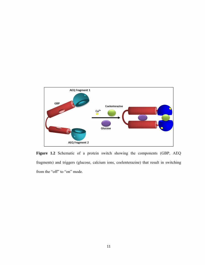
strategy is also referred to as molecular switches,30, 31 and has been utilized for inserting
MBP,29 GBP32 (Figure 1.2), and calmodulin33 within split reporter proteins such as GFP,
aequorin, and EGFP, respectively. The split protein methodology allows for two
functionally and structurally unrelated proteins to be integrated, thus combining their
characteristics; hence, it can prove to be an important tool for designing molecular
sensors for analytical and therapeutic applications.34
11
Figure 1.2 Schematic of a protein switch showing the components (GBP, AEQ
fragments) and triggers (glucose, calcium ions, coelenterazine) that result in switching
from the “off” to “on” mode.

12
Minimalist Proteins
Proteins are macromolecules comprised of hundreds of amino acids. However,
only a fraction of the protein structure or amino acid sequence in a protein is required for
maintaining a specific biological activity. Hence, efforts have been made to create a
minimum portion of a protein structure required to retain the desired function. For
example, in an enzyme or a binding protein the substrate or ligand binds to an active site
consisting of a specific sequence of amino acids; however, the amino acid sequence of
the rest of the protein helps in maintaining the required structure of the active site.35 The
truncation of proteins to generate protein fragments that contain the amino acid sequence
essential for maintaining the protein biological function has been explored in our lab.
GBP is one example of such proteins subjected to truncation.
The galactose/glucose binding protein (GBP) from E. coli is a periplasmic protein
involved in bacterial chemotaxis and active transport of glucose and galactose.36 GBP is a
monomer with a molecular weight of 33 kDa, consists of 309 amino acids, and binds to
glucose and galactose present in μM ranges.37 GBP is ellipsoidal in shape and consists of
two globular domains held together by three peptide strands referred to as the hinge
region.10 The two domains have a core of six strand β-sheet flanked on both sides by two
or three helices.38 In the open form (Figure 1.3), when glucose is not bound, the two
domains are far apart and the ligand binding site is deep within the cleft and accessible to
solvent. On the other hand, in the closed form (Figure 1.3) glucose is bound in the cleft
between the two domains and the ligand binding site is solvent-inaccessible, which
results in the formation of an extensive network of hydrogen bonds between the polar
residues of the binding site and the sugar.39 The binding of glucose is accompanied by a

13
Figure 1.3 Crystal structure of GBP without glucose (A, open form) and with glucose (B,
closed form).

14
conformational change of the protein at the hinge region. The bound sugar is stabilized
within the binding pocket by a series of interactions, including hydrogen bonds, van der
Waals interactions and salt bridges. Upon binding to the sugar, 13 strong hydrogen bonds
are formed between the sugar and the protein, involving eight polar side chains of GBP
distributed between the two domains and a water molecule. Additionally, the
glucopyranose ring is stacked on both sides with aromatic residues. The presence of
glucose bound to GBP has been found to increase the stability of the protein structure and
restore the cooperativity of temperature-induced transitions.40 In addition to the sugar
binding site, GBP also has a calcium binding site in the C-terminal domain. The calcium
binding site is constituted by a nine residue (134-142) loop.41
GBP was truncated at both the N- and the C-terminus generating protein fragments.
Three fragments of GBP were thus obtained. The largest one had amino acids 14 through
296, the second fragment had 14 through 256, and the smallest one had 87 through 271.
The smallest fragment did not bind to glucose; hence, it lost its activity upon truncation,
suggesting that the amino acids removed from the protein sequence were essential for
glucose binding. However, the two larger fragments of the full length GBP (Figure 1.4)
showed glucose binding activity.42 This observation suggested that the full length amino
acid sequence is not necessary for maintaining the activity of GBP. Therefore, utilization
of the minimalist strategy allows for the creation of smaller functional proteins. The
minimalist protein strategy can also be utilized to study folding of the protein and its
active site.43

15
Figure 1.4 Crystal structure of GBP with glucose. The red portions in the structure
indicate the parts of the amino acid sequence that have been eliminated. A: GBP
fragment with amino acids 14 through 296, B: GBP fragment with amino acids 14
through 256.42
A B

16
Incorporation of unnatural amino acids
A key advancement in the field of protein-based biosensors is the design of
proteins with improved binding selectivity, specificity and affinity for their ligands, with
enhanced stability of the protein. Protein engineering methods are generally based on
chemical and genetic methods. A more recent, less studied method of engineering
proteins involves the incorporation of unnatural amino acids in place of natural amino
acids in proteins, either
at a particular site (site-specific) or throughout the protein (global). This is a challenging
method as it plays with the natural fidelity and proof reading mechanism that living
organisms are endowed with. Nature has provided 20 natural amino acids that are vital
for the existence of organisms. Replacing the natural amino acids with unnatural amino
acids might result in the production of undesired or incorrect proteins, which can lead to
cell death. However, emerging techniques and new methods have allowed expanding the
genetic code beyond the one nature has provided.
Expanding the genetic code via incorporation of unnatural amino acids allows
exploring the structure and function of proteins by manipulating the amino acid
backbone. Also, this method allows for the tailoring or tuning of properties and functions
of proteins. For example, if leucine in a protein is replaced by an unnatural analogue of
leucine, such as fluoroleucine, then the change in the structure of the protein can be
studied. Fluorine and hydrogen have similar van der Waals radii; therefore, hydrogen can
easily be replaced with fluorine in amino acids, producing minimal steric perturbation in
proteins. However, high electronegative and hydrophobic characteristics endow proteins

17
with distinct properties. Several fluorinated amino acids, such as, fluorovaline,
fluoroleucine, fluoroisoleucine, fluorophenylalanine, and fluoroproline have successfully
been incorporated into the structure of proteins.44 Besides providing an insight into the
structure of a protein, incorporation of unnatural amino acids allows for introducing
chemical scission sites and sites for photoaffinity labeling. It also enables site-specific
conjugation and immobilization. A variety of methods for the incorporation of unnatural
amino acids in proteins have been developed, which include solid phase peptide
synthesis, semi-synthesis, in vitro site-specific incorporation, in vivo site-specific
incorporation, and global incorporation. All of these methods will be discussed in the
following sections. In our efforts towards improving the activity and stability of GBP we
utilized the global incorporation of unnatural amino acids into the structure of GBP.
Solid phase peptide synthesis
One of the most used methods for incorporation of unnatural amino acids is solid
phase peptide synthesis. This is a synthetic method for producing peptides or proteins that
are difficult to express in bacteria or to introduce unnatural amino acids in peptides. This
method employs the carboxyl group of one amino acid for coupling with the amino group
of another amino acid, while protecting the amino or carboxyl group of amino acids not
involved in peptide bond formation. In peptide synthesis an amino acid is bound to a
resin via the carboxyl group and the amino group is protected. The amino group is then
deprotected and reacted with the carboxyl group of another amino acid having the amino

18
group protected. This method involves repetitive cycles of deprotecting, coupling and
washing. The synthesized peptide remains covalently attached to the solid support until
cleaved by anhydrous hydrogen fluoride or trifluoroacetic acid. Two important protecting
groups used for protecting the α-amino group are Fmoc (9-
fluorenylmethyloxycarbonyl)45, 46 and t-Boc (tert-butoxycarbonyl).47, 48 A variety of solid
supports, such as, polystyrene or polyacrylamide resins can be used. This method is well
standardized and automated; however, it has a drawback, that is, only small peptides
comprising up to 100 amino acids can be synthesized. This chemical method can also be
used for incorporating unnatural amino acids in peptides. However, the peptide produced
may not fold properly or show the desired activity. The desired activity of proteins may
be achieved by post-translational modification of the protein within the cell. However,
the desired activity of protein cannot be obtained using the chemical synthesis of
peptides. The demand for bigger peptides or proteins still remains; to that end, semi-
synthesis of proteins can be performed.
Semi-synthesis of proteins involves chemical ligation of either two naturally
existing smaller peptides or two peptides synthesized via solid phase peptide synthesis or
one natural and one synthesized peptide. A major advancement in this field was the
chemoselective linkage49 of a peptide containing an amino-terminal cysteine with a
peptide containing a carboxy-terminal thioester, attained by the initial
transthioesterification followed by S,N-shift to produce a native amide bond. Hence, this
method can be used for generating recombinant proteins. Amino-terminal cysteines can
be introduced in proteins using proteases,50 while a thioester can be introduced into a
recombinant protein by fusion to an intein. The peptide with carboxy-terminal cysteine

19
then reacts with the protein thioester by native chemical ligation, resulting in a semi-
synthetic protein. This method is known as expressed protein ligation (EPL).51, 52 This
method allows the generation of bigger peptides/proteins and somewhat overcomes the
limitations posed by post-translational modification of proteins. A drawback of this semi-
synthesis method is that it requires the introduction of an N-terminal cysteine or the
presence of cysteine within the structure of the protein, which in some cases may perturb
the structure or activity of the protein. The incorporation of unnatural amino acids at
desired locations within proteins is also limited when using EPL methods as the unnatural
amino acid can only be introduced in the synthetic part of the peptide. Thus, the need to
incorporate unnatural amino acids without any location restrictions has been achieved by
exploiting the natural biological machinery that the cells use for manufacturing proteins.
Site-specific incorporation of unnatural amino acids
The fidelity of the genetic code depends upon the interaction of codon-anticodon
and the acylation of tRNA with the proper amino acid by aminoacyl-tRNA synthetases
(aaRSs). Thus, changing the genetic code requires a tRNA for a specific codon and a
method of acylating the tRNA with an unnatural amino acid. Nature has provided 64
three base codons in the standard genetic code. Three of these three base codons, namely,
UAG (amber), UAA (ochre) and UGA (opal) are referred to as stop codons or nonsense
codons as they signal the termination of translation. The amber codon is the least used
stop codon in E. coli, and a number of efficient ‘suppressor’ tRNAs are known.53 The

20
suppressor tRNAs are tRNAs from certain organisms that have the capability to ‘read
through’ a stop codon. Additionally, the presence of the amber suppressors in some E.
coli strains does not significantly affect cell growth rates.54
Schultz and Chamberlin have reported in vitro incorporation of unnatural amino
acids by “nonsense suppression” strategy. Nonsense suppression involves the use of
nonsense codons and suppressor tRNAs. The first step towards incorporation of unnatural
amino acids via this method is the construction of aminoacylated suppressor tRNA.
Hecht and coworkers have constructed a suppressor tRNA by the condensation of the 3'
end of an E. coli truncated tRNA and an acylated dinucleotide in the presence of T4 RNA
ligase, hence resulting in an aminoacyl-tRNA (aatRNA) charged with an unnatural amino
acid.55, 56 This methodology has been further improved by Schultz57 and Sisido.58
Dougherty and coworkers have been successful in incorporating unnatural amino
acids into proteins expressed in Xenopus oocytes via the nonsense suppression method.
The oocyte is coinjected with the modified mRNA encoding for the target protein and the
aatRNA is chemically acylated with an unnatural amino acid. This results in the
expression of the protein containing the unnatural amino acid on the surface of the
oocyte. This methodology has enabled a detailed study of the structure-function
relationships of ion channels and the incorporation of biophysical probes.59
Further, Schultz and coworkers have developed an alternative to the nonsense
supression method for incorporating unnatural amino acids into proteins in vivo. This
method (Figure 1.5) utilizes a novel tRNA/aaRS pair for each unnatural amino acid to be
incorporated. The aaRS is engineered such that it can only recognize the unnatural amino
acid and proficiently acylate the tRNA. The tRNA/aaRS pair should be orthogonal to the

21
Figure 1.5 Site-specific unnatural amino acid incorporation method. The specific site on
the protein’s gene is first mutated to TAG. The genes encoding for the orthogonal
tRNA/aminoacyl-tRNA synthetases are cotransformed into E. coli with the genes for the
TAG-mutated protein.

22
host organism, meaning that the aaRS must not recognize endogenous tRNAs and the
suppressor tRNA cannot be a substrate for endogenous aaRSs. The most commonly used
tRNA/aaRS pair has originated from Methanococcus jannaschii. The tRNA/aaRS pair
from M. jannaschii has been used to incorporate various unnatural amino acids, such as
photoactivatable amino acids60 and keto-containing amino acids,61 into proteins. Several
other orthogonal tRNA/aaRS pairs have been proposed to suppress nonsense codons in
eukaryotic cells, including mammalian cell lines.62 Schultz and coworkers have
efficiently incorporated five amino acids with high fidelity in the genetic code of
Saccharomyces cerevisiae by utilizing a unique orthogonal tRNA/aaRS pair originated
from E. coli.63
Site-specific incorporation of unnatural amino acids allows for the precise
positioning of the desired unnatural amino acid. However, the nonsense suppression
approach for incorporation of unnatural amino acids suffers from the limitation that a
maximum of two unnatural amino acids can be incorporated in a protein at a time, due to
the availability of two nonsense codons. Sisido and coworkers have overcome this
limitation by using extended codons and frame shift suppression. However, if these
extended codons are read as a three base codon by an endogenous tRNA, the reading
frame will be shifted by one base. This will eventually result in a premature encounter
with a stop codon and early termination of protein synthesis, thereby resulting in a
truncated protein. Several four and five base codons have been used to incorporate
unnatural amino acids in proteins in E.coli.64-67
The in vivo method has been used to incorporate a wide variety of unnatural
amino acids in proteins. These include chemically reactive amino acids containing

23
functional groups, such as, ketones and photoreactive groups capable of covalent
bonding, activation via photocages, and photoisomerization upon irradiation with UV
light. Fluorescent amino acids have also been incorporated. An amino acid analogue of
prodan, an environment-sensitive fluorophore, has been successfully incorporated in
yeast proteins.68 Incorporation of fluorinated amino acids in proteins causes minimal
perturbation of the protein structure because the van der Waals radii of fluorine and
hydrogen are similar. Incorporation of trifluorleucine in coiled-coil proteins has shown to
enhance stability. The 4-iodo-l-phenylalanine residue can be used for the chemoselective
modification of proteins. Rowe et al. have recently incorporated four unnatural
phenylalanines (bromo, iodo, amino and methoxy derivatives) in place of tyrosine at
position 82 in aequorin. Aequorin is a bioluminescent protein with an emission
wavelength at 473 nm. The incorporation of unnatural phenylalanine resulted in a
spectral shift of up to 44 nm in aequorin.69
Global incorporation of unnatural amino acids
Site-specific incorporation of unnatural amino acids enables the study of protein
structure and function. It also allows the introduction of new functions in the protein by
adding novel functional groups. However, the number of unnatural amino acids that can
be introduced in a protein via site-specific incorporation is very limited. The global
incorporation method allows the incorporation of unnatural amino acids throughout the
protein; hence, it helps in better understanding the structural and functional properties of
a protein. Global incorporation of β-selenolo[3,2-b]pyrrolyl-L-alanine in the small

24
protein barstar allows for solving phase problems in protein X-ray crystallography.70
Global incorporation involves the replacement of a single natural amino acid at all
positions within the protein by its unnnatural amino acid analogue. For example, if an
unnatural derivative of lysine is incorporated in a protein via the global method, then all
the lysines in the protein are theoretically replaced by the unnatural analogue. This
method requires that the unnatural analogue is structurally very similar to the natural
amino acid, so that it is readily accepted by the natural tRNA in the genetic/synthetic
machinery of the organism. An unnatural amino acid that differs considerably from the
natural amino acid might lead to no or partial incorporation in the protein. Another
requirement is the use of an auxotrophic host strain. An auxotrophic strain is a cell strain
that cannot grow in the absence of a particular amino acid. Therefore the growth of such
strains requires the addition of that particular amino acid in the growth medium. This
method utilizes the natural machinery of the cells for protein synthesis and does not
require special tRNAs or ribosomes. To incorporate an unnatural amino acid globally, the
auxotrophic strain for the amino acid that will be replaced by its unnatural analogue is
grown in a growth medium enriched with all 20 amino acids. The cells are then harvested
by centrifugation and then washed thoroughly to get rid of the growth medium. The cells
are then grown again in a medium that is enriched with all amino acids except for the one
that needs to be replaced by its unnatural analogue, followed by the addition of the
unnatural analogue. This method of global incorporation is known as the “medium shift
method.” The possibility of tuning the properties of proteins via global incorporation was
proven by incorporating norleucine in place of methionines in a cytochrome, which
increased the peroxygenase activity by two-fold70. The hydrophobicity and spectral

25
properties of barstar were tuned by replacing natural tryptophans with
aminotryptophans.71 In another example 5-hydroxytryptophan and 7-azatryptophan were
incorporated in staphylococcal nuclease. An incorporation efficiency of 98% was
reported. The secondary structure was found to be unaltered by the incorporation, but the
stability of the proteindecreased upon incorporation of unnatural analogues.72 Bae and
coworkers incorporated unnatural tryptophans in the green fluorescent protein (GFP),
thus shifting the emission spectra and achieving a “gold” variant of GFP.73 In another
example the global method was used to incorporate six unnatural amino acids, p-amino-
L-phenylalanine, 3-amino-L-tyrosine, 5-hydroxy-L-tryptophan, 3-fluoro-L-tyrosine, 5-
fluoro-L-tryptophan, and 6-fluoro-L-tryptophan, into the structure of aequorin to study
their effects on the bioluminescence properties of the photoprotein. A 54 nm shift in the
emission maximum of aequorin, when compared to the native aequorin, with a variety of
coelenterazine analogues was observed.74 Thus, global incorporation of unnatural amino
acids, such as tryptophan analogues, might facilitate altering the structure of proteins and,
consequently, their properties.
Binding Proteins
Binding proteins bind to a specific ligand or class of ligands. These proteins are
ubiquitous and, within the cell, the binding of the ligand by the proteins is generally
accompanied by the transport of the bound ligand in or out of the cell. For example, GBP
is involved in the active transport of glucose across the cell membrane from the

26
environment into the cell.75 Binding proteins have been classified on the basis of the
ligands they bind. Examples include glucose binding proteins (e.g., GBP, as mentioned
previously), calcium binding proteins (e.g., calmodulin, described in a later section),
nucleotide (DNA/RNA) binding proteins, and sulfate binding proteins. Binding proteins
are also classified on the basis of the mechanism utilized for binding the ligand. An
interesting class of binding proteins is that of the so-called hinge-motion binding
proteins. Hinge-motion binding proteins undergo a change in the conformation,
consisting of the bending of two protein domains around a hinge region, upon binding to
their ligands.76 Many hinge-motion binding proteins are found in the periplasm of
bacterial cells and are involved in the transport of molecules and ions across the cell
membrane.77 These periplasmic binding proteins consist of two globular domains that are
connected by a hinge region. The binding site is found at the interface of the two
domains.78 The ligand is bound to various residues in the binding site by hydrogen bonds,
van der Waals interactions,79, 80 π-interactions81 or ionic interactions.82 Upon binding to
the ligand the periplasmic proteins adopt a ligand-bound (closed) conformation and, in
the absence of the ligand, the protein adopts a ligand-free (open) conformation. These
two forms interconvert through a large bending motion around the hinge. An example of
such hinge-motion binding proteins is GBP, which is described in a previous section.
Protein engineers and bioanalytical chemists have harnessed the change in binding
protein conformation upon ligand binding to develop biosensing systems for several
analytes, as previously reported under the section “Mutagenesis”. Another class of
binding proteins includes antibodies or immunoglobulins, which bind to a wide range of
molecules, such as, proteins, nucleotides, drugs, pathogens, other antibodies, etc.

27
Antibodies as binding proteins
Antibodies or immunoglobulins are glycoproteins that are produced by plasma cells in
response to an immunogen or antigen. Antibodies are found in the blood and other body
fluids of vertebrates. The basic structure of antibody consists of two heavy chains (50-70
kDa) and two light chains (~30 kDa) linked by disulfide bonds (Figure 1.6). Both heavy
and light chains comprise constant and variable regions. Each heavy chain consists of one
variable (VH) and three or four constant regions (CH1, CH2, CH3, CH4) depending upon the
antibody isoytype. CH1 and CH2 are held together by a hinge region which allows
flexibility between the two arms of a “Y” shaped antibody. Carbohydrates are attached to
the CH2 region in most antibodies; however, in some cases they may also be attached at
different locations. The antigen binding site resides between the variable regions of the
light and heavy chains. The variable region is further divided into hypervariable regions
(HV) and framework regions (FR). Most of the variability in the amino acid sequence of
the variable region resides in the HV regions. HV regions are also called
complementarity determining regions (CDRs). The HV regions form a direct contact with
the antigen’s surface. The FR regions form a β-sheet structure which serves as a scaffold
to hold the HV regions in position to make contact with the antigen. Antibody fragments
produced by proteolytic digestion have been used for elucidating structure/function
relationships in antibodies. Upon digestion with papain, the immunoglobulin molecule is
cleaved at the hinge region generating two Fab fragments, each consisting of the light
chain and the VH and CH1 domains of the heavy chain, and one Fc fragment consisting of
the remaining constant domains of the heavy chain. Fab is involved in antigen binding.

28
Figure 1.6 Structure of antibody

29
Fc (so named because it can be crystallized easily) carries out the effector functions. The
digestion with pepsin leads to the formation of F(ab')2, containing the two antigen
binding sites, and to the cleaving of the Fc fragment into smaller peptides.
Immunoglobulins can be divided into five classes based on the differences between the
amino acid sequences in the constant regions of the heavy chains. These classes are IgG,
IgA, IgM, IgD and IgE. Further, the light chains can be classified as κ or λ chains, based
on the amino acid sequence in the constant region of the light chains. IgG are monomers
and are the most versatile immunoglobulins, capable of performing all the functions of an
immunoglobulin molecule. IgG are the major antibody in serum and the only antibody
that crosses the placenta. IgM mostly exist as pentamers. They are the first class of
antibody to appear in the serum after exposure to an antigen and the first to be produced
in the fetus. IgA are found in external secretions such as saliva, tears, bronchial mucus
and intestinal mucus. IgD and IgE exist as monomers. IgE bind to basophils and are
involved in allergic reactions.83
Antibodies are divided into two types based on their specificity to antigens.
Monoclonal antibodies recognize a single, specific epitope of an antigen, while
polyclonal antibodies recognize various sites on an antigen. Monoclonal antibodies are
usually produced by immunizing a mouse or a rabbit against the antigen of interest to
stimulate the production of antibodies. The antibody forming cells are then isolated from
the mouse’s spleen. These cells are then fused with mouse myeloma cells; the resulting
cells are called hybridomas. Each hybridoma produces large quantities of identical
antibody molecules. Polyclonal antibodies are produced by immunization of an animal,
such as mouse, rabbit or goat. When the animal is injected with the antigen, the B-

30
lymphocytes produce the polyclonal antibodies, which are collected from the animal
blood serum.
Antibodies have been used for several years for the detection of their
corresponding antigens. Antibodies labeled with reporter groups form the basis of
immunoassays. Immunoassay is an analytical technique for quantitative measurements of
target compounds, which found numerous applications in clinical and biomedical
diagnostics as well as environmental analysis. The ability of antibodies to form
complexes with the corresponding antigens in a highly specific manner results in the
great specificifty/selectivity of immunoassays. The most common type of immunoassay
in clinical analysis is the enzyme linked immunosorbent assay (ELISA). ELISA can be
used to detect specific antigens or antibodies present in the sample. In sandwich-type
ELISA, a solid surface (such as polystyrene) is coated with a solution of antibody against
the target antigen (capture antibody), and the sample to be analyzed is then added. After
incubation, the bound antigen is either detected by the addition of an enzyme-labeled
detection antibody specific for the captured antigen (Direct Sandwich) or by first adding
the antigen-specific antibody followed by addition of an enzyme-labeled secondary
antibody (Indirect Sandwich). The final step is the addition of a chromogenic enzyme
substrate, which produces a color whose intensity is determined by absorbance
measurements using a spectrophotometer. The label enzyme is usually conjugated to the
antibody via chemical coupling, which leads to random attachment of the enzyme to the
reactive amino acid side chains of the antibody. This nonspecific coupling of the enzyme
to the antibody can lead to various degrees of antibody denaturation or loss of antibody
activity or binding ability. To overcome this drawback a method that allows more

31
specific attachment of enzymes or other molecules to antibodies is required.
Kohler and co-workers have reported the presence of a novel site in the variable
antibody domain involving invariant residues, which binds purine containing nucleotide
photoaffinity probes with high affinity.84 This novel site can be utilized for site-specific
labeling of antibodies. The nucleotide/nucleoside affinity site is formed by parts of both
the light and heavy chains within the variable domain of the immunoglobulin. A
computer model of the antibody with adenosine diphosphate (ADP) in the
unconventional site was constructed by Kohler and his co-workers (figure 1.7).84 The
model shades light on the biochemical aspects of the interaction between the purine and
the site in the Ig molecule.84 Figure 1.7 also shows the structure and location of the
nucleotide with respect to the antigen binding site.84 The antigen binding site is
unaffected by the binding of the ADP molecule in the hydrophobic pocket that forms this
unconventional site and that is located below the antigen binding site.84, 85 The purine ring
is held in the hydrophobic pocket via stacking, while the phosphate groups and the ribose
are exposed to the solvent between the variable domains of the heavy and light chains of
the Ig.84, 85 The phosphate groups can thus be tethered to molecules like biotin, metal
chelates, antisense oligonucleotides, and peptides without affecting the Ig antigen binding
site. The purine ring is held between the rings of Trp 103 (in the Ig heavy chain) and Pro
44 (in the Ig light chain) by nonspecific stacking interactions.84, 85 The fact that these
heterocyclic amino acids are highly conserved in most antibodies, and the successful
photolabeling of various antibodies suggest that most antibodies can be labeled at this
unconventional site.84, 85 Upon photolysis a covalent bond can be formed between the
azido group of the purine ring and the hydroxyl group of Tyr 36 in the light chain as they

32
Figure 1.7 Computer generated model of the Fv region of the Ig with the inserted ADP.
A: The loops facing the top are the CDRs with the antigen binding site. The red and green
represent the light chain and heavy chain, respectively. B: Interaction of ADP with the
residues Trp-H103, Tyr-L36, and Asp-H101 in the heavy and light chains of the Ig
molecule.84

33
are in close proximity.84, 85 A similar interaction of the purine ring of the nucleotide probe
with the residues Asp 101 or Glu 100 in the heavy chain has been observed in other
antibodies.84, 85Immunoprecipitation experiments have shown that the average number of
probes attached to each antibody molecule is approximately two, and that each of the two
arms of the antibody have been labeled with the probe.84 It has been demonstrated that a
biotin molecule can be attached to ATP via the ribose ring or the γ phosphate.85, 86 These
biotin conjugates can then bind to the unconventional site of an antibody in a similar
manner as the binding of ADP to the unconventional site.85, 86 There is no steric
hindrance to the antigen binding site of the antibody by this interaction with the
biotinylated ATP. This photobiotinylation is performed under mild, physiological
conditions, and is quick due to the high affinity of the unconventional site for the
nucleotide/nucleoside probes.85, 86 Since this process involves photoactivated chemical
crosslinking, it can be controlled by time and intensity of UV exposure. Also, no residual
chemically reactive groups are obtained, as a result of photochemical crosslinking, which
would need to be removed.86 Therefore, this conserved unconventional site of antibodies
could be utilized to incorporate signal transduction for the development of biosensing
systems. These biosensing systems would be advantageous over ELISA in that they
would be ready to use and would require no washing steps. Another advantage of the
novel unconventional binding site is its utilization to tether molecules of biological and
medical interest to antibodies via coupling to the phosphate groups or the ribose in the
nucleotide moiety. We have utilized this novel binding site of antibody to develop a
universal biosensing system for biomolecules by covalently attaching a nucleotide
fluorescent analog to the nucleotide binding site.

34
Incorporation of engineered proteins in hydrogels
Hydrogels are three dimensional covalently crosslinked, hydrophilic, water
insoluble polymers that swell upon absorbing large amounts of water. Hydrogels have a
wide variety of functions related to their swelling, mechanical, permeation, surface and
optical properties. These properties make hydrogels suitable for an array of potential
applications in the fields of medicine,87, 88 agriculture89 and biotechnology.90 In addition,
hydrogels can undergo a change in response to an environmental cue or to a change in
environmental conditions. Such stimuli sensitive hydrogels are referred to as “smart” or
responsive hydrogels. Various stimuli-responsive hydrogels that respond to pH,91, 92
temperature,93, 94 ion concentration,95 electric potential,96 and solvent composition97 have
been studied. Stimuli-responsive hydrogels have been utilized in molecular switches,
sensors, drug delivery devices, specialized separation systems and artificial muscles.
“Smart” or responsive hydrogels have been made employing protein domains and
polypeptides that fold into three dimensional structures with distinct functions. Protein
engineering allows for the manipulation of DNA sequences encoding protein domains or
peptides and, therefore, allows control over the structure and function of these peptides or
domains. Incorporating such engineered proteins within hydrogels can enable
manipulation of the properties of the hydrogel. An example of incorporation of such a
genetically engineered protein into stimuli-responsive hydrogels has been demonstrated
in our lab.98 In this system, calmodulin, a calcium binding protein, was selected as the
biological recognition element in the smart hydrogel. Calmodulin undergoes a large
conformational change upon binding calcium, certain peptides and phenothiazines.
Calmodulin undergoes two conformational changes: in the presence of calcium it

35
assumes a “dumbbell” conformation, and in the presence of phenothiazines it goes from
the “dumbbell” shape to a more constrictive conformation. A unique cysteine was
introduced at the C-terminus of calmodulin by site-directed mutagenesis. This was done
to attach an allylamine moiety to the free sulfhydryl residue in the protein. Allylamine
allows oriented immobilization in hydrogels. Further, an amine derivative of
phenothiazine was reacted with N-succinimidylacrylate to form a polymerizable
phenothiazine. The hydrogel was then synthesized by free radical polymerization of
allylamine-calmodulin, polymerizable phenothiazine, acrylamide and N,N’-
methylenebisacrylamide. It was observed that in the absence of Ca2+ the hydrogel swelled
due to the release of immobilized phenothiazine from the calmodulin binding site and to
the change in the conformation of calmodulin, which modifies the hydrophobic surface of
the protein, thus altering the water uptake of the hydrogel (figure 1.8). However, in the
presence of Ca2+, the phenothiazine was bound to the protein as the phenothiazine
binding site was accessible. This creates non-covalent crosslinking in the polymer
network, shrinking the hydrogel to its original size.
Stimuli-responsive hydrogels find extensive applications in development of drug
delivery systems. An example of such systems is that of glucose sensitive hydrogels for
insulin delivery systems. Ishihara et al. developed a glucose responsive hydrogel to
control the release of insulin in response to glucose variations.99 This system consisted of
a copolymer membrane of N,N-diethylaminoethyl methacrylate and 2-hydroxypropyl
methacrylate combined with a cross-linked polyacrylamide membrane. Glucose oxidase
was immobilized within the hydrogel. Glucose oxidase is an enzyme that converts
glucose to gluconic acid. In this system glucose diffuses in the membranes and is

36
Figure 1.8 A schematic showing swelling and shrinking of calmodulin containing
hydrogel in absence and presnece of calcium. Figure reprinted by permission from Nature
Publishing Group Ehrick, J. D. et al. Nat Mater 4, 298-302.

37
oxidized by glucose oxidase. The resulting gluconic acid lowers the pH in the membrane
causing the membrane to swell due to the ionization of amine groups, thereby enhancing
the permeability of the membrane to insulin (figure 1.9). Hence, permeation of insulin
through the membrane is dependent on the concentration of glucose.
In another study an antigen responsive hydrogel was developed by Miyata et al.
utilizing chemically engineered antibody and antigen.100 In this system the antigen, rabbit
IgG, and the antibody, goat anti-rabbit IgG, were chemically modified by coupling with
N-succinimidylacrylate to introduce vinyl groups in both the antigen and the antibody.
The vinyl antibody was copolymerized with acrylamide to create a polymerized antibody
that acts as a linear chain in the hydrogel. The hydrogel was then prepared by
polymerization of vinyl antigen, acrylamide, N,N’-methylenebisacrylamide as a
crosslinker, in the presence of polymerized vinyl antibody. In the presence of free antigen
the antigen- antibody hydrogel shows swelling. The swelling mechanism was explained
as follows: in the presence of free antigen in the solution containing the antigen-antibody
hydrogel, the dissociation of antigen-antibody bonds grafted to the network is induced by
the free antigen as a result of the affinity of the antibody for the free antigen being
stronger than that for the antigen attached to the hydrogel. The dissociation of antigen-
antibody bonds in the hydrogel decreases the crosslinking, thus causing the hydrogel to
swell.

38
Figure 1.9 pH Sensitive hydrogel with glucose oxidase immobilized within the hydrogel.
The hydrogel swells as glucose binds to glucose oxidase and is converted to gluconic
acid, allowing insulin permeation. Figure adapted from Ishihara, K, et al, Polymer
Journal 16, 625-631 (1984).

39
Incorporation of Biomaterials into Sensing Devices
As shown in the previous section the integration of proteins with polymers has led
to the fabrication of protein-based biomaterials. The incorporation of biomaterials into
various platforms has led to the design of devices for a number of applications, including
sensing, drug delivery, and clinical diagnostics. Protein biochips are composed of
functional protein microarrays immobilized on a solid substrate (glass or silicone). A
variety of chips have been designed, including 3D surface, nanowell and plain glass
chips. The proteins are either directly immobilized on the solid substrate or captured in
polyacrylamide or agarose gel and then immobilized on the solid substrate.101 Protein
biochips find extensive applications in immunoassays,102 diagnosis,103 and drug
discovery.104 ProteinChip technology has been utilized to profile and compare protein
expression in normal and disease states in the areas of cancer, infectious disease and
toxicology.105
Nanomaterials are materials that incorporate structures having dimensions in the
range 1-100 nm.106 Many properties of nanomaterials are size-dependent. Nanomaterials
are classified on the basis of their morphology as nanotubes, nanowires, nanoparticles
(also referred to as quantum dots), and sheet-like two-dimensional structures.107 A wide
variety of biosensing devices based on nanomaterials have been investigated. A highly
sensitive electrochemical biosensor for cholesterol has been reported, wherein cholesterol
oxidase (ChOx) has been immobilized on well-crystallized flower-shaped ZnO structures
composed of hexagonal-shaped ZnO nanorods. These nanorods are then immobilized on
gold electrodes. The nanorods based biosensor exhibited a linear dynamic range from

40
1.0-15.0 μM and low detection limits.108 In another example, a glucose biosensor was
developed by entrapping glucose oxidase (GOD) in a poly(o-aminophenol) (POAP) film
for immobilization of the enzyme on a glassy carbon electrode modified with boron-
doped carbon nanotubes. The biosensor exhibited high sensitivity, low detection limits
(3.6 μM), a short response time of 6 s, satisfactory anti-interference ability, and good
stability as the POAP film, which is a non-conducting film, acts as a barrier to prevent
electrode fouling from hydrogen peroxide and glucose.109
Another interesting example of integration of hybrid materials and devices is
provided by microelectromechanical systems (MEMS). These are very small mechanical
devices driven by electricity. MEMS devices usually range from 20 μm to mm in size. In
a sensor application, an antibody biorecognition element was immobilized on a MEMS
cantilever. Binding of the corresponding antigen resulted in a change in the mass which
was detected as a change in the frequency of a resonating circuit fabricated as a part of
the cantilever structure.110 In another example, a MEMS thermal biosensor for
monitoring metabolites was developed. This sensor consisted of a polymer microfluidic
structure integrated with a silicon-based thermal biochip. The enzymes specific for a
particular metabolite analyte were immobilized on microbeads in the microfluidic
platform.111
A further example of incorporation of biospecific elements in sensing devices is
represented by fiber optic based sensors, which will be discussed in the following section.
41
Fiber Optic Biosensors
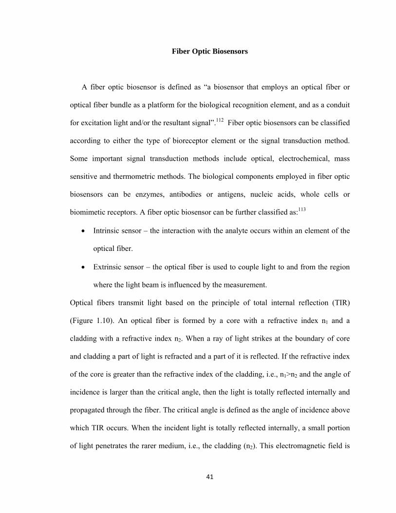
A fiber optic biosensor is defined as “a biosensor that employs an optical fiber or
optical fiber bundle as a platform for the biological recognition element, and as a conduit
for excitation light and/or the resultant signal”.112 Fiber optic biosensors can be classified
according to either the type of bioreceptor element or the signal transduction method.
Some important signal transduction methods include optical, electrochemical, mass
sensitive and thermometric methods. The biological components employed in fiber optic
biosensors can be enzymes, antibodies or antigens, nucleic acids, whole cells or
biomimetic receptors. A fiber optic biosensor can be further classified as:113
Intrinsic sensor – the interaction with the analyte occurs within an element of the
optical fiber.
Extrinsic sensor – the optical fiber is used to couple light to and from the region
where the light beam is influenced by the measurement.
Optical fibers transmit light based on the principle of total internal reflection (TIR)
(Figure 1.10). An optical fiber is formed by a core with a refractive index n1 and a
cladding with a refractive index n2. When a ray of light strikes at the boundary of core
and cladding a part of light is refracted and a part of it is reflected. If the refractive index
of the core is greater than the refractive index of the cladding, i.e., n1˃n2 and the angle of
incidence is larger than the critical angle, then the light is totally reflected internally and
propagated through the fiber. The critical angle is defined as the angle of incidence above
which TIR occurs. When the incident light is totally reflected internally, a small portion
of light penetrates the rarer medium, i.e., the cladding (n2). This electromagnetic field is

42
called evanescent wave, and has an intensity that decays exponentially with distance,
starting at the interface and extending into the medium of lower refractive index. The
evanescent wave can interact with molecules in the penetration depth, thus producing a
net flow of energy across the reflecting surface in the medium with lower refractive index
to maintain the evanescent field. This transfer of energy leads to attenuation in
reflectance called attenuated total reflection (ATR). This phenomenon has been utilized
to develop ATP absorption sensors.114
Optical fiber biosensors have been used in combination with different types of
spectroscopic techniques such as absorption, fluorescence, phosphorescence, and surface
plasmon resonance (SPR). Fiber optic bisosensors based on absorbance may consist of
the biological component immobilized close to the optical fiber or directly on its surface.
The biological component interaction with the analyte results in a change in the
absorbance which is related to the concentration of analyte. Light is directed to the optic
fiber tip where the biological element is immobilized and the resulting radiation is
directed back to the detector by the fiber. Several fiber optic biosensors based on
absorbance measurements have been developed for use in medical applications115, 116 and
molecular recognition.117
Fluorescence-based fiber optic systems consist of a biological component that is
intrinsically fluorescent or is labeled with a fluorescent molecule. In such biosensors the
biological components may also be enzymes that catalyze reactions leading to the
production or consumption of fluorescent species. For example, dehydrogenases that
catalyze nicotinamide adenine dinucleotide (NAD)-dependent reactions have been used
for the development of several fiber optic biosensors; these reactions produce NADH or

43
Figure 1.10 Total internal reflection in optical fiber.

44
NADPH, which can be detected by their fluorescence at ex = 350 nm, em = 450 nm.118,
119 In another example a fiber optic biosensor for glucose based on fluorescence
resonance energy transfer was developed.120 In this system two optical fibers were
inserted in a hollow fiber, which was filled with assay solution containing fluorescein
isothiocyanate (FITC)-Dextran as the donor and tetramethylrhodamine isothiocyanate
(TRITC) labeled ConA (TRITC-ConA) as the acceptor (quencher). In the absence of
glucose the donor and the acceptor are significantly close together for the fluorescein
signal to be quenched by rhodamine. However, in the presence of glucose the FITC-
Dextran is separated from the TRITC-ConA and the fluorescein signal increases in a
dose-dependent manner. Hence, this system enables the monitoring of glucose
concentrations as a function of changes in fluorescence.
A fluorescent fiber optic immunosensor involves the immobilization of an
antibody or antigen on the distal end of the fiber. The incident light introduced at the
proximal end of the fiber travels through the fiber via TIR causing excitation of the
fluorophore attached to either the antigen or a secondary antibody.121 A variety of fiber
optic based immunosensors for the detection of pesticides have been developed. In an
example, a hapten, a derivative of triazine (pesticide), was immobilized on the end of a
hard clad silica fiber.122 The fiber was then incubated with a fluorescently labeled
antibody and a sample containing unknown amounts of triazine. This competitive
immunoassay is based on measurements of the changes in fluorescence signal to
determine the amount of triazine present in a sample.122
Immobilization of enzyme based sol-gels on the fiber is another technique for
developing fluorescent fiber optic sensors. Doong and coworkers developed a fiber optic

45
fluorescent biosensor for acetylcholine.123 In this approach acetylcholinesterase was
immobilized in a sol-gel on the distal end of an optical fiber. The acetic acid formed as a
result of the enzymatic reaction between acetylcholinesterase and acetylcholine causes a
change in the fluorescence intensity of the pH-sensitive fluorescent dye FITC-Dextran.
This system was used for the detection of Paraoxon, an organophosphorous pesticide, as
Paraoxon inhibits acetylcholinesterase, thus increasing fluorescence.
Surface plasmon resonance (SPR) is a direct optical detection method. SPR is
based on TIR, wherein the TIR interface is coated with a thin layer of conducting metal.
Polarized light penetrates the metal layer and excites electromagnetic surface plasmon
waves propagating within the conductor surface, which is in contact with the low
refractive index medium. SPR has successfully been employed in the development of
immunosensors for simple and rapid assay of several analytes. Fiber optic SPR sensors
are small and suitable for use at remote sites. Several fiber optic SPR sensors have been
reported.124-127 Slavik et al. described the fabrication of a single-mode fiber optic SPR
device.128 The sensing element was created by coating the fiber with a 65-nm layer of
gold followed by a 19-nm layer of tantalum pentoxide. The sensor was used for detecting
IgG concentrations in nanomolar ranges. Further, in continuation of the above mentioned
work, a double layer of anti-Staphylococcal enterotoxin B (SEB) antibody crosslinked by
glutaraldehyde was immobilized on the sensor surface for the detection of SEB.129 A
fiber optic SPR immunosensor known as BIAcore probe, which enables dipstick-type
sampling, has been launched by the Swedish company BIAcore.130
One drawback of fiber optic sensors is the variation in response and signal
intensity from fiber to fiber.113 Immobilization of biological recognition elements on

46
optical fibers has led to increases in selectivity, sensitivity, and in some cases
reversibility.112 Fiber optic sensors have several advantages, in that they are usually
compact and light weight, minimally invasive, and they are not affected by
electromagnetic interferences from static electricity, strong magnetic fields, or surface
potentials.112, 113

47
STATEMENT OF RESEARCH
The goal of this work was to first create designer proteins with enhanced stability and
activity by global incorporation of unnatural amino acids into the structure of GBP and
truncated fragments of GBP. Also, modified antibodies were created by photolabeling the
unconventional region of the antibody variable region with a fluorophore conjugated
nucleotide probe. Secondly, the goal of this work was to develop fiber optic glucose
biosensor by using GBP and the unnatural amino acid incorporated proteins covalently
attached within a hydrogel and immobilized on a tip of an optical fiber. Also, modified
antibodies can be utilized to develop universal biosensing system for various
biomolecules since modifying antibodies in the unconventional region does not affect
antigen binding. The overall hypotheses for this work have ben formulated below:
Thermal stability and glucose binding activity of GBP can be altered by global
incorporation of unnatural amino acids and these unnatural proteins can be
utilized for the development of biosensor for continuous glucose monitoring in
physiologically relevant glucose concentrations.
The newly created glucose-recognition proteins can be incorporated into hydrogel
polymer networks, placed at the tip of a fiber optic system and employed in the
continuous monitoring of glucose in blood.
Antibody based biosensing systems/assays can be developed by labeling the
unconventional nucleoside binding site with an environment sensitive fluorophore
conjugated to a nucleotide/nucleoside probe, and correlating the changes in
fluorescence intensity in the presence of the analyte.

48
GBP has been extensively studied and utilized for developing glucose sensors.
However, to the best of our knowledge, no thermally stable GBP or its fragments have
been created by global incorporation of unnatural amino acids. Also no glucose biosensor
has been developed with unnatural GBP able to detect glucose in physiologically relevant
mM ranges. Moreover no fluorescent antibodies have been developed by photolabeling
the unconventional region in the variable region of antibodies. It was our belief that
global incorporation of unnatural amino acids in GBP would alter the stability and
activity of GBP and further modifying the antibodies via fluorophore conjugated
nucleotides we can develop universal biosensing system for a variety of biomolecules.
Chapter Two describes the universal strategy for designing antibody based
sensing molecules by labeling an inherent nucleoside binding site. Here as a proof
of concept Interleukin 6 was chosen as an analyte to be detected with our
modified antibody-based biosensing system.
Chapter Three describes the development of a biosensing system for monitoring
bone loss. Herein, we utilize the biosensing system based on fluorophore
modified monoclonal antibody for the detection of Osteonectin, which is a bone
biomarker.
Chapter Four describes the method of global incorporation of unnatural amino
acids in GBP and its truncated fragments. It also details the analysis of structure,
stability and activity of the unnatural proteins.
Chapter Five describes the development of fiber optic based biosensor for
glucose, wherein, GBP and unnatural amino acid incorporated GBP and GBP
fragments are immobilized in a hydrogel.

49
CHAPTER TWO
UNIVERSAL STRATEGY FOR DESIGNING ANTIBODY BASED SENSING
MOLECULES BY LABELING AN INHERENT NUCLEOSIDE BINDING SITE
The quest for specific and sensitive sensing methods for target molecules is an on-
going challenge. While the utility of biosensors has been proven, their development has
been limited to the availability of natural or artificial receptors that recognize the target
molecule. Herein, a universal approach is demonstrated for the design of antibody based
sensing molecules that overcome this limitation. In particular, the antibody is modified by
covalently attaching a nucleoside probe containing an environment-sensitive fluorophore
to the inherent non-canonical nucleoside-binding site of the antibody. The non-canonical
nucleoside binding site resides between the light and the heavy chains of the variable
domain of antibodies, and tethering nucleoside probes to this site does not affect antigen
binding.131, 132 The binding of the antigen to the antibody causes a change in the
microenvironment of the nucleoside-binding site that is manifested by an alteration in the
fluorescence properties of the fluorophore in a manner that is related to the concentration
of antigen present in the sample. This relationship constitutes the basis for the
development of the antibody based assays and biosensing systems.
Antibodies have found wide applicability in a range of immunoassay127, 133, 134 and
biosensor135-137 formats. Fluorescent antibodies with a potential to be used as biosensors
have been developed, wherein, a fluorescent stilbene derivative is bound to the antigen
binding site138-141. Besides the antigen binding site, an inherent non-canonical site is
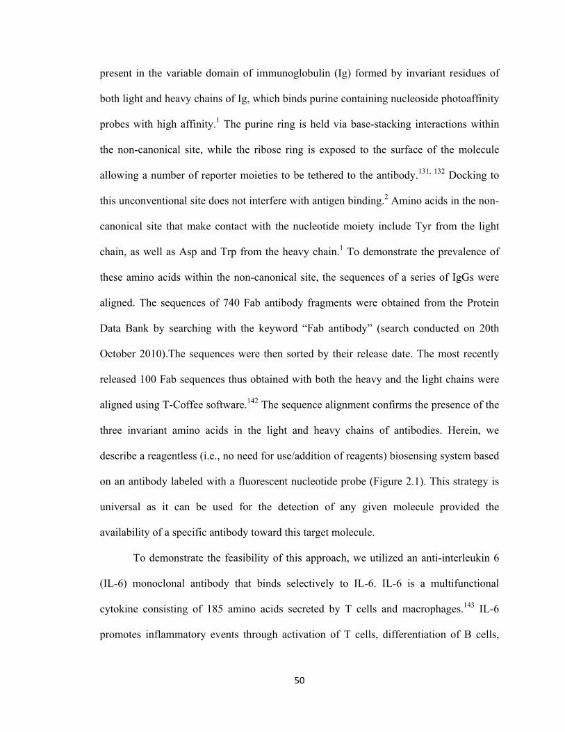
50
present in the variable domain of immunoglobulin (Ig) formed by invariant residues of
both light and heavy chains of Ig, which binds purine containing nucleoside photoaffinity
probes with high affinity.1 The purine ring is held via base-stacking interactions within
the non-canonical site, while the ribose ring is exposed to the surface of the molecule
allowing a number of reporter moieties to be tethered to the antibody.131, 132 Docking to
this unconventional site does not interfere with antigen binding.2 Amino acids in the non-
canonical site that make contact with the nucleotide moiety include Tyr from the light
chain, as well as Asp and Trp from the heavy chain.1 To demonstrate the prevalence of
these amino acids within the non-canonical site, the sequences of a series of IgGs were
aligned. The sequences of 740 Fab antibody fragments were obtained from the Protein
Data Bank by searching with the keyword “Fab antibody” (search conducted on 20th
October 2010).The sequences were then sorted by their release date. The most recently
released 100 Fab sequences thus obtained with both the heavy and the light chains were
aligned using T-Coffee software.142 The sequence alignment confirms the presence of the
three invariant amino acids in the light and heavy chains of antibodies. Herein, we
describe a reagentless (i.e., no need for use/addition of reagents) biosensing system based
on an antibody labeled with a fluorescent nucleotide probe (Figure 2.1). This strategy is
universal as it can be used for the detection of any given molecule provided the
availability of a specific antibody toward this target molecule.
To demonstrate the feasibility of this approach, we utilized an anti-interleukin 6
(IL-6) monoclonal antibody that binds selectively to IL-6. IL-6 is a multifunctional
cytokine consisting of 185 amino acids secreted by T cells and macrophages.143 IL-6
promotes inflammatory events through activation of T cells, differentiation of B cells,

51
FIGURE 2.1 Schematic showing the strategy for development of biosensing system
based on fluorophore labeled monoclonal antibody

52
and the induction of acute phase reactants by hepatocytes.144, 145 In addition, IL-6 plays a
protective role during disease, acting both as a pro- and an anti-inflammatory cytokine.146
Elevated concentrations of IL-6 in body fluids have been reported in various disease
states such as cardiac myxomas and cardiovascular diseases147-149 and is employed as a
general marker of inflammation.150
EXPERIMENTAL PROCEDURES
Materials
Mouse anti-IL-6 monoclonal antibody and recombinant Human IL-6 were
purchased from USBiological (Massachusetts, MA). Nucleotide probes 8-N3ATP and 2-
N3ATP were obtained from Affinity Labeling Technologies (ALT) Corporation
(Lexington, KY). Alexa Fluor 594 cadaverine was obtained from Invitrogen (Eugene,
OR). The Bradford protein assay kit was obtained from Bio-Rad laboratories (Hercules,
CA). Carry Eclipse Spectrophotometer (Milpitas, CA) was used for fluorescence
measurements.
Methods
Selecting the photoaffinity probe
Two radioactive nucleotide probes, [γ-32P]8-N3ATP and [γ-32P]2-N3ATP were
evaluated for their reactivity with mouse anti-interleukin 6 monoclonal antibody. For the
labeling procedure 8 µg of antibody was incubated with 300 µM of each probe in two
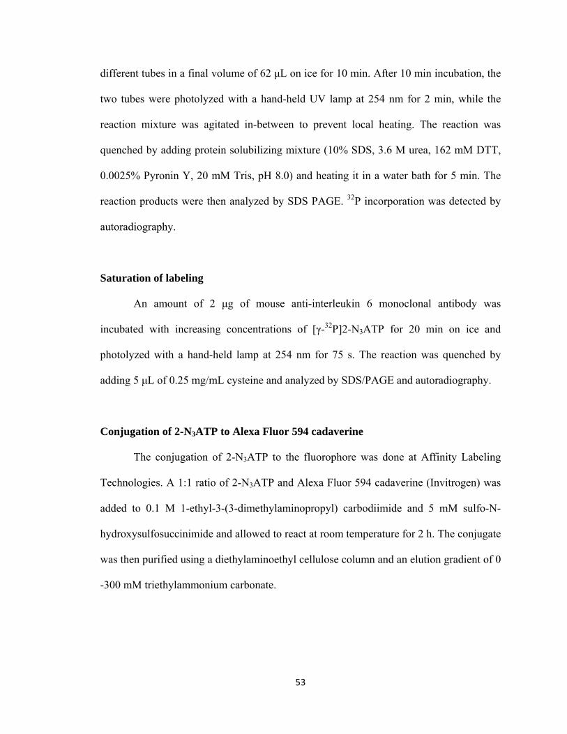
53
different tubes in a final volume of 62 μL on ice for 10 min. After 10 min incubation, the
two tubes were photolyzed with a hand-held UV lamp at 254 nm for 2 min, while the
reaction mixture was agitated in-between to prevent local heating. The reaction was
quenched by adding protein solubilizing mixture (10% SDS, 3.6 M urea, 162 mM DTT,
0.0025% Pyronin Y, 20 mM Tris, pH 8.0) and heating it in a water bath for 5 min. The
reaction products were then analyzed by SDS PAGE. 32P incorporation was detected by
autoradiography.
Saturation of labeling
An amount of 2 μg of mouse anti-interleukin 6 monoclonal antibody was
incubated with increasing concentrations of [γ-32P]2-N3ATP for 20 min on ice and
photolyzed with a hand-held lamp at 254 nm for 75 s. The reaction was quenched by
adding 5 μL of 0.25 mg/mL cysteine and analyzed by SDS/PAGE and autoradiography.
Conjugation of 2-N3ATP to Alexa Fluor 594 cadaverine
The conjugation of 2-N3ATP to the fluorophore was done at Affinity Labeling
Technologies. A 1:1 ratio of 2-N3ATP and Alexa Fluor 594 cadaverine (Invitrogen) was
added to 0.1 M 1-ethyl-3-(3-dimethylaminopropyl) carbodiimide and 5 mM sulfo-N-
hydroxysulfosuccinimide and allowed to react at room temperature for 2 h. The conjugate
was then purified using a diethylaminoethyl cellulose column and an elution gradient of 0
-300 mM triethylammonium carbonate.

54
Determination of KD of the monoclonal antibody for 2-N3ATP-fluorophore
Competitive labeling of the anti-interleukin 6 (IL-6) monoclonal antibody with 2-
N3ATP [γ 32P] and 2-N3ATP-fluorophore was done. 2-N3ATP [γ 32P] (30 µM) and 2 µg
of the antibody was added to each 10 µM, 20 µM, 40 µM, 60 µM, 80 µM, 100 µM and
120 µM of 2-N3ATP-fluorophore tube and incubated for on ice 20 min. The tubes were
then photolyzed for 75 s. The reaction was quenched by adding 5 μL of 0.25 mg/mL
cysteine and analyzed by SDS/PAGE and autoradiography. The apparent KD was
determined using GraphPad Prism 5 software.
Conjugation of the antibody to the 2-N3ATP-fluorophore
For the conjugation of 2-N3ATP-Alexa Fluor 594 cadaverine to the antibody, 4 μg
of the antibody was incubated with 30 μM 2-N3ATP labeled fluorophore in a final
volume of 50 μL for 20 min, on ice and in the dark. The reaction was photolyzed with a
hand-held UV lamp at 254 nm for 4 min. To quench the photolytic reaction, cysteine (10
μL of 1mg/ml) was added. The unbound fluorophore was separated from the antibody
bound fluorophore by dialyzing against six changes of 10 mM phosphate buffer, pH 7.0
over 24h.
Fluorescence studies of the interaction of the labeled antibody with IL-6
The effect of IL-6 binding on the fluorescence signal of the label attached to the
non-canonical site of the IL-6 antibody was studied by incubating 4 μg/mL of the labeled
antibody with different concentrations of IL-6 for 30 min at room temperature in the
dark. The sample solutions were prepared using 10 mM phosphate buffer, pH 7.0. The

55
fluorescence intensity of the samples (total volume 200 µL) was measured on a Cary
Eclipse Spectrofluorometer.
RESULTS AND DISCUSSION
Nucleotide probes conjugated with a fluorophore can be docked to the non-
canonical nucleoside binding site of the antibody without interfering with binding of
antigen to the canonical antigen-binding site. The mouse anti-human IL-6 monoclonal
antibody was labeled with two photoreactive probes, [γ-32P]-2-N3ATP (Figure 2.2a) and
[γ-32P]-8-N3ATP (Figure 2.2b) to test their reactivity toward the antibody. Both the light
and heavy chains were labeled with the radioactive probe indicating that the nucleoside
binding site is composed of amino acids located on both chains (Figure 2.3a). It was also
determined that [γ-32P]-2-N3ATP labels the antibody better than [γ-32P]-8-N3ATP (Figure
2.3b). Further, to optimize the efficiency of labeling the antibody with the photoreactive
probe, the concentration of the probe required to label the antibody was determined by
incubating anti-IL-6 monoclonal antibody with increasing concentrations of [γ-32P]-2-
N3ATP followed by exposure to UV light to activate the photolabel (Figure 2.4). The
concentration of probe selected to label the antibody in all subsequent experiments was
~25 µM, as this is relatively low concentration and generates a good signal.
After proving that [γ-32P]-2-N3ATP labeled the antibody successfully, a
fluorescent, rather than radiolabeled derivative of 2-N3ATP was synthesized. In that
regard, Alexa Fluor 594 cadaverine was selected for coupling to 2-N3ATP. This

56
Figure 2.2 Structures of nucleotide probes utilized for photolabeling of antibody. (a)
[γ32P] 2-N3ATP (b) [γ-32P] 8-N3ATP
57
0.0000 0.0005 0.0010 0.0015 0.0020 0.00250.0
0.5
1.0
1.5
2.0
2.5
3.0
3.58-N3ATP[32P]
2-N3ATP[32P]
Antibody (mg)
DL
U x
10
6
Figure 2.3 Antibody labeled with radioactive nucleotide probes. (a) autoradiogram of the
antibody labeled with the photoreactive probes [γ-32P] 2-N3ATP (A) and [γ-32P] 8-N3ATP
(B). Lanes 1, 4; 2, 5; and 3, 6 correspond to the use of 0.6 μg, 1.3 μg and 1.9 μg of
labeled antibody, respectively. (b) Normalized data obtained from the autoradiogram of
antibody labeled with the photoreactive probes.
a B
b

58
0 25 50 75 1000
50
100
150
200
2-N3ATP, M
DL
U x
10
6
Figure 2.4 Saturation of photolabeling of antibody with [γ-32P] 2-N3ATP.

59
fluorophore has long excitation and emission wavelengths (590 nm/617 nm), thus
circumventing interferences from common fluorescent compounds present in biological
samples. The amine functional group of Alexa Fluor 594 cadaverine was utilized for
conjugation to 2-N3ATP, since it was observed that 2-N3ATP labels the antibody with a
greater efficiency than the 8-N3ATP vide supra. The apparent KD for 2-N3ATP-Alexa
Fluor 594 (Figure 2.5) cadaverine to the non-canonical site was determined to be ~17
µM. The fluorescently labeled nucleotide was further conjugated to the mouse anti-
human IL-6 monoclonal antibody at the non-canonical site upon exposure to UV light.
The effect of antigen (IL-6) binding on the fluorescence signal of the 2-N3ATP-
Alexa Fluor 594 (Figure 2.6a) labeled antibody was evaluated. The apparent KD for IL-6
to the fluorophore labeled antibody was determined to be ~2.4 x 10⁻⁸ M. It was observed
that binding of IL-6 to the antibody resulted in an increase of the fluorescence intensity of
the fluorophore with increasing concentration of antigen (Figure 2.6b). The experiments
suggest that binding of IL-6 to the antigen binding site induces a change in the
conformation of the non-canonical site causing a change in the microenvironment
surrounding the fluorophore, which is manifested by a change in the fluorescence signal.
Because the site in the antibody where the fluorescent probe is bound is adjacent to the
antigen-binding site in the variable region of the antibody, there is an allosteric effect that
upon antigen binding alters the fluorescence signal of the fluorophore.

60
0 25 50 75 100 125 1500
2
4
6
8
10
12
14Heavy Chain
Light Chain
2-N3ATP-Fluorophore, M
DL
U x
10
6
Figure 2.5 Competitive labeling of anti-interleukin 6 (IL-6) monoclonal antibody with 2-N3ATP
-Alexa Fluor 594 cadaverine and 2-N3ATP [γ 32P].

61
N
N N
N
NH2
O
OHOH
HH
HH
CH2O P
O
OH
O P
O
OH
O P
O
OH
NH(CH2)5 NH
N3
C
O
COOH
O NCH3
CH3
N
CH3
H3C
H3C
CH2SO3H CH2SO3
CH3
-10.0 -9.5 -9.0 -8.5 -8.0 -7.5 -7.0 -6.5 -6.0 -5.5500
550
600
650
700
750
800
850
900
950
log (IL-6, M)
Re
lati
ve
Lig
ht
Un
its
Figure 2.6 Structure of fluorescent nucleotide probe and the effect of Il-6 on the
fluorescence intensity of the probe. (a) 2-N3ATP-Alexa Fluor 594 cadaverine (b) Dose-
response curve for IL-6. Data are the average of ± one standard deviation (n=3). Relative
standard deviations at all concentrations are less than 10%.
b
a
62
CONCLUSIONS
Herein we have developed a universal method for the design of biosensors. We
demonstrated that a fluorophore-modified nucleotide probe can bind to the non-canonical
site of the antibody without affecting antigen binding. Additionally, we have shown that
binding of the antigen to the antibody alters the fluorescence intensity of the fluorophore-
modified nucleotide probe in an allosteric fashion. The change in fluorescence intensity
of the label can be correlated to the concentration of IL-6 in the sample. Because of the
widespread nature of this non-canonical site among IgGs, and availability of antibodies
for practically any molecule of interest, this versatile biosensing approach should find
wide applicability in a variety of fields where detection of antigen-antibody interactions
is important. Among those, we can envision their use in sensing in a variety of samples,
imaging, localization of target molecules in cells, etc.

63
CHAPTER THREE
BIOSENSING SYSTEM BASED ON MODIFIED ANTIBODY FOR
MONITORING BONE LOSS
INTRODUCTION
Bone undergoes mass changes throughout life, also referred to as bone
remodeling. Bone remodeling involves cycles of bone deposition by osteoblasts and bone
resorption by osteoclasts.151 Bone remodeling allows for the renewal of bones in
vertebrates. Bone is continuously remodeled in response to mechanical and physiological
stress.152 Bone mass is maintained by a balance between the rates of bone deposition and
bone resorption.151 However, when the rate of bone resorption is faster than the rate of
bone deposition it leads to a disorder called osteoporosis.151 Osteopenia is another bone
disorder where bone mass is less than average. It is sometimes also referred to as onset of
osteoporosis. Besides these health conditions, bone mass loss is also reported in humans
after prolonged space flights and weightlessness or microgravity.153, 154 Although the
mechanism of microgravity induced bone loss is not fully known, it has been attributed to
a decrease in osteoblast function without a significant change in bone resorption.154
Several ground based studies on microgravity induced bone loss have been performed
using various models. These studies suggest that bone loss may occur because of
inhibition of osteoblast differentiation from human mesenchymal stem cells due to a
failure in cellular expression of osteonectin.
Osteonectin is a 32,000 kDa glycoprotein occurring in the noncollagenous matrix
of bone, extracellular matrices and some body compartments including blood platelets.155,

64
156 Hence, two forms of ON are known, a bone derived form and a platelet derived form.
Osteonectin is a major constituent of basement membrane matrix of many embryonic and
adult tissues.157 Osteonectin contains structural domains including an acidic domain,
which is rich in aspartic and glutamic acid, a NH2-terminal domain, a cysteine rich
domain with potential glycosylation sites, an alpha helical domain, and two calcium
binding motifs.157, 158 ON binds to type I collagen and hydroxyapatite and, consequently,
plays a key role in initiating mineral deposition and linking mineral to the matrix.159
Osteonectin deficient mice have shown decreased bone mineral density and a decreased
function of osteoblasts and osteoclasts resulting in osteopenia.151 Several animal models
of bone fragility show a decreased expression of osteonectin. Further, osteonectin has
also been determined as a salivary biomarker of alveolar bone loss.160, 161 Herein, we
present an antibody based biosensing system for osteonectin that could find application in
the prevention and treatment of bone loss for healthier life. Our biosensing system is
based on the previously developed concept of conjugating a fluorophore labeled
nucleotide at the non-canonical site of an antibody in which change in the conformation
induced by the binding of antigen at the antigen binding site causes a change in
fluorescence intensity. Specifically, we used a monoclonal antibody against osteonectin,
which was labeled with 2-N3ATP conjugated to Alexa Fluor 594 cadaverine. The
intensity of fluorescence of the label was found to increase with increasing osteonectin
concentration in the sample.

65
EXPERIMENTAL PROCEDURES
Materials
Anti-human osteonectin/SPARC monoclonal antibody was purchased from Santa
Cruz Biotechnology (Santa Cruz, CA). Human recombinant osteonectin was purchased
from ALPCO Diagnostic (Salem, NH). Nucleotide probes 8-N3ATP and 2-N3ATP were
obtained from Affinity Labeling Technologies (ALT) Corporation (Lexington, KY).
Alexa Fluor 594 cadaverine was obtained from Invitrogen (Eugene, OR). The Bradford
protein assay kit was obtained from Bio-Rad laboratories (Hercules, CA). Carry Eclipse
Spectrophotometer (Milpitas, CA) was used for fluorescence measurements
Methods Selecting the photoaffinity probe
Two radioactive nucleotide probes, [γ-32P]8-N3ATP and [γ-32P]2-N3ATP were
evaluated for their reactivity with mouse anti-osteonectin monoclonal antibody. For the
labeling procedure 8 µg of antibody was incubated with 358 µM of each probe in two
different tubes in a final volume of 58 μL on ice for 10 min. After 10 min incubation, the
two tubes were exposed to UV light with a hand-held UV lamp at 254 nm for 75 s, while
the reaction mixture was agitated in-between to prevent local heating. The reaction was
quenched by adding a protein solubilizing mixture (10% SDS, 3.6 M urea, 162 mM DTT,
0.0025% Pyronin Y, 20 mM Tris, pH 8.0) and heating it in a water bath for 5 min. The
reaction products were then analyzed by SDS PAGE. 32P incorporation was detected by
autoradiography.

66
Conjugation of 2-N3ATP to Alexa Fluor 594 cadaverine
The conjugation of 2-N3ATP to the fluorophore was performed at Affinity
Labeling Technologies. A 1:1 ratio of 2-N3ATP and Alexa Fluor 594 cadaverine was
added to 0.1 M 1-ethyl-3-(3-dimethylaminopropyl) carbodiimide and 5 mM sulfo-N-
hydroxysulfosuccinimide and allowed to react at room temperature for 2 h. The conjugate
was then purified using a diethylaminoethyl cellulose column and an elution gradient of 0
-300 mM triethylammonium carbonate.
Conjugation of the antibody to the 2-N3ATP-fluorophore
For the conjugation of 2-N3ATP-Alexa Fluor 594 cadaverine to the antibody, 0.2
mg/mL of the antibody was incubated with 90 μM 2-N3ATP labeled fluorophore in a
final volume of 100 μl for 20 min, over ice and in dark, in two flat bottom eppendorf
tubes. The reaction mixture was exposed to UV light with a hand-held UV lamp at 254
nm for 2 min. To quench the photolytic reaction, cysteine (12.5 μL of 1mg/mL) was
added. The unbound fluorophore was separated from the antibody bound fluorophore by
dialyzing against six changes of 10 mM phosphate buffer, pH 7.0 over 24h. The
concentration of fluorophore-labeled antibody (ON antibody-2-N3ATP-Alexa Fluor 594
cadaverine) was determined by Bradford assay.
Calibration curve for osteonectin antibody-2-N3ATP-Alexa Fluor 594 cadaverine
In order to generate calibration curves for the ON antibody-2-N3ATP-Alexa Fluor
594 cadaverine serial dilutions were made of fluorophore-labeled antibody from a stock
solution at concentration of 6.7 x 10⁻⁷ M with 10 mM phosphate buffer, pH 7.0.

67
Fluorescence measurements were performed in a microtiter plate with 100 μL samples.
The samples were excited at 590 nm and the emission was measured at 617 nm. A
volume of 100 μL of 10 mM phosphate buffer, pH 7.0, was used as blank. The
fluorescence intensity of the samples was measured on a Cary Eclipse
Spectrofluorometer.
Association study
A volume of 50 μL of 3 x 10⁻⁸ M fluorophore-labeled antibody and 50 μL of
varying concentrations of osteonectin ranging from 1 x 10⁻⁷ M to 1 x 10⁻¹⁰ M were
incubated in a microtiter plate for a time period ranging from 5 to 60 min at room
temperature. The fluorescence of the samples in microtiter plate were measured upon
excitation at 590 nm after every 5 min for 30 min and every 15 min from 30 to 60 min. A
sample of 100 μL fluorophore-labeled antibody was used as a control and 10 mM
phosphate buffer, at pH 7.0, was used as blank.
Fluorescence studies of the interaction of the labeled antibody with osteonectin
The effect of ON binding on the fluorescence signal of the label attached to the
non-canonical site of the ON antibody was studied by incubating 4 μg/mL of the labeled
antibody with different concentrations of ON for 30 min at room temperature in the dark.
The sample solutions were prepared by mixing 100 µL of antibody solution and 100 µL
of ON solution. The solutions were made in 10 mM phosphate buffer, pH 7.0. The
fluorescence intensity of the samples (total volume 200 µL) was measured on a Cary
Eclipse Spectrofluorometer.

68
Selectivity study
In order to determine whether the monoclonal antibody based sensor is selective
for osteonectin, 4 μg/mL fluorophore labeled antibody was incubated with different
concentrations of osteonectin and inteleukin-6 (IL6) separately in a total volume of 200
μL in a microtiter plate for 25 min in dark at room temperature. The fluorescence of the
samples was measured upon excitation at 590 nm. A sample of 100 μL fluorophore-
labeled antibody was used as a control and 10 mM phosphate buffer, pH 7.0, was used as
blank.
RESULTS AND DISCUSSION
An anti-osteonectin monoclonal antibody was labeled with two radioactive
nucleotide probes, [γ-32P]-2-N3ATP and [γ-32P]-8-N3ATP to determine the reactivity of
the probes against the osteonectin antibody. The photolabeld osteonectin antibody was
analyzed via SDS PAGE analysis to determine which nucleotide probe labels the
antibody better. The determination of 32P incorporation via autoradiography indicates that
both the light and heavy chains of osteonectin antibody are labeled with the radioactive
probe. Hence, the nucleotide binding site resides within the light and heavy chain of
antibody. Autoradiogram data (Figure 3.1) also show that [γ-32P]-2-N3ATP labels the
antibody better than [γ-32P]-8-N3ATP. Therefore, for further experiments the osteonectin
antibody was labeled with the fluorescent derivative of 2-N3ATP.

69
0.0000 0.0005 0.0010 0.0015 0.0020 0.00250.0
0.5
1.0
1.5[32P]-2-N3ATP HC
[32P]-2-N3ATP LC
[32P]-8-N3ATP LC
[32P]-8-N3ATP HC
Antibody (mg)
DL
U x
106
Figure 3.1 Data obtained from autoradiograph of monoclonal antibody against
osteonectin with [γ-32P]8-N3ATP and [γ-32P]2-N3ATP. HC= Heavy Chain, LC=Light
Chain.

70
In our biosensing system, we utilized the non-canonical nucleotide binding site to
dock a fluorescent reporter molecule covalently attached to 2-N3ATP. We utilized Alexa
Fluor 594 cadaverine as the fluorescent reporter molecule, because it has long excitation
and emission wavelengths (590 nm/617 nm), thus circumventing interferences from
common fluorescent compounds present in biological samples. The amine functional
group of Alexa Fluor 594 cadaverine was utilized for conjugation to 2-N3ATP. Alexa
Fluor 594 cadaverine labeled 2-N3ATP was further conjugated to the osteonectin
monoclonal antibody at the non-canonical site upon exposure to UV light. Haley and
coworkers have previously determined via computer modeling that the nucleoside base is
sandwiched between the rings of an invariant Trp in the heavy chain and Pro in the light
chain forming the non-canonical binding site.131 The azido group of the purine ring on the
other hand forms a covalent bond with the amino acids in the light chain of antibody
upon irradiation with UV light131. The unbound nucleotide fluorophore conjugate to the
ON antibody was separated from the fluorophore-bound antibody by dialysis, and the
protein concentration of the fluorophore-labeled antibody was determined by Bradford
assay.
The amount of fluorophore-labeled antibody required for further experiments was
determined by generating calibration curves that relate the amount of fluorescent signal to
the different concentrations of the fluorescent antibody (Figure 3.2). The concentration of
fluorescent antibody selected for further experiments was 1 X 10⁻⁸ M as this was a
relatively low concentration that generates a good detectable signal.
To determine the amount of time required to obtain the maximum possible change
in the signal upon incubating the fluorescent osteonectin antibody with osteonectin, an

71
-13 -12 -11 -10 -9 -8 -70
50
100
150
200
250
log(Fluorophore labeledantibody against osteonectin, M)
Flu
ore
scen
ce In
ten
sity
Figure 3.2 Calibration curve for 2-N3ATP-Alexa Fluor 594 labeled antibody against
osteonectin. Data are the average of ± one standard deviation (n=3). Relative standard
deviations at all concentrations are less than 10%.

72
0 10 20 30 40 50 600
25
50
75
100
125
150
175
200
0.05.00E-071.00E-075.00E-081.00E-08
5.00E-091.00E-095.00E-101.00E-10
Time (min)
Flu
ore
scen
ce In
ten
sity
Figure 3.3 Association curve for 2-N3ATP-Alexa Fluor 594 labeled antibody against
osteonectin with osteonectin. Data are the average of ± one standard deviation (n=3).
Relative standard deviations at all concentrations are less than 10%.

73
association study was performed. Figure 3.3 shows the association curve for fluorescent
osteonectin antibody with osteonectin. A maximum increase of fluorescence intensity
was obtained after 30 min incubation of fluorescent antibody and antigen. However, a
distinguishable increase of fluorescence intensity was observed after 25 min incubation
of antibody and antigen. The time study shows a lag phase over the first 15 min of
incubation of the analyte (antigen) with the fluorophore labeled antibody. This lag phase
can be because of the time required for the diffusion of the analyte in the binding site of
the antibody or the conformation change in the binding site in the presence of the analyte.
Therefore, for all subsequent experiments an incubation time of 25 min was used.
A dose-response curve (Figure 3.4) was generated to evaluate the effect of antigen
(osteonectin) binding on the fluorescence signal of the 2-N3ATP-Alexa Fluor 594 labeled
antibody. An increase in the fluorescence signal was observed with increasing
concentration of antigen. The change in the fluorescence is attributed to the change in the
microenvironment surrounding the fluorophore induced by the change in the
conformation of the non-canonical site to which the fluorophore is bound. As previously
described by Haley and coworkers this non-canonical site resides adjacent to the antigen
binding site in the variable region of the antibody. Therefore, there is an allosteric effect
that upon antigen binding alters the fluorescence signal of the fluorophore.
To evaluate the selectivity of our biosensing system for osteonectin, selectivity
studies were done by incubating the fluorophore-labeled osteonectin antibody with
varying concentrations of osteonectin and IL6 for 25 min and the change in fluorescence
intensity was measured. Figure 3.5 shows an increase in the fluorescence intensity of
osteonectin antibody with increasing concentrations of osteonectin. However, no change

74
-9.0 -8.5 -8.0 -7.5 -7.0 -6.5 -6.0440
450
460
470
480
490
500
log(Osteonectin, M)
Flu
ore
scen
ce In
ten
sity
Figure 3.4 Dose-response curve for osteonectin. Data are the average of ± one standard
deviation (n=3). Relative standard deviations at all concentrations are less than 10%.

75
-9.0 -8.5 -8.0 -7.5 -7.0 -6.5 -6.0400
425
450
475
500
525 OsteonectinIL6
log(Analyte, M)
Flu
ore
scen
ce In
ten
sity
Figure 3.5 Selectivity studies for 2-N3ATP-Alexa Fluor 594 labeled antibody against
osteonectin with osteonectin and IL6. Data are the average of ± one standard deviation
(n=3). Relative standard deviations at all concentrations are less than 10%.

76
in the fluorescence intensity is observed for osteonectin antibody with increasing
concentrations of IL6. This confirms that fluorophore-labeled osteonectin antibody binds
specifically to osteonectin and hence, the biosensing system is very specific for the
antigen.
CONCLUSIONS
In conclusion we have demonstrated the use of our previously developed
universal biosensing system for the detection of osteonectin based on labeling the non-
canonical nucleoside binding site of an anti-osteonectin antibody. We have also
demonstrated that our biosensing system based on fluorescently labeled antibody is very
selective for the antigen. Our biosensing system will help in the clinical analysis of
osteonectin. Also this type of biosensing system can be extended to different kinds of
analytes of interest.

77
CHAPTER FOUR
Designer Glucose Recognition Proteins via Global Incorporation of Unnatural
Amino Acids
INTRODUCTION
A key advancement in biotechnology has been the ability to prepare designer
proteins with a wide spectrum of chemical and physical properties. The properties of
these proteins have been tailored, thus altering their function and stability, by employing
a variety of chemical and genetic methods including the incorporation of unnatural amino
acids into the protein structure162-166. Rather recently, it has been reported that the
incorporation of unnatural amino acids allows to tune the properties and thermal stability
of proteins.72, 167 Several fluorinated non-natural amino acids such as fluorovaline,168
fluoroleucine,167-169 fluoroisoleucine,170 fluorophenylalanine171 and fluoroproline172 have
shown to enhance thermal stability and resistance to chemical denaturation. Since
fluorine has similar Van der Walls radii as hydrogen, it can be replaced in amino acids
with minimal steric perturbation of the protein.173 Fluorine in high electronegative and
hydrophobic amino acids confers distinct properties in proteins. Thus, adopting such a
strategy could endow proteins with improved properties, increased temperature stability,
increased shelf-lives, a decrease in refrigeration storing needs, and easier
packaging/transport of these reagents. In addition, it would allow for their use at RT and,
potentially, in extreme environments.
78
GBP has been proposed as a recognition element in biosensors for the detection of
glucose in a wide variety of fields, including in the management of diabetics.174, 175 GBP
is a 33 kDa periplasmic binding protein39 with two similarly folded globular domains
connected via three protein segments.10 The sugar-binding site is located within a cleft
between the two domains of the protein. Upon glucose binding, a conformational change
in the protein at the hinge region occurs. In the open form of GBP (in the absence of
glucose) the two domains are far apart and the cleft is exposed to the solvent, while in the
closed form the glucose is engulfed within the cleft.10 Construction of GBP-based
fluorescent biosensors by covalently coupling organic chemical fluorophores to various
amino acids introduced in the protein via mutagenesis, in or around the glucose binding
pocket has been reported by us and others.10, 176, 177 Although these chemically-modified
GBPs demonstrated sensitive and selective response to glucose, the need for highly stable
sensors for long-term detection of glucose in hypoglycemic, normal and hyperglycemic
ranges continues to be a challenge.
Herein, we demonstrate the production of unnatural glucose recognition proteins
(GRPs) by global incorporation of the unnatural amino acids 5-fluorotryptophan (FW)
(Figure 4.1 a) and 5,5,5-trifluoroleucine (FL) (Figure 4.1b) within the structure of a
genetically engineered GBP, GBP152, of E.coli and its truncated fragments (GRP1 and
GRP2).42 In our work we replaced the natural leucines and tryptophans in GBP152 and
its truncated fragments with FL and FW, and studied their structure, activity, and
stability. Far-UV circular dichroism (CD) of GRPs-FW/FL revealed change in the
secondary structure compared to GBP152, indicating that the incorporation of unnatural
amino acids alter the secondary structure of proteins. Unnatural GRPs could still bind to

79
Figure 4.1 Chemical structures of unnatural amino acids incorporated in glucose
recognition protein (a) 5-fluorotryptophan (b) 5,5,5-trifluorotryptophan
a b

80
glucose; however their binding constants are affected by incorporation of unnatural
amino acids. In contrast, temperature-induced CD changes showed an increase in the
thermal stability of almost all GRPs-FL/FW except GRP1,2-FW, whose thermal stability
remains unaltered.
EXPERIMENTAL PROCEDURES
Materials
M9 minimal salts was purchased from DifcoTM (Sparks,MD) Ampicillin sodium
salt, anhydrous calcium chloride, magnesium sulfate, calcium chloride, thiamine, 5-
fluorotryptophan, and 5,5,5-trifluoroleucine were purchased from Sigma-Aldrich (St.
Louis, MO). Imidazole was purchased from J.T. Baker, Inv. (Phillipsburg, NJ).
Isopropyl-β-D-thiogalactoside (IPTG) was purchased from Gold Biotechnology, Inc.
(Houstan, TX). The Bradford protein assay kit was purchased from Bio-Rad laboratories,
Inc. (Hercules, CA). Sodium Dodecyl Sulfate (SDS) was purchased from Curtin
Mathesin Scientific, Inc. (Houstan, TX). Nickel-NitriloTriAcetic acid (Ni-NTA) agarose
was purchased from Qiagen (Valencia, CA). Tris glycine SDS Page gels and 7-
diethylamino-3-((((2-maleimidyl)ethyl-amino)carbonyl)coumarin (MDCC) and YM-10
Microcon centrifugal devices were purchased from Invitrogen (Carlsbad, CA). D-
Glucose, anhydrous was purchased from Spectrum (New Brunswick, NJ).) HEPES was
purchased from VWR International (Bridgeport, NJ). All solutions were prepared using
deionized reverse osmosis (MILL-Q Water purification System, Millipore, Bedford, MA)
water.

81
Apparatus
Cell cultures were grown on an orbital shaker from Forma Scientific (Marrietta,
OH) and a Beckman J2-MI centrifuge (Palo Alto, CA) was used for harvesting the cells.
GRPs-FW/FL were obtained by lysing the cells with a 550 Sonic Dismembrator from
Fisher Scientific. Fluorescence studies of GRPs-FW/FL in solution were performed on
Varian Cary Eclipse fluorescence Spectrophotometer. CD measurements were performed
with a Jasco model J-810 spectropolarimeter (JASCO Ltd. UK).
Methods
Expression and purification of GRPs-FW/FL
Plasmid pQE70 with the gene encoding GBP152 and GRP42, 178 and pLacI were
co-transformed into the E.coli tryptophan auxotroph ATCC #27873 for expression of
GRPs-FW. Plasmid pQE70 with GBP152 and GRP42, 178 were transformed into the E. coli
leucine auxotroph HB101F΄ for expression of GRPs-FL. Protein expression was
performed using the medium shift method165. A single freshly transformed colony was
used to inoculate 5 mL of M9 media supplemented with 0.4% glucose, 1 mM MgSO4, 0.1
mM CaCl2, 1 mM Thiamine, 0.1 volume of a solution containing 0.01% (w/v) each of 19
amino acids (-Leu/ or -Trp), 40 µg/mL Leu/ or Trp and 100 µg/mL ampicillin. This
culture was allowed to grow overnight at 37 0C, 250 rpm. 500 mL culture containing the
same ingredients was inoculated with the overnight grown 5 mL culture. This culture was
then grown till the optical density (OD600) was ~0.5- 0.6. The cells were then centrifuged
at 10,000 rpm for 10 min, 25 0C, the supernatant was discarded and the cells were

82
resuspended in a 0.9% NaCl solution for washing, and this was repeated three times. The
cells were then resuspended in 500 mL M9 minimal media which was supplemented with
0.4% glucose, 1 mM MgSO4, 0.1 mM CaCl2, 1 mM Thiamine, 0.1 volume of a solution
containing 0.01% (w/v) each of 19 amino acids (-Leu/ or -Trp), 100 µg/ml ampicillin,
and grown at 37 0C for 30 min to further deplete leucine. 1 mM Isopropyl β-D-1-
thiogalactopyranoside, IPTG was then added with 0.1 mM of 5,5,5-fluoroleucine/ or 5-
fluorotryptophan to the culture and was grown overnight at 37 0C. The cells were
harvested by centrifugation and the protein was purified using Ni-NTA resin. The
purified protein was dialyzed against 3 changes of 10 mM Hepes, 0.2 mM CaCl2, pH 7.2.
Mass Spectrometry
SDS Page gel pieces of all three proteins with unnatural amino acids were
digested with trypsin, and LC-ESI-MS-MS was performed using a ThermoFinnigan LTQ.
Resulting MS-MS spectra were searched against proteins in the Swiss-Prot database
using the mascot search engine (Matrix Science). The difference in mass of GBP152 and
GRPs confirmed the incorporation of the unnatural amino acids.
Circular Dichroism
CD measurements were performed with a Jasco model J-810 spectropolarimeter
with a 1 nm bandwidth using a 0.1 cm pathlength cuvette for the far UV scans. The
concentration of proteins used for far UV scans and temperature scan were 0.10 mg/mL
of GRP-FW, 0.29 mg/mL of GRP1-FW, 0.20 mg/mL of GRP2-FW, 0.17 mg/mL of
GRP-FL, 0.18 mg/mL of GRP1-FL, and 0.29 mg/mL of GRP2-FL. The proteins were

83
dialyzed against a low salt buffer (5 mM phosphate) before CD measurements. The
thermal stability of GGBP secondary structure was monitored by heating samples over
the range 0 - 99 °C. The mean residue molar ellipticity at 222 nm was recorded at 0.2 °C
intervals using a time constant of 8 s.
Labeling GRPs-FL/FW with MDCC (N-[2-(1-maleimidyl(ethyl]-7-
(diethylamino)coumarin-3-carboxamide)
The purified protein was incubated with dithiothreitol (DTT) to a final
concentration of 2 mM at 4 0C for 2 h. Excess DTT was removed using YM-10 microcon
centrifugal device. The protein was then reacted overnight with 10X MDCC, 4 0C and in
dark. The unreacted MDCC and glucose from the protein was removed by dialyzing
against six changes of 10 mM Hepes, 0.2 mM CaCl2, pH 8.0.
Fluorescence study of GRP1-FW/FL and GRP2-FW/FL in solution
The excitation and emission wavelengths of MDCC were 425 nm and 475 nm,
respectively. The concentration of labeled proteins used in the fluorescence studies was 1
X 10-7 M. The MDCC conjugates were incubated with various glucose concentrations for
1 min. All measurements were done at RT in quartz cuvettes with sample volume of 200
µL.

84
RESULTS AND DISCUSSION
GBP has a total of 22 leucines and 5 tryptophans. None of the 22 leucines is
involved in the formation of bonds with glucose, thus incorporating FL into GBP should
not affect the glucose binding ability of the protein. However GRP1 (bigger truncated
fragment)42 has 5 tryptophans and 21 leucines while GRP2 (smaller truncated fragment)42
has 4 tryptophans and 19 leucines. However, one of all the tryptophans in GBP and its
truncated fragments is located within the glucose binding pocket of GBP152.39 Hence,
incorporation of FWs in the protein might affect the binding ability and/or stability of the
protein. Also as previously mentioned fluoroleucines have shown to enhance the thermal
stability of proteins. Therefore, we hypothesized that global incorporation of FL and FW
in GBP152, GRP1 and GRP2 may result in changes in the structure and/or stability of the
protein.
In our work we globally incorporated FL/FW into GBP152, a H152C GBP mutant
and its two fragments that contains a unique cysteine located in close proximity to the
glucose binding site. For the global incorporation of unnatural tryptophans in the
GBP152 and GRPs, the pQE70 vector with GBP152 or GRP1 or GRP2 was co-
transformed with plasmid placI in E. coli tryptophan auxotrophic strain. Plasmid pLacI
was transformed in the cells to control the expression of protein. For global incorporation
of unnatural leucines in GBP152 and GRPs, the pQE70 vector with GBP152 or GRP1 or
GRP2 was transformed into E. coli leucine auxotrophic strain. The protein expression
was then achieved by medium shift method165, 178, where the cells are grown in a minimal
medium supplemented with salts, glucose and all 20 amino acids then the cells are
washed and resuspended in minimal medium supplemented as before but lacking the 20th

85
amino acid which will be replaced by its unnatural analogue. The cells are starved for the
20th amino acid and then the unnatural amino acid is added to the medium causing the
cells to forcefully incorporate the amino acid in the protein. The cells are harvested by
centrifugation and then purified using nickel affinity due to the presence of six histidine
tag in the plasmid. To confirm the incorporation of unnatural amino acids, the unnatural
GRPs were analyzed by LC-ESI-MS-MS. The difference in protein mass obtained
confirmed the incorporation of the unnatural amino acids (Table 4.1).
Previously we demonstrated that GBP152 chemically conjugated to fluorescent probes
such as N-[2-(1-maleimidyl)ethyl]-7-(diethylamino)coumarin-3-carboxamide (MDCC)
not only can bind to glucose, but also causes a modulation of the probe’s fluorescence
directly proportional to the concentration of glucose.10 In a similar manner,
GBP152/GRPs-FL/FW were covalently attached to MDCC and the effect of glucose on
the fluorescence intensity of the probe was studied. The glucose binding activity of
GBP152-FW/FL, GRP1-FW/FL and GRP2-FW/FL was then determined in solution by
incubating the unnatural proteins with glucose solutions. The dose response curves for
the unnatural proteins with FW and FL are shown in Figure 4.2a and 4.2b, respectively.
A change in the fluorescence was observed with different concentration of glucose,
suggesting that the incorporation of fluorinated tryptophan and leucine did not affect the
glucose binding of the unnatural proteins. Apparent Kds were determined to be 5.4 x 10⁻⁵
M, 2.02 x 10⁻⁶ M, 4.1 x 10⁻⁶ M, 7.5 x10⁻⁴ M, 1.9 x 10⁻⁴ M and 1.8 x10⁻⁴ M for GBP152-
FL, GBP152-FW GRP1-FW, GRP2-FW, GRP1-FL and GRP2-FL respectively. It is also
observed that proteins with unnatural trytophan demonstrate lower detection limits
compared to the protein with unnatural leucine.

86
Table 4.1 Mass spectrometry analysis of trypsin digest of GBP152-FW/FL and GRPs-
FW/FL
Protein Peptide Fragments Mass in
GBP
Mass in
GRP
Delta
Mass
GBP152-
FW
TEQLQLDTAMWDTAQAK 1949.92 1966.91 16.99
GAADGTNWK 918.42 936.41 17.99
HWAANQGWDLNK 1438.67 1474.66 35.99
GBP152-
FL
DGQIQFVLLK 1160.67 1267.59 106.92
ESGIIQGDLIAK 1243.69 1297.21 53.52
ATFDLAK 765.414 818.32 52.91
GRP1-FW
GAADGTNWK 918.42 936.41 17.99
MDAWLSGPNANK 1303.61 1320.59 16.98
TEQLQLDTAMWDTAQAK 1949.92 1966.91 16.99
GRP2-FW
HWAANQGWDLNKDGQIQFVLLK 2600.34 2616.30 15.96
MDAWLSGPNANK 1303.61 1320.59 16.98
TEQLQLDTAMWDTAQAK 1949.92 1966.91 16.99
GRP1-FL
TEQLQLDTAMWDTAQAK 1949.92 2056.72 106.83
HWAANQGWDLNKDGQIQFVLLK 2600.34 2742.08 141.74
NLADGK 617.32 670.55 53.23
GRP2-FL
DGQIQFVLLK 1160.67 1267.49 106.82
HWAANQGWDLNKDGQIQFVLLK 2600.34 2742.08 141.74
QNDQIDVLLAK 1256.68 1363.51 106.83

87
-8 -7 -6 -5 -4 -3 -2 -1 0 1
0
20
40
60
80
100
120
GRP2 FWGRP1 FWGRP FW
log(Glucose, M)
No
rmal
ized
Flu
ore
scen
ce In
ten
sity
-8 -7 -6 -5 -4 -3 -2 -1 0
0
25
50
75
100
125
GRP1 FLGRP2 FL
GBP152 FL
log(Glucose, M)
No
rmal
ized
Flu
ore
scen
ce In
ten
sity
Figure 4.2 Dose response curves for glucose employing proteins with (a) FW (b) FL.
Data are the average of ± one standard deviation (n=3). Relative standard deviations at all
concentrations are less than 10%.
a
b

88
The secondary structure of GBP152-FW/FL, GRP1-FW/FL and GRP2-FW/FL
were then analyzed by far-UV CD spectroscopy at RT. The CD spectrum of GBP152-FW
is similar to the reported CD spectrum of GBP,36 hence, indicating that the secondary
structure of GBP152 is not altered by incorporation of unnatural tryptophans (Figure
4.3a). This is further supported by the apparent Kds of both GBP152 and GBP152-FW in
μM range. However, the CD spectrum of GBP152-FL (Figure 4.3b) is different from that
of GBP152, as the bands at 222 nm and 208 are shifted and less intense for GBP152-FL.
This suggests that the secondary structure of GBP152 is altered by incorporating
unnatural leucines and, hence, explains the change in apparent Kd as compared to
GBP152. The CD spectrum of GRP1/2-FW (Figure 4.3a) reveals that the incorporation
of fluorinated tryptophans in the trunctated proteins, i.e., GRP1/2 alter the secondary
structure of protein as compared to the full length GBP152-FW. This can be explained
again by both the position and intensity of bands at 222 nm and 208 nm. The band at 222
nm for GRP1/2-FW is less pronounced than the corresponding band for GRP152-FW
while the band at 208 nm in GBP152-FW is shifted towards lower wavelength (204 nm)
in GRP1/2-FW. Also on comparing the secondary structure of GRP1/2-FW with the
natural GRP1/242 it is evident that the band around 208 nm as seen in both the natural
GRP1/2 is shifted to a lower wavelength of 204 nm in GRP1/2-FW, suggesting a
decrease in alpha helical content. However, the CD spectrum of GRP1/GRP2-FL (Figure
4.3b) suggests that the incorporation of fluorinated leucines in GRP1/2 does not alter the
secondary structure as compared to GRP1/2, but is altered as compared to GBP152.
The Thermal stability of the GBP152, GBP152-FW/FL, (Figure 4.4a) GRP1/2-
FW (Figure 4.4b) and GRP1/2-FL (Figure 4.4c) was determined by temperature-induced

89
200 220 240 260-20
-15
-10
-5
0
5
10
GRP2 FWGRP1 FW
GBPFW
Wavelength (nm)
[ ](
X 1
0-3 d
eg c
m2 d
mo
l-1)
200 220 240 260-15
-10
-5
0
5
10
15
GRP1 FLGRP2 FLGBPFL
Wavelength (nm)
[ ](
X 1
0-3 d
eg c
m2 d
mo
l-1)
Figure 4.3 Secondary structure of proteins as determined by far-UV Circular Dichroism
spectroscopy at room temperature. (a) GBP152-FW and GRP1/2-FW (b) GBP152-FL
and GRP1/2-FL.
a
b

90
20 40 60 80 1000
20
40
60
80
100
GBP152-FW
GBP152-FL
GBP152
Temperature (C)
Per
cen
t U
nfo
ldin
g
20 30 40 50 600
20
40
60
80
100
GRP1 FWGRP2 FW
Temperature (C)
Per
cen
t U
nfo
ldin
g
20 30 40 50 60 70 80 90 1000
20
40
60
80
100
GRP1 FLGRP2 FL
Temperature (C)
Per
cen
t U
nfo
ldin
g
Figure 4.4 Thermal stability as determined by Circular Dichroism spectroscopy at 222
nm. (a) GBP152, GBP152-FW and GBP152-FL (b) GRP1/2-FW (c) GRP1/2-FL.
a
b
c

91
CD changes at 222 nm. On comparing the thermal stability of the unnatural amino
acid incorporated GRPs with the reported natural amino acid containing GRP1/242 , the
GRP1/2-FW/FL show enhanced thermal stability. Also the thermal stability of GBP152-
FW/FL was enhanced as compared to GBP152. The melting temperatures, Tm, were
determined to be 47 ⁰C, 66.1 ⁰C, 58.9 ⁰C, 41.0 ⁰C, 43.1 ⁰C, 65.3 ⁰C and 56.5 ⁰C for
GBP152, GBP152-FL, GBP152-FW, GRP1-FW, GRP2-FW, GRP1-FL and GRP2-Fl
respectively. Further, incorporation of fluorinated leucines demonstrated a larger increase
in melting temperature as compared to fluorinated tryptophans. It is also observed that
increase in Tm can be correlated to the number of leucines/tryptophans in the protein.
GRP1 has 21 leucines and GRP2 has 19 leucines. GRP2-FL, which has a total of 6
fluorines less than GRP1-FL, has a Tm that is lower by 10 ⁰C than GRP1-FL. However,
GRP1/2-FW have similar Tm as there is a difference of only one fluorine in the structure
of the two proteins.
CONCLUSIONS
In summary, we illustrated a method to incorporate unnatural amino acids into
glucose recognition proteins to alter their stability and properties. The global
incorporation of fluorinated tryptophans and fluorinated leucines in the proteins affect the
secondary structure of the proteins when compared to the secondary structure of GBP152
but the ability to bind to glucose is not compromised. More importantly, FL and FW
enhance the thermal stability of the sensing proteins. The enhanced thermal stability and
altered Kds of these newly prepared GRPs makes them especially suited for long-term

92
glucose sensing applications in a variety of platforms and devices179. Moreover, it paves
the way for the design and preparation of other binding proteins with tailor-made
characteristics for a variety of biotechnology applications.

93
CHAPTER FIVE
REAGENTLESS FIBER OPTIC BIOSENSOR FOR THE CONTINUOUS MONITORING OF GLUCOSE
INTRODUCTION
Diabetes affects millions of people worldwide resulting in a series of long term
health consequences, including cardiovascular diseases180-182, neuropathy183, 184 and
blindness.185, 186 Management of diabetes has initiated an urgent need for the close control
of glucose in body fluids. The invention of a glucose self-monitoring device in 1971
revolutionized modern diabetes care by allowing diabetics to live better lives though
greater control of their diabetes.187 In-vivo monitoring of glucose levels, therefore, plays
an important part in the management of diabetes. The current gold-standard method for
measuring blood glucose is the finger prick method, which involves detection with a
glucose biosensor. Hyperglycemia is also related to increased in-hospital mortality and
morbidity in patients with cardiovascular diseases.188, 189 However, studies have indicated
that there is a need for monitoring blood glucose levels in not only diabetics but non-
diabetic in-hospital patients.190 Attempts have been made for tight control of blood
glucose levels in-hospital patients via point-of-care (POC) devices and have
demonstrated a decrease in morbidity, mortality, and other complications for patients in
critical care conditions.191-193 Most of the commercial glucose monitoring devices utilize
electrochemical signal transduction based on the enzymatic activity reaction of either
glucose oxidase194-196 or glucose dehydrogenase.197 However, these devices suffer from a
series of drawbacks such as the presence of interferences in the blood, poor control of

94
activity due to variation in oxygen levels, and electrode fouling.198 Another limitation
related to poor sensitivity of the glucose sensors in the hypoglycemic range198 and the
somewhat limited stability of the proteins used as the recognition elements.199 Reports
regarding the elevation of blood glucose levels and hence, worsening glycemic control in
patients with diabetes under stress due to hypothermia, sepsis, etc. have underscored the
need for a more accurate and stable continuous glucose monitoring device.200
Dexcom’s SEVEN+ and Medtronic’s MiniMed Guardian are among the few
continuous glucose monitoring devices available in the market. These devices measure
subcutaneous glucose electrochemically in interstitial fluid. Yet, they suffer from
problems such as substantial sensor drift and poor reliability.201 Given the above
limitations it is not surprising that there is still a demand for glucose biosensors that are
stable over time, can perform at temperatures covering all physiological thermic states,
and are capable of sensing glucose at hypoglycemic, normal and hyperglycemic ranges.
To that end, the design and development of fluorescence-based glucose sensor is
reported. The sensor consisting of the use of a mutant of the Glucose/Galactose Binding
Protein, GBP152, or unnatural amino acids incorporated GBP152 or unnatural amino
acids incorporated truncated fragments of GBP 152, referred to as Glucose Recognition
Proteins (GRPs) as the sensing element, which is covalently attached within an optically
transparent acrylamide hydrogel immobilized on the tip of an optical fiber for detection
of glucose.
GBP of Escherichia coli is a periplasmic binding protein that binds glucose with
high affinity (KD= 0.2 μM)202 and is an attractive candidate for the development of
glucose biosensors.203, 204 The mature GBP is a 33 kDa36 ellipsoidal protein and consists

95
of two distinctly similarly folded globular domains which are connected to each other by
three peptide segments. The sugar binding site is located in a cleft formed between the
two domains of the protein.39 The binding of glucose is accompanied by a conformational
change that affects the hinge region. In the open form of GBP (absence of glucose), the
two domains are far apart and the cleft is exposed to the solvent, while in the closed form,
the glucose is engulfed within the cleft.39 GBP mutants with fluorophores in or near the
binding pocket have been successfully employed for the development of reagentless
glucose fluorescent biosensors by us and others.8, 10 GBP 152 was chosen as recognition
element in our sensor because it has been previously reported by our group that this
mutant when labeled with a fluorophore and in the presence of glucose undergoes a
quenching of 30 % in its fluorescence intensity, thus making it ideal for the development
of biosensors.
GRPs are obtained by incorporating unnatural amino acids into the structure of
GBP and its truncated fragments. In an attempt to design glucose sensing proteins with a
minimum structure of GBP required for maintaining its affinity to glucose we have
previously truncated GBP into two fragments.42 These fragments are referred to as GRP1
and GRP2 throughout the manuscript. GRP1 and GRP2 (collectively GRP1/2) have
demonstrated affinity to glucose. GRP1/2 have been assumed to bind glucose in a similar
manner to GBP. Further labeling GRP1/2 with a fluorescent probe has enabled
fluorescence based glucose sensing in glucose solutions. It has been previously
demonstrated that unnatural amino acids, 5-Fluorotryptophan (FW) and 5,5,5-
Trifluoroleucine (FL), can be incorporated in GBP178 and GRP1/2 utilizing the global
incorporation method. Incorporation of fluorinated amino acids into the structures of

96
GBP/GRP1/GRP2 have resulted in altered glucose binding affinities and enhanced
thermal stability.
The fluorescence based-biosensor was developed by covalent attachment of the
fluorophore N-[2-(1-maleimidyl)ethyl]-7-(diethylamino)coumarin-3-carboxamide,
MDCC in GBP152/GRPs-FW/FL. Further, the MDCC-labeled protein was immobilized
within the hydrogel on the tip of an optical fiber (Figure 5.1). A hydrogel was employed
to allow for high loading of the protein, and also to incorporate a diffusion barrier capable
of shifting the detection range of GBP/GRPs-FW/FL from the micromolar to the
milimolar range, which is necessary for in vivo glucose detection in hypoglycemic,
normal, and hyperglycemic ranges. The response of the fiber optic glucose biosensor was
evaluated in standard glucose solutions, in human serum and in pig blood, by monitoring
the changes in fluorescence intensity of the probe. The response of the sensor was also
studied at different temperatures, 35.5 ⁰C – 42 ⁰C, to reflect physiological situations
ranging from hypothermia to hyperthermia.

97
Figure 5.1 Schematic of a fiber optic biosensor for glucose. GBP labeled with a
fluorophore is covalently attached to the hydrogel and the hydrogel is immobilized on the
tip of the optical fiber connected to a readout device.

98
EXPERIMENTAL PROCEDURES
Materials
Luria Bertani (LB) medium was purchased from Fischer Scientific (Fair Lawn,
NJ). Ampicillin sodium salt, anhydrous calcium chloride, acrylic acid, acrylamide, 2,2-
Diethoxyacetaphenone, N,N′-Methylenebis(acrylamide), 3-(Trimethoxysilyl)propyl
methacrylate (MPTS), tetracycline and human serum were purchased from Sigma-
Aldrich (St. Louis, MO). Imidazole was purchased from J.T. Baker, Inv. (Phillipsburg,
NJ). Isopropyl-β-D-thiogalactoside (IPTG) was purchased from Gold Biotechnology, Inc.
(Houstan, TX). The Bradford protein assay kit was purchased from Bio-Rad laboratories,
Inc. (Hercules, CA). Sodium Dodecyl Sulfate (SDS) was purchased from Curtin
Mathesin Scientific, Inc. (Houstan, TX). pQE70, QIAprep Spin Miniprep kit and Nickel-
NitriloTriAcetic acid (Ni-NTA) agarose was purchased from Qiagen (Valencia, CA). Tris
glycine SDS Page gels and 7-diethylamino-3-((((2-maleimidyl)ethyl-
amino)carbonyl)coumarin (MDCC) and Reagents required for TOPO TA cloning were
purchased from Invitrogen (Carlsbad, CA). D-Glucose, anhydrous was purchased from
Spectrum (New Brunswick, NJ).) Optical fiber with core diameter of 200 µm was
obtained from Ocean Optics Inc. (Dunedin, FL).HEPES was purchased from VWR
International (Bridgeport, NJ). All solutions were prepared using deionized reverse
osmosis (MILL-Q Water purification System, Millipore, Bedford, MA) water. Swine
whole blood was purchased from Innovative Research (Southfield, MI), 1-ethyl-3-(3-
dimethylaminopropyk)carbodiimide hydrochloride (EDC) and N-Hydroxysuccinimide
(NHS) was purchased from Thermo Scientific (Waltham, MA). PFU Ultra II fusion HS

99
polymerase and other reagents required for PCR were purchased from Agilent. T4 DNA
ligase was purchased from Promega (Madison, WI).
Apparatus
Cell cultures were grown on orbital shaker from Forma Scientific (Marrietta, OH)
and Beckman J2-MI centrifuge (Palo Alto, CA) was used for harvesting the cells.
GBP152 was obtained by lysing the cells with 550 sonic dismembrator from Fisher
Scientific. Varian Carry Eclipse fluorescence Spectrophotometer was used for
fluorescence study of GBP152 in solution. UVGL-58 hand held UV lamp for
polymerization of hydrogel was obtained from UVP (Upland, CA). A thermal water
circulator from Polyscience (Niles, IL) was used for temperature study. USB4000
Miniature Fiber Optic Spectrometer, obtained from Ocean Optics (Dunedin, Fl), was used
for fluorescence measurements of hydrogel.
Construction of plasmid pSDGBP
The polymerase chain reaction (PCR) was performed to introduce a SphI
restriction site at the 5’ end and a BamHI site at the 3’ end of the GBP152 coding
sequence. pSD50210 was used as a template for PCR with PFU Ultra II fusion HS
polymerase and primers in a 30-cycle PCR. The primers used were GBPforSph: 5’-
GAGGCATGCTTGATACTCGCATTGGTGTAACAATC-3’ and GBPrevBam: 5’-
CTCGGATCCTTTCTTGCTAAATTCAGCCAGGTTGTC-3’. Cycle conditions were 95
°C x 5 min followed by 30 cycles of 95 °C x 30 s, 55 °C x 30 s, 72 °C x 1 min, 72 °C x 7
min, then Taq polymerase and dNTPs were added to the reaction mixture for TA cloning,
followed by 72 °C x 15 min. Reaction was run on TAE 1% agarose gel and the PCR

100
product was cut out and extracted from the gel. The extracted product was TOPO TA
cloned following the method provided with the kit from Invitrogen. TA clones were
grown in LB medium and then screened for the insert of the coding sequence for GBP152
by digesting with SphI and BamHI. The digested product was run on TAE 1% agarose
gel and then extracted from the gel using QIAprep Spin Miniprep kit. The extracted
product was then ligated into SphI and BamHI sites of pQE70 and thus resulting in
plasmid pSDGBP. Ligation was carried out overnight at room temperature using T4
DNA ligase. pSDGBP was then transformed into TOP10F' cells using a modified
rubidium chloride method.205
Expression and Purification of GBP and GRPs-FW/FL
For expression of GBP mutant with Cys at position 152, 10 ml LB broth
containing 100 μg/ml ampicillin and 12 μg/mL tetracycline was inoculated with a single
E.coli colony containing the pQE70 vector with GBP152. The culture was grown
overnight (17-18 h) at 370C at 250 rpm. Next day 500ml LB broth containing 100 μg/ml
ampicillin and 12 μg/ml tetracycline was inoculated with the 10 ml of the overnight
culture and was grown at 37 0C at 250 rpm to an OD600 of 0.2. The culture was then
grown at room temperature at 250 rpm to an OD600 of 0.4. The GBP expression was then
induced by adding IPTG to 1mM final concentration. The culture was then allowed to
grow for 3 h at room temperature at 250 rpm and the cells were then harvested by
centrifugation at 12,000 rpm for 15 min at 4 0C.

101
Plasmid pQE70 with GRP42, 178 and pLacI were co-transformed into the E.coli
tryptophan auxotroph ATCC #27873 for expression of GRPs-FW. Plasmid pQE70 with
GRP42, 178 were transformed into the E.coli leucine auxotroph HB101F΄ for
expression of GRPs-FL. Protein expression was performed using medium shift
method165. A single freshly transformed colony was used to inoculate 5 mL of M9 media
supplemented with 0.4% glucose, 1 mM MgSO4, 0.1 mM CaCl2, 1 mM Thiamine, 0.1
volume of a solution containing 0.01% (w/v) each of 19 amino acids (-Leu/ or -Trp), 40
µg/mL Leu/ or Trp and 100 µg/ml ampicillin. This culture was allowed to grow overnight
at 37 0C, 250 rpm. 500 mL culture containing the same ingredients was inoculated with
the overnight grown 5 mL culture. This culture was then grown till the optical density
(OD600) was ~0.5- 0.6. The cells were then centrifuged at 10,000 rpm for 10 min, 25 0C,
the supernatant was discarded and the cells were resuspended in a 0.9% NaCl solution for
washing, and this was repeated three times. The cells were then resuspended in 500 mL
M9 minimal media which was supplemented with 0.4% glucose, 1 mM MgSO4, 0.1 mM
CaCl2, 1 mM Thiamine, 0.1 volume of a solution containing 0.01% (w/v) each of 19
amino acids (-Leu/ or -Trp), 100 µg/mL ampicillin, and grown at 37 0C for 30 min to
further deplete leucine. 1 mM Isopropyl β-D-1-thiogalactopyranoside, IPTG was then
added with 0.1 mM of 5,5,5-fluoroleucine/ or 5-fluorotryptophan to the culture and was
grown overnight at 37 0C.
For the extraction of GBP/GRPs-FW/FL, the cell pellet was resuspended in 30
mL of lysis buffer (50 mM NaH2PO4, 300 mM NaCl, 10 mM imidazole, pH 8.0) and the
cytoplasmic GBP was released from the cells by sonication (5 min cycle, 10 sec ON and
10 sec OFF). The sonicated cells were removed by centrifugation at 12,000 rpm for 20
102
min at 4 0C and the supernatant was collected. 2ml of Ni-NTA agarose resin were added
to the cell lysate, which was then incubated at 4 0C on a rotor for 1 h. The cell lysate with
the resin was poured in a column and the unbound resin was allowed to flow through.
The column was then washed with 30 mL of wash buffer (50 mM NaH2PO4, 300 mM
NaCl, 20 mM imidazole, pH 8.0). The protein was eluted by adding the elution buffer (50
mM NaH2PO4, 300 mM NaCl, 250 mM imidazole, pH 8.0) to the column and 1 ml
fractions were collected. The purity of proteins was determined by SDS-PAGE, using
12% acrylamide gels that were stained with Commassie blue. The purified protein was
dialyzed against three changes of 10 mM HEPES, 0.2 mM CaCl2, pH 7.0 over 12 h. The
purified protein concentration was determined by Bradford assay.
Labeling GBP/GRPs-FW/FL with MDCC
DTT was added to the purified protein solution to a 2 mM final concentration.
The resulting solution was incubated at 4 °C to reduce any disulfide linkages that may
have formed between two protein molecules. After the reduction was complete DTT was
removed from the protein solution by running the sample through Microcon Ultracel
YM-10 and resuspending the obtained protein concentrate in 10 mM HEPES, 0.2 mM
CaCl2, pH 7.0. The conjugation of the fluorophore to the protein was achieved by mixing
1 equivalents of the protein with 10 equivalents of MDCC. The resulting solution was
incubated in dark at 4 ⁰C overnight. Then the excess fluorophore was removed from the
MDCC labeled protein by dialyzing the protein solution against six changes of 10 mM
HEPES, pH 7.00, containing 0.2 mM CaCl2, over a period of 24 h.

103
Fluorescence study of GBP152-MDCC in solution
The excitation and emission wavelengths of MDCC were 425 nm and 475 nm,
respectively. The concentration of labeled proteins used in the fluorescence studies was 1
X 10-7 M. The MDCC conjugates were incubated with various glucose concentrations for
15 min. All measurements were done at RT in quartz cuvettes in a sample volume of 200
µL.
Conjugation of MDCC-labeled GBP/GRPs-FW/FL with Acrylic acid
For the conjugation reaction, 10 equivalents of acrylic acid were added to a
solution containing 2 mM EDC and 5 mM NHS. This mixture was then incubated at
room temperature for 2 h with stirring. Then, one equivalent of MDCC labeled proteins
in 10 mM HEPES, 0.2 mM CaCl2, pH 7.0 was added to the EDC and NHS treated acrylic
acid and incubated in dark at 4 ⁰C overnight. The acrylic acid conjugated protein was
separated from the free acrylic acid, EDC, and NHS by running the sample through a
centrifugal filter device (Microcon Ultracel YM-10). The obtained protein was then
resuspended in 10 mM HEPES, 0.2 mM CaCl2, pH 8.0.
Preparation of Hydrogel Precursor Solution and Optical Fiber Surface Modification
A hydrogel precursor solution was made from the combination of 8 uL of (1.125
mg acrylamide and 4.125 ug N,N ethylenebisacrylamide in 1 ml), 4 μL 10% glycerol, 9
μL of 1.2 X 10⁻⁴ M GBP-MDCC-Acrylic acid, and 0.5 μL 2,2-diethoxyacetaphenone.
The fiber was cleaned by immersing in 1:1 concentrated HCl: MeOH for 30 min and then
rinsed in deionized water and immersed in concentrated H2SO4 for 30 min at room

104
temperature. It was rinsed again by immersing deionized water for 10 min followed by
rinsing with ethanol. The fiber was then silanized by immersing in MPTS (3-
(Trimethoxysilyl)propyl methacrylate) for 1 h at room temperature and then air dried. 0.3
µL hydrogel precursor solution was deposited on the tip of silanized fiber and
polymerized under UV for 30 min. The prepared fibers were stored at 4 ⁰C till further
use.
Optimization of sensor response time
The optical fiber with the immobilized GBP152 hydrogel was immersed in 2 mL
of glucose solutions of concentrations ranging from 2 mM to 20 mM in buffer for 30 min.
The sensor response i.e. the fluorescence was measured after 1 min, 5 min and 30 min for
each glucose solution.
Study of the effect of the length of fiber on sensor response
Two different optical fibers with lengths of 38 cm and 45 cm were used. Hydrogel
containing GBP152-MDCC was immobilized on tip of each silanized fiber. The fibers
with hydrogel were then dipped in different concentrations of glucose separately and the
fluorescence from the sensor was measured.
Sensor response
The response of the fiber based GBP152/GRPs-FW/FL hydrogel sensor was
determined by immersing the tip of the hydrogel coated fiber in 2 ml glucose (2 mM – 20
mM) solutions containing 10 mM HEPES and 0.2 mM CaCl2 with pH 8.0. The hydrogel
was washed with 2 mL of buffer for 2 min after each concentration of glucose. The

105
fluorescence intensity of the hydrogel with MDCC labeled proteins at 475 nm, excited by
the optical fiber at 425 nm, was recorded by Ocean Optics Spectrometer. Similarly, the
sensor response was also determined in human serum and in pig blood, both spiked with
different glucose concentrations (2 mM – 20 mM). The reversibility of the sensor was
determined by immersing the hydrogel coated fiber in 2 ml glucose solutions (20 mM – 2
mM) as described above. Response of the sensor was also determined at different
temperatures (35.5 ⁰C, 37 ⁰C, room temperature and 42.5 ⁰C) by immersing the fiber tip
with hydrogel in 2ml glucose solutions in glass vials kept in thermal water cycler used to
maintain the temperature. All the experiments were performed in dark.
RESULTS AND DISCUSSION
The glucose biosensor reported herein is based on the use of GBP/GRPs-FW/FL
immobilized into a hydrogel network, which in turn is positioned at the tip of an optical
fiber. The basis for the response of the glucose biosensor is the conformational changes
that GBP/GRPs undergo upon binding to glucose. Therefore, in order to investigate and
correlate the changes in conformation of protein with glucose concentrations a signal
generating molecule into the system capable of reporting such changes was required to be
incorporated into the protein structure. For that, a genetically engineered mutant of wild-
type GBP, with a unique Cys at position 152 (GBP152), was used to obtain designer
GRPs for selectively attaching an environment sensitive fluorophore, MDCC, into the
proteins.42 It has been previously shown by our group that this mutant when labeled with
MDCC, in the presence of glucose presents a 30% decrease in the intensity of the

106
fluorescence signal.10 In order to purify the protein using an easy method such as a Ni-
affinity column, the GBP152 coding sequence was removed from pSD502 and ligated
into vector pQE70 that encodes for a histidine tag at the C-terminus of the protein. For
regulating the expression of GBP152, plasmid pSDGBP was transformed into TOP10F'
cells. However, for expression of GRPs with unnatural amino acids plasmid pSDGBP
was transformed into the respective auxotrophic strain of E. coli. The purified proteins
were first analyzed by SDS-PAGE and concentration was determined by employing the
Bradford assay prior to labeleing of the proteins with MDCC using the thiol chemistry to
the Cys in the proteins.
The response of GBP152-MDCC in solution was evaluated by incubating it with
different concentrations of glucose solutions in 10 mM HEPES, 0.2 mM CaCl2, pH 8.0
and measuring the fluorescence of the samples. Figure 5.2 shows that the fluorescence of
GBP152-MDCC decreases with the increase in concentration of glucose. The response of
GRPs-FW/FL is discussed in Chapter 4.
The next step involved crosslinking the fluorophore-labeled GBP152/GRPs-
FW/FL within an acrylamide optically transparent hydrogel. This hydrogel not only
allows for the loading of larger amounts of protein, but it also serves as a diffusion
barrier. Furthermore, incorporation of the proteins in a hydrogel allows for the detection
range of glucose in physiologically relevant milimolar range. MDCC-labeled proteins
were conjugated to acrylic acid by employing well-established protocols of EDC/ NHS
mediated activation of amines in amino acids to label the lysines residues in the proteins
with acrylic acid. This reaction results in the formation of GBP152/GRPs-FW/FL-acrylic
acid conjugates, thereby, functionalizing GBP152/GRPs-FW/FL with many allyl groups,

107
Figure 5.2 Fluorescence study of GBP152-MDCC in presence of glucose. Data are the
average of ± one standard deviation (n=3). Relative standard deviations at all
concentrations are less than 10%.
a
-8 -6 -4 -2 0
20
30
40
50
60
log(Glucose, M)
Flu
ore
scen
ce In
ten
sity

108
which can be covalently attached within the hydrogel. Although there are 29 Lys in the
sequence of glucose binding protein (GBP) eight are partially buried (K11, 92, 104, 147,
189, 227, 270, and 289) and an additional 6 form ion pairs (salt bridges) with acidic
residues, including K113 with D111; K191 with D179; K79 with D53; K125 with D299;
K169 with D121, K263 with D267 (Figure 5.3a). All these interactions would hinder
their accessibility to labeling. Therefore only the more flexible and exposed lysines will
be readily labeled with acrylic acid. This accounts for the GBP/GRPs to retain the
glucose binding ability.
Furthermore, the optical fiber was silanized with MPTS, which also results in
functionalizion of the fiber with allyl groups. This was followed by polymerization
between the protein-acrylic acid, with acrylamide (forming the polymer backbone), using
bisacrylamide as the crosslinking agent, and 2,2-Diethoxyacetaphenone as the initiator.
The resulting polymer crosslinks to the surface of the fiber through the allyl groups when
it is placed on the silanized fiber and subjected to UV light to obtain covalent
immobilization of the hydrogel on the tip of the silanized fiber (Figure 5.3b).
The MDCC-labeled protein covalently immobilized within the hydrogel matrix
demonstrated its capability to emit fluorescence at 475 nm after excitation of the MDCC
fluorophore at 425 nm (Figure 5.4). To ensure that the protein is covalently bound within
the hydrogel and does not leach into the solution once the tip of the optical fiber is
immersed in buffer, the fluorescence of the GBP152 hydrogel in buffer as well as the
fluorescence of the buffer (in which the hydrogel is immersed) was measured every 10
min. Table 1 shows that both, the fluorescence of the hydrogel and that of the buffer,

109
Figure 5.3 (a) Space filled presentation of GBP. Residues other than lysine are colored as
blue for nitrogen, white for carbon and red for oxygen. The partially buried lysines
are green, the ion-pair forming ones are purple (with their acidic residue partner colored
in cyan), and the exposed ones are orange. (b) Schematic of the steps involved in the
immobilization of hydrogel containing covalently attached fluorescent GBP/GRPs.
a
b

110
Figure 5.4 Excitation and emission wavelengths (425 nm and 475 nm respectively) of
MDCC labeled GBP152 immobilized on the tip of a fiber in an optically transparent
hydrogel.
350 400 450 500 550 6000
25
50
75
100
Wavelength (nm)
No
rmal
ized
Flu
ore
scen
ceIn
ten
sity

111
Table 5.1 Leaching studies to determine the covalently bound fluorophore labeled
GBP152 within the acrylamide hydrogel.
Time (min)
Fluorescence of Buffer
Fluorescence of Hydrogel
0 19.22 583.64 10 19.32 587.18 20 18.64 583.99 40 19.66 589.65 60 20.17 588.08 80 19.07 587.36 100 18.58 588.41

112
does not change over a period of 100 min. This confirms that the fluorophore-labeled
protein is covalently bound and contained within the hydrogel. No leaching of the
fluorophore bound-protein is observed.
For the optimization of response time of the sensor, the sensor was immersed in
buffer solutions containing glucose in mM concentrations and the fluorescence change
recorded over a period of 30 min. The response time for the sensor was determined to be
1 min, as the observed change in the fluorescence intensity in the presence of glucose was
completed within 1 min (Figure 5.5)
The reversibility of the sensor was determined by measuring the change in
fluorescence when the sensor is immersed in buffer with increasing glucose
concentrations (2 mM -20 mM) followed by decreasing glucose concentrations (20 mM –
2 mM). Figure 5.6 shows that the fluorescence signal decreased with the increase in
glucose concentration to almost the same extent as the increase in signal with the
decrease in glucose concentration. Change in the fluorescence intensity observed when
going from low to high glucose concentrations is the same as the change in fluorescence
intensity observed from high to low glucose concentrations. While the signal generated
by the biosensor did not show 100% reversibility, still it returns to the baseline
completely. This can be explained due to the increase in the glucose concentration within
the buffer upon release of glucose from the hydrogel in going from high to low glucose
concentrations.
The effect of the length of optical fiber on sensor response was studied by
immobilizing the GBP152 hydrogel precursor solution on two different optical fibers

113
Figure 5.5 Sensor response at different time intervals. Data are the average of ± one
standard deviation (n=3). Relative standard deviations at all concentrations are less than
10%.
0 2 4 6 8 10 12 14 16 18 20100
125
150
175
200
225
2501 min5 min30 min
Glucose (mM)
Flu
ore
scen
ce In
ten
sity

114
Figure 5.6 Sensor reversibility. Data are the average of ± one standard deviation (n=3).
Relative standard deviations at all concentrations are less than 10%.
20 16 12 10 7 4 2 0 2 4 7 10 12 16 20
300
350
400
450
Glucose (mM)
Flu
ore
scen
ce In
ten
sity

115
with different lengths (Figure 5.7). The sensor response for each fiber was then
determined by measuring the fluorescence intensity of the covalently immobilized protein
hydrogel when dipping the hydrogel in glucose solutions. It was observed that the shorter
fiber (38 cm) showed higher fluorescence intensity at each concentration of glucose than
the longer fiber (45 cm). This can be explained by the loss of light as it travels through
the fiber.
The sensor response was also studied over a period of three days. After three days
it was observed that the mechanical integrity of the hydrogel was starting to fade. For
this, a GBP hydrogel was immobilized onto the tip of a silanized fiber and its response
was measured during three consecutive days as described previously in different glucose
solutions. The fiber was stored at 4 °C in buffer and in the dark after measuring the
response each day at 37 °C. The results (Figure 5.8) show that the sensor response was
not affected by storage in solution. The immobilized protein was still active and its
binding to glucose was not affected by storage and time for at least three days.
Response of the sensor was studied in buffer by immersing the silanized fiber
with fluorophore labeled GBP152/GRPs-FW/FL hydrogel immobilized on the tip, in
glucose solutions of concentrations within mM ranges. As shown in Figure 5.9a, 5.9b,
and 5.9c, the fluorescence intensity decreases with the increase in glucose concentrations.
The binding of glucose to GBP152/GRPs-FW/FL causes a change in the
microenvironment of the glucose binding site that is manifested by an alteration in the
fluorescence properties of the fluorophore in a directly proportional manner to the
concentration of glucose present in the sample. The development of the hydrogel

116
Figure 5.7 Response of two different sensors with GBP152 immobilized within the
hydrogel. The sensors differ in the length if optical fiber used. Data are the average of ±
one standard deviation (n=3). Relative standard deviations at all concentrations are less
than 10%.
0 2 4 6 8 10 12 14 16 18 20250
300
350
400
450
500 Fiber 1 (38 cm)Fiber 2 (45 cm)
Glucose (mM)
Flu
ore
scen
ce In
ten
sity
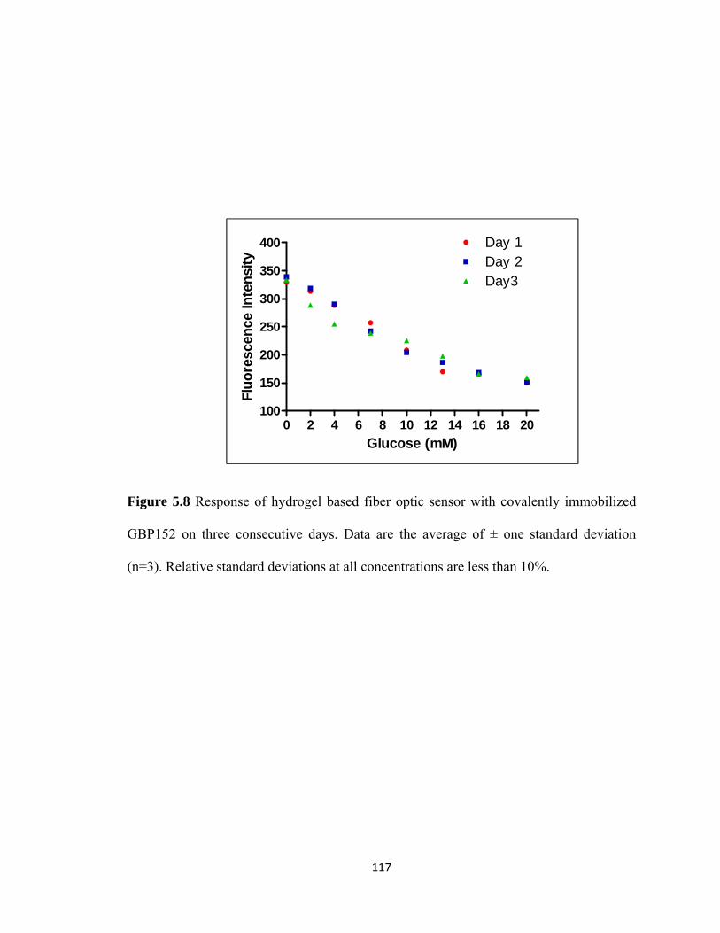
117
Figure 5.8 Response of hydrogel based fiber optic sensor with covalently immobilized
GBP152 on three consecutive days. Data are the average of ± one standard deviation
(n=3). Relative standard deviations at all concentrations are less than 10%.
0 2 4 6 8 10 12 14 16 18 20100
150
200
250
300
350
400 Day 1Day 2Day3
Glucose (mM)
Flu
ore
scen
ce In
ten
sity

118
Figure 5.9 Fluorescence study of fiber optic sensor in physiologically relevant glucose
concentrations (a) GBP152 (b) GRPs-FW (c) GRPs-FL. Data are the average of ± one
standard deviation (n=3). Relative standard deviations at all concentrations are less than
10%.
0 2 4 6 8 10 12 14 16 18 20
0
20
40
60
80
100GBP152
Glucose (mM)N
orm
aliz
edF
luo
resc
ence
Inte
nsi
ty
0 2 4 6 8 10 12 14 16 18 20
0
20
40
60
80
100 GRP-FWGRP1-FWGRP2-FW
Glucose (mM)
No
rmal
ized
Flu
ore
scen
ce In
ten
sity
0 2 4 6 8 10 12 14 16 18 20
0
20
40
60
80
100 GRP-FLGRP1-FLGRP2-FL
Glucose (mM)
No
rmal
ized
Flu
ore
scen
ce In
ten
sity
a
b
c

119
biosensor allows the detection of glucose in the desired physiological mM range by
incorporation of a diffusion barrier provided by the hydrogel matrix.
In order to study the performance of the GBP152/GRPs-FW/FL sensor in
physiological fluids, the sensor response was studied in human serum (Figure 5.10a and
5.10b) and pig blood (5.11a and 5.11b). A quenching in the fluorescence signal was
observed with the increase in glucose concentration, both in spiked human serum and pig
blood for all the sensors. Since the initial glucose concentration in pig blood and human
serum was 1.6 mM and 6.5 mM, respectively as determined by Free Style Lite (Abott)
blood glucose monitoring system, human serum and pig blood were spiked to reach the
glucose level upto 20 mM. The performance for GBP152/GRPs-FW/FL sensor was also
evaluated by measuring its response at different temperatures such as RT, 37 °C and 42.5
°C (Figure 5.12a-d and Figure 5.13a-c). The results obtained are those that we would
expect given the demonstrated thermal stability of the GBP152/GRPs-FW/FL. Since the
GBP152, GRP1-FW, and GRP2-FW have Tm at 47 ⁰C, 41.0 ⁰C, and 43.1 ⁰C
respectively, the sensors incorporating these proteins showed very little change in
fluorescence in the presence of different glucose concentrations at 42.5 ⁰C. However,
GRP-FW178, GRP-FL178, GRP1-FL and GRP2-FL with Tm at 58.9 ⁰C, 66.1 ⁰C, 65.3 ⁰C,
and 56.5 ⁰C respectively showed fluorescence quenching at both 37 ⁰C and 42.5⁰C. Thus,
GRP-FW and GRPs-FL retain glucose binding activity at higher temperatures. Thus, the
sensors incorporating designer proteins with enhanced thermal stability can be utilized
for the monitoring of glucose within a range of temperatures including those in
hypothermic and hyperthermic ranges.

120
Figure 5.10 Fluorescence study of fiber optic sensor in spiked human serum (a) GRPs-
FW (b) GRPs-FL. Data are the average of ± one standard deviation (n=3). Relative
standard deviations at all concentrations are less than 10%.
a
b

121
Figure 5.11 Fluorescence study of fiber optic sensor in spiked pig blood (a) GRPs-FW
(b) GRPs-FL. Data are the average of ± one standard deviation (n=3). Relative standard
deviations at all concentrations are less than 10%.
a
b

122
Figure 5.12 Temperature study of GBP142 and GBP-FW sensor (a) GBP152 (b) GRP-
FW (c) GRP1-FW (d) GRP2-FW. Symbols represent 35.5 °C, and 42.5 °C. Data are
the average of ± one standard deviation (n=3). Relative standard deviations at all
concentrations are less than 10%.
0 2 4 6 8 10 12 14 16 18 20275
300
325
350
375
400
425
450
475
Glucose (mM)
Flu
ore
scen
ce In
ten
sity
b a
c d

123
Figure 5.13 Temperature study of GRPs-FL sensor (a) GRP-FL (b) GRP1-FL (c) GRP2-
FL. Symbols represent 35.5 °C, and 42.5 °C. Data are the average of ± one standard
deviation (n=3). Relative standard deviations at all concentrations are less than 10%.
b
c
a

124
CONCLUSIONS
The existing need for continuous glucose monitoring in diabetics as well as in-
hospital patients instigated the development of our fiber optic-based glucose monitoring
systems. As previously mentioned, the currently available glucose sensing devices have a
number of limitations. In addition to the devices described in this chapter others such as
the,“GluCath”, an intravascular continuous monitor product from GluMetrics, Inc., is still
in the developmental stages.206 GluCath is based on fluorescent derivatives of boronates.
Although the product claims to be sensitive in hypoglycemic ranges, it suffers from some
major limitations. These limitations arise from of the fact that boronic acid derivatives
based-glucose sensors are known to be pH sensitive74 and to bind to glycosylated
proteins.207, 208 Thus, these impose a challenge when using boronates to measure glucose
in the clinic under physiological conditions.
In this research fiber optic-based reagentless fluorescence biosensor for glucose,
comprising of a miniaturized hydrogel that incorporates glucose binding protein,GBP152
or designer glucose recognition proteins, GRPs-FW/FL were developed. Immobilizing
the protein within the hydrogel network provides protection to the protein and worsens
the detection limit to our advantage. Thus, enables the sensing of glucose in
physiologically relevant mM ranges. The fiber optic glucose biosensors demonstrated a
fast response time, ~ 1 min, at different physiological temperatures ranging from 35.5 °C
to 42.5 °C. The biosensors are highly selective and sensitive to glucose. Moreover, the
biosensors were capable of glucose detection in different samples, including human
serum and pig blood, without any pretreatment of the sample. Thus, demonstrating its

125
sensitivity at physiological pH and its potential use in the monitoring of glucose as it
overcomes the limitations of the existing glucose sensing systems.

126
CHAPTER SIX
CONCLUSIONS AND FUTURE PERSPECTIVES
Protein Engineering is a valuable tool for manipulation of protein structure and
hence, for better understanding of protein structure and function. Traditional design
methods in protein engineering are based, among others, on direct evolution or rational
design. In recent years, computational protein design methods have provided further
insights into protein structure. Engineered proteins have found application in the
development of sensing devices. Fluorescence-based sensing is a powerful tool utilized
for the development of fast, reagentless, and non-invasive detection of analytes. Proteins
have been engineered to fluoresce by either fusion of the non-fluorescing proteins with
fluorescent proteins, conjugation of fluorescent molecules or incorporation of unnatural
fluorescent amino acids. In this work, we describe the development of designer proteins
and their utilization for the development of fluorescence-based biosensors for glucose
and universal biosensing systems for several biomolecules of interest.
Chapter two and three describes the development of universal biosensing systems
for the detection of biomolecules of interest. The biosensing system utilizes modified
monoclonal antibodies. The non-canonical site in the variable region of antibodies near
the antigen-binding site is modified and utilized for signal transduction. The non-
canonical site has been reported to covalently bind to nucleotides with azide
functionality. Docking to this non-canonical site does not alter the antigen-binding ability

127
of the antibody. In an effort towards design of antibodies capable of sensing molecules
for target analytes, the non-canonical site in the monoclonal antibody was covalently
conjugated to a fluorophore labeled azido-nucleotide that acts as the reporter molecule of
the biosensing element. This modification at the non-canonical site does not affect the
antigen binding, which was demonstrated by testing the biosensing system for two
different analytes. The first analyte employed in the proof of concept work was
Interleukin 6. Interleukin 6 is a pro- and anti-inflammatory cytokine, whose
concentrations is elevated in several disease states, including cardiovascular diseases. The
second analyte of interest chosen for our work is Osteonectin, a salivary bone marker.
The biosensing system thus developed is universal as it can be utilized for the detection
of a variety of analytes depending upon the availability of monoclonal antibody against
the analyte of interest.
Future work with respect to the work described in Chapters Two and Three
should include understanding the mechanism of response of the fluorescently labeled
antibody when exposed to its corresponding ligand analyte. Current studies showed a lag
time in generating a response of the antibody when exposure to its ligand. This could be
due to a change in conformation of the antibody upon binding the ligand, or simply due
to the time needed for difussion of the ligand into the binding pocket of the antibody. To
elucidate this mechanism, a time study of the response generated by a series of
concentrations (lower than that employed in the study) of fluorescently labeled antibody
to its corresponding antigen should be undertaken. This would provide an answer on
whether the lag time is due to diffusion or hint toward a delayed conformational change.
Future work can be further extended by developing detection systems utilizing the above

128
mentioned strategy for other biomolecules of interest. Some examples include cortisol,
osteocalcin and melatonin. In our work we utilized ATP-modifed probes for conjugation
of the fluorophore to the non-canonical site of the antibody. Other nucleotide probes
wherein the fluorophore is conjugated to the probe not at the γ-phosphate of ATP, but
rather covalently linked to the oxidized ribose can be utilized for labeling the antibody
unconventional site. The effect of conjugating the fluorophore at this position might
produce a fluorescence signal of similar or different intensity as in previous design and
thus, provide a better understanding of the orientation of fluorophore within the non-
canonical nucleoside binding site. The effect of antigen-binding to the antibody can be
then studied using these fluorescent probes. Further, it has been reported that the non-
canonical site has affinity for heterocyclic ring structures and, therefore, the use of other
nucleotides, nucleosides and amino acids such as tryptophan should be explored as
probes for the covalent attachment of the reporter fluorophore molecule. Also several
other fluorophores can be conjugated to the nucleotide probes, allowing for a wider range
of emission wavelengths of the reporter molecule.
Chapter four describes the global incorporation of unnatural tryptophan and
unnatural leucine into the amino acid sequence of GBP of E coli and its truncated
fragments. 5-Fluorotryptophan and 5,5,5-trifluoroleucines were incorporated separately
in the protein by using a medium shift method wherein the bacteria is first grown in a
medium enriched with all 20 amino acids, and then transferred to a medium lacking the
20th amino acid, which is then replaced by its unnatural analogue. By utilizing this
method we have been successful in replacing all the natural tryptophans or leucines by
their unnatural analogues in the Glucose/Galactose binding protein (GBP) and its

129
truncated fragments (GRPs). The effect of incorporation of these unnatural amino acids
on the secondary structure, glucose binding activity, and thermal stability of the proteins
was studied. Circular dichroism spectroscopy in the UV region at room temperature
demonstrated that the incorporation of 5-Fluorotryptophans in full length GBP does not
affect the secondary structure of GBP. However, the incorporation of fluoroleucines in
GBP does alter the secondary structure. Incorporation of either fluorotryptophans or
fluoroleucines in truncated GBP fragments does affect the secondary structure of the
proteins. This change in the secondary structure does not prevent the GRPs from binding
to glucose. However, their glucose binding ability was altered as can be inferred from the
apparent Kds of these unnatural proteins (GRPs) compared to natural GBP. The glucose
binding ability of these unnatural proteins was determined by labeling the proteins with
an environment sensitive fluorophore, MDCC, and generating dose-response curves for
glucose and other sugars. All proteins utilized for incorporation of unnatural amino acids
contained a unique cysteine that was conjugated to MDCC using the sulfhydryl group of
cysteine. Further, thermal stability of the unnatural proteins was determined by far-UV
circular dichroism spectroscopy. Incorporation of fluorinated tryptophans or leucines in
the proteins demonstrated an increase in the thermal stability of the proteins.
The work described in Chapter four can be further extended by incorporating
other unnatural amino acids that would confer these proteins with special properties and
functionalities without diminishing their glucose binding capability. Some examples of
unnatural amino acids that can be incorporated in the future include
benzoylphenylalanine, p-azidophenylalanine and azidonorleucine, which can confer a
photoreactive functionality to the protein that can be further harnessed for selective

130
modification of proteins for sensing purposes. Another example of unnatural amino acid
that can be incorporated in these proteins is 2-Ferrocenylglycine, an electroactive non-
natural amino. The incorporation of an electroactive amino acid will allow for the further
development of electrochemical sensor for glucose utilizing these unnatural electroactive
proteins.
In this work, the proteins were labeled with a fluorophore for the development of
optical sensors. A future improvement to this work would be the incorporation of a
fluorescent amino acid within the structure of the proteins, which would result in
genetically encoded fluorescent proteins. An example of such an amino acid is 2-Amino-
3-(7-methoxy-2-oxo-2H-chromen-4-yl)propanoic acid, an alanine analogue and a
fluorotryptophan. Incorporation of these unnatural amino acids would confer inherent
fluorescence to the proteins and, hence, these can be directly utilized for the development
of optical sensors.
Chapters five describe the development of fiber optic-based glucose
biosensors, wherein, the fluorophore-conjugated glucose recognition element, GBP or
GRPs, was covalently immobilized within an optically transparent acrylamide hydrogel.
GBP, as well as the unnatural amino acids incorporated GRPs, have a high affinity for
glucose with Kds in the 10-6 M to 10-5 M range. In an effort to develop a glucose
biosensor that can detect glucose in physiologically relevant mM concentrations, the
fluorescently labeled GBP/GRPs were encaptured within a hydrogel matrix. The
Hydrogel matrix serves several purposes as it provides for a hydrophilic, protein friendly
environment, allows for high loading of the protein, and serves as a diffusion barrier
capable of shifting the detection range of the proteins to the desired mM range. In our

131
work, the hydrogel was further immobilized on the tip of an optical fiber. The sensor
response demonstrated reversibility at low-to-high glucose concentration ranges and
viceversa. The ruggedness of the sensor was demonstrated by evaluating the sensor
response during three consecutive days. Further the sensor exhibited acceptable response
in body fluids such as blood and serum and at physiologically important temperatures.
Thus, the sensor performance suggests its potential use for use for in vivo glucose
monitoring.
Future work for improvement of the biosensing systems described in Chapters
five could be accomplished by further enclosing the hydrogel within a dialysis membrane
and attached onto the tip of the optical fiber. The dialysis membrane would help to retain
the hydrogel integrity over longer periods of storage and use. In order to introduce an
internal reference within the fiber optic sensor to eliminate background noise due to the
variations in intensity of light, another fluorophore that is not sensitive to the
environment can be labeled to the protein at a position that does not interfere glucose
binding. Ratiometeric study of emissions of environment sensitive and non-environment
sensitive fluorophores can then be performed. This single optical fiber optic based sensor
can be used as a catheter for in vivo applications as such. However, a dual lumen catheter
would extend its utility for in vivo applications, wherein, one lumen would contain the
sensor, while another would contain a channel through which blood may be drawn, such
that glucose readings from the same blood sample can be analyzed and compared by both
the catheter and traditional methods. Further incorporation of more than one hydrogel
with the glucose sensing element on different fibers bundled together can be utilized for
extending the life of the sensor, as after the lifetime of one sensor has expired other

132
sensor could be used for glucose recognition in blood. Study of the functioning of the
catheters in animal study would enable to determine the performance of the sensor in
vivo.
In conclusion, antibody-based sensing system for universal detection of analytes
of interest was successfully developed by labeling an inherent non-canonical nucleoside
binding site with a fluorophore conjugated nucleotide probe. Docking the non-canonical
site did not affect the antigen binding. The developed sensing system was demonstrated
to be very selective due to the use of monoclonal antibodies. Successful development of
glucose recognition proteins with enhanced thermal stability by incorporation of
unnatural amino acids in to the structure of GBP and its truncated fragments has been
reported. Further, application of the unnatural amino acid incorporated proteins for the
development of fiber optic based biosensor for in vivo glucose monitoring has also been
demonstrated.

133
REFERENCES
1. Schott, H. & Koessel, H. Synthesis of phage specific deoxyribonucleic acid
fragments. I. Synthesis of four undecanucleotides complementary to a mutated
region of the coat protein cistron of fd phage deoxyribonucleic acid. Journal of
the American Chemical Society 95, 3778-3785 (1973).
2. Hutchison, C.A., III & Edgell, M.H. Genetic Assay for Small Fragments of
Bacteriophage {varphi}X174 Deoxyribonucleic Acid. J. Virol. 8, 181-189 (1971).
3. Zoller, M.J. & Smith, M. Oligonucleotide-directed mutagenesis using M13-
derived vectors: an efficient and general procedure for the production of point
mutations in any fragment of DNA. Nucleic Acids Research 10, 6487-6500
(1982).
4. Leatherbarrow, R.J. & Fersht, A.R. Protein engineering. Protein Engineering 1, 7-
16 (1986).
5. Winter, G., Fersht, A.R., Wilkinson, A.J., Zoller, M. & Smith, M. Redesigning
enzyme structure by site-directed mutagenesis: tyrosyl tRNA synthetase and ATP
binding. Nature 299, 756-758 (1982).
6. Chusacultanachai, S. & Yuthavong, Y. in, Vol. 270 319-3332004).
7. McCullum, E.O., Williams, B.A.R., Zhang, J. & Chaput, J.C. in In Vitro
Mutagenesis Protocols, Vol. 634. (ed. J. Braman) 103-109 (Humana Press, 2010).
8. De Lorimier, R.M. et al. Construction of a fluorescent biosensor family. Protein
Science 11, 2655-2675 (2002).

134
9. Marvin, J.S. & Hellinga, H.W. Engineering Biosensors by Introducing
Fluorescent Allosteric Signal Transducers: Construction of a Novel Glucose
Sensor. Journal of the American Chemical Society 120, 7-11 (1998).
10. Salins, L.L.E., Ware, R.A., Ensor, C.M. & Daunert, S. A Novel Reagentless
Sensing System for Measuring Glucose Based on the Galactose/Glucose-Binding
Protein. Analytical Biochemistry 294, 19-26 (2001).
11. Ahern, T.J., Casal, J.I., Petsko, G.A. & Klibanov, A.M. Control of oligomeric
enzyme thermostability by protein engineering. Proceedings of the National
Academy of Sciences of the United States of America 84, 675-679 (1987).
12. Matsumura, M., Signor, G. & Matthews, B.W. Substantial increase of protein
stability by multiple disulphide bonds. Nature 342, 291-293 (1989).
13. Wilkinson, A.J., Fersht, A.R., Blow, D.M., Carter, P. & Winter, G. A large
increase in enzyme-substrate affinity by protein engineering. Nature 307, 187-188
(1984).
14. Hartman, I. Insulin Analogs: Impact on Treatment Success, Satisfaction, Quality
of Life, and Adherence. CLINICAL MEDICINE & RESEARCH 6, 54-67 (2008).
15. Tellier, C. Exploiting antibodies as catalysts: potential therapeutic applications.
Transfusion Clinique et Biologique 9, 1-8 (2002).
16. Mets, B. et al. A catalytic antibody against cocaine prevents cocaine’s reinforcing
and toxic effects in rats. Proceedings of the National Academy of Sciences of the
United States of America 95, 10176-10181 (1998).

135
17. Briscoe, R.J. et al. A catalytic antibody against cocaine attenuates cocaine's
cardiovascular effects in mice: a dose and time course analysis. International
Immunopharmacology 1, 1189-1198 (2001).
18. Wentworth, P. et al. Toward antibody-directed "abzyme" prodrug therapy,
ADAPT: carbamate prodrug activation by a catalytic antibody and its in vitro
application to human tumor cell killing. Proceedings of the National Academy of
Sciences of the United States of America 93, 799-803 (1996).
19. Wang, Q. et al. Construction of EGFP-tagged rBCG of
<i>E.tenella</i> and distribution in chickens. Science in China Series
C: Life Sciences 52, 278-283 (2009).
20. Lewis, J.C. & Daunert, S. Dual Detection of Peptides in a Fluorescence Binding
Assay by Employing Genetically Fused GFP and BFP Mutants. Analytical
Chemistry 71, 4321-4327 (1999).
21. Nilsson, J. (Tekniska högsk., Stockholm; 1996).
22. Lewis, J.C., Feliciano, J. & Daunert, S. Fluorescence binding assay for a small
peptide based on a GFP fusion protein. Analytica Chimica Acta 397, 279-286
(1999).
23. Evers, T.H., Appelhof, M.A.M., Meijer, E.W. & Merkx, M. His-tags as Zn(II)
binding motifs in a protein-based fluorescent sensor. Protein Engineering Design
and Selection 21, 529-536 (2008).
24. Takeda, S., Kamiya, N. & Nagamune, T. A novel protein-based heme sensor
consisting of green fluorescent protein and apocytochrome b562. Analytical
Biochemistry 317, 116-119 (2003).

136
25. Magliery, T.J. et al. Detecting Protein−Protein Interactions with a Green
Fluorescent Protein Fragment Reassembly Trap: Scope and Mechanism. Journal
of the American Chemical Society 127, 146-157 (2004).
26. Porter, J.R., Stains, C.I., Jester, B.W. & Ghosh, I. A General and Rapid Cell-Free
Approach for the Interrogation of Protein−Protein, Protein−DNA, and
Protein−RNA Interactions and their Antagonists Utilizing Split-Protein Reporters.
Journal of the American Chemical Society 130, 6488-6497 (2008).
27. Piehler, J. New methodologies for measuring protein interactions in vivo and in
vitro. Current Opinion in Structural Biology 15, 4-14 (2005).
28. Ghosh, I., Hamilton, A.D. & Regan, L. Antiparallel Leucine Zipper-Directed
Protein Reassembly: Application to the Green Fluorescent Protein. Journal of the
American Chemical Society 122, 5658-5659 (2000).
29. Jeong, J. et al. Monitoring of conformational change in maltose binding protein
using split green fluorescent protein. Biochemical and Biophysical Research
Communications 339, 647-651 (2006).
30. Nobuhide, D. & Hiroshi, Y. Design of generic biosensors based on green
fluorescent proteins with allosteric sites by directed evolution. FEBS letters 453,
305-307 (1999).
31. Nagai, T., Sawano, A., Park, E.S. & Miyawaki, A. Circularly permuted green
fluorescent proteins engineered to sense Ca2+. Proceedings of the National
Academy of Sciences of the United States of America 98, 3197-3202 (2001).

137
32. Teasley Hamorsky, K., Ensor, C.M., Wei, Y. & Daunert, S. A Bioluminescent
Molecular Switch For Glucose. Angewandte Chemie International Edition 47,
3718-3721 (2008).
33. Baird, G.S., Zacharias, D.A. & Tsien, R.Y. Circular permutation and receptor
insertion within green fluorescent proteins. Proceedings of the National Academy
of Sciences of the United States of America 96, 11241-11246 (1999).
34. Ostermeier, M. Engineering allosteric protein switches by domain insertion.
Protein Engineering Design and Selection 18, 359-364 (2005).
35. Toscano, M.D., Woycechowsky, K.J. & Hilvert, D. Minimalist Active-Site
Redesign: Teaching Old Enzymes New Tricks. Angewandte Chemie International
Edition 46, 3212-3236 (2007).
36. Piszczek, G., D'Auria, S., Staiano, M., Rossi, M. & Ginsburg, A. Conformational
stability and domain coupling in D-glucose/D-galactose-binding protein from
Escherichia coli. Biochem. J. 381, 97-103 (2004).
37. Scognamiglio, V. et al. D-galactose/D-glucose-binding protein from Escherichia
coli as probe for a non-consuming glucose implantable fluorescence biosensor.
Sensors 7, 2484-2491 (2007).
38. Vyas, N.K., Vyas, M.N. & Quiocho, F.A. Comparison of the periplasmic
receptors for L-arabinose, D-glucose/D-galactose, and D-ribose. Structural and
functional similarity. Journal of Biological Chemistry 266, 5226-5237 (1991).
39. Vyas, N.K., Vyas, M.N. & Quiocho, F.A. Sugar and signal-transducer binding
sites of the Escherichia coli galactose chemoreceptor protein. Science
(Washington, DC, United States) 242, 1290-1295 (1988).

138
40. Herman, P. et al. The role of calcium in the conformational dynamics and thermal
stability of the D-galactose/D-glucose-binding protein from Escherichia coli.
Proteins: Structure, Function, and Bioinformatics 61, 184-195 (2005).
41. Vyas, N.K., Vyas, M.N. & Quiocho, F.A. A novel calcium binding site in the
galactose-binding protein of bacterial transport and chemotaxis. Nature (London,
United Kingdom) 327, 635-638 (1987).
42. Turner, K. & Daunert, S. Development of a Glucose Biosensor Based Upon
Glucose Recognition Peptides Engineered by the Truncation of Glucose Binding
Protein. (2011 (in preparation)).
43. Head-Gordon, T. & Brown, S. Minimalist models for protein folding and design.
Current Opinion in Structural Biology 13, 160-167 (2003).
44. Voloshchuk, N., Zhu, A.Y., Snydacker, D. & Montclare, J.K. Positional effects of
monofluorinated phenylalanines on histone acetyltransferase stability and activity.
Bioorganic & Medicinal Chemistry Letters 19, 5449-5451 (2009).
45. Camarero, J.A., Hackel, B.J., de Yoreo, J.J. & Mitchell, A.R. Fmoc-Based
Synthesis of Peptide α-Thioesters Using an Aryl Hydrazine Support†. The
Journal of Organic Chemistry 69, 4145-4151 (2004).
46. Katritzky, A.R., Yoshioka, M., Narindoshvili, T., Chung, A. & Khashab, N.M. N-
Fmoc-Protected(α-Dipeptidoyl)Benzotriazoles for Efficient Solid-Phase Peptide
Synthesis by Segment Condensation. Chemical Biology & Drug Design 72, 182-
188 (2008).
47. Deechongkit, S., You, S.-L. & Kelly, J.W. Synthesis of All Nineteen
Appropriately Protected Chiral α-Hydroxy Acid Equivalents of the α-Amino

139
Acids for Boc Solid-Phase Depsi-Peptide Synthesis. Organic Letters 6, 497-500
(2004).
48. Zhang, Y. & Kennan, A.J. Efficient Introduction of Protected Guanidines in BOC
Solid Phase Peptide Synthesis. Organic Letters 3, 2341-2344 (2001).
49. Dawson, P.E., Muir, T.W., Clark-Lewis, I. & Kent, S.B.H. Synthesis of Proteins
by Native Chemical Ligation. Science 266, 776-779 (1994).
50. Erlanson, D.A., Chytil, M. & Verdine, G.L. The leucine zipper domain controls
the orientation of AP-1 in the NFAT·AP-1·DNA complex. Chemistry & Biology
3, 981-991 (1996).
51. Schwarzer, D. & Cole, P.A. Protein semisynthesis and expressed protein ligation:
chasing a protein's tail. Current Opinion in Chemical Biology 9, 561-569 (2005).
52. Muir, T.W., Sondhi, D. & Cole, P.A. Expressed protein ligation: A general
method for protein engineering. Proc. Natl. Acad. Sci. U. S. A. 95, 6705-6710
(1998).
53. Bossi, L. & Roth, J.R. The influence of codon context on genetic code translation.
Nature 286, 123-127 (1980).
54. Benzer, S. & Champe, S.P. A change from nonsense to sense in the genetic code.
Proc Natl Acad Sci U S A 48, 1114-1121 (1962).
55. Heckler, T.G. et al. T4 RNA ligase mediated preparation of novel "chemically
misacylated" tRNAPhes. Biochemistry 23, 1468-1473 (1984).
56. Roesser, J.R., Xu, C., Payne, R.C., Surratt, C.K. & Hecht, S.M. Preparation of
misacylated aminoacyl-tRNAPhe's useful as probes of the ribosomal acceptor
site. Biochemistry 28, 5185-5195 (1989).

140
57. Robertson, S.A., Ellman, J.A. & Schultz, P.G. A general and efficient route for
chemical aminoacylation of transfer RNAs. Journal of the American Chemical
Society 113, 2722-2729 (1991).
58. Hashimoto, N., Ninomiya, K., Endo, T. & Sisido, M. Simple and quick chemical
aminoacylation of tRNA in cationic micellar solution under ultrasonic agitation.
Chemical Communications, 4321-4323 (2005).
59. Beene, D.L., Dougherty, D.A. & Lester, H.A. Unnatural amino acid mutagenesis
in mapping ion channel function. Current Opinion in Neurobiology 13, 264-270
(2003).
60. Chin, J.W. & Schultz, P.G. In Vivo Photocrosslinking with Unnatural Amino
Acid Mutagenesis. ChemBioChem 3, 1135-1137 (2002).
61. Cornish, V.W., Hahn, K.M. & Schultz, P.G. Site-Specific Protein Modification
Using a Ketone Handle. Journal of the American Chemical Society 118, 8150-
8151 (1996).
62. Sakamoto, K. et al. Site‐specific incorporation of an unnatural amino acid into
proteins in mammalian cells. Nucleic Acids Research 30, 4692-4699 (2002).
63. Chin, J.W. et al. An Expanded Eukaryotic Genetic Code. Science 301, 964-967
(2003).
64. Hohsaka, T., Ashizuka, Y., Sasaki, H., Murakami, H. & Sisido, M. Incorporation
of Two Different Nonnatural Amino Acids Independently into a Single Protein
through Extension of the Genetic Code. Journal of the American Chemical
Society 121, 12194-12195 (1999).

141
65. Murakami, H., Hohsaka, T., Ashizuka, Y., Hashimoto, K. & Sisido, M. Site-
Directed Incorporation of Fluorescent Nonnatural Amino Acids into Streptavidin
for Highly Sensitive Detection of Biotin. Biomacromolecules 1, 118-125 (2000).
66. Taki, M., Hohsaka, T., Murakami, H., Taira, K. & Sisido, M. Position-Specific
Incorporation of a Fluorophore−Quencher Pair into a Single Streptavidin
through Orthogonal Four-Base Codon/Anticodon Pairs. Journal of the American
Chemical Society 124, 14586-14590 (2002).
67. Hohsaka, T., Ashizuka, Y., Murakami, H. & Sisido, M. Five-base codons for
incorporation of nonnatural amino acids into proteins. Nucleic Acids Research 29,
3646-3651 (2001).
68. Lee, H.S., Guo, J., Lemke, E.A., Dimla, R.D. & Schultz, P.G. Genetic
Incorporation of a Small, Environmentally Sensitive, Fluorescent Probe into
Proteins in Saccharomyces cerevisiae. Journal of the American Chemical Society
131, 12921-12923 (2009).
69. Rowe, L., Ensor, M., Mehl, R. & Daunert, S. Modulating the Bioluminescence
Emission of Photoproteins by in Vivo Site-Directed Incorporation of Non-Natural
Amino Acids. ACS Chemical Biology 5, 455-460 (2010).
70. Cirino, P.C., Tang, Y., Takahashi, K., Tirrell, D.A. & Arnold, F.H. Global
incorporation of norleucine in place of methionine in cytochrome P450 BM-3
heme domain increases peroxygenase activity. Biotechnology and Bioengineering
83, 729-734 (2003).
71. Rubini, M., Lepthien, S., Golbik, R. & Budisa, N. Aminotryptophan-containing
barstar: Structure-function tradeoff in protein design and engineering with an

142
expanded genetic code. Biochimica et Biophysica Acta (BBA) - Proteins &
Proteomics 1764, 1147-1158 (2006).
72. Wong, C.-Y. & Eftink, M.R. Biosynthetic incorporation of tryptophan analogues
into staphylococcal nuclease: Effect of 5-hydroxytryptophan and 7-azatryptophan
on structure and stability. Protein Science 6, 689-697 (1997).
73. Hyun Bae, J. et al. Expansion of the Genetic Code Enables Design of a Novel
"Gold" Class of Green Fluorescent Proteins. Journal of Molecular Biology 328,
1071-1081 (2003).
74. Dikici, E., Deo, S.K. & Daunert, S. Genetically Encoded Aequorin Variants with
Red-Shifted Emission Wavelengths by Global Incorporation of Non-Natural
Amino Acids. (to be submitted, 2011).
75. Johan & Mowbray, S.L. Sugar Recognition by a Glucose/Galactose Receptor.
Journal of Biological Chemistry 270, 9978-9981 (1995).
76. Moschou, E.A., Bachas, L.G., Daunert, S. & Deo, S.K. Hinge-Motion Binding
Proteins: Unraveling Their Analytical Potential. Analytical Chemistry 78, 6692-
6700 (2006).
77. Ames, G.F. Energy coupling in periplasmic permeases: the histidine permease as
a model system. Research in microbiology 141, 341-348 (1990).
78. Pflugrath, J.W. & Quiocho, F.A. The 2 Å resolution structure of the sulfate-
binding protein involved in active transport in Salmonella typhimurium. Journal
of Molecular Biology 200, 163-180 (1988).
79. Borths, E.L., Locher, K.P., Lee, A.T. & Rees, D.C. The structure of Escherichia
coli BtuF and binding to its cognate ATP binding cassette transporter.

143
Proceedings of the National Academy of Sciences of the United States of America
99, 16642-16647 (2002).
80. Karpowich, N.K., Huang, H.H., Smith, P.C. & Hunt, J.F. Crystal Structures of the
BtuF Periplasmic-binding Protein for Vitamin B12 Suggest a Functionally
Important Reduction in Protein Mobility upon Ligand Binding. Journal of
Biological Chemistry 278, 8429-8434 (2003).
81. Heddle, J., Scott, D.J., Unzai, S., Park, S.-Y. & Tame, J.R.H. Crystal Structures of
the Liganded and Unliganded Nickel-binding Protein NikA from Escherichia coli.
Journal of Biological Chemistry 278, 50322-50329 (2003).
82. Huang, H.-C. & Briggs, J.M. The association between a negatively charged ligand
and the electronegative binding pocket of its receptor. Biopolymers 63, 247-260
(2002).
83. Schroeder Jr, H.W. & Cavacini, L. Structure and function of immunoglobulins.
Journal of Allergy and Clinical Immunology 125, S41-S52 (2010).
84. Rajagopalan, K. et al. Novel unconventional binding site in the variable region of
immunoglobulins. Proceedings of the National Academy of Sciences of the United
States of America 93, 6019-6024 (1996).
85. Pavlinkova, G. et al. Site-specific photobiotinylation of immunoglobulins,
fragments and light chain dimers. Journal of Immunological Methods 201, 77-88
(1997).
86. Pavlinkova, G., Lou, D. & Kohler, H. Site-Specific Photobiotinylation of
Antibodies, Light Chains, and Immunoglobulin Fragments. Methods 22, 44-48
(2000).

144
87. Wang, C., Varshney, R.R. & Wang, D.-A. Therapeutic cell delivery and fate
control in hydrogels and hydrogel hybrids. Advanced Drug Delivery Reviews 62,
699-710 (2010).
88. Hoare, T.R. & Kohane, D.S. Hydrogels in drug delivery: Progress and challenges.
Polymer 49, 1993-2007 (2008).
89. Dragusin, M. et al. in Gels, Vol. 102. (ed. M. Zrínyi) 123-125 (Springer Berlin /
Heidelberg, 1996).
90. Galaev, I.Y. & Mattiasson, B. Thermoreactive water-soluble polymers, nonionic
surfactants, and hydrogels as reagents in biotechnology. Enzyme and Microbial
Technology 15, 354-366 (1993).
91. Verdejo, B., Rodriguez-Llansola, F., Escuder, B., Miravet, J.F. & Ballester, P.
Sodium and pH responsive hydrogel formation by the supramolecular system
calix[4]pyrrole derivative/tetramethylammonium cation. Chemical
Communications (2011).
92. Chen, Q. et al. Cysteine and pH-Responsive Hydrogel Based on a Saccharide
Derivative with an Aldehyde Group. Langmuir 26, 3165-3168 (2009).
93. Ma, Z., Nelson, D.M., Hong, Y. & Wagner, W.R. Thermally Responsive
Injectable Hydrogel Incorporating Methacrylate-Polylactide for Hydrolytic
Lability. Biomacromolecules 11, 1873-1881 (2010).
94. Khutoryanskaya, O.V., Mayeva, Z.A., Mun, G.A. & Khutoryanskiy, V.V.
Designing Temperature-Responsive Biocompatible Copolymers and Hydrogels
Based on 2-Hydroxyethyl(meth)acrylates. Biomacromolecules 9, 3353-3361
(2008).

145
95. Zhao, B. & Moore, J.S. Fast pH- and Ionic Strength-Responsive Hydrogels in
Microchannels. Langmuir 17, 4758-4763 (2001).
96. Jager, E.W.H., Smela, E. & Inganäs, O. Microfabricating Conjugated Polymer
Actuators. Science 290, 1540-1545 (2000).
97. Tanaka, T. et al. Phase Transitions in Ionic Gels. Physical Review Letters 45,
1636 (1980).
98. Ehrick, J.D. et al. Genetically engineered protein in hydrogels tailors stimuli-
responsive characteristics. Nat Mater 4, 298-302 (2005).
99. Ishihara, K., Kobayashi, M., Ishimaru, N. & Shinohara, I. Glucose Induced
Permeation Control of Insulin through a Complex Membrane Consisting of
Immobilized Glucose Oxidase and a Poly(amine). Polymer Journal 16, 625-631
(1984).
100. Miyata, T., Asami, N. & Uragami, T. A reversibly antigen-responsive hydrogel.
Nature 399, 766-769 (1999).
101. Zhu, H. & Snyder, M. Protein chip technology. Current Opinion in Chemical
Biology 7, 55-63 (2003).
102. Corgier, B.P., Marquette, C.A. & Blum, L.J. Direct electrochemical addressing of
immunoglobulins: Immuno-chip on screen-printed microarray. Biosensors and
Bioelectronics 22, 1522-1526 (2007).
103. Dal-Hee, M., Wei-Jen, T. & Mrksich, M. Chemical screening by mass
spectrometry to identify inhibitors of anthrax lethal factor. Nature Biotechnology
22, 717-723 (2004).

146
104. Lueking, A., Cahill, D.J. & Müllner, S. Protein biochips: A new and versatile
platform technology for molecular medicine. Drug Discovery Today 10, 789-794
(2005).
105. Fung, E.T., Thulasiraman, V., Weinberger, S.R. & Dalmasso, E.A. Protein
biochips for differential profiling. Current Opinion in Biotechnology 12, 65-69
(2001).
106. Klemer, D.P. Microelectronic Biosensors: Materials and Devices. (Biomedical
Engineering, 2009).
107. Vollath, D. Nanomaterials: An introduction to Synthesis, Properties and
Applications. (Wiley-VCH, ISBN 978-3527315314, Weinheim, Germany, 2008).
108. Umar, A., Rahman, M.M., Al-Hajry, A. & Hahn, Y.B. Highly-sensitive
cholesterol biosensor based on well-crystallized flower-shaped ZnO
nanostructures. Talanta 78, 284-289 (2009).
109. Chen, X. et al. Amperometric glucose biosensor based on boron-doped carbon
nanotubes modified electrode. Talanta 76, 763-767 (2008).
110. Marie, R., Jensenius, H., Thaysen, J., Christensen, C.B. & Boisen, A. Adsorption
kinetics and mechanical properties of thiol-modified DNA-oligos on gold
investigated by microcantilever sensors. Ultramicroscopy 91, 29-36 (2002).
111. Li, W., Sipe, D.M., Yong, X. & Qiao, L. A MEMS Thermal Biosensor for
Metabolic Monitoring Applications. Microelectromechanical Systems, Journal of
17, 318-327 (2008).
112. Monk, D.J. & Walt, D.R. Optical fiber-based biosensors. Analytical and
Bioanalytical Chemistry 379, 931-945 (2004).

147
113. Bosch, M., Sánchez, A., Rojas, F. & Ojeda, C. Recent Development in Optical
Fiber Biosensors. Sensors 7, 797-859 (2007).
114. Marazuela, M. & Moreno-Bondi, M. Fiber-optic biosensors - an overview.
Analytical and Bioanalytical Chemistry 372, 664-682 (2002).
115. Wolthuis, R.A. et al. Development of a medical fiber-optic oxygen sensor based
on optical absorption change. IEEE Transactions on Biomedical Engineering 39,
185-193 (1992).
116. Wolthuis, R., McCrae, D., Saaski, E., Hartl, J. & Mitchell, G. Development of a
medical fiberoptic ph sensor based on optical absorption. IEEE Transactions on
Biomedical Engineering 39, 531-537 (1992).
117. Li, Y.F., Y., Zhang, L. & Ma, S.Z., J. Molecular recognition of functionalized
polydiacetylene and its biosensor. Proceedings of SPIE - The International
Society for Optical Engineering 4077, 242-245 (2000).
118. Wang, A.J. & Arnold, M.A. Dual-enzyme fiber-optic biosensor for glutamate
based on reduced nicotinamide adenine dinucleotide luminescence. Analytical
Chemistry 64, 1051-1055 (1992).
119. Zhang, W., Chang, H. & Rechnitz, G.A. Dual-enzyme fiber optic biosensor for
pyruvate. Analytica Chimica Acta 350, 59-65 (1997).
120. Meadows, D.L. & Schultz, J.S. Design, manufacture and characterization of an
optical fiber glucose affinity sensor based on an homogeneous fluorescence
energy transfer assay system. Analytica Chimica Acta 280, 21-30 (1993).

148
121. Conroy, P.J., Hearty, S., Leonard, P. & O'Kennedy, R.J. Antibody production,
design and use for biosensor-based applications. Seminars in Cell &
Developmental Biology 20, 10-26 (2009).
122. Bier, F.F., Stöcklein, W., Böcher, M., Bilitewski, U. & Schmid, R.D. Use of a
fibre optic immunosensor for the detection of pesticides. Sensors and Actuators
B: Chemical 7, 509-512 (1992).
123. Doong, R.-A. & Tsai, H.-C. Immobilization and characterization of sol-gel-
encapsulated acetylcholinesterase fiber-optic biosensor. Analytica Chimica Acta
434, 239-246 (2001).
124. Jorgenson, R.C. & Yee, S.S. A fiber-optic chemical sensor based on surface
plasmon resonance. Sensors and Actuators B: Chemical 12, 213-220 (1993).
125. Bender, W.J.H., Dessy, R.E., Miller, M.S. & Claus, R.O. Feasibility of a
Chemical Microsensor Based on Surface Plasmon Resonance on Fiber Optics
Modified by Multilayer Vapor Deposition. Analytical Chemistry 66, 963-970
(1994).
126. Lin, W.B., Lacroix, M., Chovelon, J.M., Jaffrezic-Renault, N. & Gagnaire, H.
Development of a fiber-optic sensor based on surface plasmon resonance on silver
film for monitoring aqueous media. Sensors and Actuators B: Chemical 75, 203-
209 (2001).
127. Bidmanova, S., Chaloupkova, R., Damborsky, J. & Prokop, Z. Development of an
enzymatic fiber-optic biosensor for detection of halogenated hydrocarbons.
Analytical and Bioanalytical Chemistry 398, 1891-1898 (2010).

149
128. SlavIk, R., Homola, J., Ctyroký, J. & Brynda, E. Novel spectral fiber optic sensor
based on surface plasmon resonance. Sensors and Actuators B: Chemical 74, 106-
111 (2001).
129. Slavík, R., Homola, J. & Brynda, E. A miniature fiber optic surface plasmon
resonance sensor for fast detection of staphylococcal enterotoxin B. Biosensors
and Bioelectronics 17, 591-595 (2002).
130. http//www.biacore.com.
131. Rajagopalan, K. et al. Novel unconventional binding site in the variable region of
immunoglobulins. Proceedings of the National Academy of Sciences of the United
States of America 93, 6019-6024 (1996).
132. Pavlinkova, G. et al. Site-specific photobiotinylation of immunoglobulins,
fragments and light chain dimers. Journal of immunological methods 201, 77-88
(1997).
133. Araujo, L.S., Moraes, R.M., Trajman, A. & Saad, M.H.F. Assessing the IgA
immunoassay potential of the Mycobacterium tuberculosis MT10.3:MPT64
fusion protein in tuberculous pleural fluid. Clin. Vaccine Immunol., CVI.00372-
00310 (2010).
134. Theakston, R.D.G. & Reid, H.A. Enzyme-linked Immunosorbent Assay (ELISA)
in assessing antivenom potency. Toxicon 17, 511-515 (1979).
135. Blackburn, G.F. et al. Potentiometric biosensor employing catalytic antibodies as
the molecular recognition element. Analytical Chemistry 62, 2211-2216 (1990).
136. Saerens, D., Huang, L., Bonroy, K. & Muyldermans, S. Antibody Fragments as
Probe in Biosensor Development. Sensors 8, 4669-4686 (2008).

150
137. Geng, T., Uknalis, J., Tu, S.-I. & Bhunia, A. Fiber-Optic Biosensor Employing
Alexa-Fluor Conjugated Antibody for Detection of Escherichia coli O157:H7
from Ground Beef in Four Hours. Sensors 6, 796-807 (2006).
138. Simeonov, A. et al. Blue-Fluorescent Antibodies. Science 290, 307-313 (2000).
139. Wang, Q., Raja, K.S., Janda, K.D., Lin, T. & Finn, M.G. Blue Fluorescent
Antibodies as Reporters of Steric Accessibility in Virus Conjugates. Bioconjugate
Chemistry 14, 38-43 (2002).
140. Matsushita, M. et al. High-Throughput Screening by Using a Blue-Fluorescent
Antibody Sensor. Angewandte Chemie International Edition 42, 5984-5987
(2003).
141. Matsushita, M., Meijler, M.M., Wirsching, P., Lerner, R.A. & Janda, K.D. A Blue
Fluorescent Antibody−Cofactor Sensor for Mercury. Organic Letters 7, 4943-
4946 (2005).
142. O'Sullivan, O., Suhre, K., Abergel, C., Higgins Desmond, G. & Notredame, C.
3DCoffee: combining protein sequences and structures within multiple sequence
alignments. J Mol Biol 340, 385-395 (2004).
143. Somers, W., Stahl, M. & Seehra, J.S. 1.9 A crystal structure of interleukin 6:
implications for a novel mode of receptor dimerization and signaling. The EMBO
journal 16, 989-997 (1997).
144. Tokuda, H. et al. Function of Rho-kinase in prostaglandin D2-induced
interleukin-6 synthesis in osteoblasts. Prostaglandins, Leukotrienes and Essential
Fatty Acids 79, 41-46 (2008).

151
145. Hirano, T. et al. Complementary DNA for a novel human interleukin (BSF-2) that
induces B lymphocytes to produce immunoglobulin. Nature (London, United
Kingdom) 324, 73-76 (1986).
146. Leemans Jaklien, C., Vervoordeldonk Margriet, J.B.M., Florquin, S., van Kessel
Kok, P. & van der Poll, T. Differential role of interleukin-6 in lung inflammation
induced by lipoteichoic acid and peptidoglycan from Staphylococcus aureus.
American journal of respiratory and critical care medicine 165, 1445-1450
(2002).
147. Yokomuro, H. et al. The Variations in the Immunologic Features and Interleukin-
6 Levels for the Surgical Treatment of Cardiac Myxomas. Surgery Today 37, 750-
753 (2007).
148. Panoulas, V.F. et al. Association of interleukin-6 (IL-6)-174G/C gene
polymorphism with cardiovascular disease in patients with rheumatoid arthritis:
The role of obesity and smoking. Atherosclerosis (Amsterdam, Netherlands) 204,
178-183 (2009).
149. Luc, G. et al. C-Reactive Protein, Interleukin-6, and Fibrinogen as Predictors of
Coronary Heart Disease. Arteriosclerosis, Thrombosis, and Vascular Biology 23,
1255-1261 (2003).
150. Fonseca, J.E., Santos, M.J., Canhão, H. & Choy, E. Interleukin-6 as a key player
in systemic inflammation and joint destruction. Autoimmunity Reviews 8, 538-542
(2009).
152
151. Mansergh, F.C. et al. Osteopenia in Sparc (osteonectin)-deficient mice:
characterization of phenotypic determinants of femoral strength and changes in
gene expression. Physiological Genomics 32, 64-73 (2007).
152. Delany, A.M. et al. Osteopenia and decreased bone formation in osteonectin-
deficient mice. The Journal of Clinical Investigation 105, 915-923 (2000).
153. A. LeBlanc et al. Bone mineral and lean tissue loss after long duration space
flight. J Musculoskelet Neuronal Interact. 1, 157-160 (2000).
154. Zayzafoon, M., Gathings, W.E. & McDonald, J.M. Modeled Microgravity
Inhibits Osteogenic Differentiation of Human Mesenchymal Stem Cells and
Increases Adipogenesis. Endocrinology 145, 2421-2432 (2004).
155. Xie, R.-L. & Long, G.L. Elements within the First 17 Amino Acids of Human
Osteonectin Are Responsible for Binding to Type V Collagen. Journal of
Biological Chemistry 271, 8121-8125 (1996).
156. Yost, J.C. & Sage, E.H. Specific interaction of SPARC with endothelial cells is
mediated through a carboxyl-terminal sequence containing a calcium-binding EF
hand. Journal of Biological Chemistry 268, 25790-25796 (1993).
157. Kelm, R.J., Swords, N.A., Orfeo, T. & Mann, K.G. Osteonectin in matrix
remodeling. A plasminogen-osteonectin-collagen complex. Journal of Biological
Chemistry 269, 30147-30153 (1994).
158. Yan, Q. & Sage, E.H. SPARC, a Matricellular Glycoprotein with Important
Biological Functions. Journal of Histochemistry & Cytochemistry 47, 1495-1505
(1999).

153
159. Termine, J.D. et al. Osteonectin, a bone-specific protein linking mineral to
collagen. Cell 26, 99-105 (1981).
160. Ng, P.Y.B. et al. Candidate salivary biomarkers associatedwith alveolar bone
loss:cross-sectional andinvitro studies. FEMS Immunol Med Microbiol 49, 252-
260 (2007).
161. Embery, G. & Waddington, R. Gingival Crevicular Fluid: Biomarkers of
Periodontal Tissue Activity. Advances in Dental Research 8, 329-336 (1994).
162. Triola, G., Gerauer, M., Görmer, K., Brunsveld, L. & Waldmann, H. Solid-Phase
Synthesis of Lipidated Ras Peptides Employing the Ellman Sulfonamide Linker.
Chemistry – A European Journal 16, 9585-9591 (2010).
163. Young, T.S., Ahmad, I., Yin, J.A. & Schultz, P.G. An Enhanced System for
Unnatural Amino Acid Mutagenesis in E. coli. J. Mol. Biol. 395, 361-374.
164. Torrice, M.M., Bower, K.S., Lester, H.A. & Dougherty, D.A. Probing the role of
the cation-pi interaction in the binding sites of GPCRs using unnatural amino
acids. Proc. Natl. Acad. Sci. U. S. A. 106, 11919-11924, S11919/11911-
S11919/11916 (2009).
165. Cirino, P.C., Tang, Y., Takahashi, K., Tirrell, D.A. & Arnold, F.H. Global
incorporation of norleucine in place of methionine in cytochrome P 450 BM-3
heme domain increases peroxygenase activity. Biotechnology and Bioengineering
83, 729-734 (2003).
166. Cisnetti, F., Gateau, C., Lebrun, C. & Delangle, P. Lanthanide(III) Complexes
with Two Hexapeptides Incorporating Unnatural Chelating Amino Acids:

154
Secondary Structure and Stability. Chemistry – A European Journal 15, 7456-
7469 (2009).
167. Tang, Y. & Tirrell, D.A. Biosynthesis of a Highly Stable Coiled-Coil Protein
Containing Hexafluoroleucine in an Engineered Bacterial Host. Journal of the
American Chemical Society 123, 11089-11090 (2001).
168. Bilgiçer, B., Fichera, A. & Kumar, K. A Coiled Coil with a Fluorous Core.
Journal of the American Chemical Society 123, 4393-4399 (2001).
169. Tang, Y. et al. Fluorinated Coiled-Coil Proteins Prepared In Vivo Display
Enhanced Thermal and Chemical Stability. Angewandte Chemie International
Edition 40, 1494-1496 (2001).
170. Son, S., Tanrikulu, I.C. & Tirrell, D.A. Stabilization of bzip Peptides through
Incorporation of Fluorinated Aliphatic Residues. ChemBioChem 7, 1251-1257
(2006).
171. Voloshchuk, N., Zhu, A.Y., Snydacker, D. & Montclare, J.K. Positional effects of
monofluorinated phenylalanines on histone acetyltransferase stability and activity.
Bioorg Med Chem Lett 19, 5449-5451 (2009).
172. Hodges, J.A. & Raines, R.T. Stereoelectronic Effects on Collagen Stability: The
Dichotomy of 4-Fluoroproline Diastereomers. Journal of the American Chemical
Society 125, 9262-9263 (2003).
173. Beatty, K.E. & Tirrell, D.A. in Protein Engineering, Vol. 22. (eds. C. Köhrer &
U.L. RajBhandary) 127-153 (Springer Berlin Heidelberg, 2009).

155
174. Moschou, E.A., Sharma, B.V., Deo, S.K. & Daunert, S. Fluorescence Glucose
Detection: Advances Toward the Ideal In Vivo Biosensor. Journal of
Fluorescence 14, 535-547 (2004).
175. Saxl, T. et al. Fluorescence lifetime spectroscopy and imaging of nano-engineered
glucose sensor microcapsules based on glucose/galactose-binding protein.
Biosensors & Bioelectronics 24, 3229-3234 (2009).
176. De Lorimier, R.M. et al. Construction of a fluorescent biosensor family. Protein
Sci. 11, 2655-2675 (2002).
177. Khan, F., Saxl, T.E. & Pickup, J.C. Fluorescence intensity- and lifetime-based
glucose sensing using an engineered high-Kd mutant of glucose/galactose-binding
protein. Anal. Biochem. 399, 39-43 (2010).
178. Joel, S., Head, T. & Daunert, S. Designer Sensing Proteins via Global
Incorporation of Unnatural Amino Acids. (2011 (in preparation)).
179. Siegrist, J. et al. Continuous glucose sensor using novel genetically engineered
binding polypeptides towards in vivo applications. Sensors and Actuators B:
Chemical 149, 51-58 (2010).
180. Basile, J.N. Antihypertensive therapy, new-onset diabetes, and cardiovascular
disease. International Journal of Clinical Practice 63, 656-666 (2009).
181. Mahmud, I. et al. Hyperphosphataemia Is Associated with the Diabetes-related
Cardiovascular Risk Factors. Journal of Oleo Science 60, 79-85 (2011).
182. Zoungas, S. & Patel, A. Cardiovascular outcomes in type 2 diabetes: the impact of
preventative therapies. Annals of the New York Academy of Sciences 1212, 29-40
(2010).

156
183. Kamenov, Z.A., Parapunova, R.A. & Georgieva, R.T. Earlier development of
diabetic neuropathy in men than in women with type 2 diabetes mellitus. Gender
Medicine 7, 600-615 (2010).
184. Kuroda, N., Taniguchi, H., Baba, S. & Yamamoto, M. Relationship between age
and autonomic neuropathy in diabetes mellitus. Diabetes Research and Clinical
Practice 9, 49-53 (1990).
185. Jackson, G. & Barber, A. Visual Dysfunction Associated with Diabetic
Retinopathy. Current Diabetes Reports 10, 380-384 (2010).
186. Jeppesen, P. & Bek, T. The occurrence and causes of registered blindness in
diabetes patients in Arhus County, Denmark. Acta ophthalmologica Scandinavica
82, 526-530 (2004).
187. Oliver, N.S., Toumazou, C., Cass, A.E.G. & Johnston, D.G. Glucose sensors: a
review of current and emerging technology. Diabetic Medicine 26, 197-210
(2009).
188. Trence, D.L., Kelly, J.L. & Hirsch, I.B. The Rationale and Management of
Hyperglycemia for In-Patients with Cardiovascular Disease: Time for Change. J
Clin Endocrinol Metab 88, 2430-2437 (2003).
189. Sleiman, I. et al. Hyperglycemia as a Predictor of In-Hospital Mortality in Elderly
Patients without Diabetes Mellitus Admitted to a Sub-Intensive Care Unit.
Journal of the American Geriatrics Society 56, 1106-1110 (2008).
190. Van den Berghe, G. et al. Intensive Insulin Therapy in Critically Ill Patients. New
England Journal of Medicine 345, 1359-1367 (2001).

157
191. Ascione, R., Rogers, C.A., Rajakaruna, C. & Angelini, G.D. Inadequate Blood
Glucose Control Is Associated With In-Hospital Mortality and Morbidity in
Diabetic and Nondiabetic Patients Undergoing Cardiac Surgery. Circulation 118,
113-123 (2008).
192. Krinsley, J. Glycemic variability and mortality in critically ill patients: the impact
of diabetes. J. Diabetes Sci. Technol. 3, 1292-1301 (2009).
193. Grey, N. & Perdrizet, G. Reduction of nosocomial infections in the surgical
intensive-care unit by strict glycemic control. Endocr. Pract. 10, 46-52 (2004).
194. Chang, G., Tatsu, Y., Goto, T., Imaishi, H. & Morigaki, K. Glucose concentration
determination based on silica sol-gel encapsulated glucose oxidase optical
biosensor arrays. Talanta 83, 61-65 (2010).
195. Wilkins, E. & Atanasov, P. Glucose monitoring: state of the art and future
possibilities. Medical Engineering & Physics 18, 273-288 (1996).
196. Wu, B., Zhang, G., Shuang, S. & Choi, M.M.F. Biosensors for determination of
glucose with glucose oxidase immobilized on an eggshell membrane. Talanta 64,
546-553 (2004).
197. Hiratsuka, A., Fujisawa, K. & Muguruma, H. Amperometric Biosensor Based on
Glucose Dehydrogenase and Plasma-polymerized Thin Films. Anal. Sci. 24, 483 (
(2008) .).
198. Hovorka, R. Continuous glucose monitoring and closed-loop systems. Diabetic
Medicine 23, 1-12 (2006).

158
199. Vesper, H.W., Wang, P.M., Archibold, E., Prausnitz, M.R. & Myers, G.L.
Assessment of Trueness of a Glucose Monitor Using Interstitial Fluid and Whole
Blood as Specimen Matrix. Diabetes Technology & Therapeutics 8, 76-80 (2006).
200. Wing, R., Epstein, L., Blair, E. & Nowalk, M. Psychologic stress and blood
glucose levels in nondiabetic subjects. Psychosom Med 47, 558-564 (1985).
201. Kovatchev, B., Anderson, S., Heinemann, L. & Clarke, W. Comparison of the
Numerical and Clinical Accuracy of Four Continuous Glucose Monitors. Diabetes
Care 31, 1160-1164 (2008).
202. Miller, D.M., Olson, J.S., Pflugrath, J.W. & Quiocho, F.A. Rates of ligand
binding to periplasmic proteins involved in bacterial transport and chemotaxis.
Journal of Biological Chemistry 258, 13665-13672 (1983).
203. Ge, X., Rao, G. & Tolosa, L. On the Possibility of Real-Time Monitoring of
Glucose in Cell Culture by Microdialysis Using a Fluorescent Glucose Binding
Protein Sensor. Biotechnology Progress 24, 691-697 (2008).
204. Saxl, T. et al. Fluorescence lifetime spectroscopy and imaging of nano-engineered
glucose sensor microcapsules based on glucose/galactose-binding protein.
Biosensors and Bioelectronics 24, 3229-3234 (2009).
205. Hanahan, D. In: DNA Cloning. 1 D. Glover, ed., IRL Press, Ltd., London,109
(1985).
206. http://www.glumetrics.com/.
207. A, C. et al. Glycosylated proteins as an indicator of metabolic control in diabetes:
evaluation by aminophenylboronic acid affinity chromatography. Acta Diabetol
Lat. 21, 49-54 (1984).

159
208. Herold, D. et al. Measurement of glycosylated hemoglobins using boronate
affinity chromatography. Ann Clin Lab Sci 13, 482-488 (1983).

160
VITA
Smita Joel was born on May 23, 1978 in Jhansi, India. After graduating from high
school in India, she earned her Bachelor of Science (B. S.) degree in chemistry in 1999
from Isabella Thoburn College, Lucknow University, India. She then acquired a Bachelor
of Education (B. Ed.) degree from Lucknow Christian Training College, Lucknow
University, India in the year 2000. She received her Master of Science (M. S.) degree in
organic chemistry from Lucknow Christian College, Lucknow University, India in 2002.
In Fall 2004, she started the graduate program in the Department of Chemistry at
University of Kentucky (UK) in Lexington, KY. In Fall 2005, she joined the research
group of Dr. Sylvia Daunert at UK and worked on the development of biosensing system
for molecular diagnostics and biosensors using binding proteins. She was awarded the
Research Challenge Trust Fund Fellowship and Gill Fellowship during her graduate
studies at the University of Kentucky, Lexington, KY.
PATENTS
1. Daunert, Sylvia; Bachas, Leonidas G.; Haley, Boyd; Joel, Smita; Moschou,
Elizabeth; Turner, Kendrick; Rowe, Laura; Wang, Ping; Siegrist, Jonathan P.;
Madou, Marc J. Fiber-optic device including catheter and biosensor containing
glucose-binding protein for continuous in vivo detection of glucose. PCT Int.
Appl. (2009), 58pp. WO 2009021039.

161
2. Daunert, Sylvia; Turner, Kendrick; Joel, Smita; Rowe, Laura. Stabilized
variants of bacterial glucose-binding proteins for use in biosensors for
monitoring of blood glucose. PCT Int. Appl. (2009), 53pp. WO 2009021052.
3. Daunert, Sylvia; Bachas, Leonidas G.; Haley, Boyd; Joel, Smita. Semi-synthetic
antibodies as recognition elements containing purine molecular and fluorophore
bound to purine. PCT Int. Appl. (2009), 47pp. WO 2009021026.
PUBLICATIONS
1. J. Siegrist, T. Kazarian, C. Ensor, S. Joel, M. Madou, P. Wang, and S. Daunert,
“Continuous glucose sensor using novel genetically engineered binding
polypeptides towards in vivo applications.” Sensors and Actuators, B: Chemical
(2010), 149(1), 51-58.
2. S. Joel, A. Bhattacharya, B. Haley, L. G. Bachas, and S. Daunert, "Biosensors
Based on Site-Specific Labeling of Antibodies", to be submitted, 2011.
3. S. Joel, T. Head and S. Daunert. “Designer Sensing Proteins via Global
Incorporation of Unnatural Amino Acids”, submitted, 2011.
4. S. Joel, K. Turner, and S. Daunert, "Toward Designer Glucose Recognition
Proteins: Preparation of Semi-Synthetic Unnatural Glucose Recognition
Proteins", to be submitted 2011.
5. S. Joel, L. G. Bachas and S. Daunert, "Reagentless Fiber Optic Biosensors for the
Continuous Monitoring of Glucose ", in preparation, 2011.

162
CONFERENCE PRESENTATIONS
1. Poster presentation at the 238th ACS National Meeting August 16-20, 2009,
Washington, DC, United States. “Biosensors based on site-specific labeling of
antibodies.” Smita Joel, Anjan K. Bhattacharyya, Boyd Haley, Leonidas G.
Bachas, and Sylvia Daunert.
2. Oral presentation at Pittcon March 11, 2009, Chicago, IL, United States.
“Development of Universal Biosensing Systems Based on Monoclonal
Antibody.” Smita Joel, Anjan K. Bhattacharyya, Boyd Haley, Leonidas Bachas,
and Sylvia Daunert.
3. Poster presentation at the Kentucky Academy of Science, November 2009,
Northern Kentucky University, KY, United States. “Fiber Optic Biosensor for In
situ Monitoring of Hydroxylated PCB’s.” Neha Ray, Smita Joel, Kendrick
Turner, and Sylvia Daunert.
4. Poster presentation at the University of Kentucky for NSF Research Experiences
for Undergraduates 2009, Lexington, KY, United States. “Enhancing Natural
Fluorescence: Incorporation of 7-azatryptophan into Glucose Binding Protein.”
Trajen Head, Smita Joel, and Sylvia Daunert.
5. Poster presentation at the 14th Annual EPSCoR Conference, October 3, 2008,
Louisville, KY, United States. “Biosensors Based on Site-Specific Labeling of
Antibodies.” Smita Joel, Boyd E. Haley, Leonidas G. Bachas, and Sylvia
Daunert.
6. Poster presentation at the University of Kentucky for NSF Research Experiences
for Undergraduates August 2008, Lexington, KY, United States. “Creating a

163
Glucose Biosensor: Global Incorporation of 7-azatryptophan in Glucose Binding
Protein.” Jacqueline Ward, Smita Joel, and Sylvia Daunert.
7. Poster presentation at the Clinical and Translational Science Conference, June 3,
2008, Lexington, KY, United States. “Reagentless fiber optic biosensors for the
continuous monitoring of glucose.” Smita Joel, Lisa Moschou, and Sylvia
Daunert.
8. Poster presentation at the Naff Symposium, April 4, 2008, Lexington, KY, United
States. “Reagentless fiber optic biosensors for the continuous monitoring of
glucose.” Smita Joel, Lisa Moschou, and Sylvia Daunert.
9. Poster presentation at the University of Kentucky for NSF Research Experiences
for Undergraduates August 2007, Lexington, KY, United States. “Incorporation
of Unnatural Amino Acids in Glucose Binding Protein: Developing a Thermally
Stable Glucose Biosensor for in vivo Applications.” Eric Hunt, Smita Joel,
Kendrick Turner, and Sylvia Daunert.
10. Poster presentation at the 39th Central Regional Meeting of the American
Chemical Society, May 20-23, 2007, Covington, KY, United States. “Reagentless
fiber optic biosensors for the continuous monitoring of glucose.” Smita Joel, Lisa
Moschou, and Sylvia Daunert.
SMITA JOEL