endoscopic ultrasound: applications in pre-malignant and malignant disease december 20 th, 2010...
TRANSCRIPT

Endoscopic Ultrasound:Endoscopic Ultrasound:Applications in Pre-malignant Applications in Pre-malignant
and Malignant Diseaseand Malignant Disease
December 20December 20thth, 2010, 2010
Andrew T. Pellecchia, MDAndrew T. Pellecchia, MDDirector of Advanced EndoscopyDirector of Advanced Endoscopy
Jacobi Medical CenterJacobi Medical Center

EUSEUS Originally utilized to ‘clear’ the bile duct pre-Originally utilized to ‘clear’ the bile duct pre-
cholecystectomy in patients with suspected cholecystectomy in patients with suspected CBD stonesCBD stonesLess invasive alternative to ERCPLess invasive alternative to ERCPRisks similar to standard EGDRisks similar to standard EGD
EUS still used for this indicationEUS still used for this indicationLess than 20% of EUS procedures are performed Less than 20% of EUS procedures are performed
for this indication in established advanced for this indication in established advanced endoscopy centerendoscopy center

Evolution of EUSEvolution of EUS EUS as an imaging studyEUS as an imaging study EUS as a means of fluid and tissue EUS as a means of fluid and tissue
acquisitionacquisitionCancer stagingCancer stagingCyst analysisCyst analysis
EUS as an interventional/therapeutic modalityEUS as an interventional/therapeutic modalityNeurolysisNeurolysisTransmural cyst drainageTransmural cyst drainageDirect access to biliary systemDirect access to biliary systemMore…More…

OverviewOverview
Several illustrative EUS cases from JMCSeveral illustrative EUS cases from JMC Basic EUS principlesBasic EUS principles What is ‘within reach’ of EUS +/- FNA?What is ‘within reach’ of EUS +/- FNA? Brief overview of selected diseasesBrief overview of selected diseases

Patient GRPatient GR
62 y.o. woman with significant weight loss 62 y.o. woman with significant weight loss over the past 6 monthsover the past 6 months
CT a/p shows a 6 cm intra-abdominal massCT a/p shows a 6 cm intra-abdominal mass EGD/EUS/FNA planned to further evaluate EGD/EUS/FNA planned to further evaluate
lesionlesion
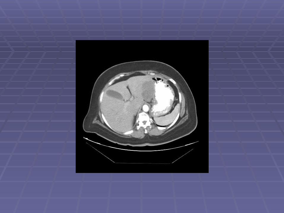


Endosonographic EvaluationEndosonographic Evaluation
EGD showed normal gastric mucosa with EGD showed normal gastric mucosa with evidence of mild external compression vs. evidence of mild external compression vs. submucosal lesion in the area of the gastric submucosal lesion in the area of the gastric incisuraincisura
EUSEUS Clear demarcation of hypoechoic mass adjacent Clear demarcation of hypoechoic mass adjacent
to left lobe of the liverto left lobe of the liver FNA was performedFNA was performed

GR-GISTH&E

GR-GIST
C-KIT (CD117)

Patient DDPatient DD
62 y.o. man with history of alcoholism and recurrent 62 y.o. man with history of alcoholism and recurrent pancreatitis since the 1970’s, admitted to an outside pancreatitis since the 1970’s, admitted to an outside hospital with jaundicehospital with jaundice
MRI showed a large pancreatic head massMRI showed a large pancreatic head mass ERCP for biliary drainage – failedERCP for biliary drainage – failed
Complicated by pancreatic tail pseudocyst formationComplicated by pancreatic tail pseudocyst formation
PTC with internalization - successfulPTC with internalization - successful Patient left AMA and came to JMCPatient left AMA and came to JMC EUS/FNA performed to obtain diagnosisEUS/FNA performed to obtain diagnosis


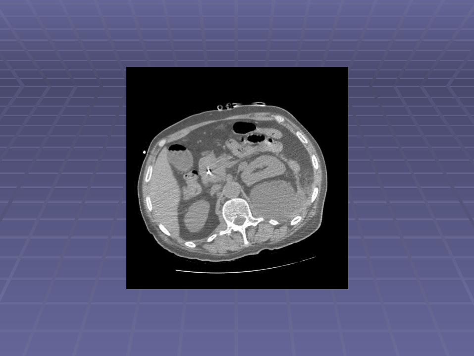

Endosonographic EvaluationEndosonographic Evaluation
EUSEUSLarge ~30mm hypoechoic pancreatic head mass Large ~30mm hypoechoic pancreatic head mass
surrounding the intrapancreatic CBD with PTC surrounding the intrapancreatic CBD with PTC drain seen within CBDdrain seen within CBD
Dilated PD to 5mm with evidence of chronic Dilated PD to 5mm with evidence of chronic pancreatitispancreatitis
FNA performedFNA performed

DD- Pancreas Ca. Pap stain

DD-Pancreas Ca.
Pap stain

Patient CEPatient CE
69 y.o. man with h/o non-small cell lung 69 y.o. man with h/o non-small cell lung cancer s/p LUL resection in 2006 who is cancer s/p LUL resection in 2006 who is referred after a chest CT showed new referred after a chest CT showed new mediastinal lymphadenopathymediastinal lymphadenopathy
EUS/FNA scheduled to evaluate for recurrent EUS/FNA scheduled to evaluate for recurrent diseasedisease


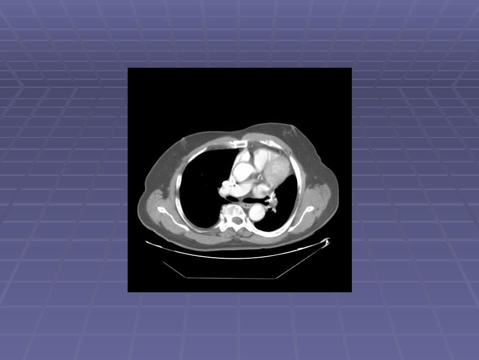


Endosonographic EvaluationEndosonographic Evaluation
EUSEUSSuspicious lymph nodes in the aortopulmonary Suspicious lymph nodes in the aortopulmonary
window, sized 6-11mmwindow, sized 6-11mmSuspicious lymph nodes in the subcarinal space, Suspicious lymph nodes in the subcarinal space,
sized 6-12mmsized 6-12mm FNA performedFNA performed

CE-Non-small cell ca.
Pap stain

CE-Non-small cell ca.Pap stain

Radial UltrasonographyRadial Ultrasonography
Oblique-viewing instruments with an ultrasound transducer Oblique-viewing instruments with an ultrasound transducer located at the tiplocated at the tip
The circumferential ultrasound image is perpendicular to the The circumferential ultrasound image is perpendicular to the long axis of the endoscopelong axis of the endoscope
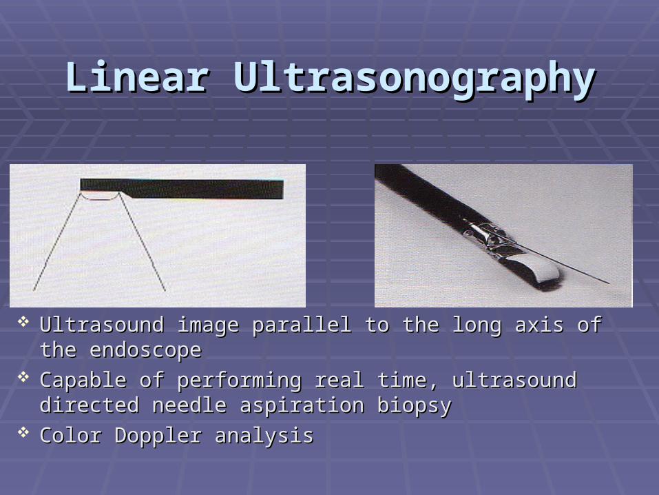
Linear UltrasonographyLinear Ultrasonography
Ultrasound image parallel to the long axis of the endoscope Ultrasound image parallel to the long axis of the endoscope Capable of performing real time, ultrasound directed needle Capable of performing real time, ultrasound directed needle
aspiration biopsyaspiration biopsy Color Doppler analysisColor Doppler analysis

Working End of Linear Working End of Linear EchoendoscopeEchoendoscope

The Scope of the The Scope of the EchoendoscopeEchoendoscope
What can be assessed by EUS with potential What can be assessed by EUS with potential FNA?FNA? Any structure within several cm of U/L GI tractAny structure within several cm of U/L GI tract Ability to see structures measuring Ability to see structures measuring 1 mm1 mm Ability to perform FNA upon structures measuring Ability to perform FNA upon structures measuring
3mm3mm
LimitationsLimitations Cannot visualize beyond air-filled structuresCannot visualize beyond air-filled structures Cannot biopsy through air-filled structures, blood Cannot biopsy through air-filled structures, blood
vessels, or the heartvessels, or the heart Lung that is non-adjacent to esophagus, trachea, aorta, Lung that is non-adjacent to esophagus, trachea, aorta,
pulmonary artery, r/l atriapulmonary artery, r/l atria

Risks of EUS FNARisks of EUS FNAPancreatitisPancreatitis
< 1:100< 1:100Significant bleedingSignificant bleeding
< 1:500< 1:500PerforationPerforation
< 1:1000< 1:1000Infection - rareInfection - rare
Antibiotics for transrectal FNA or FNA of cystsAntibiotics for transrectal FNA or FNA of cystsInadequate tissueInadequate tissue
1:101:10 to 1:5 to 1:5 Can be related to pathology of lesionCan be related to pathology of lesion
Cholangio, GISTCholangio, GIST

Thyroid MassThyroid Mass

FNA of Thyroid MassFNA of Thyroid Mass

Right Lower Pole Kidney MassRight Lower Pole Kidney Mass

EUS in Pre-Malignant DiseaseEUS in Pre-Malignant DiseasePancreatic CystsPancreatic CystsPD fluid analysisPD fluid analysisPancreatic screening in high risk Pancreatic screening in high risk
populationspopulations Chronic pancreatitisChronic pancreatitis Family history of pancreatic cancerFamily history of pancreatic cancer Cancer syndromesCancer syndromes
Submucosal lesionsSubmucosal lesions Pancreatic restsPancreatic rests

Pancreatic Cystic Fluid AnalysisPancreatic Cystic Fluid Analysis
Incidental pancreatic cysts seen in up to 20% Incidental pancreatic cysts seen in up to 20% of abdominal CT’s performed for any reasonof abdominal CT’s performed for any reason
Cystic lesions of the pancreas, even when Cystic lesions of the pancreas, even when found incidentally, may represent found incidentally, may represent malignantmalignant or or pre-malignantpre-malignant lesions lesionsThe majority of pancreatic cysts require The majority of pancreatic cysts require
evaluation by EUS/FNAevaluation by EUS/FNAFNA measurement of CEA, amylase, genetic markersFNA measurement of CEA, amylase, genetic markersRelatively sensitive and specific for differentiating Relatively sensitive and specific for differentiating
mucinous cysts (IPMN, MCA) from non-mucinous cysts mucinous cysts (IPMN, MCA) from non-mucinous cysts (SCA, Pseudocyst)(SCA, Pseudocyst)

HOP Serous CystadenomaHOP Serous Cystadenoma
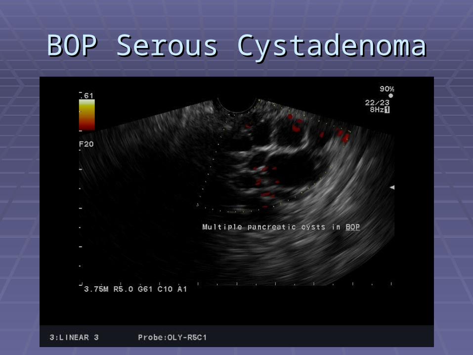
BOP Serous CystadenomaBOP Serous Cystadenoma

Oncology Consult?Oncology Consult?(FNA benign: Island of normal pancreatic tissue within serous cystadenoma)(FNA benign: Island of normal pancreatic tissue within serous cystadenoma)

Patient PSPatient PS Media reports state that the actor was Media reports state that the actor was
diagnosed with an IPMNdiagnosed with an IPMN IPMN is a IPMN is a pre-cancerouspre-cancerous lesion lesion Conclusion: the IPMN had already progressed Conclusion: the IPMN had already progressed
to adenocarcinoma prior to to adenocarcinoma prior to diagnosis/resectiondiagnosis/resection
Resected IPMNs often have foci of Resected IPMNs often have foci of adenocarcinomaadenocarcinoma
Lesson: ALL pancreatic cysts need to be Lesson: ALL pancreatic cysts need to be referred for risk stratificationreferred for risk stratification

EUS in Malignant DiseaseEUS in Malignant Disease Non-small cell lung cancerNon-small cell lung cancer Pancreatic cancerPancreatic cancer Esophageal and gastric cancerEsophageal and gastric cancer CholangiocarcinomaCholangiocarcinoma Rectal adenocarcinomaRectal adenocarcinoma Metastatic diseaseMetastatic disease
Lymph nodes: aortopulmonary, subcarinal, para-Lymph nodes: aortopulmonary, subcarinal, para-esophageal, celiac, intra-abdominalesophageal, celiac, intra-abdominal
Left lobe of liverLeft lobe of liver Left adrenalLeft adrenal And beyondAnd beyond – right lobe of liver, right adrenal, ... – right lobe of liver, right adrenal, ...

EUS and Lung CancerEUS and Lung Cancer
““We really do not need additional proof before We really do not need additional proof before EUS-FNA is considered the gold standard for EUS-FNA is considered the gold standard for invasive staging of non-small cell lung cancer invasive staging of non-small cell lung cancer and for diagnosis of posterior mediastinal and for diagnosis of posterior mediastinal lesions; there is little to lose and much to lesions; there is little to lose and much to gain.”gain.”
--P. Vilmann and S.S. Larsen, Eur Respir J P. Vilmann and S.S. Larsen, Eur Respir J 20052005; 25: 400–401; 25: 400–401

EUS and Lung CancerEUS and Lung Cancer
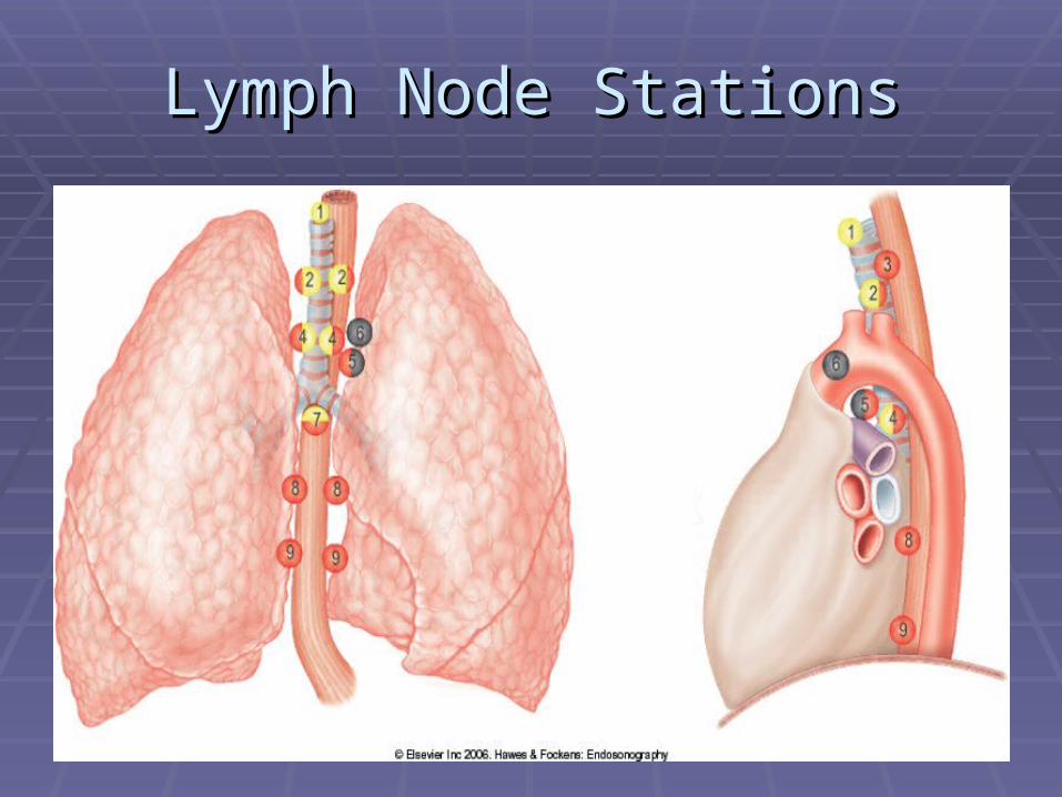
Lymph Node StationsLymph Node Stations

Normal AP WindowNormal AP Window
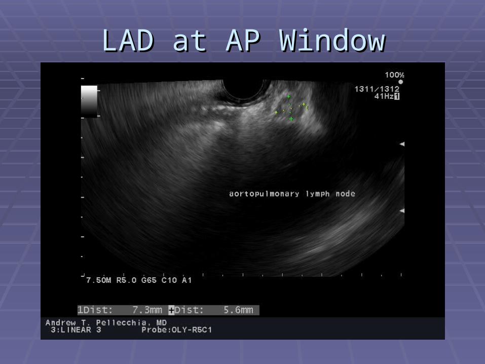
LAD at AP WindowLAD at AP Window

FNA at AP WindowFNA at AP Window

Subcarinal SpaceSubcarinal Space

LAD in Subcarinal SpaceLAD in Subcarinal Space

Likely Benign Abd LADLikely Benign Abd LAD
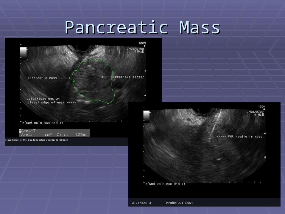
Pancreatic MassPancreatic Mass

Pancreatic Mass at CTPancreatic Mass at CT
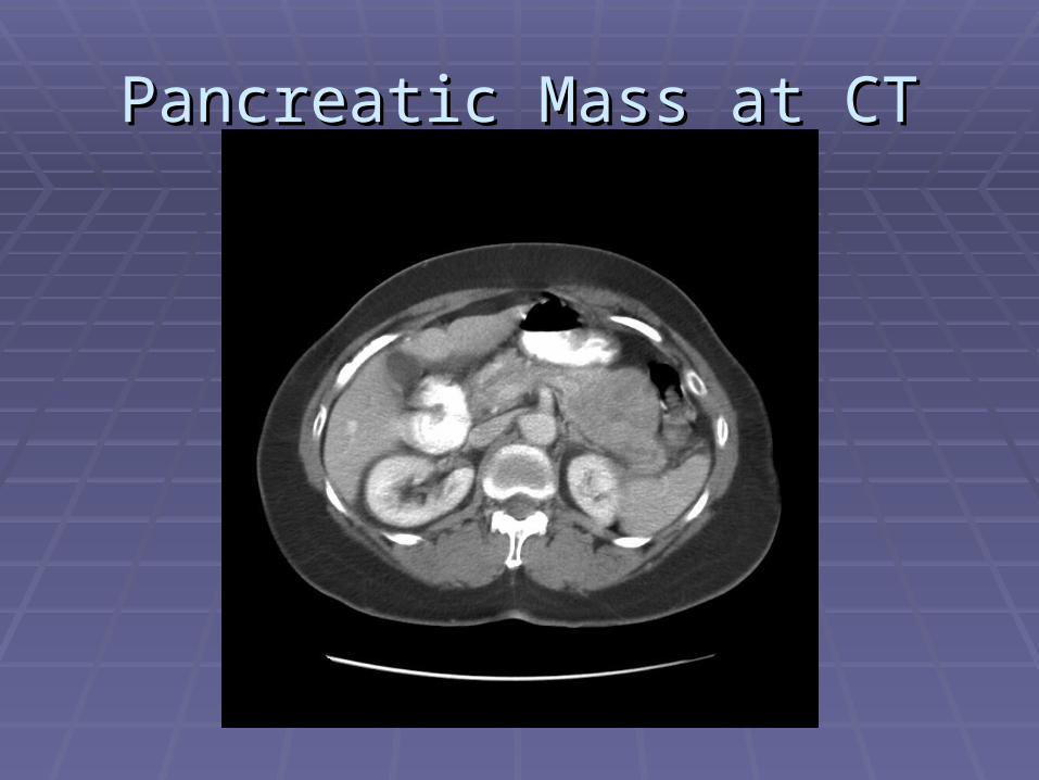
Pancreatic Mass at CTPancreatic Mass at CT

'Pancreatic' Mass at EUS'Pancreatic' Mass at EUS

FNA of Peri-pancreatic MassFNA of Peri-pancreatic Mass
Metastatic LeiomyosarcomaMetastatic Leiomyosarcoma

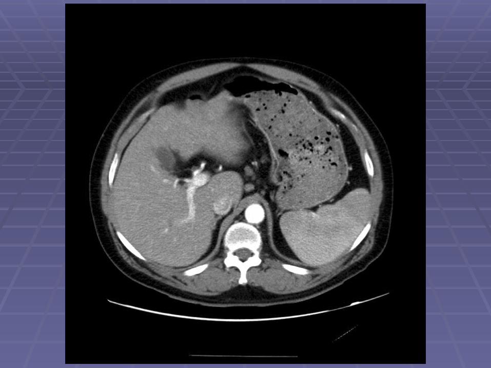
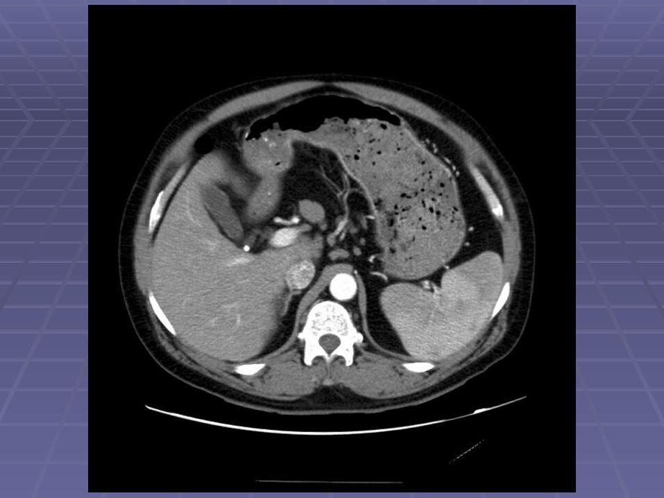
Liver Mass Liver Mass

FNA of Liver MassFNA of Liver Mass
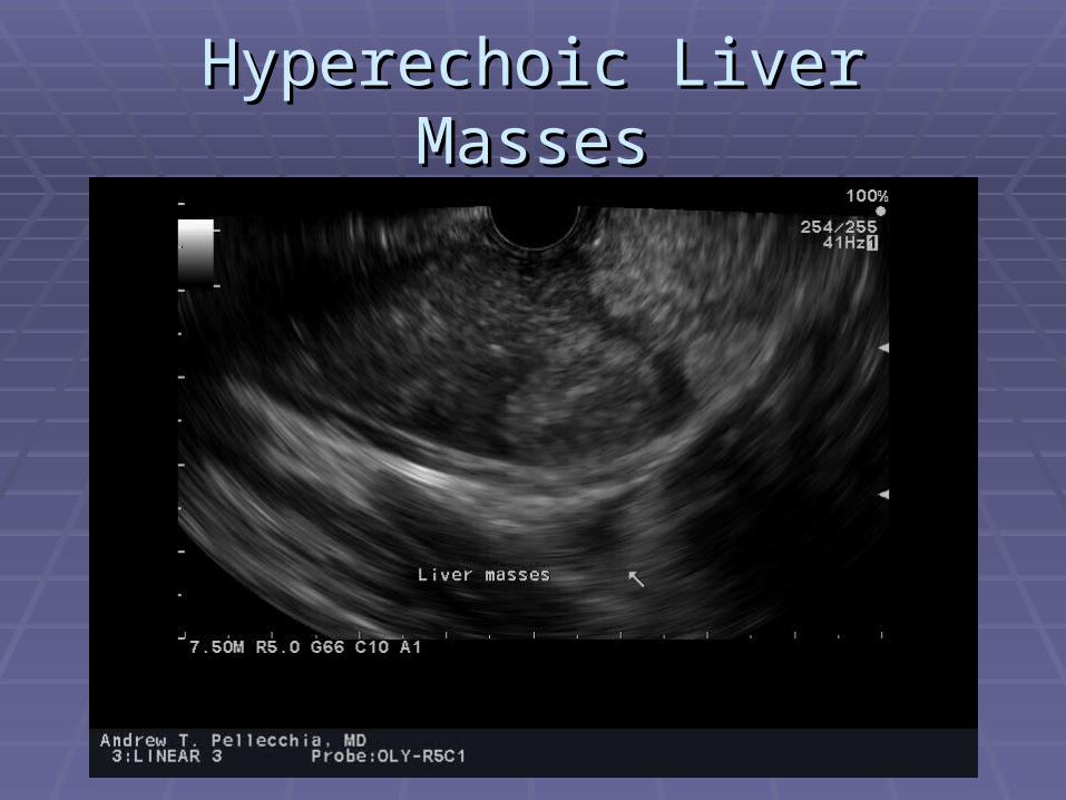
Hyperechoic Liver MassesHyperechoic Liver Masses

FNA of Hyperechoic Liver MassFNA of Hyperechoic Liver Mass


EUS Evaluation of Left Lobe of LiverEUS Evaluation of Left Lobe of Liver

Abdominal LADAbdominal LAD
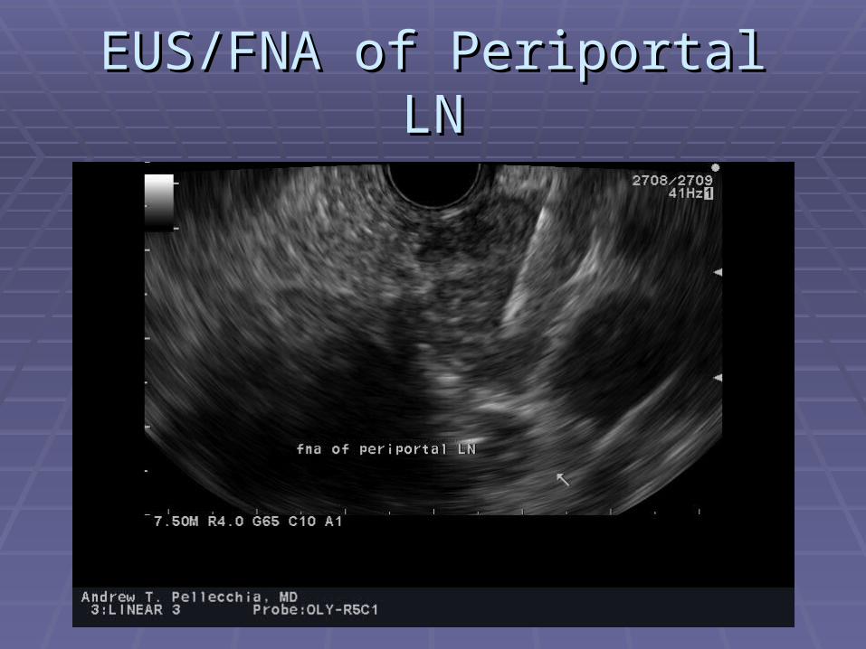
EUS/FNA of Periportal LNEUS/FNA of Periportal LN

Primary Target Fail…Primary Target Fail…

……Secondary Target AcquiredSecondary Target Acquired(Carcinoma at FNA)(Carcinoma at FNA)
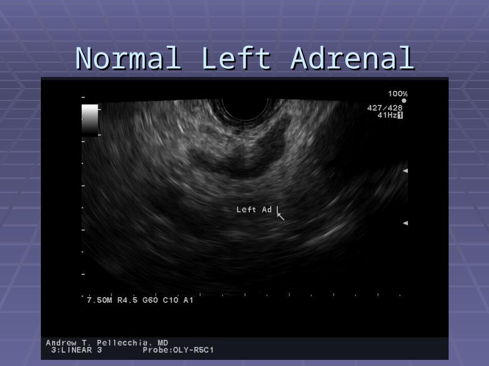
Normal Left AdrenalNormal Left Adrenal

Left Adrenal Met in NSCLCLeft Adrenal Met in NSCLC
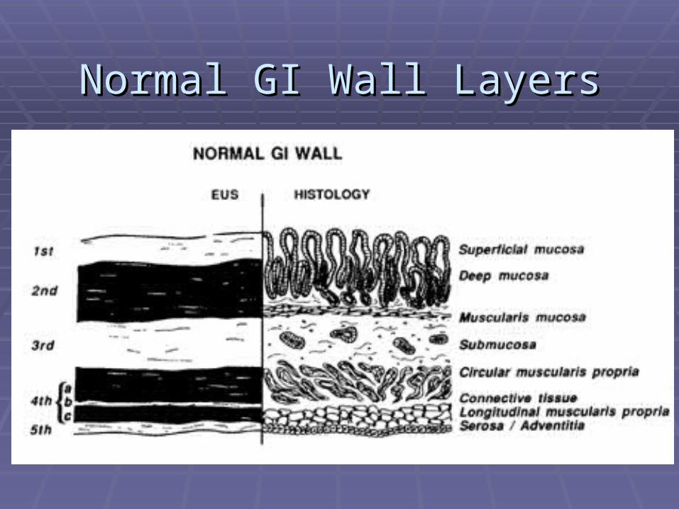
Normal GI Wall LayersNormal GI Wall Layers

Normal Esophagus and CystNormal Esophagus and Cyst

Distal Esophageal LesionDistal Esophageal Lesion

Normal Gastric Wall LayersNormal Gastric Wall Layers

Mucosal LesionMucosal Lesion

Mucosal LesionMucosal Lesion

Malt LymphomaMalt Lymphoma

Gastric LipomaGastric Lipoma

T2 Gastric AdenocarcinomaT2 Gastric AdenocarcinomaInvasion of Muscularis With Intact SerosaInvasion of Muscularis With Intact Serosa

T3 Gastric CancerT3 Gastric Cancer

T1 Rectal Cancer by EUST1 Rectal Cancer by EUS

T2 Rectal CancerT2 Rectal Cancer

Rectal Mass at CT: T4?Rectal Mass at CT: T4?(Apparent invasion of uterus)(Apparent invasion of uterus)
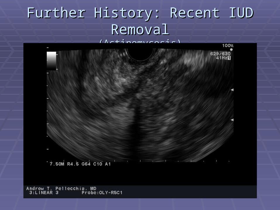
Further History: Recent IUD RemovalFurther History: Recent IUD Removal(Actinomycosis)(Actinomycosis)

Celiac Plexus NeurolysisCeliac Plexus Neurolysis

Celiac AxisCeliac Axis

Key PointsKey Points All patients with pancreatic cysts should have All patients with pancreatic cysts should have
consultation for possible EUS/FNAconsultation for possible EUS/FNA EUS/FNA is the standard of care in the loco-regional EUS/FNA is the standard of care in the loco-regional
staging of many cancersstaging of many cancers LungLung EsophagealEsophageal GastricGastric PancreaticPancreatic CholangiocarcinomaCholangiocarcinoma Rectal adenocarcinomaRectal adenocarcinoma

Key Points, ContinuedKey Points, Continued
EUS is minimally invasiveEUS is minimally invasiveReduces need for mediastinoscopy, surgical Reduces need for mediastinoscopy, surgical
biopsy, bronchoscopy, CT guided biopsybiopsy, bronchoscopy, CT guided biopsy Reduces morbidity/mortality while reducing Reduces morbidity/mortality while reducing
health care costshealth care costsAppropriate cancer stagingAppropriate cancer staging
Prevents unnecessary surgical resectionsPrevents unnecessary surgical resections Identifies patients who will benefit from pre-op Identifies patients who will benefit from pre-op
chemo/xrtchemo/xrt

Cutting Edge EUS ApplicationsCutting Edge EUS Applications
Role for EUS is expandingRole for EUS is expandingEUS placement of fiducials for radiation therapyEUS placement of fiducials for radiation therapyEUS rendezvous procedure for accessing CBDEUS rendezvous procedure for accessing CBDEUS directed brachytherapyEUS directed brachytherapyEUS guided hepaticogastrostomy for malignant EUS guided hepaticogastrostomy for malignant
CBD obstructionCBD obstruction