endocrine system - ice.jefferson.edu 16... · there are typically 4 parathyroid glands, located...
TRANSCRIPT

Endocrine System Page 1 of 12 Dr. Fenderson
ENDOCRINE SYSTEM
Lecturer: Dr. Bruce Fenderson
READINGS
Chapter 20: Endocrine System, In: Basic Histology: Text & Atlas, 14th Edition.
Junqueira, JC and Carneiro J, McGraw-Hill Companies, Inc. 2016.
Educational Goals
Understand structure - function relationships in the endocrine system.
Investigate the microscopic anatomy of the pituitary gland.
Understand the hypothalamo-hypophyseal system (vascular network and sites of
hormone production, storage, and release).
Investigate the microscopic anatomy of the adrenal cortex and medulla.
Investigate the microscopic anatomy of the endocrine pancreas.
Investigate the microscopic anatomy of the thyroid gland.
Relate morphological variations in the thyroid epithelium to changes in function.
Investigate the microscopic anatomy of the parathyroid glands.
Key Words
Adrenal glands (cortex and medulla)
Beta () cells (insulin-producing cells of endocrine pancreas)
Chief cells (parathyroid - parathyroid hormone)
Follicular cells (thyroid - thyroglobulin)
Hypophysis (pituitary, anterior and posterior)
Hypothalamic releasing hormones
Hypothalamo-hypophyseal neuroendocrine system
Islets of Langerhans
Oxyphil cells (parathyroid)
Parafollicular cells (thyroid - calcitonin)
Pars distalis (anterior pituitary)
Pars nervosa (posterior pituitary)
Pituicyte
Spongyocyte
Zona glomerulosa, fasiculata, reticularis
1

Endocrine System Page 2 of 12 Dr. Fenderson
Introduction to the Endocrine System
Hormones are potent chemical signaling substances (messengers) that are produced by
secretory cells and released into the circulatory system. The hormone is subsequently
delivered via the circulation to target cells with appropriate cellular receptors. The
secretory cells and target cells may lie in close proximity to one another, or they may be
scattered widely throughout the body. Based on distance, mechanisms of chemical
signaling are often described as: endocrine, paracrine or autocrine. Secretory cells of the
endocrine system are in most cases linked to the hypothalamo-hypophyseal
(neuroendocrine) system via feedback loops. The endocrine glands that we will explore
in class this week include:
Pituitary
Adrenals
Islets of Langerhans
Thyroid
Parathyroids
Pituitary Gland (Hypophysis)
Lies in a cavity of the sphenoid bone (sella turcica).
Normal weight 0.5 gm.
Develops embryologically from oral ectoderm (Rathke’s pouch) and floor of
diencephalon (see Figure below).
Derivatives of Oral Ectoderm = Adenohypophysis = Anterior Pituitary
Pars distalis (anterior pituitary)
Pars tuberalis (surrounds neural stalk)
Pars intermedia (vestigial)
Derivatives of Diencephalon = Neurohypophysis = Posterior Pituitary Neural stalk (infundibulum) includes stem and median eminence
Pars nervosa (posterior pituitary)
2
Endocrine System Page 3 of 12 Dr. Fenderson
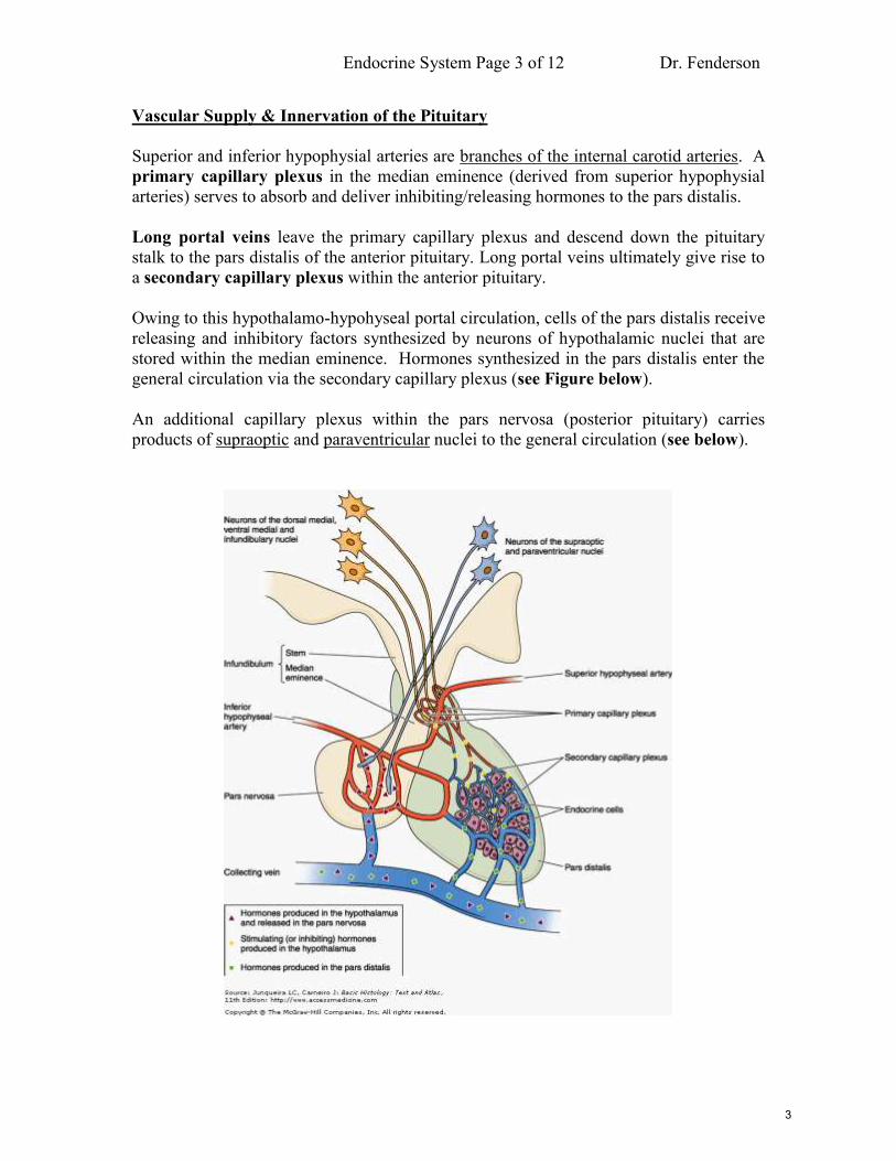
Vascular Supply & Innervation of the Pituitary
Superior and inferior hypophysial arteries are branches of the internal carotid arteries. A
primary capillary plexus in the median eminence (derived from superior hypophysial
arteries) serves to absorb and deliver inhibiting/releasing hormones to the pars distalis.
Long portal veins leave the primary capillary plexus and descend down the pituitary
stalk to the pars distalis of the anterior pituitary. Long portal veins ultimately give rise to
a secondary capillary plexus within the anterior pituitary.
Owing to this hypothalamo-hypohyseal portal circulation, cells of the pars distalis receive
releasing and inhibitory factors synthesized by neurons of hypothalamic nuclei that are
stored within the median eminence. Hormones synthesized in the pars distalis enter the
general circulation via the secondary capillary plexus (see Figure below).
An additional capillary plexus within the pars nervosa (posterior pituitary) carries
products of supraoptic and paraventricular nuclei to the general circulation (see below).
3

Endocrine System Page 4 of 12 Dr. Fenderson
Hypothalamo-Hypophyseal Neuroendocrine System
There are 3 sites for production of these polypeptide, neuroendocrine hormones:
(1) Peptides synthesized by secretory neurons in the supraoptic and paraventricular
nuclei are transported along axons to the neurohypophysis (posterior pituitary).
(2) Peptides (releasing hormones) synthesized by neurons in the hypothalamus
(dorsal-medial, ventral-medial and infundibular nuclei) are released in the median
eminence and transported to the anterior pituitary via a capillary portal system.
(3) Polypeptide hormones synthesized by cells of the pars distalis (anterior pituitary).
Microscopic Anatomy of the Adenohypophysis (Anterior Pituitary)
Pars Distalis
Cords of glandular cells surrounded by capillaries
Chromophobes and Chromophils (Acidophils & Basophils)
Hormones include: somatotrophin (GH), prolactin, FSH, LH, MSH, TSH, ACTH
Pars Intermedia
Rudimentary region in humans
Weakly basophilic cells
Pars Tuberalis
Cells arranged in cords along blood vessels - surrounds the neural stalk
Most of these cells secrete FSH or LH
Microscopic Anatomy of the Neurohypophysis (Posterior Pituitary)
Neurons of the supraoptic and paraventricular nuclei produce hormones that are stored
and liberated from the posterior pituitary. Axons of neurons within these nuclei extend
down through the median eminence into the posterior pituitary (pars nervosa). Hormones
synthesized by neurons of these nuclei are transported along axons and occupy storage
granules (Herring bodies) within the pars nervosa. The stored hormones, typically bound
to a carrier protein - neurophysin, are released into adjacent capillaries as a result of
neuronal stimulation from other regions in the brain.
Cells of the Neurohypophysis (Axons and Pituicytes)
Axons of hypothalamic neurons (from supraoptic and paraventricular nuclei)
25% of volume of neurohypophysis = pituicytes (highly branched glial cells)
Herring bodies = aggregates of neurosecretory granules in dilated axon terminals
4

Endocrine System Page 5 of 12 Dr. Fenderson
Hormones of the Neurohypophysis (Posterior Pituitary)
Oxytocin
Vasopressin (arginine vasopressin / antidiuretic hormone)
Binding protein for each = neurophysin
Released from storage via neuronal impulses from the hypothalamus
Clinical correlation: diabetes insipidus (patient has polyuria due to antidiuretic
hormone deficiency)
Adrenal (Suprarenal) Glands
Paired organs near the superior poles of the kidneys. They are embedded in adipose
tissue. Their weight and size varies with age and physiology. Closer examination reveals
2 concentric layers: an outer (yellow) cortex and a central (red-brown) medulla (see
Figure below).
The adrenal glands are derived during development from 2 sources: coelomic epithelium
and neural crest.
The metabolic activity of the adrenal cortex is regulated by ACTH from the anterior
pituitary, whereas the metabolic and secretory activity of the adrenal medulla is
controlled by preganglionic sympathetic nerve fibers. Adrenal cortical hormones
regulate metabolism (corticosteroids), maintain normal electrolyte balance (aldosterone),
and influence reproductive organs (generation of weak androgens). Hormonal targets for
epinephrine and norepinephrine include: glandular epithelial cells, cardiac muscle, and
smooth muscle of blood vessels & viscera.
5

Endocrine System Page 6 of 12 Dr. Fenderson
Vascular Supply to the Adrenal Glands
The adrenals are highly vascular organs. Blood vessels enter at various points around the
gland. Capillaries eventually drain through a common suprarenal vein. The arteries can
be divided into three groups:
Capsular arteries provide an extensive subcapsular network
Cortical arteries irrigate the cortex
Medullary arteries pass through the cortex directly to the medulla
Cortical and medullary arterioles originate as branches of capsular arteries. Together,
they supply the parenchyma of the gland. The medullary capillary bed receives blood
from two sources: cortical veins and medullary arterioles. Capillaries of the cortical and
medullary arterials form a single suprarenal vein.
Lymphatic vessels are only associated only with capsule and connective tissue around the
large blood vessels. Lymphatics are not associated with parenchymal elements.
Microscopic Anatomy of the Adrenal Cortex
Cells of the adrenal cortex have the typical ultrastructure of steroid-secreting cells (foam
cells with lipid droplets). Cords of glandular tissue are grouped along capillaries. There
are 3 concentric zones with different physiological functions (salt/sugar/sex):
Zona Glomerulosa (salt - aldosterone)
Zona Fasiculata (sugar - cortisol)
Zona Reticularis (sex hormones)
Zona Glomerulosa
Thin outer zone adjacent to the capsule
Small columnar or pyramidal cells arranged in clusters
Continuous with cords of cells in subjacent region
Zona Fasiculata
Large polyhedral cells arranged in parallel columns with lipid droplets
Columns of these spongyocytes are separated by cortical sinusoids
Zona Reticularis
Innermost layer of cortex
Lipofuchsin granules present
Developmental Considerations: Fetal (Provisional) Cortex
Large by midgestation - produces precursors to placental estrogen (helps mother)
Regresses postnatally
Zones of adult cortex develop during first 3 postnatal years
6

Endocrine System Page 7 of 12 Dr. Fenderson
Summary of Adrenal Cortical Hormones
Aldosterone
Mineralocorticoid that helps regulate systemic blood pressure
Primary site of synthesis is zona glomerulosa
Cortisol (hydrocortisone)
Glucocorticoid helps regulate metabolism
Primary site of synthesis is zona fasiculata
Androgens
Small amounts produced by cells of zona reticularis
Minute amounts also produced by cells of zona fasiculata
Weak androgens (e.g., androstenedione)
Adrenal Medulla: Microscopic Anatomy
Like the cortex, the adrenal medulla is composed of polyhedral cells in cords with a
surrounding network of capillaries.
Cells of the medulla are considered to be modified postganglionic neurons of the
sympathetic nervous system. These neural crest derived cells are referred to as
chromaffin cells.
Summary of Adrenal Medullary Hormones
Catecholamines (epinephrine and norepinephrine)
These similar hormones are synthesized by 2 different populations of cells
Stored in membrane-bound, electron-dense, secretory granules
7

Endocrine System Page 8 of 12 Dr. Fenderson
Endocrine Pancreas
Islets of Langerhans are multi-hormonal micro-organs located in the pancreas. They are
approximately 100-200 m in diameter. Each islet includes several hundred cells. They
are embedded within pancreatic exocrine tissue. There are approximately 1 million islets
per pancreas.
Islets: Microscopic Anatomy
Spherical configuration of islets
Composed of rounded /polygonal cells
Fenestrated capillary network
Separated from exocrine tissue by reticular fibers
10% of cells innervated by autonomic nerve fibers
Islet Cell Types (immunocytochemical methods are used to identify types)
Quantities not uniform within or between islets
A cells (20%) - synthesize glucagon
B cells (70%) – synthesize insulin
D cells (<5%) --synthesize somatostatin
F cells (rare) - pancreatic polypeptide
8

Endocrine System Page 9 of 12 Dr. Fenderson
Thyroid Gland
The thyroid gland is located anterior to the larynx. It originates during development from
endoderm of the primitive gut. It synthesizes thyroid hormones (T3 and T4) that are
important for growth, differentiation, and control of metabolic rate (e.g., oxygen
consumption).
The gland is composed of 20-30 million follicles, that are lined by a simple epithelium.
The central cavity stores extracellular hormone precursor (thyroglobulin) in a gelatinous
matrix called colloid. Thyroid follicles contain a 3-month store of thyroid hormone.
Thyroid Gland: Microscopic Anatomy
Loose connective tissue capsule with septae
Highly vascular organ
Fenestrated capillaries (functional significance)
Follicles (spherical shape; simple epithelium with variable morphology)
o squamous to low columnar
o follicular diameter is variable (depends on functional state)
Colloid (thyroglobulin stains with PAS due to high sugar content)
9

Endocrine System Page 10 of 12 Dr. Fenderson
Effect of Thyroid-Stimulating Hormone (TSH) on the Thyroid
Stimulates thyroid hormone synthesis
Increases height of follicular epithelium
Decreases quantity of colloid as well as follicle diameter
Functional Aspects of Follicular Morphology
Larger diameter
distended with colloid
cuboidal to squamous epithelium
in hypoactive thyroid the most common epithelium is squamous
Smaller diameter
less colloid
simple columnar epithelium
associated with higher metabolic activity (hyperactive thyroid)
clear vacuoles in the colloid next to the follicular epithelium (scalloping of
the colloid) indicates rapid uptake of thyroglobulin (hyperfunction)
Parafollicular or C-Cells: Microscopic Anatomy
Clear cells may form part of follicular epithelium, or….
May exist as isolated clusters between follicles
Larger and less intensely stained compared to follicular cells
Arise from infiltrating neural crest cells
Parafollicular or C-Cells: Endocrine Function
Synthesize and secrete the hormone - calcitonin
Calcitonin decreases blood calcium levels by inhibiting bone resorption
Secretion of calcitonin stimulated by increased levels of blood calcium
(hypercalcemia)
Clinical Correlations
Nodular goiter (common and benign)
Graves hyperthyroidism (an autoimmune disease)
Hashimoto thyroiditis (associated with hypothyroidism)
Benign and malignant neoplasms (adenoma vs carcinoma)
10

Endocrine System Page 11 of 12 Dr. Fenderson
Parathyroid Glands
There are typically 4 parathyroid glands, located behind the thyroid gland at each upper
and lower pole (see Figure below). They are usually found within the capsule that
covers the lobes of the thyroid; however, they may be found embedded in the thyroid
parenchyma. The glands are derived during development from the 3rd and 4th
pharyngeal pouches. Ectopic glands are found in the mediastinum, beside the thymus.
Each gland contains a connective tissue capsule with endocrine cells arranged in cords.
The two types of parathyroid parenchymal cells are chief cells and oxyphil cells. Chief
cells secrete parathyroid hormone in response to hypocalcemia.
Parathyroid Glands: Microscopic Anatomy
Parenchymal component includes chief cells and oxyphil cells
Chief cells are smaller and produce parathyroid hormone
Oxyphil cells are larger polygonal cells, smaller population, of unknown function
Stromal component includes connective tissue capsule and septae
With increasing age, adipose tissue comes to replace the secretory cells
Summary of Parathyroid Hormone Function
Increases blood calcium levels (acts on osteoclasts)
Reduces blood phosphate levels (acts on kidneys)
Promotes increased calcium absorption from the gut
11

Endocrine System Page 12 of 12 Dr. Fenderson
NOTES:
12