endocrine nuclear medicine - up.ac.za · thyroid cancer • ablation therapy: 6 weeks post...
TRANSCRIPT
Endocrine Nuclear Medicine
Outline of Lecture Organs: • Thyroid • Parathyroid • Adrenal Gland Nuclear Medicine: • Tracers, technical aspects • Relationship to patient diagnostic pathways and
other imaging modalities • Contribution to management and treatment

Functional imaging
• The aim of nuclear medicine is to identify and track physiological actions using a “tracer” labelled with a radioisotope
• Anatomical information may be inferred from the physiological image but this is secondary
• Imaging methods should be standardised-reproducible

The Thyroid Gland
•
Image : ABC Health and Wellbeing Website

Thyroid Hormones Negative Feedback System
Image : ABC Health and Wellbeing Website

Thyroid Gland

Production of Thyroid Hormones T3 and T4

Thyroid imaging
• When should it be performed? • How does it help diagnosis? • What alternatives are there for imaging the
thyroid? • How do the results of the nuclear medicine
scan affect treatment?

Functional Imaging of Thyroid
• Thyroid Gland – Overactive – Underactive – Malignancy

The Scan Patient preparation: • Patient letter/leaflet
• Stop relevant medication Carbimazole (CBZ) : 48 hrs Propothyruracil (PTU) : 48hrs T4 : 4-6 weeks T3 : 3 weeks Other factors in patient history may affect scan

Factors affecting uptake of 123I, 131I and 99mTc-04
-
• Exogenous thyroid hormone • Medication (CBZ) and (PTU) • Iodine containing radiological contrast
agents (wait 6-8 weeks) • High level of intake of Kelp products • Amiodarone All the above will decrease uptake : ASK
the patient!!!!!!

Iodine and Pertechnetate
Both Iodine and pertechnetate have similar size and charge

The Scan
Radiopharmaceutical • 99mTc pertechnetate: cheap, not organified scan
that day (ARSAC DRL =80MBq). Scan 20 mins post injection
• 123I: more expensive, scan next day if oral prep (ARSAC DRL= 20 MBq)
• Measure syringe activity before and after injection for % uptake calculation
• (accurate camera sensitivity required. Activities decay corrected etc)

The Scan Scan Parameters • Single or dual headed camera • Camera: standard FOV • Collimator: Pinhole, LEHR Patient position • Supine, neck extended, standard ( eg 10
cms) from collimator. Optimise comfort!
The Scan:
Views: • Anterior (include salivary glands)
100-200K counts • Obliques • +/- Lateral (vital in infant if looking for lingual thyroid) • +/- Large FOV 100K counts • Suprasternal notch (SSN) – Co source marker
60 secs to check for retrosternal extension
Causes of Hyperthyroidism • Graves
• Solitary or Multiple Autonomous Nodules (toxic adenoma, Plummer’s Disease) • Thyroid Hormone ‘Leak’ thyroiditis, Hashimotos thyroiditis (early), subacute(=De Quervains) thyroiditis, post partum thyroiditis • XS thyroid hormone ingestion eg thyroxine, ‘slimming’ drugs
• Thyroid hormone or TSH secreting tumour eg some ovarian
• Pituitary gland malfunction

Grave’s
• Primary diagnosis by history, examination • Diagnosis established by biochemistry and
immunology • Functional imaging confirmatory • May be of particular use if thyroid
abnormal: – Nodules – Previous surgery – 131I Therapy being considered

Graves Disease
• Autoimmune disease ie antibodies made to ‘self’
• Up to 10 different Abs described so far • Abs to TSH receptor on thyroid cell stimulates hormone production • Abs stimulating thyroid growth (or other
tissues e.g. front of shins, retro-orbital fat) • Clinical manifestations depend on Abs
present

Graves Disease
• Women>>men • 20-40 years • Genetic predisposition (other auto-immune
conditions may co-exist) HLA B81, DR2 and DR3 in Caucasians BW35 and BW 36 in Asians • 50% have family history

Graves Disease: Clinical Picture • Increased metabolic rate: weight loss, increased
bowel transit
• Sweating
• Sympathomimetic effects: fast heart rate, palpitations, tremor, anxiety
• Immune mediated effects: dysthyroid eye disease, pretibial myxoedema
• Other: e.g. proximal muscle wasting

Pretibial Myxoedema
Skin is thickened and inelastic due to deposition of excess glycosaminoglycans
Image: DermNet NZ

Graves Dysthyroid Eye Disease
• Affects up to 50% of patients
• Proptosis, diplopia and compression of optic nerve
• Infiltration of fat and occular muscles with muccopolysaccharides
Images: Handbook of Ocular Disease Management www.revoptom.com Visitech Eye Centre

Normal Thyroid Gland
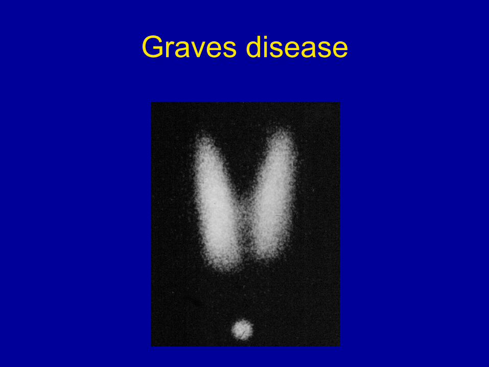
Graves disease

Graves Disease

Hypothyroidism
• NM: Not so useful as uptake low • Especially difficult to see nature of nodes • Ultrasound is probably better
• Hashimoto’s Thyroidtis is most common cause of hypothyroidism - autoimmune condition (can be toxic in very early stage)
- scan appearances vary with stage - chronic : inhomogeneous tracer uptake

Thyroiditis
Subacute thyroidits (also known as de Quervains) • NM: Very good test as Iodine and pertechnetate
are not taken up in acute phase (first 4 weeks after onset of symptoms)
• Patient initially toxic • Reduced uptake persists 4-8 weeks • Tends to be normal by 12 weeks • Scan these within 10 days of request • NB This patient is NOT treated with 131I for ‘toxic’ state

Thyroiditis

Thyroiditis

Thyroid Nodules • Common – F>>M and ↑ with age • 95% of nodules are cold
(‘nonfunctioning’) • Cold nodule is not normally cancer
however risk of malignancy 1.5-38%, most quoted value ≈ 10%
-patient should have USS +/- FNA • Less than 1% hot (‘functioning’) nodules
are malignant
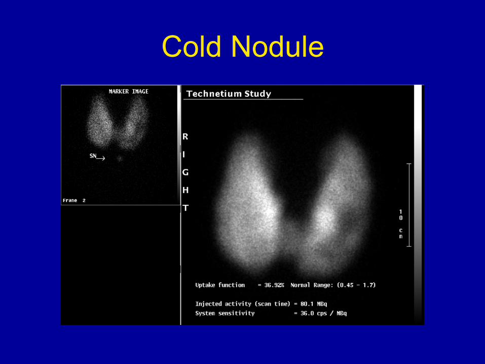
Cold Nodule

Thyroid Nodules
Cold Nodule • Colloid Nodule • Cyst • Adenoma • Haemorrhage • Focal Thyroiditis • Abscess • Parathyroid adenoma
Hot Nodule Adenoma

Hot Nodule • May become autonomous (not responsive
to feedback loop)
• Rest of gland suppressed
• If patient ‘toxic’ (i.e. ↑T4 and/or ↓TSH) due to functioning nodules, then they have ‘Plummers Disease

Hot Nodule

?HOT nodule
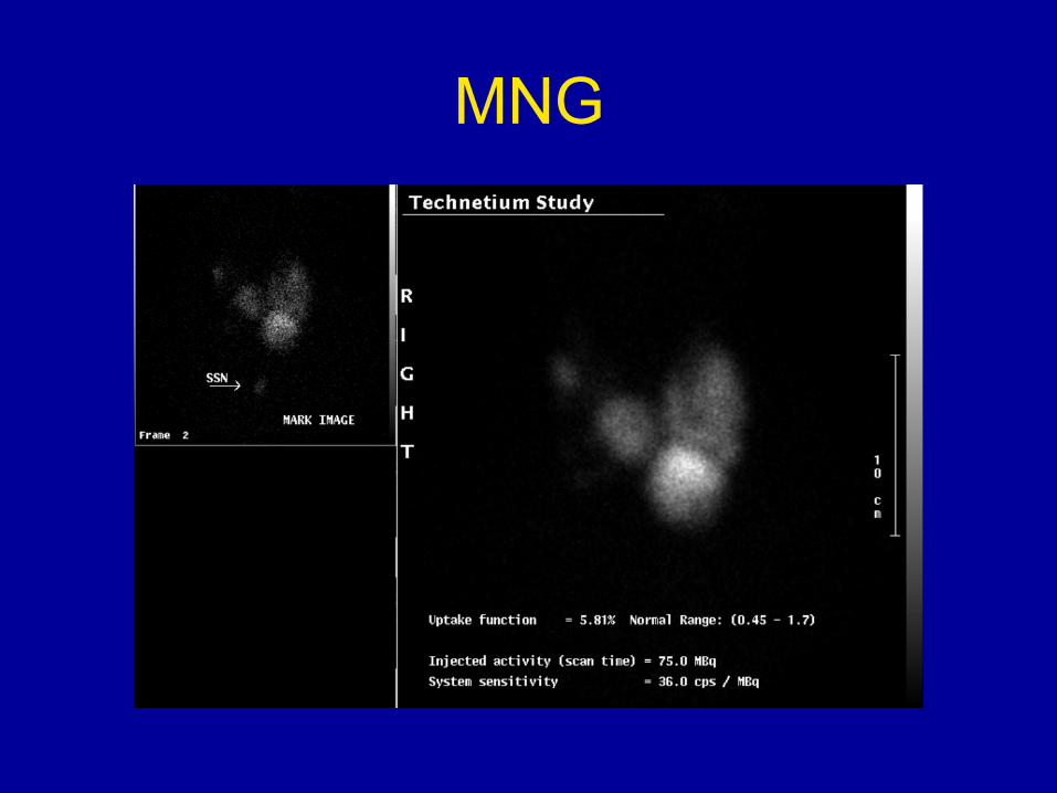
MNG

Treatment of Benign Thyroid Disease
Conditions • Graves • Toxic Nodules – high activity required (600MBq) • MNG – high activity required (600MBq) Treatment : 131I • Discuss with patient: treatment options e.g. surgery • Informed consent – risk of hypothyroidism • Radiation protection issues: exposing family members
and public (time and distance!!) Restrictions last up to ≈ 3 weeks e.g. separate bed from
partners, avoid pregnancy for 6 months Lifelong follow up (regular thyroid blood tests)
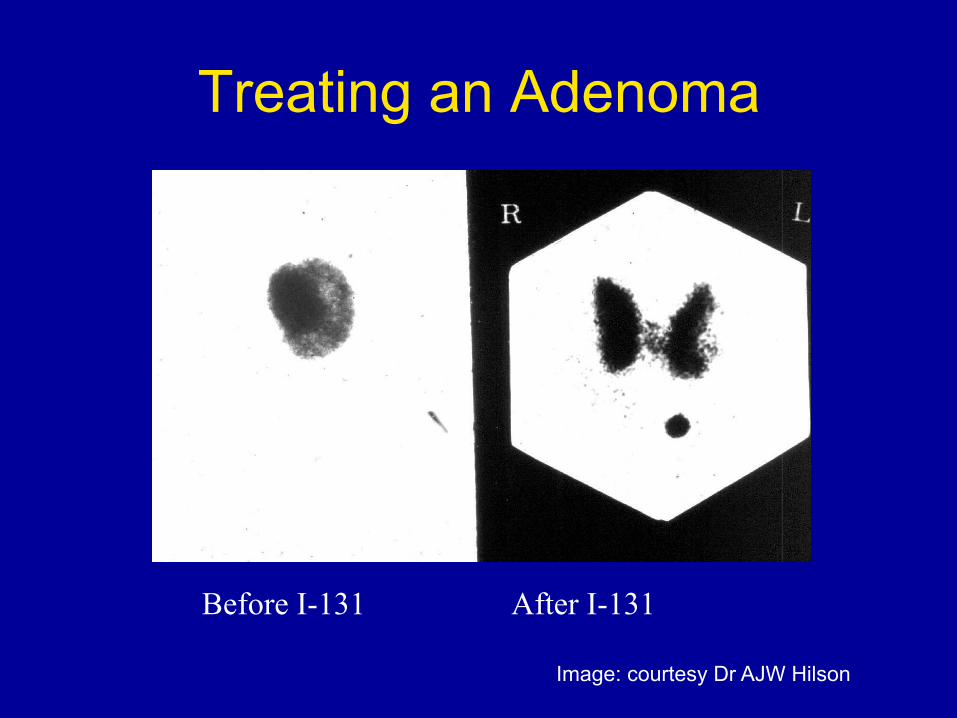
Treating an Adenoma
Before I-131 After I-131
Image: courtesy Dr AJW Hilson
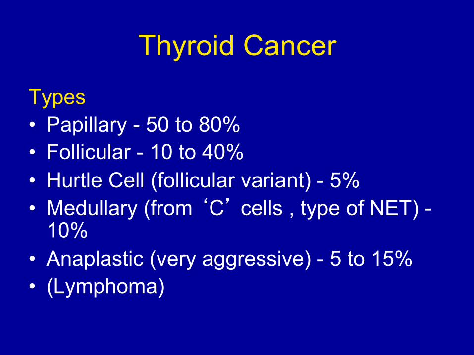
Thyroid Cancer
Types • Papillary - 50 to 80% • Follicular - 10 to 40% • Hurtle Cell (follicular variant) - 5% • Medullary (from ‘C’ cells , type of NET) -
10% • Anaplastic (very aggressive) - 5 to 15% • (Lymphoma)

Thyroid cancer • Ablation Therapy: 6 weeks post thyroidectomy
(papillary and follicular ca, T2 and above) give 3-5GBq 131I ablation therapy
• Have to stop T4 for 4weeks, T3 for 10 days • Can be given with TRH, rTSH (£1000) • Scan at 48-72 hours
• Repeat therapies till thyroid bed and any mets disappear 3-6 monthly intervals
• Post treatment image is used to stage patient. • If uptake is low, consider ‘tracer’ dose (123I prior
to next therapy – 400MBq)
NB: has NO role in anaplastic ca or lymphoma

Multiple Metastases on 1st Dose 131I

Thyroid Ca: Multiple Metastases

Other Tracers Used for Detecting Ca Thyroid (if Iodine Scan Negative)
• 99m Tc MIBI or tetrafosmin
useful with SPECT of neck • 18F FDG • 111In octreotide • 99mTcDMSA(V) – ‘pentavalent DMSA’ • 201Tl

111In Octreotide in papillary Ca Thyroid

F-18 FDG in thyroid cancer
Image: Atlas of Clinical PET, 2006, Eds Barrington et al

Imaging Medullary Carcinoma of the Thyroid (MCT)
• Tc-99m DMSA (V) • 123I mIBG - Therapy version available with 131I
mIBG • 111In Octreotide - Therapy version available with 90Y Octreotide • 18F- FDG PET/CT
Mainly used for staging

123I-MIBG in MCT

Parathyroid Glands : Role of Nuclear Medicine
• Diagnosis – Renal patients: primary vs secondary
• Localisation – Assist surgeon in reducing surgical operating times – May help reduce morbidity – Aids use of minimally invasive techniques
• ‘Second look’ ! – Missed adenoma – Ectopic adenoma
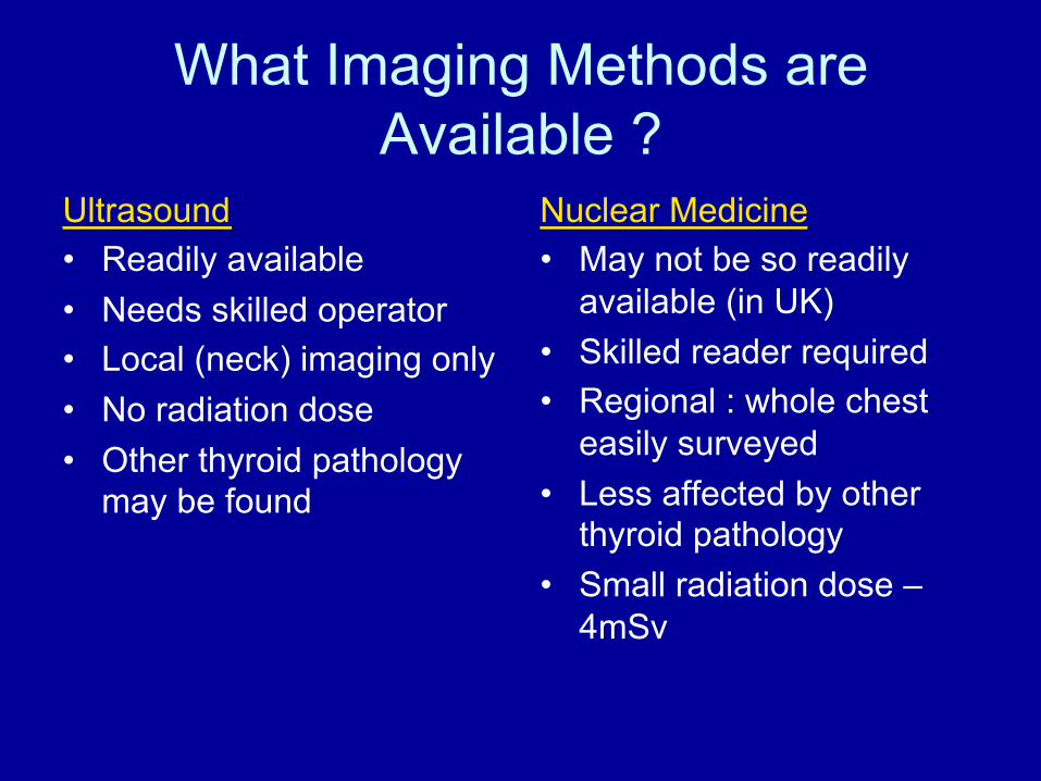
What Imaging Methods are Available ?
Ultrasound • Readily available • Needs skilled operator • Local (neck) imaging only • No radiation dose • Other thyroid pathology
may be found
Nuclear Medicine • May not be so readily
available (in UK) • Skilled reader required • Regional : whole chest
easily surveyed • Less affected by other
thyroid pathology • Small radiation dose –
4mSv

Nuclear Medicine
• Exploits functional aspects of tumour • Ideally need an agent taken up only by
parathyroids but no such agent currently available
• Some agents only have uptake in thyroid and others in both thyroid and parathyroid
• Others have initial uptake in both organs but “washout” of normal thyroid

Subtraction technique • Inject agent: taken up by thyroid and parathyroid
(Tl-201 or Tc-99m MIBI/TF) • Wait 30 minutes, then scan neck • Keep patient under camera, inject agent taken
up by only thyroid (123I, 99mTc pertechnetate) • Wait 15 minutes, then rescan • Subtract images

Washout technique
• Inject agent which washes out of thyroid but not parathyroid (99m Tc MIBI)
• Wait 15 minutes • Perform planar and/or SPECT images • Wait a further 2 hours • Repeat planar and/or SPECT images • Review images.

Normal (Negative) Washout Scan
Early
Late

Parathyroid Adenoma

Ectopic Parathyroid Adenoma

Advantages of SPECT in parathyroid imaging
• Allows increased contrast (fewer overlapping structures)
• Better localisation • Should find lesions 7mm
and above • Interactive display
possible

SPECT alone


Other uses of 99mTc MIBI Peri-Operative Use • Inject 50MBq of 99mTc MIBI (10% of usual activity)
• Localise uptake with gamma probe in theatre at time of surgery to localise adenoma
• Surgery can be pre-planned e.g. just one side explored • Scar size and surgery time are reduced
• Ugar et al Ankara (2006) showed significantly improved surgical localisation using probe in 35 patients vs usual imaging protocol then surgery
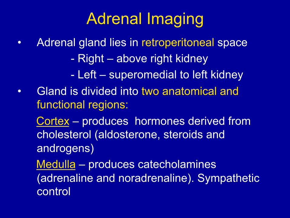
Adrenal Imaging • Adrenal gland lies in retroperitoneal space - Right – above right kidney - Left – superomedial to left kidney • Gland is divided into two anatomical and
functional regions: Cortex – produces hormones derived from
cholesterol (aldosterone, steroids and androgens)
Medulla – produces catecholamines (adrenaline and noradrenaline). Sympathetic control

Adrenal Glands on CT
RIGHT LEFT
Imaging of Adrenal Gland Adrenal Cortex • Nuclear medicine very rarely used in imaging of
the adrenal cortex. • Biochemical tests e.g. serum cortisol levels,
together with anatomical imaging (CT or MRI) usually used.
• Tracers – limited availability 131 I-19 Iodocholesterol (75Se-6-beta-selenomethyl –norcholesterol)
11C metomidate
Incorporated into synthesis pathway • Imaged at 5 days • High(ish) dose to patient 6mSv

C-11 metomidate in small adrenal adenoma in medial limb of right adrenal

Imaging of the Adrenal Gland Adrenal Medulla • Indication: localisation of phaeochromocytoma
(should have +ve catecholamine in urine)
• Tracer: 123I MIBG
• Method of uptake: amine uptake transporter mechanism present in neuroectodermal tissue
• May need to stop drugs which reduce uptake of 123I MIBG - reserpine, cocaine(!) and labetolol and some anti-depressants
• Give thyroid blockade: e.g. potassium iodide 60mg bd for 3 days. Start at least 1hr prior to injection

The Scan
• Inject up to 400MBq 123I MIBG • Image at 24 hrs • Parameters: LEHR • Planar • SPECT images e.g 2 headed camera 60 projections at 3° 20-30 secs per projection

Phaeochromocytoma • Neoplasm arising from
adrenal medulla • Triad (paroxysmal headache, ↑BP,
palpitations)
‘10%’ • 10% malignant • 10% bilateral • 10% ectopic • 10% found in children • 10% associated with syndrome • 10% neg MIBG scan

Pre Surgery Post Surgery Recurrence


Malignant Metastatic Phaeochromocytoma
Treatment High dose (5GBq) x3 131I-
MIBG if 123IMIBG scan is positive