endocrine disruption in fish || disruption of male reproductive function
TRANSCRIPT
Chapter 6
Disruption of Male Reproductive Function
6.1 Introduction
A decreased gonadosomatic index resulting from exposure of fish to xenobiotics has been described by many authors, but it is frequently not clear whether this is due to disruption via the pituitary-hypothalamic system or to direct action on the testis itself. Such general results will not be discussed in detail in this Chapter, but are listed in the Appendix. Direct effects on the testis can be considered either as cytotoxicological, where the disruption is caused by damage to the cellular integrity or function of gonadal cells in general, or endocrine in which the function of specific cells are disrupted due to an endocrine malfunction. The latter could be due to altered pituitary secretion or to alterations in the chain of enzymes within the testis which lead to synthesis of the testicular hormones. Such disruption of steroidogenesis can have secondary effects on the testicular somatic and germ cells which are dependent upon the correct hormonal milieu for normal functioning. Furthermore other functions of the male, such as secondary sexual characteristics and courtship behaviour are hormonally dependent and can be disrupted in the absence of normal circulating hormone levels. The recent concern with environmental estrogens and their effect on male fertility (Colborn and Clement, 1992; Purdom et aI., 1994; Sharpe and Skakkebaek, 1993) is particularly relevant in this section. Many organochlorine pesticides and phenolics are known to be weakly estrogenic and dysfunction of male reproduction has been attributed (Colborn and Clement, 1992) to estrogenicity of these pollutants. Pesticides are, however, designed specifically as toxic agents and if they disrupt the cellular functions of pests they are also likely to have similar effects on non-target organisms such as fish or
D. E. Kime, Endocrine Disruption in Fish© Kluwer Academic Publishers 1998
122 Chapter 6
mammals. Reproductive tissues may well be affected, but such action may have nothing to do with estrogenic activity. It is therefore important to compare the effects on the male which are caused both by estrogenic and nonestrogenic pollutants so that the true cause of the damage may be ascertained. It is possible, for example, to expose the fish or its tissue to a level of estradiol, or the synthetic analogue ethynylestradiol, that has equivalent estrogenic potency to the pollutant in, for example, stimulation of vitellogenesis. The concentration of the estrogen will, perhaps, be less than 1/1000 of that of the pollutant and its effects can be considered purely as estrogenic without any toxicological side effects. Any effects caused by the pollutant which are not apparent with the estrogen will therefore be due to toxic rather than estrogenic activity. The relative estrogenic and toxic activities will also vary from one pollutant to another which should be reflected in the nature of the disruption caused to the reproductive system. Popular concern with environmental estrogens may well be misdirected if it leads to restriction only of pollutants which are estrogenic, while compounds which actually cause more damage to the reproductive system through their toxicological activity continue in use. Even nonylphenol, which is of recent concern as an environmental estrogen has a 96 h LCso of only 0.12 mg/l in salmon (McLeese et al., 1981), a level lower than some pesticides. Furthermore, restrictions on one compound will inevitably lead to increased use of other chemicals which may be more toxic. It is important to realise that "environmental estrogens", "endocrine disruptors" and "disruptors of reproduction" are not synonymous and that the latter term is what really matters when the effect on the population as a whole is considered. Although some of the toxic effects found for xenobiotics on the testis, or sperm, do not fit clearly within the definition of "endocrine disruptor", their effects on the fertility of the affected fish are similar and use of strict definitions is irrelevant. The important factor to consider is whether the testis is affected at concentrations at which the rest of the fish is unharmed. A similar consideration of course applies to female reproduction.
Organochlorines pose a particular danger to the testis since they are both lipid soluble and bioaccumulate, and are only very slowly biodegradable to inactive material. Other chemicals may pose dangers if they are permanently present in the aquatic ecosystem, either free in the water, or as part of the diet. Furthermore, release into the water for only a short period can cause serious damage if it is co-incident with a crucial phase of testicular activity. This is especially important during the final stages of sperm maturation and courtship where synchrony with female development is essential, and there may be direct effects on the viability of sperm released into the water during spawning or held within the sperm ducts ready for release. The use of organophosphate sheep dips during the spring spawning season may be particularly hazardous in this respect.
6. Disruption of Male Reproductive Function 123
The effects of pollutants on the male are for convenience divisible into those effects that are apparent in changes of the structure of the testis and developing sperm, those that result in changes in the hormones secreted, and those that directly affect the essential role of the male in provision of sperm capable of fertilising the eggs produced by the female. These three types of effect are discussed separately in this Chapter, although quite clearly all three are interdependent.
6.2 Effects on testicular morphology
6.2.1 Heavy metals
Exposure of Colisa fasciatus for 30 days to 2 mgll arsenic, the concentration of effluent from a fertiliser factory, had little effect on testicular structure, but at 14 mgll there were degenerative changes in the lobules and dissolution of their walls, and significant reductions in diameters of the interstitial cells (Shukla and Pandey, 1984b). The nucleic acid content of the testis was also decreased by arsenic exposure possibly by generating chromosomal abnormalities or inhibition of DNA repair (Shukla and Pandey, 1984c).
Cadmium caused extensive damage to the structure of testes of the brook trout after only 24 h exposure to concentrations as low as 10 Ilgll (10 ppb) (Sangalang and O'Halloran, 1972, 1973). Seven days after a 24 h exposure to 25 1lg/1 of cadmium there was evidence of testicular damage with extensive discolouration suggestive of massive haemorrhage. Histological examination showed extensive haemorrhage, necrosis and disintegration of the lobule boundary cells, which had pyknotic nuclei, although there was no detectable damage to the primordial germ cells.
Other non-salmonid studies have used much higher concentrations of cadmium which would probably have been toxic to a salmonid. 225 mg/l of cadmium, which was considered a safe level for Lebistes reticuiaris, caused an arrest of spermatogenesis, a decrease in numbers of spermatogenic cysts, necrosis of interstitial cells and hyperplasia of Sertoli cells after 30 days (Sehgal and Pandey, 1984; Sehgal et ai., 1984). In Puntius sarana, exposure to 20 mgll cadmium for 10 days gave a disorganisation of the testicular lobules and an accumulation of a yellow-brown pigment. The vascular bed was severely damaged at lower concentrations (2 mg/l) than that which caused damage to the pituitary gonadotrophs suggesting that the primary effect was on the testis rather than the pituitary (Kumari and Dutt, 1991). Damage to the cytoplasm and nucleus of interstitial cells, vacuolisation and destruction of spermatogonia and primary spermatocytes, lobular rupture and sperm duct damage were found in the related Puntius ticto after 4 days
124 Chapter 6
exposure to 26 mg/l cadmium at different periods of the reproductive cycle (Pundir and Saxena, 1990). Injection of Clarias batrachus with 0.5 J.l.g cadmium chloride per 100 g body weight arrested spermatogenesis at the secondary spermatocyte level and caused vacuolisation and degeneration of the Sertoli cells, damage to the lobular walls, and pycnosis of the interstitial cells (Ahsan and Ahsan, 1974).
Lead caused similar structural damage to the testis. Exposure of Colisa !asciatus to 15 mg/llead for 4 days caused reduced spermatogenic activity, dilation and rupture of the testicular blood vessels, and collapse of the lobules as a result of necrosis, shrinkage and dissolution of the germinal epithelium (Srivastava, 1987). Dilation of testicular blood capillaries with necrosis and disintegration of the seminiferous tubules was also found in Puntius conchonius exposed to 0.1 mg/l of lead or 11 mg/l zinc for 4 months. In rainbow trout the transformation of spermatogonia to spermatocytes was inhibited after a 12 day exposure to 10 J.l.g/I zinc (Ruby et aI., 1993a).
Copper caused only a transient arrest in spermatogenesis in Puntius conchonius during the first two months exposure to 0.2 mg/l (Kumar and Pant, 1984), while in Lebistes reticularis at 0.5 mg/l damage was similar to that caused by 225 mg/l cadmium (Sehgal et al., 1984) although it also caused an agglutination of sperm heads.
Testes of Channa punctatus exposed to 0.01 mg/l methyl mercury for six months during gonadal recrudescence showed only secondary spermatogonia in their testes while control fish had mature sperm. There was no evidence of morphological damage and the inhibition of development in exposed fish was correlated with inactivity of pituitary gonadotrophs (Ram and Sathyanesan, 1983). Both organic and inorganic mercury at 0.04 - 0.05 mg/l decreased the GSI of Clarias batrachus, inhibited the transformation of spermatids into functional spermatozoa, decreased the size of the seminiferous tubules, and caused degeneration of the interstitial cells (Kirubagaran and Joy, 1992). The differences between these two studies in stage of spermatogenic arrest is probably attributable to differences in the exact stage of the cycle at which fish were exposed.
6.2.2 Pesticides
This class covers a very wide range of pollutants including the organochlorines, organophosphates and carbamate pesticides.
Exposure of tilapia (Oreochromis mossambicus) to 2 mg/l benzene hexachloride for 10 days produced vacuolated cells and necrosis in the seminiferous tubules, atrophy of the interstitial cells, and thickening of the wall of the sperm duct (pandey and Shukla, 1980). At 0.02 and 0.005 mg/l this pesticide had no effect on gonadal morphology of male medaka but disrupted ovarian function (Hirose, 1975). DDT at only 0.001 mg/l caused
6. Disruption of Male Reproductive Function 125
deformation of the seminiferous lobules, degeneration of the germinal cells, primary and secondary spermatocytes and atrophy of the interstitial cells of tilapia (Oreochromis mossambicus), but this damage was reversed after 30 days in clean water (Bhattacharya and Pandey, 1989).
The organochlorine chlordecone (Kepone) at 0.02 mg/l for 20 days, or 0.04 mg/l for 4 days caused flattening, degeneration and desquamation of the germinal epithelium of the seminiferous tubules, thinning of the intertubular epithelium, cytolysis of spermatids and sperms and atrophy and vacuolisation of the interstitial cells in Heteropneustes fossilis (Srivastava and Srivastava, 1994). No effect was observed at 0.008 mg/l for 60 days. Whether these effects are due to the toxic effect of this pesticide or to the estrogenic effects which have been demonstrated in mammals (Eroschenko and Palmiter, 1980) is not clear.
In a comparison of the effects of the organochlorine endosulfan (0.00075 and 0.001 mg/I) and the carbamate carbaryl (10 and 20 mg/l) on testes of Channa punctatus, rupture of the peritoneal lining and walls of the seminiferous lobules, failure of spermatogenesis at the spermatid stage, necrosis of interstitial cells, fibrosis of connective tissue and reduced GSI were observed with both pesticides (Arora and Kulshrestha, 1984). The effects of the two pesticides were progressive with exposure, from 2 to 30 days and with increased concentration, and differed in extent rather than in the nature of the damage caused. Since the chemical structures of these two pesticides differ so greatly and no estrogenic activity has been demonstrated for carbamates, it is more probable that these effects and those of chlordecone and other organochlorines are toxicological rather than estrogenic. A similar study in which vitellogenin levels were also compared as an indicator of estrogenic activity would be of great interest.
The low toxicity of carbamide (urea) which is released from fertiliser factories was demonstrated by Shukla and Pandey (1984d) in which doses of 2,500 mgll for 15 days were without effect on the testes of Colisa fasciatus, and only after 30 days did dissolution of the germinal epithelium and necrosis and degeneration of the interstitial cells occur. Carbamide derivatives such as carbofuran which are used as pesticides are considerably more toxic and at 0.5 mg/l caused disintegration of the walls of the seminiferous tubules and clumping of spermatids in testes of Clarias batrachus after 30 days exposure, while the organophosphate malathion was without effect at this concentration (Sadhu and Mukhopadhyay, 1985). At higher concentrations both carbofuran (5 mgll) and the organophosphate fenitrothion (1.5 mg/I) caused testicular damage in Channa punctatus after 120 days (Saxena and Mani, 1985, 1987). Malathion at 2 or 4 mg/l also caused histophysiological damage in Oreochromis mossambicus after 10 days (Pandey and Shukla, 1982). Cythion at 2 mgll caused similar damage in Channa punctatus after 6 months (Ram and Sathyanesan, 1987b), while parathion at 0.01 mg/l caused
126 Chapter 6
degeneration of all categories of germinal cells in the testis only after 40 days (Billard and de Kinkeln, 1970). While most of these pesticides affected both sexes, male Poecilia reticulata are more sensitive than females to the organophosphate TEPA which at 24 mgll caused inhibition of testicular but not ovarian function (Stock and Cope, 1969).
Both j3-HCH and nonylphenol induce intersex in medaka which are exposed from hatch, and can stimulate vitellogenin production in juvenile male guppies (Wester and Canton, 1986; Wester, 1991; Gray and Metcalf, 1997) (see Chapter 8), but there are no reports of the effects of these estrogenic pollutants on the testicular morphology of adult males.
6.2.3 Industrial and other chemicals
Despite the wide variety of industrial pollutants, few have been examined for possible morphological damage to the reproductive system of male fish.
Bleached kraft mill effluent decreased testicular weight and maturity but did not cause any damage to germ cells or reproductive tissues (Sandstrom et al., 1988). Testicular size was also decreased in white suckers below kraft mill effluents in Canada (McMaster et al., 1992; Gagnon et al., 1994a).
In Mystus vittatus, thiourea decreased the size of the testicular lobules and decreased the number of sperm mother cells and spermatids while increasing the numbers of primary and secondary spermat.ocytes and increasing the thickness of interlobular septa (Misra and Pandey, 1985). Cytolysis, degeneration and reduced diameter of the interstitial cells also occurred which may be due to suppression of pituitary function leading to decreased steroidogenesis.
Cod (Gadus morhua) fed a diet of herring contaminated with PCBs (1-50 J.lglg) had similar liver and testis PCB concentrations to those found in some cod and herring caught in the North Sea and Baltic Sea (Chapter 3) and exhibited severe testicular damage (Sangalang et al., 1981; Freeman et aI., 1982). Abnormalities were present only in fish at either functional maturity or in a stage of rapid spermatogenic proliferation, and not in testes that were immature or regressed. Damage included disorganisation of the lobules and spermatogenic elements, inhibition of spermatogenesis, fibrosis of lobule walls, fatty necrosis and in one case a total disintegration of the elements in many lobules. The fatty degeneration of the spermatogenic elements mimicked the pathological response of the liver to chlorinated hydrocarbons.
Injection of25 mglkg body weight ofthe PCB Arochlor 1254 into rainbow trout and carp gave no observable changes in testicular morphology by light microscopy, but under the electron microscope there was evidence of some alterations in sub-cellular organelles (Sivarajah et al., 1987a). Spermatozoa were damaged in the head region but this was attributed to the decreased androgen level rather than to a direct effect on the spermatozoa.
6. Disruption of Male Reproductive Function 127
Despite stories in the popular press, there is no scientific evidence that adult male fish can be turned into females as a result of exposure to environmental estrogens, although intersex fish may result from such exposure during embryonic or juvenile development (Chapter 8). Although Gimeno et al. (1998) found evidence of an early ovotestis in mature male carp exposed to estradiol, the levels used (1 Jlg/I) caused greater testicular atrophy and vitellogenin stimulation than even 1 mg/l of 4-tert-pentylphenol. and suggests that environmentally unrealistic levels of 100 mg/l of the alkyl phenol would be needed to even initiate sex reversal.
(A) 10'
- 10 7
E 10 • -C)
I: 10 • -I: 'c 10 •
III 10 • ~
S 10 2
'> 10 ' Initial final EE2 OP NP NP1EC NP2EO
(8) controls
1.0
0.8
en 0.6 (!)
0.4
0.2
0.0 Initial final EE2 OP NP NP1EC NP2EO
controls
treatment
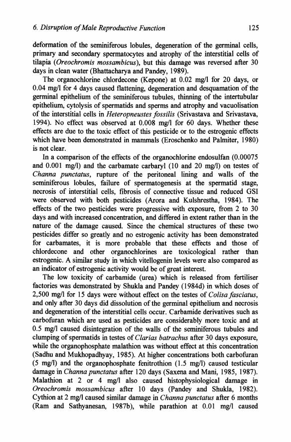
Figure 6.1. Effect of estrogenic alkylphenolic compounds (0.03 mgll) and ethynylestradiol (EE2; 2 ngll) on (A) plasma vitellogenin and (B) testicular growth in male rainbow trout exposed for 3 weeks. • is significantly different from the final control values. (OP =
octylphenol; NP = nonylphenol; NPlEC = nonylphenoxycarboxylic acid; NP2EO = nonylphenoldiethoxylate). (Reprinted with kind permission from Environmental Toxicology and Chemistry, 1996. lnhibition of testicular growth in rainbow trout (Oncorhynchus mykiss) exposed to estrogenic alkylphenolic chemicals, by Jobling, S., Sheahan, D., Osborne, J. A, Matthiessen, P. and Sumpter, J. P. Volume 15, pp. 194-202. Copyright Society of Environmental Toxicology and Chemistry (SETAC), Pensacola, FL, 1996).
128 Chapter 6
A B C '. 30 CONTROL
30 30
20 20 20 --Q 10 10 10 ... ~
~ 0 0 0 GA GB _pcyA spcyB .ptd GA GB speyA .peyB sptd GA GB _pcyA speyB sptd
0 E
ell EE2 NPlEO ... 30 30 30 - '. ell '. ~ -'$. 20 20 20
10 10 10
0 0 0 GA G8 _pcyA speyB sptd GA GB spcyA spcyB sptd GA GB speyA speyB sptd
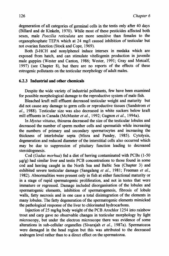
Figure 6.2. The effect of alkyl phenolic compounds on spermatogenesis. Data from testes of control rainbow trout and those exposed to octylphenol (OP), nonylphenol (NP), nonylphenoxy carboxylic acid (NPlEC) and nonylphenoldiethoxylate (NP2EO) at approximately 30 Ilg/I, or ethynylestradiol(EE2) at 2 ng/I for 3 weeks are shown. The five germ cell stages were:spermatogonia A (GA), spermatogonia B (GB), spermatocyte A (spcyA), spermatocyte B (spcyB), and spermatid (sptd). The proportional distribution of these cell types are illustrated. *+ denotes significantly higher than control; *- denotes significantly lower than control (P<0.05) (Reprinted with kind permission from Environmental Toxicology and Chemistry, 1996. Inhibition oftesticular growth in rainbow trout (Oncorhynchus mykiss) exposed to estrogenic alkylphenolic chemicals, by Jobling, S., Sheahan, D., Osborne, J. A., Matthiessen, P. and Sumpter, J. P. Volume 15, pp. 194-202. Copyright Society of Environmental Toxicology and Chemistry (SETAC), Pensacola, FL, 1996).
While the earlier studies (Job ling and Sumpter, 1994; Purdom et aI., 1994) with environmental estrogens, such as the alkylphenolics, concentrated on the stimulation of hepatic vitellogenesis (Chapter 9), a more recent study has shown that a wide range of alkylphenolics can also affect testicular development at levels similar to that which stimulates vitellogenin (Jobling et ai., 1996). In rainbow trout exposed for three weeks to 0.03 mg/loctylphenol, nonylphenol, nonylphenoldiethoxylate and nonylphenoxycarboxylic acid towards the end of gonadal recrudescence, testicular growth was inhibited to a similar extent to that induced by 0.000002 mg/l (2 ng/l; 0.002 ppb) ethynylestradiol. Of the alkylphenolics, octylphenol was the most potent in both inhibiting testicular growth and in inducing hepatic vitellogenin synthesis (Figure 6.1). This decreased gonadal growth resulted from increased numbers of the early stages of gamete development such as spermatogonia A and decreased numbers of spermatocytes, a pattern which was mimicked by
6. Disruption of Male Reproductive Function 129
exposure to estradiol (Figure 6.2). It is not clear whether these compounds acted directly on the testis or whether they acted indirectly via hypothalamic or pituitary receptors and inhibited gonadotrophin secretion. Similar inhibitory effects on spermatogenesis were found in mature male carp exposed to 4-tert-pentylphenol (Gimeno et aI., 1998).
While these compounds are undoubtedly estrogenic, as shown by their stimulation of vitellogenin synthesis, it is important to note that their effect on the testis, in arresting spermatogenesis at early stages, is comparable to many of the other pollutants discussed in this Chapter, many of which have no estrogenic activity. Even cyanide, which has not the slightest resemblance to an estrogen, decreased the transformation of spermatogonia into spermatocytes, probably acting via the pituitary (Ruby et aI., 1993a), while low pH decreased the numbers of mature sperm and development of testicular cysts (Ruby et ai., 1978).
6.3 EtTects on testicular hormone production
6.3.1 In vitro studies
Studies of in vitro steroidogenesis by the testis can provide valuable information on how plasma steroid hormone concentrations might be affected in the intact animal exposed to pollutants. They must, however, be used with some discretion and followed up by confIrmatory studies in vivo. In vitro incubations are an artifIcial system and it is difficult to relate the concentrations of xenobiotic added to an incubation flask to those that might be present in the tissue of exposed fIsh. Even in unexposed fIsh, results obtained in vitro do not necessarily relate to those pertaining in the intact animal. Furthermore, in vitro studies examine only the pollutant itself, whereas in vivo the effects could be due to products formed by hepatic or peripheral metabolism.
It is important to differentiate between the two types of in vitro study which use exogenous and endogenous precursors. In the simplest case, tissue is incubated with a suitable exogenous steroid precursor (e.g. 17-hydroxyprogesterone) and the effects of addition of different amounts of added pollutant on the pattern of metabolites monitored. For simplicity of detection and identifIcation, radiolabelled precursors C4C or 3H) are frequently used in such experiments. It is also possible to compare incubations of tissue taken from fIsh which have been exposed to different concentrations of pollutants (the ex vivo approach). Such a study may show which enzyme is most sensitive to pollutant challenge. For example if addition of a pollutant caused decreased II-ketotestosterone, but increased testosterone production this is indicative of inhibition of the 11j3-hydroxylase
130 Chapter 6
enzyme (see Figure 4.3). Studies of this type require use of radioisotopes and specialised instrumentation and separation techniques which are not generally available. Such studies only indicate changes affecting the precursor chosen, even though earlier steps in the biosynthetic sequence could also be affected. Use of endogenous precursors by incubating testes in the presence of gonadotrophin and assaying the steroids produced would determine whether any stage of the biosynthesis, or its stimulation by gonadotrophin is affected, but it is important to remember that products other than those which are assayed could confuse the picture. The major advantage of in vitro studies is that they permit comparison of a range of pollutants and concentrations on identical tissue without using large numbers of fish.
6.3.1.1 Heavy metals Apart from cadmium, the effects of few heavy metals have been examined
in vitro. Testes from brook trout which had been exposed to 25 Jlg/l cadmium and showed morphological damage (see Section 6.2) did not convert [14C]pregnenolone into 11-ketotestosterone, and production of 11 13-hydroxytestosterone and testosterone were lower than in unexposed fish (Freeman and Sangalang, 1977a; Freeman et al., 1984b; Sangalang and o 'Halloran, 1972; 1973). Addition of 10, 100 and 1000 Jlg cadmiumlg testis from unexposed fish also resulted in inhibition of ll-ketotestosterone production and decreased conversion of radiolabelled pregnenolone. These results indicate that the 1113-HSD enzyme (conversion of 1113-hydroxytestosterone to 11-ketotestosterone) was preferentially affected, although other steps in the conversion of pregnenolone to testosterone were also partially inhibited. Cadmium also affected endogenous production of 11-oxygenated androgen production in testes of rainbow trout, but in this case the effect was stimulatory (Kime, 1984) possibly due to the effects of cadmium on calcium flows as discussed for pituitary function (Section 5.3.1).
Another approach to steroidogenesis is to determine 313HSD activity in tissue. This requires less sophisticated technology than that described above but provides less information. 313HSD is a key enzyme in conversion of pregnenolone to progesterone (Figure 4.3) and decreases in enzyme activity are assumed to correlate with decreased steroidogenic activity. Exposure to 0.05 and 0.5 mg/l methyImercuric chloride and emisan-6 (6% methoxyethyl mercuric chloride) respectively reduced 313HSD activity in interstitial cells of Clarias batrachus (Kirubagaran and Joy, 1988a).
6.3.1.2 Pesticides The effects of y-hexachlorocyclohexane (lindane) has been extensively
studied in vitro. With testes of the roach Rutilus rutilus it had only a small impact on the metabolism of radiolabelled 17-hydroxyprogesterone, predominantly by inhibiting glucuronidation of 17,2013P, while with
6. Disruption of Male Reproductive Function 131
endogenous precursors it significantly inhibited ll-ketotestosterone and 17-hydroxyprogesterone production while stimulating that of testosterone and its glucuronide (Kime and Singh, 1996). In spermiating goldfish there was also little effect on the metabolism of exogenous radio labelled precursors while production of deoxycortisol and testosterone from endogenous precursors was stimulated (Kime and Singh, 1996). These results suggest that the pesticide acts predominantly on the early stages of steroidogenesis, rather than on the enzymes that convert 17-hydroxyprogesterone. Much clearer results were obtained with incubations of testes from goldfish which had been exposed to 0.01 mg/l y-HCH for 4 weeks (Singh et aI., 1994). Testosterone, testosterone glucuronide and ll-ketotestosterone were all significantly suppressed while production of I1-deoxycortisol increased. The results suggest an inhibition of androgen synthesis, possibly due to inhibited side chain cleavage (17,20-lyase) activity at the testicular level, but whether the lower GSI in exposed fish was a cause or effect of inhibited steroidogenesis is not clear.
After 20 days exposure of Oreochromis mossambicus to 0.001 mg/l DDT 313 and 1713-HSD activity was absent from interstitial cells of the testes but after 30 days exposure to freshwater, activity had returned concomitant with repair to the morphological damage (Bhattacharya and Pandey, 1989). These enzyme activities were also significantly decreased in testes of Clarias batrachus exposed to 0.025 mg/l of the organophosphorus quinalphos for 4 days (Bagchi et ai., 1990). 313HSD activity was also partially reduced in testes of carp exposed to 0.3 mg/l fenitrothion (Kapur et al., 1978).
Cythion and hexadrin exposed Heteropneustes fossilis had a lower testicular 32p uptake than unexposed fish which might be indicative of decreased steroidogenesis (Singh and Singh, 1980a). The carbofuran and fenitrothion induced changes in testicular cholesterol and phospholipids in Channa punctatus may also have had a similar cause (Saxena et al., 1986).
6.3.1.3 Industrial The only industrial pollutant which has received significant attention in
vitro is PCB. Unlike y-HCH, PCB exposure of brook trout (Salvelinus fontinalis) stimulated production of 1113-hydroxytestosterone from [14C]pregnenolone by testes in vitro but did not affect yields of testosterone or ll-ketotestosterone (Freeman and Idler, 1975). Plasma ll-ketotestosterone was unaffected by exposure, but unfortunately 1113-hydroxytestosterone was not measured so it is difficult to corroborate in vivo and in vitro data. In cod fed a diet of herring containing 1-10 J.lg/g of the PCB Arochlor 1254 for 5Yz months conversion of [14C]progesterone to l1-ketotestosterone was inhibited (Freeman et al., 1980, 1982).
132
5
4 -~ 3 c -CD c e 2
~ 0 -;; 1 ~
_50
:€ C)
.5.40 CD c e
30 S fI)
~ S 20 0 ti ~
I 10 ..... .....
Chapter 6
/x /
'. / --•• x/ treated I e, /x__ / Cd-
,x~._. ._. ! _--X""" \. Control
.-------- =: -- - \..
o 0 0 10 27 41 56 77 93 DAYS
June 19 Aug 22 Sept5 Sept 15 Oct2 Oct 16 Oct 31 No.21 Dec 7 DATE
/ /
Cd -treated
_-x----x x---I
I _x-_ tr--. x-" 7 ~ontrol
// '" I • •
• '/
--------------.---. -0 o 0 10 27 41 56 77 93 DAYS
June 19 Aug 22 SeptS Sept 15 Dc: 2 Oct 16 Oct 31 No.21 Oec7 DATE
Date and duration of exposure
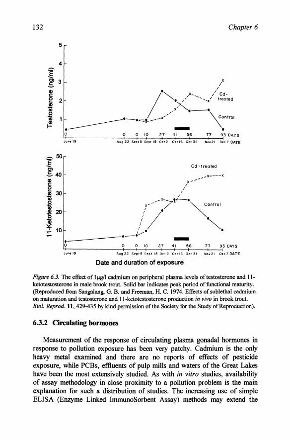
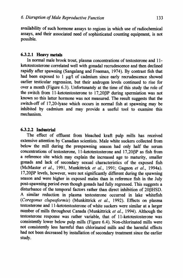
Figure 6.3. The effect of IJ.1g11 cadmium on peripheral plasma levels of testosterone and 11-ketotestosterone in male brook trout. Solid bar indicates peak period of functional maturity. (Reproduced from Sangalang, O. B. and Freeman, H. C. 1974. Effects of sublethal cadmium on maturation and testosterone and II-ketotestosterone production in vivo in brook trout. Bioi. Reprod. 11, 429-435 by kind permission of the Society for the Study of Reproduction).
6.3.2 Circulating hormones
Measurement of the response of circulating plasma gonadal hormones in response to pollution exposure has been very patchy. Cadmium is the only heavy metal examined and there are no reports of effects of pesticide exposure, while PCBs, eftluents of pulp mills and waters of the Great Lakes have been the most extensively studied. As with in vitro studies, availability of assay methodology in close proximity to a pollution problem is the main explanation for such a distribution of studies. The increasing use of simple ELISA (Enzyme Linked ImmunoSorbent Assay) methods may extend the
6. Disruption of Male Reproductive Function 133
availability of such hormone assays to regions in which use of radiochemical assays, and their associated need of sophisticated counting equipment, is not possible.
6.3.2.1 Heavy metals In normal male brook trout, plasma concentrations of testosterone and 11-
ketotestosterone correlated well with gonadal recrudescence and then declined rapidly after spawning (Sangalang and Freeman, 1974). By contrast fish that had been exposed to 1 f.1g/l of cadmium since early recrudescence showed earlier testicular regression, but their androgen levels continued to rise for over a month (Figure 6.3). Unfortunately at the time of this study the role of the switch from ll-ketotestosterone to 17 ,20~P during spermiation was not known so this latter hormone was not measured. The result suggests that the switch-off of 17,20-lyase which occurs in normal fish at spawning may be inhibited by cadmium and may provide a useful tool to examine this mechanism.
6.3.2.2 Industrial The effect of eftluent from bleached kraft pulp mills has received
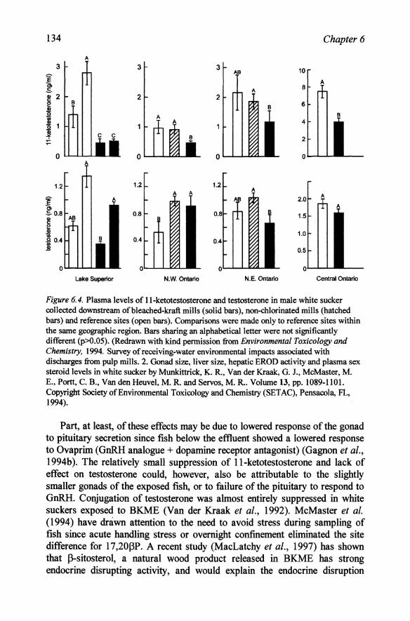
extensive attention by Canadian scientists. Male white suckers collected from below the mill during the prespawning season had only half the serum concentrations of testosterone, l1-ketotestosterone and 17 ,20 ~P as fish from a reference site which may explain the increased age to maturity, smaller gonads and lack of secondary sexual characteristics of the exposed fish (McMaster et al., 1991, Munkittrick et al., 1991; Gagnon et al., 1994a). 17 ,20~P levels, however, were not significantly different during the spawning season and were higher in exposed males than in reference fish in the July post-spawning period even though gonads had fully regressed. This suggests a disturbance of the temporal factors rather than direct inhibition of 20~HSD. A similar reduction in plasma testosterone occurred in lake whitefish (Coregonus clupeaformis) (Munkittrick et aI., 1992). Effects on plasma testosterone and l1-ketotestosterone of white suckers were similar at a larger number of mills throughout Canada (Munkittrick et al., 1994). Although the testosterone response was rather variable, that of l1-ketotestosterone was consistently lower below pulp mills (Figure 6.4). Non-chlorinated mills were not consistently less harmful than chlorinated mills and the harmful effects had not been decreased by installation of secondary treatment since the earlier study.
134 Chapter 6
3 3 AS 10
A A 8
2 2 6
B 4
2
o L..J..-J...L...L 0 0 o
1.2 1.2 1.2 A
~ A $0.8 0.8 0.8
I~ B 0.4 0.4 0.5
oL..J..-J...L...L 0 0 o lake Superior N.W. Ontario N.E. Ontario Central Ontario
Figure 6.4. Plasma levels of ll-ketotestosterone and testosterone in male white sucker collected downstream of bleached-kraft mills (solid bars), non-chlorinated mills (hatched bars) and reference sites ( open bars). Comparisons were made only to reference sites within the same geographic region. Bars sharing an alphabetica1letter were not significantly different (p>O.05). (Redrawn with kind permission from Environmental Toxicology and Chemistry. 1994. Survey of receiving-water environmental impacts associated with discharges from pulp mills. 2. Gonad size, liver size, hepatic EROD activity and plasma sex steroid levels in white sucker by Munkittrick, K. R., Van der Kraak, G. J., McMaster, M. E., Portt, C. B., Van den Heuvel, M. R. and Servos, M. R.. Volume 13, pp. 1089-1101. Copyright Society of Environmental Toxicology and Chemistry (SETAC), Pensacola, FL, 1994).
Part, at least, of these effects may be due to lowered response of the gonad to pituitary secretion since fish below the effluent showed a lowered response to Ovaprim (GnRH analogue + dopamine receptor antagonist) (Gagnon et al., 1994b). The relatively small suppression of ll-ketotestosterone and lack of effect on testosterone could, however, also be attributable to the slightly smaller gonads of the exposed fish, or to failure of the pituitary to respond to GnRH. Conjugation of testosterone was almost entirely suppressed in white suckers exposed to BKME (Van der Kraak et aI., 1992). McMaster et al. (1994) have drawn attention to the need to avoid stress during sampling of fish since acute handling stress or overnight confinement eliminated the site difference for 17,20I3P. A recent study (MacLatchy et al., 1997) has shown that j3-sitosterol, a natural wood product released in BKME has strong endocrine disrupting activity, and would explain the endocrine disruption
6. Disruption of Male Reproductive Function 135
apparent in fish exposed to effluent from mills in which chlorination is no longer used and in which dioxins and other chlorinated aromatics are less likely to be present. j3-sitosterol decreased both plasma levels of androgens and the response to gonadotrophin (hCG), while GtH levels remained unchanged. Although j3-sitosterol has been shown to be estrogenic in some binding studies (Tremblay et ai., 1995; Mellanen et ai., 1996), the effects in vivo differed markedly from those of estradiol. Gonadal cholesterol was significantly decreased in [3-sitosterol treated fish, but not in those exposed to estradiol. The authors conclude that [3-sitosterol can cause endocrine disruption either by decreasing the availability of cholesterol to the P 450scc
enzyme that converts cholesterol to pregnenolone, or by decreasing the activity of this enzyme. Such a mechanism appears much more probable than that due to any estrogenic activity given the close structural similarity between [3-sitosterol and cholesterol. It also warns against assuming too readily that the action of an endocrine disruptor is due to its estrogenic activity if this has been demonstrated using screening procedures involving binding affinities.
The field studies in Canada have been supported by laboratory studies on effluent from Finnish pulp mills. Mature prespawning male rainbow trout exposed to 0.5-0.75% BKME for 2 weeks had plasma testosterone levels 50% lower than control fish (Lindstrom-Seppa and Oikari, 1989a). Intraperitoneal injection of 2~g/kg body weight of 2,3,7,8-tetrachlorodibenzo-p-dioxin (TCCD), a constituent of pulp mill effluent did not significantly affect plasma testosterone, but fish in this second experiment did not respond either to BKME exposure which suggests that ll-ketotestosterone or 17,20[3P might have been a more appropriate hormone to measure at this stage of maturity.
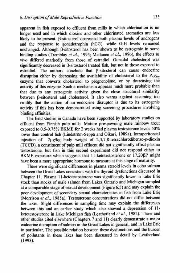
There were significant differences in plasma steroid levels in coho salmon between the Great Lakes consistent with the thyroid dysfunctions discussed in Chapter 11. Plasma ll-ketotestosterone was significantly lower in Lake Erie stock than stocks of male salmon from Lakes Ontario and Michigan sampled at a comparable stage of sexual development (Figure 6.5) and may explain the poor development of secondary sexual characteristics in fish from Lake Erie (Morrison et al., 1985a). Testosterone concentrations did not differ between the lakes. Slight differences in sampling time may explain the differences between this and an earlier study which also showed a depression of 11-ketotestosterone in Lake Michigan fish (Leatherland et ai., 1982). These and other studies cited elsewhere (Chapters 7 and 11) clearly demonstrate a major endocrine disruption of salmon in the Great Lakes in general, and in Lake Erie in particular. The possible relation between these dysfunctions and the burden of pollutants in these lakes has been discussed in detail by Leatherland (1993).
136 Chapter 6
it.-\;;';:1.-
::::.:':.::::
ur""~
1.~
Figure 6.5. Plasma testosterone and ll-ketotestosterone in coho salmon collected from. Lakes Ontario (2 sites), Erie or Michigan Vertical bars indicate 95% confidence limit, and numbers of samples are shown at the base of the bars. (Redrawn from Compo Biochem. Physiol. SOA, Morrison, P. F., Leatherland, J. F. and Sonstegard, R. A Plasma cortisol and sex steroid levels in Great Lakes coho salmon (Oncorhynchus kisutch Walbaum) in relation to fecundity and egg survival, pp. 61-68, Copyright 1985, with kind permisssion from. Elsevier Science.
The Great Lakes, like the North Sea is contaminated by high levels of PCBs and several laboratory studies have examined the effects of these pollutants on steroid levels in male fish. Intraperitoneal administration of 25 mglkg body weight Arochlor 1254 to rainbow trout or common carp at weekly intervals for 4 weeks resulted in a steady decrease in testosterone compared with control fish (Sivarajah et al., 1978). Carp given a single intraperitoneal administration of Arochlor 1248 also had lower plasma testosterone than control fish after 6 days (Yano and Matsuyama, 1986). Feeding experiments with PCBs did not give conclusive results with male cod, but androgen levels remained low in fish which had accumulated high levels of PCBs in their testes (Freeman et al., 1984b).
Exposure of male salmon (Salmo salar) and flounder (Pleuronectes platessa) to crude oil generally caused a decrease in testosterone and 11-ketotestosterone but the results for the salmon were not consistent. Conjugation of the androgens may have been preferentially affected in the flounder and in both species II-hydroxylation was particularly sensitive (Truscott et al., 1983). In further studies with the flounder using sediment containing crude oil (Truscott et al., 1992), free androgens were not affected, but the glucuronide levels were significantly decreased although it is not clear whether this was due to decreased UDP-glucuronyl transferase activity in the testis or liver. The increased liver size in exposed fish, however, indicated that this organ had been affected by the pollutant.
6. Disruption of Male Reproductive Function 137
Plasma androgens did not differ between carp held in outflows from sewage lagoons and those held in a reservoir (Sivarajah et ai., 1979), but this study took place before the widespread use of alkylphenolics and other potential estrogens which have recently been shown to affect fish below such discharges (Purdom et al., 1994).
Atlantic salmon in the acidic Westfield River (pH 4.7) had significantly lower testosterone and II-ketotestosterone than those in the less acid Medway River (pH 5.6) (Freeman et ai., 1983), but rainbow trout exposed in the laboratory to a range of pHs from 4.5 to 7.1 did not show any differences (Weiner et ai., 1986). This suggests that other factors may have caused the suppression in the Westfield River fish.
6.4 Effects on sperm viability
6.4.1 Sperm motility
For fertilisation of eggs it is clearly essential for the male to produce motile sperm that can both locate the eggs and then the micropyle for entry into the oocyte. The motility and morphology of fish sperm can be affected by xenobiotics in several ways: 1) by alteration of the internal hormonal environment during it's development, 2) by alteration in the function of the Sertoli cells which nurture the developing germ cells, 3) by changes in the seminal fluid or sperm maturation as a result of changes in 17,20(3P synthesis and 4) by direct effects on the sperm itself which can result in either cytological damage or changes in the efficiency of mitochondrial energy production. Measurement of sperm motility therefore assesses the totality of the impact of any pollutant on the male reproductive system as a whole.
Fish sperm differ from those of mammals in three important aspects, all related to the prevalence of external fertilisation. Firstly they are immotile within the testis or sperm ducts and attain motility only on ejaculation when the milt is diluted with water. Secondly, this motility is of only short duration and rarely exceeds 2 minutes. Thirdly they do not penetrate the oocyte wall, but enter via a special channel, the micropyle. The rapid decrease in velocity of the sperm within such a short time makes motility measurement very difficult. Fish milt is a viscous liquid and is only mixed with water with some difficulty under a microscope. The two step dilution used by Billard and Cosson (1992), involving initial dilution with an extender solution, in which sperm remain immotile, followed by a second dilution with pure water on the microscope stage itself to initiate motility has greatly facilitated such studies. Furthermore this technique permits the exposure of sperm for a longer period. The initial dilution may be made with extender to which pollutant has been added and the final dilution and observation made 24 h later since little loss of
138 Chapter 6
motility occurs during this period in extender. Such exposure would mimic exposure of sperm to pollutants in the seminal fluids which may reflect body burdens within exposed fish and the results may be compared with observations made on sperm to which the final dilution is made with polluted solution analogous to that which occurs during spawning in polluted water (Kime et al., 1996).
Early studies on sperm motility used a subjective measure of motility such as the time taken for most sperm to stop moving or by rating activity on a scale of 1 to 5. This gave a very rough and ready assessment since not all sperm within a sample have the same behaviour and all do not cease swimming at the same instant. In such experiments it is usually necessary for several observers to give independent assessments. Using a scale from 1 (poor motility with 0-20% spermatozoa moving slowly) to 5 (excellent motility, 80-100% sperm moving rapidly) the motility score was significantly lower (3.1) for sperm from white sucker exposed to bleached kraft mill effluent than for control fish (4.0) (McMaster et aI., 1992). Milt volume, spermatocrit, mineral content and osmolarity did not differ significantly. There was no difference in fertilisation rate (>95%) by sperm from the two populations which suggests that the relatively small differences in motility were not important provided that sufficient milt was produced. Ratings of 3 and 4 (40-60% rapid vs. 60-80% very rapid sperm) are also very difficult to differentiate objectively. By use of a similar 4 point scale Khan and Weis (1987a, b) found that motility of mummichog sperm was decreased when exposed to 0.05 mgtl of either mercuric chloride or methylmercury. This study used only a single dilution step since mummichog sperm normally remains motile for 10-20 min after dilution.
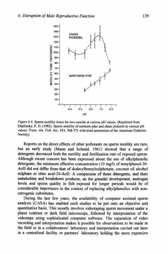
The duration of sperm motility has also been used by several groups as a measure of motility, but it is not clear from such studies how many of the sperm were initially moving and how precise such measurement is since individual sperm cease movement at a wide range of times and one or two sperm still move very fast some time after the majority have stopped (Ebrahimi and Kime, Personal observations). pH had very different effects on sperm motility in two esocids, the chain pickerel and the northern pike (Duplinsky, 1982). The duration of motility of northern pike sperm decreased by 50% as pH decreased from 8.0 to 5.5, while that of chain pickerel decreased sharply above 7.0 and more gradually to pH 5.0 (Figure 6.6), but still retained a high duration of motility down to pH 4.0. By contrast white sucker sperm was little affected by pH and motility time decreased linearly from 77 sec at pH 7.0 to 52 sec at pH 3.0 (Mohr and Chalanchuk, 1985). Duration of motility in the Atlantic salmon also decreased gradually to pH 4.5, but then declined so that there was no motility at pH 4.0 (Daye and Glebe, 1984).
6. Disruption of Male Reproductive Function
150
140
13
120
(i) 110 Cl
6 100 U W 90 (/) -w 80 :E i= 70
> 60 I-:J l- SO 0 :E 40
30
20
CHAIN PICKEREL
NORTHERN PIKE
I' I I I I I I 4.0 5.0 6.0 7.0 8.0
pH
139
Figure 6.6. Sperm motility times for two esocids at various pH values. (Reprinted from Duplinsky, P. D. (1982). Sperm motility of northern pike and chain pickerel at various pH values. Trans. Am. Fish. Soc. 111, 768-771 with kind permission of the American Fisheries Society).
Reports on the direct effects of other pollutants on sperm motility are rare, but an early study (Mann and Schmid, 1961) showed that a range of detergents decreased both the motility and fertilisation rate of exposed sperm. Although recent concern has been expressed about the use of alkylphenolic detergents, the minimum effective concentration (10 mg/l) ofnonylphenol-30-AcO did not differ from that of dodecylbenzylsulphonate, coconut oil alcohol sulphate or oleic acid-20-AcO. A comparison of these detergents, and their metabolites and breakdown products, on the gonadal development, androgen levels and sperm quality in fish exposed for longer periods would be of considerable importance in the context of replacing alkylphenolics with nonestrogenic substitutes.
During the last few years, the availability of computer assisted sperm analysis (CASA) has enabled such studies to be put onto an objective and quantitative basis. This usually involves videotaping sperm movement under a phase contrast or dark field microscope, followed by interpretation of the videotape using sophisticated computer software. The separation of video recording and interpretation makes it possible for observations to be made in the field or in a collaborators' laboratory and interpretation carried out later in a centralised facility or partners' laboratory holding the more expensive
140 Chapter 6
tracker. A number of trackers are available for such studies, ranging from those capable of tracking individually tagged sperm on a frame-by-frame basis to those which are capable of simultaneously tracking several hundred sperm during a real-time run of the video tape. Most instruments produce a large number of parameters, of which the most useful are straight-line velocity (VSL; the straight-line distance between the start and end points of the track divided by the time of the track), curvilinear velocity (VeL; the sum of the incremental distances moved in each frame along the sampled path divided by the total time of the track) and angular path velocity (V AP; a derived path based on an average number of points and divided by the time of the track), and percentage of sperm that is motile.
o 1 10 50 100 200 400 soo 10002000
concentration (ppm)
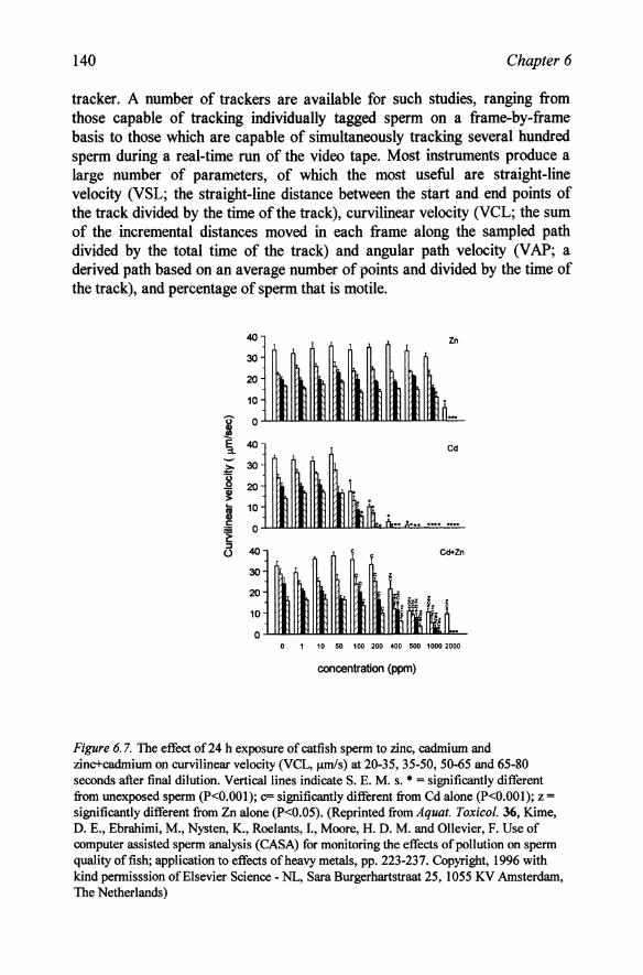
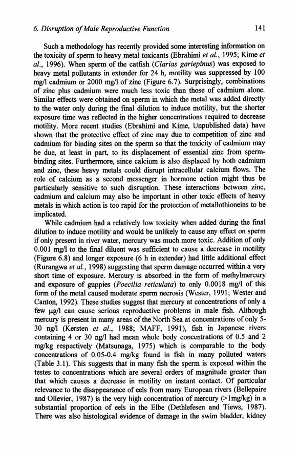
Figure 6. 7. The effect of 24 h exposure of catfish sperm to zinc, cadmium and zinc+cadmium on curvilinear velocity (VCL, J.UDis) at 20-35, 35-50, 50-65 and 65-80 seconds after final dilution. Vertical lines indicate S. E. M. s. • = significantly different from unexposed sperm (P<O.OOI); c= significantly different from Cd alone (P<O.OOI); z = significantly different from Zn alone (P<0.05). (Reprinted from Aquat. Toxicol. 36, Kime, D. E., Ebrahimi, M., Nysten, K., Roelants, I., Moore, H. D. M. and Ollevier, F. Use of computer assisted sperm analysis (CASA) for monitoring the effects of pollution on sperm quality offish; application to effects of heavy metals, pp. 223-237. Copyright, 1996 with kind permisssion of Elsevier Science - NL, Sara Burgerhartstraat 25, 1055 KV Amsterdam, The Netherlands)
6. Disruption of Male Reproductive Function 141
Such a methodology has recently provided some interesting information on the toxicity of sperm to heavy metal toxicants (Ebrahimi et al., 1995; Kime et aI., 1996). When sperm of the catfish (Clarias gariepinus) was exposed to heavy metal pollutants in extender for 24 h, motility was suppressed by 100 mgll cadmium or 2000 mgll of zinc (Figure 6.7). Surprisingly, combinations of zinc plus cadmium were much less toxic than those of cadmium alone. Similar effects were obtained on sperm in which the metal was added directly to the water only during the final dilution to induce motility, but the shorter exposure time was reflected in the higher concentrations required to decrease motility. More recent studies (Ebrahimi and Kime, Unpublished data) have shown that the protective effect of zinc may due to competition of zinc and cadmium for binding sites on the sperm so that the toxicity of cadmium may be due, at least in part, to its displacement of essential zinc from spermbinding sites. Furthermore, since calcium is also displaced by both cadmium and zinc, these heavy metals could disrupt intracellular calcium flows. The role of calcium as a second messenger in hormone action might thus be particularly sensitive to such disruption. These interactions between zinc, cadmium and calcium may also be important in other toxic effects of heavy metals in which action is too rapid for the protection of metallothioneins to be implicated.
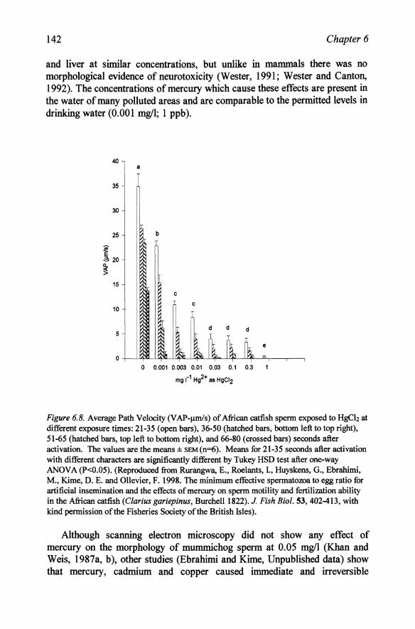
While cadmium had a relatively low toxicity when added during the final dilution to induce motility and would be unlikely to cause any effect on sperm if only present in river water, mercury was much more toxic. Addition of only 0.001 mgll to the final diluent was sufficient to cause a decrease in motility (Figure 6.8) and longer exposure (6 h in extender) had little additional effect (Rurangwa et aI., 1998) suggesting that sperm damage occurred within a very short time of exposure. Mercury is absorbed in the form of methylmercury and exposure of guppies (Poecilia reticulata) to only 0.0018 mgll of this form of the metal caused moderate sperm necrosis (Wester, 1991; Wester and Canton, 1992). These studies suggest that mercury at concentrations of only a few J.lgli can cause serious reproductive problems in male fish. Although mercury is present in many areas ofthe North Sea at concentrations of only 5-30 ngll (Kersten et al., 1988; MAFF, 1991), fish in Japanese rivers containing 4 or 30 ngll had mean whole body concentrations of 0.5 and 2 mglkg respectively (Matsunaga, 1975) which is comparable to the body concentrations of 0.05-0.4 mglkg found in fish in many polluted waters (Table 3.1). This suggests that in many fish the sperm is exposed within the testes to concentrations which are several orders of magnitude greater than that which causes a decrease in motility on instant contact. Of particular relevance to the disappearance of eels from many European rivers (Bellepaire and Ollevier, 1987) is the very high concentration of mercury (>lmglkg) in a substantial proportion of eels in the Elbe (Dethlefesen and Tiews, 1987). There was also histological evidence of damage in the swim bladder, kidney
142 Chapter 6
and liver at similar concentrations, but unlike in mammals there was no morphological evidence of neurotoxicity (Wester, 1991; Wester and Canton, 1992). The concentrations of mercury which cause these effects are present in the water of many polluted areas and are comparable to the permitted levels in drinking water (0.001 m!ifl; 1 ppb).
40 a
35
30
25
~ .2: 20
~ 15
10
5
0
b
c c
d d d
o 0.001 0.003 0.01 0.03 0.1 0.3
mg r1 Hg2+ as HgCI2
e
Figure 6.8. Average Path Velocity (V AP-,.lInls) of African catfish sperm exposed to HgCh at different exposure times: 21-35 (open bars), 36-50 (hatched bars, bottom left to top right), 51-65 (hatched bars, top left to bottom right), and 66-80 (crossed bars) seconds after activation. The values are the means::l:: sEM(n=6). Means for 21-35 seconds after activation with different characters are significantly different by Tukey HSD test after one-way ANOVA (P<O.05). (Reproduced from Rurangwa, E., Roelants, 1, Huyskens, G., Ebrahimi, M., Kime, D. E. and Ollevier, F. 1998. The minimum effective spermatozoa to egg ratio for artificial insemination and the effects of mercury on sperm motility and fertilization ability in the African catfish (Clarius gariepinus, Burchell 1822). J. Fish Bioi. 53, 402-413, with kind permission of the Fisheries Society of the British Isles).
Although scanning electron microscopy did not show any effect of mercury on the morphology of mummichog sperm at 0.05 m!ifl (Khan and Weis, 1987a, b), other studies (Ebrahimi and Kime, Unpublished data) show that mercury, cadmium and copper caused immediate and irreversible
6. Disruption of Male Reproductive Function 143
morphological damage to carp sperm. Modern advances in computer technology now make quantitative measurement of such morphological changes possible, and will be particularly useful in assessing the effects of endocrine disruptors on the development of sperm as a result of action either on the Sertoli cells or on the hormonal support during spermatogenesis. Such motility and morphology measurements may be used either to compare the sperm from wild fish populations in polluted and non polluted environments, or that of laboratory exposed and control fish. This would be of particular interest if it were also related to changes in the morphology of the gonads and pituitary and their secretion of hormones.
Not all of the effects of pollutants may be apparent from a simple examination of sperm. Macek (1968) found that the cumulative mortality for brook trout fry from parents in which only the male was exposed to DDT was greater than in controls even though the fertilisation rates did not differ. Although motility was not directly measured, sperm volume was unaffected by the organochlorine insecticide. It is not clear whether mortality resulted from chromosomal damage or to other biochemical changes in the sperm, but the rapid increase in mortality 7 weeks after fertilisation would suggest that some key developmental stage had been affected.
6.4.2 Fertilising ability
While sperm motility measurements can provide a rapid and readily quantifiable parameter for potentially harmful effects of pollutants without the need for direct exposure of experimental animals, only the much more labour intensive fertilisation of eggs by sperm can at present show how this actually affects fertility. As yet there have been no reports of parallel studies on sperm motility and fertilising capability, but a number of studies have clearly shown that fertilising ability is affected by similar pollutants to those which decrease motility.
Exposure of male brown trout to bleached kraft mill effluent at 0.2-0.5% concentration had no significant effect on the fertilising ability of their milt although the eggs produced by exposed females were less well fertilised at the same concentration (Vuorinen and Vuorinen, 1985). This suggests that in this case, eggs rather sperm are the most sensitive to the pollutant. In contrast, using in vitro exposure of gametes for 40 min, Billard and Roubaud (1985) found that the toxicity of chromium, iron, cyanide and zinc was greater for sperm than ova of the rainbow trout. Sperm of the mummichog were also more sensitive than eggs to mercuric chloride and methyl mercury (Khan and Weis, 1987 a, b). Fertilisation success was decreased after exposure of sperm for 2 min to 0.01 mgll of the pollutant, while eggs exposed to 0.5 mgll for 25 min were unaffected. The difference between these studies may reflect
144 Chapter 6
different pollutants, or that the male and female gametes may have different relative sensitivities during development (in vivo exposure) and when fully mature (in vitro exposure). Methylmercury at 1 mg/l also decreased fertilisation by steelhead trout (Oncorhynchus mykiss) sperm (McIntyre, 1973). Copper at 0.11 mg/l decreased fertilisation by exposed sperm by 50%, but higher exposures of embryos and larvae were required to affect their viability (Anderson et ai., 1991).
The percentage hatch rates of rainbow trout eggs was lower when fertilised by sperm from males exposed to ambient pH 4.5 to 5.5 than to pH 6.5-7.1 although steroid hormone levels and other physiological parameters in the exposed males were unaffected (Weiner et ai., 1986). The means by which this was effected is not clear since, unlike other pollutants, acid water does not contain bioaccumulating material and under laboratory conditions the effects could not have been caused by changes in water mineral content induced by high acidity. Stress during critical stages of gamete formation is perhaps the most likely explanation.
In none of these reports is any data given on the sperm:egg ratio used, but clearly if sperm is present in say a 100 fold excess, then 99% could be rendered unviable and still leave sufficient for complete fertilisation. Such an excess of sperm is more likely to be present during in vitro fertilisation procedures than in the wild and these experiments may well underestimate the toxicity of the pollutants used. A necessary pre-requisite of such experiments should, therefore, be a determination of the minimum sperm:egg ratio required for complete fertilisation of the eggs. A recent such study (Rurangwa et ai., 1998) has shown that the spermitoxicity of a pollutant, such as mercury, on fertilisation rate is in fact very dependent upon the sperm:egg ratio. This study showed that increasing the sperm:egg ratios above 3000:1 had no effect on the rate of fertilisation and hatch of catfish eggs, while the rates decreased rapidly if less sperm were used. This was reflected in the toxicity of mercury in that fertilisation and hatching rates were unaffected by addition of 1 mg/l mercuric chloride to the extender in which sperm was added to the eggs if a sperm:egg ratio of 9000:1 was employed, while at 3000:1 there was a significant decrease and at 1500:1 fertilisation and hatch rates were effectively zero (Figure 6.9). This study shows that if an excess of sperm is present, then it doesn't matter if the pollutant destroys the viability of large numbers of sperm, provided that the minimum number remain sufficiently motile to ensure fertilisation of the eggs, but if only this minimum is present initially even very low losses will be reflected in a decreased fertilisation rate.
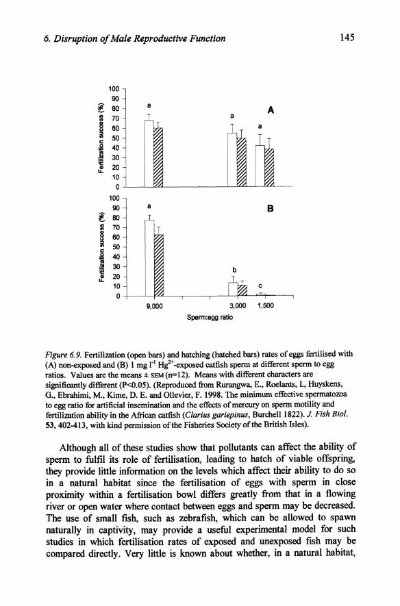
6. Disruption of Male Reproductive Function 145
100 90
~ 80 a A
:l 70 a § 60 iii 50 c: :B 40 J! 30 1::
20 Q) u..
10 0
100 90 a B
~ 80 II) 70 II)
B 60 u ::J II) 50 c: :B 40 .~ 30 b := 1:: Q) 20 u..
10 c 0
9,000 3,000 1,500
Spenn:egg ratio
Figure 6.9. Fertilization (open bars) and hatching (hatched bars) rates of eggs fertilised with (A) non-exposed and (B) 1 mg rl Hi+ -exposed catfish sperm at different sperm to egg ratios. Values are the means ± sEM(n=12). Means with different characters are significantly different (P<0.05). (Reproduced from Rurangwa, E., Roelants, 1, Huyskens, G., Ebrahimi, M., Kime, D. E. and Ollevier, F. 1998. The minimum effective spermatozoa to egg ratio for artificial insemination and the effects of mercury on sperm motility and fertilization ability in the African catfish (Clarius gariepinus, Burchell 1822). J. Fish Bioi. 53,402-413, with kind permission of the Fisheries Society of the British Isles).
Although all of these studies show that pollutants can affect the ability of sperm to fulfil its role of fertilisation, leading to hatch of viable offspring, they provide little information on the levels which affect their ability to do so in a natural habitat since the fertilisation of eggs with sperm in close proximity within a fertilisation bowl differs greatly from that in a flowing river or open water where contact between eggs and sperm may be decreased. The use of small fish, such as zebrafish, which can be allowed to spawn naturally in captivity, may provide a useful experimental model for such studies in which fertilisation rates of exposed and unexposed fish may be compared directly. Very little is known about whether, in a natural habitat,
146 Chapter 6
male fish release more sperm than is required to fertilise the eggs of the female, and if so how great an excess is released. Recent studies on coral reef fish (Warner et ai., 1995; Warner, 1997), however, suggest that males can regulate the amount of sperm that they release to correspond to the size of the female, or to the number of females with whom they intend to mate. They may accept a fertilisation rate slightly lower than maximal in order to conserve sufficient sperm to fertilise an increased number of females. This suggests that the amount of sperm released is not in excess, and that the in vitro fertilisation tests using a minimal sperm:egg ratio may be a good approximation to the situation in wild fish. In group spawning species, in which sperm competition between males may play an important role, the situation may be different and sperm may be produced in greater excess by dominant males to increase their competitive advantage. In such cases, pollutants might be expected to alter the relative fertilising success among competing males. They may also differentially alter the reproductive success of different species within the same ecosystem if there are marked interspecific differences in the amount of sperm released during spawning. In such cases pollution might have marked effects on the population ecology of the habitat. There have been no studies on sperm release in the species which are under the greatest threat in polluted habitats. Such studies are, however, essential to understand the vulnerability of the male and how his ability to fertilise eggs may be affected by pollutants. Recent concern about the sustainability of North Sea cod has concentrated solely on the effects of overfishing (Cook et al., 1997), but if the cod is a species which does not produce a sufficient excess of sperm to compensate for the effects of pollution, then the outlook for the cod population is indeed bleak.
6.5 Other effects on the male
6.5.1 Courtship behaviour.
Courtship patterns are controlled by both hormonal and neural factors, and are an essential preliminary to spawning in many species. Both lindane and water from the Munich wastewater purification plant modified the behaviour of male guppies (Poecilia reticulata) which, surprisingly, led to greater reproductive success of the exposed fish than controls (SchrOder and Peters, 1988). Breeding behaviour in tilapia was delayed by exposure to 0.5 ~g/l endosulfan, a concentration which did not cause morphological changes to the gonads or significant changes in gonadal steroid production (Matthies sen and Logan, 1984). While brook trout were unaffected by 3.4 ~g/l cadmium during the 24 week prespawning period, the males suddenly began swimming erratically, lost equilibrium, lapsed into paralysis and died
6. Disruption of Male Reproductive Function 147
within minutes once the females began their nest building activity in the gravel substrate (Benoit et ai., 1976). Although the cause of this sudden mortality was not clear it cautions against the notion of a safe dose as this clearly can become lethal as a result of social interactions.
There is increasing recognition that in addition to visible behaviour, fish also use olfactory signals (pheromones) as cues (Canario and Power, 1995). Such cues not only signal the stage of sexual maturation of the female, but directly stimulate the male endocrine system. Of the ovarian pheromones, the most studied are the steroids and the prostaglandins. The effects of pollution on ovarian steroid synthesis will be discussed in Chapter 7, but there have been no studies on how prostaglandin synthesis might be affected although it would also presumably be affected by ovarian dysfunction. To detect such pheromones the male must possess functional olfactory receptors and be able to interpret their response by an elevation of his own hormones. Recent studies have shown that such olfactory function can be disrupted by pollutants. The organophosphate pesticide diazinon at 1).lg/l significantly decreased the electrophysiological response of the olfactory epithelium to prostaglandin F2a (Moore and Waring, 1996a, b) and exposure to the pesticide for 5 days significantly reduced the levels of plasma 17,20j3P, testosterone and gonadotrophin II after priming with urine from ovulated female salmon which is known to play a role in synchronising reproductive physiology and behaviour of the sexes. Carbofuran at a concentration of only 0.001 mg/l also impaired the ability of male Atlantic salmon to detect and respond to the reproductive priming pheromone, prostaglandin F2a which is released by the female at ovulation (Waring and Moore, 1997). 0.003 mg/l of the pesticide significantly reduced plasma concentrations of 17,20j3P, testosterone and ll-ketotestosterone, and the volume of expressible milt after priming with this pheromone. The acidity of holding water also has a deleterious effects on the males ability to detect important odorants and pheromones which may be important mediators of reproductive physiology and behaviour (Moore, 1994). It is not clear whether such pollution induced dysfunction is due to direct action on the function of the olfactory receptors themselves or whether it is due to disruption to the transmission and interpretation of the signal.
6.6 Conclusions
Morphological damage to the testis can be caused by a wide range of pollutants, such as heavy metals, organochlorines, organophosphates, carbamates and a range of industrial chemicals. The literature is extremely patchy, covering a wide range of pollutants, species, doses and exposure times, but there have been few concerted attempts to compare the relative
148 Chapter 6
cytotoxicity and endocrine disrupting potential of different pollutants under standardised conditions. Such a study requires choice of a suitable species and then examination of a number of pollutants over a wide range of concentrations. Morphological changes caused by the different xenobiotics should then be examined to determine whether they really differ in their basic nature or whether such differences are simply quantitative and reflect the different potencies of the pollutants and would disappear if different relative doses were used. While morphological data clearly shows disruption of testicular function, it does not indicate whether the testis is the primary site or whether changes in its structure are a secondary effect of pituitaryhypothalamic dysfunction, or of the failure of key steroidogenic enzymes. A truly integrative study would combine morphological criteria with measurement of pituitary and steroidogenic activity and could provide invaluable data on the mechanism of action of xenobiotics as well as information on the safest chemical for the designated use. Clearly, comparisons also have to be made between species to determine whether sensitivities to all effects change in similar proportions from species to species. Furthermore, for practical application to pesticide choice and usage it is necessary to relate the toxic effects on the fish to the concentrations that need to be used on the target pest.
While studies of the effects of pesticides and heavy metals are very patchy, those for the large numbers of industrial pollutants which are released into the waterways are notable by their absence, apart from some studies on pulp mill effluents and on fish from the Great Lakes. Although studies on morphology and endocrine function may provide explanations for reduced fertility, the bottom line is whether the sperm is capable of fertilisation and whether the male is capable of delivering it to the female. Determination of fertilisation rates are very labour intensive, and a simple method for relating sperm motility to its fertilising capability would be of great potential use. Study of the effects of pollution on courtship behaviour, and especially of the release and detection of pheromones is in its infancy, but is vitally important in ensuring delivery of sperm to eggs.