encoding of self‐referential pain catastrophizing in the posterior … · 2018-10-16 · encoding...
TRANSCRIPT

Encoding of Self-Referential Pain Catastrophizing in thePosterior Cingulate Cortex in Fibromyalgia
Jeungchan Lee ,1 Ekaterina Protsenko,1 Asimina Lazaridou,2 Olivia Franceschelli,2
Dan-Mikael Ellingsen,1 Ishtiaq Mawla,1 Kylie Isenburg,1 Michael P. Berry,1 Laura Galenkamp,2
Marco L. Loggia,1 Ajay D. Wasan,3 Robert R. Edwards,2 and Vitaly Napadow1
Objective. Pain catastrophizing is a common fea-ture of chronic pain, including fibromyalgia (FM), and isstrongly associated with amplified pain severity anddisability. While previous neuroimaging studies havefocused on evoked pain response modulation by catastro-phizing, the brain mechanisms supporting pain catastro-phizing itself are unknown. We designed a functionalmagnetic resonance imaging (fMRI)–based pain catas-trophizing task whereby patients with chronic painengaged in catastrophizing-related cognitions. We under-took this study to test our hypothesis that catastrophizingabout clinical pain would be associated with amplifiedactivation in nodes of the default mode network (DMN),which encode self-referential cognition and show alteredfunctioning in chronic pain.
Methods. During fMRI, 31 FM patients reflectedon how catastrophizing (CAT) statements (drawn fromthe Pain Catastrophizing Scale) impact their typical FMpain experience. Response to CAT statements was com-pared to response to matched neutral (NEU) statements.
Results. During statement reflection, higher fMRIsignal during CAT statements than during NEU state-ments was found in several DMN brain areas, includingthe ventral (posterior) and dorsal (anterior) posteriorcingulate cortex (vPCC and dPCC, respectively). Patients’ratings of CAT statement applicability were correlatedsolely with activity in the vPCC, a main DMN hub sup-porting self-referential cognition (r = 0.38, P < 0.05).Clinical pain severity was correlated solely with activityin the dPCC, a PCC subregion associated with cognitivecontrol and sensorimotor processing (r = 0.38, P < 0.05).
Conclusion. These findings provide evidence thatthe PCC encodes pain catastrophizing in FM and suggestdistinct roles for different PCC subregions. Understand-ing the brain circuitry encoding pain catastrophizing inFM will prove to be important in identifying and evaluat-ing the success of interventions targeting negative affectin chronic pain management.
Negative affect plays a key role in the pathophysi-ology of chronic pain and shapes individual differences inpain treatment outcomes. Pain catastrophizing, in particu-lar, is a pain-specific psychosocial construct comprisingcognitive and emotional processes such as helplessness,pessimism, rumination about pain-related symptoms, andmagnification of pain complaints (1). Catastrophizing isalso a major contributor to pain severity in fibromyalgia(FM) (2), a chronic functional pain disorder characterizedby maladaptive brain plasticity (3). While catastrophizingcorrelates positively with other measures of negativeaffect such as anxiety, it also shows a unique and specificinfluence on pain-related outcomes (2). Overall, highercatastrophizing is a risk factor for long-term pain and fordisproportionately negative pain sequelae (e.g., physicaldisability) (2,4). Studies of musculoskeletal pain suggestthat catastrophizing is the single most important pretreat-ment risk factor that impairs the effectiveness of pain-relieving interventions (5). Our own work suggests that
Clinical Trials.gov identifier: NCT01345344.Supported by the NIH (National Center for Complementary
and Integrative Health grants R01-AT-007550, R61-AT-009306, and P01-AT-006663, National Institute of Arthritis and Musculoskeletal and SkinDiseases grant R01-AR-064367, and National Center for ResearchResources grants P41-RR-14075, S10-RR-021110, and S10-RR-023043).
1Jeungchan Lee, PhD, Ekaterina Protsenko, BS, BA, Dan-Mikael Ellingsen, PhD, Ishtiaq Mawla, BA, Kylie Isenburg, BS,Michael P. Berry, BS, Marco L. Loggia, PhD, Vitaly Napadow, PhD:Massachusetts General Hospital, Boston; 2Asimina Lazaridou, PhD,Olivia Franceschelli, BS, Laura Galenkamp, BS, Robert R. Edwards,PhD: Harvard Medical School, Brigham and Women’s Hospital, andMassachusetts General Hospital, Boston; 3Ajay D. Wasan, MD, MSc:University of Pittsburgh Medical Center, Pittsburgh, Pennsylvania,and Massachusetts General Hospital, Boston.
Drs. Edwards and Napadow contributed equally to this work.Address correspondence to Jeungchan Lee, PhD, Martinos
Center for Biomedical Imaging, 149 Thirteenth Street, Suite 2301,Charlestown, MA 02129. E-mail: [email protected].
Submitted for publication November 3, 2017; accepted inrevised form March 20, 2018.
1308
ARTHRITIS & RHEUMATOLOGYVol. 70, No. 8, August 2018, pp 1308–1318DOI 10.1002/art.40507© 2018, American College of Rheumatology

catastrophizing amplifies pain sensitivity and brainresponse and interferes with pain modulation in patientswith many chronic pain conditions, including FM (6–8).While trait pain catastrophizing has been shown to shapebrain responses to evoked pain stimuli (9), the neural cir-cuitry supporting the pain catastrophizing state itselfremains poorly understood. No functional neuroimagingstudies have attempted to directly identify the neuralunderpinnings of chronic pain patients’ engagement inthe cognitive/emotional processes of catastrophizing.
Several studies have attempted to experimentallyinduce a pain catastrophizing state, with mixed results. In2 separate studies, healthy adults were asked to rehearsecatastrophizing self-statements while undergoing coldpressor testing, with no impact on pain sensitivity or toler-ance (10,11). However, inducing catastrophizing by exper-imentally manipulating the perceived threat of painsignificantly impacted cold pain responses (12). Further-more, a study in chronic pain patients found a large reduc-tion in cold pain tolerance when they verbally recitedcatastrophizing, relative to positive statements (13). Otherstudies in patients with chronic musculoskeletal pain haveinduced a catastrophizing state by asking patients to imag-ine their pain worsening in the future (14,15). While thesestudies are limited by the lack of a randomized design oran active control condition, the cognitive manipulationsdid enhance sensitivity to experimentally applied noxiousstimuli. Collectively, while prior studies have applied ca-tastrophizing modulation of brain responses to evokedpain or stress in healthy adults (16) and migraine patients(17), no study has applied neuroimaging-based assessmentof brain function during actual catastrophizing about clini-cal pain.
In this study in FM patients, we investigatedphysiologic (heart rate [HR]) and functional magneticresonance imaging (fMRI) brain responses to a catastro-phizing state induced by visually presenting validated state-ments from the Pain Catastrophizing Scale (PCS) (18) andasking the patients to reflect on the degree to which thesestatements mirrored their experiences with their ownrecalled, clinical pain. Matched neutral statements wereused as controls, and patients rated the applicability ofstatements to their clinical pain. As multiple studies havelinked altered neurophysiology in the brain’s default modenetwork (DMN) to clinical pain severity in FM (19,20),and since DMN regions are known to encode self-referen-tial cognitive processing (21), we hypothesized thatpatients would activate DMN regions during catastrophiz-ing, and that catastrophizing-specific regions would showan association between fMRI response and ratings ofapplicability of the catastrophizing statements to patients’clinical pain.
PATIENTS AND METHODS
Patients. Thirty-five female patients meeting the Ameri-can College of Rheumatology (ACR) criteria for a diagnosisof FM (22) were recruited through Clinical Trials listings(clinicaltrials.partners.org), a Partners Healthcare medical recordsdatabase, and physician referral. The protocol was approved bythe Human Research Committee of Partners Healthcare andMassachusetts General Hospital, and patients provided writteninformed consent prior to beginning study procedures.
All patients completed telephone prescreening to deter-mine their eligibility for the study, and all patients were assessedfor the following inclusion criteria: be ages 18–65 years andfemale; meet the ACR diagnostic criteria for FM for at least 1year (22); receive stable doses of medication prior to enteringthe study; have baseline pain intensity of at least 4 of 10 on aver-age and report pain for at least 50% of days; be able to providewritten informed consent; and be fluent in English. Exclusioncriteria were comorbid acute or chronic pain conditions ratedmore painful than FM; use of stimulant medications for fatigueassociated with sleep apnea or shift work; psychiatric disorderswith history of psychosis; psychiatric hospitalization within 6months prior to enrollment; current or recent use of recreationaldrugs; active suicidal ideation; participation in other therapeutictrials; lower limb vascular surgery or current lower limb vasculardysfunction; pregnant or nursing; history of significant headinjury (e.g., with substantial loss of consciousness); history ofanxiety disorders interfering with fMRI procedures (e.g., panic);and contraindications to MRI.
Behavioral visit. All patients completed a behavioralsession on a separate day from the MRI scan and were intro-duced to study procedures, including rating scales and the paincatastrophizing task described below. Additionally, patientscompleted the following questionnaires: PCS (18); Brief PainInventory (BPI) (23), to assess pain severity and interference;and Patient-Reported Outcomes Measurement InformationSystem 29-Item Profile (PROMIS-29), to assess several healthoutcomes (24).
Pain catastrophizing (CAT) task. The pain CAT task(Figure 1) consisted of 6 statements drawn from the validatedPCS (18) and 6 Velten-type affectively neutral statements takenfrom a validated set used as a control in prior cognitive/affectiveresearch (25,26) and modified to have lexical difficulty and wordcount similar to PCS statements (see Figure 1 for a full list ofstatements). Only PCS statements containing the word “pain”were selected, and 2 statements were chosen from each of 3 sub-scales: helplessness, magnification, and rumination. Statementswere presented in a block design, with separate blocks for PCSsubscales (CAT statements) and matched neutral (NEU) state-ments. Each statement block was presented for 20 seconds(10 seconds/statement, including a 1-second fade-in for smoothvisual transition between statements), separated by a 20-secondcross-hair fixation rest period. Patients were instructed to readeach statement and reflect on the degree to which they had thesethoughts or feelings during a recent typical day of FM pain. Nomotor responses were required; patients were instructed to sim-ply reflect on their FM-related experiences with these thoughtsand feelings. This general methodology (i.e., participants readand reflect on affectively themed statements in order to induce acertain mood) has been used in multiple pain studies to alterboth self-reported mood and pain sensitivity (27). Additionally,each run contained a single 5-second “catch trial” (CATCH) in
PCC ENCODES PAIN CATASTROPHIZING IN FIBROMYALGIA 1309
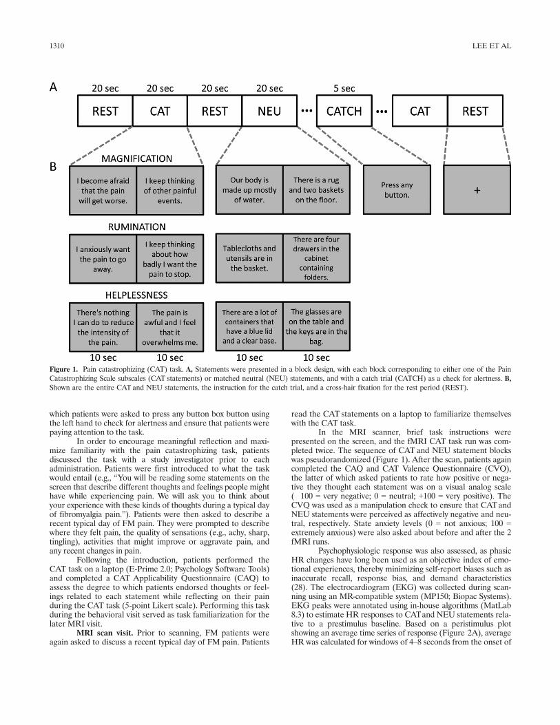
which patients were asked to press any button box button usingthe left hand to check for alertness and ensure that patients werepaying attention to the task.
In order to encourage meaningful reflection and maxi-mize familiarity with the pain catastrophizing task, patientsdiscussed the task with a study investigator prior to eachadministration. Patients were first introduced to what the taskwould entail (e.g., “You will be reading some statements on thescreen that describe different thoughts and feelings people mighthave while experiencing pain. We will ask you to think aboutyour experience with these kinds of thoughts during a typical dayof fibromyalgia pain.”). Patients were then asked to describe arecent typical day of FM pain. They were prompted to describewhere they felt pain, the quality of sensations (e.g., achy, sharp,tingling), activities that might improve or aggravate pain, andany recent changes in pain.
Following the introduction, patients performed theCAT task on a laptop (E-Prime 2.0; Psychology Software Tools)and completed a CAT Applicability Questionnaire (CAQ) toassess the degree to which patients endorsed thoughts or feel-ings related to each statement while reflecting on their painduring the CAT task (5-point Likert scale). Performing this taskduring the behavioral visit served as task familiarization for thelater MRI visit.
MRI scan visit. Prior to scanning, FM patients wereagain asked to discuss a recent typical day of FM pain. Patients
read the CAT statements on a laptop to familiarize themselveswith the CAT task.
In the MRI scanner, brief task instructions werepresented on the screen, and the fMRI CAT task run was com-pleted twice. The sequence of CAT and NEU statement blockswas pseudorandomized (Figure 1). After the scan, patients againcompleted the CAQ and CAT Valence Questionnaire (CVQ),the latter of which asked patients to rate how positive or nega-tive they thought each statement was on a visual analog scale(�100 = very negative; 0 = neutral; +100 = very positive). TheCVQ was used as a manipulation check to ensure that CATandNEU statements were perceived as affectively negative and neu-tral, respectively. State anxiety levels (0 = not anxious; 100 =extremely anxious) were also asked about before and after the 2fMRI runs.
Psychophysiologic response was also assessed, as phasicHR changes have long been used as an objective index of emo-tional experiences, thereby minimizing self-report biases such asinaccurate recall, response bias, and demand characteristics(28). The electrocardiogram (EKG) was collected during scan-ning using an MR-compatible system (MP150; Biopac Systems).EKG peaks were annotated using in-house algorithms (MatLab8.3) to estimate HR responses to CATand NEU statements rela-tive to a prestimulus baseline. Based on a peristimulus plotshowing an average time series of response (Figure 2A), averageHR was calculated for windows of 4–8 seconds from the onset of
Figure 1. Pain catastrophizing (CAT) task. A, Statements were presented in a block design, with each block corresponding to either one of the PainCatastrophizing Scale subscales (CAT statements) or matched neutral (NEU) statements, and with a catch trial (CATCH) as a check for alertness. B,Shown are the entire CAT and NEU statements, the instruction for the catch trial, and a cross-hair fixation for the rest period (REST).
1310 LEE ET AL
each CATor NEU statement. The averaged HR responses werethen normalized with respect to the average HR from a 5-secondbaseline preceding each statement block. Differences in normal-ized HR response between CAT and NEU statement blockswere calculated, and significance (P < 0.05) was determinedusing a paired t-test (SPSS software version 10.0.7).
MRI data acquisition. MRI data were obtained on a3.0T Siemens Skyra (Siemens Medical) equipped with a 32-channelhead coil at the Athinoula A. Martinos Center for BiomedicalImaging, Massachusetts General Hospital. T1-weighted structuralimages were obtained using a 3-dimensional magnetization-preparedrapid gradient-echo pulse sequence (repetition time [TR] 2,530msec, echo time [TE] 1.64 msec, flip angle 7°, field of view[FOV] 256 9 256 mm, spatial resolution 1 9 1 9 1 mm). Func-tional data (228 volumes/run) were obtained with a simultane-ous multislice imaging pulse sequence for improvedspatiotemporal resolution (acceleration factor 5, TR 1,250msec, TE 33 msec, flip angle 65°, FOV 196 9 196 mm, voxeldimensions 2 9 2 9 2 mm, 75 axial slices with no gap) (29).
MRI data processing and analysis. Functional MRIdata processing was carried out using FMRIB Software Library(FSL [fsl.fmrib.ox.ac.uk]), Analysis of Functional NeuroImages(afni.nimh.nih.gov/afni), and FreeSurfer (https://surfer.nmr.mgh.harvard.edu/fswiki). The first 3 fMRI volumes were removed,and data were then corrected for head motion (FSL-MCFLIRT)and B0 inhomogeneities (FSL-PRELUDE and FSL-FUGUE),skull stripped (FSL-BET), spatially smoothed (Gaussian kernel,full-width half-maximum 5 mm), and temporal high-pass filtered(cutoff 80 seconds) to remove signal drift noise. We used the fol-lowing head motion exclusion criteria: framewise displacementand rotation >2 mm and 2°, respectively. For coregistration ofstructural and functional data to standard Montreal Neurologi-cal Institute (MNI) space (FSL-FNIRT), structural images werealigned to fMRI data (BBREGISTER).
A first-level, within-subject, generalized linear model(GLM) analysis was performed by modeling CAT and NEUstatement blocks, as well as the CATCH trial block, convolvedwith the canonical double-gamma hemodynamic response
function (FSL-FEAT). The first-level parameter estimates andcorresponding variance maps from the 2 fMRI runs were thencombined with a second-level analysis using a fixed-effectsmodel (FSL-FEAT). Resulting parameter estimates and vari-ance maps were then registered to standard space (MNI152)using the FMRIB Nonlinear Image Registration Tool (FNIRT)and used for group analysis (i.e., CATand NEU statement groupmap, and CAT versus NEU statement difference map) usingFMRIB Local Analysis of Mixed Effects (FLAME 1+2, cluster-corrected for multiple comparisons, Z > 2.3, P < 0.05).
The CAT versus NEU statement difference map wasused to identify regions of interest (ROIs), defined as 3-mmdiameter spheres centered at the cluster peak voxel. ROIaverage percent signal change was then used to investigate asso-ciations with relevant pain and catastrophizing behavioral out-comes (e.g., PCS, CAQ, BPI, and PROMIS-29).
FreeSurfer and Caret (https://www.nitrc.org/projects/caret/) were used for visualization of results on inflated cerebraland cerebellar surfaces, respectively. As we hypothesized DMNinvolvement in the brain circuitry encoding catastrophizing,results were visualized on a brain surface with an outline of thepublicly available resting-state fMRI parcellation based on 1,000subjects (30).
RESULTS
Of 35 enrolled FM patients, 31 were included in theanalyses (mean � SD age 43.74 � 11.71 years; 26 Cau-casians, 2 AfricanAmericans, 1 Asian, 1 CapeVerdean, and1 of unknown ethnicity) (for medications the 31 patientsused at the time of scanning, see Supplementary Table 1,available on theArthritis & Rheumatologyweb site at http://onlinelibrary.wiley.com/doi/10.1002/art.40507/abstract). FourFM patients were excluded from analyses (2 due to struc-tural brain abnormalities, 1 due to inability to comply with
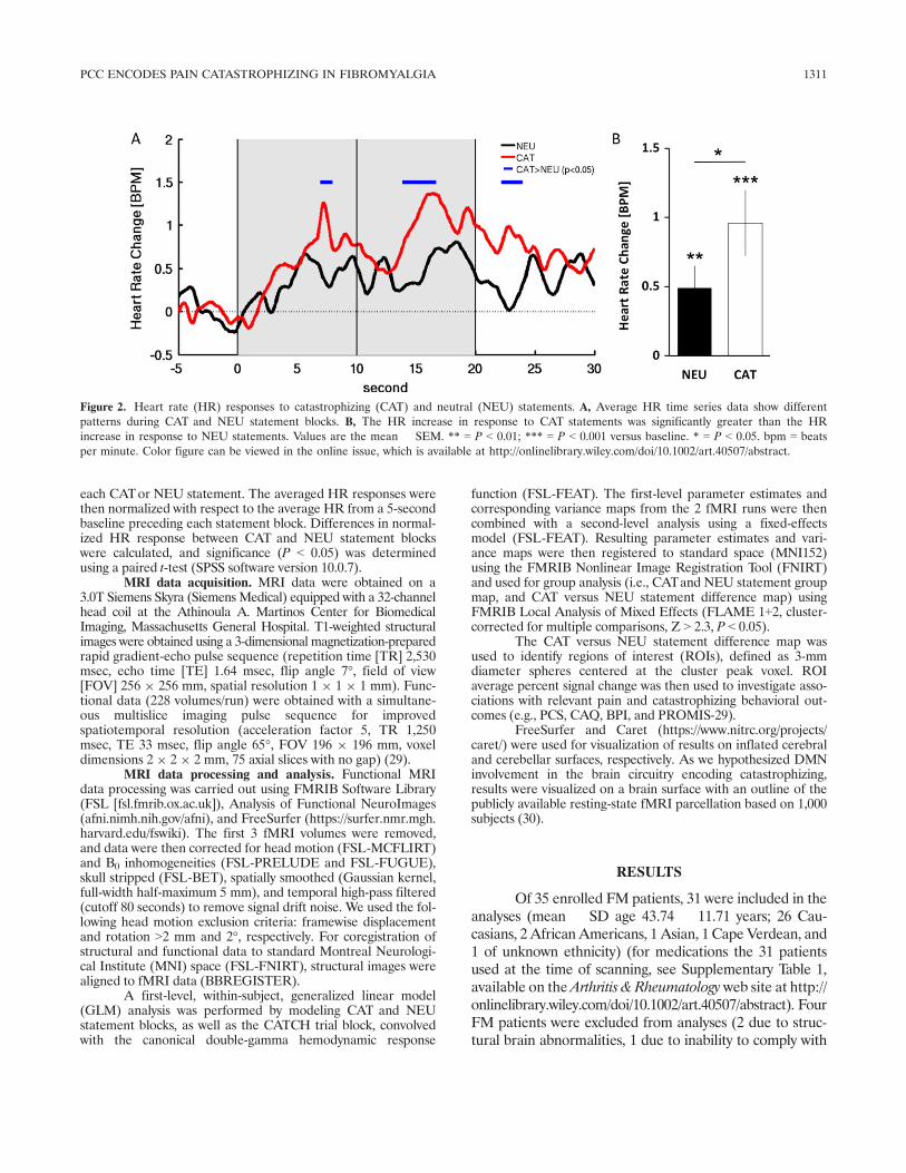
Figure 2. Heart rate (HR) responses to catastrophizing (CAT) and neutral (NEU) statements. A, Average HR time series data show differentpatterns during CAT and NEU statement blocks. B, The HR increase in response to CAT statements was significantly greater than the HRincrease in response to NEU statements. Values are the mean � SEM. ** = P < 0.01; *** = P < 0.001 versus baseline. * = P < 0.05. bpm = beatsper minute. Color figure can be viewed in the online issue, which is available at http://onlinelibrary.wiley.com/doi/10.1002/art.40507/abstract.
PCC ENCODES PAIN CATASTROPHIZING IN FIBROMYALGIA 1311
MRI safety requirements, and 1 because of falling asleepduring fMRI [the patient failed to respond to the catch trial,which the patient confirmed retrospectively]).
Clinical measures. FM patients reported moder-ate-to-high scores on the PCS (mean � SD 21.03 � 12.92),BPI subscales (4.74 � 1.87 for severity, 5.13 � 2.41 forinterference), and PROMIS-29 subscales (39.89 � 6.96 forphysical function, 56.73 � 8.13 for anxiety, 53.68 � 8.97 fordepression, 58.73 � 8.13 for sleep disturbance, 46.96 �13.91 for satisfaction with participation in social roles) atthe initial behavioral visit. After 2 CAT task fMRI runs,patients reported higher CAQ (statement applicability)scores for CAT statement blocks compared to CAQ scoresfor NEU statement blocks (mean � SD CAQCAT score6.90 � 4.64 versus CAQNEU score 2.03 � 2.93; P <0.0001). Mean � SD CVQ scores demonstrated that CATstatements were indeed perceived as affectively negative(CVQCAT score �57.86 � 37.68), while NEU statementswere not perceived as having negative valence (CVQNEU
score 7.88� 19.00) (P < 0.00001 for CATstatements versusNEU statements) (Table 1). Mean � SD state anxietylevels showed no significant difference between before andafter 2 CAT task fMRI runs (21.25 � 18.54 for beforeversus 21.32 � 20.02 for after; P = 0.93 [n = 28 patients]).
PCS scores correlated positively with CAQCAT
scores (r = 0.81, P < 0.0001), the BPI interference subscale(r = 0.45, P < 0.05), and PROMIS-29 subscales (for anxi-ety, r = 0.73, P < 0.0001; for depression, r = 0.66, P <0.0001). Furthermore, CAQCATscores correlated positivelywith the BPI interference subscale (r = 0.52, P < 0.01) andPROMIS-29 subscales (for anxiety, r = 0.69, P < 0.0001;for depression, r = 0.60, P < 0.0001; for sleep disturbance,r = 0.39, P < 0.05; for satisfaction with participation insocial roles, r = �0.37, P < 0.05).
Brain and psychophysiologic responses duringcatastrophizing. Data from all 31 FM patients were avail-able for fMRI analysis, as all patients passed head motionexclusion criteria. To assess psychophysiologic arousal, wequantified HR responses and found greater HR increasesduring CAT statements (mean � SEM 0.96 � 0.24 beatsper minute) than during NEU statements (0.49 � 0.16beats per minute) (P = 0.03) (Figure 2B).
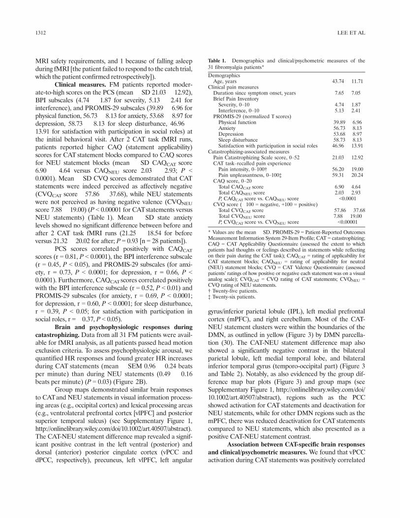
Group maps demonstrated similar brain responsesto CATand NEU statements in visual information process-ing areas (e.g., occipital cortex) and lexical processing areas(e.g., ventrolateral prefrontal cortex [vlPFC] and posteriorsuperior temporal sulcus) (see Supplementary Figure 1,http://onlinelibrary.wiley.com/doi/10.1002/art.40507/abstract).The CAT-NEU statement difference map revealed a signif-icant positive contrast in the left ventral (posterior) anddorsal (anterior) posterior cingulate cortex (vPCC anddPCC, respectively), precuneus, left vlPFC, left angular
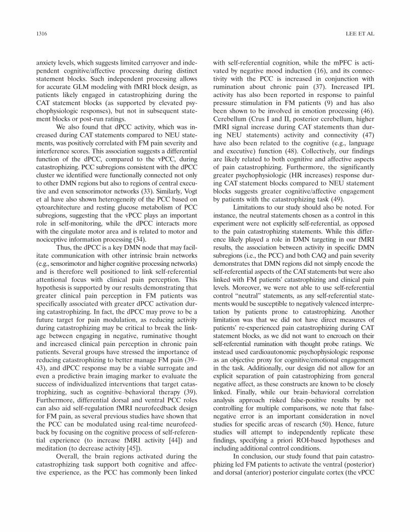
gyrus/inferior parietal lobule (IPL), left medial prefrontalcortex (mPFC), and right cerebellum. Most of the CAT-NEU statement clusters were within the boundaries of theDMN, as outlined in yellow (Figure 3) by DMN parcella-tion (30). The CAT-NEU statement difference map alsoshowed a significantly negative contrast in the bilateralparietal lobule, left medial temporal lobe, and bilateralinferior temporal gyrus (temporo-occipital part) (Figure 3and Table 2). Notably, as also evidenced by the group dif-ference map bar plots (Figure 3) and group maps (seeSupplementary Figure 1, http://onlinelibrary.wiley.com/doi/10.1002/art.40507/abstract), regions such as the PCCshowed activation for CAT statements and deactivation forNEU statements, while for other DMN regions such as themPFC, there was reduced deactivation for CATstatementscompared to NEU statements, which also presented as apositive CAT-NEU statement contrast.
Association between CAT-specific brain responsesand clinical/psychometric measures. We found that vPCCactivation during CATstatements was positively correlated
Table 1. Demographics and clinical/psychometric measures of the31 fibromyalgia patients*
DemographicsAge, years 43.74 � 11.71
Clinical pain measuresDuration since symptom onset, years 7.65 � 7.05Brief Pain InventorySeverity, 0–10 4.74 � 1.87Interference, 0–10 5.13 � 2.41
PROMIS-29 (normalized T scores)Physical function 39.89 � 6.96Anxiety 56.73 � 8.13Depression 53.68 � 8.97Sleep disturbance 58.73 � 8.13Satisfaction with participation in social roles 46.96 � 13.91
Catastrophizing-associated measuresPain Catastrophizing Scale score, 0–52 21.03 � 12.92CAT task–recalled pain experiencePain intensity, 0–100† 56.20 � 19.00Pain unpleasantness, 0–100‡ 59.31 � 20.24
CAQ score, 0–20Total CAQCAT score 6.90 � 4.64Total CAQNEU score 2.03 � 2.93P, CAQCAT score vs. CAQNEU score <0.0001
CVQ score (�100 = negative, +100 = positive)Total CVQCAT score �57.86 � 37.68Total CVQNEU score 7.88 � 19.00P, CVQCAT score vs. CVQNEU score <0.00001
* Values are the mean � SD. PROMIS-29 = Patient-Reported OutcomesMeasurement Information System 29-Item Profile; CAT = catastrophizing;CAQ = CAT Applicability Questionnaire (assessed the extent to whichpatients had thoughts or feelings described in statements while reflectingon their pain during the CAT task); CAQCAT = rating of applicability forCAT statement blocks; CAQNEU = rating of applicability for neutral(NEU) statement blocks; CVQ = CAT Valence Questionnaire (assessedpatients’ ratings of how positive or negative each statement was on a visualanalog scale); CVQCAT = CVQ rating of CAT statements; CVQNEU =CVQ rating of NEU statements.† Twenty-five patients.‡ Twenty-six patients.
1312 LEE ET AL
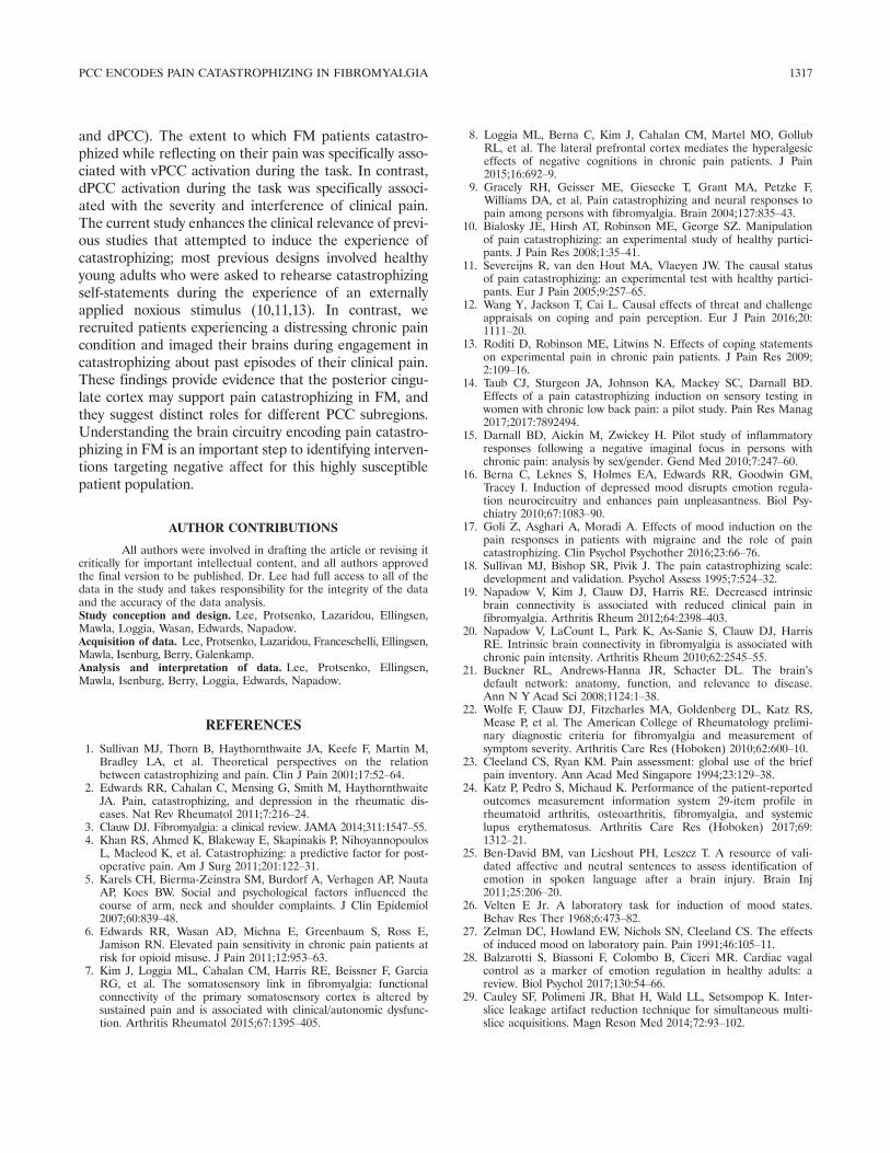
with the total CAQCAT score (r = 0.38, P < 0.05), whiledPCC activation during CATstatements was positively cor-related with the BPI subscales (r = 0.38, P < 0.05 for bothseverity and interference) (Figure 4). No other brain ROIshowed significant correlation with these clinical/psycho-metric measures (e.g., for vPCC versus BPI severity sub-scale, r = 0.11, P = 0.57; for vPCC versus PCS score, r =0.20, P = 0.27; for vPCC versus PROMIS-29 anxiety sub-scale, r = 0.20, P = 0.30; for vPCC versus PROMIS-29depression subscale, r = 0.17, P = 0.37; for dPCC versusCAQCATscore, r = 0.28, P = 0.13).
DISCUSSION
Pain catastrophizing plays a substantial role in thepathophysiology of FM. In this study, we investigated theneural circuitry supporting pain catastrophizing in FM.Patients reflected on their experiences with pain-referentialcatastrophizing statements during a recent episode ofFM pain. These procedures allowed patients prone tocatastrophizing to engage in such ruminative cognitionswhile fMRI tracked brain activity. These FM patientsreported, on average, high PCS scores, with relatively
Figure 3. Brain responses during catastrophizing (CAT). A, Most of the clusters with positive contrast (more responses to CAT statements than toneutral [NEU] statements) in the cerebral cortex, including the ventral and dorsal posterior cingulate cortex (vPCC and dPCC, respectively), themedial prefrontal cortex (mPFC), and the angular gyrus/inferior parietal lobule (AnG/IPL), were within the boundary of the default mode net-work, as defined by the resting-state functional magnetic resonance imaging (fMRI) parcellation based on 1,000 subjects (yellow outlines) (see ref.30). B, The right cerebellum demonstrated significant positive contrast (more responses to CAT statements than to NEU statements). Values arethe mean � SEM. L = left hemisphere; R = right hemisphere.
PCC ENCODES PAIN CATASTROPHIZING IN FIBROMYALGIA 1313

high interpatient variability. Compared to neutral state-ments, reflection on pain catastrophizing statements wasencoded by activation of the ventral (posterior) and dor-sal (anterior) posterior cingulate cortex (vPCC anddPCC) in addition to other, mainly DMN, brain regions.Importantly, the vPCC and dPCC were the only brainareas that showed a positive correlation between catas-trophizing-related activation (CAT-NEU statements)and clinical measures of catastrophizing and FM pain.Specifically, patients who found the catastrophizingstatements most applicable while reflecting on their painalso showed greater vPCC activation. In addition, theseverity and interference of FM clinical pain (i.e., theBPI score) were associated with dPCC activation. Takentogether, these findings suggest that the posterior cingu-late cortex encodes pain catastrophizing in FM.
While previous neuroimaging studies have ex-plored how catastrophizing influences brain response toevoked pain stimuli, our study directly investigated thebrain activity supporting catastrophizing cognitions inFM patients. The striking involvement of multipleDMN regions in pain catastrophizing is consistent withprevious research. For instance, induced negative mood(using depressing music and visual lexical cues) acti-vates the mPFC and increases the PCC response toevoked pain stimuli in healthy adults (16). In fact, pre-vious research has consistently shown reduced DMN
deactivation in chronic pain patients in response to arange of externally focused tasks (31,32). Our resultssuggest that reduced PCC deactivation in chronic painpatients may be due to ongoing catastrophizing-associated activity in this brain area while processingpain-related external stimuli.
We found not only greater brain activation in thevPCC in response to catastrophizing about FM pain butalso a significant association between vPCC activity andthe CAQ score. The PCS score was positively correlatedwith the CAQ score; hence, patients who reported greatertrait catastrophizing also reported greater applicabilityand endorsement of catastrophizing statements to theirown pain during recall. In turn, greater applicability ofcatastrophizing statements was associated with greatervPCC activation, closely linking this subregion of the pos-terior cingulate cortex to the catastrophizing state. ThevPCC is a cardinal node of the DMN (21) and has beenstrongly linked with self-referential cognition, attentionalfocus, and arousal (33). The vPCC has been differentiatedfrom the more anterior dPCC subregion based on cytoar-chitecture (34) and connectivity to canonical brain net-works. Specifically, while both PCC subregions showstrong connectivity to the DMN, the dPCC also showsgreater connectivity to dorsal attention, central executive,and even salience networks (35), suggesting that thedPCC also plays a broader role in attentional focus,
Table 2. Brain response on functional magnetic resonance imaging during catastrophizing task*
Size, mm3
Location, mm†
Z score
Z score, mean � SD
X Y Z CAT statements NEU statements
CAT statements > NEU statementsCerebellum, right hemisphere 21,448 32 �74 �38 5.26 1.79 � 1.51 0.04 � 1.22dPCC, left hemisphere 2,576 �2 �20 36 5.10 0.64 � 1.66 �1.16 � 1.93vPCC, left hemisphere 6,848 �4 �42 26 4.95 1.32 � 1.83 �0.90 � 2.32PC, left hemisphere 6,848 �4 �72 38 4.65 1.34 � 2.36 �0.45 � 2.90Angular gyrus, left hemisphere 10,512 �44 �62 36 4.43 0.77 � 1.79 �0.60 � 2.05Pre-SMA/SMA, left hemisphere 6,440 �14 20 62 4.02 1.33 � 1.55 0.27 � 1.78vlPFC, left hemisphere 1,984 �50 22 0 4.27 2.78 � 2.06 1.38 � 1.68dlPFC, left hemisphere 6,240 �20 56 28 4.00 1.03 � 1.68 �0.39 � 1.56mPFC, left hemisphere 6,240 �12 54 10 3.75 �0.02 � 1.33 �1.17 � 1.60
CAT statements < NEU statementsMTL, left hemisphere 30,248 �34 �30 �20 �5.21 �0.96 � 1.37 1.26 � 2.06IPL, left hemisphere 21,128 �28 �70 36 �4.99 2.59 � 2.94 4.86 � 3.32IPL, right hemisphere 14,576 42 �76 26 �4.05 �1.60 � 2.55 �0.55 � 2.29SPL, left hemisphere 21,128 �16 �56 58 �3.75 �0.69 � 1.99 0.32 � 2.08SPL, right hemisphere 14,576 12 �60 58 �3.87 �1.02 � 2.44 0.31 � 2.38ITG, left hemisphere 30,248 �46 �54 �14 �5.73 1.80 � 2.57 3.87 � 3.05ITG, right hemisphere 6,312 60 �60 �14 �3.80 �0.74 � 1.25 0.37 � 1.45Premotor area, left hemisphere 7,504 �31 �4 66 �3.48 �0.07 � 1.31 0.66 � 1.36Premotor area, right hemisphere 6,288 28 14 58 �4.02 �1.23 � 1.89 0.04 � 1.94M1, left hemisphere 7,504 �42 �10 46 �3.48 0.68 � 1.39 1.47 � 1.78
* CAT = catastrophizing; NEU = neutral; dPCC = dorsal posterior cingulate cortex; vPCC = ventral posterior cingulate cortex; PC =precuneus; SMA = supplementary motor area; vlPFC = ventrolateral prefrontal cortex; dlPFC = dorsolateral prefrontal cortex; mPFC =medial prefrontal cortex; MTL = medial temporal lobe; IPL = inferior parietal lobule; SPL = superior parietal lobule; ITG = inferiortemporal gyrus; M1 = primary motor cortex.† By standard Montreal Neurological Institute space.
1314 LEE ET AL

modulating dynamic interactions between the DMN andheteromodal cognitive control and attention networks. Incontrast, the vPCC shows greater linkage to medial tem-poral lobe regions of the DMN (e.g., hippocampus) andmay thus play a greater role in self-referential cognitionand autobiographical memory (33).
Interestingly, previous neuroimaging studies havelinked PCC neurophysiology with trait pain catastrophiz-ing, as assessed by the PCS questionnaire. For instance,Fayed et al used magnetic resonance spectroscopy andfound increased combined levels of glutamate and glu-tamine (Glx) in FM for a PCC subregion consistent withthe vPCC, suggesting increased excitatory neurotransmis-sion in this area (36). Importantly, greater levels of Glxwere positively correlated with greater trait PCS scores.Another study noted that increased connectivity betweenthe vPCC and mPFC in chronic pain patients was stronglyassociated with trait PCS rumination about pain (37).Similarly, fMRI data collected during a self-appraisal taskshowed not only greater activation in the PCC, IPL, and
mPFC during the self-appraisal condition but also greaterPCC activation in depressed individuals compared tohealthy controls (38).
These findings suggest that the trait tendency forpain-associated rumination and catastrophizing leads toincreased vPCC connectivity to other DMN areas andstems from or even induces increased excitatory neuro-transmission in the vPCC subregion. Thus, we suggestthat such altered vPCC neurophysiology may be main-tained by ongoing vPCC activity while engaged in self-referential rumination (i.e., the pain catastrophizingstate). Interestingly, while PCS and CAQ scores werehighly correlated with each other (r = 0.81) in our study,the PCS score was not associated with vPCC activationin response to our CAT task, suggesting that greater acti-vation (compared to neutral statements) is more of astate phenomenon, while altered connectivity and Glxlevels for the vPCC may reflect more stable trait catas-trophizing. Additional support for a phasic response isprovided by the lack of a post-run increase in general
Figure 4. Regions from the catastrophizing–neutral (CAT-NEU) statement difference map demonstrating an association with clinical/psychometricmeasures. The response of the dorsal posterior cingulate cortex (dPCC) to CATstatements was positively correlated with clinical fibromyalgia pain sever-ity on the Brief Pain Inventory (BPI), and the response of the ventral posterior cingulate cortex (vPCC) to CATstatements was positively correlated withratings of catastrophizing applicability (i.e., the CAT Applicability Questionnaire [CAQ] score). fMRI = functional magnetic resonance imaging. Colorfigure can be viewed in the online issue, which is available at http://onlinelibrary.wiley.com/doi/10.1002/art.40507/abstract.
PCC ENCODES PAIN CATASTROPHIZING IN FIBROMYALGIA 1315
anxiety levels, which suggests limited carryover and inde-pendent cognitive/affective processing during distinctstatement blocks. Such independent processing allowsfor accurate GLM modeling with fMRI block design, aspatients likely engaged in catastrophizing during theCAT statement blocks (as supported by elevated psy-chophysiologic responses), but not in subsequent state-ment blocks or post-run ratings.
We also found that dPCC activity, which was in-creased during CAT statements compared to NEU state-ments, was positively correlated with FM pain severity andinterference scores. This association suggests a differentialfunction of the dPCC, compared to the vPCC, duringcatastrophizing. PCC subregions consistent with the dPCCcluster we identified were functionally connected not onlyto other DMN regions but also to regions of central execu-tive and even sensorimotor networks (33). Similarly, Vogtet al have also shown heterogeneity of the PCC based oncytoarchitecture and resting glucose metabolism of PCCsubregions, suggesting that the vPCC plays an importantrole in self-monitoring, while the dPCC interacts morewith the cingulate motor area and is related to motor andnociceptive information processing (34).
Thus, the dPCC is a key DMN node that may facil-itate communication with other intrinsic brain networks(e.g., sensorimotor and higher cognitive processing networks)and is therefore well positioned to link self-referentialattentional focus with clinical pain perception. Thishypothesis is supported by our results demonstrating thatgreater clinical pain perception in FM patients wasspecifically associated with greater dPCC activation dur-ing catastrophizing. In fact, the dPCC may prove to be afuture target for pain modulation, as reducing activityduring catastrophizing may be critical to break the link-age between engaging in negative, ruminative thoughtand increased clinical pain perception in chronic painpatients. Several groups have stressed the importance ofreducing catastrophizing to better manage FM pain (39–43), and dPCC response may be a viable surrogate andeven a predictive brain imaging marker to evaluate thesuccess of individualized interventions that target catas-trophizing, such as cognitive–behavioral therapy (39).Furthermore, differential dorsal and ventral PCC rolescan also aid self-regulation fMRI neurofeedback designfor FM pain, as several previous studies have shown thatthe PCC can be modulated using real-time neurofeed-back by focusing on the cognitive process of self-referen-tial experience (to increase fMRI activity [44]) andmeditation (to decrease activity [45]).
Overall, the brain regions activated during thecatastrophizing task support both cognitive and affec-tive experience, as the PCC has commonly been linked
with self-referential cognition, while the mPFC is acti-vated by negative mood induction (16), and its connec-tivity with the PCC is increased in conjunction withrumination about chronic pain (37). Increased IPLactivity has also been reported in response to painfulpressure stimulation in FM patients (9) and has alsobeen shown to be involved in emotion processing (46).Cerebellum (Crus I and II, posterior cerebellum, higherfMRI signal increase during CAT statements than dur-ing NEU statements) activity and connectivity (47)have also been related to the cognitive (e.g., languageand executive) function (48). Collectively, our findingsare likely related to both cognitive and affective aspectsof pain catastrophizing. Furthermore, the significantlygreater psychophysiologic (HR increases) response dur-ing CAT statement blocks compared to NEU statementblocks suggests greater cognitive/affective engagementby patients with the catastrophizing task (49).
Limitations to our study should also be noted. Forinstance, the neutral statements chosen as a control in thisexperiment were not explicitly self-referential, as opposedto the pain catastrophizing statements. While this differ-ence likely played a role in DMN targeting in our fMRIresults, the association between activity in specific DMNsubregions (i.e., the PCC) and both CAQ and pain severitydemonstrates that DMN regions did not simply encode theself-referential aspects of the CATstatements but were alsolinked with FM patients’ catastrophizing and clinical painlevels. Moreover, we were not able to use self-referentialcontrol “neutral” statements, as any self-referential state-ments would be susceptible to negatively valenced interpre-tation by patients prone to catastrophizing. Anotherlimitation was that we did not have direct measures ofpatients’ re-experienced pain catastrophizing during CATstatement blocks, as we did not want to encroach on theirself-referential rumination with thought probe ratings. Weinstead used cardioautonomic psychophysiologic responseas an objective proxy for cognitive/emotional engagementin the task. Additionally, our design did not allow for anexplicit separation of pain catastrophizing from generalnegative affect, as these constructs are known to be closelylinked. Finally, while our brain–behavioral correlationanalysis approach risked false-positive results by notcontrolling for multiple comparisons, we note that false-negative error is an important consideration in novelstudies for specific areas of research (50). Hence, futurestudies will attempt to independently replicate thesefindings, specifying a priori ROI-based hypotheses andincluding additional control conditions.
In conclusion, our study found that pain catastro-phizing led FM patients to activate the ventral (posterior)and dorsal (anterior) posterior cingulate cortex (the vPCC
1316 LEE ET AL
and dPCC). The extent to which FM patients catastro-phized while reflecting on their pain was specifically asso-ciated with vPCC activation during the task. In contrast,dPCC activation during the task was specifically associ-ated with the severity and interference of clinical pain.The current study enhances the clinical relevance of previ-ous studies that attempted to induce the experience ofcatastrophizing; most previous designs involved healthyyoung adults who were asked to rehearse catastrophizingself-statements during the experience of an externallyapplied noxious stimulus (10,11,13). In contrast, werecruited patients experiencing a distressing chronic paincondition and imaged their brains during engagement incatastrophizing about past episodes of their clinical pain.These findings provide evidence that the posterior cingu-late cortex may support pain catastrophizing in FM, andthey suggest distinct roles for different PCC subregions.Understanding the brain circuitry encoding pain catastro-phizing in FM is an important step to identifying interven-tions targeting negative affect for this highly susceptiblepatient population.
AUTHOR CONTRIBUTIONS
All authors were involved in drafting the article or revising itcritically for important intellectual content, and all authors approvedthe final version to be published. Dr. Lee had full access to all of thedata in the study and takes responsibility for the integrity of the dataand the accuracy of the data analysis.Study conception and design. Lee, Protsenko, Lazaridou, Ellingsen,Mawla, Loggia, Wasan, Edwards, Napadow.Acquisition of data. Lee, Protsenko, Lazaridou, Franceschelli, Ellingsen,Mawla, Isenburg, Berry, Galenkamp.Analysis and interpretation of data. Lee, Protsenko, Ellingsen,Mawla, Isenburg, Berry, Loggia, Edwards, Napadow.
REFERENCES
1. Sullivan MJ, Thorn B, Haythornthwaite JA, Keefe F, Martin M,Bradley LA, et al. Theoretical perspectives on the relationbetween catastrophizing and pain. Clin J Pain 2001;17:52–64.
2. Edwards RR, Cahalan C, Mensing G, Smith M, HaythornthwaiteJA. Pain, catastrophizing, and depression in the rheumatic dis-eases. Nat Rev Rheumatol 2011;7:216–24.
3. Clauw DJ. Fibromyalgia: a clinical review. JAMA 2014;311:1547–55.4. Khan RS, Ahmed K, Blakeway E, Skapinakis P, Nihoyannopoulos
L, Macleod K, et al. Catastrophizing: a predictive factor for post-operative pain. Am J Surg 2011;201:122–31.
5. Karels CH, Bierma-Zeinstra SM, Burdorf A, Verhagen AP, NautaAP, Koes BW. Social and psychological factors influenced thecourse of arm, neck and shoulder complaints. J Clin Epidemiol2007;60:839–48.
6. Edwards RR, Wasan AD, Michna E, Greenbaum S, Ross E,Jamison RN. Elevated pain sensitivity in chronic pain patients atrisk for opioid misuse. J Pain 2011;12:953–63.
7. Kim J, Loggia ML, Cahalan CM, Harris RE, Beissner F, GarciaRG, et al. The somatosensory link in fibromyalgia: functionalconnectivity of the primary somatosensory cortex is altered bysustained pain and is associated with clinical/autonomic dysfunc-tion. Arthritis Rheumatol 2015;67:1395–405.
8. Loggia ML, Berna C, Kim J, Cahalan CM, Martel MO, GollubRL, et al. The lateral prefrontal cortex mediates the hyperalgesiceffects of negative cognitions in chronic pain patients. J Pain2015;16:692–9.
9. Gracely RH, Geisser ME, Giesecke T, Grant MA, Petzke F,Williams DA, et al. Pain catastrophizing and neural responses topain among persons with fibromyalgia. Brain 2004;127:835–43.
10. Bialosky JE, Hirsh AT, Robinson ME, George SZ. Manipulationof pain catastrophizing: an experimental study of healthy partici-pants. J Pain Res 2008;1:35–41.
11. Severeijns R, van den Hout MA, Vlaeyen JW. The causal statusof pain catastrophizing: an experimental test with healthy partici-pants. Eur J Pain 2005;9:257–65.
12. Wang Y, Jackson T, Cai L. Causal effects of threat and challengeappraisals on coping and pain perception. Eur J Pain 2016;20:1111–20.
13. Roditi D, Robinson ME, Litwins N. Effects of coping statementson experimental pain in chronic pain patients. J Pain Res 2009;2:109–16.
14. Taub CJ, Sturgeon JA, Johnson KA, Mackey SC, Darnall BD.Effects of a pain catastrophizing induction on sensory testing inwomen with chronic low back pain: a pilot study. Pain Res Manag2017;2017:7892494.
15. Darnall BD, Aickin M, Zwickey H. Pilot study of inflammatoryresponses following a negative imaginal focus in persons withchronic pain: analysis by sex/gender. Gend Med 2010;7:247–60.
16. Berna C, Leknes S, Holmes EA, Edwards RR, Goodwin GM,Tracey I. Induction of depressed mood disrupts emotion regula-tion neurocircuitry and enhances pain unpleasantness. Biol Psy-chiatry 2010;67:1083–90.
17. Goli Z, Asghari A, Moradi A. Effects of mood induction on thepain responses in patients with migraine and the role of paincatastrophizing. Clin Psychol Psychother 2016;23:66–76.
18. Sullivan MJ, Bishop SR, Pivik J. The pain catastrophizing scale:development and validation. Psychol Assess 1995;7:524–32.
19. Napadow V, Kim J, Clauw DJ, Harris RE. Decreased intrinsicbrain connectivity is associated with reduced clinical pain infibromyalgia. Arthritis Rheum 2012;64:2398–403.
20. Napadow V, LaCount L, Park K, As-Sanie S, Clauw DJ, HarrisRE. Intrinsic brain connectivity in fibromyalgia is associated withchronic pain intensity. Arthritis Rheum 2010;62:2545–55.
21. Buckner RL, Andrews-Hanna JR, Schacter DL. The brain’sdefault network: anatomy, function, and relevance to disease.Ann N Y Acad Sci 2008;1124:1–38.
22. Wolfe F, Clauw DJ, Fitzcharles MA, Goldenberg DL, Katz RS,Mease P, et al. The American College of Rheumatology prelimi-nary diagnostic criteria for fibromyalgia and measurement ofsymptom severity. Arthritis Care Res (Hoboken) 2010;62:600–10.
23. Cleeland CS, Ryan KM. Pain assessment: global use of the briefpain inventory. Ann Acad Med Singapore 1994;23:129–38.
24. Katz P, Pedro S, Michaud K. Performance of the patient-reportedoutcomes measurement information system 29-item profile inrheumatoid arthritis, osteoarthritis, fibromyalgia, and systemiclupus erythematosus. Arthritis Care Res (Hoboken) 2017;69:1312–21.
25. Ben-David BM, van Lieshout PH, Leszcz T. A resource of vali-dated affective and neutral sentences to assess identification ofemotion in spoken language after a brain injury. Brain Inj2011;25:206–20.
26. Velten E Jr. A laboratory task for induction of mood states.Behav Res Ther 1968;6:473–82.
27. Zelman DC, Howland EW, Nichols SN, Cleeland CS. The effectsof induced mood on laboratory pain. Pain 1991;46:105–11.
28. Balzarotti S, Biassoni F, Colombo B, Ciceri MR. Cardiac vagalcontrol as a marker of emotion regulation in healthy adults: areview. Biol Psychol 2017;130:54–66.
29. Cauley SF, Polimeni JR, Bhat H, Wald LL, Setsompop K. Inter-slice leakage artifact reduction technique for simultaneous multi-slice acquisitions. Magn Reson Med 2014;72:93–102.
PCC ENCODES PAIN CATASTROPHIZING IN FIBROMYALGIA 1317
30. Yeo BT, Krienen FM, Sepulcre J, Sabuncu MR, Lashkari D,Hollinshead M, et al. The organization of the human cerebralcortex estimated by intrinsic functional connectivity. J Neurophys-iol 2011;106:1125–65.
31. Baliki MN, Geha PY, Apkarian AV, Chialvo DR. Beyond feeling:chronic pain hurts the brain, disrupting the default-mode networkdynamics. J Neurosci 2008;28:1398–403.
32. Weissman-Fogel I, Moayedi M, Tenenbaum HC, Goldberg MB,Freeman BV, Davis KD. Abnormal cortical activity in patientswith temporomandibular disorder evoked by cognitive and emo-tional tasks. Pain 2011;152:384–96.
33. Leech R, Sharp DJ. The role of the posterior cingulate cortex incognition and disease. Brain 2014;137:12–32.
34. Vogt BA, Vogt L, Laureys S. Cytology and functionally correlatedcircuits of human posterior cingulate areas. Neuroimage 2006;29:452–66.
35. Leech R, Kamourieh S, Beckmann CF, Sharp DJ. Fractionatingthe default mode network: distinct contributions of the ventraland dorsal posterior cingulate cortex to cognitive control. J Neu-rosci 2011;31:3217–24.
36. Fayed N, Andres E, Rojas G, Moreno S, Serrano-Blanco A, RocaM, et al. Brain dysfunction in fibromyalgia and somatization dis-order using proton magnetic resonance spectroscopy: a controlledstudy. Acta Psychiatr Scand 2012;126:115–25.
37. Kucyi A, Moayedi M, Weissman-Fogel I, Goldberg MB, FreemanBV, Tenenbaum HC, et al. Enhanced medial prefrontal-defaultmode network functional connectivity in chronic pain and itsassociation with pain rumination. J Neurosci 2014;34:3969–75.
38. Davey CG, Breakspear M, Pujol J, Harrison BJ. A brain modelof disturbed self-appraisal in depression. Am J Psychiatry 2017;174:895–903.
39. Lazaridou A, Kim J, Cahalan CM, Loggia ML, Franceschelli O,Berna C, et al. Effects of cognitive-behavioral therapy (cbt) onbrain connectivity supporting catastrophizing in fibromyalgia. ClinJ Pain 2017;33:215–21.
40. Edwards RR, Bingham CO III, Bathon J, Haythornthwaite JA.Catastrophizing and pain in arthritis, fibromyalgia, and otherrheumatic diseases. Arthritis Rheum 2006;55:325–32.
41. Burns JW, Day MA, Thorn BE. Is reduction in pain catastrophiz-ing a therapeutic mechanism specific to cognitive-behavioral ther-apy for chronic pain? Transl Behav Med 2012;2:22–9.
42. Mansell G, Kamper SJ, Kent P. Why and how back pain interven-tions work: what can we do to find out? Best Pract Res ClinRheumatol 2013;27:685–97.
43. Tran ST, Guite JW, Pantaleao A, Pfeiffer M, Myer GD, Sil S,et al. Preliminary outcomes of a cross-site cognitive–behavioraland neuromuscular integrative training intervention for juvenilefibromyalgia. Arthritis Care Res (Hoboken) 2017;69:413–20.
44. Brewer JA, Garrison KA, Whitfield-Gabrieli S. What about the“self” is processed in the posterior cingulate cortex? Front HumNeurosci 2013;7:647.
45. Garrison KA, Scheinost D, Worhunsky PD, Elwafi HM, ThornhillTA, Thompson E, et al. Real-time fMRI links subjective experi-ence with brain activity during focused attention. Neuroimage2013;81:110–8.
46. Engelen T, de Graaf TA, Sack AT, de Gelder B. A causal role forinferior parietal lobule in emotion body perception. Cortex2015;73:195–202.
47. Krienen FM, Buckner RL. Segregated fronto-cerebellar circuitsrevealed by intrinsic functional connectivity. Cereb Cortex2009;19:2485–97.
48. Stoodley CJ, Schmahmann JD. Functional topography in thehuman cerebellum: a meta-analysis of neuroimaging studies. Neu-roimage 2009;44:489–501.
49. Weis PP, Herbert C. Bodily reactions to emotional words refer-ring to own versus other people’s emotions. Front Psychol 2017;8:1277.
50. Lee EC, Whitehead AL, Jacques RM, Julious SA. The statisticalinterpretation of pilot trials: should significance thresholds bereconsidered? BMC Med Res Methodol 2014;14:41.
DOI 10.1002/art.40658
Erratum
In the article by Saag et al in the January 2017 issue of Arthritis & Rheumatology (pages 203�212), text inthe fourth paragraph on page 211, regarding lesinurad prescribing information, was incorrect. The para-graph should have read “Of note, the US prescribing information for lesinurad recommends assessment ofrenal function prior to initiation of therapy, with initiation contraindicated in patients with creatinineclearance <45 ml/minute. Renal function should be assessed periodically after initiation of therapy, withdiscontinuation recommended if creatinine clearance persistently falls below 45 ml/minute. Lesinurad iscontraindicated in subjects with severe renal impairment, end-stage renal disease, kidney transplantrecipients, and patients on dialysis.”
We regret the error.
1318 LEE ET AL