emerging technologies in the cath & ep labs ep labs · case presentation 76yo, female, pvc from...
TRANSCRIPT
EP Labs
Emerging Technologies in the Cath & EP Labs
Mauricio Arruda, MDJohn R. Antonucci Master Clinician of Cardiovascular Innovation
Director of Electrophysiology Center
UH Harrington Heart & Vascular Institute
Associate Professor of Medicine
Case Western Reserve University
School of Medicine
Ohio-ACC 27th Annual Meeting
Disclosures
Consultant / Speaker / Advisor
• Biosense Webster
• Abbott
• Stereotaxis
No Investigational Technologies
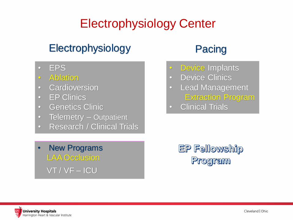
Electrophysiology Center
Electrophysiology Pacing
• EPS
• Ablation
• Cardioversion
• EP Clinics
• Genetics Clinic
• Telemetry – Outpatient
• Research / Clinical Trials
• New Programs
LAA Occlusion
VT / VF – ICU
• Device Implants
• Device Clinics
• Lead Management
Extraction Program
• Clinical Trials
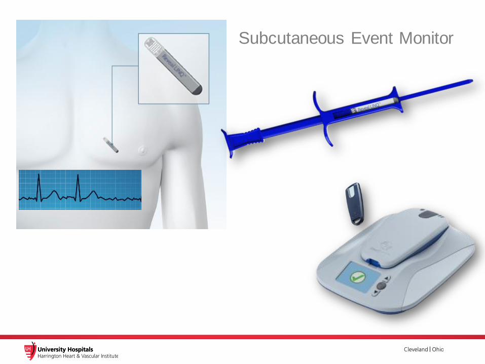
Subcutaneous Event Monitor
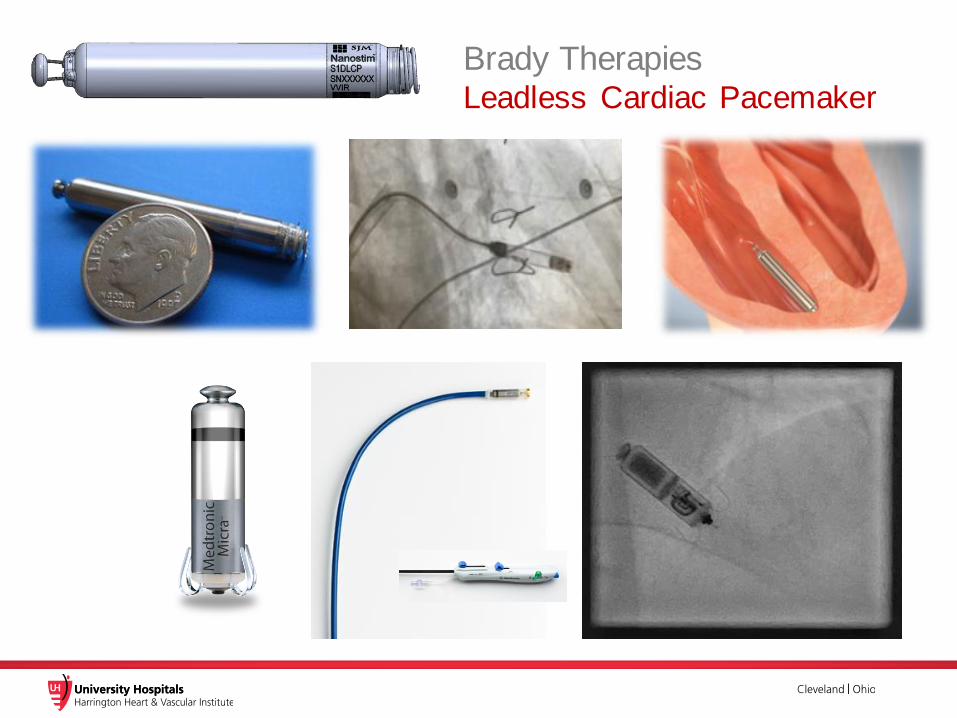
Brady TherapiesLeadless Cardiac Pacemaker
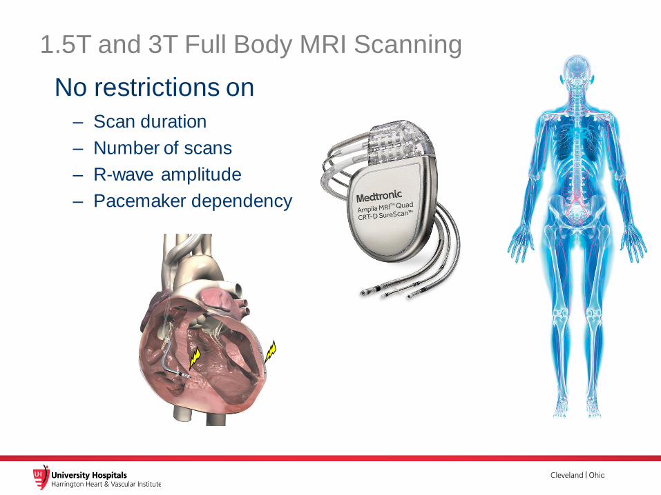
1.5T and 3T Full Body MRI Scanning
No restrictions on
– Scan duration
– Number of scans
– R-wave amplitude
– Pacemaker dependency
6
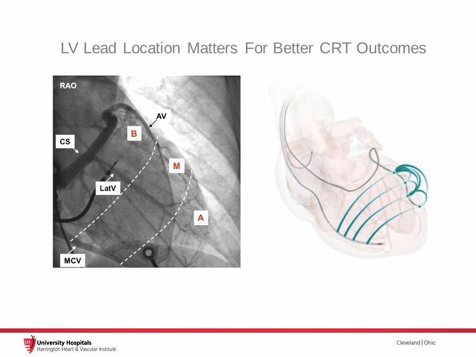
LV Lead Location Matters For Better CRT Outcomes

Subcutaneous ICD
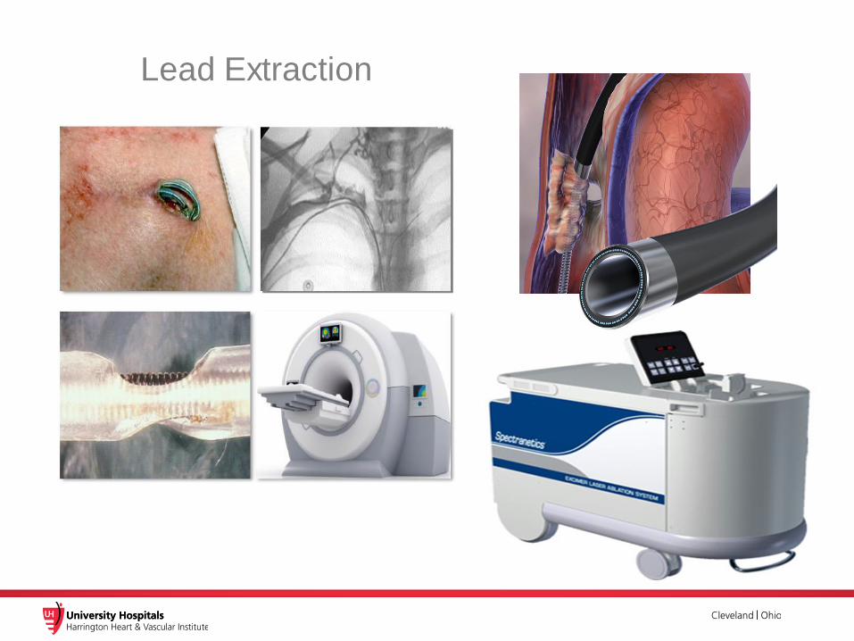
Lead Extraction
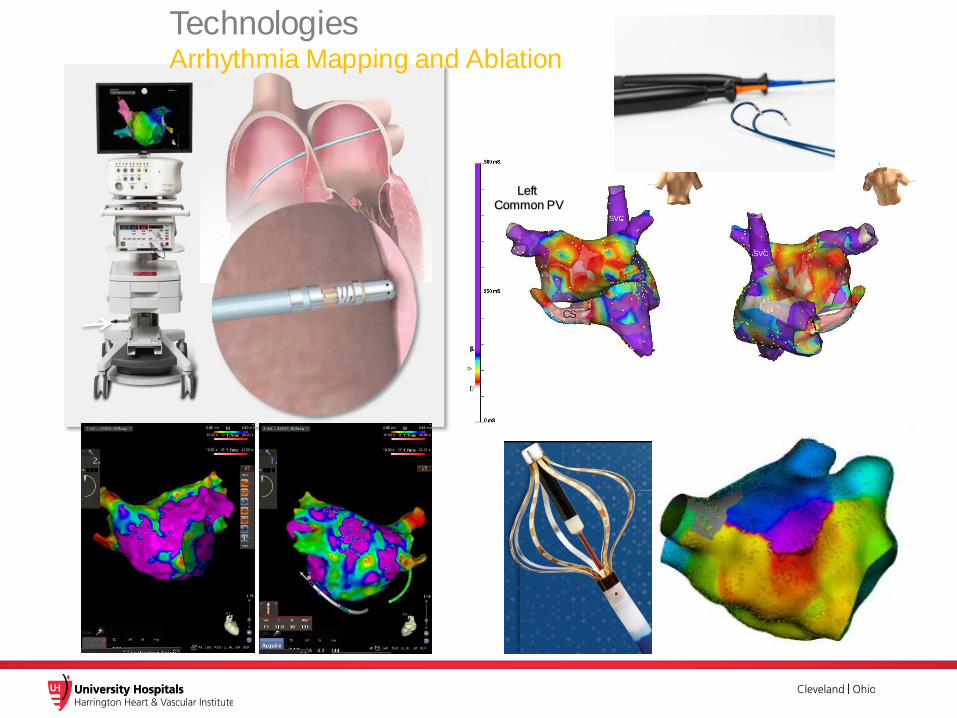
CARTO® SMARTTOUCH® Technology
CS
Left Common PV
SVC
SVC
Technologies Arrhythmia Mapping and Ablation

Robotic Magnetic Navigation Arrhythmia Mapping and Ablation
Catheter Ablation for Rhythm Control
Atrial FibrillationNon-Pharmacologic Management

1. Craig T. January, MD, PhD, FACC; L. Samuel Wann, MD, MACC, FAHA; Joseph S. Alpert, MD, FACC, FAHA; Hugh Calkins, MD, FACC,ET AL. 2014 AHA/ACC/HRS Guideline
for the Management of Patients With Atrial Fibrillation: Executive Summary. J Am Coll Cardiol. 2014;64(21):2246-2280
The 2014 AHA/ACC/HRS Guidelines for Afib Management provide the highest level
of recommendation (Class: 1; Level of Evidence: A) for catheter ablation as
treatment for drug-refractory, symptomatic paroxysmal Afib.
ClassRECOMMENDATION
THE STRONGEST RECOMENDATION
1 LevelLEVEL OF EVIDENCE
THE HIGHEST LEVEL OF EVIDENCE
A
Society Guidelines for AF Ablation

Rate vs. Rhythm ControlTimeline
AFFIRM StudyNo survival
advantage of
Rate vs. Rhythm control
2002 2006 20142012
Class
III
Level C
2001
Class II
Level B
Ionescu Mortality among
patients on rhythm
control gradually decreased relative
to rate control
Class I
Level
A*
Ghanbari NSR after RFCA is
associated with
60% reduction in CV mortality
Society Guidelines for Afib Ablation
*Class I Level A for PAF with no or minimal heart disease
2010
Wilber RFCA is superior in
efficacy and safety
to AAD
2004
Corley (AFFIRM sub-analysis)
NSR associated
with 47% reduction on risk of death
2003
DIAMOND Study
NSR leads to
significant reduction in
mortality
2002

✓ Paroxysmal AF 167pts ➢Catheter Ablation: 106➢AADs: 61
✓ PVI 100.0%, CTI 35.9%, Linear Ablation 22.3%, SVC 16.5%, Non-PV trigger 16.5%.
✓ Freedom from recurrence at 9 months.➢Ablation: 66%➢AAD: 16%Log-rank P<0.01
Antiarrhythmic Drugs vs. Ablation (PVI) for Paroxysmal AF
ThermoCool AF
Wilber et al. JAMA 2010;303:333-340.
PVI by Ablation
AAD
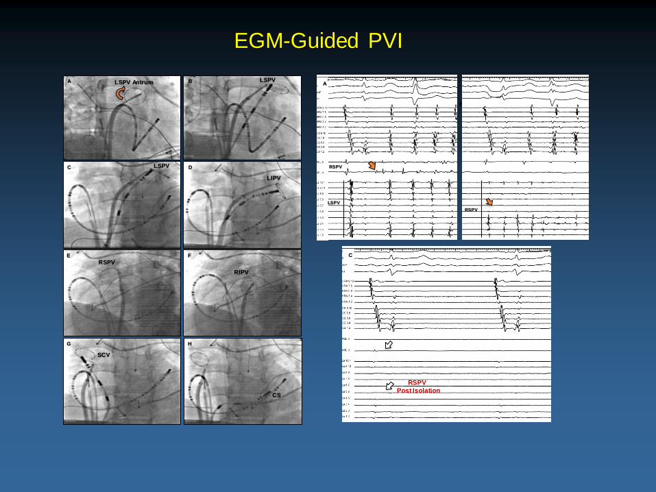
B
RSPV
Pre Isolation
A
RSPV
LSPV
RSPV
Post Isolation
C
A B
C D
E F
G H
LSPV Antrum LSPV
LSPV
LIPV
RSPV
RIPV
SCV
CS
EGM-Guided PVI
RSPV

CFAEs and Linear Ablation for Persistent AF
STAR AF II
Verma et al. N Engl J Med 2015;372:1812-22.
✓ Persistent AF 589 pts (PVI: 67, PVI + CFAE: 263, PVI + Roof + Mitral Line: 259)✓ No difference in long-term outcome between the 3 strategies.
Freedom from recurrence at 18 months (Log-rank P=0.15). ➢ PVI : 59%➢ PVI + CFAE : 49%➢ PVI + Roof + Mitral Line : 46%
Left Atrial Fibrotic Substrate Ablation
as an Adjunct to PVI
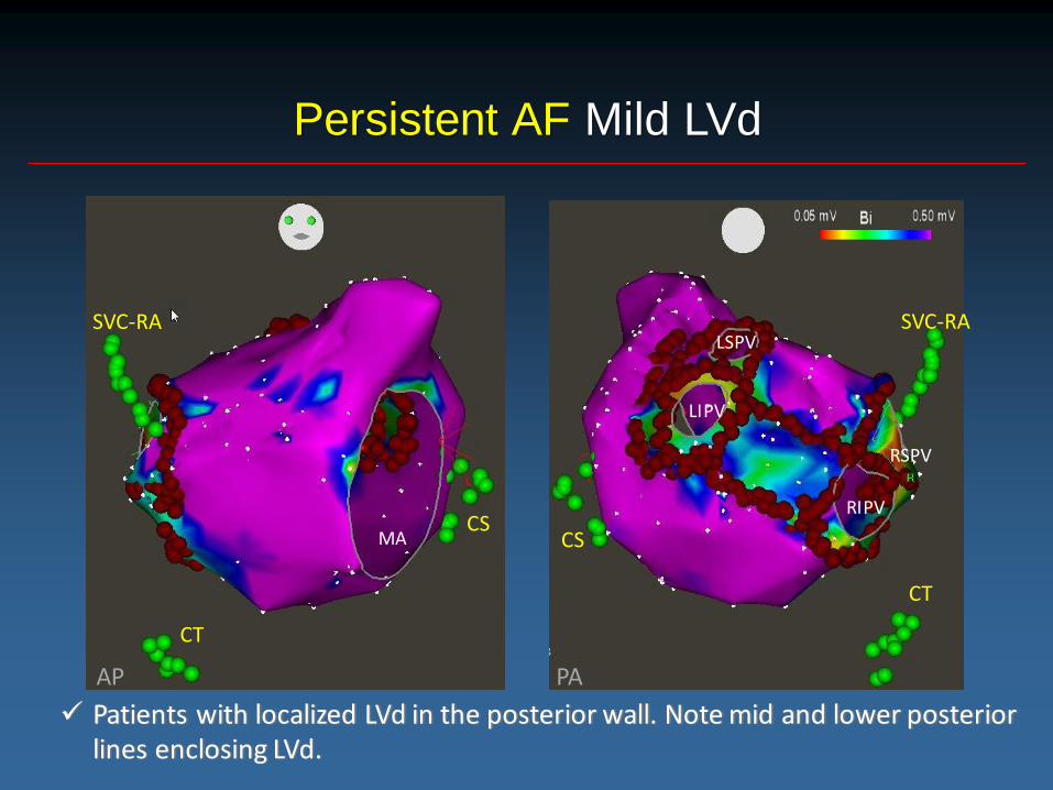
Persistent AF Mild LVd
LSPV
RSPV
RIPV
LIPV
SVC-RA
CT
CSCS
SVC-RA
CT
PAAP
MA
✓ Patients with localized LVd in the posterior wall. Note mid and lower posterior lines enclosing LVd.

Case Presentation
• 78yo, male Pt with persistent AF
• Miral valvuloplasty (2004)
• Broad low voltage area in the LA on Voltage map
AP PA

ⅡⅠ
V1
RA
ABL 1-2ABL 3-4
LSPV
aVF
CS

AF Termination during Ablation at the LA Roof
ⅡⅠ
V1
RA
ABL 1-2
LSPV
aVF
CS
ABL 3-4
PA

Ventricular Fibrotic Substrate Ablation
VT / VF
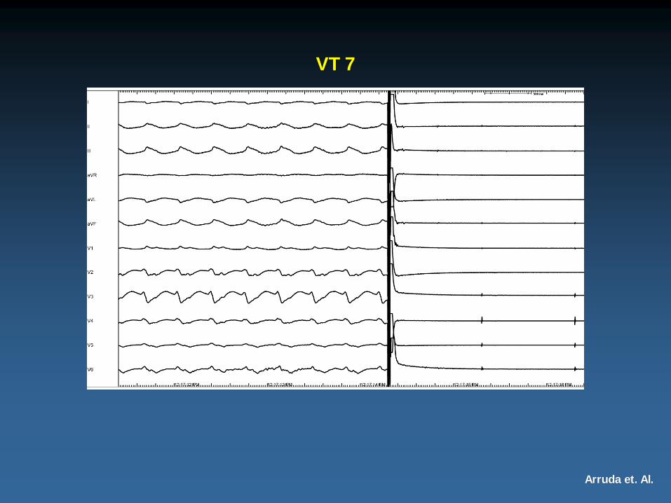
VT 7
Arruda et. Al.

Arruda et. Al.
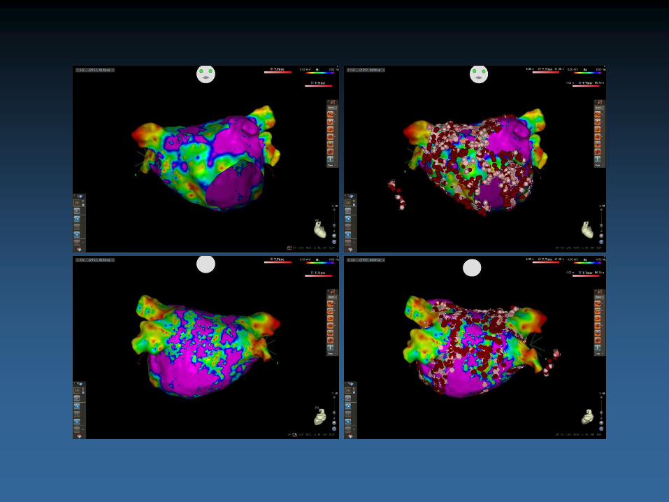
LV and RV Endocardial
Electroanatomic Voltage Mapping

Arruda et. Al.
Endocardial and Epicardial
Electroanatomic Voltage Mapping
Arruda et. Al.
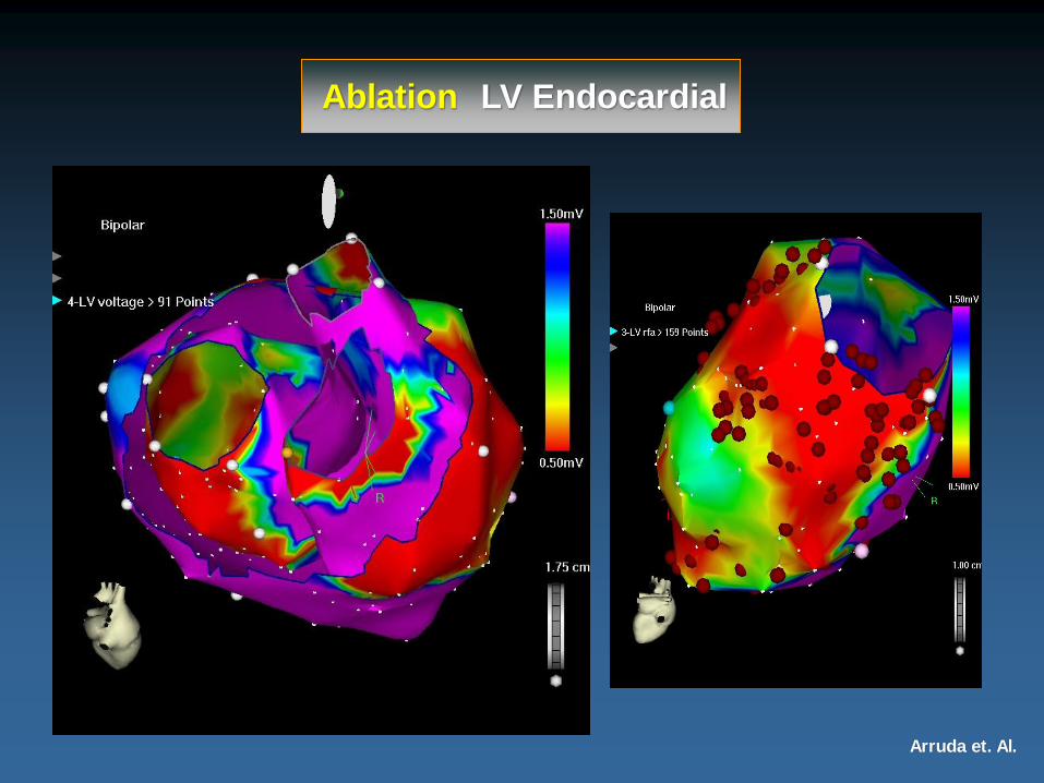
Ablation LV Endocardial
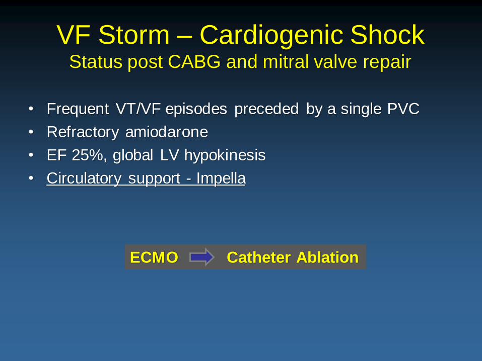
• Frequent VT/VF episodes preceded by a single PVC
• Refractory amiodarone
• EF 25%, global LV hypokinesis
• Circulatory support - Impella
VF Storm – Cardiogenic ShockStatus post CABG and mitral valve repair
ECMO Catheter Ablation

Monomorphic VT (RBBB Morphology and superior Axis)
changed to Fast Polymorphic VT
V1
V2
V3
V4
V5
V6
I
II
III
aVR
aVF
aVL
500ms

Monomorphic VT(RBBB Morphology and superior Axis)
changed to VF
V1
V2
V3
V4
V5
V6
I
II
III
aVR
aVF
aVL
500ms
DC Shock

Elimination of VT/VF After
Fibrotic Substrate Homogenization
RAO
LAO
LV Voltage map
RAO
LAO
Scar Homogenization
V1
V2
V3
V4
V5
V6
I
II
III
aVR
aVF
aVL
VT PM at LV septum
V1
V2
V3
V4
V5
V6
II
III
aVR
aVF
aVL

Intracardiac Echo (ICE)
In The EP Lab

ICE-Guided Pulmonary Vein Electrical Isolation

ViewFlex™ Xtra ICE Catheter
Left / Right
Anterior / Posterior
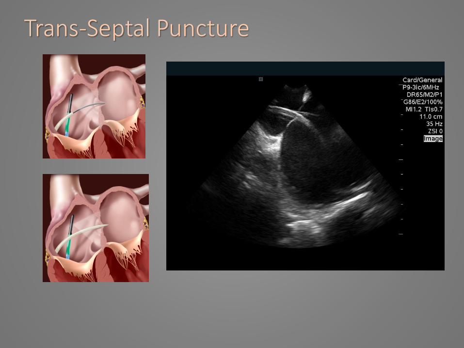
Trans-Septal Puncture


ICE Assessment of Catheter Contact
Case Presentation76yo, Female, PVC from Anterior Papillary Muscle

Clinical PVC RBBB / Inferior Axis Morphology
V1
V2
V3
V4
V5
V6
I
II
III
aVR
aVF
aVL
500ms

Activation Mapping at the APMPVCSR
-24
V1
III
aVF
RA
ABLd
p
CS
d
p

ICE Imaging to guide Ablation at Pappilary Muscle

V1
V2
V3
V4
V5
V6
I
II
III
aVR
aVF
aVL
V1
V2
V3
V4
V5
V6
I
II
III
aVR
aVF
aVL
500ms
PMPVC
Pace Mapping at the APM
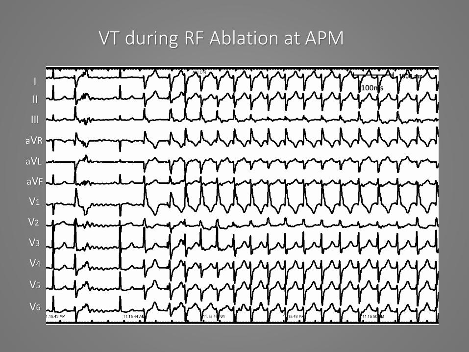
VT during RF Ablation at APM
V1
V2
V3
V4
V5
V6
I
II
III
aVR
aVF
aVL
100ms
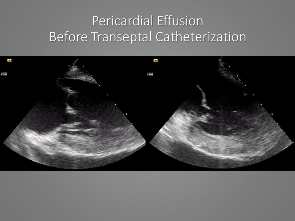
Pericardial Effusion Before Transeptal Catheterization

Pericardial Effusion After
Pericardiocentesis

ICE Imaging Miscellaneous

Saline Irrigation - tip of Ablation Catheter

RF Lesion Formation in the LA
During RFA After RFA

Left Atrial Appendage Occlusion Stroke Prevention
Atrial FibrillationNon-Pharmacologic Management

LAA Closure for Stroke Prevention in NVAF
LAA
STROKE
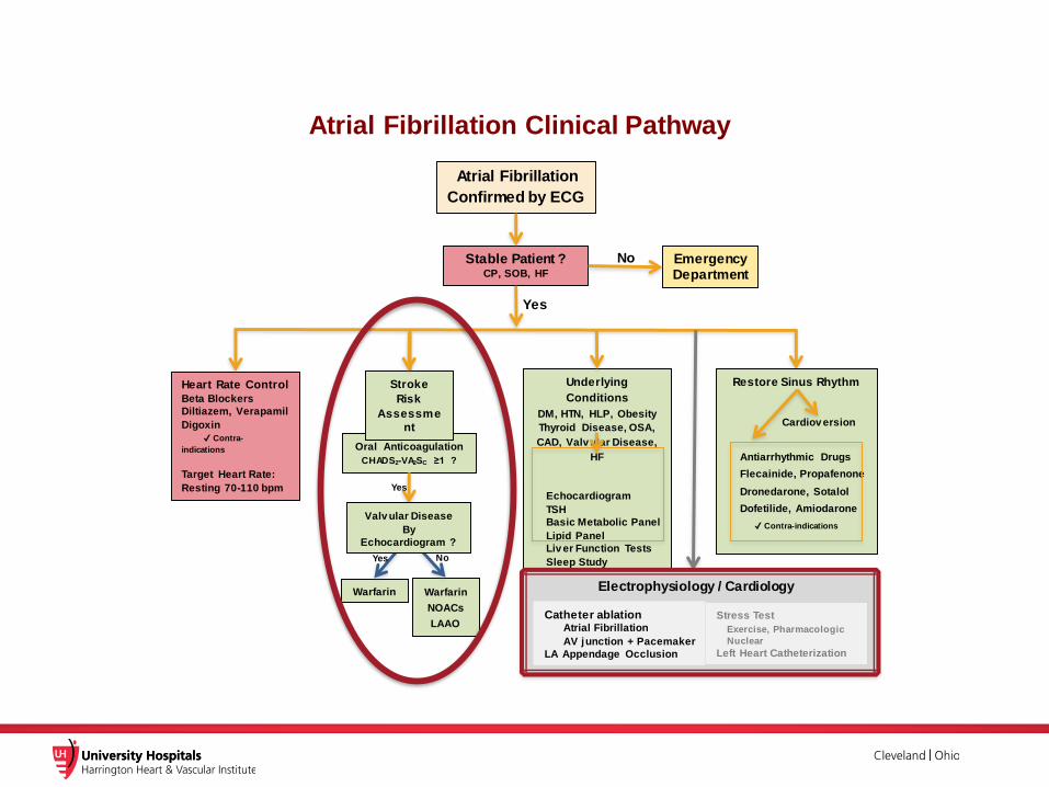
Atrial Fibrillation Clinical Pathway
Atrial Fibrillation
Confirmed by ECG
Stable Patient ? CP, SOB, HF
Oral Anticoagulation
CHADS2-VA2SC ≥1 ?
Warfarin Warfarin
NOACs
LAAO
No
Underlying
Conditions
DM, HTN, HLP, Obesity
Thyroid Disease, OSA,
CAD, Valv ular Disease,
HF
Echocardiogram
TSH
Basic Metabolic Panel
Lipid Panel
Liv er Function Tests
Sleep Study
EmergencyDepartment
Heart Rate Control
Beta Blockers
Diltiazem, Verapamil
Digoxin
✔ Contra-
indications
Target Heart Rate:
Resting 70-110 bpm
Yes
No
Yes
Yes
Restore Sinus Rhythm
Cardiov ersion
Antiarrhythmic Drugs
Flecainide, Propafenone
Dronedarone, Sotalol
Dofetilide, Amiodarone
✔ Contra-indications
Stroke
Risk
Assessme
nt
Stress Test
Exercise, Pharmacologic
Nuclear
Left Heart Catheterization
Catheter ablation Atrial Fibrillation
AV junction + Pacemaker
LA Appendage Occlusion
Electrophysiology / Cardiology
Valv ular Disease
By
Echocardiogram ?

53
LAA Closure Devices
Endocardial
• PLAATO
• Watchman
• ACP
• Amulet
• Coherex
• Prolipsis
• Occlutech
• PFM Medical
• Lifetech
• Cardia• Gore
• SentreHeart
• AEGIS
• AtriCure
• Medtronic
• Maquet
CE, FDACECE - US trial - starting
CE, FDA 510K
CE
Epicardial
Surgical
Cactus (30%) Windsock (19%)Chicken Wing (48%) Cauliflower (3%)
Di Biase et al. JACC 2012, 60: 531-538
LAA Anatomy - LAA were were distributed into 4 morphologies 932 AF patients

• Nitinol frame radially expands to maintain position in LAA
• 10 fixation anchors engage LAA tissue for stability and retention
• Polyethylene terephthalate (PET) membrane designed to block emboli from exiting the LAA
Anchors
160 Micron Membrane
Designed specifically for the left atrial appendage
WATCHMAN LAAC Device Overview

55
Assessment of LAA size for proper device choice
00 900
1350
22mm
20mm
20mm
21mm
LCX
450
a a
Tug test Color Flow around device20% Compression

WATCHMAN™ Device Endothelialization
Canine Model – 30 Day
Canine Model – 45 DayHuman Pathology - 9 Months Post-implant
(Non-device related death)
Images on file at Boston Scientif ic Corporation.
Results in animal models may not necessarily be indicat ive of clinical outcomes.

Procedural Success
Implant success defined as deployment and release of the device into the LAA; no leak ≥ 5 mm* The EWOLUTION Registry is a European prospectiv e registry which reflects CE Mark indications for use which differ from the FDA indications for use.
1 Boersma, L.et al. EHJ; published online Jan 2016 in press; 2 Reddy VY, Holmes DR, et al. JACC 2016; Article in press
~50% new operators ~70% new operators

AMPLATZER™
Amulet™
Device
Lobe▪ Inside the LAA neck
▪ Designed to conformto LAA anatomy
Stabilizing
Wires▪ Engage with LAA wall▪ Help hold the device in place
Waist▪ Maintains tension between lobe and disc
▪ Allows device to self-orient
Disc• Completely seal
at the orifice
“Amulet is an investigational device in the US and not approved by FDA for commercial distribution.”

Amplatzer™ Amulet™
Device Implant Procedure
Measure LAA orifice, landing zone, depth Deploy LOBE in landing zone
Deploy the DISC, to cover the ostium Release
1 2
3 4
“Amulet is an investigational device in the US and not approved by FDA for commercial distribution.”

Coherex WAVECREST LAA Closure Device

Permanent Ligation Approximation Closure and Exclusion
LARIAT® Suture Delivery Device
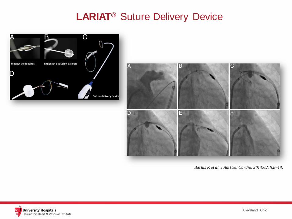
Bartus K et al. J Am Coll Cardiol 2013;62:108–18.
LARIAT® Suture Delivery Device

Thank You
Emerging Technologies in the Cath & EP Labs
Ohio-ACC 27th Annual Meeting