emerging roles for cysteine proteases in human
TRANSCRIPT

P1: sbs
August 28, 1956 22:38 Annual Reviews CHAPCHPT.DUN AR25-04
Annu. Rev. Physiol. 1997. 59:63–88Copyright c© 1997 by Annual Reviews Inc. All rights reserved
EMERGING ROLES FOR CYSTEINEPROTEASES IN HUMAN BIOLOGY
Harold A. Chapman, Richard J. Riese, and Guo-Ping ShiDepartment of Medicine, Brigham and Women’s Hospital, and Harvard MedicalSchool, Boston, Massachusetts 02115
KEY WORDS: thiol protease, cathepsin, lung, MHC class II, macrophage
ABSTRACT
Cysteine proteases have traditionally been viewed as lysosomal mediators of ter-minal protein degradation. However, recent findings refute this limited view andsuggest a more expanded role for cysteine proteases in human biology. Sev-eral newly discovered members of this enzyme class are regulated proteaseswith limited tissue expression, which implies specific roles in cellular physiol-ogy. These roles appear to include apoptosis, MHC class II immune responses,prohormone processing, and extracellular matrix remodeling important to bonedevelopment. The ability of macrophages and other cells to mobilize elastolyticcysteine proteases to their surfaces under specialized conditions may also lead toaccelerated collagen and elastin degradation at sites of inflammation in diseasessuch as atherosclerosis and emphysema. The development of inhibitors of spe-cific cysteine proteases promises to provide new drugs for modifying immunity,osteoporosis, and chronic inflammation.
INTRODUCTION
Proteases are enzymes that catalyze hydrolysis of amide bonds. Although pro-teins may undergo many reversible posttranslational modifications during theirlifespan, e.g. phosphorylation and allosteric transitions, proteolysis is irre-versible. Once proteins are hydrolyzed, the only means available for rebuildingthe intact molecule is to translate more mRNA. Based on the nature of pro-teolysis, it is not surprising that proteolytic enzymes have evolved to mediateprocesses that are themselves frequently irreversible: coagulation, digestion,maturation of cytokines and prohormones, apoptosis, and breakdown of in-tracellular proteins. Proteolysis is a ubiquitous mechanism the cell employs
630066-4278/97/0315-0063$08.00

P1: sbs
August 28, 1956 22:38 Annual Reviews CHAPCHPT.DUN AR25-04
64 CHAPMAN, RIESE & SHI
to regulate the function and fate of proteins (1, 2). Accordingly, the numberof proteases identified in and around cells is enormous, and many are vitalfor normal homeostasis. This is also true for the respiratory system. Sincethe demonstration of emphysema following intratracheal instillation of papainin experimental animals (3), much of what has been reported about proteasesand the respiratory system has centered on the potential for proteases to causedamage in the lungs and airways. However, proteases are as vital to normallung function as anywhere else. Indeed, the lung airways normally contain freeproteases and peptidases (urokinase, Factor VII, neutral endopeptidase), andlining cells and stromal cells of the lung depend on regulated protease activityfor their “housekeeping” functions, as well as for responses to the frequentinjurious insults to which this organ is subjected (4–6). Clearance of organicparticulates and microorganisms from the lung is dependent on intracellularproteases and occurs daily without any evident injury. Injury more often takesplace when proteases are unable to effect clearance, such as occurs after inhala-tion of inorganic dusts and cigarette smoke.
All proteases share in common the general mechanism of a nucleophilic at-tack on the carbonyl-carbon of an amide bond (7). This results in a generalacid-base hydrolytic process that disrupts the covalent bond. Different pro-teases utilize different strategies to generate the nucleophile and to juxtaposethe nucleophile with the targeted bond. These distinctions serve as a usefulclassification scheme, and on this basis proteases can be grouped into four ma-jor classes: serine, cysteine, aspartate, and metallo. The latter two groups ofenzymes utilize aspartate residues and heavy metals, respectively, to immobi-lize and polarize a water molecule so that the oxygen atom in water becomesthe nucleophile (8). Serine and cysteine proteases utilize their HO- and HS-side chains, respectively, directly as nucleophiles. Although not identical, thecatalytic mechanisms of serine and cysteine proteases are remarkably similar.In general, these enzymes are folded into two relatively large globular domainssurrounding a cleft containing the active site residues. Substrate entry into thecleft is a prerequisite for cleavage, and efficient entry is dictated by the struc-tural fit between the potential substrate and the topology of the cleft, a majordeterminant of enzyme specificity. The formation of a spatial fit between atargeted bond of the substrate and the active site nucleophile is obviously alsoa critical determinant of substrate specificity. Crystallographic analysis of sev-eral members of the serine and cysteine class enzymes reveals detailed structureof the active site regions and the importance of additional amino acids to thecatalytic mechanism (9, 10). In both serine and cysteine proteases, the forma-tion of an oxyanion or thiolate anion (the nucleophile), respectively, is criticalto catalysis, and the formation of these anions appears to be dependent on ionpair formation between the active site amino acid and neighboring basic amino

P1: sbs
August 28, 1956 22:38 Annual Reviews CHAPCHPT.DUN AR25-04
CATHEPSIN BIOLOGY 65
acids (histidine). Several recent reviews detailing the mechanism of catalysisby serine and cysteine proteases are available (2, 11).
This review focuses on the role of cysteine proteases in cellular physiology.These enzymes have been a major interest of this laboratory and many devel-opments have occurred within this class of enzymes in the last several years.Importantly, the elucidation of new members of the cysteine protease class ap-pears to be a preface to delineation of novel roles for these enzymes in humanbiology, affecting the function of the respiratory systems as well as other or-gans. A distinguishing feature of the newer proteases is their restricted tissueexpression and regulated behavior, which probably accounts for the fact thatmost were not identified by standard biochemical methods but, instead, requiredthe advent of RNA and DNA screening techniques for characterization. Cor-respondingly, a view of the biological role of cysteine proteases must take intoaccount the function of these new enzymes. The presence of regulated enzymeswith restricted tissue distribution implies specific cellular functions rather thansimply cooperative mediation of terminal protein degradation. This is an impor-tant change in the conceptual view of the role of cysteine proteases in humanbiology because, if true, therapeutic targeting of these enzymes could affectspecific changes in cell function without broad inhibition of lysosomal func-tion. Where possible, this review will attempt to highlight specific functionsfor cysteine proteases, even if these putative functions are based on preliminaryevidence, in the hope of stimulating further investigation and insight.
CLASSIFICATION OF CYSTEINE PROTEASES
Cysteine proteases can be grouped into two superfamilies: the family of en-zymes related to interleukin 1β converting enzyme (ICE), and the papain su-perfamily of cysteine proteases (12). Distinctive features of their structures andfunctions are summarized in Table 1. Although each superfamily of enzymesemploys an active site cysteine for nucleophilic attack, important evolutionaryand structural differences distinguish them. The ICE superfamily of enzymes,other than the active site cysteine itself, shares no sequence homology withthe papain superfamily (13). They are remarkable in their specificity for as-partate as the SI amino acid, an uncommon cleavage site among proteases.Their emerging role in inflammation and programmed cell death has been re-cently reviewed and is not discussed further here (14). The calpains are agroup of cytoplasmic cysteine proteases within the papain superfamily whoseactivity is strictly calcium dependent but whose protease domain is nonethelessvery much like that of papain. The calcium sensitivity results from the ances-tral fusion of a papain-type protease domain with a calmodulin-like domain(15, 16). These enzymes are implicated in limited proteolysis of a number of

P1: sbs
August 28, 1956 22:38 Annual Reviews CHAPCHPT.DUN AR25-04
66 CHAPMAN, RIESE & SHI
Table 1 Structural and functional features of human cysteine proteases
Enzyme Interleukin-1β convertingfamilya enzyme (ICE) Calpain Papain
Active site motifs 279–V I I IQ A C R G D S— –19N Q G C G S C W A F Sb—Members ICE m-calpain cathepsin B, H, L, S, O,
Cpp32, others µ-calpain K, others
Preferred –Y/D-V–A–D–X- -X-I/V-L/R–X –R/K–X–X (CAT B-like)c
cleavage sites –L/I—X–X- (CAT L-like)Location Cytoplasm Cytoplasm Endosomes/lysosomes
inner membranesFunction IL-1-β release Regulation of Digestion
membrane signaling Antigen presentationHormone processing
Apoptosis Matrix remodeling? Tumor invasion
aAn additional group of enzymes in the papain superfamily not listed in the table are the bleomycin hydrolases. Seetext for discussion.bSequence shown is for cathepsin B. See Figure 1 for sequence homologies among the cathepsins.cSubstrate specificity for the papain group of enzymes is determined primarily by amino acid preferences in the S2subsite rather than the S1 cleavage site (arrowheads). Cathepsin B-like enzymes accomodate basic amino acids into theS2 subsite and efficiently cleave proteins after Arg-Arg or Lys-Arg sequences. By contrast, cathepsin L-like enzymesstrongly prefer hydrophobic or branched chain amino acids in the S2 subsite. Only these enzymes are efficient elastases.
intracellular proteins in association with rises in intracellular calcium concen-tration. Protease activation appears to correlate with membrane binding andis followed quickly by autolysis. Although the exact physiological role ofthese enzymes is still being elucidated, the demonstration of limited cleavageof several regulatory proteins, such as protein kinase C, actin-binding proteins,and integrin cytoplasmic tails, by calpains, makes a regulatory role for theseenzymes in cellular signaling likely (16, 17). The discovery of tissue-specificcalpains, e.g. a muscle-specific calpain, has further opened the physiologicalpossibilities (18). Recent linkage of limb-girdle dystrophy with mutations inthis calpain underscore this point (19). Additional calpains are almost certain tobe forthcoming, along with a better view of their biological role. The structureand function of calpains including the newer enzymes are also the subject ofseveral recent reviews (15, 16).
A second group of enzymes in the papain superfamily, not listed in Table 1,are the bleomycin hydrolases (20). These enzymes were identified originallyas an activity in rabbit and bovine lung extracts that mediate bleomycin in-activation and were subsequently reported to protect human tumor cells frombleomycin toxicity (21, 22). Isolation and molecular cloning of the rabbit en-zyme demonstrated the activity to be due to a papain-type cysteine protease (20).
P1: sbs
August 28, 1956 22:38 Annual Reviews CHAPCHPT.DUN AR25-04
CATHEPSIN BIOLOGY 67
The enzyme appears to self-assemble into hexamers of its 50-kDa single chain,reminiscent of proteasome organization and, because there is no signal peptide,to localize to the cytoplasm. Recently, a yeast homologue of the mammalianenzyme was crystallized and found to contain both DNA-binding and papain-type motifs in each of its five chains, implying that DNA-binding and proteasefunctions of the enzyme are intertwined (23). Although identified on the basisof its bleomycin hydrolase activity, this enzyme appears to be the first exampleof a mammalian protease with DNA-binding and presumably transcriptionalregulatory functions. Further characterization of this activity and elucidationof other proteins with similar properties should determine whether this enzymeprovides a new paradigm for transcriptional regulation.
The papain family itself (the third enzyme group within the papain super-family) has been extensively studied, with over 80 distinct and complete entriesin sequence databases (12). Papain (fromCarica papaya) and, more recently,cathepsin B have been analyzed by X-ray crystallography and their functionalproperties have been examined (10, 24). Until recently, information about mam-malian members of the papain family has been more limited. The discovery ofmammalian papain-type cysteine proteases can now be divided roughly into twoeras. Prior to 1990, the known enzymes (cathepsins B, H, L, and S) were entirelycharacterized by standard protein isolation of enzyme activities and subsequentphysical characterization. Although bovine cathepsin S had been isolated as anenzyme activity, complete sequence data were available for only B, L, and H(25–29). These enzymes had been purified from adult solid organs, where theyconstitute the most abundant lysosomal enzymes. Perhaps, in part, becauseof their strong homology to papain (common meat tenderizer) and because ofthe long-standing view of lysosomes as terminal degradative organelles, theseenzymes had been viewed largely as collective mediators for terminal digestionof endocytized and endogenous proteins entering lysosomes (30). This was notunreasonable because nonspecific inhibitors of cysteine proteases have beenreported to inhibit up to 40% of total cellular protein turnover (31).
Recently, the techniques of molecular biology have been employed to in-vestigate papain-type cysteine proteases. What has emerged is at least fivenew human enzymes of the papain family and an evolving view of the roleof these enzymes in biology. In 1990, our laboratory utilized degenerate nu-cleotide primers spanning the highly conserved amino acid sequences within thecatalytic domains of known human cysteine proteases and reverse-transcribedRNA from human alveolar macrophages to search by polymerase chain am-plification for new cysteine protease sequences. With this technique we wereable to isolate partial cDNA sequences for all of the known human enzymes(cathepsins H, B, and L), as well as three new sequences. One of these, cathep-sin S, had previously been purified and partially sequenced (25), whereas the

P1: sbs
August 28, 1956 22:38 Annual Reviews CHAPCHPT.DUN AR25-04
68 CHAPMAN, RIESE & SHI
other two, now designated cathepsins K and F, had not been observed. Fullsequences for these enzymes were subsequently obtained by screening appro-priate cDNA libraries (32, 33). Wiederanders and colleagues independentlyobtained a full cDNA for cathepsin S (34). This technique was also appliedby other investigators to reveal additional human and rodent members of thepapain family (35, 36). Figure 1 summarizes the sequence alignment of theknown human enzymes (25–29, 32, 33, 36). There are almost certainly addi-tional sequences forthcoming. Another enzyme not listed in Table 1 (dipeptidylpeptidase I or cathepsin C) has been fully sequenced in rodents and found tobe a typical papain-type enzyme, albeit exhibiting only aminodipeptidase ac-tivity (37). To date no human sequence for this enzyme has been entered into adatabase. Dipeptidylpeptidase I is found in various myleoid cells and functionsas a processing enzyme for activation of several serine proteases (38).
Inspection of the sequence alignments reveals several interesting points:
1. All enzymes shown contain a signal peptide and a propiece, which is re-moved at maturation. This propiece is important because enzymes, e.g. cathep-sin B, expressed without the propiece are not properly folded and remain in-active (39). Moreover, the isolated propieces themselves inhibit their matureenzymes, suggesting that the propiece functions as a chaperone to permit properfolding and to block the active site cleft until the enzyme is in an activation en-vironment (40, 40a) . Interestingly, one of the newer sequences identified in ourlaboratory is a typical papain-type protease except that it lacks a characteristicsignal peptide (cathepsin F). Where this enzyme localizes and functions withincells will be interesting to explore.
2. Cathepsin B has an additional≈30-amino acid sequence inserted proximalto the active site histidine. In the crystal structure of cathepsin B, this sequenceloops over the active site cleft in the mature enzyme and restricts access ofpotential substrates (24). This probably accounts in part for the relativelyweak endoprotease activity of cathepsin B compared with other members ofthis family. By contrast, cathepsin B has particularly good carboxypeptidaseactivity. Three-dimensional modeling of cathepsin H also reveals a closedactive site cleft, which may in part account for its predominant function as anaminopeptidase (41).
3. Two regions of marked sequence similarity (denoted by ts) are evident inthe Figure 1. The proximal region surrounds the active site cysteine 25 and the
−−−−−−−−−−−−−−−−−−−−−−−−−−−−−−−−−−−−−−−−−−−−−−−−−−−−−−→Figure 1 Amino acid sequence alignment of the known human cysteine proteases. Conservedamino acid residues in the human sequences relevative to papain are denoted with an asterisk. Thedash indicates a gap relative to the papain sequence. Numbering shown is that for papain.

P1: sbs
August 28, 1956 22:38 Annual Reviews CHAPCHPT.DUN AR25-04
CATHEPSIN BIOLOGY 69
P1: sbs
August 28, 1956 22:38 Annual Reviews CHAPCHPT.DUN AR25-04
70 CHAPMAN, RIESE & SHI
distal region surrounds the active site histidine 159, which functions to form anion pair with the cysteine in the active enzyme. Three-dimensional modelingshows the targeted carbonyl-carbon, and these amino acids are juxtaposed in aplane (24, 41). Of note, the asparagine 175 is also highly conserved and hasbeen considered a possible member of a “catalytic triad” for cysteine proteasesanalogous to the serine-histidine-aspartate triad of serine proteases. However,recent mutagenesis studies demonstrate that this asparagine is not critical forenzymatic function, although mutation to alanine results in an≈100-fold lossof enzymatic activity (42).
4. There is little sequence homology in the regions between the active siteamino acids, with the notable exception of several glycine residues (positions64, 65) that are markedly conserved among all members of the papain super-family (12). Amino acids in this region must confer functional idiosyncrasiesupon the enzymes, which for the most part remain to be defined.
Therefore, members of the papain superfamily of cysteine proteases sharethe basic building blocks of a signal peptide, a propiece, and a protease domain.In addition, the calpains have one or two other domains conferring calcium sen-sitivity. Although each of the enzymes is structurally and functionally distinct,in no case has it been shown that any of these enzymes has a single, specificsubstrate, as is the case for many serine proteases.
Regulation of Cysteine Protease ActivityFor many types of proteases, especially those with only limited proteolyticpotential, activity is regulated by the balance between the amount of active en-zyme present and the amount of active inhibitors. Dysregulation implies eitheran overabundance or a deficiency of enzyme relative to inhibitors. However,regulation of cysteine proteases is more complicated. Aside from the determi-nants of gene expression, numerous factors govern the proteolytic activity ofcysteine proteases:
1. pH. Most cysteine proteases are unstable and weakly active at neutral pHand thus are optimized to function in acidic intracellular vesicles.
2. Redox potential. The active site cysteine is readily oxidized, and hencethese enzymes are most active in a reducing environment. Endosomes specifi-cally accumulate cysteine to maintain such an environment (43).
3. Synthesis as an inactive precursor. All enzymes require proteolytic activa-tion. Activation generally requires an acidic pH, thus preventing indiscriminateactivation following accidental secretion.
4. Targeting of enzymes to endosomes and lysosomes. All of the knownenzymes possess N-glycosylation sites that are subsequently mannosylated andtargeted on the basis of phosphomannosyl residues, which promote binding to

P1: sbs
August 28, 1956 22:38 Annual Reviews CHAPCHPT.DUN AR25-04
CATHEPSIN BIOLOGY 71
mannose-6-phosphate receptors, the major receptor for lysosomal targeting ofproteins in the secretory pathway.
5. The presence of cysteine protease inhibitors. In the case of the papain-type cysteine proteases all of these factors combine to tightly compartmental-ize protease activity. Protease inhibitors appear to function predominantly toinhibit active enzyme that escapes compartmentalization by the mechanismslisted above. Accordingly, the cytoplasm and extracellular spaces are endowedwith cysteine protease inhibitors in high stoichiometric excess over enzyme.Nonetheless, some cells, especially macrophages, appear capable of mobiliz-ing the active enzymes within endosomal and/or lysosomal compartments to thecell surface under special circumstances (44). An important point underscoredby this study is that simple expression of a cysteine protease does not mean cellswill utilize the protease in matrix remodeling. To do so also requires mobiliza-tion of acid, enzymes, and possibly other unknown factors to the cell surface.In this case, the cell surface/substrate interface becomes a compartment fromwhich inhibitors are excluded and can be viewed as a physiological extension ofthe lysosome. This type of physiology is an innate trait of osteoclasts (45–47),a bone macrophage, and, as discussed below, may also be exploited by othermacrophages or cells in the context of inflammation.
Protease InhibitorsNumerous inhibitors of cysteine proteases have been described. The mostabundant is the superfamily of cystatins: the intracellular type lacking a sig-nal peptide (Type 1, cystatin A and B), commonly termed stefins; the abundantsecreted, extracellular inhibitor, cystatin C (Type II); and the circulating kinino-gens (Type III) (11, 48). These proteins interact with active cysteine proteasesthrough multiple sites on the inhibitor, implying a more complex mechanism ofinteraction than that of serine protease inhibitors (49) (see below). Nonetheless,they bind tightly and essentially irreversibly. Combined with the other factorslisted above, these inhibitors appear to protect cells, tissues, and the circulationfrom unwarranted cysteine protease activity. The first genetic deficiency of acystatin was recently reported (50). Loss of cystatin B activity was found tounderlie a congenital seizure disorder, raising the question of what cytoplasmicprotease is left unprotected. It is hoped that this finding will lead to further elu-cidation of the role of calpains and other intracytoplasmic cysteine proteases incellular function.
The recent discovery of two new cysteine protease inhibitors highlights thesimilarity between serine and cysteine proteases. Two new members of theserpin family (a family of serine protease inhibitors) appear to possess po-tent inhibitory activity toward cysteine proteases. Crm A is a viral serpin first

P1: sbs
August 28, 1956 22:38 Annual Reviews CHAPCHPT.DUN AR25-04
72 CHAPMAN, RIESE & SHI
discovered because of its ability to inhibit ICE and block the apoptotic process(51). This inhibitor has strong amino acid sequence homology with plasmino-gen activator inhibitor type II and the ovalbumin subfamily of serpins. Similarto all members of the serpin family, the inhibitor employs a reactive site loopthat serves as a bait for protease attack, following which the inhibitor changesconformation and forms a tight inhibitory complex with enzyme. The discoveryof Crm A has triggered a search for mammalian analogues with ICE inhibitoryactivity, but to date none has been reported. It is intriguing that several PAI-IItype serpins lack signal peptides and are found predominantly in the cytoplasm.
A novel serpin of the PAI-II type was also identified as a tumor cell markerof squamous carcinoma and termed squamous cell carcinoma antigen (SSCA)(52). This serpin was subsequently found to inhibit cathepsin L (53). Recently,Silverman and colleagues reported the localization of two SSCA genes withina serpin cluster on chromosome 18q21.3 (53a). They have also studied theirexpression and function (G Silverman, unpublished observations). SSCA1appears to be expressed in a highly restricted fashion limited to squamouscells of the skin and the conducting airways of the lung. Interestingly, in thelung, this inhibitor localizes almost exclusively to ciliated columnar epithelialcells. Functionally, SSCA was found to be a high-affinity inhibitor not only forcathepsin L but also for cathepsins S, K, and papain itself. Why would cellsexpress such an inhibitor, again predominantly intracellularly? It is possiblethat the inhibitor primarily functions in the setting of injury to protect the upperairway from unrestricted cysteine protease activity. The preliminary evidencethat these cells also produce cathepin S would be consistent with this notion(see below). However, its constitutive expression in specific cells also suggestsa more fundamental role in the normal function of these cells; this remains anenigma.
PHYSIOLOGICAL ROLES OF CYSTEINE PROTEASES
Almost all cells express some level of papain-type lysosomal proteases. Thisappears to be required for the housekeeping function of lysosomes in proteinturnover by cells. Cathepsin B is the most abundant and widely expressed ofthis family and its role appears to be reflected by the housekeeping nature of itspromoter. The delineation of novel cathepsin sequences has been paralleled bynew information regarding the physiological roles of these enzymes in biology,although several of the newer enzymes are still being characterized, and littlemature information is available. For example, cathepsin O is a typical papain-type enzyme first isolated from a breast cancer cDNA library but then foundto be widespread in its tissue distribution (36). To date its role is completelyobscure. A cathepsin L-like enzyme expressed mainly in human thymus seems

P1: sbs
August 28, 1956 22:38 Annual Reviews CHAPCHPT.DUN AR25-04
CATHEPSIN BIOLOGY 73
particularly interesting in terms of ontological development of immunity, butno functional work has been completed to date (D Bromme, unpublished ob-servations). The same is true for cathepsin F, alluded to above, which has theinteresting property of no obvious signal peptide (GP Shi, unpublished obser-vations). The roles of these new enzymes in human biology await detailedfunctional studies.
We focus on two enzymes, cathepsin S and K, for which new functional infor-mation is available. Recent observations regarding these enzymes seem partic-ularly relevant to the respiratory system. General reviews of the biochemistryand function of papain-type cysteine proteases have been published recently(11, 54).
CATHEPSIN K
Cathepsin K was first discovered as a cDNA prominent in rabbit osteoclastsand referred to as OC-2 (55). One of the papain-type cDNA sequences wehad identified by RT-PCR of human lung macrophage mRNA proved to bethe human orthologue of this enzyme. In collaboration with Weiss et al, weobtained the full coding sequence of this enzyme and studied its functionalproperties (33). Independently, Inaoka et al, as well as other investigators, alsoreported the full coding sequence of the human enzyme (56, 57). The enzymewas given different names by the various groups describing the human ortho-logue, but we refer to the enzyme as cathepsin K, as suggested by Inaoka etal (56). Cathepsin K is a typical cysteine protease with a signal peptide, shortpropiece, and a catalytic domain characteristic of the papain family. The pro-tein shares highest DNA and amino acid sequence homology with cathepsinsS and L, and these three enzymes can reasonably be considered a subfamilywithin the human group of papain cysteine proteases. This is borne out by re-cent studies of the gene structure for this cathepsin, which is quite similar to thatof cathepsins S and L. Moreover, cathepsin K maps physically to chromosome1q21, essentially next to cathepsin S (GP Shi & C Mort, unpublished observa-tions). This is the first pair of cysteine proteases found to be clustered in thegenome and highlights the concept of gene duplication as the basic mechanismunderlying the appearance of many cathepsins in mammals.
Expression of cathepsin K is both restricted and regulated. Although we iden-tified cathepsin K in human lung macrophages by PCR, Northern blot analysisreveals little mRNA, and immunostaining of lung sections shows only weak im-munoreactivity in nonsmokers (33). In contrast, cathepsin K is highly expressedin ovaries and osteoclasts (57). Retinoic acid is reported to induce transcriptionand protein accumulation in osteoclastic cell lines (58). Moreover, cathepsin Kappears to be upregulated at sites of inflammation. Macrophages from cigarette
P1: sbs
August 28, 1956 22:38 Annual Reviews CHAPCHPT.DUN AR25-04
74 CHAPMAN, RIESE & SHI
smokers contain approximately twofold increase in mRNA and more proteinthan nonsmokers (GP Shi, unpublished observations). Normal human vascularsmooth muscle cells contain no detectable cathepsin K by immunostaining, butcells within atherosclerotic plaques are clearly positive, as are macrophages(GP Shi & P Libby, unpublished observations). Hence, whereas tissue expres-sion of cathepsin K is normally quite low outside bone, the enzyme has nowbeen observed in several cell types within the context of inflammation.
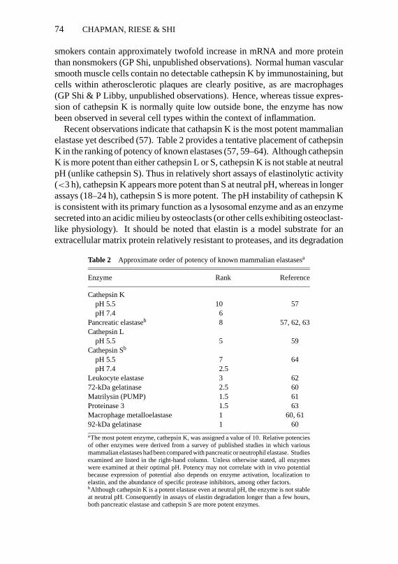
Recent observations indicate that cathapsin K is the most potent mammalianelastase yet described (57). Table 2 provides a tentative placement of cathepsinK in the ranking of potency of known elastases (57, 59–64). Although cathepsinK is more potent than either cathepsin L or S, cathepsin K is not stable at neutralpH (unlike cathepsin S). Thus in relatively short assays of elastinolytic activity(<3 h), cathepsin K appears more potent than S at neutral pH, whereas in longerassays (18–24 h), cathepsin S is more potent. The pH instability of cathepsin Kis consistent with its primary function as a lysosomal enzyme and as an enzymesecreted into an acidic milieu by osteoclasts (or other cells exhibiting osteoclast-like physiology). It should be noted that elastin is a model substrate for anextracellular matrix protein relatively resistant to proteases, and its degradation
Table 2 Approximate order of potency of known mammalian elastasesa
Enzyme Rank Reference
Cathepsin KpH 5.5 10 57pH 7.4 6
Pancreatic elastaseb 8 57, 62, 63Cathepsin L
pH 5.5 5 59Cathepsin Sb
pH 5.5 7 64pH 7.4 2.5
Leukocyte elastase 3 6272-kDa gelatinase 2.5 60Matrilysin (PUMP) 1.5 61Proteinase 3 1.5 63Macrophage metalloelastase 1 60, 6192-kDa gelatinase 1 60
aThe most potent enzyme, cathepsin K, was assigned a value of 10. Relative potenciesof other enzymes were derived from a survey of published studies in which variousmammalian elastases had been compared with pancreatic or neutrophil elastase. Studiesexamined are listed in the right-hand column. Unless otherwise stated, all enzymeswere examined at their optimal pH. Potency may not correlate with in vivo potentialbecause expression of potential also depends on enzyme activation, localization toelastin, and the abundance of specific protease inhibitors, among other factors.bAlthough cathepsin K is a potent elastase even at neutral pH, the enzyme is not stableat neutral pH. Consequently in assays of elastin degradation longer than a few hours,both pancreatic elastase and cathepsin S are more potent enzymes.

P1: sbs
August 28, 1956 22:38 Annual Reviews CHAPCHPT.DUN AR25-04
CATHEPSIN BIOLOGY 75
identifies these cathepsins as potent endoproteases. As such, cathepsin K, aswell as cathepsins S and L, is also a potent collagenase and gelatinase.
Expression of cathepsin K has recently been correlated with a degradativephenotype of macrophages (33, 65). Freshly explanted monocytic cells exhibitalmost no cathepsin K mRNA. Within 2 to 3 days of in vitro culture in the pres-ence of human serum, the levels of cathepsins B, L, and S increase in the cells,but the cells nonetheless do not degrade extracellular particulate elastin. How-ever, beginning on days 9–11 of culture, the monocyte-derived macrophagesbegin to secrete large amounts of acid and acidic hydrolases, including cathep-sins L and S, into the extracellular space and degrade large amounts of elastin(44). The process is stimulated rather than inhibited by the presence of serum.At this time, there is a marked induction of cathepsin K mRNA. Because thereare several elastases being secreted at once, it is unclear which, if any , is pre-dominantly mediating degradation. Nonetheless, these observations illustratethat under some conditions macrophages are quite capable of using cathep-sins to degrade extracellular matrix protein, as had been previously postulated(4, 65), and that under these culture conditions, the appearance of cathepsin Kcorrelates with the expression of this potential.
That cathepsin K (and by inference other cathepsins) is actually importantto extracellular matrix remodeling has recently been verified by the identifi-cation of mutations in the coding sequence of cathepsin K in individuals withpycnodysostosis (66). Pyknodysostosis is an autosomal recessive disorder char-acterized by premature closure of long bone growth, facial hypoplasia (espe-cially micrognathia), and brittle, dense long bones with osteosclerosis (67).Patients have fractures and the hallmark skeletal features of the disorder. Ob-structive sleep apnea is also a clinical problem (68). Pycnodysostosis maps tochromosome 1q21 in several distinct family pedigrees (69, 70). Screening ofcDNA and genomic DNA obtained by PCR from lymphoblastoid cells revealsdistinct mutations in affected members of three separate families (66). One ofthe mutations transcribes a premature stop codon near the active site cysteine.A second large family with 16 affected members carries a mutation in the na-tive stop codon that results in the predicted addition of 18 amino acids to thecarboxy-terminal end, but in fact results in misfolded or mistargeted proteinthat is unstable and undetectable in cells expressing this mutant mRNA. Thedemonstration of altered bone formation and growth in individuals deficientin cathepsin K is the first direct demonstration of a critical role for cathepsinsin extracellular matrix remodeling and provides a rationale for inhibition ofcathepsin K in bone disorders such as osteoporosis.
There are several clinical situations in which the mobilization of cathepsinK or its closely related partners could be relevant. Large amounts of elastinare degraded rather quickly in the context of vascular inflammation, especially

P1: sbs
August 28, 1956 22:38 Annual Reviews CHAPCHPT.DUN AR25-04
76 CHAPMAN, RIESE & SHI
giant cell arteritis, leading to aneurysm formation. Immunostaining of bothatherosclerotic plaques and sites of elastin degradation in giant cell aortitis re-veal vivid immunostaining for cathepsins S and K in smooth muscle cells andgiant cells, respectively (G Sukhova, unpublished observations). Because largeamounts of elastin are degraded in these disorders, these proteases are good can-didates for mediators of the process. This also may be true in other disordersassociated with extensive elastin degradation such as lymphangiomyomatosis.Histologic studies indicate extensive lung elastin remodeling in the setting oflymphangiomyomatosis (71). In this disorder there is abnormal proliferationof smooth muscle cells and extensive matrix remodeling leading to emphyse-matous changes and airway obstruction. To date, the presence of cathepsin Kor other potent elastases in smooth muscle cells from patients with this disorderhas not been tested.
The elastolytic cathepsins (K, L, and S) may also be important to elastindestruction in the more common disorder of smokers’ lung. Although theparadigm of protease inhibitor deficiency exemplified by alpha-1-antitrypsindeficiency is still attractive as an etiologic mechanism for emphysema (72),the proteases mainly involved in this process may have little to do with alpha-1-antitrypsin. In spite of thirty years of trying to fit smoking-related injuryinto a model of functional deficiency of alpha-1-antitrypsin as the sole cause ofemphysema, this model remains much in doubt (73, 74). The recent demonstra-tion of a longer time for inhibition of neutrophil elastase by alpha-1-antitrypsinobtained by bronchoalveolar lavage from cigarette smokers over that of non-smokers (75) is of uncertain significance, as an even longert1/2 would bepredicted in individuals with the MZ phenotype, and yet there is little or noincreased risk for emphysema in this genotype. Instead, the list of proteasesin the lung that could mediate emphysema independently of neutrophil elas-tase continues to grow (Table 2). The group of metalloenzymes, especiallymacrophage metalloelastase, along with the elastolytic cathepsins all have thepotential to mediate elastin and other matrix protein destruction without im-pugning a deficiency of protease inhibitors. This is because these enzymes canbe compartmentalized by macrophages to degrade matrix proteins with whichthey are in direct contact. Unfortunately, it remains unclear which if any of thepotentially destructive proteases is actually important. This uncertainty may beresolved by molecular genetics. The generation of mice specifically deficient ina single protease would allow the direct test of whether the enzyme is necessaryfor lung injury in the context of smoking or other inflammatory disorders ofthe lung. Mice subjected to smoke inhalation for several weeks are reportedto develop pathologic features of emphysema (76). Indeed, several proteasegenes, including both metalloenzymes and cathepsins, have now been disruptedin mice and are being studied in this context.

P1: sbs
August 28, 1956 22:38 Annual Reviews CHAPCHPT.DUN AR25-04
CATHEPSIN BIOLOGY 77
A second approach is to better delineate the genetics of human emphysema.Surprisingly, this may be made possible by the interest in lung transplantationfor chronic obstructive lung disease (COPD). The referral of young people withend-stage emphysema to transplant centers has revealed numerous probands(age less than 50 years) with smoking-related severe emphysema and normalalpha-1-antitrypsin levels (77). The incidence of reduced lung function in theirfamily members is much higher than that of the general population. Althoughemphysema in this setting is likely a complex trait, identification of genesthat underlie early-onset disease may help elucidate the major pathways ofdestruction in this disorder. It would be surprising if this did not also reveal newinformation about susceptibility to tissue destruction in chronic inflammatorydisorders involving other organs, e.g. arthritis.
The attempts to elucidate the role of cathepsin K and the other elastinolyticcathepsins in human disease is not without therapeutic importance. Severalclasses of nontoxic specific inhibitors of cysteine proteases are becoming avail-able. What is critically missing is the elucidation of a biological role justifyingtheir use. One example of the use of these types of inhibitors to delineate aspecific function for a cathepsin is discussed below.
Cathepsin SCathepsin S was originally identified as a distinct enzyme activity in lymphnodes and was found to be prominently expressed in and subsequently puri-fied from spleen (25). The human orthologue of this enzyme was identifiedby DNA sequence homologies to cathepsins B and L and cloned in a humanlung macrophage cDNA library (32). The full coding sequence was also ob-tained independently by Wiederanders from a cDNA library screen (34). Ouroriginal intent on isolating the human enzyme was to identify new elastolyticenzymes and, indeed, cathepsin S proved to be a potent elastase with substan-tial enzymatic activity and stability at neutral pH (Table 2). Moreover, thisenzyme also exhibited restricted and regulated tissue expression and was foundto be inducible by cytokines such as interferon-gamma and interleukin 1β. Inrats, cathepsin S is expressed in thyroid tissue and is inducible by thyroid-stimulating hormone, which suggests a possible specific role in intracellularthyroglobulin processing for the release of thyroid hormone (35). Cathepsin Sis also highly expressed in the spleen and antigen-presenting cells, includingB lymphocytes, macrophages, and dendritic cells (32, 78, 79). Because ofits high expression in spleen (and lymph nodes) and inducibility by cytokinesknown to be involved in major histocompatibility complex (MHC) class IIantigen expression, we explored the role of this enzyme in class II antigenpresentation.

P1: sbs
August 28, 1956 22:38 Annual Reviews CHAPCHPT.DUN AR25-04
78 CHAPMAN, RIESE & SHI
Figure 2 Participation of lysosomal proteases in MHC class II antigen presentation pathway.Lysosomal proteases are essential for two steps: (a) the degradation of Ii to CLIP (residues 81–104of Ii) to permit dissociation of CLIP from class II molecules and subsequent peptide binding; and(b) the generation of antigenic peptide fragments from larger polypeptide/protein moieties.
MHC Class II Antigen Presentation and Cathepsin SLysosomal proteases play an essential role in the MHC class II antigen presen-tation pathway, as schematically reviewed in Figure 2. Proteases are involved intwo critical steps: the degradation of the class II chaperone, the invariant chain(Ii), prior to its removal from the class II peptide binding cleft; and the genera-tion of antigenic peptides (13–26 amino acids in length) capable of replacing theinvariant chain in the peptide-binding grove of the class II molecules. Class IIαβ dimers associate with Ii in the endoplasmic reticulum to form nonamercomplexes consisting of a scaffold of homotrimers associated with up to threeclass IIαβ dimers (80, 81). These complexes traverse the Golgi apparatus andare targeted to intracellular compartments where degradation of the Ii occurs,followed by binding of exogenous, antigenic peptides (82–86). Ii associateswith class II molecules via direct interaction of residues 81–104 of its lumenaldomain (87–90), designated CLIP (class II-associated invariant chain peptides),with the antigen-binding groove of class II (91). Most class II alleles requirean additional class II-like molecule, HLA-DM, to liberate the peptide-bindinggroove of CLIP and to facilitate loading with antigenic peptide (92–94). The

P1: sbs
August 28, 1956 22:38 Annual Reviews CHAPCHPT.DUN AR25-04
CATHEPSIN BIOLOGY 79
αβ-peptide complexes formed by this pathway are then transported to the cellsurface to initiate MHC class II-restricted T cell recognition (95).
Proteolysis of Ii fromαβ-Ii complexes and formation ofαβ-CLIP is re-quired prior to class II peptide association because matureαβ-Ii heterodimersare unable to load peptides (96). Moreover, Roche & Cresswell (97) havedemonstrated that proteolysis of Ii fromαβ-Ii complexes promotes peptidebinding in vitro. Of the known lysosomal proteases, cysteine proteases havebeen most clearly implicated in Ii proteolysis. Cysteine protease inhibitionwith leupeptin impairs Ii breakdown and results in accumulation of Ii frag-ments in B-lymphoblastoid cells (98–100). Also, lysosomotropic agents suchas chloroquine (101) and concanamycin B (102) interrupt Ii proteolysis andcause accumulation of Ii fragments, presumably by neutralizing endosomal pHand disrupting protease activity. Accumulation of the Ii breakdown intermedi-ates has been shown to impair peptide loading onto MHC class II moleculesleading to diminished SDS-stableαβ-peptide complexes (103, 104), decreasedMHC class II cell surface expression (103) and attenuation of antigen-stimulatedT cell proliferation (105, 106).
Cathepsin S has recently been demonstrated to play an essential role in Iiprotelolysis and peptide loading (107). Convincing evidence for participationof cathepsin S in Ii processing was provided by using a novel, specific cathepsinS inhibitor (morpholinurea-leucine-homophenylalanine-vinylsulfone-phenyl;LHVS). LHVS has an≈67-fold increased activity toward cathepsin S overcathepsin L and≈6000-fold increase over cathepsin B (108). Specific inhibi-tion of cathepsin S with 1 nM and 5 nM LHVS in B lymphoblastoid (HOM2)cells results in accumulation of a class II-associated 13-kDa Ii fragment and aconcomitant reduction in peptide loading of class II molecules, as evidencedby a marked decrease in formation of SDS-stable complexes migrating at≈50kDa (Figure 3, lanes 2, 3). This 50-kDa band represents class II moleculesassociated with antigenic peptides. The class II-peptide complex is stable inSDS at room temperature but not when boiled (Figure 3, compare lane 1 withlane 4). Inhibition of all cysteine proteases with the cysteine-class inhibitor 2S,3S-trans-epoxysuccinyl-L-leucylamido-3-methylbutane ethyl ester (E64D) re-sults in a buildup of a class II-associated 23-kDa Ii fragment with a decreasein SDS-stable dimer formation (Figure 3, lane 4). This suggests that cathep-sin S acts on a relatively late Ii breakdown intermediate and is required forefficient proteolysis of Ii necessary for subsequent peptide loading. Further-more, purified cathepsin S, but not cathepsin B, H, or D, specifically digests Iifrom αβ-Ii trimers, generatingαβ-CLIP complexes capable of binding exoge-nously added peptide in vitro (107). The finding that a single cysteine proteasemay be crucial for Ii proteolysis and subsequent class II-peptide binding rein-forces the emerging view that lysosomal proteases may play specific roles inbiologic systems.

P1: sbs
August 28, 1956 22:38 Annual Reviews CHAPCHPT.DUN AR25-04
80 CHAPMAN, RIESE & SHI
Figure 3 Specific inhibition of cathepsin S impairs class II-associated Ii proteolysis and peptideloading. HOM2 (B lymphoblastoid) cells were labeled with35S-methionine/cysteine and chasedfor 5 h without inhibitor (lanes 1, 5), in the presence of 1 nM LHVS (lanes 2, 6); 5 nM LHVS (lanes3, 7); and 20µM E64D (lanes 4, 8). Class II-Ii complexes were immunoprecipitated from celllysates with monoclonal antibody T¨u36 and analyzed by 14% SDS-PAGE under mildly denaturingconditions (non-boiled, non-reduced) (lanes 1–4) and denaturing conditions (lanes 5–8).
Lysosomal proteases are also essential for generation of the antigenic peptidespresented to T cells on the class II molecules (Figure 2). Proteins may enter theendocytic pathway by binding to membrane-bound immunoglobulin on B cells,or by pinocytosis primarily in dendritic cells and macrophages (109). Peptideprocessing of endocytosed antigens has been localized to dense compartmentscolocalizing with lysosomes (110) and low-density endosomal compartmentsdistinct from the denser lysosomes (111). Once in the endocytic pathway, theseproteins are broken down into peptides and loaded onto class IIαβ dimers. It isunclear whether free peptides are generated first followed by class II binding orwhether class II molecules bind larger peptide/polypeptide fragments that are
P1: sbs
August 28, 1956 22:38 Annual Reviews CHAPCHPT.DUN AR25-04
CATHEPSIN BIOLOGY 81
then digested to smaller peptide fragments while bound to class II molecules.The carboxypeptidase and aminopeptidase activities of cathepsins B and H,respectively, could be functionally important at this point. In this way the classII binding groove may act as a protective pocket preventing terminal proteolysisof presented peptides.
Both cysteine class and aspartyl class proteases have been implicated in gen-eration of antigenic epitopes. The ability of the cysteine protease inhibitor,leupeptin, to alter ovalbumin and tetanus toxin processing appears to be epi-tope dependent (112, 113). In vitro digestion of ovalbumin by the aspartylprotease cathepsin D, but not by the cysteine protease cathepsin B, generatedpeptides capable of stimulating T cells in association with class II molecules(114). Cathepsin D from bovine alveolar macrophages also produces epitopescapable of binding to class II molecules, which suggests a structural relation-ship between the antigenic motif generated by cathepsin D digestion and theantigenic structure recognized by MHC class II molecules (115). A specificinhibitor of the nonlysosomal aspartyl protease, cathepsin E, inhibited the pro-cessing of ovalbumin in a murine antigen-presenting cell line (116). These datasuggest that several enzymes from the cysteine and aspartyl protease classesmay be important in generating suitable peptide epitopes for presentation byclass II molecules, dependent on epitope structure and mode of entry into thesecretory pathway.
In summary, lysosomal protease involvement is required for Ii degradationso that efficient class II-Ii dissociation and peptide loading may occur and forgeneration of the antigenic peptides presented on the class IIαβ dimers. Cys-teine proteases, and specifically cathepsin S, appear to mediate Ii processing,whereas several cysteine and aspartyl proteases may participate in antigenicpeptide generation.
Antigen presentation is an important function of the lung. Recent studiesindicate a network of dendritic cells within the epithelium of lung airwaysthat are repeatedly exposed to antigenic agents (117, 118). The surprisingfinding that a single cysteine protease is essential in antigen presentation raisesthe possibility that targeted inhibition of this enzyme may be beneficial insettings in which exaggerated immune responses to exogenous antigens mediatedisease: transplantation, asthma, hypersensitivity pneumonitis, and potentiallyautoimmune disorders.
Role for Cathepsin S in Cilial Function?Immunostaining of normal human lung with cathepsin S antibodies also sug-gests an additional previously unsuspected role for this enzyme in lung biology.As illustrated in Figure 4, monospecific antibodies to cathepsin S vividly stainthe cilia of conducting airway cells for cathepsin S antigen (Panel A). In contrast

P1: sbs
August 28, 1956 22:38 Annual Reviews CHAPCHPT.DUN AR25-04
82 CHAPMAN, RIESE & SHI
Figure 4 Immunostaining of human lung airways with antiserum against cathepsin S (A) andcathepsin K (B).

P1: sbs
August 28, 1956 22:38 Annual Reviews CHAPCHPT.DUN AR25-04
CATHEPSIN BIOLOGY 83
neither cathepsin S antibodies adsorbed with antigen (not shown) nor monospe-cific antibodies to cathepsin K stain these structures (Panel B). This result raisesthe intriguing possibility that because of its stability at neutral pH and potentialfor broad endoprotease activity, ciliated cells have captured the enzyme ontotheir surfaces to promote motility of their cilia. Indeed, airway inflammationis known to produce dysfunctional ciliary motion. One could envision thatplasma-derived proteins, in the setting of inflammation, could bind and impaircilial motility and that cathepsin S would be protective. If so, this would repre-sent another example of the importance of protease activity, even of nonspecificendoproteases, to normal lung function. Thus far no functional studies havebeen performed to test this hypothesis.
FUTURE DIRECTIONS
Remarkable advances in the last twenty years in understanding the catalyticmechanism and fine structural features of proteases and their inhibitors havehad important implications for medicine. The detailed view of the active sitepockets of numerous proteases now available makes the rational design of pro-tease inhibitors feasible. Indeed, the limiting step in the use of novel proteaseinhibitors in medicine is not so much the discovery of an effective inhibitorbut elucidation of the exact physiological role of the protease in the biology ofthe cell and the intact organism. Where successfully understood and applied,both proteases and protease inhibitors have proven to be therapeutically useful.Angiotensin-converting-enzyme inhibitors and, more recently, HIV proteaseinhibitors, as well as the proteases urokinase and tissue plasminogen activa-tor, are good examples of merging molecular and cell biology for therapeuticadvance. In this regard, the identification of new cysteine proteases and theirinhibitors in the last five years alone poses a big challenge for cell biology. Inthis review, we have summarized recent advances in understanding the role ofcysteine proteases in both the physiology of the lung as well as in other organsystems. The field is energized by these findings; yet much of what is presentedis new and the importance too early to judge. Still, there is promise that thecontinued elucidation of specific physiological functions for cysteine proteaseswill presage new therapeutic tools.
ACKNOWLEDGMENTS
Work in the investigators’ laboratory (HAC) was supported by National Insti-tutes of Health grant HL48261. The authors thank D Bromme, GA Silverman,G Sukhova, and P Libby for communicating results prior to publication.

P1: sbs
August 28, 1956 22:38 Annual Reviews CHAPCHPT.DUN AR25-04
84 CHAPMAN, RIESE & SHI
Literature Cited
1. Neurath H, Walsh KA. 1976. Role ofproteolytic enzymes in biological regula-tion.Proc. Natl. Acad. Sci. USA73:3825–32
2. Polgar L. 1989. General aspects of pro-teases. InMechanisms of Protease Action,ed. L Polar, pp. 43–76. Boca Ratan, FL:CRC Press
3. Gross P, Babyak MA, Tolker E, KaschakM. 1964. Enzymatically produced pul-monary emphysema: a preliminary re-port.J. Occup. Med.6:481
4. Chapman HA, Stone OL, Vavrin Z. 1984.Degradation of fibrin and elastin by hu-man alveolar macrophages in vitro. Char-acterization of a plasminogen activatorand its role in matrix degradation.J. Clin.Invest.73:806–15
5. Chapman HA Jr, Stahl M, Fair DS, AllenCL. 1988. Regulation of the procoagulantactivity within the alveolar compartmentof normal human lung.Am. Rev. Resp.Dis. 37:1417–25
6. Nadel JA. 1991. Neutral endopeptidasemodulates neurogenic inflammation.Eur.Resp. J.4:745–54
7. Polgar L, ed. 1989. Metalloproteases. InMechanisms of Protease Action,pp. 208–210. Boca Ratan, FL: CRC Press
8. Menard R, Storer A. 1992. Oxyanionhole interactions in serine and cysteineproteases.Hoppe-Seyler’s Z. Biol. Chem.373:393–400
9. Matthews BW, Sigler PB, HendersonR, Blow DM. 1967. Three-dimensionalstructure of tosyl-α-chymotrypsin.Na-ture214:652–56
10. Varughese KL, Ahmed FR, Careys PR,Hasnain S, Huber CP, Storer AC. 1989.Crystal structure of papain-E-64 complex.Biochemistry28:1330–32
11. Mason RW, Wilcox D. 1993. Chemistryof lysosomal cysteine proteases.Adv. CellMol. Biol. Membr.1:81–116
12. Berti PJ, Storer AC. 1995. Align-ment/phylogeny of the papain superfam-ily of cysteine proteases.J. Mol Biol.246:273–83
13. Thornberry N, Bull HG, Calaycay JR,Chapman KT, Howard AD, et al. 1992.A novel heterodimeric cysteine proteaseis required for inteleukin-1 beta pro-cessing in monocytes.Nature 356:768–74
14. Henkart PA. 1996. ICE family proteases:mediators of all apoptotic cell death?Im-munity4:194–201
15. Saido TC, Sorimachi H, Suzuki K. 1994.
Calpain: new perspectives in moleculardiversity and physiological-pathologicalinvolvement.FASEB J.8:814–22
16. Croall DE, DeMartino GN. 1991.Calcium-activated neutral protease (cal-pain) system: structure, function, andregulation.Physiol. Rev.71:813–47
17. Du X, Saido TC, Tsubuki S, Indig FE,Wiklliams MJ, Ginsberg MH. 1995. Cal-pain cleavage of the cytoplasmic domainof the integrin beta 3 subunit.J. Biol.Chem.270:26146–51
18. Sorimachi H, Saido TC, Suzuki K. 1994.New era of calpain research. Discoveryof tissue-specific calpains.FEBS Lett.343:1–5
19. Richard I, Broux O, Allamand V, Fouger-ousse F, Chiannilkulchai N, et al. 1995.Mutations in the proteolytic enzymecalpain 3 cause limb-girdle muscular dys-trophy type 2A.Cell 81:27–40
20. Sebti SM, Mignano JE, Jani JP, Sri-matkandada S, Lazo JS. 1989. Bleomycinhydrolase: molecular cloning, sequenc-ing, and biochemical studies reveal mem-bership in the cysteine proteinase family.Biochemistry28: 6544–48
21. Sebti SM, DeLeon JC, Lazo JS. 1987.Purification, characterization, and aminoacid composition of rabbit pulmonarybleomycin hydrolase.Biochemistry26:4213–19
22. Lazo JS, Boland CJ, Schwartz PE. 1982.Bleomycin hydrolase activity and cyto-toxicity in human tumors.Cancer Res.42:4026–31
23. Joshua-Tor L, Xu HE, Johnston SA,Rees DC. 1995. Crystal structure ofa conserved protease that binds DNA:the bleomycin hydrolase, Gal6.Science269:945–50
24. Musil D, Zucic D, Turk D, Engh RA,Mayr L, et al. 1991. The refined 2.15AA X-ray crystal structure of human livercathepsin B: the structural basis for itsspecificity.EMBO J.10:2321–30
25. Kirschke H, Weideranders B, Bromme D,Rinne A. 1989. Cathepsin from bovinespleen. Purification, distribution, intracel-lular localization and action on proteins.Biochem. J.264:467–73
26. Fuchs R, Gassen HG. 1989. Nucleotidesequence of human preprocathepsin H,a lysosomal cysteine proteinase.NucleicAcids Res.17:9471
27. Joseph LJ, Chang LC, Stemenkovich D,Sukhatme VP. 1988. Complete nucleotideand deduced amino acid sequence of hu-
P1: sbs
August 28, 1956 22:38 Annual Reviews CHAPCHPT.DUN AR25-04
CATHEPSIN BIOLOGY 85
man and murine preprocathepsin L.J.Clin. Invest.81:1621–29
28. Chan SJ, Segundo BS, McCormick MB,Steiner DF. 1986. Nucleotide and pre-dicted amino acid sequence of clonedhuman and mouse preprocathepsin BcDNAs. Proc. Natl. Acad. Sci. USA83:7721–28
29. Fong D, Calhoun DH, Hsieh W-T, LeeB, Wells RD. 1986. Isolation of a cDNAclone for the human lysosomal proteinasecathepsin B.Proc. Natl. Acad. Sci. USA83:2909–13
30. Barrett AJ, Kirschke H. 1981. CathepsinsB, H, and L.Meth. Enzymol.80:535–61
31. Shaw E, Dean RT. 1980. The inhibition ofmacrophage protein turnover by a selec-tive inhibitor of thiol proteases.Biochem.J. 186:385–90
32. Shi GP, Munger JS, Meara JP, RichDH, Chapman HA. 1992. Molecularcloning and expression of human alve-olar macrophage cathepsin S, an elasti-nolytic cysteine protease.J. Biol. Chem.267:7258–62
33. Shi GP, Chapman HA, Bhairi SM,DeLeeuw C, Reddy VY, Weiss SJ. 1995.Molecular cloning of human cathepsin O,a novel endoproteinase and homologue ofrabbit OC-2.FEBS Lett.357:129–34
34. Wiederanders B, Bromme D, KirschkeH, Kalkkiner N, Rinne A, et al. 1991.Primary structure of bovine cathepsin S.Comparison to cathepsins L, H, and B.FEBS Lett.286:189–92
35. Petanceska S, Devi L. 1992. Sequenceanalysis, tissue distribution, and expres-sion of rat cathepsin S.J. Biol. Chem.267:26038–43
36. Velasco G, Ferrando AA, Puente XS,Sanchez LM, Lopez-otin C. 1994. Hu-man cathepsin O. Molecular cloning froma breast carcinoma, production of the ac-tive enzyme inEscherichia coli,and ex-pression analysis in human tissues.J. Biol.Chem.269:27136–42
37. McGuire MJ, Lipsky PE, Thiele DL.1992. Purification and characterization ofdipeptidyl peptidase I from human spleen.Arch. Biochem. Biophys. 295:280–88
38. Dikov MM, Springman EB, Yeola S,Serafin WE. 1994. Processing of pro-carboxypeptidase A and other zymogensin murine mast cells.J. Biol. Chem.269:25897–904
39. Vernet T, Berti PJ, de Montigny C, MusilR, Tessier DC, et al. 1995. Processing ofthe papain precursor. The ionization stateof a conserved amino acid motif withinthe pro region participates in the regula-
tion of the intramolecular processing.J.Biol. Chem.270:10838–46
40. Fox T, de Miguel E, Mort JS, Storer AC.1992. Potent slow-binding inhibition ofcathepsin B by its propeptide.Biochem-istry 31:12571–76
40a. Tao K, Stearns NA, Dong J, Wu QL, Saha-gian GG. 1994. The pro region of cathep-sin L is required for proper folding, stabil-ity, and ER exit.Arch. Biochem. Biophys.311:19–27
41. Baudys M, Meloun T, Gan-Erdene T,Fusek M, Mares M, et al. 1991. S-Sbridges of cathepsin B and H from bovinespleen: a basis for cathepsin B modelbuilding and possible functional implica-tions for discrimination between exo- andendopeptidase activities among cathep-sins B, H and L.Biomed. Biochim. Acta50:569–77
42. Vernet T, Tessier DC, Chatellier J, PlouffeC, Lee TS, et al. 1995. Structural andfunctional roles of asparagine 175 in thecysteine protease papain.J. Biol. Chem.270:16645–52
43. Pisoni RL, Acker TL, Lisowski KM,Lemons RM, Theone JG. 1990. A cys-teine-specific lysosomal transport systemprovides a major route for the delivery ofthiol to human fibroblast lysosomes: pos-sible role in supporting lysosomal prote-olysis.J. Cell Biol.110: 327–35
44. Reddy VY, Zhang Q-Y, Weiss SJ. 1995.Pericellular mobilization of the tissue-destructive cysteine proteases, cathepsinsB, L, and S, by human macrophages.Proc.Natl. Acad. Sci. USA92:3849–53
45. Baron R, Neff L, Louvard D, Courtoy PJ.1985. Cell mediated extracellular acidifi-cation and bone resorption: evidence toa low pH in resorbing lacunae and local-ization of a 100 kD lysosomal membraneprotein at the osteoclast ruffled border.J.Cell Biol. 101:2210–28
46. Baron R. 1989. Molecular mechanisms ofbone resorption by the osteoclast.Anat.Rec.224:2317–429
47. Dalaisse JM, Eeckhout Y, Vaes G. 1980.Inhibition of bone resorption in culture byinhibitors of thiol proteinases.Biochem. J.192:365–68
48. Barrett AJ. 1987. The cystatins: a newclass of peptidase inhibitors.TrendsBiochem. Sci.12:193–96
49. Lindahl P, Ripoll D, Abrahamson M, MortJS, Storer AC. 1994. Evidence for the in-teraction of valine-10 in cystatin C withthe S2 subsite of cathepsin B.Biochem-istry 33:4384–92
50. Penacchio LA, Lehesjoki AE, Stone NE,Willour VL, Virtaneva K, et al. 1996. Mu-

P1: sbs
August 28, 1956 22:38 Annual Reviews CHAPCHPT.DUN AR25-04
86 CHAPMAN, RIESE & SHI
tations in the gene encoding cystatin B inprogressive myoclonus epilepsy (EPM1).Science271:1731–34
51. Ray CA, Black RA, Kronheim SR, Green-street TA, Sleath PR, et al. 1992. Viral in-hibition of inflammation: cowpox virusencodes an inhibitor of the interleukin-1beta converting enzyme.Cell69:597–604
52. Suminami Y, Kishi F, Sekiguchi K, KatoH. 1991. Squamous cell carcinoma anti-gen is a new member of the serine pro-tease inhibitors.Biochem. Biophys. Res.Commun.181:51–58
53. Takeda A, Yamamoto T, Nakamura Y,Takahashi T, Hibino T. 1995. Squamouscell carcinoma antigen is a potent in-hibitor of cysteine proteinase cathepsin L.FEBS Lett.359:78–80
53a. Schneider SS, Schick C, Fish KE, MillerE, Pena JG, et al. 1995. A serine pro-teinase inhibitor locus at 18q21.3 containsa tandem duplication of the human squa-mous cell carcinoma antigen gene.Proc.Natl. Acad. Sci. USA92:3147–51
54. Chapman HA, Munger JS, Shi GP. 1994.Role of thiol proteases in tissue injury.Am. J. Resp. Crit. Care Med.150:S155–59
55. Tezuka K, Tezuka Y, Maejima A, Sato T,Nemoto K, et al. 1994. Molecular cloningof a possible cysteine proteinase predom-inantly expressed in osteoclasts.J. Biol.Chem.269:1106–9
56. Inaoka T, Bilbe G, Ishibashi O, TezukaK, Kumegawa M, et al. 1995. Molecu-lar cloning of human cDNA for cathep-sin K: novel cysteine proteinase predom-inantly expressed in bone.Biochem. Bio-phys. Res. Commun.206:89–96
57. Bromme D, Okamoto K, Wang BB, BirocS. 1996. Human cathepsin O2, a ma-trix protein-degrading cysteine proteaseexpressed in osteoclasts.J. Biol. Chem.271:2126–32
58. Saneshige S, Mano H, Tezuka K, KakudoS, Mori Y, et al. 1995. Retinoic acid di-rectly stimulates osteoclastic bone resorp-tion and gene expression of cathepsinK/OC-2.Biochem. J.309:721–24
59. Mason RW, Johnson D, Barret AJ, Chap-man HA Jr. 1986. Elastolytic activity ofhuman cathepsin L.Biochem. J.122:925–27
60. Senior RM, Griffin GL, Fliszar CJ,Shapiro SD, Goldberg GI, Welgus HG.1991. Human 92 kDA and 72 kDA TypeIV collagenases are elastases.J. Biol.Chem.266:7870–75
61. Murphy G, Cockett ML, Ward RV,Docherty AJP. 1991. Matrix metallopro-teinase degradation of elastin, type IV
collagen, and proteoglycan.Biochem. J.277:277–79
62. Baugh RJ, Travis J. 1976. Human leuko-cyte granule elastase: rapid isolation andcharacterization.Biochemistry 15:836–41
63. Kao RC, Wehmer NG, Skubitz KM, GrayBH, Hoidal JR. 1988. Proteinase 3. A dis-tinct human polymorphonuclear leuko-cyte proteinase that produced emphysemain hamsters.J. Clin. Invest.82:1963–73
64. Xin XQ, Gunesekera B, Mason RW. 1992.The specificity and elastinolytic activi-ties of bovine cathepsins S and H.Arch.Biochem. Biophys.299:334–39
65. Chapman HA, Stone OL. 1984. Compari-son of live human neutrophil and alveolarmacrophage elastolytic activity in vitro:relative resistance of macrophage elas-tolytic activity to serum and alveolar pro-tease inhibitors.J. Clin. Invest.74:1693–700
66. Gelb B, Shi GP, Chapman HA, DesnickRJ. 1996. Pycnodysostosis is caused bya deficiency of cathepsin K.Science.273:1236–38
67. Edelson JG, Obad S, Geiger R, On A, Ar-tul HJ. 1992. Pycnodysostosis. Orthope-dic aspects with a description of 14 newcases.Clin. Orth.280:263–76
68. Aronson DC, Heymans HS, Bijlmer RP.1984. Cor pulmonale and acute livernecrosis, due to upper airway obstructionas part of pycnodysostosis.Eur. J. Pediatr.141:251–53
69. Gelb BD, Edelson JG, Desnick RJ. 1995.Linkage of pycnodysostosis to chromo-some 1q21 by homozygosity mapping.Nat. Genet.10:235–37
70. Polymeropoulos MH, Ortiz De Luna RI,Ide SE, Torres R, et al. 1995. The genefor pycnodysostosis maps to human chro-mosome 1cen-q21.Nat. Genet.10:238–39
71. Fukuda Y, Kawamoto M, YamamotoA, Ishizaki M, Basset F, Masugi Y.1990. Role of elastic fiber degrada-tion in emphysema-like lesions of pul-monary lymphangiomyomatosis.Hum.Pathol.21:1252–61
72. Janoff A. 1985. Elastases and emphy-sema. Current assessment of the protease-antiprotease hypothesis.Am. Rev. Resp.Dis. 132:417–33
73. Tetley TD. 1993. New perspectives on ba-sic mechanisms in lung disease. 6. Pro-teinase imbalance: its role in lung disease.Thorax48:560–65
74. Snider GL. 1992. Emphysema: the firsttwo centuries—and beyond. A histori-cal overview, with suggestions for future

P1: sbs
August 28, 1956 22:38 Annual Reviews CHAPCHPT.DUN AR25-04
CATHEPSIN BIOLOGY 87
research: Part 2.Am. Rev. Resp. Dis.146:1615–22
75. Ogushi F, Hubbard RC, Vogelmeier C,Fells GA, Crystal RG. 1991. Risk fac-tors for emphysema. Cigarette smokingis associated with a reduction in the as-sociation rate constant of lung alpha-1-antitrypsin for neutrophil elastase.J. Clin.Invest.87:1060–65
76. Belaaouaj A, Shapiro SD. 1996. Identi-fication of differentially expressed genesin lungs of mice following exposure tocigarette smoke.Resp. Crit. Care Med.153:A30 (Abstr.)
77. Silverman P, Chapman H, Drazen J,O’Donnell W, Reilly J, et al. 1996. Early-onset chronic obstructive pulmonary dis-ease (COPD): preliminary evidence forgenetic factors other than PI type.Resp.Crit. Care Med.153:A48 (Abstr.)
78. Shi GP, Webb AC, Foster KE, Knoll JHM,Lemere CA, et al. 1994. Human cathepsinS: chromosomal localization, gene struc-ture, and tissue distribution.J. Biol. Chem.269:11530–36
79. Morton PA, Zacheis ML, Giacoletto KS,Manning JA, Schwartz BD. 1995. De-livery of nascent MHC class II-invariantchain complexes to lysosomal compart-ments and proteolysis of invariant chainby cysteine proteases precedes peptidebinding in B-lymphoblastoid cells.J. Im-munol.154:137–50
80. Roche PA, Marks MS, Cresswell P. 1991.Formation of a nine-subunit complex byHLA class II glycoproteins and the invari-ant chain.Nature354:392–394
81. Lamb C, Cresswell P. 1992. Assem-bly and transport properties of invari-ant chain trimers and HLA-DR-invariantchain complexes.J. Immunol.148:3478–82
82. Guagliardi LE, Koppelman B, Blum JS,Marks MS, Cresswell P, Brodsky FM.1990. Co-localization of molecules in-volved in antigen processing and presen-tation in an early endocytic compartment.Nature343:133–39
83. Peters PJ, Neefjes JJ, Oorschot V, PloeghHL, Geuze HJ. 1991. Segregation ofMHC class II molecules from MHC classI molecules in the Golgi complex fortransport to lysosomal compartments.Na-ture349:669–75
84. Amigorena S, Drake JR, Webster P, Mell-man I. 1994. Transient accumulation ofnew class II MHC molecules in a novel en-docytic compartment in B lymphocytes.Nature349:113–20
85. Tulp A, Verwoerd D, Dobberstein B,Ploegh HL, Peters J. 1994. Isolation and
characterization of the intracellular MHCclass II compartment.Nature349:120–26
86. West MA, Lucocq JM, Watts C. 1994.Antigen processing and class II MHCpeptide-loading compartments in humanB-lymphoblastoid cells.Nature369:147–51
87. Bijlmakers M-JE, Benaroch P, PloeghHL. 1994. Mapping functional regionsin the lumenal domain of the class II-associated invariant chain.J. Exp. Med.180:623–29
88. Rudensky AY, Preston-Hurlburt P, HongSC, Barlow A, Janeway CA Jr. 1991.Sequence analysis of peptides boundto MHC class II molecules.Nature353:622–27
89. Riberdy JM, Newcomb JR, Surman MJ,Barbosa JA, Cresswell P. 1992. HLA-DRmolecules from an antigen-processingmutant cell line are associated with invari-ant chain peptides.Nature360:474–76
90. Chicz RM, Urban RG, Lane WS, GorgaJC, Stern LJ, et al. 1992. Predominantnaturally processed peptides bound toHLA-DR1 are derived from MHC-relatedmolecules and are heterogeneous in size.Nature358:764–68
91. Ghosh P, Amaya M, Merlins E, Wiley DC.1995. The structure of an intermediate inclass II maturation: CLIP bound to HLA-DR3.Nature378:457–62
92. Denzin LK, Cresswell P. 1995. HLA-DMinduces CLIP dissociation from MHCclass II alpha beta dimers.Cell 82:155–65
93. Sherman MA, Weber DA, Jenson PE.1995. DM enhances peptide binding toclass II MHC by release of invariantchain-derived peptide.Immunity3:197–205
94. Sloan VS, Cameron P, Porter G, Gam-mon M, Amaya M, et al. 1995. Mediationby HLA-DM of dissociation of peptidesfrom HLA-DR. Nature375:802–6
95. Cresswell P. 1994. Assembly, transport,and function of MHC class II molecules.Annu. Rev. Immunol.12:259–93
96. Roche PA, Cresswell P. 1990. Invari-ant chain association with HLA-DRmolecules inhibits immunogenic peptidebinding.Nature345:615–18
97. Roche PA, Cresswell P. 1991. Proteolysisof the class II-associated invariant chaingenerates a peptide binding site in intra-cellular HLA-DR molecules.Proc. Natl.Acad. Sci. USA88:3150–54
98. Blum JS, Cresswell P. 1988. Role for in-tracellular proteases in the processing andtransport of class II HLA antigens.Proc.Natl. Acad. Sci. USA85:3975–79

P1: sbs
August 28, 1956 22:38 Annual Reviews CHAPCHPT.DUN AR25-04
88 CHAPMAN, RIESE & SHI
99. Nguyen QV, Knapp W, Humphreys RE.1988. Inhibition by leupeptin and antipainof the intracellular proteolysis of Ii.Hum.Immunol.24:153–63
100. Nguyen QV, Humphreys RE. 1989. Timecourse of intracellular associations, pro-cessing, and cleavages of Ii forms andclass II major histocompatibility complexmolecules.J. Biol. Chem.264:1631–37
101. Humbert M, Bertolino P, Forquet F,Rabourdine-Comb C, Gerlier D, et al.1993. Major histocompatibility complexclass II-restricted presentation of secretedand endoplasmic reticulum resident anti-gens requires the invariant chains and issensitive to lysosomotropic agents.Eur. J.Immunol.23:3167–72
102. Benaroch P, Mamadi Y, Raposo G, Ito K,Miwa K, et al. 1995. How MHC class IImolecules reach the endocytic pathway.EMBO J.14:37–49
103. Neefjes JJ, Ploegh HL. 1992 Inhibitionof endosomal proteolytic activity byleupeptin blocks surface expression ofMHC class II molecules and their con-version to SDS resistantαβ heterodimersin endosomes. EMBO J. 11:411–16
104. Demotz S, Danieli C, Wallny H-J, Maj-dic O. 1994. Inhibition of peptide bindingto DR molecules by a leupeptin-inducedinvariant chain fragment.Mol. Immunol.31:885–93
105. Buus S, Werdelin O. 1986. A group-specific inhibitor of lysosomal cysteineproteinases selectively inhibits both pro-teolytic degradation and presentation ofthe antigen dinitrophenyl-poly-L-lysineby guinea pig accessory cells to T cells.J. Immunol.136:452–58
106. Diment S. 1990. Different roles forthiol and aspartyl proteases in antigenpresentation of ovalbumin.J. Immunol.145:417–22
107. Riese RJ, Wolf PR, Bromme D, NatkinLR, Villadangos JA, et al. 1996. Essen-tial role for cathepsin S in MHC class II-associated invariant chain processing andpeptide loading.Immunity4:357–65
108. Palmer JT, Rasnick D, Klaus JL, BrommeD. 1995. Vinyl sulfones as mechanism-
based cysteine protease inhibitors.J. Med.Chem.38:3193–96
109. Lanzavecchia A. 1990. Receptor-medi-ated antigen uptake and its effect onantigen presentation to class II-restrictedT lymphocytes. Annu. Rev. Immunol.8:773–93
110. Qiu Y, Xu X, Wandinger-Ness A, DalkeDP, Pierce SK. 1994. Separation of sub-cellular compartments containing func-tional forms of MHC class II.J. Cell Biol.119:531–42
111. Barnes KA, Mitchell RN. 1995. Detectionof functional class II-associated antigen:role of a low density endosomal compart-ment in antigen processing.J. Exp. Med.181:1715–27
112. Vidard L, Rock KL, Benacerraf B. 1991.The generation of immunogenic peptidescan be selectively increased or decreasedby proteolytic enzyme inhibitors.J. Im-munol.147:1786–91
113. Demotz S, Matricardi PM, Irle C, Pan-ina P, Lanzavecchia A, Corradin G.1989. Processing of tetanus toxin by hu-man antigen-presenting cells. Evidencefor donor and epitope-specific processingpathways.J. Immunol.143:3881–86
114. Rodriguez GM, Diment S. 1992. Roleof cathepsin D in antigen presentation ofovalbumin.J. Immunol.149:2884–98
115. van Noort JM, Boon J, van der DriftACM, Wagenaar JPA, Boots AMH, BoogCJP. 1991. Antigen processing by endo-somal proteases determines which sitesof sperm-whale myoglobin are eventuallyrecognized by T cells.Eur J. Immunol.21:1989–96
116. Bennett K, Levine T, Ellis JS, PeanaskyRJ, Samloff IM, et al. 1992. Antigen pro-cessing for presentation by class II ma-jor histocompatibility complex requirescleavage by cathepsin E.Eur. J. Immunol.22:1519–24
117. Heft PG, Heining S, Nelson DJ, Sedg-wick JD. 1994. Origin and steady-stateturnover of class II MHC-bearing den-dritic cells in the epithelium of conductingairways.J. Immunol.153:256–61
118. Holt PG. 1993. Regulation of antigen-presenting cell function(s) in lung and air-way tissues.Eur. Res. J.6:120–29