emerging pathogens in meat and poultry report from sept 2016 emerging pathogens in meat and poultry...
TRANSCRIPT

Sept 2016A report from
Emerging Pathogens in Meat and PoultryU.S. must step up efforts to rapidly detect and control new foodborne hazards

The Pew Charitable TrustsSusan K. Urahn, executive vice president Allan Coukell, senior director
Safe food projectSandra Eskin, director Karin Hoelzer, officer
External reviewersThe report benefited from the insights and expertise of external peer reviewers Colin Parrish, Ph.D., the John M. Olin professor of virology and director of the Baker Institute for Animal Health and the Feline Health Center, both at Cornell University; and Ewen C.D. Todd, Ph.D., president of Ewen Todd Consulting, and former professor, Michigan State University. Neither the peer reviewers nor their organizations necessarily endorse the conclusions provided in this report.
Acknowledgments The authors are grateful to J. Glenn Morris, Jr., M.D., M.P.H. and T.M., director of the Emerging Pathogens Institute and professor of infectious diseases, University of Florida; and Michael Batz, assistant director for food safety programs, Emerging Pathogens Institute, University of Florida. Both critically reviewed the topic and provided in-depth information regarding the risks posed by emerging pathogens in meat and poultry and the ways to address them. We appreciate the assistance of Betsy Towner Levine with fact-checking and Lisa Plotkin and Erika Compart with copy editing. Thank you to the following current and former Pew colleagues for their contributions to this report: Juliana Ruzante, Molly Mathews, Demetra Aposporos, Kimberly Burge, Matt Mulkey, and Elise Walter for their editorial input; and Kristin Centrella and Carol Conroy for their work preparing this report for publication.
Any opinions and conclusions expressed herein are those of The Pew Charitable Trusts and do not necessarily represent the views of the above individuals.
Contact: Matt Mulkey, communications manager Email: [email protected] Project website: pewtrusts.org/safefood
The Pew Charitable Trusts is driven by the power of knowledge to solve today’s most challenging problems. Pew applies a rigorous, analytical approach to improve public policy, inform the public, and invigorate civic life.
For further information, please visit: pewtrusts.org/safefood

Contents
1 Overview
3 BackgroundMechanisms of disease emergence 8New and emerging foodborne pathogens 11
17 Major meat- and poultry-associated pathogens with emerging strains Campylobacter 17Shiga toxin-producing E. coli (STEC) 20Antimicrobial-resistant non-STEC E. coli 24Listeria monocytogenes 29Non-typhoidal Salmonella 30Toxoplasma gondii 35
38 EPs with potential transmission through meat and poultry Bacterial pathogens 39
Arcobacter butzleri 39Clostridium difficile 39Helicobacter 40Methicillin-resistant Staphylococcus aureus 41Mycobacterium avium subspecies paratuberculosis 41Yersinia 42
Viruses 42Avian influenza virus 42Crimean-Congo hemorrhagic fever virus 43Hepatitis E virus 43Middle East respiratory syndrome coronavirus and related coronaviruses 44Rift Valley fever virus 45
Parasites and prions 45Cryptosporidium parvum 45BSE and new variant Creutzfeld-Jacobs Disease (vCJD) 46
47 Recommendations and conclusions
55 Abbreviations and acronyms
56 Glossary of terms
59 Endnotes

1
OverviewMeat and poultry are among the leading vehicles for foodborne illnesses around the world and are responsible for sickening more than 2 million Americans each year. The pathogens that cause these infections are typically zoonotic (meaning they can be transmitted between animals and humans) and can be introduced at any point along the food chain—from when the animal is raised, to the day of slaughter and beyond, up to the moment the meat or poultry product is consumed.
A significant number of pathogens can be transmitted to humans through meat and poultry, and the risks have changed over time. The public health threat posed by some pathogens has diminished, while others have persisted for decades. New, often more virulent strains of existing disease agents continue to emerge, along with previously unknown pathogens such as the Middle East respiratory syndrome coronavirus (MERS-CoV). When such truly new pathogens emerge in one part of the world, many questions remain initially unanswered, including how they are transmitted, which animal species they can infect, whether they may extend their geographic range, and whether transmission through meat and poultry may be possible. MERS-CoV, for instance, is currently emerging in camels and humans in the Middle East; what, if any, risk this virus may pose to the U.S. meat supply going forward is difficult to predict. Some emerging pathogens, such as E. coli O157:H7, have eventually developed into major food safety concerns. For others, such as the hepatitis E virus (HEV), the importance of exposure through meat is still uncertain. Given the dynamic properties of foodborne pathogens, one thing appears certain: New foodborne risks inevitably will develop.
Ensuring the continued safety of America’s meat and poultry supply requires an agile, adaptable system that is able to detect, assess, and control both emerging and established risks. Because the pathogens that may emerge in the future cannot be known today, that is the central challenge to building a strong food safety oversight system.
This study reviews microbial hazards and risks in the U.S. meat and poultry supply that have emerged, are emerging, or that evidence suggests may emerge in the future. The study’s goals are to identify factors that favor the occurrence of emerging pathogens (EPs) and pinpoint traits that EPs transmitted through meat and poultry may share; characterize the challenges these pose, be they scientific, technological, or regulatory; and determine mechanisms that might facilitate the expeditious detection, characterization, and control of such EPs.
For the purposes of this report, EPs are defined as new microbial hazards to which significant exposure to the public through meat or poultry is possible or likely, known hazards to which new or increased exposure is possible or likely, or known hazards to which human susceptibility is increasing.1 Unlike other definitions of EPs,2 this one includes pathogens such as Salmonella that have not increased in overall occurrence but have strains with new traits that continue to emerge.
The history of how microbial hazards have emerged as foodborne pathogens provides valuable lessons learned for the study of future disease emergence. For this reason, this report includes pathogens, such as Listeria monocytogenes or Yersinia enterocolitica, that have become widely established in the meat and poultry supply and are no longer typically thought of as emerging. Studying the history of their emergence provides useful insights about the detection, characterization, and control of EPs. The report also includes EPs that have the potential to emerge in meat and poultry in the future—because they have recently emerged in other parts of the world, for instance—even if questions remain about transmission through these food products. The EPs discussed in the report can be broadly divided into six categories:

2
• New, previously unknown pathogens.
• Evolving strains of established pathogens.
• Known pathogens with a stronger transmission component through meat and poultry than previously understood.
• Foodborne pathogens affecting susceptible subgroups of the population that are growing in size.
• Previously unknown pathogens with suspected, but not yet established, transmission through meat and poultry.
• Pathogens common in other parts of the world that may present a future emergence threat in the U.S. meat and poultry supply.
These categories are more fully explained in the Background section.
Because many EPs have traits in common, this report begins with a general discussion of EPs, primarily those associated with meat and poultry, and then focuses on issues resulting from the emergence of five major foodborne pathogens for which considerable data are available: Campylobacter, pathogenic E. coli (Shiga toxin-producing E. coli [STEC] as well as antimicrobial-resistant non-STEC E. coli), Listeria monocytogenes, Salmonella, and Toxoplasma gondii. The report discusses the potential effects of changes in population susceptibility and evolving strains for each pathogen, including the development of any antimicrobial resistance.
The report also explores a number of EPs known or believed to be hazards associated with the consumption or consumer handling of meat and poultry products, as well as EPs that do not occur in the U.S. today but that may extend in range and therefore pose a potential threat in the future.
The focus of this report is EPs in meat and poultry products derived from major (i.e., cattle, swine, chicken, and turkey) as well as minor (e.g., sheep, goat, ostrich, and duck) food-producing species. However, the report is not limited to these. To assess the potential impact of new EPs on all aspects of society, and to gain insights into the effectiveness of EP response, selected examples beyond the scope of meat and poultry are discussed, where appropriate, including the 1998 Nipah virus outbreak that occurred in Malaysia, the 2003 SARS–coronavirus epidemic, and the 2009 H1N1 pandemic. A discussion of the Zika virus outbreak is not included in this report because, at the time of publication, the Zika epidemic was still ongoing. When the epidemic ends, the analysis is likely to hold valuable lessons for the study of disease emergence and pandemic preparedness and response. This study ends with recommendations for implementing a more holistic approach to food safety, one that acknowledges the dynamic and interconnected ecology of microorganisms, humans, food animals, and wildlife and their respective environments.
These recommendations fall into four categories. All are explored in detail at the report’s conclusion:
• Prediction. Support efforts to understand what factors lead to the emergence of new pathogens and how the government and stakeholders have responded to such pathogens in the past. This will provide valuable insights for predicting and mitigating the risk of future disease emergence. It includes supporting research to uncover weaknesses in current food production practices and identifying and monitoring trends that may lead to disease emergence in the food chain and beyond.
• Detection. Build or enhance surveillance systems and diagnostic tools that are able to detect EPs early and that can reliably distinguish them from other microbes that do not pose a public health risk. This includes collaborating with veterinarians, the food industry, and academia to improve detection and surveillance.
• Capacity building. Develop agile regulatory approaches, tools, and infrastructure to foster quick EP responses

3
in the face of uncertainty. This includes building necessary relationships before a new outbreak occurs, so stakeholders can collaborate quickly and efficiently, along with improving coordination of emerging disease preparedness efforts among local, state, federal, and international partners.
• Leadership and oversight. Determine where responsibility lies for the oversight of emerging disease preparedness activities and how such efforts will be evaluated. This includes identifying strategies for reconciling competing and possibly conflicting priorities such as the allocation of scarce resources between preparing for low-probability/high-risk emergence events and monitoring common occurrences such as emerging multidrug-resistant strains.
BackgroundMeat and poultry are among the leading vehicles for foodborne illnesses in the United States.3 A study focusing on 14 of the most important foodborne pathogens in the U.S. found that beef, pork, and poultry products are responsible for more than 2 million Americans getting sick each year, with an annual cost4 exceeding $5.7 billion.5 The U.S. Centers for Disease Control and Prevention (CDC) has found that meat and poultry commodities account for 40 percent of bacterial foodborne illnesses.6 Specific public health risks associated with meat and
Scope and Focus of This Report
This report discusses microbial hazards associated with meat and poultry consumption. While many of the pathogens described in the report can also be transmitted through other food vehicles (e.g., contaminated produce, raw milk, or undercooked eggs), the report focuses on direct human health risks associated with the handling or consumption of meat and poultry products in the United States. Excluded from this report are issues exclusively associated with seafood, produce, game meats, eggs, and other foods, as well as concerns primarily related to imported foods or agricultural production outside the United States. Environmental impacts not directly affecting human health are also excluded. However, because of data scarcity and knowledge gaps for many newly emerged pathogens, we chose to discuss selected relevant observations associated with related food vehicles beyond meat and poultry (e.g., milk and dairy products) that are informative for evaluating the public health risks associated with EPs in meat and poultry. In addition, selected non-foodborne epidemics are discussed as they illustrate the impact of new EPs on society and the effectiveness of rapid pandemic response.
The report does not discuss the impact of climate change on EPs—an overarching, complex topic that warrants its own discussion, which is well underway elsewhere. For similar reasons, the report limits its discussion of antimicrobial resistance to the direct health risks associated with resistant pathogens entering the food supply; indirect effects, such as the potential sharing of resistance genes with environmental pathogens, are not discussed. Emerging animal diseases such as porcine epidemic diarrhea (PED) that do not pose a human health risk are also beyond the scope of the report and therefore not discussed here.

4
poultry have changed over time, however. This report focuses on emerging and potentially emerging microbial hazards to human health that are associated with meat and poultry produced in the United States.
For the purpose of this report, hazards are biological agents (i.e., bacteria, viruses, parasites, and prions) that are reasonably likely to cause harm if present in meat or poultry destined for human consumption. By contrast, risk is defined as the probability that exposure to a hazard will lead to human harm, and therefore reflects both the hazard and the likelihood of exposure. Following the definition of emerging risks proposed by the European Food Safety Authority (EFSA),7 and in alignment with the definition used by the U.S. Department of Agriculture Food Safety and Inspection Service (USDA-FSIS),8 this report discusses pathogens that present new hazards to human health (e.g., due to the acquisition of new traits), as well as known pathogens whose importance to public health has recently surged, either because human exposure has increased or the population has become more susceptible to them. USDA-FSIS defines an emerging food safety risk as “resulting from a newly identified hazard to which significant exposure may occur or from an unexpected new or increased significant exposure and/or susceptibility to a known hazard.”9
Notably, while some of these EPs are relatively well documented, the public health risks from other pathogens remain unclear, and new risks and hazards are likely to continue to emerge.
We have included foodborne pathogens that have emerged since 1970 because the history of their emergence is illustrative of the challenges associated with the detection, characterization, and control of EPs. As a result, this report classifies certain well-established pathogens, such as Listeria monocytogenes or Yersinia enterocolitica, as emerging. Concerns about increases in population susceptibility or exposure may eventually lead to a recharacterization of these well-established pathogens as emerging.
While microbial hazards in meat and poultry today appear similar to those of a decade ago, there are some specific and important differences. The risks associated with meat and poultry have not remained static; while some risks have been successfully controlled or eliminated, new risks have emerged. Newly emerging hazards are often not well understood initially: A pathogen’s specific implications for food safety and public health might materialize almost immediately, as in the case of E. coli O157:H7, or may remain opaque for quite some time. The latter was, in fact, the case for some pathogens, such as Listeria monocytogenes, that are now unanimously accepted as major food safety risks.
Beef, pork, and poultry products are responsible for more than 2 million Americans getting sick each year, with an annual cost exceeding $5.7 billion.

5
The public health risks associated with meat and poultry products, science’s understanding of their causes and human health impacts, and the various regulatory responses to them have changed considerably over time.
The emergence of new foodborne risks can, for instance, be driven by the acquisition of new virulence or resistance characteristics, by demographic changes and associated increases in population susceptibility, or by the emergence of pathogens in a new host species or geographic region. In addition, new scientific knowledge may lead to the re-evaluation and re-prioritization of known risks.
This report discusses a number of EPs specific to the U.S. meat and poultry supply. The EPs can be broadly divided into six categories, with distinct risk factors and opportunities for lessons learned:
1. Pathogens such as Yersinia enterocolitica, bovine spongiform encephalopathy (BSE), and Cryptosporidium, which have emerged as food safety concerns since the 1970s, for which the proliferation is documented in contemporaneous reports, and the epidemiology has been studied fairly extensively;
2. Evolving strains of established foodborne pathogens—such as Campylobacter species or multidrug-resistant non-O157 STEC and Salmonella strains—that have recently emerged and are likely to continue presenting new food safety challenges;
3. Known pathogens such as Toxoplasma gondii, about which modern research suggests a potentially stronger foodborne transmission component, or greater danger to public health, than was previously understood;
4. Pathogens such as Listeria monocytogenes that primarily affect highly susceptible population subgroups. Their importance may be increasing as susceptibility expands in the U.S. population due to an aging population or other demographic factors, or as the importance of new food vehicles and exposures is being recognized;
5. EPs whose transmission through meat and poultry may be possible but has not yet been firmly established, such as Arcobacter butzleri, Clostridium difficile, Helicobacter pylori, Mycobacterium paratuberculosis, multidrug-resistant Staphylococcus aureus, and hepatitis E virus; and
6. Pathogens not currently found in the U.S. that are emerging in other parts of the world and that may be transmissible through meat or poultry, and therefore potentially pose a future threat to the U.S.; these include human-pathogenic avian influenza virus, Crimean-Congo hemorrhagic fever virus, MERS-CoV, and Rift Valley fever virus.
The public health risks associated with meat and poultry products, science’s understanding of their causes and human health impacts, and the various regulatory responses to them have changed considerably over time. In A.D. 900, Byzantium’s Emperor Leo VI forbade—under threat of exile and loss of all possessions—the making and eating of blood sausage, prepared in pigs’ stomachs and then smoked.10 It was believed that miasmas, or infectious vapors, emitted from the sausages would result in a paralytic infection. This disease was called “botulism,” after “botulus,” the Latin word for sausage.11 Botulism’s true cause, a toxin produced by a bacterium named Clostridium botulinum, would not be discovered until 1895, when Emile Pierre van Ermengem at the University of Ghent in Belgium studied an outbreak associated with smoked ham.

6
Figure 1
How and Why Pathogens EmergeE. coli O157:H7 and ground beef in the U.S.
The population becomes more susceptible to an existing pathogen, and more people fall ill.
An existing pathogen acquires new traits and becomes a greater threat to public health.
A pathogen suddenly appears in a new host—a pathogen that infected one species now infects a new species.
A pathogen emerges in a new geographic region that is not equipped to deal with it.
E. coli O157:H7 and Ground Beef in the U.S.There is perhaps no better example of an emerging foodborne pathogen than E. coli O157:H7, also known as Shiga toxin-producing E. coli, or STEC.
In 1975, E. coli O157:H7 was isolated from a patient but was not recognized as a foodborne pathogen until seven years later.*
In 1982, the Centers for Disease Control and Prevention (CDC) identified E. coli O157:H7 as a new foodborne pathogen after it caused two outbreaks of infections associated with ground beef hamburgers. Illnesses were increasingly detected over the next few years.†
In January 1993, four children died and more than 500 people were sickened after eating undercooked contaminated hamburgers from Jack in the Box restaurants.‡
In 1993, the Food and Drug Administration (FDA) revised the Model Food Code for restaurants with new cooking temperature guidelines for ground beef.‡
In 1994, E. coli O157:H7 became a nationally notifiable disease—in other words, collecting regular information on E.coli infections was considered necessary to protect public health.‡
Also in 1994, the U.S. Department of Agriculture (USDA) declared E. coli O157:H7 in raw ground beef an “adulterant” (i.e., contamination renders ground beef unfit for human consumption).†
Emergence
Government response

7
In 1996, USDA mandated prevention-based regulations for meat and poultry plants known as the Pathogen Reduction/Hazard Analysis and Critical Control Point (PR/HACCP) rule.†
In October 1994, USDA’s Food Safety and Inspection Service began a microbial testing program in raw ground beef; this reduced the risk of contaminated product reaching the consumer.†
Since 1997, CDC has included E. coli O157:H7 in its foodborne pathogen fingerprinting database, and has improved surveillance of outbreaks.‡
The National Livestock and Meat Board developed objective measures for the “doneness” of hamburgers and encouraged automated cooking systems.‡
Industry implemented control steps, such as carcass rinses and steam vacuum systems, to minimize the risk of E. coli O157:H7 contamination during slaughter.§
Beef industry response
All of the measures together led to an estimated 50 percent decrease in E. coli O157:H7 infections from 1997 to 2011.||
Government response (continued)
* Lee W. Riley et al., “Hemorrhagic Colitis Associated With a Rare Escherichia coli Serotype,” New England Journal of Medicine 308, no. 12 (1983): 681-685, doi: 10.1056/ NEJM198303243081203.
† M. Ellin Doyle et al., “Human Illness Caused by E. coli 0157:H7 From Food and Non-Food Sources,” University of Wisconsin Food Research Institute Briefings (2006), https://fri.wisc.edu/files/Briefs_File/FRIBrief_EcoliO157H7humanillness.pdf.
‡ Centers for Disease Control and Prevention, “Epidemiology of Escherichia coli 0157:H7 Outbreaks, United Sates, 1982-2002,” Emerging Infectious Disease Journal 11, no. 4 (2005), http://wwwnc.cdc.gov/eid/article/11/4/04-0739_article.
§ Institute of Food Technologists, “Foodborne Disease Significance of Escherichia coli 0157:H7 and Other Enterohemorrhagic E. coli,” Food Technology (October 1997), http://www.ift.org/knowledge-center/read-ift-publications/science-reports/scientific-status-summaries/ foodborne-disease-significance-of-escherichia-coli.aspx.
|| “Food Safety: Foodborne Infections, 1997-2011,” http://www.healthypeople.gov/2020/topics-objectives/topic/food-safety/national-snapshot.
© 2016 The Pew Charitable Trusts
In the United States, the current regulatory system for meat and poultry products has been likewise shaped by outbreaks of human disease. Twenty years ago a particularly deadly strain of the common bacterium E. coli, known as E. coli O157:H7, became a national priority after a major outbreak associated with undercooked hamburgers at Jack in the Box restaurants in the western United States. The outbreak sickened over 700 people and killed four children, resulting in significant changes to the U.S. food safety system.12
In the mid-1990s, the USDA-FSIS declared E. coli O157:H7 to be an illegal contaminant (“adulterant”) in ground beef, a determination that prohibits any producer from selling a product contaminated with it. This resulted in an accelerated path for the landmark Pathogen Reduction/Hazard Analysis and Critical Control Point (PR/HACCP) rule, which modernized food safety management in meat and poultry processing facilities. At the same

8
Routes of Exposure
This report distinguishes the following four broad categories of human foodborne exposure:
Direct foodborne exposure: from consuming or handling raw or undercooked beef, pork, or poultry products.
Direct non-foodborne exposure: through contact with or proximity to live animals or their environments.
Indirect foodborne exposure: through produce contaminated with manure via environmental pathways (e.g., wind, water) or farming practices (e.g., application of manure to crop fields).
Indirect non-foodborne exposure: via interactions with natural environments (water, soil, or wild animals) polluted by nearby animal production facilities.
It is very difficult to characterize the overall and relative importance of these pathways. Due to limitations in data and knowledge, this report focuses on direct foodborne exposure, although the report does touch on other exposure routes where necessary.
time, CDC, in collaboration with 10 state health departments, USDA-FSIS, and FDA, initiated the foodborne illness surveillance systems we rely upon today to monitor human infections with select pathogens commonly transmitted through food.13
Now-established food safety risks such as botulism and E. coli O157:H7 continue to be concerns and will probably remain so for a long time to come, and E. coli O157:H7 is an example of just how quickly a new foodborne risk can materialize. Emerging food safety risks pose important challenges to the food production system and to government agencies responsible for providing its regulatory oversight. Governing policies and surveillance systems must be able to respond and adapt to emerging public health threats.
Complicating surveillance is the complexity of the U.S. food system—a dynamic and interconnected web of individual farmers, food processors, food distributors, retailers, restaurants, and consumers. While vertical integration is common in some areas, such as the poultry sector, other parts of the food system remain highly fragmented. Hazards may enter the food supply at any point in the farm-to-fork continuum, and there are often multiple pathways that can lead to human exposure (see box). In addition, various external factors can affect food safety risks, including consumers’ tastes, consumption habits, product preferences that change over time, the adoption of new business and agricultural practices, and an aging population. Within this ever-changing landscape, pathogens continue to adapt and evolve. New risks emerge, while others are effectively contained or managed.
Mechanisms of disease emergenceEmerging infections have commonly been defined as hav[ing] newly appeared in a population or hav[ing] existed but are rapidly increasing in incidence or geographic range.”14 A broader definition includes a new hazard to which significant exposure is possible or likely, a known hazard for which new or increased exposure is possible
9
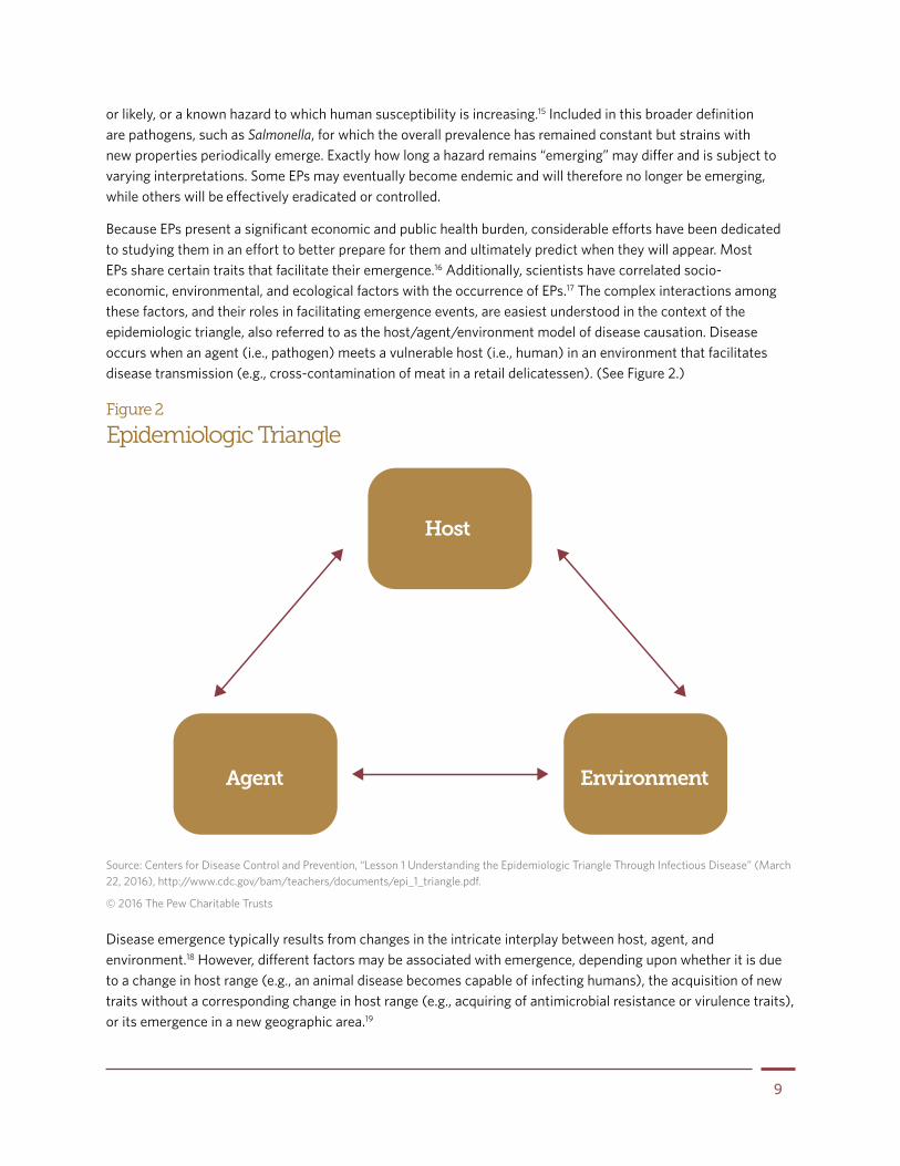
Source: Centers for Disease Control and Prevention, “Lesson 1 Understanding the Epidemiologic Triangle Through Infectious Disease” (March 22, 2016), http://www.cdc.gov/bam/teachers/documents/epi_1_triangle.pdf.
© 2016 The Pew Charitable Trusts
Figure 2
Epidemiologic Triangle
Host
Agent Environment
or likely, or a known hazard to which human susceptibility is increasing.15 Included in this broader definition are pathogens, such as Salmonella, for which the overall prevalence has remained constant but strains with new properties periodically emerge. Exactly how long a hazard remains “emerging” may differ and is subject to varying interpretations. Some EPs may eventually become endemic and will therefore no longer be emerging, while others will be effectively eradicated or controlled.
Because EPs present a significant economic and public health burden, considerable efforts have been dedicated to studying them in an effort to better prepare for them and ultimately predict when they will appear. Most EPs share certain traits that facilitate their emergence.16 Additionally, scientists have correlated socio-economic, environmental, and ecological factors with the occurrence of EPs.17 The complex interactions among these factors, and their roles in facilitating emergence events, are easiest understood in the context of the epidemiologic triangle, also referred to as the host/agent/environment model of disease causation. Disease occurs when an agent (i.e., pathogen) meets a vulnerable host (i.e., human) in an environment that facilitates disease transmission (e.g., cross-contamination of meat in a retail delicatessen). (See Figure 2.)
Disease emergence typically results from changes in the intricate interplay between host, agent, and environment.18 However, different factors may be associated with emergence, depending upon whether it is due to a change in host range (e.g., an animal disease becomes capable of infecting humans), the acquisition of new traits without a corresponding change in host range (e.g., acquiring of antimicrobial resistance or virulence traits), or its emergence in a new geographic area.19

10
Traits associated with the pathogen (agent)
Microorganisms are constantly evolving. Changes in genetic structure may make certain strains of a pathogen more efficient and better able to survive within changing environments or may allow them to survive in new environments. Pathogens can evolve into novel strains, become more virulent, or acquire resistance to antimicrobial drugs or other treatments. New pathogens may be discovered, or existing pathogens may acquire the ability to infect new hosts. Similarly, a pathogen that was previously controlled or had naturally declined can re-emerge or be reintroduced.
Certain microbial characteristics are thought to increase the risk of emergence. Zoonotic pathogens (i.e., those that can be transmitted between animals and humans) have been responsible for approximately 60 percent of infectious diseases emerging in a new host species since 194020 and are thought to pose a particularly high risk.21 Likewise, microbes that can infect multiple species, evolve at an inherently high mutational rate (e.g., RNA viruses), or are predisposed to the acquisition of genetic material (e.g., through horizontal gene transfer or reassortment) are thought to be at increased risk of emergence events.22
A number of foodborne EPs have surfaced over the past 40 years, and in recent years novel strains of them, as well as new pathogens that potentially could be transmitted through meat and poultry, continue to be discovered. Serotypes (i.e., specific subtypes of the bacterium defined by serological measures) associated with human illness have shifted over time, new virulent subtypes have emerged, and many strains have acquired antimicrobial-resistance genes.
Traits associated with the exposed individual or population (host)
How easily a pathogen can invade a human or animal is thought to be an important factor in disease emergence.23 A variety of risk factors have been identified, both on the individual level (e.g., age, underlying disease) and across populations (e.g., population structure, community composition, resilience toward invasion). Individuals may, for example, differ in their susceptibility to infection because of their immunological history (e.g., vaccination, prior exposure), genetic predisposition, exposure to pharmaceutical drugs (e.g., antacids, immune-modulating drugs), general health status (e.g., obesity, nutritional health), or because they have been affected by other diseases or conditions (i.e., “comorbidities” such as HIV/AIDS or diabetes mellitus).
Susceptibility to infection with foodborne pathogens increases with older age, diabetes mellitus and other comorbidities, as well as conditions such as obesity that lead to decreased immune responses.24 In the U.S., the number of people aged 65 and older is expected to increase 135 percent between 2000 and 2050, with the subpopulation of people aged 85 and older increasing almost 350 percent.25 Additionally, more than one-third of Americans are obese, and between 1990 and 2008 the rate of diabetes doubled.26
Many foodborne pathogens disproportionately affect the young, old, pregnant, and immunocompromised. In the future, demographic shifts resulting from population movements or differences in birth rates, as well as changes in the prevalence of underlying diseases or conditions, may lead to shifts in the susceptibility to foodborne infections.
Traits associated with the environment
A variety of environmental factors have been linked to disease emergence, including climatic factors (e.g., temperature, rainfall, and humidity), seasonality, and the presence of environmental barriers such as rivers or mountain ranges.27 Environmental factors may, for instance, prolong pathogen survival in the environment

11
or contribute to a pathogen’s dispersal through wind or rain runoff. The environment can also affect the seasonal presence of vectors such as ticks and mosquitoes, reduce host resistance (e.g., through heat stress or malnutrition), or lead to increased contact rates among people and/or animals (e.g., crowding during the winter). Therefore the contributions of host, pathogen, and environment to disease emergence are intricately linked.
A foodborne pathogen’s “environment” consists of the complete exposure pathway from its introduction into the food supply, to its eventual consumption by people. Increased exposure to microbial hazards can result from changes in food production or consumption, which can open new avenues for the introduction, proliferation, and transmission of pathogens.
Changes in food production and consumption patterns may lead to complex changes in disease epidemiology, even if these changes may not always be directly apparent from an analysis of the overall disease burden. For example, even if the overall incidence of a certain foodborne disease is not changing, the risks associated with one specific food commodity may be emerging. If the risks associated with other food commodities are simultaneously becoming better controlled, this can mask increases in disease incidence caused by the emerging risk. Demographic shifts can result in changes in food preparation and consumption and may affect associated risks. To better understand and be able to quickly react to an emerging foodborne risk, it is imperative for scientists to analyze emerging trends, in both overall pathogen incidence and in food vehicles associated with particular pathogens. It is equally important to have reliable methods for linking foodborne illnesses back to food vehicles—an often challenging task—and to understand the root causes that ultimately came together to cause the illnesses.
It is important to note that in some cases the increased recognition of an established hazard—for example, as a result of improved methods for pathogen detection or characterization, or through expanded surveillance—may be difficult to distinguish from the emergence of a new hazard. In fact, in many cases evidence suggests that pathogens that emerge in a new host species require time to reach full adaptation and often circulate for months or years before being recognized.28
Another point worth clarifying is that many EPs, by their very nature, are not as well understood as long-standing problems. Scientific evidence may be scarce, and expert communities may be split in their assessments of risk. For instance, the zoonotic potential and host range of EPs (i.e., the ability of a new animal disease to infect humans, and the risk of foodborne transmission) are often uncertain for newly emerged pathogens.
New and emerging foodborne pathogensFoodborne diseases are a significant public health challenge in the United States. The CDC estimates that microorganisms transmitted through food sicken one in six Americans each year, resulting in about 128,000 hospitalizations and 3,000 deaths.29
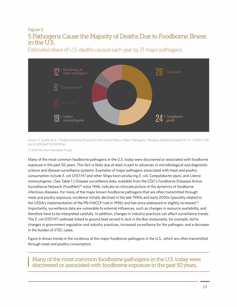
Of the 9.4 million cases of foodborne illness that the CDC ascribes to 31 major pathogens, over 90 percent are due to only five—norovirus, Salmonella, Clostridium perfringens, Campylobacter, and Staphylococcus aureus. (See Figure 3.)30 Similarly, five pathogens (Salmonella, norovirus, Campylobacter, Toxoplasma gondii, and Shiga toxin-producing E. coli) are estimated to cause 88 percent of hospitalizations due to these pathogens. (See Figure 4.) And five pathogens (Salmonella, T. gondii, Listeria monocytogenes, norovirus, and Campylobacter) are estimated to cause 88 percent of deaths attributable to these pathogens. (See Figure 5.) Beef, pork, poultry, and other animal products are estimated to be among the most important food vehicles for all of these pathogens, with the exception of norovirus and Staphylococcus aureus.31

12
58% Norovirus
11% Salmonella
10% Clostridiumperfringens
9% Campylobacter
3% Staphylococcus aureus
9% Remaining 26 major pathogens
26% Norovirus
35% Salmonella12% Remaining 26 major pathogens
15% Campylobacter
8% Toxoplasmagondii
4% STEC E. coli
Source: E. Scallan et al., “Foodborne Illness Acquired in the United States—Major Pathogens,” Emerging Infectious Diseases 17, no. 1 (2011): 7–15, doi:10.3201/eid1701.091101p1.
© 2016 The Pew Charitable Trusts
Source: E. Scallan et al., “Foodborne Illness Acquired in the United States—Major Pathogens,” Emerging Infectious Diseases 17, no. 1 (2011): 7–15, doi:10.3201/eid1701.091101p1.
© 2016 The Pew Charitable Trusts
Figure 3
5 Pathogens Cause the Majority of Foodborne Illnesses in the U.S.Estimated share of U.S. foodborne illnesses caused each year by 31 major pathogens
Figure 4
5 Pathogens Cause the Majority of Hospitalizations Due to Foodborne Illness in the U.S.Estimated share of U.S. hospitalizations caused each year by 31 major pathogens
13
Many of the most common foodborne pathogens in the U.S. today were discovered or associated with foodborne exposure in the past 50 years. This fact is likely due at least in part to advances in microbiological and diagnostic science and disease surveillance systems. Examples of major pathogens associated with meat and poultry consumption include E. coli O157:H7 and other Shiga toxin-producing E. coli, Campylobacter jejuni, and Listeria monocytogenes. (See Table 1.) Disease surveillance data, available from the CDC’s Foodborne Diseases Active Surveillance Network (FoodNet)32 since 1996, indicate an intricate picture of the dynamics of foodborne infectious diseases. For many of the major known foodborne pathogens that are often transmitted through meat and poultry exposure, incidence initially declined in the late 1990s and early 2000s (possibly related to the USDA’s implementation of the PR/HACCP rule in 1996) and has since plateaued or slightly increased.33 Importantly, surveillance data are vulnerable to external influences, such as changes in resource availability, and therefore have to be interpreted carefully. In addition, changes in industry practices can affect surveillance trends. The E. coli O157:H7 outbreak linked to ground beef served in Jack in the Box restaurants, for example, led to changes in government regulation and industry practices, increased surveillance for the pathogen, and a decrease in the burden of STEC cases.
Figure 6 shows trends in the incidence of the major foodborne pathogens in the U.S., which are often transmitted through meat and poultry consumption.
Many of the most common foodborne pathogens in the U.S. today were discovered or associated with foodborne exposure in the past 50 years.
11% Norovirus
28% Salmonella12% Remaining 26 major pathogens
6% Campylobacter
24% Toxoplasmagondii19% Listeria
monocytogenes
Source: E. Scallan et al., “Foodborne Illness Acquired in the United States—Major Pathogens,” Emerging Infectious Diseases 17, no. 1 (2011): 7–15, doi:10.3201/eid1701.091101p1.
© 2016 The Pew Charitable Trusts
Figure 5
5 Pathogens Cause the Majority of Deaths Due to Foodborne Illness in the U.S.Estimated share of U.S. deaths caused each year by 31 major pathogens

14
0.4
Rela
tive
rate
0.6
0.8
1
1.2
1.4
‘96-’98 ‘99 ‘00 ‘01 ‘02 ‘03 ‘04 ‘05 ‘06 ‘07 ‘08 ‘09 ‘10 ‘11 ‘12 ‘13 ‘14
Campylobacter O157:H7 STEC E. coli Listeria Salmonella
Source: Centers for Disease Control and Prevention, Foodborne Diseases Active Surveillance Network (FoodNet), “Data for Figures—2014,” data for Figure 2, Feb. 17, 2016, http://www.cdc.gov/foodnet/trends/data-for-figures-2014.html#ui-id-1.
© 2016 The Pew Charitable Trusts
Figure 6
Trends in Incidence of Major Foodborne PathogensRelative rates of laboratory-confirmed infections in the U.S. per year, compared with rates for 1996-98
New serotypes and molecular subtypes of common foodborne pathogens have emerged in recent years and continue to do so, with growing concerns about the impact on public health and importance of these strains. Examples include non-O157:H7 STEC and Campylobacter strains other than C. jejuni. In addition, pathogens have emerged with resistance to one or more antimicrobial drugs. These have been responsible for a number of notable foodborne outbreaks34 and reflect another emerging public health concern.
A variety of pathogens are newly emerging in the U.S. or around the world. Others have only recently become a concern for meat and poultry exposure, either from ingestion (i.e., oral exposure) or other food-associated exposure pathways—for instance, dermal contact between consumers and the contaminated meat they handle.
Which pathogens should be considered as emerging or potentially emerging hazards in the U.S. meat and poultry supply is somewhat subjective. The pathogens included in this report were selected because they were identified in one of the source documents in Table 1, or because they were identified as important potential hazards by two subject matter experts involved in writing this report based on their interpretation of the scientific literature (see box for more details).
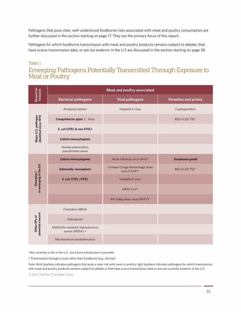
Table 1 shows emerging hazards of potential concern with exposure to meat or poultry products. As described in the overview, these include pathogens present in the U.S. as well as ones that may pose a risk if they are introduced to the U.S. at a future time. The hazards entail foodborne pathogens as well as pathogens such as Methicillin-resistant Staphylococcus aureus (MRSA) that may pose a food-handling risk.
15
*Not currently a risk in the U.S., but future introduction is possible.
† Transmission through a route other than foodborne (e.g., dermal).
Note: Bold typeface indicates pathogens that pose a clear risk with meat or poultry; light typeface indicates pathogens for which transmission with meat and poultry products remains subject to debate or that have scarce transmission data or are not currently endemic in the U.S.
© 2016 The Pew Charitable Trusts
Table 1
Emerging Pathogens Potentially Transmitted Through Exposure to Meat or Poultry
Meat and poultry associated
Bacterial pathogens Viral pathogens Parasites and prions
Arcobacter butzleri Hepatitis E virus Cryptosporidium
Campylobacter jejuni, C. Fetus BSE/vCJD/TSE*
E. coli (STEC & non-STEC)
Listeria monocytogenes
Yersinia enterocolitica, pseudotuberculosis
Listeria monocytogenes Avian influenza virus (AIV)* Toxoplasma gondii
Salmonella, monophasic Crimean-Congo hemorrhagic fever virus (CCHF)* BSE/vCJD/TSE*
E. coli, VTEC /STEC Hepatitis E virus
MERS-CoV*
Rift Valley fever virus (RVFV)*
Clostridium difficile
Helicobacter
Methicillin-resistant Staphylococcus aureus (MRSA) +
Mycobacterium paratuberculosis
Maj
or U
.S. p
atho
gen
iden
tified
sin
ce 19
70Re
ason
for
Incl
usio
n O
ther
EPs
of
pote
ntia
l con
cern
Emer
ging
/
re-e
mer
ging
in th
e EU
Pathogens that pose clear, well-understood foodborne risks associated with meat and poultry consumption are further discussed in the section starting on page 17. They are the primary focus of this report.
Pathogens for which foodborne transmission with meat and poultry products remains subject to debate, that have scarce transmission data, or are not endemic in the U.S are discussed in the section starting on page 38.

16
Methodology Used to Develop Table 1
A review of the scientific literature did not generate one single, comprehensive list of pathogens of emerging concern for the U.S. meat and poultry supply, defined for the purpose of this study as products derived from major (i.e., cattle, swine, chicken, and turkey) as well as minor (e.g., sheep, goat, ostrich, and duck) food-producing species. However, two relevant lists were identified in scientific publications. (Pathogens that appeared on both lists were included twice in Table 1.) The first, falling under reason for inclusion #1 (based on Emerging Foodborne Pathogens and Problems: Expanding Prevention Efforts Before Slaughter or Harvest*), lists pathogens that have emerged as foodborne risks since the 1970s and is based on a literature review. The second, identified by reason for inclusion #2 (based on “Drivers of Emerging Risks and Their Interactions in the Domain of Biological Risks to Animal, Plant and Public Health: A Pilot Study”†), highlights emerging biological risks in the European Union, based on the opinion of two expert panels (the Biological Hazard and Animal Health and Welfare panels) convened by the European Food Safety Authority to advise on scientific issues relevant to food safety and foodborne diseases in the European Union. Emerging issues considered in this opinion include new pathogens introduced or reintroduced in the EU, pathogens with possibly increased exposure, and pathogens identified because of changed susceptibility in the population.
We included only those pathogens from the two lists that may be of concern to the U.S. meat and poultry supply, using the following critiera for exclusion: those that cannot be transmitted to humans through meat or poultry (e.g., because they can infect a limited range of animal species such as fish, are animal but not human pathogens, or are known to be transmitted only through routes other than food); pathogens associated with game meat or certain nonconventional production systems that have decreased in incidence in the U.S. in recent decades (e.g., Trichinella spiralis); and pathogens not present in Europe, but commonly occurring in the U.S. (e.g., Corynebacterium paratuberculosis), because they are an established, rather than emerging, risk in the U.S. General risks identified in the two lists but not associated with a specific pathogen (e.g., “existing viruses becoming foodborne”) were not included in the table but are discussed in the report.
The condensed list generated by this method was supplemented to capture other risks that our two experts who contributed to this study consider to be of emerging or potentially emerging concern in the U.S. meat and poultry supply.
* C.B. Behravesh, I.T. Williams, and R.V. Tauxe, Emerging Foodborne Pathogens and Problems: Expanding Prevention Efforts Before Slaughter or Harvest, vol. A14, Improving Food Safety Through a One Health Approach: Workshop Summary (Washington: National Academies Press, 2012).
† European Food Safety Authority, “Drivers of Emerging Risks and Their Interactions in the Domain of Biological Risks to Animal, Plant and Public Health: A Pilot Study,” EFSA supporting publication (2014): EN-588.

17
Major meat- and poultry-associated pathogens with emerging strains In this section, the report focuses on the most common pathogens associated with meat and poultry products: Campylobacter, pathogenic E. coli, Listeria monocytogenes, Salmonella, and Toxoplasma gondii. The report discusses the risks these pathogens pose to people today, as well as emergence factors that may define how science will address them tomorrow. Because the threat and public health relevance of STECs differ from that of antimicrobial-resistant non-STEC E. coli, we discuss these two separately. Notably, some of these pathogens have emerged since 1970 but have now become well-established food safety risks in the U.S. Because of their value for lessons learned, we have included these hazards in the report even though they may no longer be typically considered EPs.
These pathogens were chosen for one of two reasons (see Table 1 and the corresponding methodology text box for a full rationale of inclusion criteria):
Reason #1: They have been recognized as foodborne pathogens only since 197035 and may therefore contain valuable lessons learned.
Reason #2: Expert panels convened by the European Food Safety Authority identified them as emerging or re-emerging diseases.36 (A comparable expert panel convened by U.S. authorities was not available at the time the report was written.)
CampylobacterReason for inclusion: #1
Although infections with Campylobacter species have probably caused human illness for centuries, it was not until 1968 that this bacterium was first isolated from stool samples of patients with diarrhea,37 and Campylobacter jejuni was first identified as a human diarrheal pathogen in 1973.38 The study of campylobacteriosis was made possible in large part by diagnostic advancements. Selective growth media39 and controlled growth conditions (e.g., under controlled gas atmospheres) were developed and increasingly used in the 1970s; by the late 1980s, Campylobacter was recognized as the leading cause of bacterial gastroenteritis in the world.40 In the United States, C. jejuni remained the leading cause of bacterial foodborne illness until about 2001, when it was surpassed by Salmonella.41
Recent changes in the epidemiology of foodborne campylobacteriosis were brought about by emerging strains beyond C. jejuni and Campylobacter coli. In addition, new data indicate that the use of certain prescription drugs may lead to an increased disease risk. New scientific knowledge has also led to a reconsideration of the most important transmission pathways.
Disease burden
The cost of foodborne campylobacteriosis is estimated at over $1.9 billion each year in the United States alone, from an estimated 850,000 cases of illness, over 8,000 hospitalizations, and nearly 80 deaths.42 In a 2012 ranking of major foodborne pathogens across all food commodities, Campylobacter in poultry was found to cause a greater disease burden than any other pathogen/food combination, being responsible for an estimated $1.3 billion in costs of illness annually.43 One reason for the high cost associated with Campylobacter infection is the

18
risk that after infection, patients can develop Guillain-Barré syndrome, an autoimmune disorder that leads to temporary paralysis and may cause permanent nerve damage.44
Campylobacter control is complicated by the low infectious dose (i.e., low number of bacteria needed to cause infection) and the resulting high risk of cross-contamination during and after slaughter (e.g., through shared cutting boards). However, the U.S. rate of campylobacteriosis dropped by half at the beginning of the 21st century, coinciding with and likely resulting, at least in part, from new practices implemented in poultry processing operations, such as the chlorination of water baths and chiller tanks, and improved sanitation during slaughtering.45 In 1997, the reported incidence peaked at 24.6 illnesses per 100,000 people per year, and subsequently fell to a low of 12.6 per 100,000 in 2003. As shown in Figure 7, the incidence has remained around 13 to 14 cases per 100,000 per year for more than a decade.46 Therefore, little progress has been made recently in reducing the rate of Campylobacter infections. Even though attributing Campylobacter illnesses to food vehicles has remained challenging, and raw milk is also an important food vehicle,47 these data do suggest that more needs to be done to control the Campylobacter risk associated with poultry.
Source: Centers for Disease Control and Prevention, Foodborne Diseases Active Surveillance Network (FoodNet), “Number and Incidence of Infections by Year, 1996-2014,” Table 2b, Feb. 17, 2016, http://www.cdc.gov/foodnet/trends/2014/number-of-infections-by-year-1996-2014.html#table2b
© 2016 The Pew Charitable Trusts
Figure 7
Trends in Culture-Confirmed Campylobacter InfectionsIncidence (per 100,000 population) of such infections in the U.S., by year
0
5
10
15
20
25
30
‘96 ‘97 ‘98 ‘99 ‘00 ‘01 ‘02 ‘03 ‘04 ‘05 ‘06 ‘07 ‘08 ‘09 ‘10 ‘11 ‘12 ‘13 ‘14
Inci
denc
e (p
er 10
0,00
0 po
pula
tion)

19
Emerging strains
Emerging Campylobacter species
While C. jejuni and C. coli are estimated to be responsible for more than 95 percent of campylobacteriosis cases, a number of emerging species have been associated with human illness.48 More than 10, including C. concisus, C. lari, C. upsaliensis, and C. ureolyticus, have been isolated from patients with gastroenteritis.49
Many of these emerging species have complex nutrient requirements, making them difficult to grow in bacterial culture, even some culture media routinely used for C. jejuni.50 Thus, these strains are likely under-detected in clinical isolates and underestimated in terms of disease burden. It is possible that an as-yet-unidentified species of Campylobacter may ultimately be found to be responsible for a considerable share of the large number of gastroenteritis cases without known etiology.51
Furthermore, C. concisus and some other species have been recently associated with Crohn’s disease, although much more research is needed to establish causality.52 Exposure routes for emerging species are not well defined, but some strains have been isolated from many of the same foods as C. jejuni and C. coli.53
Antimicrobial-resistant strains
Drug-resistant Campylobacter presents a considerable public health challenge and appears to be linked, at least in part, to antimicrobial drug use during food production. For example, the emergence of quinolone-resistant Campylobacter jejuni infections in the U.S. quickly followed FDA approval of fluoroquinolones for poultry use in 1995.54
Minnesota public health officials documented a rise in quinolone-resistant human C. jejuni isolates from 1.3 percent in 1992 to 10.2 percent in 1998. In 1997, they were able to isolate quinolone-resistant C. jejuni from 14 percent of retail chicken samples, and matched these strains to human illnesses using molecular subtyping.55 The CDC began tracking quinolone-resistant C. jejuni in 1997 and by 2002 found 20 percent of clinical isolates to be quinolone-resistant.56
International studies also support the role of agricultural drug use in the development of resistance: Countries with low or no fluoroquinolone usage on farms have a low prevalence of resistant infections despite the use of fluoroquinolones in human medicine, while countries with high fluoroquinolone farm usage have correspondingly higher rates of resistance in both human and animal isolates.57 A case-control study conducted by FoodNet found that people infected with fluoroquinolone-resistant Campylobacter were more likely than control groups to have consumed chicken or turkey.58
Concerns about the direct role of the agricultural uses of antimicrobial drugs in the emergence of resistant Campylobacter led the FDA to withdraw approval for use of fluoroquinolones in drinking water for poultry effective in 2005. The bacterium’s resistance to ciprofloxacin is variable but trending down. Based on surveillance of retail chicken samples by the National Antimicrobial Resistance Monitoring System (NARMS) for enteric bacteria, rates of C. coli resistance to ciprofloxacin, one of the most commonly used fluoroquinolones in humans, dropped from nearly 30 percent in 2005 to approximately 20 percent in 2013. Ciprofloxacin resistance rates for C. jejuni in retail chicken are at an all-time low, decreasing from about 15 percent at the time of the ban to 11 percent in 2013.59 Rates of quinolone resistance in human isolates peaked at 26 percent in 2007 and were at 23 percent in 2013.60
20
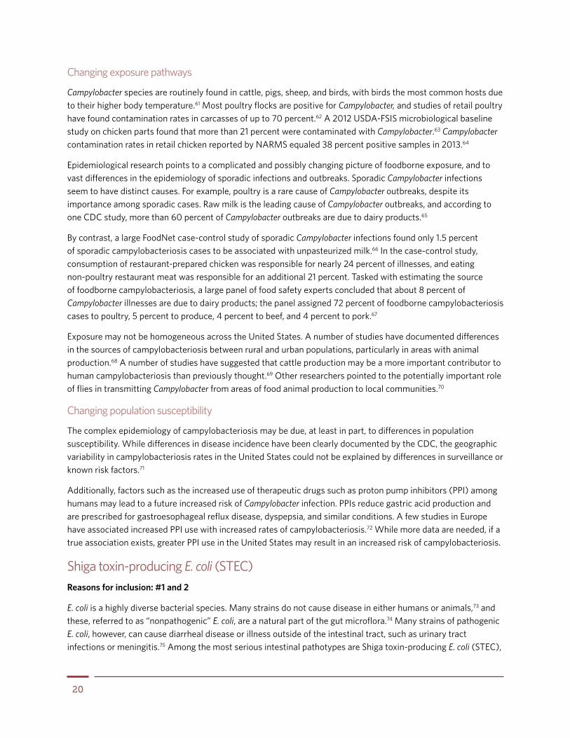
Changing exposure pathways
Campylobacter species are routinely found in cattle, pigs, sheep, and birds, with birds the most common hosts due to their higher body temperature.61 Most poultry flocks are positive for Campylobacter, and studies of retail poultry have found contamination rates in carcasses of up to 70 percent.62 A 2012 USDA-FSIS microbiological baseline study on chicken parts found that more than 21 percent were contaminated with Campylobacter.63 Campylobacter contamination rates in retail chicken reported by NARMS equaled 38 percent positive samples in 2013.64
Epidemiological research points to a complicated and possibly changing picture of foodborne exposure, and to vast differences in the epidemiology of sporadic infections and outbreaks. Sporadic Campylobacter infections seem to have distinct causes. For example, poultry is a rare cause of Campylobacter outbreaks, despite its importance among sporadic cases. Raw milk is the leading cause of Campylobacter outbreaks, and according to one CDC study, more than 60 percent of Campylobacter outbreaks are due to dairy products.65
By contrast, a large FoodNet case-control study of sporadic Campylobacter infections found only 1.5 percent of sporadic campylobacteriosis cases to be associated with unpasteurized milk.66 In the case-control study, consumption of restaurant-prepared chicken was responsible for nearly 24 percent of illnesses, and eating non-poultry restaurant meat was responsible for an additional 21 percent. Tasked with estimating the source of foodborne campylobacteriosis, a large panel of food safety experts concluded that about 8 percent of Campylobacter illnesses are due to dairy products; the panel assigned 72 percent of foodborne campylobacteriosis cases to poultry, 5 percent to produce, 4 percent to beef, and 4 percent to pork.67
Exposure may not be homogeneous across the United States. A number of studies have documented differences in the sources of campylobacteriosis between rural and urban populations, particularly in areas with animal production.68 A number of studies have suggested that cattle production may be a more important contributor to human campylobacteriosis than previously thought.69 Other researchers pointed to the potentially important role of flies in transmitting Campylobacter from areas of food animal production to local communities.70
Changing population susceptibility
The complex epidemiology of campylobacteriosis may be due, at least in part, to differences in population susceptibility. While differences in disease incidence have been clearly documented by the CDC, the geographic variability in campylobacteriosis rates in the United States could not be explained by differences in surveillance or known risk factors.71
Additionally, factors such as the increased use of therapeutic drugs such as proton pump inhibitors (PPI) among humans may lead to a future increased risk of Campylobacter infection. PPIs reduce gastric acid production and are prescribed for gastroesophageal reflux disease, dyspepsia, and similar conditions. A few studies in Europe have associated increased PPI use with increased rates of campylobacteriosis.72 While more data are needed, if a true association exists, greater PPI use in the United States may result in an increased risk of campylobacteriosis.
Shiga toxin-producing E. coli (STEC)Reasons for inclusion: #1 and 2
E. coli is a highly diverse bacterial species. Many strains do not cause disease in either humans or animals,73 and these, referred to as “nonpathogenic” E. coli, are a natural part of the gut microflora.74 Many strains of pathogenic E. coli, however, can cause diarrheal disease or illness outside of the intestinal tract, such as urinary tract infections or meningitis.75 Among the most serious intestinal pathotypes are Shiga toxin-producing E. coli (STEC),
21
also referred to as Verocytotoxin-producing E. coli (VTEC), which cause severe gastroenteritis characterized by bloody diarrhea and vomiting. STEC infection can cause hemolytic uremic syndrome (HUS), which can result in acute kidney failure and death. HUS is also associated with long-term sequelae, including chronic kidney disease, end-stage renal disease, hypertension, and deficits to many organ systems.76
There is perhaps no better example than STEC to study the emergence of a foodborne pathogen associated with meat. In 1982, CDC investigations of two outbreaks of severe bloody diarrhea associated with ground beef hamburgers led to the identification of a strain of E. coli that produced a toxin similar to the Shiga toxin of Shigella dysenteriae as a foodborne hazard.77 Foodborne outbreaks caused by this strain—E. coli O157:H7— began to be identified with increasing frequency, but it was not until January 1993 that the pathogen received public attention in the wake of an outbreak at the fast-food chain Jack in the Box, which sickened over 700 people and killed four children.78 The USDA-FSIS subsequently declared E. coli O157:H7 an adulterant, meaning a contaminant in a product that makes it unfit for human consumption, and shortly thereafter mandated the prevention-based regulations for meat and poultry plants known as the Pathogen Reduction/Hazard Analysis and Critical Control Point (PR/HACCP) rule.
More than 100 additional STEC strains have since been identified, and these non-O157 STECs are of increasing public health concern. While many are associated with milder forms of diarrheal disease, some can cause the same severe acute and long-term sequelae as O157, including hemorrhagic colitis, hemolytic uremic syndrome, and end-stage renal disease.
Disease burden
Foodborne E. coli O157:H7 is estimated to cause over 60,000 illnesses in the United States each year, resulting in about 2,000 hospitalizations and 20 deaths, while non-O157 STEC is estimated to cause over 100,000 foodborne illnesses annually, resulting in about 300 hospitalizations but no deaths.79 Foodborne STEC infections in the U.S. are estimated to cause approximately $300 million in economic costs.80 Worldwide, STECs are estimated to cause 2.8 million acute illnesses each year.81
CDC surveillance data show an increase in reported O157 infections from 1996 to 2000, as depicted in Figure 8, likely driven by increased reporting, followed by a decline in the early 2000s.82 Over the past decade, O157 disease incidence has remained largely unchanged.83
While the incidence of O157 disease has plateaued, reported cases of non-O157 STECs are on the rise, potentially in part because of improved
Foodborne E. coli O157:H7 is estimated to cause over 60,000 illnesses in the United States each year, resulting in about 2,000 hospitalizations and 20 deaths, while non-O157 STEC is estimated to cause over 100,000 foodborne illnesses annually, resulting in about 300 hospitalizations.

22
surveillance for non-O157 STECs. Figure 9 shows the sharply increasing rate of non-O157 outbreaks in the past 25 years. In another study of non-O157 STEC infections in the U.S. between 2000 and 2010, over half of the outbreaks occurred in the last four years of data.84 Within FoodNet, the number of reported isolates of non-O157 STEC equaled that of O157 in 2010. The extent to which these recorded increases reflect actual increases in disease incidence is unclear, partially because major improvements in reporting and detection, as well as an increase in testing for these strains, took place during this period.
Increases in detection and reporting capabilities were partially driven by regulatory changes. In response to an increasing incidence of infections linked to non-O157 STECs, USDA-FSIS declared the “Big Six” strains of STECs as adulterants in 2011. These are E. coli O26, O45, O103, O111, O121, and O145, which CDC had estimated to be responsible for more than 70 percent of non-O157 STEC infections.85 Simultaneously, the agency increased testing for these strains.
Even within these six major serogroups (i.e., serologically defined groups) of non-O157 STECs, however, the associated severity of disease varies considerably. For example, although only 16 percent (152 of 940) of all non-O157 human isolates sent to CDC in a 20-year period were found to be E. coli O111, this strain caused almost half (10 of 21) of the associated HUS cases.86 E. coli O111 is also the leading non-O157 strain associated with STEC outbreaks.87
Source: S.V. Sodha et al., “National Patterns of Escherichia coli O157 Infections, USA, 1996-2011,” Epidemiology and Infection 143, no. 2 (2014): 1–7, doi:10.1017/S0950268814000880.
© 2016 The Pew Charitable Trusts
Figure 8
Trends in Reported E. coli O157 InfectionsAnnual isolation rates in the U.S.
Num
ber o
f iso
late
s (p
er 10
0,00
0 po
pula
tion)
‘96 ‘97 ‘98 ‘99 ‘00 ‘01 ‘02 ‘03 ‘04 ‘05 ‘06 ‘07 ‘08 ‘09 ‘10 ‘110.2
0.4
0.6
0.8
1.0
1.2
1.4

23
Differences in clinical outcomes associated with non-O157 STEC infections are caused by a number of virulence factors. Of the two currently known major types of Shiga toxin (stx1 and stx2), STEC strains with the stx2 gene are associated with more severe clinical outcomes than are strains with the stx1 gene.88 Likewise, STEC strains expressing the virulence factor intimin (eae), which allows the pathogen to adhere to intestinal epithelial cells, have been found to cause more severe disease than strains not expressing eae.89 Other currently uncharacterized virulence factors may also be important in determining clinical outcomes.
Heterogeneity in strain-associated disease severity raises questions about how best to target public health interventions, including whether to focus on the serogroups most frequently isolated from human cases or on non-O157 strains most likely to cause severe disease (such as those expressing virulence genes stx2 and eae) regardless of their frequency of isolation.
Because of its magnitude, the 2011 outbreak of E. coli O104:H4 in Germany provides perhaps the strongest argument for focusing on virulence factors associated with severe clinical illness, even though the occurrence was not associated with meat or poultry.90 This outbreak, eventually traced back to contaminated sprouts, caused over 4,000 E. coli cases, 908 HUS cases, and 50 deaths.91 The novel strain that caused the outbreak was a particularly virulent hybrid of two pathogenic E. coli—a strain of an enteroaggregative E. coli (normally associated with severe diarrhea) that had acquired stx2 Shiga toxin genes from an STEC strain. It was also found to carry several antimicrobial-resistance genes, making treatment very difficult.
Source: R.E. Luna-Gierke et al., “Outbreaks of Non-O157 Shiga Toxin-Producing Escherichia coli Infection: USA,” Epidemiology and Infection 142, no. 11 (2014): 2270–80, doi:10.1017/S0950268813003233.
© 2016 The Pew Charitable Trusts
Figure 9
Reported Non-O157 Outbreaks Over Time
0
2
4
6
8
Num
ber o
f out
brea
ks
10
12
‘90 ‘91 ‘92 ‘93 ‘94 ‘95 ‘96 ‘97 ‘98 ‘99 ‘00 ‘01 ‘02 ‘03 ‘04 ‘05 ‘06 ‘07 ‘08 ‘09 ‘10
E. coli only E. coli and other pathogens

24
When determining the risk of future emergence events it is important to note that horizontal gene transfers (i.e., the transfer of genetic material among bacteria in a manner other than traditional reproduction), as seen in the German E. coli O104:H4 outbreak, are far from uncommon. As a case in point, Shiga toxins have been found contained within the DNA of bacteriophages,92 or viruses that infect bacteria. Bacteriophages can excise DNA from a host bacterium and may transfer the DNA to another bacterial cell; phages may, for instance, transfer virulence genes from a pathogenic bacterial strain to a nonpathogenic strain that does not otherwise cause illness.
Changing exposure pathways
Beef is the leading cause of foodborne infections with O157:H7 STEC in the U.S.93 Farm visits and exposure to cattle have also been identified as important risk factors.94 In the past several years, however, there has also been increased concern about STEC contamination of leafy greens and other produce, especially after a major outbreak of E. coli O157:H7 infections linked to prepackaged spinach in 2006.95 In this case, the outbreak strain isolated from patients was matched to bags of spinach, as well as to environmental samples, feces from cattle near the spinach farm, and fecal samples from feral swine.96 This example emphasizes the complicated and dynamic ecology of STEC infections and the central role food animals may play in both meat and produce safety.
Although cattle, and to a lesser extent other ruminants, have been established as natural reservoirs for O157:H7 STECs, the causes of emergence are unknown. Considerable research into cattle carriage and fecal shedding of O157 has found a highly dynamic system in which prevalence within farms is extremely variable over time.97 Diet is also important; the type of grain and processing method, forage quality, and use of distillers grains (particularly wet distillers grains with solubles) have all been associated with E. coli O157 prevalence in cattle feces.98 Results, however, are often conflicting, and other factors likely play a role. The factors that ultimately led to the emergence of STEC as a new pathogen in the 1980s have so far remained unclear.
Antimicrobial-resistant non-STEC E. coliReasons for inclusion: #1 and 2
Antimicrobial-resistant E. coli other than STECs (“non-STEC E. coli”) pose a distinct food safety issue, but their appearance in foods of animal origin is also an emerging public health concern.
Nonpathogenic E. coli are a natural and constant component of gut microflora and have a demonstrated ability to acquire, carry, and transfer resistance genes to pathogens that may be present in animals, the environment, or the human intestinal tract.99 The presence of drug-resistant nonpathogenic E. coli on meat or poultry products may therefore conceivably lead to the transmission of resistance genes to other bacteria, including potential pathogens, present on these foods or, after ingestion, inside the human gut.100
Notably, even though STECs are among the pathogenic E. coli of greatest public health concern, various other E. coli strains are also associated with severe disease in humans, including extraintestinal pathogenic E. coli (ExPEC). For example, uropathogenic E. coli cause urinary tract infections (UTIs), and meningitis-associated E. coli are associated with meningitis and sepsis.101 E. coli strains can quite readily exchange genetic material, including antimicrobial resistance or virulence genes, and it is believed that the emergence of drug-resistant ExPEC infections, including recurrent UTIs, may be caused by foodborne exposure, particularly to retail poultry.102

25
Emerging Antimicrobial Resistance
Assessing the relationship between antimicrobial use on farms and non-foodborne drug-resistant infections in humans is beyond the scope of this report, as well as a complicated issue: The horizontal transfer of resistance genes among unrelated bacteria; the spread of clonal strains among humans, animals, and the environment; and antimicrobial selective pressure from both human and animal use, make for a complex matter that cannot be easily dissected and studied.
Nevertheless, we know that agricultural antimicrobial use is associated with antimicrobial-resistant (AMR) bacteria in food animals, which can be shed into the farming environment and spread downwind or downstream.* As a result, AMR bacteria can colonize or infect farmworkers, or those living nearby.† As described elsewhere in the report, AMR pathogens can and do enter the food supply and cause human illness. ‡ The CDC estimates that drug-resistant Campylobacter and Salmonella, both primarily transmitted via foods of animal origin, are responsible for a combined 410,000 illnesses (i.e., 310,000 due to drug-resistant Campylobacter and 100,000 due to drug-resistant non-typhoidal Salmonella) and nearly 70 deaths (i.e., 40 due to drug-resistant non-typhoidal Salmonella and 28 due to drug-resistant Campylobacter) annually.§
In the 1980s and 1990s, multidrug-resistant strains of Salmonella emerged, particularly among serotypes Typhimurium and Newport. In particular, multidrug-resistant Salmonella Typhimurium DT104 emerged as a global epidemic in both animals and humans.¶ Since 1996, NARMS has been monitoring antimicrobial resistance among enteric bacteria isolated from humans, retail meats, and food animals.** NARMS shows the proportion of human isolates of non-typhoidal Salmonella resistant to two or more classes of antimicrobial drugs has dropped since the late 1990s, although about 1 in 10 infections were still multidrug-resistant in 2013. ††
Continued on next page
* T.R. Kelley et al., “Antibiotic Resistance of Bacterial Litter Isolates,” Poultry Science 77 (1998); J.C. Chee-Sanford et al., “Fate and Transport of Antibiotic Residues and Antibiotic Resistance Genes Following Land Application of Manure Waste,” Journal of Environmental Quality 38 (2009); A. Jindal et al., “Antimicrobial Use and Resistance in Swine Waste Treatment Systems,” Applied Environmental Microbiology 72 (2006); A. Chapin et al., “Airborne Multidrug-Resistant Bacteria Isolated From a Concentrated Swine Feeding Operation,” Environmental Health Perspectives 113 (2005); A.R. Sapkota et al., “Antibiotic-Resistant Enterococci and Fecal Indicators in Surface Water and Groundwater Impacted by a Concentrated Swine Feeding Operation,” Environmental Health Perspectives 115 (2007); R.I. Mackie et al., “Tetracycline Residues and Tetracycline Resistance Genes in Groundwater Impacted by Swine Production Facilities,” Animal Biotechnology 17 (2006); M.E. Anderson and M.D. Sobsey, “Detection and Occurrence of Antimicrobially Resistant E. coli in Groundwater on or Near Swine Farms in Eastern North Carolina,” Water Science and Technology 54 (2006); S.G. Gibbs et al., “Isolation of Antibiotic-Resistant Bacteria From the Air Plume Downwind of a Swine Confined or Concentrated Animal Feeding Operation,” Environmental Health Perspectives 114, no. 7 (2006); J. Schulz et al., “Longitudinal Study of the Contamination of Air and of Soil Surfaces in the Vicinity of Pig Barns by Livestock-Associated Methicillin-Resistant Staphylococcus aureus,” Applied and Environmental Microbiology 78 (2012).

26
† S.B. Levy, G.B. FitzGerald, and A.B. Macone, “Spread of Antibiotic-Resistant Plasmids From Chicken to Chicken and From Chicken to Man,” Nature 260 (1976); S.D. Holmberg et al., “Drug-Resistant Salmonella From Animals Fed Antimicrobials,” New England Journal of Medicine 311 (1984); R. Hummel, H. Tschäpe, and W. Witte, “Spread of Plasmid-Mediated Nourseothricin Resistance Due to Antibiotic Use in Animal Husbandry,” Journal of Basic Microbiology 26 (1986); Smith et al., “Quinolone-Resistant Campylobacter jejuni Infections in Minnesota, 1992-1998. Investigation Team”; F.M. Aarestrup et al., “Comparison of Antimicrobial Resistance Phenotypes and Resistance Genes in Enterococcus faecalis and Enterococcus faecium From Humans in the Community, Broilers, and Pigs in Denmark,” Diagnostic Microbiology and Infectious Disease 37 (2000); T.E. Besser et al., “Multiresistant Salmonella typhimurium DT104 Infections of Humans and Domestic Animals in the Pacific Northwest of the United States,” Epidemiology and Infection 124, no. 20 (2000); P.D. Fey et al., “Ceftriaxone-Resistant Salmonella Infection Acquired by a Child From Cattle,” New England Journal of Medicine 342 (2000); L.B. Price et al., “Elevated Risk of Carrying Gentamicin-Resistant Escherichia coli Among U.S. Poultry Workers,” Environmental Health Perspectives 115 (2007); I. Overdevest et al., “Extended-Spectrum β-Lactamase Genes of Escherichia coli in Chicken Meat and Humans, the Netherlands,” Emerging Infectious Diseases 17 (2011); M. Carrel et al., “Residential Proximity to Large Numbers of Swine in Feeding Operations Is Associated With Increased Risk of Methicillin-Resistant Staphylococcus aureus Colonization at Time of Hospital Admission in Rural Iowa Veterans,” Infection Control and Hospital Epidemiology 35 (2014); K. Smith, “Antimicrobial Resistance From Farm to Fork: Observations on the Impact of Antimicrobial Use in Animal Agriculture” (Minnesota Department of Health, 2015a).
‡ I. Chen, P.J. Christie, and D. Dubnau, “The Ins and Outs of DNA Transfer in Bacteria,” Science 310 (2005); N.M. M’ikanatha et al., “Multidrug-Resistant Salmonella Isolates From Retail Chicken Meat Compared With Human Clinical Isolates,” Foodborne Pathogens and Disease 7 (2010).
§ Centers for Disease Control and Prevention, “Antibiotic Resistance Threats in the United States, 2013” (Atlanta, GA, 2013).
¶ C.M. Parry, “Antimicrobial Drug Resistance in Salmonella enterica,” Current Opinion in Infectious Diseases 16 (2003).
** NARMS is a collaboration of CDC, FDA, and USDA focusing on Salmonella, Campylobacter, generic E. coli, and Enterococcus, as these organisms are ubiquitous in humans and food animals and can serve as reservoirs for resistance genes. The human program also includes Shigella, typhoidal Salmonella, and Vibrio.
†† Centers for Disease Control and Prevention, “National Antimicrobial Resistance Monitoring System: Enteric Bacteria, Human Isolates Final Report,” 2013.
Disease burden
Analyses of E. coli isolates from humans and animals suggest food and animal sources for the colonization and infection of humans with antimicrobial-resistant E. coli.103 The presence of antimicrobial-resistant E. coli in food-producing animals, their environments, and their end products therefore likely has direct relevance for the human disease burden.
Many studies have documented antimicrobial-resistant E. coli in retail meats and poultry, sometimes at high prevalence.104 For instance, from 2002 to 2008, NARMS recovered and examined E. coli isolates for antimicrobial susceptibility from nearly 12,000 food samples representing retail chicken parts, ground turkey, ground beef, and pork chops. As shown in Table 2, NARMS found in 2013 that 75 percent of retail chicken samples, and 78 percent of ground turkey samples, were positive for E. coli. Moreover, they found that resistance profiles differed according to animal origin. More than half of turkey isolates (59 percent) were multidrug-resistant (defined as resistance to at least three drug classes), compared with 31 percent of chicken, 14 percent of pork, and 8 percent of beef isolates.

27
* Based on data in Table 85a in the NARMS summary tables; Table 86a provides contradictory numbers.
Source: Centers for Disease Control and Prevention, National Antimicrobial Resistance Monitoring System for Enteric Bacteria (NARMS), Tables 83 (page 165) and 85 (pages 170 – 173), Feb. 18, 2016, http://www.fda.gov/downloads/AnimalVeterinary/SafetyHealth/AntimicrobialResistance/NationalAntimicrobialResistanceMonitoringSystem/UCM453387.pdf
© 2016 The Pew Charitable Trusts
Table 2
Prevalence of Antimicrobial Resistance Among E. coli From Different Meats
Percent resistance
Antimicrobial Chicken Breast (n=480)
Ground Turkey (n=478)
Ground Beef (n=480) Pork Chops (n=480)
Overall E. coli prevalence 75 78.2 47.3 43.3
MDR >= 5 drugs 2.8 9.6 2.2 1
MDR >= 3 drugs 31.4 59.4 7.9 13.9
Gentamicin 30.8 27 0 1
Kanamycin* 10.3 24.2 1.2 5.6
Streptomycin 38.9 54.2 8.4 17.8
Ampicillin 20.8 54 4.8 11.5
Amoxicillin-clavulanic acid 5.6 8.8 1.8 1
Cefoxitin 5 7.8 1.3 1
Ceftriaxone 4.4 6.7 2.2 1.4
Ceftiofur 4.4 6.4 1.8 1.4
Sulfisoxazole 39.2 50 7.9 10.1
Trimethoprim- sulfamethoxazole 3.1 3.7 1.8 1.4
Chloramphenicol 1.7 5.3 4 2.4
Ciproflaxin 0.6 0.3 0 0
Nalidixic acid 2.5 1.9 0.4 0
Tetracycline 43.3 74.3 22.5 51.4

28
Emerging strains
Trends suggest that strains of E. coli present in meat and poultry are becoming more resistant to frontline drugs used for treatment in humans, including third-generation cephalosporins, fluoroquinolones, and trimethoprim-sulfamethoxazole.105
Of particular public health concern among antimicrobial-resistant E. coli are strains that produce ESBLs, or extended-spectrum β-lactamases.106 These are a family of enzymes that confer resistance to third-generation (extended-spectrum) cephalosporins, as well as almost all other β-lactam antibiotics. ESBL genes in E. coli are typically found on mobile genetic elements, facilitating their carriage, accumulation, and transfer to other bacteria.107 ESBLs can be produced by a number of Enterobacteriaceae, but the most common are Klebsiella and E. coli. ESBL-producing Enterobacteriaceae are typically susceptible only to carbapenems and cephamycins, drugs of last resort. Carbapenem-resistant Enterobacteriaceae present one of the most pressing antimicrobial-resistance challenges facing human medicine today. The CDC estimates that health care-associated infections from carbapenem-resistant Klebsiella and E. coli are responsible for 9,000 infections and 600 deaths in the U.S. annually.108
ESBL-producing E. coli have been documented in animals and retail meat, although the proportion of overall drug-resistant infections transmitted to humans by food is unknown.109 A Dutch study of ESBL-producing E. coli isolates found strong similarities in ESBL genes and multi-locus sequence typing (MLST) strains between isolates from hospitalized patients and retail chicken.110 This study is not likely representative of the U.S. but is indicative of the possibility of ESBL genes disseminating from animals to humans via food.
Changing exposure pathways
Epidemiological and microbiological evidence suggests that some exposures to ESBL-producing E. coli in food may have led to extraintestinal infections such as urinary tract infections. Of the 6 million to 8 million UTIs diagnosed in the U.S. each year, more than 85 percent are caused by ExPEC infections.111 In recent years, drug-resistant ExPEC have rapidly emerged, including those producing ESBLs, making treatment more difficult and less likely to succeed.112 In a number of unusual community outbreaks suggesting a common exposure source, foodborne exposure was suspected but never confirmed.113
The potential role of food as a reservoir or source for these bacteria is supported by a number of studies finding ExPEC on chicken, turkey, pork, beef, and other foods.114 Some of these studies have identified close genetic similarities between strains from food animals, particularly chicken, and clinical isolates.115 A case-control study of women with UTIs found that higher rates of chicken consumption were associated with multidrug-resistant infections than with infections with strains that are susceptible to all antimicrobials.116 Furthermore, strong genetic similarities and similar antimicrobial resistance patterns have been documented between ExPECs and avian pathogenic E. coli (APEC), which causes extraintestinal colibacillosis in chickens.117
ESBL genes have been detected in APEC and the fecal E. coli of healthy poultry.118 Some studies suggest that the drug-resistance of APEC and poultry-associated ExPEC strains may be a result of antimicrobial use, for example cephalosporins, that may be administered to prevent or treat poultry diseases.119 There are too little data, however, to systematically assess risk factors for ESBL emergence and evaluate potential associations with antimicrobial use on farms.
As with cephalosporin-resistant Salmonella, the most promising starting point for controlling ESBL-producing E. coli in poultry and other food animals may be to limit the use of cephalosporins, as was recently proposed by the

29
EFSA Panel on Biological Hazards, and as implemented by the FDA.120 Still, because ESBL-producing E. coli strains have been found in the environment and isolated from wildlife, stopping cephalosporin use on farms may not be sufficient to reduce carriage in food animals.121
Listeria monocytogenesReasons for inclusion: #1 and 2
Although L. monocytogenes was first isolated from animals in the 1920s, its relevance to human health did not begin to be understood until 1949, as the result of an epidemic among newborns in Germany.122 However, questions about its epidemiology and transmission routes remained. During the 1980s, after a number of outbreaks in Canada and the United States, L. monocytogenes became recognized as a prominent foodborne pathogen. The risk of infection linked to the consumption of certain foods such as ready-to-eat meat appears to be declining, but an aging population will likely mean an increased public health impact in the future.
Disease burden
Foodborne listeriosis is a rare but serious infection, causing about 1,600 illnesses and 250 deaths annually in the U.S.123 Infections with Listeria monocytogenes are the third-leading cause of foodborne deaths in the U.S., behind Salmonella and T. gondii.124 Listeriosis has the highest costs of illness per case among all major foodborne pathogens: The estimated annual burden equals $2.8 billion a year, and the average cost per case has been estimated at $1.8 million.125
The overall incidence rate of listeriosis was estimated to be roughly 0.8 cases per 100,000 people in 1989, and more recent FoodNet data from 2004 to 2009 suggest a rate between 0.25 and 0.32 cases per 100,000.126 The number of recorded outbreaks has increased, however, from about one every four years prior to the creation of PulseNet127 in 1998 to about 2.2 outbreaks per year since then.128
Listeriosis is almost exclusively a disease of high-risk population subgroups, including the elderly, pregnant women and their newborns, and people with immune-compromising conditions.129 Within these high-risk population subgroups, listeriosis can cause a variety of severe clinical symptoms including septicemia, meningitis, and encephalitis.130 During pregnancy, listeriosis can result in miscarriage, stillbirth, preterm birth, septic conditions in newborns, and severe acute diseases with a heightened risk of serious neurodevelopmental sequelae.131 About 20 percent of symptomatic listeriosis cases result in death.132
Evolving understanding of strain-specific differences in public health risk
Although all strains of the species L. monocytogenes are considered pathogenic, experimental and surveillance data suggest that certain subtypes pose a greater threat to public health than others.133 Of the 13 currently known L. monocytogenes serotypes, only three (1/2a, 1/2b, 4b) are responsible for over 95 percent of human infections; of these, serotype 4b has been mainly associated with large outbreaks, while serotype 1/2a has been mostly associated with sporadic cases.134 Serotype 4b has a higher reported hospitalization and case-fatality rate than 1/2a or 1/2b.135
Changing exposure pathways
L. monocytogenes is a ubiquitous environmental bacterium, part of the fecal flora of many mammals, and widely distributed in many environments. It is also hardy, able to survive and grow in many conditions, including a wide range of pH values, high salt concentrations, and cold temperatures. Therefore, L. monocytogenes can

30
persist in cool, wet environments such as those experienced in some food manufacturing plants as well as most refrigerators.
The foods responsible for foodborne listeriosis have changed over the past 30 years, although some risks remain the same. A large Mexican-style cheese outbreak occurred in California in 1985. Over 140 people were infected, including 93 pregnant women, and 48 deaths occurred: 30 fetuses or newborns and 18 adults.136 Today, soft unripened cheeses remain a leading cause of listeriosis outbreaks.
Contaminated ready-to-eat meats have been another major source of listeriosis outbreaks in the U.S., although these appear to have peaked in the early 2000s and may now be declining. The decline is likely a direct result of concerted efforts by the food industry and regulatory agencies. Major successes have been achieved over the past decade in reducing contamination of deli meats through improved manufacturing practices in processing plants that minimize the risk of cross-contamination and in increasing formulation of products with substances that limit or prevent L. monocytogenes growth. USDA-FSIS and others have found the risks from retail-sliced deli meats to be up to five times greater than those from prepackaged cold cuts.137 In addition, simulation studies have shown that limiting environmental cross-contamination and preventing L. monocytogenes growth on contaminated products (e.g., through formulation with growth inhibitors or strict refrigeration at adequate temperatures) are the most promising interventions for reducing the risk of listeriosis associated with products handled at retail.138
Changing population susceptibility
Exposure to Listeria monocytogenes is believed to be quite common,139 however, most normal, healthy adults will not show clinical symptoms. Members of at-risk population subgroups, though, such as the elderly, pregnant women, and immunocompromised individuals, can suffer severe, potentially fatal illnesses. Based on U.S. surveillance data from 2004 to 2009, the incidence of listeriosis in the U.S. in the total population equals about 0.27 cases per 100,000 people, compared with 3.42 cases per 100,000 pregnant women and 1.21 cases per 100,000 individuals older than 65.140
Although the incidence of listeriosis is not increasing, emergence remains a future concern. Because listeriosis risk increases with age and the U.S. population is aging (i.e., the proportion of elderly individuals is increasing), there are concerns that the incidence of listeriosis could increase in the short to medium term. The number of people 65 and older is expected to increase by 135 percent between 2000 and 2050, with the subpopulation of those 85 and older expected to increase by almost 350 percent.141
Non-typhoidal SalmonellaReason for inclusion: #2
While Salmonella is not a new pathogen, new strains are emerging—including some that are resistant to antimicrobial drugs. In addition, our understanding of food vehicles associated with foodborne Salmonella infection is changing.
Disease burden
With very few exceptions (e.g., serotypes gallinarum and pullorum), non-typhoidal Salmonella serotypes are zoonotic (i.e., capable of infecting animals and humans), even though individual serotypes differ in host range and some (e.g., serotype Dublin) are primarily associated with animals. Non-typhoidal Salmonella strains are responsible for acute gastroenteritis, are primarily foodborne, and have been around a long time as well.

31
Recognition of their importance to human public health grew in the 1950s, and the CDC has been tracking Salmonella infections since 1962.142
Salmonella is the leading cause of foodborne bacterial disease in the United States today, and the CDC estimates that this pathogen alone is responsible for over a million illnesses, 20,000 hospitalizations and nearly 400 deaths a year.143 The approximate annual cost of illness exceeds $3.7 billion.144
Progress in reducing salmonellosis has been inadequate. In 1997, the annual reported incidence rate for Salmonella was 13.6 infections per 100,000 people. That rate became the baseline for the federal government’s Healthy People 2010 program, which set a target infection rate of 6.8 for 2010. By 2010, however, the reported incidence rate from FoodNet exceeded 15.145 Healthy People 2020 set a revised target rate of 11.4 cases per 100,000. However, the reported rate for 2013 was 15.9 infections per 100,000 people per year,146 about 17 percent higher than in 1997 and far above the new target rate.
Emerging strains
Changing serotypes
The somewhat static picture of overall salmonellosis incidence belies a dynamic picture of the strains responsible for disease. Hundreds of Salmonella serotypes are associated with human illness, many of which occur quite infrequently. Moreover, while some strains are in decline, others are emerging as important public health threats.
Figures 10 and 11 show incidence trends for the most common Salmonella serotypes rates. Figure 10 shows trends in the most commonly reported serotypes isolated by CDC’s FoodNet in 2014. Data are provided from 1999 to 2014 and are relative to the 1996-98 reference period. Figure 11 shows the relative change in the top Salmonella serotypes isolated in 2014, relative to 1996-98.
Both figures show declines in the frequency of S. Typhimurium and S. Heidelberg. Although S. Typhimurium had been the dominant serotype since surveillance began in the 1960s, its incidence has steadily declined; since 2007, S. Enteritidis has been the leading Salmonella serotype reported in lab-based surveillance and notifiable disease surveillance, and identified in outbreaks. Isolations of S. Newport, S. Saintpaul, and S. Infantis appear to be increasing. Likewise, in Figure 11, the best estimates for the incidence rates of S. Muenchen and S. Oranienberg show increases, though the confidence intervals for many of these are quite wide and cross the “no change” line, indicating that changes cannot be statistically proven.
Salmonella is the leading cause of foodborne bacterial disease in the United States today, and the CDC estimates that this pathogen alone is responsible for over a million illnesses, 20,000 hospitalizations and nearly 400 deaths a year.

32
Less common Salmonella strains are responsible for an increasing portion of reported human salmonellosis cases. Today, the leading serotypes reflect a lower proportion of overall isolates than they did in the past; the top five serotypes were responsible for about 65 percent of isolates submitted to CDC in 1987, but reflect only about 55 percent of isolates captured in the CDC’s Laboratory-Based Enteric Disease Surveillance (LEDS) system in 2011.147
Salmonella serotypes differ, sometimes substantially, in pathogenicity and host range. Salmonella serotypes have been demonstrated to have different relative rates of invasive infection, and differ in the probability of severe outcomes, including hospitalization and death.148 Some emerging serotypes, for example Newport and Javiana, have relatively milder average courses of illness compared with leading serotypes like Typhimurium and Enteritidis, while others such as serotype 4,[5],12:i:- may be associated with worse clinical outcomes.149 There may also be variation in associated disease severity within serotypes;150 genetic methods of identification can be used to further differentiate strains within them.
Source: S.V. Sodha et al., “National Patterns of Escherichia coli O157 Infections, USA, 1996-2011,” Epidemiology and Infection 143, no. 2 (2014): 1–7, doi:10.1017/S0950268814000880
© 2016 The Pew Charitable Trusts
Figure 10
Relative Rates of Culture-Confirmed Salmonella Infections With the Top Salmonella Serotypes in 2014 Compared With 1996-98 Rates, by Year
0
0.5
1.0
1.5
2.0
2.5
3.0
3.5
4.0
‘96-’98 ‘99 ‘00 ‘01 ‘02 ‘03 ‘04 ‘05 ‘06 ‘07 ‘08 ‘09 ‘10 ‘11 ‘12 ‘13 ‘14
Enteritidis Typhimurium Newport Infantis Heidelberg Saintpaul Muenchen
Rela
tive
rate
(log
scal
e)

33
* The estimates and confidence intervals presented indicate the relative change in the incidence of that serotype compared with 1996-98. The actual incidences of these infections cannot be determined from the graph.
† Data are preliminary.
Source: Centers for Disease Control and Prevention, Foodborne Diseases Active Surveillance Network (FoodNet), “Percentage Change in Incidence of Salmonella Infections,” Table 9, Feb. 18, 2016, http://www.cdc.gov/foodnet/trends/2014/percentage-change-of-salmonella-infections-2014.html.
© 2016 The Pew Charitable Trusts
Figure 11
Percentage Change in Incidence* of Culture-Confirmed Infections With the Top Salmonella Serotypes in 2014† Compared With Average Annual Incidence During 1996–98
-150
-100
-50
0
50
100
150
200
250
300
Enteritidis
Upper 95% confidence interval
Perc
ent c
hang
e
Point estimate Lower 95% confidence interval
Typhimurium Newport Infantis Heidelberg Saintpaul Muenchen
43%
-63%
176%
63%
-43%
54%
20%
Antimicrobial-resistant strains
Outbreaks of drug-resistant Salmonella are an emerging concern. Salmonella was the most common cause of antimicrobial-resistant foodborne outbreaks between 1973 and 2011 (48 of 55 outbreaks).151 Ground beef (n=10), poultry (n=7), and pork (n=1) together accounted for a considerable share of the 55 outbreaks with antibiotic-resistant bacteria.
Since 2012, there have been three additional notable foodborne outbreaks involving antimicrobial-resistant bacteria, all due to S. Heidelberg in chicken. One of the outbreaks sickened over 600 people and hospitalized more than 200 between March 2013 and July 2014. Of the 68 isolates tested during that outbreak, 44 (65 percent) were antimicrobial-resistant, of which more than half (i.e., 24 isolates) were multidrug-resistant.152
Drug-resistant Salmonella emerged as an international problem in the 1980s and 1990s. In particular, multidrug-resistant Salmonella Typhimurium DT104 emerged as a global epidemic in both animals and humans.153 Although the incidence of DT104 has been declining, other resistant strains have emerged.

34
For example, the rise of cephalosporin-resistant Salmonella among both human and animal isolates is an emerging concern. Similarly, resistance to ceftiofur (a veterinary cephalosporin) in animal isolates raises concerns about the future efficacy of ceftriaxone, a related drug used as a first-line therapy to treat human salmonellosis and other infections, including infections with Klebsiella and E. coli.154
Resistance to ceftiofur rose significantly among animal Salmonella isolates between 1997 and 2010: in cattle, from 0 percent to 21.5 percent; in chickens, from 0.5 percent to 11.9 percent; in turkeys, from 3.7 percent to 15.2 percent; and in pigs, from 0 percent to 1.8 percent.155 Some of the top Salmonella serotypes have seen increased resistance to ceftiofur: According to NARMS 2010 data, rates of resistance among Typhimurium isolates were 32 percent for retail chickens and 18 percent for cattle, and for Heidelberg they were 24 percent in chickens and 36 percent in turkeys. Over 70 percent of S. Dublin isolated from cattle were resistant. Although Dublin is not a major serotype in human disease, it has been associated with outbreaks and sporadic illnesses.156
Resistance to ceftriaxone, a related antibiotic, has also increased in human isolates over the same period.157 Across all serotypes of non-typhoidal Salmonella, resistance rose from 0.2 percent in 1996 to 2.9 percent in 2010, while increases were more pronounced for serotypes Typhimurium (from 0 percent to 4.7 percent), and Heidelberg (from 1.4 percent to 24.2 percent).
Regulatory Response to the Emergence of Cephalosporin Resistance
In response to concerns about the future efficacy of human cephalosporin therapy, the FDA first issued a broad ban on so-called extra-label* uses of cephalosporin in 2008.† This ban was revoked a short time later under criticism from the pharmaceutical and animal industries. In 2012, the agency issued a narrower ban on cephalosporins in animals, prohibiting most extra-label uses, including non-therapeutic uses, in cattle, swine, chickens, and turkeys.‡ The new rule does not cover the use of cephapirin, an older cephalosporin drug not considered by the agency to contribute substantially to antimicrobial resistance development, or use to treat minor food-producing species such as ducks or rabbits. The rule also allows for limited extra-label use of cephalosporins in cattle, swine, chickens, and turkeys if it follows label instructions regarding dose, frequency of administration, duration of treatment, and administration route.§
* As specified in the Animal Medicinal Drug Use Clarification Act of 1994 (AMDUCA), veterinarians are, under certain circumstances, permitted to prescribe certain approved veterinary or human drugs for extra-label use, meaning in a manner not in accordance with the approved labeling. Extra-label use may deviate from labeled use in a variety of ways, as in the species to which the drug is administered, the disease or condition the drug is used to treat, or the dosage levels, frequency, or routes of administration.
† C.W. Schmidt, “FDA Proposes to Ban Cephalosporins from Livestock Feed,” Environmental Health Perspectives 120 (2012).
‡ Ibid.
§ Ibid.

35
Canadian experiences with ceftiofur resistance support the view that ceftiofur use in animals may result in extended-spectrum cephalosporin resistance among bacteria isolated from animals and people.158 In Quebec in 2004, 62 percent of Salmonella Heidelberg isolates from chicken were ceftiofur-resistant, as were 36 percent of human isolates. In 2005, Quebec hatcheries voluntarily stopped the extra-label use of ceftiofur in eggs. By 2008, ceftiofur resistance among S. Heidelberg isolates dropped by two-thirds in both chickens and humans, to 18 percent and 12 percent, respectively. Ceftiofur-resistant E. coli in chicken dropped as well, from 34 percent in 2004 to 18 percent in 2008.159 Notably, corresponding reductions in the prevalence of resistance were also observed in neighboring jurisdictions. Whether this reflects indirect consequences of the ban in Quebec (e.g., shipment of eggs from Quebecois hatchers to neighboring provinces) or the impact of some external factors that affected multiple provinces has remained unclear.
Changing exposure pathways
Just as serotypes differ in associated disease severity, they also differ in transmission pathways and host range. Some Salmonella serotypes and subtypes are found solely in specific food animal species and their meat and animal products, while others are found in a wide variety of foods. Thus, serotypes have variable pathways to human exposure.
S. Dublin, for example, is almost entirely associated with cattle.160 Although this serotype is not a major cause of foodborne disease in the U.S., it has caused sporadic illnesses and outbreaks, and may be more likely to lead to severe clinical disease than other serotypes.161 Similarly, S. Enteritidis is primarily, yet not exclusively, associated with eggs and poultry, as are S. Heidelberg and S. Hadar.162
Overall, poultry and eggs are considered the leading causes of foodborne salmonellosis based on case-control studies, outbreak analysis, and other epidemiological studies. A recent CDC study of foodborne outbreaks found distinct food attribution patterns for serotypes Enteritidis, Heidelberg, Newport, Javiana, Typhi, and Typhimurium, as well as for all others combined.163 While poultry and eggs cause the greatest share of illnesses caused by serotypes Typhimurium, Enteritidis, Heidelberg, and Hadar, the serotypes Newport and Javiana are largely associated with produce.164
The findings from these and other studies165 point to an increasing role of food commodities other than meat, poultry, and eggs in foodborne salmonellosis. Salmonella can survive for long periods in secondary habitats outside of animal hosts, particularly aquatic environments. For example, Salmonella has been found to persist year-round and at high rates in surface waters of the southeastern United States.166 It also has been found to persist within agricultural environments despite efforts at decontamination.167 Moreover, flies and birds have been shown to be important vectors for transmitting Salmonella from farms to wildlife and local populations.168 More research, however, is needed to truly understand the complex natural cycles and interdependencies underlying environmental and foodborne exposure to Salmonella.
Toxoplasma gondiiReason for inclusion: #2
Toxoplasma gondii is a parasite that was first identified in 1908, although its complete life cycle was not understood until 1970.169 In recent years, toxoplasmosis has been of emerging concern because foodborne transmission is increasingly recognized as a primary exposure pathway and because of rising concerns about public health impacts associated with latent infections.
36
Disease burden
T. gondii, a protozoan parasite, is one of the most universal human parasites on the planet, found in every human population that has been investigated. About one in five Americans have been found to have serological evidence of prior infection, although this rate is estimated to be in decline.170
The CDC estimates that foodborne toxoplasmosis kills over 300 people in the U.S. each year, making it the second deadliest foodborne pathogen in the United States behind Salmonella. Largely due to high rates of mortality among the most susceptible population subgroup (i.e., embryos and fetuses), the annual costs of illness associated with foodborne Toxoplasma is estimated at $3.3 billion, second only to Salmonella.171
In most people, infection is asymptomatic or mild, resulting in swollen lymph nodes, fever, malaise, aches and pains, and other flu-like symptoms. In the immunocompromised, however, disease manifestation is usually very severe, including encephalitis, myocarditis, and pneumonia. In these cases, death is almost certain without treatment. It should be noted that most such severe cases in immunocompromised individuals are believed to be due to reactivated latent infections, not new ones.
If a woman becomes infected during pregnancy, tachyzoites can be transmitted to the fetus, resulting in congenital infection (i.e., the baby is born infected). The likelihood of transmission is higher at later stages of pregnancy, but earlier transmission is associated with more severe disease. Estimates of the rate of congenital toxoplasmosis vary widely, from 1 to 100 cases per 100,000 births, equaling up to 4,000 cases per year.172 Many of these children are asymptomatic at birth, although they can develop chorioretinitis later in life. Miscarriage or neonatal death can also occur, as can severe outcomes such as blindness, mental retardation, and other central nervous system abnormalities.
To correctly understand infection risks it is important to understand the complex life cycle of this pathogen. T. gondii has three primary life-cycle stages: oocysts, tachyzoites, and bradyzoites. (See Figure 12.) Sexual reproduction occurs solely in cats, where oocysts are produced and shed with the feces in very high numbers, during brief periods of time. After one to five days, these oocysts become infective and can survive for 12 to 18 months in the environment. Sporulated (infective) oocysts ingested by a mammal (e.g., rodent, pig, or sheep) or bird transform into tachyzoites, a rapidly dividing stage that spreads infection inside this intermediate host. Eventually, tachyzoites localize in skeletal muscle, heart, brain, and eyes, where they convert to tissue cysts, or bradyzoites. The parasite remains in these tissues indefinitely, suspended in a largely inactive state. If the bradyzoites are ingested by a cat, infection is followed by sexual reproduction and the cycle begins anew. However, when tissue cysts in meat are ingested by “accidental hosts” such as humans, bradyzoites transform back into tachyzoites, infecting the new host, but sexual reproduction cannot follow. The life cycle is thus interrupted.
Humans can become infected through direct contact with cat feces or fecally contaminated food (e.g., fruits, vegetables); they can also become infected with bradyzoites by ingesting meat or offal from infected animals, such as undercooked pork or lamb. In addition, humans can be infected through blood transfusions or organ transplants from infected individuals, or from a pregnant woman to the fetus.
Emerging health impact
In the past decade, new and controversial research suggests that latent T. gondii infection in the brain may affect human personality and behavior. For example, a number of studies have associated schizophrenia with prior infection by T. gondii.173 The illness has also been associated with higher rates of depression, suicide, risky
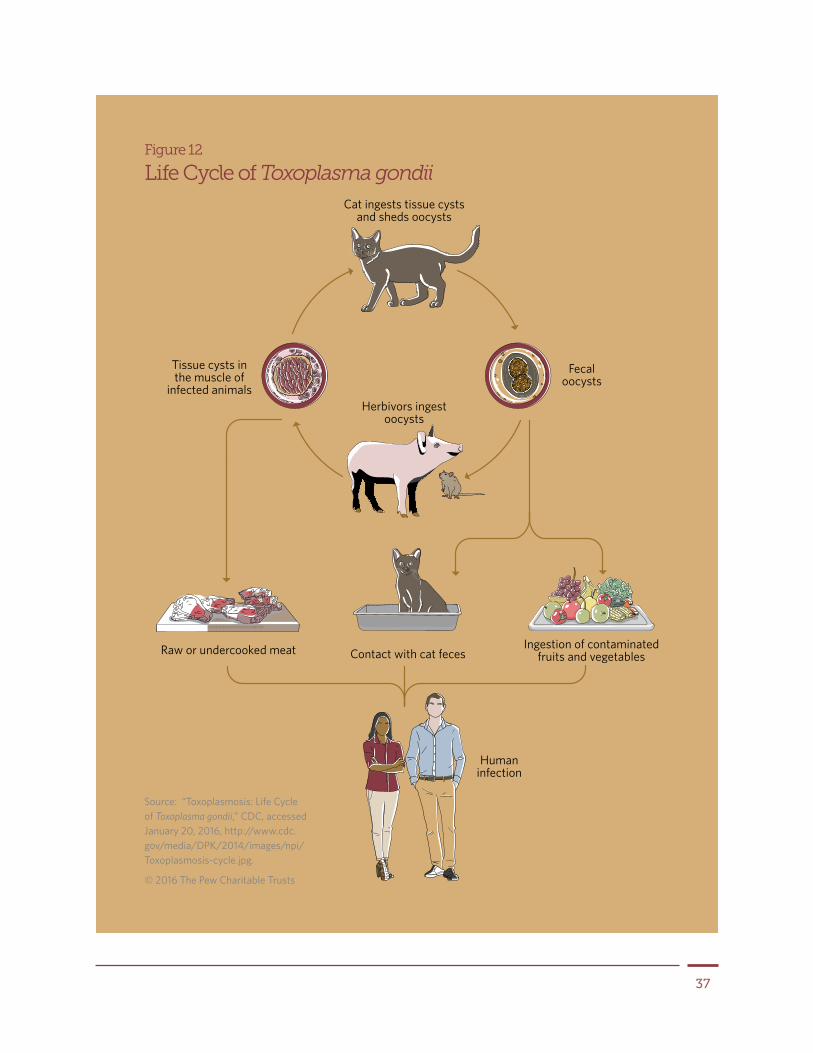
37
Humaninfection
Raw or undercooked meat
Cat ingests tissue cystsand sheds oocysts
Herbivors ingestoocysts
Fecaloocysts
Tissue cysts in the muscle of
infected animals
Ingestion of contaminated fruits and vegetablesContact with cat feces
Figure 12
Life Cycle of Toxoplasma gondii
Source: “Toxoplasmosis: Life Cycle of Toxoplasma gondii,” CDC, accessed January 20, 2016, http://www.cdc.gov/media/DPK/2014/images/npi/Toxoplasmosis-cycle.jpg.
© 2016 The Pew Charitable Trusts

38
behavior, personality shifts, changes to physical appearance associated with testosterone and estrogen levels, cognitive deficits, and reduced concentration.174
Much more research is needed to verify and solidify these associations and establish causality. If T. gondii infection does in fact contribute to any or all of these outcomes, the disease burden associated with foodborne toxoplasmosis, which is already quite high, could substantially increase.
Changing exposure pathways
The historic picture of T. gondii infection was that of direct contact with cat feces, typically through a litter box or soil. However, over the past 15 years scientists have begun to understand the critical role of foodborne exposure. The CDC now estimates foodborne exposure to be responsible for about half of all Toxoplasma infections in the United States each year.175
Foodborne exposure in people occurs from the ingestion of tissue cysts (bradyzoites) in raw or undercooked meat, tachyzoites in unpasteurized milk, or sporulated oocysts on plants or other foods that have been contaminated via water or soil. Routes of exposure are difficult to pin down in part because most acute infections are mild or asymptomatic.
Primary vehicles for foodborne transmission are estimated to be the ingestion of tissue cysts from raw or undercooked pork, lamb, and wild game.176 Unpasteurized goat’s milk, raw ground beef, raw shellfish, and fecal-contaminated soil on raw, unwashed fruits and vegetables have also been identified as risk factors.177 Numerous epidemiological studies have found undercooked meat to be an important risk factor, including a study that uncovered toxoplasmosis rates twice as high among meat eaters as among vegetarians.178 Surveys of retail pork, poultry, and beef have found low rates of contamination, although the prevalence of T. gondii has been found to be quite high in meat from other animals such as sheep, lamb, and organically raised pork and chicken.179 A recent study in Wisconsin found that children who live on farms are five times as likely to show signs of previous infections (i.e., antibodies) as those who do not live on farms.180
The importance of T. gondii in meat and poultry is likely substantial, but it is hard to measure. The overall incidence of T. gondii infection is uncertain, as are proportions due to consumption of meat, poultry, or the numerous other routes of exposure.
The CDC now estimates foodborne exposure to be responsible for about half of all Toxoplasma infections in the United States each year.
EPs with potential transmission through meat and poultry This section discusses the remaining pathogens identified in Table 1 as previously emerged or as potential emerging hazards to the U.S. meat and poultry supply. These bacterial, viral, and parasitic pathogens, as well as atypical prions, were chosen for one of three reasons (see Table 1 and the corresponding methodology text box for a full rationale of inclusion criteria):
Reason #1: They have been recognized as foodborne pathogens only since 1970181 and may therefore contain valuable lessons learned.

39
Reason #2: Expert panels convened by the European Food Safety Authority identified them as emerging or re-emerging diseases.182 (A comparable expert panel convened by U.S. authorities was not available at the time the report was written.)
Reason #3: They were identified as potential emerging threats by the experts involved in drafting this report.
For some of these pathogens, the potential for transmission with meat and poultry consumption has remained unclear or controversial. Other pathogens are not present in the U.S. but may conceivably be introduced in the country at a future time. These important caveats are highlighted where applicable. Notably, this section includes definitive and potential foodborne pathogens as well as pathogens that may be transmitted through contact with meat and poultry, such as MRSA.
Bacterial pathogensArcobacter butzleri
Reason for inclusion: #1
Arcobacter is a relatively newly identified genus of bacteria, related to Campylobacter. A. butzleri (formerly referred to as C. butzleri) is the most prominent of three species of Arcobacter associated with human enteric disease. A. butzleri shares similar microbiological and clinical features with Campylobacter jejuni but is more often associated with persistent, watery diarrhea than bloody diarrhea.183
General foodborne infection has almost never been proven for A. butzleri, but it has been isolated from meats including raw chicken, pork, beef, and lamb, with the highest prevalence among them in chicken.184 A. butzleri was only recently identified as the likely cause of a 2008 gastroenteritis outbreak after consumption of chicken at a wedding reception in Wisconsin.185 Notably, targeted tests for Arcobacter species were explored only after the comprehensive culture and polymerase chain reaction (PCR) testing of five stool samples failed to identify more common pathogens, emphasizing the potential for underdiagnoses of Arcobacter and other emerging or uncommon pathogens.
Like many of the emerging Campylobacter species, pathogenic Arcobacter may play an important role in many outbreaks and cases of sporadic disease for which an etiologic agent is not identified.
Clostridium difficile
Reason for inclusion: #3
Clostridium difficile is an emerging gastrointestinal pathogen. Although foodborne transmission is not confirmed, meat and poultry may serve as an important pathway for infection, as discussed below. C. difficile has been isolated from food animals and from retail meats and poultry, among other foods.186
C. difficile has historically been associated with hospital-acquired gastrointestinal infection, of which it is now the leading cause.187 Symptomatic C. difficile infections (CDI) generally follow disruptions to the intestinal microbiome. These interruptions are most commonly caused by treatments with antimicrobials, but they can also be the result of medical procedures, antacids, and more. Pathogenic strains of C. difficile can then overrun the gut and release toxins that cause bloating, diarrhea, and severe abdominal pain, sometimes with severe inflammation of the colon. These symptoms may last for weeks or months.188 Although treatment with antimicrobials can be successful in limiting symptoms, as many as 25 percent of patients have reported recurrences.189

40
C. difficile is a prototypical example of an EP: Disease incidence was stable until the mid-to-late 1990s, but starting in 2000 many studies have documented increasing CDI incidence, disease severity, recurrence, and antimicrobial resistance.190 At the same time, CDI is increasingly recognized as a cause of diarrhea not only in hospitals but also in communities, where it is affecting younger people and those without traditional CDI risk factors.191 More than 20 percent of CDI is now estimated to be community-associated, with some studies reporting a rate over 40 percent.192
CDIs were once believed to be almost entirely spread through person-to-person transmission via hands, shared hospital rooms, or other surfaces. More recent research, including examinations of hospital outbreak isolates using whole-genome sequencing and MLST, has found great diversity and nonclonal strains (i.e., not all strains being genetically essentially identical), thereby undermining this transmission model.193 Many infections previously thought to be nosocomial (i.e., hospital-acquired) may have actually been caused by foodborne exposure through hospital meals or via asymptomatic carriage of C. difficile into the institution from the community.194
Estimates of the share of adults who are asymptomatic carriers of toxigenic C. difficile vary from less than 2 percent to more than 7 percent, with causes of infection largely unknown.195 Potential risk factors for community-acquired CDI include asymptomatic carriers, use of proton pump inhibitors and other acid suppression medication, animal contact, and foodborne and waterborne exposure.196
Although colonization and infection via foodborne exposure has not been firmly established, C. difficile has been isolated from the feces of production animals, retail meats, poultry, seafood, and vegetables.197 Some, but not all, molecular studies have found similar strains in humans and animals, suggesting zoonotic transmission or a shared environmental reservoir.198 Among the most common strains found in food are PCR ribotype 078 and PCR ribotype 027, important causes of community-acquired CDI.199 Further research is needed to characterize the role meat and poultry consumption plays in CDI infections.
Helicobacter
Reason for inclusion: #3
Like Arcobacter, the genus Helicobacter was differentiated from Campylobacter, in this case in 1989. Over 35 Helicobacter species have been identified, with more likely to be discovered imminently. The most prominent species is H. pylori, for which humans are the reservoir. It is harbored in the upper gastrointestinal tract by as much as half of the world’s population and is a known cause of gastric ulcers and stomach cancer.200
Research into the role of H. pylori and other Helicobacter species in clinical human disease has expanded in recent years. Species other than H. pylori have been found in the gastrointestinal tracts and hepatic systems of other animals, and some have been associated (to varying degrees) with animal or human disease or both.201 Foodborne exposure, while hypothesized, has so far not been proven.202 For example, H. pullorum has been isolated from broiler chickens and laying hens and has been isolated from human patients with and without gastroenteritis.203 Still, for the most part, evidence linking pylori and non-pylori Helicobacter infection to human disease is limited. Far more research is necessary to understand the potential risks posed by this bacterium.
Clostridium difficile is an emerging gastrointestinal pathogen. Although foodborne transmission is not confirmed, meat and poultry may serve as an important pathway for infection.

41
Methicillin-resistant Staphylococcus aureus
Reason for inclusion: #3
Like C. difficile, invasive methicillin-resistant Staphylococcus aureus (MRSA) is a serious and emerging public health threat with potential but unconfirmed foodborne exposure.204 Transmission between food production animals and people has been substantiated, and MRSA has been isolated from a wide variety of meat and poultry products. Notably, the risk of MRSA infection is distinct from the risk of S. aureus food poisoning, a common food safety problem recognized since the 19th century.205 The latter occurs if certain S. aureus strains capable of generating toxins contaminate and multiply in food under the right conditions for toxin formation (e.g., temperature and food environment). The clinical symptoms of S. aureus food poisoning depend on the toxin and the ingested dose, but are usually relatively mild gastrointestinal symptoms that spontaneously resolve within 24 to 48 hours without requiring specific treatments.206
By contrast, invasive MRSA can cause skin and wound infections, or more severe illnesses such as pneumonia and bloodstream infections resulting in sepsis. An estimated 80,000 MRSA infections occurred in the United States in 2011, resulting in over 11,000 fatalities.207
In addition to hospital-associated strains, MRSA has found a reservoir in livestock, particularly swine, as well as horses, cattle, dogs, and cats.208 MRSA subtype 398 (ST398) was discovered in the early 2000s and has emerged as an important cause of human infection, often associated with exposure to livestock. A large study involving more than 30 authors from 20 institutions employed molecular methods on a diverse set of 89 ST398 isolates from humans and animals in 19 countries.209 The authors determined that this lineage originated as methicillin-sensitive S. aureus (MSSA) in humans, crossed the species barriers to pigs, and subsequently acquired tetracycline and methicillin resistance. Using state-of-the-art analyses, another group of researchers subsequently determined the existence of two distinct strains of MRSA ST398—one associated with humans and one with animals. The livestock-associated strain was found in a hospital environment and in newborn babies.210
Numerous studies in the United States and elsewhere make a compelling case for transmission of MRSA strains, including ST398, from livestock to humans. Some of these studies have found higher colonization and/or infection rates of MRSA among farmworkers, veterinarians, and others in close proximity to animals, potentially indicating direct zoonotic transmission.211
Of perhaps greater concern for public health are studies showing that proximity to animal production confers a risk of infection to the broader community. A recent study at an Iowa Veterans Affairs hospital found a threefold increased risk of MRSA colonization among people living within one mile of large swine facilities.212 Proximity to the application of swine manure has also been found to be a significant risk factor for MRSA infection.213
MRSA is not typically considered a foodborne pathogen, although community-associated and livestock-associated strains have been isolated at high rates in recent years from retail meat in the U.S. and elsewhere.214 Even if food consumption does not develop as a significant risk factor, handling raw meat may pose a transmission risk for MRSA from the farm to the community.
Mycobacterium avium subspecies paratuberculosis
Reason for inclusion: #3
Mycobacterium avium subspecies paratuberculosis (MAP) is the etiologic cause of paratuberculosis in ruminants, a fatal gastrointestinal disease that largely affects cattle. It is also a suspected cause of Crohn’s disease in

42
humans.215 Numerous epidemiologic studies have found associations between MAP and Crohn’s disease, but causality has not been confirmed and remains in dispute.216
If MAP causes Crohn’s, it could have implications for the safety of meat, milk, and dairy products.217 MAP is widespread in dairy herds, where it is shed in milk and feces; it has been found in milk supplies and in high concentrations can survive commercial pasteurization and cheese production processes.218 It has also been detected on carcasses and in organ meats. Because fecal shedding of MAP is much higher in older animals, risks of meat contamination are higher in the carcasses of culled dairy cows (i.e., cows that have been removed from the herd and sent to slaughter) than from younger animals (less than 3 years old) raised for meat.219 Limited studies have found that some MAP cells may survive cooking.220
Yersinia
Reason for inclusion: #1
The genus Yersinia spp. comprises pathogenic and nonpathogenic species.221 Among the human pathogens are enteropathogens such as Y. enterocolitica and Y. pseudotuberculosis.222 Infections of Y. enterocolitica and pseudotuberculosis primarily cause fever, diarrhea, and abdominal pain that can be misdiagnosed as appendicitis. Septicemia, skin rash, and other complications have been reported but are rare.223 Despite Y. enterocolitica’s first being isolated in 1934, its importance as a foodborne pathogen was not recognized until 1976.224 According to CDC estimates, approximately 90 percent of yersiniosis cases in the U.S. are foodborne. An estimated 97,656 foodborne yersiniosis cases are acquired in the U.S. each year.225 One study (based on expert elicitation) showed approximately 72 percent of U.S. yersiniosis cases attributable to pork, followed by 12 percent attributed to dairy and 5 percent to seafood.226
Y. enterocolitica is not emerging in the U.S. Incidence of infection with Y. enterocolitica has declined from a rate of about 1 in 100,000 in 1996 to about 0.28 in 2014.227 It was included in this report because of the potential lessons learned. Its recognition as a foodborne pathogen as late as 1976 demonstrates the fact that even for important foodborne pathogens the role of foodborne transmission may remain unclear for years or even decades. Y. enterocolitica is believed to be a commensal organism (i.e., an organism that benefits from another organism without affecting it either positively or negatively) of swine,228 so as the feral swine population increases in density and expands in range,229 Y. enterocolitica could conceivably become a larger risk to public health in the future.
VirusesAvian influenza virus
Reason for inclusion: #2
Avian influenza virus (AIV) is primarily a veterinary public health concern even though instances of foodborne transmission have been documented.230 A variety of influenza viruses can infect poultry as well as wild and pet birds.231 These viruses are typically classified as either low pathogenicity (LPAI) or high pathogenicity (HPAI) based on the disease they cause in poultry.232
HPAI viruses result in large outbreaks and severe illness, with very high mortality rates in poultry flocks.233 In fact, between December 2014 and June 2015, the U.S. experienced a devastating HPAI outbreak that severely affected parts of the U.S. turkey and egg industry.234 LPAI viruses can also cause outbreaks among poultry but are generally not characterized by severe clinical disease.235 However, through spontaneous genetic changes, LPAI

43
viruses can develop high pathogenicity. Most HPAI and LPAI viruses appear to be largely apathogenic in wild birds, which are believed to be an important reservoir for poultry infections and may disseminate the virus along migration routes.236
AI viruses generally do not infect humans; however, human-pathogenic AI viruses of the H5N1 and H7N9 subtypes have been emerging since 2003.237 These infections can be severe and potentially fatal. Human cases appear to be primarily sporadic and have been reported in Canada and several countries in Africa, southeast and central Asia, and the Middle East.238
Many questions remain about the epidemiology, transmission dynamics, and foodborne risks associated with these human-pathogenic strains. In general, contact with live or dead birds, their respiratory secretions, blood, feces, organs, or their environment is believed to be the primary risk factor for human infection, with a few cases also linked to consuming food made from raw poultry blood.239
Starting in December 2014, several HPAI strains have caused avian influenza outbreaks in commercial and backyard poultry flocks as well as wild bird populations in the U.S. While none of these strains are believed to be human-pathogenic, the outbreaks demonstrate the risk of HPAI virus introduction.240
Crimean-Congo hemorrhagic fever virus
Reason for inclusion: #2
Crimean-Congo hemorrhagic fever virus (CCHFV) is the cause of a potentially fatal hemorrhagic fever in humans, with a fatality rate of 10 to 40 percent.241 CCHFV occurs sporadically in Africa, Asia, the Middle East, and parts of Europe, and it may be emerging or re-emerging in several geographic regions including India and parts of southern Europe.242
CCHFV is a tick-borne virus in the family Bunyaviridae.243 A variety of domestic animal species, including cattle, sheep, and goats, are susceptible to infection but are not believed to exhibit clinical symptoms.244 Human infections are thought to occur primarily as a result of contact with the blood or tissues of infected livestock, or from the bite of an infected tick.245 A variety of tick species are believed to be vectors for CCHFV.246 Many epidemiological aspects associated with the establishment of disease foci, and in particular with a limited geographic spread in some areas, remain unclear.247
The potential for foodborne transmission of CCHFV is currently unknown, but risk is generally believed to exist through contact with meat or organs, at least immediately after slaughter.248 There have been no known domestically acquired CCHFV infections in the United States, but the possibility for its introduction to the U.S. cannot be excluded.
Hepatitis E virus
Reasons for inclusion: #1 and 2
Hepatitis E virus (HEV) is a cause of acute hepatitis in humans.249 While many infections are believed to be self-limiting, HEV infection can lead to liver failure, particularly in pregnant women and patients with chronic liver disease. It can cause chronic hepatitis, primarily among immunosuppressed individuals.250 In addition, infection during pregnancy can lead to premature birth, fetal loss, and health problems for the newborn such as hepatitis or hypoglycemia.251
HEV is a small, non-enveloped virus with a positive-sense, single-stranded RNA genome.252 Four genotypes are

44
currently recognized—HEV-1, HEV-2, HEV-3, and HEV-4—all of which infect humans, and the distinction of additional genotypes has been proposed.253 The first two types, HEV-1 and HEV-2, generally cause more severe clinical symptoms.254
HEV was first isolated from swine in the U.S. in 1997, and infection is believed to be asymptomatic.255 Despite numerous questions about epidemiology and transmission dynamics, the zoonotic nature of HEV-3 and HEV-4 is generally accepted, and the pathogen has been isolated from a variety of animal species, including swine, cattle, sheep, deer, and rabbits.256
Potential foodborne transmission of HEV from pork and pork products has received increased attention in recent years.257 The virus appears to be endemic among swine in the U.S. and other countries, and it has been isolated from commercial pig livers sold in U.S. grocery stores.258 In 2007 and again in 2013, outbreaks of HEV in France were attributed to the consumption of raw pork liver sausages.259
Many questions remain about HEV’s potential foodborne transmission through pork or other meats. However, if a foodborne risk is firmly established, HEV could present an emerging global food safety risk.
Middle East respiratory syndrome coronavirus and related coronaviruses
Reason for inclusion: #2
Middle East respiratory syndrome coronavirus (MERS-CoV) causes a primarily respiratory disease in humans260 and is emerging in the Middle East. Infection can lead to pneumonia, respiratory failure, and in some cases other symptoms, including diarrhea.261 Current World Health Organization (WHO) estimates place the case-fatality rate up to 36 percent,262 though numbers may be skewed by the underdiagnosis of milder infections.
MERS-CoV is a small, enveloped virus with RNA genome; it is a relative of SARS-CoV, which caused the severe acute respiratory syndrome (SARS) pandemic of 2003.263 MERS-CoV was first isolated from individuals with acute respiratory illness in Saudi Arabia in 2012.264 As of June 2016, 1,768 laboratory-confirmed cases had been reported to the WHO, including at least 630 deaths.265 They were reported from various countries in the Middle East, and some presumably travel-related cases have been reported in the United States, France, Germany, Italy, the United Kingdom, Malaysia, the Philippines, and elsewhere.266
Many aspects concerning the emergence, epidemiology, transmission dynamics, and host range of MERS-CoV so far remain unclear. However,
HEV appears to be endemic among swine in the U.S. and other countries, and it has been isolated from commercial pig livers sold in U.S. grocery stores. In 2007 and again in 2013, outbreaks of HEV in France were attributed to the consumption of raw pork liver sausages.

45
serological and virological evidence suggests that the virus may have originated in bats, that dromedary camels may be an important source of human infections,267 and that the virus may have been circulating in the camel population for as long as several decades.268 Dromedary camels have been shown to shed MERS-CoV in their feces and milk,269 and people who work closely with camels appear to be at a heightened risk of infection.270 Whether the consumption of undercooked camel meat poses a transmission risk remains to be determined. Serological surveys of other animals, including goats, sheep, and cows, have as yet provided no evidence of infection.271
MERS-CoV does not seem to pose a threat to the U.S. meat supply. However, should host range and geographic distribution change, MERS-CoV may pose an emerging hazard in the future. Notably, it has been theorized that the related SARS-CoV entered the human population through foodborne exposure, including consumption of civet cats, an intermediate host, and potentially bat meat.272
Rift Valley fever virus
Reason for inclusion: #2
Rift Valley fever virus (RVFV) causes an illness in humans273 believed to manifest most often as a benign flu-like sickness. In rare cases, however, severe forms can be associated with meningoencephalitis, hemorrhagic fever, and retinal lesions, which may result in permanent loss of vision.274
RVFV, a member of the Bunyaviridae, is a small, enveloped virus with RNA genome.275 It can infect humans and a broad range of animals, including cattle, sheep, and goats.276 Mosquitoes and infectious wildlife are thought to be the primary sources of infection for livestock. For humans, consumption of meat and possibly raw milk have been proposed as potential transmission routes, with mosquito bites and contact with infected animals or their secretions another potential risk.277 RVFV is endemic in tropical regions of eastern and southern Africa, where it is believed to primarily circulate among ruminant wildlife reservoirs and hematophagous (i.e., blood-sucking) mosquitoes.278 Explosive outbreaks among livestock occur periodically (i.e., every five to 25 years), probably driven by a combination of climatic factors (i.e., precipitation resulting in explosive hatching of mosquito eggs) and demographic ones (i.e., availability of naive hosts that have not previously been in contact with the virus).
RVFV is not endemic in the U.S.279 However, a broad range of hematophagous mosquitoes, including certain Aedes, Anopheles, and Culex species, are believed to be capable of transmitting RVFV.280 The emergence of West Nile virus, a mosquito-borne flavivirus introduced in the U.S. in 1999 that has since become endemic, demonstrates the potential risk of introducing vector-borne diseases into the U.S. if a competent vector (i.e., one that can effectively transmit the disease) naturally occurs or is introduced.281
Parasites and prionsCryptosporidium parvum
Reason for inclusion: #1
Cryptosporidiosis has been recognized as a potential foodborne disease of humans and livestock since the 1970s and emerged as an important zoonotic human pathogen in the wake of the HIV/AIDS epidemic.282
Cryptosporidium parvum is a zoonotic parasite commonly shed by cattle and other livestock species.283 It causes diarrheal illness in humans as well as livestock, particularly neonatal calves and lambs, around the world.284 In immunocompetent individuals, this illness is typically self-limiting and non-life-threatening, but for those who are

46
immunocompromised, particularly HIV/AIDS patients, disease manifestations are severe and potentially fatal.285 For these patients, severe extraintestinal manifestations such as biliary tract disease, respiratory infections, and pancreatitis are possible.286
Cryptosporidium parvum is believed to be transmitted primarily through the ingestion of food or water that has been contaminated with fecal material shed by infected livestock.287 Consumption of water and fresh produce presumably contaminated by livestock manure has been identified as a source of Cryptosporidium exposure in various outbreaks and case-control studies, indicating a clear risk associated with manure runoffs.288
Consumption of contaminated uncooked or undercooked meat may also pose some risk of infection. An expert elicitation289 study examining the sources of foodborne illness in the U.S. attributed 60 percent of cryptosporidiosis cases to produce, 9 percent to beverages, 7 percent to beef, and 6 percent to dairy.290
BSE and new variant Creutzfeld-Jacobs Disease (vCJD)
Reason for inclusion: #2
Variant Creutzfeld-Jacobs Disease (vCJD), first described in 1996, is a rare, invariably fatal neurodegenerative disease that is presumably linked to the consumption of beef from cattle infected with bovine spongiform encephalopathy (BSE), even though the time between consumption and illness probably spans many years.291 A very small number of classic CJD cases used to occur prior to the emergence of vCJD, either as sporadic cases or linked to genetic predisposition or iatrogenic exposures (i.e., related to medical treatments).292 Multiple aspects distinguish vCJD from classical CJDs, including disease onset in younger patients (median death at 28 years compared with 68 years), a longer duration of disease, specific clinical symptoms, and distinctive neuropathological changes.293
CJD and BSE are conditions classified as transmissible spongiform encephalopathies (TSEs); they have long been known to occur with low prevalence among humans (e.g., kuru among cannibals) and other species such as sheep and goats (i.e., scrapie), deer (i.e., chronic wasting disease), or mink (i.e., transmissible mink encephalopathy), and some risk of cross-species transmission may exist.294
TSEs are generally believed to be caused by prions, “misfolded”295 proteins that do not contain genetic material but that can “replicate” by inducing other proteins to misfold, even though some scientific controversy remains about the causative agent, and many aspects related to pathogenicity, transmission dynamics, genetic predisposition, strain differences, and emergence are still unclear.296
BSE was first diagnosed in U.K. cattle in the 1980s and presumably emerged through feed-borne exposure of cattle to infectious scrapies prions in meat and bone meal (MBM).297 BSE is a progressive neurodegenerative disease that primarily affects cattle and exhibits a long incubation period, averaging four to five years in cattle.298 Other livestock species, including sheep and goats, appear susceptible to infection, and intentional infections of laboratory animals by scientists indicate a potential for transmission to species such as pigs, cats, mink, and primates.299
Feed-borne exposure to MBM is thought to be the primary transmission route for BSE among cattle, even though maternal transmission cannot be completely excluded.300 Control options implemented to contain the spread of BSE among cattle included a ban on the use of animal-derived proteins presumably at high risk for transmission (such as MBM) in bovine feed, the implementation of a passive animal disease surveillance system, and an active surveillance system based on the routine diagnostic testing of cattle at slaughter, paired with trace-back investigations on positive samples.301

47
Diagnostic tests are more sensitive toward the end of the incubation period because by then considerable amounts of atypical prions (abnormal PrP) have been accumulated.302 Because of the long incubation period for BSE among cattle and the potential slaughtering of cattle in some production systems and geographic regions before diagnostic tests could detect BSE, incidence rates may not be comparable across countries or periods in time.303 The impact that changing concentrations of atypical prions during the incubation period may have on infectivity and the risk to human transmission is difficult to quantify.
The incidence of diagnosed BSE cases among livestock has been declining.304 Today, only a small number of cases are diagnosed in cattle in the U.K. and around the world, and the U.S. has been categorized by the World Organisation for Animal Health as having a negligible BSE risk.305
Control options implemented to contain the spread of BSE among cattle included a ban on the use of animal-derived proteins such as meat and bone meal in bovine feed, the implementation of a passive animal disease surveillance system, and an active surveillance system based on the routine diagnostic testing of cattle at slaughter, paired with trace-back investigations on positive samples.
Recommendations and conclusions
Emerging pathogen preparednessWhile today’s microbial risks in meat and poultry appear similar overall to what they were a decade or more ago, some specific and important differences exist. Food safety risks have not remained static; some microbial hazards, many parasitic diseases in particular, have been successfully controlled or eliminated, while some new microbial hazards have emerged. It is important to recognize that newly emerging hazards often are not well understood initially. The specific implications for food safety and public health may materialize almost immediately, as in the case of E. coli O157:H7 in undercooked hamburgers, or may remain opaque for quite some time. The latter was, in fact, the case for some pathogens such as Listeria monocytogenes that are now unanimously accepted as major food safety risks.
The emergence of new foodborne hazards can be driven by the microbe’s acquisition of virulence or resistance characteristics, by demographic changes and associated increases in population susceptibility, or by a pathogen’s emergence in a new host species or geographic region. In addition, new scientific knowledge may lead to re-evaluating and re-prioritizing of known risks.
Detecting the emergence of pathogens can pose tremendous challenges. Existing surveillance systems may not be adequate to detect EPs in a timely manner. Diagnostic tools may be incapable of distinguishing the EPs from any existing ones, and determining causal relationships between the presence of a pathogen and clinical disease can be extremely difficult. Sufficient epidemiological evidence may not be available to pinpoint transmission routes, especially if disease dynamics are complicated by multiple hosts and exposure pathways. The public health risks posed by the EP are often unclear, and data to assess these risks are scarce. Even if these challenges can be overcome, adequate regulatory policies are rarely in place to address the issue, and the relevant food industry segments may be ill-prepared to address the emerging hazards.

48
In this report, we discuss a number of emerging microbial hazards that may be transmissible through the U.S. meat and poultry supply. These include bacteria, viruses, parasites, and prions. For the purpose of emerging disease preparedness, these hazards can be broadly categorized into three distinct classes:
1. Completely new, previously unknown pathogens
The emergence of these zoonotic pathogens is an exceedingly rare event, but one with immense consequences for public health and the economy.306 This type of emergence is typically caused by a mechanism called “host jumping,” where a pathogen acquires the ability to infect a new species. For example, BSE emerged from scrapie or a related prion disease, and some avian influenza viruses acquired the ability to infect humans. Often, these emergence events are initially limited in geographic distribution, but they have the ability to expand quickly in geographic range. For example, the H5N1 avian influenza virus emerged in Asia and BSE emerged in the U.K., but both soon posed a pandemic threat.
Because these pathogens are truly “new” pathogens, many aspects related to epidemiology, transmission dynamics, and public health risk are initially unknown, and vaccines or treatments are not available. These emergence events tend to receive extensive public attention and media coverage. For instance, the emergence of SARS coronavirus in 2003 prompted the U.S. CDC to handle more than 10,000 press calls, 21 telebriefings, 23 health alerts, more than 2,000 clinical consultations, and three satellite broadcasts; there were eight laboratory-confirmed SARS cases in the U.S., all travel-associated, and no fatalities.307
In many cases, when a new pathogen emerges, civic life is uprooted, trade is disrupted, and the economy is devastated. For instance, the emergence of Nipah308 virus, first recognized in Malaysia in 1998, caused at least 257 confirmed human infections and 115 fatalities, affected more than 1,000 pig farms, and led to the culling of more than 1 million pigs. Government compensations for the destroyed pigs alone equaled an estimated $35 million in U.S. dollars. The cost of the national control program equaled $136 million, and lost tax revenue has been estimated at $105 million.309 An estimated 36,000 people became unemployed. More than 700 homes and businesses were evacuated. Malaysian pork exports came to a halt, and local pork consumption dropped by up to 80 percent. Financial losses to the pork industry during the outbreak alone totaled an estimated $124 million.
Because of the considerable societal impact, these emergence events often lead to transformative regulatory changes. For instance, as a result of the Nipah outbreak, Malaysia now permits pig farming only in “identified pig farming areas.”310 After the emergence of human-pathogenic avian
The emergence of new foodborne hazards can be driven by the microbe’s acquisition of virulence or resistance characteristics, by demographic changes and associated increases in population susceptibility, or by a pathogen’s emergence in a new host species or geographic region.

49
influenza viruses, at least some live bird markets in Indonesia implemented WHO recommendations to reduce the transmission risk.311 The emergence of BSE led to fundamental changes in cattle feeding and slaughter practices around the world, including restrictions on the repurposing of offal for ruminant feed and a prohibition on the slaughter of “downer cows” that are unable to walk.
Given the potentially enormous impacts these types of emergence events may have on all aspects of society, considerable research efforts have focused on trying to predict them. Certain risk factors, such as some pathogens’ broad host range, zoonotic nature, and “genetic plasticity” (e.g., high mutational rates or predisposition to horizontal gene transfer); predisposing climatic factors; health care system attributes; and structural factors (in particular those that affect contact rates between animals and humans) have been linked to an increased risk of emergence, but so far these types of emergence events have remained largely unpredictable.
Detecting EPs rapidly poses another challenge. For instance, evidence suggests that Nipah virus caused illnesses in humans and pigs for at least a year before it was recognized, and MERS-CoV may have been around in camels for decades before it was detected. However, technological advances such as next-generation (or high-throughput) sequencing and new phylogenetic methods, coupled with improved surveillance and collaboration among public and private entities around the world, allow EPs to be characterized and controlled more rapidly than ever. For instance, when a new, swine-origin, human-transmissible H1N1 influenza virus was isolated from two patients in California in March 2009, 45 public health laboratories in the U.S. were already using diagnostic methods capable of detecting the new strain.312 By late April 2009, swine were identified as the likely source of the outbreak strain, but evidence showed that transmission during the outbreak occurred person-to-person and that the initial outbreak occurred in Mexico.313 By April 21, CDC had begun developing a candidate virus for a new influenza vaccine,314 and by the end of the month, CDC was granted permission by FDA to manufacture and distribute diagnostic kits specific to the new strain. Within weeks kits began to be distributed to clinical and public health laboratories in all U.S. states and in more than 150 countries around the world.315 As new H1N1 viruses were isolated from case patients, information about the genetic makeup was made available to the public, starting in April 2009.316 Soon, independent academic researchers were able to confirm that the new virus indeed derived from several viruses that circulated in swine and that the initial transmission to humans had occurred months before the outbreak was recognized.317 On Sept. 15 of the same year, FDA announced the approval of several vaccines specific to the new H1N1 strain,318 and the first doses were administered in early October. Supplies were initially limited,319 but by the end of December, the vaccine was available to anyone who wanted it, although H1N1 activity had markedly decreased by then.320 Thus, even if it may not yet be possible to predict these types of emergence events, stopping them in their tracks may be a real possibility.
2. New strains of previously known pathogens
These strains typically emerge through the acquisition of virulence or antimicrobial resistance genes or both. In some cases, such emergence events were completely unanticipated and had a tremendous impact on public health, as in the case of the emergence of E. coli O157:H7 in ground beef. In many other instances, however, the emergence of new strains was perhaps more predictable. For instance, several new Salmonella strains with antimicrobial resistance have emerged over time, indicating a foreseeable pattern.
In many cases, the emerging traits have limited effects on the fundamental aspects of epidemiology and host range, and diagnostic methods are typically available to detect the pathogen, even though they may be inappropriate for discriminating an emerging strain from one that is more established. Regulatory policies and existing interventions, however, may be inappropriate for addressing the altered public health risk posed by the emerging strain, and the associated public health risks may not be fully understood.
50
While it may not be possible to predict the exact strain that will emerge, an understanding of the driving selection forces (e.g., antimicrobial use) and underlying genetic determinants may allow for a prediction of which pathogens and characteristics are more likely to emerge in the future. For example, various Salmonella strains with new antimicrobial resistance patterns have emerged in the past and are likely to continue to do so under current antimicrobial use practices.
3. Known strains to which susceptibility has been increasing, or that are expanding in geographic range
These emergence events are not directly driven by the pathogen or the food supply; driving forces may include increased immune suppression in the human population (e.g., due to aging, therapeutic drug use, or comorbidities) or an expanded geographic distribution of vectors, such as ticks for vector-borne diseases. The primary drivers here tend to be outside of the food chain. Existing diagnostic tools typically continue to be appropriate for the detection and characterization of the pathogen, and epidemiology and transmission dynamics are often relatively well understood. However, existing regulatory policies may be inappropriate for addressing the new risks. A thorough understanding of risk factors for infection and underlying population dynamics may allow the emergence of these pathogens to be more readily predicted than the preceding two types of EPs.
Regardless of the underlying mechanism, new microbial hazards will continue to emerge, and some of these will be transmissible through the meat and poultry supply. Improvements in basic research and risk assessment will allow prediction of certain emergence events, such as the emergence of new strains with new antimicrobial resistance or virulence patterns. Surveillance systems can be, and often already are, designed to detect these hazards as they emerge. Better surveillance across the food chain can expedite the detection of these hazards. Risk assessments, basic research, and related data such as on-farm antimicrobial use patterns can expedite and streamline the regulatory and industry response to emergence.
The emergence of completely new pathogens, however, is an extremely rare event with high impact but exceedingly low probability. Currently, it is not possible to predict emergence events ahead of time. However, general steps such as improvements in diagnostic capabilities and surveillance across the food chain will expedite the detection of these events, and risk assessments may prove useful in assessing the risk posed to consumers by a specific new pathogen. As recent history proves, rapid responses to such emergence events may be possible and may allow us to stop a new pathogen in its tracks.
New hazards will continue to emerge, and some of them will affect our food supply. Some we will be better able to prepare for than others. Improvements in surveillance, diagnostics, basic research, and risk assessment can help us be better prepared for the inevitable next emergence events. Being ready ahead of time will be crucial to allow for an expeditious, coordinated, and appropriate response to new emergence events.
Surveillance systems can be, and often already are, designed to detect new hazards as they emerge. Better surveillance across the food chain can expedite the detection of these hazards.
RecommendationsPreparing for disease emergence has to be premised on the fact that not every emergence event can be predicted or prevented, and not every emergence will be detected immediately. The desire to learn from the past has to be balanced against the understanding that every disease emergence event is different, shaped by conditions at a
51
certain place and time and by specific pathogens and animal species involved. The allocation of scarce resources to emerging disease preparedness has to acknowledge that some EPs go on to cause devastating pandemics that require global coordinated disease control efforts, while others remain restricted in geographic location and size. Yet predicting the magnitude of the associated public health problem is often extremely challenging. Therefore, the cost-effectiveness of any EP preparedness effort is nearly impossible to evaluate, as it depends on inherently unknown factors: the cost associated with the averted emergence event, the probability that the emergence in fact occurred, the cost of the implemented measure, and the potential opportunity cost of diverting scarce resources from other areas. More research to quantify the cost of previous emergence events as well as the cost of surveillance and the potential opportunity cost associated with the reallocation of scarce resources could be a first step toward evaluating the cost-effectiveness of EP preparedness. Nonetheless, as this report has shown, the economic, societal, and public health implications of an EP event can be devastating, as can the impact on the affected food industry as a result of eroded consumer trust. Therefore, EP preparedness is warranted.
As a first step toward preparedness for EPs, the U.S. should adopt a more holistic approach to food safety that directly acknowledges the dynamic and interconnected ecology of microorganisms, humans, food animals, and wildlife, and their respective environments. Such an approach will necessarily focus more heavily on the farm and feedlot than current ones do. Emerging disease preparedness should focus resources on multiple fronts:
1. Prediction
a. Support efforts to understand what factors lead to the emergence of new pathogens, in order to identify clues for predicting and mitigating the risk of future disease emergence. Additional efforts should strive to analyze how regulatory and industry stakeholders have responded to such pathogens in the past to develop an understanding of what has, and has not, worked in different situations, and why.
b. Support research (e.g., risk assessment, “systems thinking”) that, through conceptual approaches, helps to identify weaknesses in the current food production practices that may give rise to EPs.
c. Identify and monitor trends in the food chain and beyond that may lead to disease emergence, such as demographic changes; changes in agricultural and animal husbandry; food manufacturing or processing changes; or changes in food preparation and consumption practices.
2. Detection
a. Build surveillance systems and diagnostic tools that are able to detect EPs early, and that can reliably distinguish them from other microbes that occasionally occur but never pose a public health risk. This will allow for rapid response when necessary and minimize the risk of false alarms that waste resources and can erode public trust. Support efforts to determine when and where surveillance should be focused.
b. Invest in collaboration with veterinarians, food industry scientists, and academics to improve detection, including syndromic surveillance and the use of innovative crowdsourcing approaches where applicable.
3. Capacity building
a. Develop agile regulatory approaches, tools, and infrastructure (e.g., rapid risk assessments, expert panels) to foster quick responses in the face of large uncertainty. Develop approaches that will allow

52
for mitigation options to be implemented quickly and efficiently along the food chain, such as rapid approval processes for vaccines or therapeutics.
b. Invest in building the necessary relationships before a new outbreak occurs, so stakeholders can collaborate quickly and efficiently—be they in the U.S. or abroad; focused on humans, animals, or food; and regardless of whether they are employed by regulatory agencies, academia, or private industry.
c. Improve the coordination of emerging disease preparedness efforts among local, state, and federal officials as well as international partners. Involve key stakeholders in academia and private industry early, and often, in these planning activities.
4. Leadership and oversight
a. Determine where responsibility lies for oversight of emerging disease preparedness activities, and how such efforts will be evaluated. How will conflicting priorities be reconciled?
Implementing such an approach will face logistical and regulatory challenges. In the current regulatory system, few incentives exist to understand the complex, interconnected epidemiology of major foodborne pathogens; there is an incentive for a given agency to focus only on illnesses directly connected to a certain product falling under a certain regulatory program.
A better, more comprehensive, interdisciplinary approach is needed. Regulatory reform in particular is warranted to enable food safety agencies to monitor and address risks to the food supply beyond those occurring during slaughter or processing, in particular those originating on farms or feedlots. Here data collection and research should be focused on better understanding the complex ecology of pathogens, and on detecting emerging microbial hazards as they arise. On-farm interventions may reduce the risk of introducing new pathogens through the food system and may also have large indirect public health benefits such as lower disease transmission rates to nearby communities, lower environmental dissemination of pathogens, and lower risks of contamination to downstream produce production.
These are clearly complex goals that may not be achievable in the short or even medium term. However, concrete, incremental system-wide improvements in some of the areas mentioned above are possible within a tangible time frame and would ultimately allow the system to move toward attaining these long-term goals, even if they may not lead to immediate, radical improvements in emerging disease preparedness. To achieve these realistic but somewhat conservative changes, the U.S. should focus on the following areas:
1. Improved surveillance, specifically:
a. Expand sampling of food animals, food production environments, and retail meats for antimicrobial-resistant bacteria, including E. coli, Salmonella, Campylobacter, and Staphylococcus aureus; these efforts, though existing, should be significantly expanded. This should include expanding NARMS sampling as well as targeted research studies. Further data are needed, particularly in the farm and feedlot environment, where it is critical to obtain a better understanding of the interplay between antimicrobial drug use, the prevalence and levels of antimicrobial-resistant pathogens in animals and the production environment, and the dissemination of these pathogens via wind, water, insects, and other pathways.

53
b. Continue developing improved surveillance systems (for instance, enhance whole-genome sequencing capacity), rapid detection tools, and systematic approaches for assessing vulnerabilities, both to intentional adulteration and to novel food safety hazards that so far have not been detected but that may occur in the future.
2. Improved and more timely source attribution methods, specifically:
a. Refine methods for source attribution that will help clarify the true risks posed by pathogens such as Campylobacter, for which outbreak data seem insufficient in explaining sporadic infections. Launch new studies to inform source attribution for Campylobacter and other pathogens, possibly utilizing new subtyping methods such as MLST and whole-genome sequencing.
3. Amplified data on antimicrobial use on farms that link with surveillance data, specifically:
a. Gather systematic on-farm data on animal drug use in cattle, poultry, and swine. This will enable better characterization of the risks of developing antimicrobial resistance among foodborne pathogens. These data should be collected and reported in a manner enabling a link to data on the prevalence and levels of antimicrobial-drug-resistant bacteria in these animals and in the foods they generate. FDA’s final rule to collect antimicrobial sales and distribution data by animal species is a promising first step in this direction.
4. Improved diagnostic methods, including rapid methods, specifically:
a. Enhanced molecular diagnostic tools, used for future regulatory decision-making, should remain a strategic goal of the USDA. For example, the agency should develop molecular tools for identifying which STEC strains are the most hazardous and should be considered adulterants regardless of whether they belong to the “Big Six” non-O157 STECs. Focusing only on the Big Six non-O157 STECs (O26, O45, O103, O111, O121, and O145) may currently make sense, given the existing regulatory environment, current laboratory capacities, and costs, but new high-throughput molecular approaches are ultimately needed to detect other non-O157 STECs that pose important human health risks. In the meantime, important STECs that are not covered now, such as the O104 strain that caused the German outbreak, should be considered for inclusion in surveillance. New molecular tools could be based on identifying strains through genetic characteristics that have been associated with a particular public health significance—for instance, due to increases in virulence and disease severity (e.g., stx2 and eae for STEC)—or may utilize a combination of methods such as serotyping and genotyping, or may ultimately rely on whole-genome sequencing.
5. Enhanced basic research (e.g., evolution and antimicrobial resistance development), specifically:
a. Fund further research to understand when, why, and how antimicrobial resistance emerges, and how this emergence can be predicted. Which drugs and uses pose a disproportional risk of resistance development, and which resistance mechanisms are more likely to be shared with other bacteria? In which cases will it be difficult if not impossible to reverse the emergence of resistance?
6. Strengthened epidemiological research (e.g., transmission routes), specifically:
a. Increase research that characterizes and quantifies the foodborne risks posed by pathogens for which
54
foodborne transmission is unclear. Examples include C. difficile, multidrug-resistant Staphylococcus aureus, and hepatitis E virus, to name just a few. Microbiological testing and epidemiological research for these pathogens must be increased in food animals, production environments, and food products. Equally important are microbiological and epidemiological studies that better characterize and quantify human exposure risks via food. Hazard assessment models can be used to help characterize the potential foodborne risk, capture uncertainties, and target and evaluate the efficacy of intervention measures.
b. Fund additional research to investigate potential foodborne transmission to humans of important pathogens, such as drug-resistant extraintestinal pathogenic E. coli (ExPEC) strains, that are not typically considered foodborne.
7. Improved capabilities for rapid risk assessments that can be used for regulatory decision-making when data are scarce and conclusions are uncertain, specifically:
a. Achieve adequate and timely assessments of potential public health risks posed by EPs and strains (for instance, new strains of Campylobacter spp.); this will require strong commitments to improved detection methods, surveillance (including increases in testing of animal, food, and clinical isolates), and risk assessment.
ConclusionJust as the food system itself is not static, neither are the hazards transmitted through food. Risks may decline due to interventions or other changes in the food supply, or they may emerge or re-emerge due to factors related to the epidemiologic triangle: changes in disease agents, changes in human populations, or changes in exposure pathways. Some of these risks can be predicted better than others, but often being ready to respond quickly and effectively to an emerging microbial hazard may be more feasible than trying to predict and prevent its emergence, at least for now.
Microbial hazards in food are always changing. Humans, farm animals, wildlife, and microorganisms—both pathogenic and Nonpathogenic—live in an evolving environment and constitute a dynamic and interconnected ecosystem. Changes in food production practices or consumption patterns can result in new possibilities for the introduction, proliferation, and transmission of pathogens. Food production practices and consumption patterns change over time. New pathogens may emerge, or known pathogens may evolve to exhibit new traits. Pathogen strains may adapt to new animal reservoirs, acquire factors that increase their virulence, or acquire genes that confer resistance to antimicrobials critical to the treatment of disease in humans and animals. Demographic shifts also may determine which risks might emerge or re-emerge in the future. Aging populations are more susceptible to disease, as are young children and populations with decreased immunological status or declining overall health. In addition, our scientific understanding of the pathogens that cause foodborne infections is increasing and may cause a re-prioritization of known foodborne risks.
This report makes a number of specific recommendations. Some central themes underlie all of them: New risks will continue to emerge, and it is important to improve and integrate surveillance to be able to detect and assess

55
emerging hazards of both newly recognized EPs and emerging strains of known pathogens. Surveillance should span food, humans, food-producing animals, and the environments in which they live. The expeditious detection and effective control of emerging threats critically relies on the ability to recognize patterns and trends as they begin to develop; this is not possible without data that adequately capture the history and current status of the interconnected system in which we live.
Abbreviations and acronyms
CDC Centers for Disease Control and Prevention
EP Emerging pathogen
ESBL Extended-spectrum β-lactamases: an enzyme that enables microbes to resist drugs like penicillin, cephalosporins, and related beta-lactam medications.
FDA U.S. Food and Drug Administration
FoodNet Foodborne Diseases Active Surveillance Network: a CDC network that monitors foodborne disease in the United States. Established in 1995, the network includes the CDC, 10 health departments, USDA-FSIS, and FDA, with the geographic areas under surveillance covering approximately 15 percent of the U.S. population. FoodNet is an “active surveillance” network because public health officials routinely contact the more than 650 clinical laboratories within the surveillance area to make sure they appropriately report all cases to FoodNet.
USDA-FSIS U.S. Department of Agriculture Food Safety and Inspection Service, an agency that regulates meat, poultry, and processed egg products.
HGT Horizontal gene transfer: a mechanism by which unrelated organisms share genetic information, including genetic determinants of antimicrobial resistance.
HUS Hemolytic uremic syndrome: a life-threatening condition typically caused by infections with certain E. coli strains and characterized by the massive destruction of red blood cells, which clog the kidney’s filtration system and lead to acute kidney failure.
LEDS The CDC’s Laboratory-Based Enteric Disease Surveillance, an electronic system that collects surveillance data from individual state and local public health laboratories.
MLST Multi-locus sequence typing: a diagnostic method that characterizes bacteria based on their genetic makeup.

56
NARMS National Antimicrobial Resistance Monitoring System: a national public health surveillance network that tracks changes in the antimicrobial susceptibility of foodborne pathogens over time by testing bacteria isolated from humans, retail meats, and food animals to determine the bacteria’s susceptibility to antimicrobial drugs.
PR/HACCP USDA-FSIS’s Pathogen Reduction/Hazard Analysis and Critical Control Point rule, finalized in 1996, which requires meat and poultry slaughter and processing establishments to take responsibility for identifying, controlling, reducing, or eliminating foodborne hazards in their facilities.
PCR Polynucleotide chain reaction: a method that amplifies select fragments of genetic information (e.g., one gene or part of a gene), and generates sufficient copies of the fragment to allow for sequencing (or other applications).
PPI Proton pump inhibitors: potent antacid drugs that inhibit the secretion of gastric acid.
PulseNet CDC’s national laboratory network that identifies foodborne outbreaks by comparing pathogens from foodborne-illness patients based on their pulsed-field gel electrophoresis (PFGE) profiles.
STECs Shiga toxin-producing E. coli, lethal strains of E. coli that produce shiga toxins, which can severely damage organs; some of these strains can cause HUS.
Glossary of termsAntimicrobial drug A therapeutic drug used to treat microbial infections (e.g., antibiotics).
Antimicrobial resistance
A microbe’s ability to survive and/or grow despite the presence of a drug that would ordinarily prevent this.
Bacteriophage A virus that infects and multiplies within a bacterium, which it can also kill.
Ceftiofur An antimicrobial drug of the cephalosporin class (see below) used to treat bacterial infections.
Cephalosporin A class of antimicrobial drugs that can effectively treat infections with a broad spectrum of bacteria; cephalosporins are closely related to penicillin.
Clinical isolate The pathogen strain (or strains) isolated from clinical specimens (e.g., blood, urine, rectal swab) collected from a patient with a foodborne illness.

57
Clonal strain A group of bacteria that share a common ancestry and are essentially identical; a clonal strain suggests the recent emergence of a new epidemic strain.
Commensal A relationship between two organisms in which one partner benefits and the other is neither harmed nor benefited; commensals are normally present in the gut and on the skin.
Culture media A liquid or gel that supports the growth of microorganisms or other cells in the laboratory; it is an instrumental diagnostic tool.
Disease burden The impact of foodborne pathogen-related infections on public health, potentially measured as mortality rates, morbidity rates (e.g., illnesses), economic cost (e.g., treatment expenses, lost wages), or other indicators.
Endemic Diseases that regularly occur in a particular population, for instance in a specific country or geographic region.
Extra-label use The administration of a therapeutic drug in a way that differs from its approved use (i.e., the “label use”).
Fluoroquinolone/Quinolone
A class of antimicrobial drugs that can treat infections with a reasonably broad spectrum of bacteria.
Frontline drug Drugs of first choice for treating human infections.
Host range The range of species a pathogen can infect.
Incidence A measure of disease frequency, expressed as the number of new cases within a specified time period.
Methicillin An antimicrobial drug related to penicillin and used to treat penicillin-resistant infections with the bacterium Staphylococcus aureus.
Multidrug-resistant A microbe’s ability to survive and grow despite the presence of multiple antimicrobial drugs, making infections from such microbes particularly challenging to treat.
Next-generation sequencing
A fundamentally novel method for determining genetic information, which allows the sequencing of large amounts of genetic information within a reasonable time frame.
Nonclonal strain The opposite of a clonal strain (see definition above); it provides some evidence contrary to recent emergence.

58
Nosocomial A hospital-acquired infection.
Pathogen A microorganism that can cause disease (e.g., bacteria, viruses, and parasites).
Pathogenicity The ability of an organism to cause disease; many microorganisms contain pathogenic and Nonpathogenic species (e.g., E. coli).
Prevalence A measure of disease frequency expressed as the number of cases during a specified time period, including those cases with disease onset prior to the specified time interval.
Reservoir The long-term “nest” of a pathogen (e.g., water, soil, wildlife) that can be the source of spillover infections into human or domestic animal populations.
Serogroup Serotypes that have one or more antigens (i.e., structures to which antibodies react) in common.
Serotype A strain of a microorganism as defined by serological methods; this is typically based on its surface properties, such as the presence of certain structures on a microorganism’s surface.
Source attribution The process of estimating the most common food sources responsible for specific foodborne illnesses.
Species barriers The mechanism that restricts a pathogen to its host range.
Subtype A subset of microorganisms, usually within a serotype, that share certain properties, such as similar PFGE profiles.
Tetracycline A class of antimicrobial drugs that can treat infections with a relatively broad range of bacteria.
Trimethoprim-sulfamethoxazole
A combination of antimicrobial drugs used to treat bacterial infections with a relatively broad range of bacterial species.
Vector An organism (e.g., a tick or mosquito) that transmits pathogens from one host to another; vectors often play an important role in the epidemiology of vector-borne diseases, for example because certain stages of the pathogen can develop only inside specific vectors.

59
Vehicle An inanimate object (also called a “fomite”) that becomes contaminated with a pathogen and transmits it to a new host (e.g., hands, clothing, trucks, and farm equipment). While vectors play an active role in disease transmission, vehicles typically play a passive one and are not required for the transmission of the pathogen.
Zoonotic pathogen A pathogen that can be transmitted between animals and humans.
Endnotes
1 European Food Safety Authority, "Drivers of Emerging Risks and Their Interactions in the Domain of Biological Risks to Animal, Plant and Public Health: A Pilot Study," EFSA supporting publication (2014): EN-588.
2 S.S. Morse, “Factors in the Emergence of Infectious Diseases,” Emerging Infectious Diseases 1, no. 1 (1995).
3 M.B. Batz, S. Hoffmann, and J.G. Morris, “Ranking the Disease Burden of 14 Pathogens in Food Sources in the United States Using Attribution Data From Outbreak Investigations and Expert Elicitation,” Journal of Food Protection 75 (2012).
4 Cost of illness estimate.
5 Batz, Hoffmann, and Morris, “Ranking the Disease Burden of 14 Pathogens in Food Sources in the United States Using Attribution Data From Outbreak Investigations and Expert Elicitation.”
6 J.A. Painter et al., “Attribution of Foodborne Illnesses, Hospitalizations, and Deaths to Food Commodities by Using Outbreak Data, United States, 1998–2008,” Emerging Infectious Diseases 19 (2013).
7 European Food Safety Authority, "Drivers of Emerging Risks and Their Interactions in the Domain of Biological Risks to Animal, Plant and Public Health: A Pilot Study."
8 Janell Kause (scientific advisor for risk assessment, USDA-FSIS), pers. commun.; based on T. Robinson et al., “EFSA’s Approach to Identifying Emerging Risks in Food and Feed: Taking Stock and Looking Forward,” EFSA Journal 10, no. 10 (2012): s1015, doi:10.2903/j.efsa.2012.s1015.
9 Kause, pers. commun.; based on Robinson et al., “EFSA’s Approach to Identifying Emerging Risks in Food and Feed.”
10 F.J. Erbguth, “Historical Notes on Botulism, Clostridium Botulinum, Botulinum Toxin, and the Idea of the Therapeutic Use of the Toxin,” Movement Disorders 19 Suppl 8 (2004); G.T. Keusch, “Perspectives in Foodborne Illness,” Infectious Disease Clinics of North America 27, no. 3 (2013).
11 Erbguth, “Historical Notes on Botulism, Clostridium Botulinum, Botulinum Toxin, and the Idea of the Therapeutic Use of the Toxin.”
12 J. Tuttle et al., "Lessons From a Large Outbreak of Escherichia coli O157:H7 Infections: Insights Into the Infectious Dose and Method of Widespread Contamination of Hamburger Patties," Epidemiology & Infection 122, no. 2 (1999).
13 Centers for Disease Control and Prevention, “About FoodNet,” last updated Dec. 7, 2015, http://www.cdc.gov/foodnet/about.html.
14 Morse, “Factors in the Emergence of Infectious Diseases.”
15 European Food Safety Authority, "Drivers of Emerging Risks and Their Interactions in the Domain of Biological Risks to Animal, Plant and Public Health: A Pilot Study."
16 Morse, “Factors in the Emergence of Infectious Diseases.” Emerg. Inf. Dis. Vol. 1 No. 1 (1995).
17 A. Engering, L. Hogerwerf, and J. Slingenbergh, "Pathogen-Host-Environment Interplay and Disease Emergence," Emerging Microbes & Infections 2, no. 2 (2013), doi:10.1038/emi.2013.5.
18 Ibid.
19 Ibid.
20 K.E. Jones et al., “Global Trends in Emerging Infectious Diseases,” Nature 451, no. 7181 (2008).
21 Morse, “Factors in the Emergence of Infectious Diseases.”
22 Engering, Hogerwerf, and Slingenbergh, “Pathogen-Host-Environment Interplay and Disease Emergence.”
23 Ibid.

60
24 B.M. Lund and S.J. O’Brien, “The Occurrence and Prevention of Foodborne Disease in Vulnerable People,” Foodborne Pathogens and Disease 8, no. 9 (2011): doi:10.1089/fpd.2011.0860; M.E. Falagas and M. Kompoti, “Obesity and Infection,” The Lancet Infectious Diseases 6, no. 7 (2016): 438–46, http://dx.doi.org/10.1016/S1473-3099(06)70523-0.
25 J.M. Wiener and J. Tilly, “Population Ageing in the United States of America: Implications for Public Programmes,” International Journal of Epidemiology 31, no. 4 (2002).
26 Centers for Disease Control and Prevention, “Adult Obesity Facts,” last updated Sept. 21, 2015, http://www.cdc.gov/obesity/data/adult.html; L.S. Geiss et al., “Prevalence and Incidence Trends for Diagnosed Diabetes Among Adults Aged 20 to 79 Years, United States, 1980–2012,” JAMA 312, no. 12 (2014).
27 For example, rivers and other natural boundaries can restrict wildlife movement and limit the spread of EPs in the habitat, at least for some time; Engering, Hogerwerf, and Slingenbergh, “Pathogen-Host-Environment Interplay and Disease Emergence”; B.A. Jones et al., “Zoonosis Emergence Linked to Agricultural Intensification and Environmental Change,” Proceedings of the National Academy of Sciences 110, no. 21 (2013); Morse, “Factors in the Emergence of Infectious Diseases.”
28 C.R. Parrish et al., “Cross-Species Virus Transmission and the Emergence of New Epidemic Diseases,” Microbiology and Molecular Biology Reviews 72, no. 3 (2008): doi:10.1128/MMBR.00004-08.
29 E. Scallan et al., "Foodborne Illness Acquired in the United States—Unspecified Agents," Emerging Infectious Diseases 17, no. 1 (2011): 16–22, doi:10.3201/eid1701.P21101.
30 E. Scallan et al., “Foodborne Illness Acquired in the United States—Major Pathogens,” Emerging Infectious Diseases 17, no. 1 (2011): 7–15, doi:10.3201/eid1701.091101p1.
31 Batz, Hoffmann, and Morris, “Ranking the Disease Burden of 14 Pathogens in Food Sources in the United States Using Attribution Data From Outbreak Investigations and Expert Elicitation.”
32 The Foodborne Diseases Active Surveillance Network (FoodNet) conducts surveillance for Campylobacter, Cryptosporidium, Cyclospora, Listeria monocytogenes, Salmonella, Shiga toxin-producing Escherichia coli (STEC) O157 and non-O157, Shigella, Vibrio, and Yersinia. The network is a collaborative program among CDC, 10 state health departments, the U.S. Department of Agriculture’s Food Safety and Inspection Service (USDA-FSIS), and the Food and Drug Administration (FDA). The surveillance area includes 15 percent of the United States population (48 million people).
33 Centers for Disease Control and Prevention, “Foodborne Diseases Active Surveillance Network (FoodNet) Number and Incidence of Infections by Year, 1996–2014,” http://www.cdc.gov/foodnet/trends/2014/number-of-infections-by-year-1996-2014.html#table2b.
34 C.S. DeWaal, C. Roberts, and C. Catella, “Antibiotic Resistance in Foodborne Pathogens: Evidence of the Need for a Risk Management Strategy,” ed. Center for Science in the Public Interest (Washington, 2012).
35 C.B. Behravesh, I.T. Williams, and R.V. Tauxe, Emerging Foodborne Pathogens and Problems: Expanding Prevention Efforts Before Slaughter or Harvest, Table A14.
36 European Food Safety Authority, "Drivers of Emerging Risks and Their Interactions in the Domain of Biological Risks to Animal, Plant and Public Health: A Pilot Study."
37 J.P. Butzler, “Campylobacter, From Obscurity to Celebrity,” Clinical Microbiology and Infection 10 (2004).
38 S.F. Altekruse et al., “Campylobacter jejuni—an Emerging Foodborne Pathogen,” Emerging Infectious Diseases 5, no. 1 (1999).
39 A diagnostic tool that allows for the selective culture of Campylobacter from a sample, thereby minimizing competition with other background bacteria that often outcompete Campylobacter in traditional growth media, making it almost impossible to detect.
40 B.M. Allos, “Campylobacter jejuni Infections: Update on Emerging Issues and Trends,” Clinical Infectious Diseases 32, no. 8 (2001).
41 Scallan et al., “Foodborne Illness Acquired in the United States—Major Pathogens”; Centers for Disease Control and Prevention, "Foodborne Diseases Active Surveillance Network (FoodNet) Number and Incidence of Infections by Year, 1996-2014."
42 U.S. Department of Agriculture Economic Research Service, “Cost Estimates of Foodborne Illnesses—Overview,” http://www.ers.usda.gov/data-products/cost-estimates-of-foodborne-illnesses.aspx; Scallan et al., “Foodborne Illness Acquired in the United States—Major Pathogens.”
43 Batz, Hoffmann, and Morris, “Ranking the Disease Burden of 14 Pathogens in Food Sources in the United States Using Attribution Data From Outbreak Investigations and Expert Elicitation.”
44 N. McCarthy and J. Giesecke, “Incidence of Guillain-Barré Syndrome Following Infection With Campylobacter jejuni,” American Journal of Epidemiology 153, no. 6 (2001); I. Nachamkin, B.M. Allos, and T. Ho, “Campylobacter Species and Guillain-Barré Syndrome,” Clinical Microbiology Reviews 11, no. 3 (1998).
45 C.B. Behravesh, I.T. Williams, and R.V. Tauxe, Emerging Foodborne Pathogens and Problems: Expanding Prevention Efforts Before Slaughter or Harvest, Table A14.

61
46 Centers for Disease Control and Prevention, “Foodborne Diseases Active Surveillance Network (FoodNet) Number and Incidence of Infections by Year, 1996–2014.”
47 Batz, Hoffmann, and Morris, “Ranking the Disease Burden of 14 Pathogens in Food Sources in the United States Using Attribution Data From Outbreak Investigations and Expert Elicitation.”
48 Butzler, “Campylobacter, from Obscurity to Celebrity.”
49 S.M. Man, “The Clinical Importance of Emerging Campylobacter Species,” Nature Reviews Gastroenterology and Hepatology 8 (2011).
50 A.J. Lastovica, “Emerging Campylobacter Spp.: The Tip of the Iceberg,” Clinical Microbiology Newsletter 28 (2006).
51 Man, “The Clinical Importance of Emerging Campylobacter Species.”
52 L. Zhang et al., “Detection and Isolation of Campylobacter Species Other Than C. jejuni From Children With Crohn’s Disease,” Journal of Clinical Microbiology 47, no. 2 (2009).
53 C.M. Logue et al., “The Incidence of Campylobacter Spp. on Processed Turkey From Processing Plants in the Midwestern United States,” Journal of Applied Microbiology 95 (2003); Lastovica, “Emerging Campylobacter Spp.: The Tip of the Iceberg”; S. Zhao et al., “Antimicrobial Resistance of Campylobacter Isolates From Retail Meat in the United States Between 2002 and 2007,” Applied and Environmental Microbiology 76, no. 24 (2010).
54 J. Engberg et al., “Quinolone and Macrolide Resistance in Campylobacter jejuni and C. coli: Resistance Mechanisms and Trends in Human Isolates,” Emerging Infectious Diseases 7, no. 1 (2001).
55 K.E. Smith et al., “Quinolone-Resistant Campylobacter jejuni Infections in Minnesota, 1992–1998. Investigation Team,” New England Journal of Medicine 340 (1999).
56 Centers for Disease Control and Prevention, “National Antimicrobial Resistance Monitoring System for Enteric Bacteria (NARMS): Human Isolates Final Report, 2013,” ed. U.S. Department of Health and Human Services (Atlanta, GA, 2013).
57 J.M. Nelson et al., “Fluoroquinolone-Resistant Campylobacter Species and the Withdrawal of Fluoroquinolones From Use in Poultry: A Public Health Success Story,” Clinical Infectious Diseases 44, no. 7 (2007); L.J.V. Piddock, "Quinolone Resistance and Campylobacter," Clinical Microbiology and Infection 5, no. 5 (1999): 239–43, doi:10.1111/j.1469-0691.1999.tb00135.x.
58 H.D. Kassenborg et al., “Fluoroquinolone-Resistant Campylobacter Infections: Eating Poultry Outside of the Home and Foreign Travel Are Risk Factors,” Clinical Infectious Diseases 38 Suppl 3 (2004); J.M. Nelson et al., “Fluoroquinolone-Resistant Campylobacter Species and the Withdrawal of Fluoroquinolones From Use in Poultry: A Public Health Success Story.”
59 National Antimicrobial Resistance Monitoring System, “NARMS Integrated Report: 2012–2013,” ed. Food and Drug Administration (Washington, 2015).
60 Centers for Disease Control and Prevention, “National Antimicrobial Resistance Monitoring System for Enteric Bacteria (NARMS): Human Isolates Final Report, 2013.”
61 J. Silva et al., “Campylobacter Spp. as a Foodborne Pathogen: A Review,” Frontiers in Microbiology 2 (2011).
62 C. Zhao et al., “Prevalence of Campylobacter Spp., Escherichia coli, and Salmonella Serovars in Retail Chicken, Turkey, Pork, and Beef From the Greater Washington, D.C., Area,” Applied and Environmental Microbiology 67, no. 12 (2001a); A. Williams and O.A. Oyarzabal, “Prevalence of Campylobacter Spp. in Skinless, Boneless Retail Broiler Meat From 2005 Through 2011 in Alabama, USA,” BMC Microbiology 12 (2012).
63 Food Safety and Inspection Service, “The Nationwide Microbiological Baseline Data Collection Program: Raw Chicken Parts Survey,” ed. Office of Public Health Science, U.S. Department of Agriculture (Washington, 2012).
64 National Antimicrobial Resistance Monitoring System, “NARMS Integrated Report: 2012–2013.”
65 Painter et al., “Attribution of Foodborne Illnesses, Hospitalizations, and Deaths to Food Commodities by Using Outbreak Data, United States, 1998–2008.”
66 C.R. Friedman et al., “Risk Factors for Sporadic Campylobacter Infection in the United States: A Case-Control Study in FoodNet Sites,” Clinical Infectious Diseases 38 Suppl 3 (2004).
67 Batz, Hoffmann, and Morris, “Ranking the Disease Burden of 14 Pathogens in Food Sources in the United States Using Attribution Data From Outbreak Investigations and Expert Elicitation.”
68 Y. Doorduyn et al., “Risk Factors for Indigenous Campylobacter jejuni and Campylobacter coli Infections in the Netherlands: A Case-Control Study,” Epidemiology and Infection 138 (2010); S. Lévesque et al., “Campylobacteriosis in Urban Versus Rural Areas: A Case-Case Study Integrated With Molecular Typing to Validate Risk Factors and to Attribute Sources of Infection,” PLOS ONE 8 (2013); B. Zappe Pasturel et al., “Impact of Rurality, Broiler Operations, and Community Socioeconomic Factors on the Risk of Campylobacteriosis in Maryland,” American Journal of Public Health 103, no. 2 (2013); R.C. Potter, J.B. Kaneene, and W.N. Hall, “Risk Factors for Sporadic Campylobacter jejuni Infections in Rural Michigan: A Prospective Case-Control Study,” American Journal of Public Health 93 (2003); B.J. Gilpin et al., “Comparison of

62
Campylobacter jejuni Genotypes from Dairy Cattle and Human Sources From the Matamata-Piako District of New Zealand,” Journal of Applied Microbiology 105, no. 5 (2008).
69 G.D. Inglis, L.D. Kalischuk, and H.W. Busz, “Chronic Shedding of Campylobacter Species in Beef Cattle,” Journal of Applied Microbiology 97 (2004); Silva et al., “Campylobacter Spp. as a Foodborne Pathogen: A Review”; N.J.C. Strachan et al., “Identifying the Seasonal Origins of Human Campylobacteriosis,” Epidemiology and Infection 141 (2013b); M.A. Davis et al., “Risk Factors for Campylobacteriosis in Two Washington State Counties With High Numbers of Dairy Farms,” Journal of Clinical Microbiology 51 (2013).
70 K. Ekdahl, B. Normann, and Y. Andersson, “Could Flies Explain the Elusive Epidemiology of Campylobacteriosis?” BMC Infectious Diseases 5 (2005); G.L. Nichols, “Food-Borne Protozoa,” British Medical Bulletin 56 (2000).
71 E. Ailes et al., “Do Differences in Risk Factors, Medical Care Seeking, or Medical Practices Explain the Geographic Variation in Campylobacteriosis in Foodborne Diseases Active Surveillance Network (FoodNet) Sites?” Clinical Infectious Diseases 54 Suppl 5 (2012).
72 Doorduyn et al., “Risk Factors for Indigenous Campylobacter jejuni and Campylobacter coli Infections in the Netherlands: A Case-Control Study”; N.J.C. Strachan et al., “Operationalising Factors That Explain the Emergence of Infectious Diseases: A Case Study of the Human Campylobacteriosis Epidemic,” PLOS ONE 8 (2013a); M. Bouwknegt et al., “Potential Association Between the Recent Increase in Campylobacteriosis Incidence in the Netherlands and Proton-Pump Inhibitor Use—an Ecological Study,” Eurosurveillance 19 (2014).
73 J.B. Kaper, J.P. Nataro, and H.L. Mobley, “Pathogenic Escherichia coli,” Nature Reviews Microbiology 2, no. 2 (2004).
74 Ibid.
75 Ibid.; D. Pierard et al., “O157:H7 and O104:H4 Vero/Shiga Toxin-Producing Escherichia coli Outbreaks: Respective Role of Cattle and Humans,” Veterinary Research 43, no. 1 (2012).
76 A.X. Garg et al., “Long-Term Renal Prognosis of Diarrhea-Associated Hemolytic Uremic Syndrome: A Systematic Review, Meta-Analysis, and Meta-Regression,” JAMA 290, no. 10 (2003); Mayo Clinic, “Diseases and Conditions: Hemolytic Uremic Syndrome (HUS),” http://www.mayoclinic.org/diseases-conditions/hemolytic-uremic-syndrome/basics/complications/con-20029487; Centers for Disease Control and Prevention, “E. coli (Escherichia coli): General Information,” http://www.cdc.gov/ecoli/general/index.html/.
77 G.L. Armstrong, J. Hollingsworth, and J.G. Morris, “Emerging Foodborne Pathogens: Escherichia coli O157:H7 as a Model of Entry of a New Pathogen Into the Food Supply of the Developed World,” Epidemiologic Reviews 18, no. 1 (1996).
78 Tuttle et al., "Lessons From a Large Outbreak of Escherichia coli O157:H7 Infections: Insights Into the Infectious Dose and Method of Widespread Contamination of Hamburger Patties."
79 Scallan et al., “Foodborne Illness Acquired in the United States—Major Pathogens.”
80 U.S. Department of Agriculture Economic Research Service, “Cost Estimates of Foodborne Illnesses—Overview.”
81 S.E. Majowicz et al., “Global Incidence of Human Shiga Toxin-Producing Escherichia coli Infections and Deaths: A Systematic Review and Knowledge Synthesis,” Foodborne Pathogens and Disease (2014).
82 S.V. Sodha et al., “National Patterns of Escherichia coli O157 Infections, USA, 1996–2011,” Epidemiology and Infection 143, no. 2 (2014): 1–7, doi:10.1017/S0950268814000880.
83 Ibid.; and Centers for Disease Control and Prevention, “Incidence and Trends of Infection with Pathogens Transmitted Commonly Through Food—Foodborne Diseases Active Surveillance Network, 10 U.S. Sites, 2006-2013,” Morbidity and Mortality Weekly Report 63 (2014). The rate of 0.76 laboratory-confirmed isolates per 100,000 people reported in 2011 compares to a rate of 0.77 in 2003. Similarly, the most recent FoodNet data shows increases in incidence in 2011, 2012, and 2013, with an incidence rate in 2013 about the same as in the 2006-08 baseline.
84 L.H. Gould et al., “Increased Recognition of Non-O157 Shiga Toxin-Producing Escherichia coli Infections in the United States During 2000-2010: Epidemiologic Features and Comparison With E. coli O157 Infections,” Foodborne Pathogens and Disease 10, no. 5 (2013): 453–60, doi:10.1089/fpd.2012.1401.
85 Ibid.; Brooks et al., “Non-O157 Shiga Toxin-Producing Escherichia coli Infections in the United States, 1983–2002,” Journal of Infectious Diseases 192 (2005); G. Goetz, “The Big Six: USDA’s Newest Banned Bugs,” Food Safety News, http://www.foodsafetynews.com/2011/09/meet-the-big-six-usdas-newest-cast-of-adulterants/#.VkuDcE2o671.
86 Brooks et al., “Non-O157 Shiga Toxin-Producing Escherichia coli Infections in the United States, 1983–2002.”
87 R.E. Luna-Gierke et al., “Outbreaks of Non-O157 Shiga Toxin-Producing Escherichia coli Infection: USA,” Epidemiology and Infection 142, no. 11 (2014): 2270–80, doi:10.1017/S0950268813003233.
88 Gould et al., “Increased Recognition of Non-O157 Shiga Toxin-Producing Escherichia coli Infections in the United States During 2000–2010: Epidemiologic Features and Comparison With E. coli O157 Infections”; D. Orth et al., “The Shiga Toxin Genotype Rather Than the Amount of Shiga Toxin or the Cytotoxicity of Shiga Toxin in Vitro Correlates With the Appearance of the Hemolytic Uremic Syndrome,” Diagnostic Microbiology and Infectious Disease 59 (2007).

63
89 Brooks et al., “Non-O157 Shiga Toxin-Producing Escherichia coli Infections in the United States, 1983–2002”; S. Ethelberg et al., “Virulence Factors for Hemolytic Uremic Syndrome, Denmark,” Emerging Infectious Diseases 10 (2004); A.W. Friedrich et al., “Escherichia coli Harboring Shiga Toxin 2 Gene Variants: Frequency and Association With Clinical Symptoms,” Journal of Infectious Diseases 185 (2002); M.E. Wickham et al., “Bacterial Genetic Determinants of Non-O157 STEC Outbreaks and Hemolytic-Uremic Syndrome After Infection,” Journal of Infectious Diseases 194, no. 6 (2006).
90 U. Buchholz et al., “German Outbreak of Escherichia coli O104:H4 Associated With Sprouts,” New England Journal of Medicine 365 (2011).
91 Centers for Disease Control and Prevention, “Outbreak of Escherichia coli 0104:H4 Infections Associated With Sprout Consumption—Europe and North America, May-July 2011,” Morbidity and Mortality Weekly Report (2013).
92 C.M. McGannon, C.A. Fuller, and A.A. Weiss, “Different Classes of Antibiotics Differentially Influence Shiga Toxin Production,” Antimicrobial Agents and Chemotherapy 54 (2010); W.L. Zhang et al., “Molecular Characteristics and Epidemiological Significance of Shiga Toxin-Producing Escherichia coli O26 Strains,” Journal of Clinical Microbiology 38, no. 6 (2000).
93 Batz, Hoffmann, and Morris, “Ranking the Disease Burden of 14 Pathogens in Food Sources in the United States Using Attribution Data From Outbreak Investigations and Expert Elicitation”; Painter et al., “Attribution of Foodborne Illnesses, Hospitalizations, and Deaths to Food Commodities by Using Outbreak Data, United States, 1998–2008.”
94 Centers for Disease Control and Prevention, “Outbreaks of Escherichia coli O157:H7 Infections Among Children Associated With Farm Visits—Pennsylvania and Washington, 2000,” Morbidity and Mortality Weekly Report (2001); Kassenborg et al., “Fluoroquinolone-Resistant Campylobacter Infections: Eating Poultry Outside of the Home and Foreign Travel Are Risk Factors.”
95 F. Charatan, “FDA Warns U.S. Consumers Not to Eat Spinach After E. coli Outbreak,” BMJ 333, no. 7570 (2006).
96 M.T. Jay et al., “Escherichia coli O157:H7 in Feral Swine Near Spinach Fields and Cattle, Central California Coast,” Emerging Infectious Diseases 13, no. 12 (2007).
97 H. Pennington, “Escherichia coli O157,” Lancet 376 (2010).
98 M.E. Jacob, T.R. Callaway, and T.G. Nagaraja, “Dietary Interactions and Interventions Affecting Escherichia coli O157 Colonization and Shedding in Cattle,” Foodborne Pathogens and Disease 6, no. 7 (2009); J.E. Wells et al., “Impact of Reducing the Level of Wet Distillers Grains Fed to Cattle Prior to Harvest on Prevalence and Levels of Escherichia coli O157:H7 in Feces and on Hides,” Journal of Food Protection 74 (2011).
99 S. Zhao et al., “Identification and Expression of Cephamycinase bla(CMY) Genes in Escherichia coli and Salmonella Isolates From Food Animals and Ground Meat,” Antimicrobial Agents and Chemotherapy 45, no. 12 (2001b); P.L. Winokur et al., “Evidence for Transfer of CMY-2 AMPC Beta-Lactamase Plasmids Between Escherichia coli and Salmonella Isolates From Food Animals and Humans,” Antimicrobial Agents and Chemotherapy, no. 10 (2001); D.P. Blake et al., “Transfer of Antibiotic Resistance Between Commensal and Pathogenic Members of the Enterobacteriaceae Under Ileal Conditions,” Journal of Applied Microbiology 95 (2003); A.G. Mathew et al., “Evidence of Class 1 Integron Transfer Between Escherichia coli and Salmonella Spp. on Livestock Farms,” Foodborne Pathogens and Disease 6 (2009); C. Poppe et al., “Acquisition of Resistance to Extended-Spectrum Cephalosporins by Salmonella enterica Subsp. Enterica Serovar Newport and Escherichia coli in the Turkey Poult Intestinal Tract,” Applied and Environmental Microbiology 71 (2005).
100 H. Kruse and H. Sorum, “Transfer of Multiple Drug Resistance Plasmids Between Bacteria of Diverse Origins in Natural Microenvironments,” Applied and Environmental Microbiology 60, no. 11 (1994).
101 Kaper, Nataro, and Mobley, “Pathogenic Escherichia coli.”
102 J.R. Johnson et al., “Isolation and Molecular Characterization of Nalidixic Acid-Resistant Extraintestinal Pathogenic Escherichia coli From Retail Chicken Products,” Antimicrobial Agents and Chemotherapy 47 (2003); J.R. Johnson et al., “Antimicrobial Drug-Resistant Escherichia coli From Humans and Poultry Products, Minnesota and Wisconsin, 2002–2004,” Emerging Infectious Diseases 13 (2007); C. Vincent et al., “Food Reservoir for Escherichia coli Causing Urinary Tract Infections,” Emerging Infectious Diseases 16 (2010); L. Bélanger et al., “Escherichia coli from Animal Reservoirs as a Potential Source of Human Extraintestinal Pathogenic E. coli,” FEMS Immunology and Medical Microbiology 62, no. 1 (2011); C.R. Bergeron et al., “Chicken as Reservoir for Extraintestinal Pathogenic Escherichia coli in Humans, Canada,” Emerging Infectious Diseases 18, no. 3 (2012); X. Xia et al., “Identification and Antimicrobial Resistance of Extraintestinal Pathogenic Escherichia coli From Retail Meats,” Journal of Food Protection 74 (2011); A.R. Manges et al., “Retail Meat Consumption and the Acquisition of Antimicrobial Resistant Escherichia coli Causing Urinary Tract Infections: A Case-Control Study,” Foodborne Pathogens and Disease 4, no. 4 (2007); A.R. Manges and J.R. Johnson, “Food-Borne Origins of Escherichia coli Causing Extraintestinal Infections.,” Clinical Infectious Diseases 55 (2012).
103 Johnson et al., “Antimicrobial Drug-Resistant Escherichia coli From Humans and Poultry Products, Minnesota and Wisconsin, 2002–2004”; A.R. Vieira et al., “Association Between Antimicrobial Resistance in Escherichia coli Isolates From Food Animals and Blood Stream Isolates From Humans in Europe: An Ecological Study,” Foodborne Pathogens and Disease 8 (2011).
104 Zhao et al., “Prevalence of Campylobacter Spp., Escherichia coli, and Salmonella Serovars in Retail Chicken, Turkey, Pork, and Beef From the Greater Washington, D.C., Area”; C.M. Schroeder et al., “Isolation of Antimicrobial-Resistant Escherichia coli From Retail Meats Purchased in Greater Washington, D.C., USA,” International Journal of Food Microbiology 85, no. 1–2 (2003); Johnson et al., “Antimicrobial Drug-Resistant Escherichia coli From Humans and Poultry Products, Minnesota and Wisconsin, 2002–2004”; M.C. Li, F. Wang, and F. Li, “Identification and

64
Molecular Characterization of Antimicrobial-Resistant Shiga Toxin-Producing Escherichia coli Isolated From Retail Meat Products,” Foodborne Pathogens and Disease 8 (2011); W. Ju et al., “Non-O157 Shiga Toxin-Producing Escherichia coli in Retail Ground Beef and Pork in the Washington D.C. Area,” Food Microbiology 32 (2012); A.A. Sheikh et al., “Antimicrobial Resistance and Resistance Genes in Escherichia coli Isolated From Retail Meat Purchased in Alberta, Canada,” Foodborne Pathogens and Disease 9 (2012); National Antimicrobial Resistance Monitoring System, “NARMS Integrated Report: 2012–2013.”
105 C.M. Schroeder, D.G. White, and J. Meng, “Retail Meat and Poultry as a Reservoir of Antimicrobial-Resistant Escherichia coli,” Food Microbiology 21, no. 3 (2004); National Antimicrobial Resistance Monitoring System, “NARMS Integrated Report: 2012–2013.”
106 S. Nathisuwan, D.S. Burgess, and J.S. Lewis, “Extended-Spectrum β-Lactamases: Epidemiology, Detection, and Treatment,” Pharmacotherapy 21 (2001); J.D. Pitout et al., “Emergence of Enterobacteriaceae Producing Extended-Spectrum Beta-Lactamases (ESBLs) in the Community,” Journal of Antimicrobial Chemotherapy 56 (2005); EFSA Panel on Biological Hazards, “Scientific Opinion on the Public Health Risks of Bacterial Strains Producing Extended-Spectrum β-Lactamases and/or AMPC β-Lactamases in Food and Food-Producing Animals,” EFSA Journal 9, no. 8 (2011).
107 L. Nordstrom, C.M. Liu, and L.B. Price, “Foodborne Urinary Tract Infections: A New Paradigm for Antimicrobial-Resistant Foodborne Illness,” Frontiers in Microbiology 4 (2013).
108 H.J. Morrill et al., "Treatment Options for Carbapenem-Resistant Enterobacteriaceae Infections," Open Forum Infectious Diseases 2, no. 2 (2015): doi:10.1093/ofid/ofv050.
109 V. Blanc et al., “ESBL- and Plasmidic Class C Beta-Lactamase-Producing E. coli Strains Isolated From Poultry, Pig and Rabbit Farms,” Veterinary Microbiology 118, no. 3–4 (2006); Sheikh et al., “Antimicrobial Resistance and Resistance Genes in Escherichia coli Isolated From Retail Meat Purchased in Alberta, Canada.”
110 Overdevest et al., “Extended-Spectrum β-Lactamase Genes of Escherichia coli in Chicken Meat and Humans, the Netherlands.”
111 Bergeron et al., “Chicken as Reservoir for Extraintestinal Pathogenic Escherichia coli in Humans, Canada.”
112 Ibid.; Manges and Johnson, “Food-Borne Origins of Escherichia coli Causing Extraintestinal Infections.”
113 A.R. Manges et al., “Endemic and Epidemic Lineages of Escherichia coli That Cause Urinary Tract Infections,” Emerging Infectious Diseases 14, no. 10 (2008); Manges et al., “Retail Meat Consumption and the Acquisition of Antimicrobial Resistant Escherichia coli Causing Urinary Tract Infections: A Case-Control Study”; A.R. Manges et al., “The Changing Prevalence of Drug-Resistant Escherichia coli Clonal Groups in a Community: Evidence for Community Outbreaks of Urinary Tract Infections,” Epidemiology and Infection 134, no. 02 (2006); M. Ramchandani et al., “Possible Animal Origin of Human-Associated, Multidrug-Resistant, Uropathogenic Escherichia coli,” Clinical Infectious Diseases 40, no. 2 (2005).
114 Johnson et al., “Isolation and Molecular Characterization of Nalidixic Acid-Resistant Extraintestinal Pathogenic Escherichia coli From Retail Chicken Products”; J.R. Johnson et al., “Antimicrobial-Resistant and Extraintestinal Pathogenic Escherichia coli in Retail Foods,” Journal of Infectious Diseases 191 (2005); Johnson et al., “Antimicrobial Drug-Resistant Escherichia coli From Humans and Poultry Products, Minnesota and Wisconsin, 2002–2004”; C. Vincent et al., “Food Reservoir for Escherichia coli Causing Urinary Tract Infections,” Journal of Infectious Diseases 16 (2010); Bélanger et al., “Escherichia coli From Animal Reservoirs as a Potential Source of Human Extraintestinal Pathogenic E. coli”; Bergeron et al., “Chicken as Reservoir for Extraintestinal Pathogenic Escherichia coli in Humans, Canada.”
115 Xia et al., “Identification and Antimicrobial Resistance of Extraintestinal Pathogenic Escherichia coli From Retail Meats”; Manges and Johnson, “Food-Borne Origins of Escherichia coli Causing Extraintestinal Infections.”
116 Manges et al., “Retail Meat Consumption and the Acquisition of Antimicrobial Resistant Escherichia coli Causing Urinary Tract Infections: A Case-Control Study.”
117 Manges and Johnson, “Food-Borne Origins of Escherichia coli Causing Extraintestinal Infections.”
118 V. Bortolaia et al., “Potential Pathogenicity and Host Range of Extended-Spectrum Beta-Lactamase-Producing Escherichia coli Isolates From Healthy Poultry,” Applied and Environmental Microbiology 77 (2011); T. Asai et al., “Phylogenetic Groups and Cephalosporin Resistance Genes of Escherichia coli From Diseased Food-Producing Animals in Japan,” Acta Veterinaria Scandinavica 53, no. 1 (2011).
119 L. Dutil et al., “Ceftiofur Resistance in Salmonella enterica Serovar Heidelberg From Chicken Meat and Humans, Canada,” Emerging Infectious Diseases 16 (2010); M. Mellata, “Human and Avian Extraintestinal Pathogenic Escherichia coli: Infections, Zoonotic Risks, and Antibiotic Resistance Trends,” Foodborne Pathogens and Disease 10, no. 11 (2013).
120 EFSA Panel on Biological Hazards, “Scientific Opinion on the Public Health Risks of Bacterial Strains Producing Extended-Spectrum β-lactamases and/or AmpC β-lactamases in Food and Food-Producing Animals”; Food and Drug Administration, “Guidance for Industry: The Judicious Use of Medically Important Antimicrobial Drugs in Food-Producing Animals,” ed. U.S. Department of Agriculture (Rockville, MD, 2012).
121 S. Guenther, C. Ewers, and L.H. Wieler, “Extended-Spectrum Beta-Lactamases Producing E. coli in Wildlife, yet Another Form of Environmental Pollution?” Frontiers in Microbiology 2 (2011).

65
122 H. Hof, “History and Epidemiology of Listeriosis,” FEMS Immunology and Medical Microbiology 35 (2003).
123 Scallan et al., “Foodborne Illness Acquired in the United States—Major Pathogens.”
124 Ibid.
125 U.S. Department of Agriculture Economic Research Service, “Economic Burden of Major Foodborne Illnesses Acquired in the United States," Economic Information Bulletin, no. 140 (2015).
126 B.J. Silk et al., “Invasive Listeriosis in the Foodborne Diseases Active Surveillance Network (FoodNet), 2004–2009: Further Targeted Prevention Needed for Higher-Risk Groups,” Clinical Infectious Diseases 54, no. suppl 5 (2012).
127 PulseNet is a national network of 87 public health laboratories and an active database of clinical isolates from all over the country that are “fingerprinted” using a standardized method of pulsed-field gel electrophoresis (PFGE). PulseNet data are used to rapidly detect and define outbreaks using these PFGE fingerprints, which can also be compared to isolates sampled from suspected food or other sources.
128 E.J. Cartwright et al., “Listeriosis Outbreaks and Associated Food Vehicles, United States, 1998–2008,” Emerging Infectious Diseases 19 (2013).
129 Silk et al., “Invasive Listeriosis in the Foodborne Diseases Active Surveillance Network (FoodNet), 2004–2009: Further Targeted Prevention Needed for Higher-Risk Groups.”
130 Ibid.
131 Ibid.
132 Ibid.
133 B. Swaminathan and P. Gerner-Smidt, “The Epidemiology of Human Listeriosis,” Microbes and Infection 9, no. 10 (2007).
134 A. Lianou and K.P. Koutsoumanis, “Strain Variability of the Behavior of Foodborne Bacterial Pathogens: A Review,” International Journal of Food Microbiology 167 (2013).
135 Cartwright et al., “Listeriosis Outbreaks and Associated Food Vehicles, United States, 1998–2008.”
136 M.J. Linnan et al., “Epidemic Listeriosis Associated With Mexican-Style Cheese,” New England Journal of Medicine 319 (1988).
137 D.E. Gombas et al., “Survey of Listeria monocytogenes in Ready-to-Eat Foods,” Journal of Food Protection 66, no. 4 (2003); S. Endrikat et al., “A Comparative Risk Assessment for Listeria monocytogenes in Prepackaged Versus Retail-Sliced Deli Meat,” Journal of Food Protection 73, no. 4 (2010).
138 R. Pouillot et al., "Listeria monocytogenes in Retail Delicatessens: An Interagency Risk Assessment—Model and Baseline Results," Journal of Food Protection 78, no.1 (2015), doi:10.4315/0362-028X.JFP-14-235.
139 Hof, “History and Epidemiology of Listeriosis.”
140 Silk et al., “Invasive Listeriosis in the Foodborne Diseases Active Surveillance Network (FoodNet), 2004–2009: Further Targeted Prevention Needed for Higher-Risk Groups.”
141 Wiener and Tilly, “Population Ageing in the United States of America: Implications for Public Programmes.”
142 S.J. Olsen et al., “The Changing Epidemiology of Salmonella: Trends in Serotypes Isolated From Humans in the United States, 1987–1997,” Journal of Infectious Diseases 183, no. 5 (2001).
143 Scallan et al., “Foodborne Illness Acquired in the United States—Major Pathogens.”
144 U.S. Department of Agriculture Economic Research Service, “Cost Estimates of Foodborne Illnesses—Overview.”
145 Centers for Disease Control and Prevention, "Foodborne Diseases Active Surveillance Network (FoodNet) 2010 Surveillance Report," ed. U.S. Department of Health and Human Services (2011).
146 “Foodborne Diseases Active Surveillance Network (FoodNet) Number and Incidence of Infections by Year, 1996–2014.”
147 “Laboratory-Based Enteric Disease Surveillance (LEDS),” http://www.cdc.gov/Salmonella/reportspubs/surveillance.html; Olsen et al., “The Changing Epidemiology of Salmonella: Trends in Serotypes Isolated From Humans in the United States, 1987–1997.”
148 A.R. Sarwari et al., “Serotype Distribution of Salmonella Isolates From Food Animals After Slaughter Differs From That of Isolates Found in Humans,” Journal of Infectious Diseases 183 (2001); T.F. Jones et al., “Salmonellosis Outcomes Differ Substantially by Serotype,” Journal of Infectious Diseases 198 (2008).
149 T.F. Jones et al., “Salmonellosis Outcomes Differ Substantially by Serotype.”
150 T. Hald, D.M. Lo Fo Wong, and F.M. Aarestrup, “The Attribution of Human Infections With Antimicrobial Resistant Salmonella Bacteria in Denmark to Sources of Animal Origin,” Foodborne Pathogens and Disease 4, no. 3 (2007): 313–26, doi:10.1089/fpd.2007.0002.
151 DeWaal, Vaughn Grooters, “Antibiotic Resistance in Foodborne Pathogens: 2013.”
66
152 Pew Charitable Trusts, “Weaknesses in FSIS’s Salmonella Regulation” (2013); Centers for Disease Control and Prevention, “Outbreak of Salmonella Heidelberg Infections Linked to Tyson Brand Mechanically Separated Chicken at a Correctional Facility (Final Update),” Feb. 24, 2014, http://www.cdc.gov/Salmonella/heidelberg-01-14; Centers for Disease Control and Prevention, "Multistate Outbreak of Multidrug-Resistant Salmonella Heidelberg Infections Linked to Foster Farms Brand Chicken (Final Update)," last updated July 31, 2014, http://www.cdc.gov/salmonella/heidelberg-10-13.
153 Parry, “Antimicrobial Drug Resistance in Salmonella enterica.”
154 Centers for Disease Control and Prevention, “Antibiotic Resistance Threats in the United States, 2013.”
155 National Antimicrobial Resistance Monitoring System, “NARMS Integrated Report: 2012–2013.”
156 Ibid.; K.J. Cummings et al., “Clinical Features of Human Salmonellosis Caused by Bovine-Associated Subtypes in New York,” Foodborne Pathogens and Disease 9, no. 9 (2012).
157 National Antimicrobial Resistance Monitoring System, “NARMS Integrated Report: 2012–2013.”
158 Dutil et al., “Ceftiofur Resistance in Salmonella enterica Serovar Heidelberg From Chicken Meat and Humans, Canada.”
159 Ibid.
160 D.N. Taylor et al., “Salmonella Dublin Infections in the United States, 1979–1980,” Journal of Infectious Diseases 146 (1982).
161 Cummings et al., “Clinical Features of Human Salmonellosis Caused by Bovine-Associated Subtypes in New York.”
162 A.C. Voetsch et al., “Analysis of the FoodNet Case-Control Study of Sporadic Salmonella Serotype Enteritidis Infections Using Persons Infected With Other Salmonella Serotypes as the Comparison Group,” Epidemiology and Infection 137 (2009); B.R. Jackson et al., “Outbreak-Associated Salmonella enterica Serotypes and Food Commodities, United States, 1998–2008,” Emerging Infectious Diseases 19 (2013a).
163 Painter et al., “Attribution of Foodborne Illnesses, Hospitalizations, and Deaths to Food Commodities by Using Outbreak Data, United States, 1998–2008.”
164 Jackson et al., “Outbreak-Associated Salmonella enterica Serotypes and Food Commodities, United States, 1998–2008.”
165 J.M. Van Doren et al., “Prevalence, Serotype Diversity, and Antimicrobial Resistance of Salmonella in Imported Shipments of Spice Offered for Entry to the United States, FY2007-FY2009,” Food Microbiology 34, no. 2 (2013).
166 B.J. Haley, D.J. Cole, and E.K. Lipp, “Distribution, Diversity, and Seasonality of Waterborne Salmonellae in a Rural Watershed,” Applied and Environmental Microbiology 75 (2009); R. McEgan et al., “Predicting Salmonella Populations From Biological, Chemical, and Physical Indicators in Florida Surface Waters,” Applied and Environmental Microbiology 79 (2013).
167 M.D. Winfield and E.A. Groisman, “Role of Nonhost Environments in the Lifestyles of Salmonella and Escherichia coli,” Applied and Environmental Microbiology 69, no. 7 (2003).
168 Ibid.
169 E.A. Innes, “A Brief History and Overview of Toxoplasma gondii,” Zoonoses and Public Health 57 (2010); J.P. Dubey, “The History of Toxoplasma gondii—the First 100 Years,” Journal of Eukaryot Microbiology 55, no. 6 (2008).
170 J.L. Jones, M.E. Parise, and A.E. Fiore, “Neglected Parasitic Infections in the United States: Toxoplasmosis,” American Journal of Tropical Medicine and Hygiene 90 (2014); J.L. Jones and G.N. Holland, “Annual Burden of Ocular Toxoplasmosis in the U.S.,” American Journal of Tropical Medicine and Hygiene 82, no. 3 (2010).
171 U.S. Department of Agriculture Economic Research Service, “Cost Estimates of Foodborne Illnesses—Overview.”
172 Jones, Parise, and Fiore, “Neglected Parasitic Infections in the United States: Toxoplasmosis”; A. Lopez et al., “Preventing Congenital Toxoplasmosis,” Morbidity and Mortality Weekly Report 49 (2000).
173 B.T. Cenci-Goga et al., “Toxoplasma in Animals, Food, and Humans: An Old Parasite of New Concern,” Foodborne Pathogens and Disease 8 (2011); R.H. Yolken, F.B. Dickerson, and E. Fuller Torrey, “Toxoplasma and Schizophrenia,” Parasite Immunology 31, no. 11 (2009).
174 A. Mendy et al., “Toxoplasma gondii Seropositivity and Cognitive Functions in School-Aged Children,” Parasitology 142, no. 9 (2015); J. Flegr, “Influence of Latent Toxoplasma Infection on Human Personality, Physiology and Morphology: Pros and Cons of the Toxoplasma—Human Model in Studying the Manipulation Hypothesis,” Journal of Experimental Biology 216, no. 1 (2013); R.A. Hurley and K.H. Taber, “Latent Toxoplasmosis gondii: Emerging Evidence for Influences on Neuropsychiatric Disorders,” Journal of Neuropsychiatry and Clinical Neurosciences 24, no. 4 (2012).
175 Scallan et al., “Foodborne Illness Acquired in the United States—Major Pathogens.”
176 J.L. Jones and J.P. Dubey, “Foodborne Toxoplasmosis,” Clinical Infectious Diseases 55 (2012); J.L. Jones et al., “Risk Factors for Toxoplasma gondii Infection in the United States,” Clinical Infectious Diseases 49, no. 6 (2009).

67
177 Jones, Parise, and Fiore, “Neglected Parasitic Infections in the United States: Toxoplasmosis”; F. Robert-Gangeux and M.L. Dardé, “Epidemiology of and Diagnostic Strategies for Toxoplasmosis,” Clinical Microbiology Reviews 25, no. 2 (2012): 264-96, doi: 10.1128/CMR.05013-11.
178 M.C. Roghmann et al., “Decreased Seroprevalence for Toxoplasma gondii in Seventh Day Adventists in Maryland,” American Journal of Tropical Medicine and Hygiene 60 (1999).
179 Dubey and Jones, “Toxoplasma gondii Infection in Humans and Animals in the United States”; J.P. Dubey et al., “Prevalence of Viable Toxoplasma gondii in Beef, Chicken, and Pork From Retail Meat Stores in the United States: Risk Assessment to Consumers,” Journal of Parasitology 91, no. 5 (2005); J.P. Dubey et al., “High Prevalence and Genotypes of Toxoplasma gondii Isolated From Goats, From a Retail Meat Store, Destined for Human Consumption in the USA,” International Journal for Parasitology 41, no. 8 (2011); J. P. Dubey, “Toxoplasma gondii Infections in Chickens (Gallus domesticus): Prevalence, Clinical Disease, Diagnosis and Public Health Significance,” Zoonoses Public Health 57, no. 1 (2009).
180 C. Muñoz-Zanzi, J. Williams-Nguyen, and E.A. Belongia, “A Sero-Survey of Toxoplasmosis in Farm and Non-Farm Children From Wisconsin, United States, 1997–1999,” BMC Public Health 13 (2013).
181 C.B. Behravesh, I.T. Williams, and R.V. Tauxe, Emerging Foodborne Pathogens and Problems: Expanding Prevention Efforts Before Slaughter or Harvest, Table A14.
182 European Food Safety Authority, "Drivers of Emerging Risks and Their Interactions in the Domain of Biological Risks to Animal, Plant and Public Health: A Pilot Study."
183 O. Vandenberg et al., “Arcobacter Species in Humans,” Emerging Infectious Diseases 10 (2004).
184 L. Collado, J. Guarro, and M.J. Figueras, “Prevalence of Arcobacter in Meat and Shellfish,” Journal of Food Protection 72 (2009).
185 V. Lappi et al., “An Outbreak of Foodborne Illness Among Attendees of a Wedding Reception in Wisconsin Likely Caused by Arcobacter butzleri,” Foodborne Pathogens and Disease 10 (2013).
186 F.C. Lessa, “Community-Associated Clostridium difficile Infection: How Real Is It?” Anaerobe 24 (2013); D.G. Hoover and A. Rodriguez-Palacios, “Transmission of Clostridium difficile in Foods,” Infectious Disease Clinics of North America 27 (2013).
187 S.S. Magill et al., “Multistate Point-Prevalence Survey of Health Care-Associated Infections,” New England Journal of Medicine 370 (2014).
188 C.P. Kelly and J.T. LaMont, “Clostridium difficile—More Difficult Than Ever,” New England Journal of Medicine 359, no. 18 (2008).
189 Hoover and Rodriguez-Palacios, “Transmission of Clostridium difficile in Foods”; O. A. Cornely et al., “Treatment of First Recurrence of Clostridium difficile Infection: Fidaxomicin Versus Vancomycin,” Clinical Infectious Diseases 55 Suppl 2 (2012).
190 R.M. Dallal et al., “Fulminant Clostridium difficile: An Underappreciated and Increasing Cause of Death and Complications,” Annals of Surgery 235 (2002); A. Gupta and S. Khanna, “Community-Acquired Clostridium difficile Infection: An Increasing Public Health Threat,” Infection and Drug Resistance 7 (2014); Hoover and Rodriguez-Palacios, “Transmission of Clostridium difficile in Foods”; M.D. Zilberberg, A.F. Shorr, and M.H. Kollef, “Increase in Clostridium difficile-Related Hospitalizations Among Infants in the United States, 2000–2005,” Pediatric Infectious Disease Journal 27, no. 12 (2008).
191 M.H. Wilcox et al., “A Case-Control Study of Community-Associated Clostridium difficile Infection,” Journal of Antimicrobial Chemotherapy 62, no. 2 (2008); S. Khanna et al., “The Epidemiology of Community-Acquired Clostridium difficile Infection: A Population-Based Study,” American Journal of Gastroenterology 107 (2012); Gupta and Khanna, “Community-Acquired Clostridium difficile Infection: An Increasing Public Health Threat.”
192 Khanna et al., “The Epidemiology of Community-Acquired Clostridium difficile Infection: A Population-Based Study”; I. Vesteinsdottir et al., “Risk Factors for Clostridium difficile Toxin-Positive Diarrhea: A Population-Based Prospective Case-Control Study,” European Journal of Clinical Microbiology & Infectious Diseases 31 (2012); Lessa, “Community-Associated Clostridium difficile Infection: How Real Is It?”; Hoover and Rodriguez-Palacios, “Transmission of Clostridium difficile in Foods”; Gupta and Khanna, “Community-Acquired Clostridium difficile Infection: An Increasing Public Health Threat.”
193 Hoover and Rodriguez-Palacios, “Transmission of Clostridium difficile in Foods.”
194 Ibid.
195 L.C. McDonald et al., “Recommendations for Surveillance of Clostridium difficile-Associated Disease,” Infection Control and Hospital Epidemiology 28 (2007); C. Ghose, "Clostridium difficile Infection in the Twenty-First Century," Emerging Microbes & Infections 2, no. 9 (2013): e62, doi:10.1038/emi.2013.62.
196 L.C. McDonald et al., “Recommendations for Surveillance of Clostridium difficile-Associated Disease”; Gupta and Khanna, “Community-Acquired Clostridium difficile Infection: An Increasing Public Health Threat”; J.J. Heidelbaugh, K.L. Goldberg, and J.M. Inadomi, “Adverse Risks Associated With Proton Pump Inhibitors: A Systematic Review,” Journal of Gastroenterology and Hepatology 5, no. 10 (2009); J. Freeman et al., “The Changing Epidemiology of Clostridium difficile Infections,” Clinical Microbiology Reviews 23, no. 3 (2010).

68
197 Lessa, “Community-Associated Clostridium difficile Infection: How Real Is It?”; Hoover and Rodriguez-Palacios, “Transmission of Clostridium difficile in Foods.”
198 L.G. Arroyo et al., “PCR Ribotyping of Clostridium difficile Isolates Originating From Human and Animal Sources,” Journal of Medical Microbiology 54, no. Pt 2 (2005); L.H. Gould and B. Limbago, “Clostridium difficile in Food and Domestic Animals: A New Foodborne Pathogen?” Clinical Infectious Diseases 51, no. 5 (2010).
199 Hoover and Rodriguez-Palacios, “Transmission of Clostridium difficile in Foods”; Gupta and Khanna, “Community-Acquired Clostridium difficile Infection: An Increasing Public Health Threat”; Gould and Limbago, “Clostridium difficile in Food and Domestic Animals: A New Foodborne Pathogen?”
200 N. Skovgaard, “New Trends in Emerging Pathogens,” International Journal of Food Microbiology 120 (2007); J. Parsonnet et al., "Helicobacter pylori Infection and the Risk of Gastric Carcinoma," New England Journal of Medicine 325, no. 16 (1991): 1127–31, doi:10.1056/NEJM199110173251603.
201 N. Skovgaard, “New Trends in Emerging Pathogens”; B. Mateos-Munoz et al., “Enterohepatic Helicobacter Other Than Helicobacter pylori,” Spanish Journal of Gastroenterology 105, no. 8 (2013).
202 L.M. Brown, “Helicobacter pylori: Epidemiology and Routes of Transmission,” Epidemiologic Reviews 22 (2000); A.G. Herrera, “Helicobacter pylori and Food Products: A Public Health Problem,” Methods in Molecular Biology 268 (2004).
203 G. Manfreda et al., “Prevalence of Helicobacter pullorum in Conventional, Organic, and Free-Range Broilers and Typing of Isolates,” Applied and Environmental Microbiology 77, no. 2 (2011); L. Ceelen et al., “Prevalence of Helicobacter pullorum Among Patients With Gastrointestinal Disease and Clinically Healthy Persons,” Journal of Clinical Microbiology 43, no. 6 (2005).
204 While this section refers to strains of Staphylococcus aureus resistant to methicillin, many MRSA strains are actually multidrug-resistant.
205 J.A. Hennekinne, M.L. De Buyser, and S. Dragacci, “Staphylococcus aureus and Its Food Poisoning Toxins: Characterization and Outbreak Investigation,” FEMS Microbiology Reviews 36, no. 4 (2012).
206 Ibid.
207 R. Dantes et al., “National Burden of Invasive Methicillin-Resistant Staphylococcus aureus Infections, United States, 2011,” JAMA Internal Medicine 173 (2013).
208 G.J. Smith et al., “Origins and Evolutionary Genomics of the 2009 Swine-Origin H1N1 Influenza A Epidemic,” Nature 459, no. 7250 (2009); T.C. Smith et al., “Methicillin-Resistant Staphylococcus aureus in Pigs and Farm Workers on Conventional and Antibiotic-Free Swine Farms in the USA,” PLOS ONE 8, no. 5 (2013); A. Pantosti, “Methicillin-Resistant Staphylococcus aureus Associated With Animals and Its Relevance to Human Health,” Frontiers in Microbiology 3 (2012).
209 L.B. Price et al., “Staphylococcus aureus CC398: Host Adaptation and Emergence of Methicillin Resistance in Livestock,” mBio 3, no. 1 (2012).
210 M.J. Ward et al., "Time-Scaled Evolutionary Analysis of the Transmission and Antibiotic Resistance Dynamics of Staphylococcus aureus Clonal Complex 398," Applied and Environmental Microbiology 80, no. 23 (2014): 7275–82, doi:10.1128/AEM.01777-14.
211 B. Bisdorff et al., “MRSA-ST398 in Livestock Farmers and Neighbouring Residents in a Rural Area in Germany,” Epidemiology and Infection 140, no. 10 (2012); J.L. Rinsky et al., “Livestock-Associated Methicillin and Multidrug Resistant Staphylococcus aureus Is Present Among Industrial, Not Antibiotic-Free Livestock Operation Workers in North Carolina,” PLOS ONE 8(2013); Carrel et al., “Residential Proximity to Large Numbers of Swine in Feeding Operations Is Associated With Increased Risk of Methicillin-Resistant Staphylococcus aureus Colonization at Time of Hospital Admission in Rural Iowa Veterans,” Infection Control and Hospital Epidemiology 35 (2014); S.E. Wardyn et al., “Swine Farming Is a Risk Factor for Infection With and High Prevalence of Carriage of Multidrug-Resistant Staphylococcus aureus,” Clinical Infectious Diseases 61, no. 1 (2015): 59–66, doi:10.1093/cid/civ234; Smith, “Antimicrobial Resistance from Farm to Fork: Observations on the Impact of Antimicrobial Use in Animal Agriculture”; Smith et al., “Methicillin-Resistant Staphylococcus aureus in Pigs and Farm Workers on Conventional and Antibiotic-Free Swine Farms in the USA”; Smith et al., “Origins and Evolutionary Genomics of the 2009 Swine-Origin H1N1 Influenza A Epidemic”; X. Huijsdens et al., “Community-Acquired MRSA and Pig-Farming,” Annals of Clinical Microbiology and Antimicrobials 5, no. 1 (2006); M. Wulf et al., “Prevalence of Methicillin-Resistant Staphylococcus aureus Among Veterinarians: An International Study,” Clinical Microbiology and Infection 14, no. 1 (2008); M. Wulf et al., “Methicillin-Resistant Staphylococcus aureus in Veterinary Doctors and Students, the Netherlands,” Emerging Infectious Diseases 12, no. 12 (2006); A. Moodley et al., “High Risk for Nasal Carriage of Methicillin-Resistant Staphylococcus aureus Among Danish Veterinary Practitioners,” Scandanavian Journal of Work, Environment and Health 34, no. 2 (2008).
212 Carrel et al., “Residential Proximity to Large Numbers of Swine in Feeding Operations Is Associated With Increased Risk of Methicillin-Resistant Staphylococcus aureus Colonization at Time of Hospital Admission in Rural Iowa Veterans.”
213 J.A. Casey et al., “High-Density Livestock Operations, Crop Field Application of Manure, and Risk of Community-Associated Methicillin-Resistant Staphylococcus aureus Infection in Pennsylvania,” JAMA Internal Medicine 173 (2013).
214 S. Pu, F. Han, and B. Ge, “Isolation and Characterization of Methicillin-Resistant Staphylococcus aureus Strains From Louisiana Retail Meats,” Applied Environmental Microbiology 75 (2009); J.S. Weese, B.P. Avery, and R.J. Reid-Smith, “Detection and Quantification of Methicillin-Resistant Staphylococcus aureus (MRSA) Clones in Retail Meat Products,” Letters in Applied Microbiology 51 (2010); A.E. Waters et al.,

69
“Multidrug-Resistant Staphylococcus aureus in U.S. Meat and Poultry,” Clinical Infectious Diseases 52 (2011); C.R. Jackson, J.A. Davis, and J.B. Barrett, “Prevalence and Characterization of Methicillin-Resistant Staphylococcus aureus Isolates From Retail Meat and Humans in Georgia,” Journal of Clinical Microbiology 51 (2013b); K. Bhargava et al., “Methicillin-Resistant Staphylococcus aureus in Retail Meat, Detroit, Michigan, USA,” Emerging Infectious Diseases 17, no. 6 (2011); E. de Boer et al., “Prevalence of Methicillin-Resistant Staphylococcus aureus in Meat,” International Journal of Food Microbiology 134, no. 1–2 (2009); A.M. O’Brien et al., “MRSA in Conventional and Alternative Retail Pork Products,” PLOS ONE 7, no. 1 (2012).
215 M. Feller et al., “Mycobacterium avium Subspecies paratuberculosis and Crohn’s Disease: A Systematic Review and Meta-Analysis,” Lancet Infectious Diseases 7 (2007); E.S. Pierce, “Ulcerative colitis and Crohn’s disease: Is Mycobacterium avium subspecies paratuberculosis the common villain?”, Gut Pathogens 2 (2010), doi:10.1186/1757-4749-2-21.
216 Ibid.; M.A. Behr and V. Kapur, “The Evidence for Mycobacterium paratuberculosis in Crohn’s Disease,” Current Opinion in Gastroenterology 24, no. 1 (2008).
217 C.O. Gill, L. Saucier, and W.J. Meadus, “Mycobacterium avium Subsp. paratuberculosis in Dairy Products, Meat, and Drinking Water,” Journal of Food Protection 74 (2011); G. Rosenfeld and B. Bressler, “Mycobacterium avium paratuberculosis and the Etiology of Crohn’s Disease: A Review of the Controversy From the Clinician’s Perspective,” Canadian Journal of Gastroenterology 24, no. 10 (2010).
218 Feller et al., “Mycobacterium avium Subspecies paratuberculosis and Crohn’s Disease: A Systematic Review and Meta-Analysis”; E.S. Pierce, "Ulcerative Colitis and Crohn's Disease: Is Mycobacterium avium Subspecies paratuberculosis the Common Villain?"
219 Gill, Saucier, and Meadus, “Mycobacterium avium Subsp. paratuberculosis in Dairy Products, Meat, and Drinking Water.”
220 Ibid.
221 C. Nunes-Alves, “Bacterial Evolution: Parting of the Ways for Yersinia,” Nature Reviews Microbiology 12, no. 6 (2014).
222 Ibid.
223 Centers for Disease Control and Prevention, “National Center for Emerging and Zoonotic Infectious Diseases—Yersinia,” http://www.cdc.gov/nczved/divisions/dfbmd/diseases/yersinia/.
224 E.J. Bottone, “Yersinia enterocolitica: The Charisma Continues,” Clinical Microbiology Reviews 10, no. 2 (1997).
225 Scallan et al., “Foodborne Illness Acquired in the United States—Major Pathogens.”
226 S.A. Hoffmann et al., “Using Expert Elicitation to Link Foodborne Illnesses in the United States to Foods,” Journal of Food Protection 70 (2007).
227 K.L. Ong et al., “Changing Epidemiology of Yersinia enterocolitica Infections: Markedly Decreased Rates in Young Black Children, Foodborne Diseases Active Surveillance Network (FoodNet), 1996–2009,” Clinical Infectious Diseases 54 Suppl 5 (2012); Centers for Disease Control and Prevention, “Foodborne Diseases Active Surveillance Network (FoodNet) Number and Incidence of Infections by Year, 1996–2014.”
228 S. Bhaduri, I. V. Wesley, and E. J. Bush, “Prevalence of Pathogenic Yersinia enterocolitica Strains in Pigs in the United States,” Applied and Environmental Microbiology 71, no. 11 (2005).
229 USDA National Wildlife Research Center, “Feral Swine: Are They a Disease Threat to Livestock in the United States?” (University of Nebraska-Lincoln, 2003).
230 World Health Organization, “Influenza at the Human-Animal Interface (HAI),” http://www.who.int/influenza/human_animal_interface/en; World Health Organization, "Highly Pathogenic H5N1 Avian Influenza Outbreaks in Poultry and in Humans: Food Safety Implications" (2005), http://www.who.int/foodsafety/fs_management/No_07_AI_Nov05_en.pdf.
231 E. Mumford et al., “Avian Influenza H5N1: Risks at the Human-Animal Interface,” Food and Nutrition Bulletin 28, no. 2 Suppl (2007).
232 Ibid.; World Health Organization, “Influenza at the Human-Animal Interface (HAI).”
233 World Organisation for Animal Health, “Chapter 2.3.4. Avian Influenza,” OIE Terrestrial Manual 2015 (2015b).
234 U.S. Department of Agriculture, “Highly Pathogenic Avian Influenza (HPAI) Response Plan: The Red Book (Draft August 2015),” ed. Animal and Plant Health Inspection Service (2015).
235 World Organisation for Animal Health, "Chapter 2.3.4 Avian Influenza.”
236 Mumford et al., “Avian Influenza H5N1: Risks at the Human-Animal Interface”; World Organisation for Animal Health, "Chapter 2.3.4 Avian Influenza”; World Health Organization, "Avian Influenza Fact Sheet" (March 2014), http://www.who.int/mediacentre/factsheets/avian_influenza/en.
237 World Health Organization, “Influenza at the Human-Animal Interface (HAI)”; World Health Organization, "Cumulative Number of Confirmed Human Cases of Avian Influenza A (H5N1) Reported to WHO," last updated June 13, 2016, http://www.who.int/influenza/human_animal_interface/H5N1_cumulative_table_archives/en.
238 Ibid.

70
239 Mumford et al., “Avian Influenza H5N1: Risks at the Human-Animal Interface”; World Health Organization, “Influenza at the Human-Animal Interface (HAI).”
240 U.S. Department of Agriculture, “Avian Influenza Disease,” in Animal Disease Information, ed. Animal and Plant Health Inspection Service (2015).
241 S.B. Appannanavar and B. Mishra, “An Update on Crimean Congo Hemorrhagic Fever,” Journal of Global Infectious Diseases 3, no. 3 (2011); World Health Organization, “Crimean-Congo Haemorrhagic Fever,” Fact Sheet No. 208 (2013).
242 H.C. Maltezou et al., “Crimean-Congo Hemorrhagic Fever in Europe: Current Situation Calls for Preparedness,” Eurosurveillance 15, no. 10 (2010); P.D. Yadav et al., “Emergence of Crimean-Congo Hemorrhagic Fever in Amreli District of Gujarat State, India, June to July 2013,” International Journal of Infectious Diseases 18 (2014); World Health Organization, "Crimean-Congo Haemorrhagic Fever," Fact Sheet No. 208 (January 2013), http://www.who.int/mediacentre/factsheets/fs208/en.
243 C.A. Whitehouse, “Crimean-Congo Hemorrhagic Fever,” Antiviral Research 64, no. 3 (2004).
244 H.A.A. Aidaros, “Public and Animal Health Importance of Crimean-Congo Haemorrhagic Fever and Other Tick-Transmitted Diseases of Animals in the Middle East,” Conference OIE (2001).
245 Ibid.
246 Center for Food Security & Public Health, “Crimean-Congo Hemorrhagic Fever” (2007); European Food Safety Authority, “Drivers of Emerging Risks and Their Interactions in the Domain of Biological Risks to Animal, Plant and Public Health: A Pilot Study.”
247 Ibid.; C.A. Whitehouse, “Crimean-Congo Hemorrhagic Fever.”
248 World Health Organization, “Crimean-Congo Haemorrhagic Fever.”
249 R. Aggarwal, “Clinical Presentation of Hepatitis E,” Virus Research 161, no. 1 (2011).
250 Ibid.
251 Ibid.
252 International Committee on Taxonomy of Viruses, “Virus Taxonomy: 2015 Release,” http://www.ictvonline.org/virustaxonomy.asp; D.B. Smith, M.A. Purdy, and P. Simmonds, "Genetic Variability and the Classification of Hepatitis E Virus," Journal of Virology 87, no. 8 (2013): 4161–69, doi:10.1128/JVI.02762-12.
253 Ibid.
254 L. Christou and M. Kosmidou, “Hepatitis E Virus in the Western World—a Pork-Related Zoonosis,” Clinical Microbiology and Infection 19, no. 7 (2013).
255 S.U. Khan et al., “Epidemiology, Geographical Distribution, and Economic Consequences of Swine Zoonoses: A Narrative Review,” Emerging Microbes & Infections 2 (2013).
256 D.M. Yugo and X.J. Meng, “Hepatitis E Virus: Foodborne, Waterborne and Zoonotic Transmission,” International Journal of Environmental Research and Public Health 10, no. 10 (2013).
257 Ibid.
258 Ibid.; A.R. Feagins et al., “Inactivation of Infectious Hepatitis E Virus Present in Commercial Pig Livers Sold in Local Grocery Stores in the United States,” International Journal of Food Microbiology 123, no. 1–2 (2008).
259 R. Christophe, R. Anne-Marie, and P. Nicole, “Foodborne Transmission of Hepatitis E Virus From Raw Pork Liver Sausage, France,” Emerging Infectious Diseases 20, no. 11 (2014); E.C. Todd and J.D. Greig, “Viruses of Foodborne Origin: A Review,” Virus Adaptation and Treatment 2015, no. 7 (2015): 25–45, doi:10.2147/VAAT.S50108.
260 World Health Organization, “Middle East Respiratory Syndrome Coronavirus (MERS-CoV)—Saudi Arabia,” Disease Outbreak News (October 2014).
261 Ibid.
262 “Middle East Respiratory Syndrome Coronavirus (MERS-CoV),” http://www.who.int/mediacentre/factsheets/mers-cov/en/.
263 C.M. Coleman and M.B. Frieman, “Emergence of the Middle East Respiratory Syndrome Coronavirus,” PLOS Pathogens 9, no. 9 (2013); International Committee on Taxonomy of Viruses, “Virus Taxonomy: 2015 Release.”
264 A.S. Abdel-Moneim, “Middle East Respiratory Syndrome Coronavirus (MERS-CoV): Evidence and Speculations,” Archives of Virology 159, no. 7 (2014); Coleman and Frieman, “Emergence of the Middle East Respiratory Syndrome Coronavirus.”
265 World Health Organization, “Middle East Respiratory Syndrome Coronavirus (MERS-CoV)—Saudi Arabia.”

71
266 Ibid.; Centers for Disease Control and Prevention, "Middle Eastern Respiratory Syndrome (MERS)—MERS in the U.S.," last updated Dec. 8, 2015, http://www.cdc.gov/coronavirus/mers/us.html.
267 C.B. Reusken et al., “Geographic Distribution of MERS Coronavirus Among Dromedary Camels, Africa,” Emerging Infectious Diseases 20, no. 8 (2014); C. B. Reusken et al., “Middle East Respiratory Syndrome Coronavirus Neutralising Serum Antibodies in Dromedary Camels: A Comparative Serological Study,” Lancet Infectious Diseases 13, no. 10 (2013); T. Briese et al., “Middle East Respiratory Syndrome Coronavirus Quasispecies That Include Homologues of Human Isolates Revealed Through Whole-Genome Analysis and Virus Cultured From Dromedary Camels in Saudi Arabia,” mBio 5, no. 3 (2014); N. Nowotny and J. Kolodziejek, “Middle East Respiratory Syndrome Coronavirus (MERS-CoV) in Dromedary Camels, Oman, 2013,” Eurosurveillance 19, no. 16 (2014).
268 Todd and Greig, “Viruses of Foodborne Origin: A Review.”
269 Briese et al., “Middle East Respiratory Syndrome Coronavirus Quasispecies That Include Homologues of Human Isolates Revealed Through Whole-Genome Analysis and Virus Cultured From Dromedary Camels in Saudi Arabia”; Nowotny and Kolodziejek, “Middle East Respiratory Syndrome Coronavirus (MERS-CoV) in Dromedary Camels, Oman, 2013”; Reusken et al., “Geographic Distribution of MERS Coronavirus Among Dromedary Camels, Africa.”
270 World Health Organization, “Middle East Respiratory Syndrome Coronavirus (MERS-CoV)—Saudi Arabia.”
271 Ibid.; Reusken et al., "Geographic Distribution of MERS Coronavirus Among Dromedary Camels, Africa”; N. van Doremalen et al., “Stability of Middle East Respiratory Syndrome Coronavirus in Milk,” Emerging Infectious Diseases 20, no. 7 (2014).
272 Todd and Greig, “Viruses of Foodborne Origin: A Review.”
273 World Organisation for Animal Health, “Rift Valley Fever,” Technical disease card (2015c).
274 Ibid.
275 International Committee on Taxonomy of Viruses, “Virus Taxonomy: 2015 Release.”
276 World Organisation for Animal Health, “Rift Valley Fever.”
277 Ibid.
278 Ibid.
279 Ibid.
280 Ibid.
281 W.K. Reisen, “Ecology of West Nile Virus in North America,” Viruses 5, no. 9 (2013).
282 M. Bouzid et al., “Cryptosporidium Pathogenicity and Virulence,” Clinical Microbiology Reviews 26, no. 1 (2013).
283 H.V. Smith et al., “Cryptosporidium and Giardia as Foodborne Zoonoses,” Veterinary Parasitology 149, no. 1–2 (2007).
284 Bouzid et al., “Cryptosporidium Pathogenicity and Virulence.”
285 Ibid.
286 Ibid.
287 Smith et al., “Cryptosporidium and Giardia as Foodborne Zoonoses.”
288 Ibid.; Bouzid et al., “Cryptosporidium Pathogenicity and Virulence.”
289 Expert elicitation is a scientific method of capturing and synthesizing qualitative or quantitative opinions of “experts” on a topic (e.g., source attribution) in a structured way. This is often used to bridge data gaps, for instance for risk assessments.
290 Batz, Hoffmann, and Morris, “Ranking the Disease Burden of 14 Pathogens in Food Sources in the United States Using Attribution Data From Outbreak Investigations and Expert Elicitation.”
291 World Health Organization, “Variant Creutzfeldt-Jakob Disease” Fact sheet N°180 (2012).
292 Ibid.
293 Ibid.; EFSA Panel on Biological Hazards, “Joint Scientific Opinion on Any Possible Epidemiological or Molecular Association Between TSEs in Animals and Humans.”
294 EFSA Panel on Biological Hazards, “Joint Scientific Opinion on Any Possible Epidemiological or Molecular Association Between TSEs in Animals and Humans.”
295 Proteins are macromolecules that consist of large chains of amino acids, often thousands of amino acids long; when proteins are formed, the “nascent” amino acid chain initially consists of individual amino acids that are arranged linearly, similar to beads on a string; however, as a result of many different molecular forces that act on the nascent amino acid chain (e.g., negative or positive side charges, hydrophobic effects,
72
Van der Waals interactions, and the like), the protein will spontaneously “fold” into some three-dimensional form that is energetically more favorable than the arrangement in a linear string. Proteins can assume more than one three-dimensional form that is somewhat favorable energetically, but proteins typically will spontaneously fold into the one specific most energetically favorable form in a given environment (e.g., a human cell). The function of proteins is closely linked to their three-dimensional form. Once folded, proteins exist in a “semi-stable” state, where proteins typically do not undergo major changes in their three-dimensional structure. Various external forces, however, such as heat, certain chemicals, or drastic changes in pH or solvent can cause proteins to temporarily or permanently change their three-dimensional form (this is, incidentally, the fundamental principle underlying “perms” and hair-straightening treatment). Such structural changes can render proteins unable to fulfill their functions. Atypical prions are proteins that have been folded “incorrectly,” and, upon contact, can induce certain other proteins that may occur normally in the body of animals or humans to change their form and become “misfolded” themselves. The newly misfolded proteins can induce other proteins to misfold, triggering a chain reaction. These misfolded proteins can no longer fulfill their functions. In addition, the misfolded prions form aggregates (also called “fibrils”) that grow in size as more proteins are misfolded. These fibrils ultimately interfere with cell and tissue functions, causing damage to the cells and tissues in which the fibrils are formed (primarily in the central nervous system).
296 World Health Organization, “Variant Creutzfeldt-Jakob Disease.”
297 EFSA Panel on Biological Hazards, “Joint Scientific Opinion on Any Possible Epidemiological or Molecular Association Between TSEs in Animals and Humans”; C. Ducrot et al., “Review on the Epidemiology and Dynamics of BSE Epidemics,” Veterinary Research 39, no. 4 (2008).
298 World Organisation for Animal Health, “Bovine Spongiform Encephalopathy (BSE),” (2015a).
299 Ibid.; EFSA Panel on Biological Hazards, “Joint Scientific Opinion on Any Possible Epidemiological or Molecular Association Between TSEs in Animals and Humans.”
300 Ducrot et al., “Review on the Epidemiology and Dynamics of BSE Epidemics.”
301 Ibid.
302 World Organisation for Animal Health, “Bovine Spongiform Encephalopathy (BSE)”; EFSA Panel on Biological Hazards, “Joint Scientific Opinion on Any Possible Epidemiological or Molecular Association Between TSEs in Animals and Humans.”
303 Ibid.
304 EFSA Panel on Biological Hazards, “Joint Scientific Opinion on Any Possible Epidemiological or Molecular Association Between TSEs in Animals and Humans.”
305 World Organisation for Animal Health, “Bovine Spongiform Encephalopathy (BSE).”
306 Notably, as discussed in the methodology of the study, because these emergence events are exceedingly rare and because the emergence and response to other zoonotic diseases provide valuable lessons learned, this part of the discussion does intentionally include selected examples beyond those transmissible through meat or poultry.
307 Centers for Disease Control and Prevention, “Remembering SARS: A Deadly Puzzle and the Efforts to Solve It,” http://www.cdc.gov/about/history/sars/feature.htm.
308 Nipah virus is a zoonotic virus, but transmission is limited to direct contact with infected animals. Therefore, Nipah virus is not discussed in its own report section. However, this emergence event clearly demonstrates the unsettling effect that disease emergence can have on all aspects of society and is therefore discussed here.
309 Food and Agriculture Organization of the United Nations, “Manual on the Diagnosis of Nipah Virus Infection in Animals,” ed. Regional Office for Asia and the Pacific (Bangkok, Thailand 2002).
310 Ibid.
311 G. Samaan et al., “Application of a Healthy Markets Guide to Two Indonesian Markets to Reduce Transmission of ‘Avian Flu,’” Bulletin of the World Health Organization 90, no. 4 (2012).
312 Centers for Disease Control and Prevention, “Swine Influenza A (H1N1) Infection in Two Children—Southern California, March-April 2009,” Morbidity and Mortality Weekly Report 58 (2009); D.B. Jernigan et al., “Detecting 2009 Pandemic Influenza A (H1N1) Virus Infection: Availability of Diagnostic Testing Led to Rapid Pandemic Response,” Clinical Infectious Diseases 52 Suppl 1 (2011).
313 Ibid.
314 “The 2009 H1N1 Pandemic: Summary Highlights, April 2009-April 2010,” http://www.cdc.gov/h1n1flu/cdcresponse.htm.
315 Jernigan et al., “Detecting 2009 Pandemic Influenza A (H1N1) Virus Infection: Availability of Diagnostic Testing Led to Rapid Pandemic Response.”
316 Ibid.

73
317 Smith et al., “Origins and Evolutionary Genomics of the 2009 Swine-Origin H1N1 Influenza A Epidemic”; Centers for Disease Control and Prevention, “Swine Influenza A (H1N1) Infection in Two Children—Southern California, March-April 2009”; Jernigan et al., “Detecting 2009 Pandemic Influenza A (H1N1) Virus Infection: Availability of Diagnostic Testing Led to Rapid Pandemic Response.”
318 Food and Drug Administration, “FDA Approves Vaccines for 2009 H1N1 Influenza Virus, Approval Provides Important Tool to Fight Pandemic,” last updated April 17, 2013, http://www.fda.gov/newsevents/newsroom/pressannouncements/ucm182399.htm.
319 Centers for Disease Control and Prevention, “Swine Influenza A (H1N1) Infection in Two Children—Southern California, March-April 2009.”
320 “The 2009 H1N1 Pandemic: Summary Highlights, April 2009-April 2010.”
