emergence of a viral rna polymerase variant during gene...
TRANSCRIPT

Emergence of a Viral RNA PolymeraseVariant during Gene Copy NumberAmplification Promotes Rapid Evolutionof Vaccinia Virus
Kelsey R. Cone,a Zev N. Kronenberg,a,b* Mark Yandell,a,b Nels C. Eldea
Department of Human Genetics, University of Utah, Salt Lake City, Utah, USAa; Utah Center for GeneticDiscovery, University of Utah, Salt Lake City, Utah, USAb
ABSTRACT Viruses are under relentless selective pressure from host immune de-fenses. To study how poxviruses adapt to innate immune detection pathways, weperformed serial vaccinia virus infections in primary human cells. Independentcourses of experimental evolution with a recombinant strain lacking E3L revealedseveral high-frequency point mutations in conserved poxvirus genes, suggesting im-portant roles for essential poxvirus proteins in innate immune subversion. Two dis-tinct mutations were identified in the viral RNA polymerase gene A24R, which seemto act through different mechanisms to increase virus replication. Specifically, aLeu18Phe substitution encoded within A24R conferred fitness trade-offs, includingincreased activation of the antiviral factor protein kinase R (PKR). Intriguingly, thisA24R variant underwent a drastic selective sweep during passaging, despite en-hanced PKR activity. We showed that the sweep of this variant could be acceleratedby the presence of copy number variation (CNV) at the K3L locus, which in multiplecopies strongly reduced PKR activation. Therefore, adaptive cases of CNV can facili-tate the accumulation of point mutations separate from the expanded locus. Thisstudy reveals how rapid bouts of gene copy number amplification during accrual ofdistant point mutations can potently facilitate poxvirus adaptation to host defenses.
IMPORTANCE Viruses can evolve quickly to defeat host immune functions. For pox-viruses, little is known about how multiple adaptive mutations emerge in popula-tions at the same time. In this study, we uncovered a means of vaccinia virus adap-tation involving the accumulation of distinct genetic variants within a singlepopulation. We identified adaptive point mutations in the viral RNA polymerasegene A24R and, surprisingly, found that one of these mutations activates the nucleicacid sensing factor PKR. We also found that gene copy number variation (CNV) canprovide dual benefits to evolving virus populations, including evidence that CNV fa-cilitates the accumulation of a point mutation distant from the expanded locus. Ourdata suggest that transient CNV can accelerate the fixation of mutations conferringmodest benefits, or even fitness trade-offs, and highlight how structural variationmight aid poxvirus adaptation through both direct and indirect actions.
KEYWORDS RNA polymerase, experimental evolution, genome analysis, innateimmunity, poxvirus, vaccinia virus
Although the mutation rates of animal viruses are much higher than those of theirhosts, the point mutation rate varies greatly between different types of viruses
(1–4). For example, some double-stranded DNA (dsDNA) viruses have point mutationrates that are orders of magnitude lower than those of RNA viruses (3). Poxviruses, forinstance, are predicted to have relatively low point mutation rates due to 3=-5=proofreading activity of the viral DNA polymerase (3, 5, 6). While recent estimates
Received 19 July 2016 Accepted 29November 2016
Accepted manuscript posted online 7December 2016
Citation Cone KR, Kronenberg ZN, Yandell M,Elde NC. 2017. Emergence of a viral RNApolymerase variant during gene copy numberamplification promotes rapid evolution ofvaccinia virus. J Virol 91:e01428-16. https://doi.org/10.1128/JVI.01428-16.
Editor Grant McFadden, The BiodesignInstitute, Arizona State University
Copyright © 2017 American Society forMicrobiology. All Rights Reserved.
Address correspondence to Nels C. Elde,[email protected].
* Present address: Zev N. Kronenberg,Department of Genome Sciences, University ofWashington School of Medicine, Seattle,Washington, USA.
GENETIC DIVERSITY AND EVOLUTION
crossm
February 2017 Volume 91 Issue 4 e01428-16 jvi.asm.org 1Journal of Virology
on February 3, 2017 by U
NIV
OF
UT
AH
http://jvi.asm.org/
Dow
nloaded from
suggest a higher point mutation rate for poxviruses than for other dsDNA viruses (7, 8),these rates are still lower than those for most RNA viruses. Observations that viruseswith various mutation rates flourish in shared hosts strongly predict that successfuladaptation of dsDNA viruses, including poxviruses, relies on mechanisms in addition tothe rapid sampling of point mutations.
Vaccinia virus (VACV) provides a useful model system for studying poxvirus evolu-tion due to the vast repertoire of available molecular tools and numerous well-characterized interactions between VACV-encoded factors and the host innate immunesystem. One key interface involves interactions between VACV and the host nucleic acidsensor protein kinase R (PKR). Upon binding viral dsRNA, PKR phosphorylates theeukaryotic translation initiation factor eIF2�. Phosphorylation of eIF2� leads to a severeblock in protein translation and to attenuated viral replication. Like many poxviruses,VACV encodes E3L and K3L, which inhibit PKR via different mechanisms (9–11).
Consistent with their role as key host range factors, VACV E3L and K3L vary in theability to block PKRs from different host species (9, 12). In particular, K3L is a poorinhibitor of human PKR, such that a VACV strain lacking E3L (ΔE3L) (13) exhibits a severereplication defect during infection of HeLa cells (12). This deficiency places strongselective pressure on the virus to adapt to counteract PKR. Previous courses ofexperimental evolution of ΔE3L in HeLa cells revealed a recombination-based “genomicaccordion” mechanism, in which copy number variation (CNV) of the K3L gene allowedrapid adaptation by inhibiting the activity of PKR (14). Although whole-genome se-quencing of VACV revealed that each of the three replicate populations in thisexperiment acquired an increased K3L copy number, there were differences betweenpopulations in the recombination breakpoints as well as unique high-frequency pointmutations throughout the genome. These differences suggest that experimental evo-lution is just beginning to uncover adaptive mutations and mechanisms contributing topoxvirus evolution. Further analysis of evolving populations under different conditionsmight reveal unrecognized means of virus adaptation.
In this study, we identified adaptive mutations in VACV genomes that arose duringserial infections of primary human fibroblast (HF) cells. Interestingly, two nonsynony-mous point mutations, from independent replicate populations, were identified withinthe A24R gene, which encodes a catalytic subunit of the viral RNA polymerase (vRNAP).Experimental rescue analysis indicates that either a Leu18Phe or Lys452Asn amino acidchange in the A24R gene product is sufficient to provide a replication gain to the ΔE3Lvirus in HF cells. Additionally, the A24R mutations we found seem to act throughmechanisms distinct from those of previously identified adaptive A24R mutations toimprove viral fitness. We also show that by blocking PKR activation, the K3L CNV arisingin our virus populations enhanced the accumulation of a point mutation in A24R. Thiswork provides a new view of how the rapid fixation of a beneficial point mutation, oftendescribed as a selective sweep, can occur concurrently with recombination-mediatedadaptation in a viral population, and it illuminates a fundamental mechanism for howstructural variants (SVs) might enhance poxvirus adaptation.
RESULTSReplication gains following serial infection of primary HF cells. To study mech-
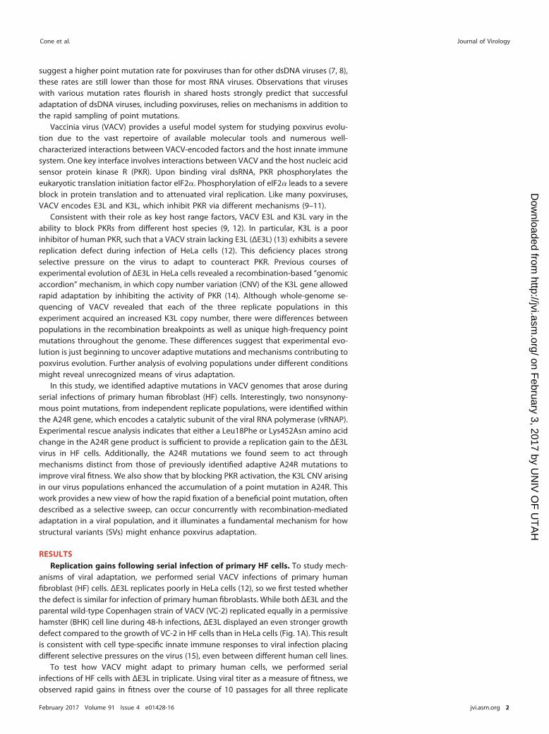
anisms of viral adaptation, we performed serial VACV infections of primary humanfibroblast (HF) cells. ΔE3L replicates poorly in HeLa cells (12), so we first tested whetherthe defect is similar for infection of primary human fibroblasts. While both ΔE3L and theparental wild-type Copenhagen strain of VACV (VC-2) replicated equally in a permissivehamster (BHK) cell line during 48-h infections, ΔE3L displayed an even stronger growthdefect compared to the growth of VC-2 in HF cells than in HeLa cells (Fig. 1A). This resultis consistent with cell type-specific innate immune responses to viral infection placingdifferent selective pressures on the virus (15), even between different human cell lines.
To test how VACV might adapt to primary human cells, we performed serialinfections of HF cells with ΔE3L in triplicate. Using viral titer as a measure of fitness, weobserved rapid gains in fitness over the course of 10 passages for all three replicate
Cone et al. Journal of Virology
February 2017 Volume 91 Issue 4 e01428-16 jvi.asm.org 2
on February 3, 2017 by U
NIV
OF
UT
AH
http://jvi.asm.org/
Dow
nloaded from
virus populations (Fig. 1B). Despite modest replication of the parental ΔE3L virus in HFcells compared to HeLa cells, we observed comparable gains in replication of ΔE3L viruspopulations over the course of our infections of HF cells (Fig. 1B) and HeLa cells, aspreviously reported (14).
High-frequency point mutations in evolved virus populations. To define geneticchanges that might account for increases in viral fitness, we used deep sequencing ofviral genomes from each of the replicate populations after 10 rounds of serial infection(P10). We obtained an average of �2,000� coverage across the genome for each P10population. Excluding the inverted terminal repeat regions (1 to 5,000 bp and 186,737to 191,737 bp), we identified nine single nucleotide polymorphisms (SNPs) not presentin the ΔE3L parent virus at a frequency of �1% for any P10 population, in addition to20 shared differences compared to the VC-2 reference strain (Table 1). The nine SNPspresent in at least one of the three P10 populations but not in the ΔE3L parent virusrepresent potentially adaptive mutations. All nine SNPs lie within open reading frames,and notably, seven of them result in nonsynonymous amino acid changes or frame-shifts (Fig. 2A). Remarkably, the highest-frequency SNP in each population caused asubstitution in an essential gene conserved among poxviruses, resulting in a Leu18Pheamino acid substitution in A24R, encoding a catalytic subunit of the viral RNA poly-merase (vRNAP) (16); a Glu495Gly amino acid substitution in E9L, encoding the viralDNA polymerase (17, 18); and a Trp44Cys amino acid substitution in F10L, encoding akinase required for virion morphogenesis (19). These core genes are all involved inreplication and assembly, suggesting a common adaptive advantage through al-tered replication cycle kinetics under nonoptimal conditions in which nucleic acidsensors are activated. This may represent an indirect selection for altered replicationand assembly in addition to the direct influence of innate signaling pathways. Further-more, another A24R mutation, resulting in a Lys452Asn amino acid substitution, wasidentified in a replicate population independently of the Leu18Phe variant. This resultsuggests that the A24R gene may be a common target for beneficial mutations in VACV,consistent with previous reports of other adaptive A24R mutations (20–22). Together,these high-frequency mutations suggest a common role for poxvirus genes encodingessential viral functions in adaptation to activated host innate immune responses andan altered cellular environment.
SVs in evolved virus populations. In addition to point mutations, genetic changesin the form of gene copy number variation (CNV) have previously been shown to playan adaptive role during poxvirus adaptation (14). To identify potentially adaptivestructural variants (SVs) in our evolved virus populations, we analyzed the P10 se-quences by using the SV analysis implemented in the program Wham (23). We foundseven SVs with �10 reads to define both the 5= and 3= locations of recombination
AV
irus
Tite
r (P
FU
/mL)
106
104
VC-2
108
1010
HFBHK HeLa
ΔE3L
B
B
A
C
2 4 6 8 10
108
109
Passage
Viru
s T
iter
(PF
U/m
L)
107
1010
ΔE3L
FIG 1 Rapid adaptation of ΔE3L during experimental evolution in HF cells. (A) Cells were infected withwild-type VC-2 or ΔE3L virus (MOI � 0.1) for 48 h. (B) Triplicate populations of ΔE3L virus were passaged10 times in HF cells. Equal volumes of virus from every other passage were expanded in BHK cells for 48h and titrated simultaneously. The similarly expanded parental ΔE3L virus titer is indicated by the dottedline. All viral titers were measured in BHK cells by 48-h plaque assays performed in triplicate, and dataare mean PFU per milliliter � standard deviations.
Rapid Rise of a Viral RNA Polymerase Variant Journal of Virology
February 2017 Volume 91 Issue 4 e01428-16 jvi.asm.org 3
on February 3, 2017 by U
NIV
OF
UT
AH
http://jvi.asm.org/
Dow
nloaded from

breakpoints in the virus populations (Fig. 2B; Table 2). Sanger sequencing corroboratedeach of these SVs to within one nucleotide of the putative breakpoint. The single SVidentified in the ΔE3L parent virus was also present in all three P10 populations(breakpoint 7 in Table 2). This variant corresponds to the 11K vaccinia virus promoterintroduced during the deletion of E3L (13), resulting in two copies of this sequence,found at different genomic locations (Fig. 2B). The remaining 6 SVs were present onlyin the P10 virus populations, and each of them was associated with the K3L locus. Therewas a corresponding increase in sequencing depth surrounding K3L for each P10 viruspopulation, but this was absent in the parent population (Fig. 2B). Three of the SNPsfrom variant calling were located 1 bp from a structural variant breakpoint, and wetherefore categorized these as false-positive calls due to misaligned reads (Table 1). Thisfinding illustrates that SVs in poxvirus genomes should be considered when usingstandard variant calling methods to identify SNPs.
A similar copy number amplification of the K3L locus was observed following serialinfections of HeLa cells with ΔE3L (14). In the previous study, we showed that CNV atthe K3L locus corresponds to an increased amount of K3L protein, which impairs PKRactivation. Interestingly, two of the seven breakpoints identified in this study wereidentical to those observed in virus populations passaged in HeLa cells (14), suggestingthe presence of these variants at a level below the limit of detection in the ΔE3L parentvirus or indicating that these are preferential sites for recombination. These results,combined with studies demonstrating CNV in other poxvirus genes in response toselective pressure (24–26), continue to reveal CNV as a mechanism for rapid adaptationof VACV.
TABLE 1 Point mutations in virus populations relative to the VC-2 reference sequence
Position ORFa
Nucleotide(s) Allele frequencyb
Reference Variant �E3L A P10 B P10 C P10
16033 A G 1.00 1.00 1.00 1.0023443 C T 1.00 1.00 1.00 1.0024256 G C 1.00 1.00 1.00 1.0025525 G C 1.00 1.00 1.00 1.0030295c K2L G A 0.00 0.14 0.00 0.0230752 K4L GT G 0.00 0.00 0.00 0.1135080 C G 0.85 0.69 0.67 0.6335081 G C 0.86 0.80 0.78 0.7540751 F10L C A 0.00 0.00 0.00 0.6044312 A G 1.00 1.00 1.00 1.0046730c T A 0.07 0.17 0.18 0.1346742 C G 1.00 1.00 1.00 1.0046743 G C 1.00 1.00 1.00 1.0051482c A C 1.00 0.96 1.00 1.0058304 E9L T C 0.00 0.00 0.80 0.0069922 C T 0.09 0.06 0.08 0.0477258 A T 1.00 1.00 1.00 1.0081834 G A 1.00 1.00 1.00 1.0085139 T C 1.00 1.00 1.00 1.0095128 T C 0.32 0.38 0.37 0.40104656 C T 1.00 1.00 1.00 1.00120212 A7L G T 0.00 0.00 0.31 0.00134369 A24R G T 0.00 0.91 0.00 0.00135671 A24R G T 0.00 0.00 0.00 0.14145840 T A 1.00 1.00 1.00 1.00148905 A T 1.00 1.00 1.00 1.00152700 G GC 1.00 1.00 1.00 1.00172688 B13R G T 0.00 0.00 0.00 0.18175621 B17L CT C 0.00 0.00 0.25 0.00aThe ORF containing the mutation is shown for variant alleles (bold) not present in the ΔE3L parent virus(also see Fig. 2A).
bAlleles with frequencies of �0.01 are shown.cPredicted false-positive variant stemming from a misaligned read at a structural variant breakpoint (seeTable 2).
Cone et al. Journal of Virology
February 2017 Volume 91 Issue 4 e01428-16 jvi.asm.org 4
on February 3, 2017 by U
NIV
OF
UT
AH
http://jvi.asm.org/
Dow
nloaded from

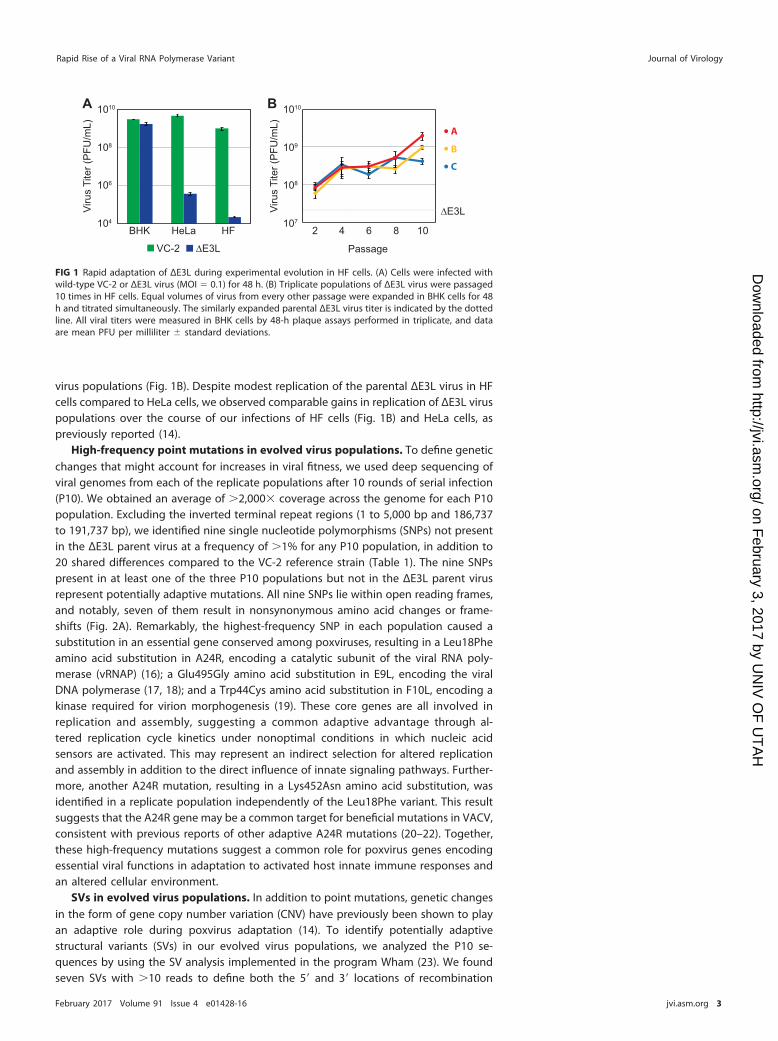
A24R mutations increase viral fitness through distinct mechanisms. Following10 passages in HF cells, each population of viruses harbored a unique set of mutationsthat might contribute to increased fitness. The most drastic example was a Leu18Pheamino acid substitution encoded within A24R that was nearly fixed (frequency � 0.91)in one virus population after 10 passages (Fig. 2A). The lack of other nonsynonymouspoint mutations in this replicate population above a frequency of 1% strongly suggeststhat this single mutation contributed to the observed increase in fitness (Fig. 1B). Totest this hypothesis, we generated a recombinant virus with this A24R mutation in theparental ΔE3L strain (A24RLeu18Phe). Growth curve analysis with the permissive BHK cellline revealed that the A24RLeu18Phe and ΔE3L viruses replicated to similar titers (Fig. 3A).However, in HF cells, the A24RLeu18Phe virus exhibited a significant increase in titerrelative to that of ΔE3L from 48 to 72 h postinfection. Thus, the single nucleotidechange in A24R in the ΔE3L genetic background was sufficient to enhance viral fitnessunder selective pressure in HF cells.
To examine how a single amino acid substitution encoded within A24R mightincrease viral fitness, we mapped A24R mutations onto solved structures of RNA
Rel
ativ
e D
epth
Genomic Position (kbp)
K3LB
A
0 1Allele Frequency
K4L F10L A24R A24R B13R
ΔE3L
C P10
B P10
A P10
FS W44C L18F K452N SynAA change:
ORF: E9LE495G
A7LF288L
B17LFS
K2LSyn*
11K
50 100 150
1
2
3
ΔE3L
1
2
3A P10
1
2
3
B P10
1
2
3
C P10
FIG 2 Genetic changes in virus populations following experimental evolution. (A) Allele frequencies wereobtained by deep sequencing of viral genomic DNA from passage 10 virus populations compared to the ΔE3Lparent virus. Alleles with a frequency of �0.01 that were not present in the parent virus are shown. The VACVopen reading frame (ORF) and resulting amino acid change are listed at the top. FS, frameshift; Syn,synonymous. *, the K2L allele is likely a miscalled structural variant. (B) Relative depths of coverage across theviral genome are shown in black, excluding the inverted terminal repeat regions. Breakpoint positions ofstructural variants called by Wham are shown as vertical lines, with 5= positions in red and 3= positions inorange. The genomic locations of the K3L gene and the duplicated 11K promoter are indicated at the top.
Rapid Rise of a Viral RNA Polymerase Variant Journal of Virology
February 2017 Volume 91 Issue 4 e01428-16 jvi.asm.org 5
on February 3, 2017 by U
NIV
OF
UT
AH
http://jvi.asm.org/
Dow
nloaded from

polymerases. Previous work used the crystal structure of Saccharomyces cerevisiae RNApolymerase II to map A24R mutations encoding amino acid substitutions onto thehomologous subunit in yeast, RBP2 (22). RBP2 is the second largest subunit of yeastRNA polymerase II and forms an active site of the enzyme with RBP1 (27). Using a similarapproach, we generated an amino acid alignment between the VACV A24R geneproduct and S. cerevisiae RBP2 to predict the locations of amino acid substitutionsencoded within A24R mutants on the S. cerevisiae RNA polymerase II crystal structure(PDB entry 1I50) (28). This analysis suggested that the Leu18Phe substitution encodedby the corresponding A24R mutant is located on a solvent-exposed surface of thepolymerase distal from the active site of the enzyme (Fig. 3B). This surface might beinvolved in binding other factors to the polymerase, such that a Leu18Phe substitutionmay alter a protein-protein interaction(s). However, little is currently known about viralor host proteins that bind to the A24R gene product, making it difficult to predict thefunctional consequences of this mutation.
Distinct adaptive mutations in A24R demonstrate how vRNAP variation can affectviral fitness. Two A24R mutations were shown to influence transcription elongation,resulting in the production of short virus transcripts in response to isatin-�-thiosemicarbazone (IBT) selection (21, 22). IBT-resistant mutations have also beenshown to reduce the activation of the host dsRNA sensor oligoadenylate synthetase(OAS) (29, 30). These studies suggest that changes to transcript length, dictated byvRNAP, can influence the activation of innate immune dsRNA sensors in the cell.Indeed, another A24R mutation was recently identified that reduces the activation ofthe dsRNA sensor PKR (20). Without E3L, viruses are more vulnerable to PKR and OASactivities, and thus we hypothesized that the A24R substitutions we identified might actthrough a similar mechanism to reduce the activation of dsRNA sensors.
To test whether either of the A24R variants from our evolved virus populations issufficient to alter innate immune responses, we first determined whether dsRNAproduction was affected by the vRNAP variants. A dot blot of dsRNAs from infected cellsrevealed less total dsRNA in cells infected with either the A24RLeu18Phe or A24RLys452Asn
recombinant virus than that in cells infected with the ΔE3L virus (Fig. 3C). Viruses withthe Leu18Phe substitution produced a notable reduction in dsRNA, which might reflectchanges to transcript length or stability, or even changes to a subset of transcripts.Additionally, the overall reduction in dsRNA might have affected the activation ofdsRNA sensors and contributed to the fitness increases observed for viruses harboringthe A24R variants.
To test whether a reduction in dsRNA affects downstream nucleic acid sensingpathways, we measured the activities of the OAS/RNase L and PKR pathways in cellsinfected with recombinant viruses harboring the A24R mutant-encoded Leu18Phe orLys452Asn substitution. We did not detect any notable difference in RNase L activity asjudged by rRNA degradation following infection with either the A24RLeu18Phe orA24RLys452Asn recombinant virus (Fig. 3D). This result suggests that the OAS/RNase Lpathway was not significantly affected by these A24R substitutions, despite the reduc-tion in dsRNA. We next tested whether either of the A24R variants reduced PKR
TABLE 2 Structural variants in virus populations relative to the VC-2 reference sequence
Breakpoint
Genomicpositiona Read support (no. of reads [5=, 3=])
5= 3= �E3L A P10 B P10 C P10
1 26302 35775 0, 0 0, 0 0, 0 112, 1022 27105 32160 0, 0 0, 0 74, 70 0, 03 27322 33524 0, 0 0, 0 0, 0 105, 734 28875 33568 0, 0 0, 0 114, 74 0, 05 30287 30840 0, 0 0, 0 164, 125 0, 06 30296 31725 0, 0 190, 137 0, 0 96, 857 46731 51483 179, 260 159, 465 179, 472 200, 389aPositions supported by �10 reads are shown.
Cone et al. Journal of Virology
February 2017 Volume 91 Issue 4 e01428-16 jvi.asm.org 6
on February 3, 2017 by U
NIV
OF
UT
AH
http://jvi.asm.org/
Dow
nloaded from
activation as measured by changes in phosphorylated PKR and phosphorylated eIF2�
protein levels in infected HF cells. Counter to our expectation, immunoblot analysisindicated that HF cells infected with the A24RLeu18Phe virus repeatedly showed in-creased levels of both phosphorylated PKR and eIF2� (Fig. 3E). While the A24RLeu18Phe
virus conferred a replication benefit at late time points that may account for an increasein PKR activation, this is unlikely given the equivalent viral titers between theA24RLeu18Phe and ΔE3L viruses at the 6-h time point (Fig. 3A) when we harvested totalprotein. In contrast, there were no substantial changes in PKR activation upon infectionwith the A24RLys452Asn virus compared to infection with ΔE3L. These data suggest thatthe A24R variants we identified work through mechanisms distinct from those for otherknown A24R substitutions to enhance viral fitness. Moreover, the increase in PKR
A
C D
BLeu18Phe
Lys452AsnΔE3L (BHK)
ΔE3L (HF)
A24RLeu18Phe (BHK)
A24RLeu18Phe (HF)
ΔE3L
A24RLe
u18P
he
A24RLy
s452
Asn
Moc
kA P
10
Active site
P-eIF2α
eIF2α
P-PKR
PKR
Poly(I:
C)
ΔE3L
A24RLe
u18P
he
A24RLy
s452
Asn
Moc
k
28S
18S
ΔE3L
A24RLe
u18P
he
A24RLy
s452
Asn
Moc
k
2.25
2.00
1.75
1.50
1.25
1.00
0.75
μgRNA
E
Viru
s T
iter
(PF
U/m
L)
Time (hrs)
109
107
105
103
24 48 72
ΔE3L
A24RLe
u18P
he
A24RLy
s452
Asn
Moc
kA P
10
******
FIG 3 A24R mutations increase fitness through distinct mechanisms. (A) Single-step growth curveanalysis (MOI � 5.0) was performed in triplicate with either the ΔE3L or A24RLeu18Phe recombinant virusin BHK or HF cells. Viral titers were measured in BHK cells by a 72-h plaque assay, and data are mean PFUper milliliter � standard deviations. ***, P � 0.005 relative to ΔE3L within each cell type, by 2-way analysisof variance (ANOVA) followed by Bonferroni’s multiple-comparison test. (B) Positions of A24R mutationsidentified in this study (red spheres) and previously published data (yellow spheres) were mapped ontothe homologous S. cerevisiae RNAP II structure (PDB entry 1I50). The RBP2 protein (homologous to theA24R gene product) is shown as blue ribbons in the context of the multisubunit polymerase shown ingray (the active site is shown in green). (C) dsRNA dot blot with decreasing amounts of total RNA fromHF cells left untreated (mock) or infected with the ΔE3L, A24RLeu18Phe, or A24RLys452Asn virus (MOI � 10.0)for 13 h. An image representative of three independent blots is shown. (D) rRNA degradation in HF cellsleft untreated (mock), transfected with poly(I·C), or infected with the ΔE3L, A24RLeu18Phe, or A24RLys452Asn
virus (MOI � 5.0) for 6 h. Filled arrowheads indicate 28S and 18S rRNAs, and open arrowheads indicatedegradation products. (E) Immunoblot of phosphorylated or total PKR and phosphorylated or total eIF2�in HF cells infected with the ΔE3L, A24RLeu18Phe, A24RLys452Asn, or replicate A passage 10 virus (MOI � 5.0)for 6 h. An image representative of five independent blots is shown.
Rapid Rise of a Viral RNA Polymerase Variant Journal of Virology
February 2017 Volume 91 Issue 4 e01428-16 jvi.asm.org 7
on February 3, 2017 by U
NIV
OF
UT
AH
http://jvi.asm.org/
Dow
nloaded from
activation with the A24RLeu18Phe virus was paradoxical, considering the reduction indsRNA and the replication advantage we observed in HF cells.
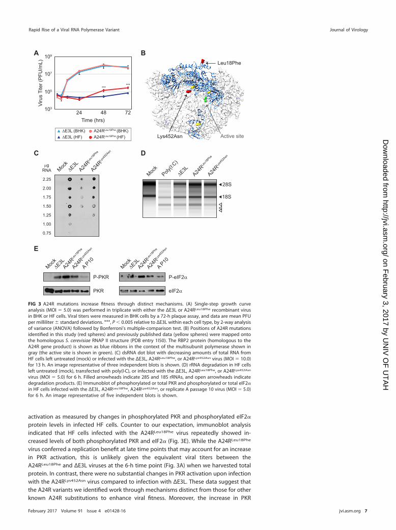
The A24R Leu18Phe variant confers fitness trade-offs. Given that E3L and K3L actas host range factors, blocking innate immune activation in some hosts but not others(12), we tested the impact of A24R variation during infections of cells from otherprimate species. We performed triplicate 48-h infections in two human and two OldWorld primate cell lines: HF cells, HeLa cells, rhesus macaque fibroblasts, and Africangreen monkey fibroblasts. In each of the four cell lines tested, the parental ΔE3L virusexhibited considerably reduced replication compared to that in a permissive BHK cellline (Fig. 4A). The A24RLys452Asn virus replicated equally to or better than ΔE3L in allprimate cells tested, suggesting that the fitness increase provided by the A24RLys452Asn variant is not species or cell type specific. In contrast, while A24RLeu18Phe
virus replication showed a 43-fold increase in HF cells relative to replication of theΔE3L virus, titers were significantly reduced in HeLa cells and African green monkeyfibroblasts. The clear differences in A24RLeu18Phe viral titers between cells from twoOld World monkey species and also between two human cell lines reveal a potentialfitness trade-off for viruses harboring this substitution.
Consistent with a fitness trade-off in some cell types, the A24RLeu18Phe virus dis-played a small-plaque phenotype in permissive BHK cells (Fig. 4B). This phenotype isassociated with defects in cell-to-cell spread (31–33), although the A24RLeu18Phe virusproduced approximately the same number of infectious particles as ΔE3L in BHK cells(Fig. 3A). The small-plaque phenotype in conjunction with differential replicationamong cell lines supports the idea that the A24R Leu18Phe variant is beneficial onlyunder certain conditions. Furthermore, the growth benefit in HF cells and the growthdefect in HeLa cells highlight that specific, species-independent differences betweenthe cell lines can contribute to the success or failure of the A24RLeu18Phe virus.
Accelerated sweep of the A24R Leu18Phe variant during adaptive gene copynumber amplification. The A24R Leu18Phe variant rose to near fixation in a viralpopulation during experimental evolution of ΔE3L in HF cells (Fig. 2; Table 1). The rapiddominance of the variant in this population was unusual given the slower accumulationdynamics of mutations within other replicate populations. For example, the A24RLys452Asn variant provided a similar replication increase alone (Fig. 4A) yet reached afrequency of only 14%, in contrast to the 91% frequency of the A24R Leu18Phe variant(Fig. 5A). Furthermore, we might have predicted that the increased PKR activation
ΔE3L A24RLeu18Phe
Pla
que
size
(pi
xels
)
ΔE3L A24RLeu18Phe0
10
20
***
BHK
A BV
irus
Tite
r (P
FU
/mL)
HFBHK HeLa Rh AGM
Cell Line
108
106
1010
104
102
***
******
*
*
ΔE3LA24RLeu18Phe
A24RLys452Asn
FIG 4 The A24R Leu18Phe variant confers fitness trade-offs. (A) Cell lines were infected in triplicate withthe ΔE3L, A24RLeu18Phe, or A24RLys452Asn virus (MOI � 0.1) for 48 h. Viral titers were measured in BHK cellsby a 72-h plaque assay, and data are mean PFU per milliliter � standard deviations. *, P � 0.05; **, P �0.01; ***, P � 0.005 relative to ΔE3L within each cell type, by one-way ANOVA followed by Dunnett’smultiple-comparison test. Rh, rhesus macaque fibroblasts; AGM, African green monkey fibroblasts. (B)Average plaque size � standard deviation for three independent wells of BHK cells infected with theΔE3L or A24RLeu18Phe virus (MOI � 0.1) for 48 h. A representative image is shown. ***, P � 0.005 by2-tailed t test.
Cone et al. Journal of Virology
February 2017 Volume 91 Issue 4 e01428-16 jvi.asm.org 8
on February 3, 2017 by U
NIV
OF
UT
AH
http://jvi.asm.org/
Dow
nloaded from

induced by the A24R Leu18Phe variant (Fig. 3E) would prevent the mutation fromreaching fixation so rapidly. To more carefully determine how this mutation arose, weanalyzed earlier passages during the course of experimental evolution. Remarkably, theA24R Leu18Phe variant was detectable only starting at passage 8 as judged bysequencing of A24R amplicons in each passage, prompting us to deep sequence viruspopulations from passages 7 to 9. Genome sequence analysis revealed that themutation rapidly increased in frequency with each successive passage late in theexperiment (Fig. 5A). Consistent with our earlier analysis, this was the only verified SNP
A
C
20
10
7
54
kbp
2ΔE3L 4 6 8 10Passage
Replicate A K3Lcopies
5
1
2
3
...
A24RLeu18Phe:ΔE3L
A24RLeu18Phe+CNV:ΔE3L
1%
1%
A24RLeu18Phe Abundance
45.2%
83.6% 98.8%
2.0%
CNV:ΔE3L
ΔE3L:ΔE3L
P7
Rep
licat
e A
Rep
licat
e C
P8
P9
P10
BK4L F10L A24R A24R B13RFS W44C L18F K452N SynAA change:
ORF:
2
108
Passage:
Viru
s T
iter
(PF
U/m
L)
106
104
10
1010
Wildtype
Breakpoint 6
500bp
K2L K4L3 K4L3
K2L
Breakpoint 1
1000bpK3L M1LF3L K3L
Breakpoint 3
1000bpK3L M1LF1L K3L
D
0.05
0.27
0.66
0.91
0.00
0.05
0.09
0.14
P7
P8
P9
P10
0 1Allele Frequency
Replicate A
Replicate C
F3L
K3L
M1L
M2L
K2L
K1L
K4L
K5L
K6L
K7R
F1L
F2L
F4L
N2L 1000bp
Breakpoint 6
500bp
K2L K4L3 K4L3
K2L
FIG 5 Copy number variation enhances the sweep of a point mutation. (A) Allele frequencies of �0.01 forreplicates A and C were obtained by deep sequencing of P7 to P10 virus populations, and positions in ORFsand resulting amino acid changes are listed at the top, as in Fig. 2A. FS, frameshift; Syn, synonymous. (B)Genome structures from direct sequencing across the CNV breakpoints identified by Wham (Table 2) forreplicate A and C passage 10 populations. (C) Southern blotting using a K3L-specific probe was performed ondigested viral DNAs from the indicated viral populations. Sizes (left) and numbers of K3L copies (right) areshown. (D) Viruses were mixed as listed, at a ratio of 1:100, and passaged twice for 48 h in HF cells (MOI � 0.1).Titers were measured in BHK cells by 72-h plaque assays performed in triplicate, and data are mean PFU permilliliter � standard deviations. A24R Leu18Phe variant abundances are listed below the graph, as input (P0)or as measured by deep sequencing (P1 and P2).
Rapid Rise of a Viral RNA Polymerase Variant Journal of Virology
February 2017 Volume 91 Issue 4 e01428-16 jvi.asm.org 9
on February 3, 2017 by U
NIV
OF
UT
AH
http://jvi.asm.org/
Dow
nloaded from
present at frequencies above 1% in passages 7 to 10 (replicate A), which is in starkcontrast to the findings for replicate C, in which multiple mutations across the genomefluctuated in frequency from passages 7 to 10 (Fig. 5A). The rapid accumulation of theA24R Leu18Phe variant, as well as a lack of any other mutations across the genome, isconsistent with a strong selective sweep of the mutation in the replicate A population.However, because the A24RLeu18Phe virus also increased PKR activation, the questionremained as to how the mutation induces increased virus replication.
The increased copy number of the K3L gene in the P10 population provided a clueto how the A24R mutation might rapidly sweep to fixation despite increasing PKRactivation. There was only one recombination breakpoint at P10 for replicate A, asopposed to multiple breakpoints for replicates B and C (Fig. 5B; Table 2). This suggeststhat when the selective sweep of the A24R Leu18Phe variant occurred, these virusesalso contained a single K3L CNV breakpoint. To determine whether the K3L gene copynumber amplification or the A24R Leu18Phe mutation arose first in the virus popula-tion, we used Southern blot analysis to measure K3L CNV during experimental evolu-tion of replicate A. We first detected K3L CNV at passage 4, and the proportion ofviruses with the population harboring CNV seemed to remain steady through passage10, as indicated by the consistent intensities of bands within each lane (Fig. 5C). Sincethe A24R Leu18Phe substitution did not emerge until passage 7, the earlier appearanceof K3L CNV might have preemptively blocked PKR activation that would otherwise beinduced by the A24R Leu18Phe variant, facilitating its rapid fixation. Indeed, in cellsinfected with the replicate A P10 virus, which contained both the A24R Leu18Phesubstitution and K3L CNV, there was a marked reduction in PKR activation (Fig. 3E). Thisresult suggests that K3L CNV can compensate for the activation of PKR induced by theA24R Leu18Phe variant.
To test whether rapid accumulation of the A24R Leu18Phe variant was influenced bythe K3L copy number, we wanted to track increases of the A24R Leu18Phe variant invirus populations with and without K3L CNV. To do this, we infected cells with virusescontaining the A24R Leu18Phe substitution alone (A24RLeu18Phe), K3L CNV alone (CNV),or the combination of the two genetic changes (A24RLeu18Phe�CNV), each starting at aratio of 1:100 with the parental ΔE3L virus (see Materials and Methods for strain details).Compared to ΔE3L alone after two passages in HF cells, the A24RLeu18Phe:ΔE3L viruspopulation replicated �10-fold better, CNV:ΔE3L �1,000-fold better, and A24RLeu18Phe:CNV:ΔE3L �10,000-fold better (Fig. 5D). These data suggest that while either the A24RLeu18Phe variant or K3L CNV is sufficient for a fitness benefit in HF cells, the combi-nation is additive and may therefore facilitate fixation of the A24R Leu18Phe variant. Wenext analyzed the abundance of the A24R Leu18Phe substitution in the virus popula-tions to determine if it accumulated faster in the presence of K3L CNV. Starting at 1%of the population, the mutation reached 98.8% abundance after only two passageswhen multiple copies of K3L were present, compared to 45.2% abundance at passage2 with a single copy of K3L (Fig. 5D). The increased accumulation of the variant in thepresence of CNV is consistent with the added fitness benefit for viruses carrying bothgenetic changes. Thus, the A24R Leu18Phe substitution accumulated markedly faster inthe presence of K3L CNV, which suggests that the reduction in PKR activation providedby increased K3L expression may have facilitated the rapid rise of the distant A24Rmutation.
DISCUSSION
In this study, we found a new means of poxvirus adaptation to innate immuneresponse pathways. Vaccinia virus has proven to be a useful model for experimentalevolution and continues to reveal the genetic basis of various poxvirus adaptations (14,20, 25). We charted the rise of multiple point mutations over the course of 10 serialinfections, which is consistent with adaptive evolution through several independentmechanisms. Most notable among these was the rapid accumulation of a Leu18Phesubstitution resulting from a mutation within A24R, which encodes a subunit of theviral RNA polymerase. Unlike in other evolved virus populations, the rise of the A24R
Cone et al. Journal of Virology
February 2017 Volume 91 Issue 4 e01428-16 jvi.asm.org 10
on February 3, 2017 by U
NIV
OF
UT
AH
http://jvi.asm.org/
Dow
nloaded from
Leu18Phe variant appeared in the near absence of other point mutations (Fig. 5A), anobservation reminiscent of clonal interference, where strongly adaptive point muta-tions on separate genomes can compete and transiently dominate within asexualpopulations (34, 35). However, a case of clonal interference seemed unlikely given thehigh rates of recombination in poxviruses (36–39) and the modest replicative advan-tage we measured in a recombinant strain containing only the A24R Leu18Phe muta-tion (Fig. 3A). We therefore considered the impact of K3L gene copy number amplifi-cation as a facilitating event for the rapid accumulation of the A24R Leu18Phe variantduring this course of experimental evolution.
We previously found that genomes harboring multiple copies of K3L produced moreprotein product, which resulted in fitness gains for viruses under selective pressure toovercome the antiviral factor PKR (14). In two populations evolved in HeLa cells, weobserved the additional emergence of a beneficial point mutation in K3L during serialinfections, which we predict is more likely to occur in viruses with multiple copies of thegene (14). However, as we did not observe K3L mutations in every evolved population,adaptive mutations likely still arise at a low frequency. In this study, we observed abeneficial point mutation in a gene distant from the expanded K3L locus (�100,000 bpapart) that may benefit from the presence of virus genomes harboring multiple copiesof K3L. We speculate that the Leu18Phe substitution encoded within the A24R variantalters virus transcription to promote virus replication and found that while the vRNAPvariant does reduce total dsRNA, it also activates PKR (Fig. 3C and E). These results showthat the A24RLeu18Phe virus produces less dsRNA and suggest a complicated mechanismin which the polymerase variant alters RNA production in a way that activates PKRdespite the reduction in dsRNA. Further analysis of viral transcripts may reveal changesto transcript length or other modifications to specific transcripts that modulate immunesensing.
Despite providing modest fitness gains and activating PKR, the A24R Leu18Phemutation can rapidly sweep to fixation in virus populations in the presence of multiplecopies of K3L that block PKR activation. Consistent with this idea, we observed rapidfixation of the A24R Leu18Phe variant following K3L copy number amplification (Fig. 5Aand C) and a 2-fold increase in the accumulation of the A24R Leu18Phe variant in thepresence of multiple K3L copies compared to that in populations with a single copy ofK3L (Fig. 5D). These data suggest that adaptive copy number variation can facilitatethe rapid accumulation of otherwise modestly beneficial mutations, such as the A24RLeu18Phe variant. In this way, recombination-based CNV may enhance the viability ofan expanded set of beneficial mutations that otherwise suffer from trade-offs (e.g.,activation of PKR) and might otherwise be unable to sweep through populations. Giventhat copy number amplification events are likely to be transient (14), this foothold maybe temporary, as suggested by previous work describing the accumulation of beneficialpoint mutations causing the collapse of CNV (20). In the current study, CNV of the K3Llocus persisted through passage 10 (Fig. 5C), but it might collapse to a single copy afterfurther rounds of infection. In any case, copy number variation provides an opportunityfor mutations to sweep rapidly through genes both undergoing CNV and distant fromCNV, despite small initial fitness gains relative to those for other mutations in thepopulation. Given long periods of evolutionary time, the presence of seemingly simple,beneficial point mutations in virus populations may belie a more volatile history offixation involving the aid of adaptive yet transient gene copy number amplificationevents.
MATERIALS AND METHODSCells and viruses. Primary human fibroblast (HF) cells derived from human foreskin were a gift from
Adam Geballe (Fred Hutchinson Cancer Research Center). HF, HeLa, and BHK cells were maintained inDulbecco’s modified Eagle’s medium (DMEM; HyClone) supplemented with 10% fetal bovine serum(HyClone), 1% penicillin-streptomycin (GE Lifesciences), and 1% stable L-glutamine (GE Lifesciences).Rhesus fibroblasts (from Macaca mulatta; Coriell Institute for Medical Research) and African greenmonkey fibroblasts (from Cercopithecus aethiops; Coriell Institute for Medical Research) were maintainedin minimum essential medium, alpha modification (MEM-alpha; HyClone), supplemented as described
Rapid Rise of a Viral RNA Polymerase Variant Journal of Virology
February 2017 Volume 91 Issue 4 e01428-16 jvi.asm.org 11
on February 3, 2017 by U
NIV
OF
UT
AH
http://jvi.asm.org/
Dow
nloaded from
above for DMEM. The Copenhagen strain of vaccinia virus (VC-2) and the E3L deletion virus (ΔE3L) (13)were generous gifts from Bertram Jacobs (Arizona State University).
Experimental evolution. For each infection during experimental evolution, 150-mm dishes wereseeded with an aliquot from the same stock of HF cells (5 � 106 cells/dish). Triplicate dishes of cells wereinfected (multiplicity of infection [MOI] � 1.0 for P1 and 0.1 for subsequent passages) from a single stockof ΔE3L virus for 2 h in a minimal volume and then supplemented with medium. At 48 h, cells werecollected, washed, pelleted, and resuspended in 1 ml of medium. Virus was released by one freeze-thawcycle followed by sonication. Viral titers were determined by a 48-h plaque assay with BHK cells betweenpassages. Following 10 passages, equal volumes of virus from every other passage were expanded in BHKcells for 48 h, with viral titers determined by a 48-h plaque assay with BHK cells, or for 72 h for replicateA passage 10 due to a small-plaque phenotype. An equal volume from the input parental ΔE3L virusstock was similarly expanded for comparison.
VACV whole-genome deep sequencing. Total viral genomic DNA was collected following a 24-hinfection of BHK cells (MOI � 0.1) as previously described (41). Libraries were constructed using a NexteraXT DNA sample prep kit (Illumina, Inc.). Barcoded libraries were combined and sequenced using anIllumina MiSeq instrument at the High-Throughput Genomics Core (University of Utah). Reads weremapped to the VC-2 reference genome (accession number M35027.1; modified on http://poxvirus.org)(42) by using BWA mem (v0.7.10) (40) in default mode. PCR duplicates were removed, and the read depthwas calculated using samtools (v0.1.18) (43). We utilized the Genome Analysis Toolkit (v3.2-2) (44) forbase quality score recalibration, indel realignment, and variant calling across all samples (45, 46). Weutilized Wham (v1.7.0-272-g078c-dirty) for structural variant calling (23). SNP and depth plots weregenerated in R (https://www.r-project.org/).
Recombinant virus generation. A sequence of 500 bp of homology surrounding the Leu18Phe-encoding mutation in A24R was amplified by PCR from replicate A passage 10 viral DNA by using primersA24R_1F (5=-CCTCCTCTCGAGCCCTCTCTGTTAGATGAGGATAGC) and A24R_1R (5=-CCTCCTACTAGTCAGTGAACGTGGCTAATGCG). A sequence of 500 bp of homology surrounding the Lys452Asn-encoding muta-tion in A24R was amplified by PCR from VC-2 viral DNA by using primers A24R_2F (5=-CCTCCTCTCGAGCGTTGGCACATGATGAATTAGAGAATTAC) and A24R_2R (5=-CCTCCTACTAGTGAGATGCGACTAGAGCATTTTCTATAGTG). The resulting PCR products were digested with XhoI and SpeI (New England BioLabs), gelpurified, and cloned into pEQ1422 (a gift from A. Geballe, Fred Hutchinson Cancer Research Center) (20)cut with the same enzymes to generate pEQ1422-Leu18Phe and pEQ1422-A24R_2. The Lys452Asn-encoding mutation was introduced into pEQ1422-A24R_2 by use of site-directed mutagenesisprimers A24R_Lys452Asn_F (5=-GTTGGATTTTATCCGGATCAAGTAAATATTTCAAAGATGTTTTCTGTCA) andA24R_Lys452Asn_R (5=-TGACAGAAAACATCTTTGAAATATTTACTTGATCCGGATAAAATCCAAC) (pEQ1422-Lys452Asn).
BHK cells were infected with ΔE3L (MOI � 1.0) and then transfected at 1 h postinfection withpEQ1422-Leu18Phe or pEQ1422-Lys452Asn by use of FuGENE6 (Promega) according to the manufactur-er’s protocol. Infected cells were collected at 48 h postinfection, and viruses were released by onefreeze-thaw cycle followed by sonication. Resulting viruses were selected using transient dominantselection (47). Briefly, viruses were plaque purified three times in the presence of 600 �g/ml hygromycinB (Sigma-Aldrich), followed by three rounds of plaque purification without hygromycin B. The presenceof the Leu18Phe or Lys452Asn substitution and loss of the Hygr phenotype were confirmed by PCRfollowed by Sanger sequencing. Viruses were amplified and titers measured in BHK cells.
One-step growth curve. BHK or HF cells were infected with VACV ΔE3L or A24RLeu18Phe (MOI � 5.0)in triplicate, and the virus was replaced with fresh medium after 2 h. Cells were harvested at 2, 6, 12, 24,48, and 72 h postinfection, and viral titers were determined by a 72-h plaque assay with BHK cells.
Modeling. Amino acid alignment between the VACV A24R gene product and S. cerevisiae RBP2 wasgenerated using Clustal Omega (v1.2.1). Corresponding A24R variant residues were then mapped ontothe S. cerevisiae RNAP II crystal structure (PDB entry 1I50) (28) by use of Chimera software (48;http://www.cgl.ucsf.edu/chimera/).
dsRNA dot blotting. HF cells were left untreated (mock) or infected with the ΔE3L, A24RLeu18Phe, orA24RLys452Asn virus (MOI � 10) for 13 h. Total RNA was collected from infected cells, and dilutions werespotted onto nylon membranes (GE Lifesciences). Membranes were allowed to dry before two rounds ofUV cross-linking. Blots were blocked for 1 h in phosphate-buffered saline with Tween (PBST) plus 5% milkand then incubated with M�dsRNA J2 (1:1,000; Scicons) for 1 h followed by G�M-IgG-HRP (1:50,000;Millipore) for 1 h. Blots were then activated with WesternBright ECL reagent (Advansta) and exposed toautoradiography film (GeneMate), which was developed in a Mini-Med 90 film processor (AFP Imaging).
rRNA degradation assay. HF cells were pretreated with interferon alpha for 24 h. Cells were thenleft untreated (mock), transfected with poly(I·C), or infected with the ΔE3L, A24RLeu18Phe, or A24RLys452Asn
virus (MOI � 5.0) for 6 h. Total RNA was harvested, and a total of 225 ng/lane was run on an Agilent 2200TapeStation instrument.
Immunoblot analysis. HF cells were left untreated (mock) or infected with the ΔE3L, A24RLeu18Phe,A24RLys452Asn, or replicate A P10 virus (MOI � 5.0) for 6 h. Protein lysates were collected in RIPA lysisbuffer, and total protein concentrations were quantified by Bradford assay by use of a Synergy HT platereader (BioTek). Equivalent amounts of lysate were separated in a precast Mini-Protean TGX gel (Bio-Rad)and transferred to a polyvinylidene difluoride (PVDF) membrane (Immobilon). Blots were blocked for 30min in PBST plus 5% milk, or for 10 min followed by three washes for phospho-specific antibodies. Blotswere then incubated with the following primary antibodies overnight at 4°C: M�PKR B-10 (1:200; SantaCruz), R�Phospho-PKR E120 (1:500; Abcam), R�eIF2� (1:1,000; Cell Signaling), and M�Phospho-eIF2�
(1:250; Cell Signaling). Blots were probed with the appropriate secondary antibody for 1 h at room
Cone et al. Journal of Virology
February 2017 Volume 91 Issue 4 e01428-16 jvi.asm.org 12
on February 3, 2017 by U
NIV
OF
UT
AH
http://jvi.asm.org/
Dow
nloaded from

temperature, i.e., G�M-IgG-HRP (1:50,000; Millipore) or G�R-IgG-HRP (1:50,000; Millipore), activated withWesternBright ECL reagent (Advansta), and exposed to autoradiography film (GeneMate), which wasdeveloped in a Mini-Med 90 film processor (AFP Imaging).
Plaque size analysis. BHK cells were infected with the ΔE3L or A24RLeu18Phe virus (MOI � 0.1) for 48h and then stained with crystal violet. Plates were imaged on a Gel Doc XR� system (Bio-Rad), andplaque size was quantified using ImageJ v1.48 software (Rasband). Three independent wells, for a totalof 350 plaques, were analyzed for each virus.
Southern blot analysis. Total viral DNA was collected as described above, and �2 �g per samplewas digested with BspEI (New England BioLabs). Digested DNAs were separated by agarose gelelectrophoresis and transferred to nylon membranes (GE Lifesciences) by vacuum transfer, followed byUV cross-linking. The resulting blots were probed with PCR-amplified K3L by use of a DIG High-PrimeDNA Labeling & Detection starter kit II (Roche) according to the manufacturer’s protocol.
Mutation accumulation assay. The replicate A P10 virus was passaged an additional three times inHF cells until the A24R Leu18Phe variant was fixed (A24RLeu18Phe�CNV virus). The K3L CNV-only viruscontains the same CNV breakpoint but has no mutations in A24R (CNV virus). BHK cells were infectedwith viruses mixed at a ratio of 1:100, i.e., A24RLeu18Phe:ΔE3L, CNV:ΔE3L, or A24RLeu18Phe�CNV:ΔE3L, fortwo passages in BHK cells (MOI � 0.1). All passages were collected after 48 h as described above andtitrated by a 72-h plaque assay with BHK cells.
Accession number(s). All deep-sequencing data are available at the Sequence Read Archive underaccession number SRP073123.
ACKNOWLEDGMENTSExperiments were designed by K.R.C. and N.C.E. Experiments were performed by
K.R.C. Data were analyzed by K.R.C. and Z.N.K. Analysis tools were contributed by Z.N.K.and M.Y. The paper was written by K.R.C. and N.C.E.
We thank Adam Geballe (Fred Hutchinson Cancer Research Center) and BertramJacobs (University of Arizona) for reagents. We also thank Adam Geballe for valuableinsights and critical assessment of the manuscript. We thank the High-ThroughputGenomics Core (University of Utah) for technical assistance.
This study was supported by NIH grants R01GM114514 (N.C.E.), R01GM104390(M.Y.), T32AI055434 (K.R.C.), and T32GM007464 (Z.N.K.). N.C.E. was supported by thePew Biomedical Scholars program and the Mario R. Capecchi Endowed Chair inGenetics.
The experimental design, data collection and analysis, tools, and decision to submitfor publication are our own, and independent of the National Institutes of Health.
REFERENCES1. Duffy S, Shackelton LA, Holmes EC. 2008. Rates of evolutionary change
in viruses: patterns and determinants. Nat Rev Genet 9:267–276. https://doi.org/10.1038/nrg2323.
2. Gago S, Elena SF, Flores R, Sanjuan R. 2009. Extremely high mutation rateof a hammerhead viroid. Science 323:1308. https://doi.org/10.1126/science.1169202.
3. Sanjuán R, Nebot MR, Chirico N, Mansky LM, Belshaw R. 2010. Viralmutation rates. J Virol 84:9733–9748. https://doi.org/10.1128/JVI.00694-10.
4. Lynch M. 2010. Evolution of the mutation rate. Trends Genet 26:345–352.https://doi.org/10.1016/j.tig.2010.05.003.
5. Challberg MD, Englund PT. 1979. Purification and properties of thedeoxyribonucleic acid polymerase induced by vaccinia virus. J Biol Chem254:7812–7819.
6. Qin L, Evans DH. 2014. Genome scale patterns of recombination be-tween co-infecting vaccinia viruses. J Virol 88:5277–5286. https://doi.org/10.1128/JVI.00022-14.
7. Firth C, Kitchen A, Shapiro B, Suchard MA, Holmes EC, Rambaut A. 2010.Using time-structured data to estimate evolutionary rates of double-stranded DNA viruses. Mol Biol Evol 27:2038 –2051. https://doi.org/10.1093/molbev/msq088.
8. Kerr PJ, Ghedin E, DePasse JV, Fitch A, Cattadori IM, Hudson PJ, TscharkeDC, Read AF, Holmes EC. 2012. Evolutionary history and attenuation ofmyxoma virus on two continents. PLoS Pathog 8:e1002950. https://doi.org/10.1371/journal.ppat.1002950.
9. Davies MV, Chang HW, Jacobs BL, Kaufman RJ. 1993. The E3L and K3Lvaccinia virus gene products stimulate translation through inhibition ofthe double-stranded RNA-dependent protein kinase by different mech-anisms. J Virol 67:1688 –1692.
10. Chang HW, Watson JC, Jacobs BL. 1992. The E3L gene of vaccinia virusencodes an inhibitor of the interferon-induced, double-stranded RNA-dependent protein kinase. Proc Natl Acad Sci U S A 89:4825– 4829.https://doi.org/10.1073/pnas.89.11.4825.
11. Davies MV, Elroy-Stein O, Jagus R, Moss B, Kaufman RJ. 1992. Thevaccinia virus K3L gene product potentiates translation by inhibitingdouble-stranded-RNA-activated protein kinase and phosphorylation ofthe alpha subunit of eukaryotic initiation factor 2. J Virol 66:1943–1950.
12. Langland JO, Jacobs BL. 2002. The role of the PKR-inhibitory genes, E3Land K3L, in determining vaccinia virus host range. Virology 299:133–141.https://doi.org/10.1006/viro.2002.1479.
13. Beattie E, Denzler KL, Tartaglia J, Perkus ME, Paoletti E, Jacobs BL. 1995.Reversal of the interferon-sensitive phenotype of a vaccinia virus lackingE3L by expression of the reovirus S4 gene. J Virol 69:499 –505.
14. Elde NC, Child SJ, Eickbush MT, Kitzman JO, Rogers KS, Shendure J,Geballe AP, Malik HS. 2012. Poxviruses deploy genomic accordions toadapt rapidly against host antiviral defenses. Cell 150:831– 841. https://doi.org/10.1016/j.cell.2012.05.049.
15. van Boxel-Dezaire AHH, Rani MRS, Stark GR. 2006. Complex modulationof cell type-specific signaling in response to type I interferons. Immunity25:361–372. https://doi.org/10.1016/j.immuni.2006.08.014.
16. Amegadzie BY, Holmes MH, Cole NB, Jones EV, Earl PL, Moss B. 1991.Identification, sequence, and expression of the gene encoding thesecond-largest subunit of the vaccinia virus DNA-dependent RNApolymerase. Virology 180:88 –98. https://doi.org/10.1016/0042-6822(91)90012-Z.
17. Jones EV, Moss B. 1984. Mapping of the vaccinia virus DNA polymerasegene by marker rescue and cell-free translation of selected RNA. J Virol49:72–77.
Rapid Rise of a Viral RNA Polymerase Variant Journal of Virology
February 2017 Volume 91 Issue 4 e01428-16 jvi.asm.org 13
on February 3, 2017 by U
NIV
OF
UT
AH
http://jvi.asm.org/
Dow
nloaded from

18. Traktman P, Sridhar P, Condit RC, Roberts BE. 1984. Transcriptionalmapping of the DNA polymerase gene of vaccinia virus. J Virol 49:125–131.
19. Betakova T, Wolffe EJ, Moss B. 1999. Regulation of vaccinia virusmorphogenesis: phosphorylation of the A14L and A17L membrane pro-teins and C-terminal truncation of the A17L protein are dependent onthe F10L kinase. J Virol 73:3534 –3543.
20. Brennan G, Kitzman JO, Shendure J, Geballe AP. 2015. Experimentalevolution identifies vaccinia virus mutations in A24R and A35R thatantagonize the protein kinase R pathway and accompany collapse of anextragenic gene amplification. J Virol 89:9986 –9997. https://doi.org/10.1128/JVI.01233-15.
21. Condit RC, Easterly R, Pacha RF, Fathi Z, Meis RJ. 1991. A vaccinia virusisatin-beta-thiosemicarbazone resistance mutation maps in the viralgene encoding the 132-kDa subunit of RNA polymerase. Virology 185:857– 861. https://doi.org/10.1016/0042-6822(91)90559-T.
22. Prins C, Cresawn SG, Condit RC. 2004. An isatin-beta-thiosemicarbazone-resistant vaccinia virus containing a mutation in the second largestsubunit of the viral RNA polymerase is defective in transcription elon-gation. J Biol Chem 279:44858 – 44871. https://doi.org/10.1074/jbc.M408167200.
23. Kronenberg ZN, Osborne EJ, Cone KR, Kennedy BJ, Domyan ET, ShapiroMD, Elde NC, Yandell M. 2015. Wham: identifying structural variants ofbiological consequence. PLoS Comput Biol 11:e1004572. https://doi.org/10.1371/journal.pcbi.1004572.
24. Slabaugh MB, Roseman NA, Mathews CK. 1989. Amplification of theribonucleotide reductase small subunit gene: analysis of novel joints andthe mechanism of gene duplication in vaccinia virus. Nucleic Acids Res17:7073–7088. https://doi.org/10.1093/nar/17.17.7073.
25. Brennan G, Kitzman JO, Rothenburg S, Shendure J, Geballe AP. 2014.Adaptive gene amplification as an intermediate step in the expansion ofvirus host range. PLoS Pathog 10:e1004002. https://doi.org/10.1371/journal.ppat.1004002.
26. Erlandson KJ, Cotter CA, Charity JC, Martens C, Fischer ER, Ricklefs SM,Porcella SF, Moss B. 2014. Duplication of the A17L locus of vaccinia virusprovides an alternate route to rifampin resistance. J Virol 88:11576 –11585. https://doi.org/10.1128/JVI.00618-14.
27. Cramer P, Bushnell DA, Fu J, Gnatt AL, Maier-Davis B, Thompson NE,Burgess RR, Edwards AM, David PR, Kornberg RD. 2000. Architecture ofRNA polymerase II and implications for the transcription mechanism.Science 288:640 – 649. https://doi.org/10.1126/science.288.5466.640.
28. Cramer P, Bushnell DA, Kornberg RD. 2001. Structural basis oftranscription: RNA polymerase II at 2.8 angstrom resolution. Science292:1863–1876. https://doi.org/10.1126/science.1059493.
29. Cohrs RJ, Condit RC, Pacha RF, Thompson CL, Sharma OK. 1989. Modu-lation of ppp(A2=p)nA-dependent RNase by a temperature-sensitivemutant of vaccinia virus. J Virol 63:948 –951.
30. Bayliss CD, Condit RC. 1993. Temperature-sensitive mutants in the vac-cinia virus A18R gene increase double-stranded RNA synthesis as a resultof aberrant viral transcription. Virology 194:254 –262. https://doi.org/10.1006/viro.1993.1256.
31. Sanderson CM, Frischknecht F, Way M, Hollinshead M, Smith GL. 1998.Roles of vaccinia virus EEV-specific proteins in intracellular actin tailformation and low pH-induced cell-cell fusion. J Gen Virol 79:1415–1425.https://doi.org/10.1099/0022-1317-79-6-1415.
32. Röttger S, Frischknecht F, Reckmann I, Smith GL, Way M. 1999. Interac-
tions between vaccinia virus IEV membrane proteins and their roles inIEV assembly and actin tail formation. J Virol 73:2863–2875.
33. Wolffe EJ, Weisberg AS, Moss B. 1998. Role for the vaccinia virus A36Router envelope protein in the formation of virus-tipped actin-containingmicrovilli and cell-to-cell virus spread. Virology 244:20 –26. https://doi.org/10.1006/viro.1998.9103.
34. Fisher RA. 1930. The genetical theory of natural selection. ClarendonPress, Oxford, United Kingdom.
35. Muller HJ. 1932. Some genetic aspects of sex. Am Nat 66:118 –138.https://doi.org/10.1086/280418.
36. Ball LA. 1987. High-frequency homologous recombination in vacciniavirus DNA. J Virol 61:1788 –1795.
37. Evans DH, Stuart D, McFadden G. 1988. High levels of genetic recombi-nation among cotransfected plasmid DNAs in poxvirus-infected mam-malian cells. J Virol 62:367–375.
38. Spyropoulos DD, Roberts BE, Panicali DL, Cohen LK. 1988. Delineation ofthe viral products of recombination in vaccinia virus-infected cells. J Virol62:1046 –1054.
39. Merchlinsky M. 1989. Intramolecular homologous recombination in cellsinfected with temperature-sensitive mutants of vaccinia virus. J Virol63:2030 –2035.
40. Li H, Durbin R. 2009. Fast and accurate short read alignment withBurrows-Wheeler transform. Bioinformatics 25:1754 –1760. https://doi.org/10.1093/bioinformatics/btp324.
41. Esposito J, Condit R, Obijeski J. 1981. The preparation of orthopoxvirusDNA. J Virol Methods 2:175–179. https://doi.org/10.1016/0166-0934(81)90036-7.
42. Goebel SJ, Johnson GP, Perkus ME, Davis SW, Winslow JP, Paoletti E.1990. The complete DNA sequence of vaccinia virus. Virology 179:247–266, 517–563. https://doi.org/10.1016/0042-6822(90)90294-2.
43. Li H. 2011. A statistical framework for SNP calling, mutation discovery,association mapping and population genetical parameter estimationfrom sequencing data. Bioinformatics 27:2987–2993. https://doi.org/10.1093/bioinformatics/btr509.
44. McKenna A, Hanna M, Banks E, Sivachenko A, Cibulskis K, Kernytsky A,Garimella K, Altshuler D, Gabriel S, Daly M, DePristo MA. 2010. TheGenome Analysis Toolkit: a MapReduce framework for analyzing next-generation DNA sequencing data. Genome Res 20:1297–1303. https://doi.org/10.1101/gr.107524.110.
45. DePristo MA, Banks E, Poplin R, Garimella KV, Maguire JR, Hartl C,Philippakis AA, del Angel G, Rivas MA, Hanna M, McKenna A, Fennell TJ,Kernytsky AM, Sivachenko AY, Cibulskis K, Gabriel SB, Altshuler D, DalyMJ. 2011. A framework for variation discovery and genotyping usingnext-generation DNA sequencing data. Nat Genet 43:491– 498. https://doi.org/10.1038/ng.806.
46. Van der Auwera GA, Carneiro MO, Hartl C, Poplin R, del Angel G,Levy-Moonshine A, Jordan T, Shakir K, Roazen D, Thibault J, Banks E,Garimella KV, Altshuler D, Gabriel S, DePristo MA. 2013. From FastQ datato high confidence variant calls: the Genome Analysis Toolkit bestpractices pipeline. Curr Protoc Bioinformatics 11:11.10.1–11.10.33.https://doi.org/10.1002/0471250953.bi1110s43.
47. Falkner FG, Moss B. 1990. Transient dominant selection of recombinantvaccinia viruses. J Virol 64:3108 –3111.
48. Pettersen EF, Goddard TD, Huang CC, Couch GS, Greenblatt DM, MengEC, Ferrin TE. 2004. UCSF Chimera—a visualization system for explor-atory research and analysis. J Comput Chem 25:1605–1612. https://doi.org/10.1002/jcc.20084.
Cone et al. Journal of Virology
February 2017 Volume 91 Issue 4 e01428-16 jvi.asm.org 14
on February 3, 2017 by U
NIV
OF
UT
AH
http://jvi.asm.org/
Dow
nloaded from