“elucidation of phytochemical and antimicrobial ...jsscacs.edu.in/sites/default/files/files/seema...
TRANSCRIPT
JSS MAHAVIDYAPEETHA
JSS COLLEGE OF ARTS, COMMERCE AND SCIENCE
(An Autonomous College of University of Mysore; Re-accredited by Naac with ‘A’ Grade)
B.N.ROAD, MYSURU-25
UGC-MINOR RESEARCH PROJECT
“Elucidation of Phytochemical and Antimicrobial
characteristics of Betel vine (Piper betle L.)”
Submitted to
UNIVERSITY GRANTS COMMISSION
South Western Regional Office
Prasana Kumar Block, Palace Road
Bangalore - 560 009
Karnataka
Submitted by
Dr. M. Seema Department of Microbiology(UG)
JSS College of Arts,Commerce and Science
(An Autonomous College of University of Mysore)
Ooty Road, Mysore-570 025
Re-accredited by Naac with ‘A’ Grade
Phone:0821-2548236,Fax:0821-2548238,[email protected]

JSS MAHAVIDYAPEETHA JSS COLLEGE OF ARTS, COMMERCE & SCIENCE
(An Autonomous College of University of Mysore)
OOTY ROAD, MYSORE-570 025 KARNATAKA
Re-accredited by NAAC with ‘A’ grade
Recognised by UGC as “College with Potential for Excellence”
Ph: 0821-2548236 & 2548380. FAX: 0821-2548238 E-mail: [email protected] Website: www.jsscacs.edu.in
DECLARATION
I hereby declare that the Minor research project (MRP(S)-1499-MRP/14-
15/KAMY013/UGC-SWRO) entitled “Elucidation of Phytochemical and
Antimicrobial characteristics of Betel vine (Piper betle L.)” is the result of
bonafide work carried out by me.
I further declare that the results are not submitted for the award of any other
degree or fellowship.
PRINCIPAL INVESTIGATOR

JSS MAHAVIDYAPEETHA JSS COLLEGE OF ARTS, COMMERCE & SCIENCE
(An Autonomous College of University of Mysore)
OOTY ROAD, MYSORE-570 025 KARNATAKA
Re-accredited by NAAC with ‘A’ grade
Recognised by UGC as “College with Potential for Excellence”
Ph: 0821-2548236 & 2548380. FAX: 0821-2548238 E-mail: [email protected] Website: www.jsscacs.edu.in
CERTIFICATE
I hereby certify that the Minor Research Project (MRP(S)-1499-
MRP/14-15/KAMY013/UGC-SWRO) entitled “Elucidation of
Phytochemical and Antimicrobial characteristics of Betel vine
(Piper betle L.)” carried out by Dr. M. Seema, Assistant Professor,
Department of Microbiology, JSS College, B.N.Road, Mysuru-25. The
project report has been kept in the college library and the executive
summary of the study has been posted on the website of the college.
PRINCIPAL
ACKNOWLEDGMENTS
I immensely thank JSS MahavidyaPeetha for permitting and
encouraging me to take up UGC -MRP. I am grateful to Prof. B.V.
Sambashivaiah, Cheif executive and Prof. M. Mahadevappa,
Principal, JSS College, B.N. Road, Mysuru-25 for extending necessary
facilities for my research work.
I am greatly indebted to Dr. N.S. Devaki, Associate Professor
and Course Co-ordinator, Department of Molecular Biology,
Yuvaraja’s College, University of Mysore, Mysore for her excellent
guidance, encouragement throughout the course of this investigation
and inspiring me to work in this field of research.
I am extremely thankful to Dr. M. S. Manjunath, Assistant
professor and Head, Department of Biochemistry and Dr.Rekha N.D,
Assistant professor, Department of Biotechnology, JSS College, B.N.
Road, Mysuru.
I would remiss if I did not acknowledge the help of Amog P Urs,
Senior Research Fellow, DOS in Biochemistry, University of Mysuru ,
Mysuru during the course of my research work.
I am thankful to University Grant Commission, Bangalore for
Financial support (UGC, Minor Research Project).
Dr. M. SEEMA
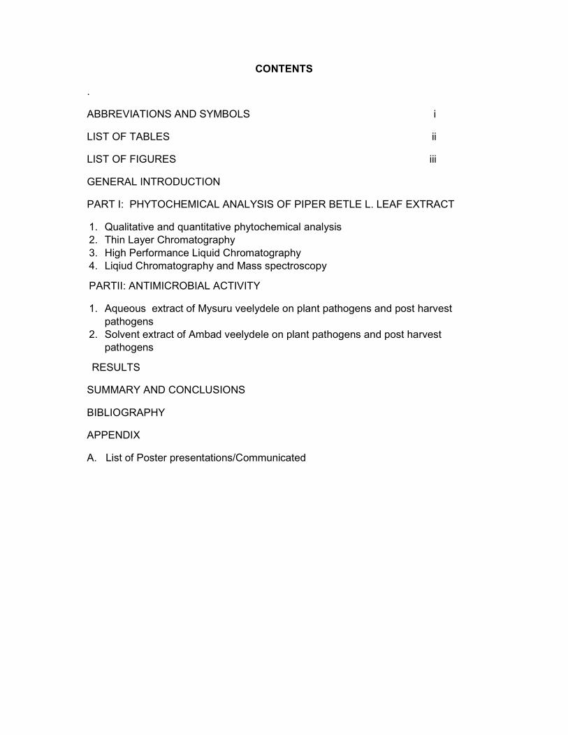
CONTENTS
.
ABBREVIATIONS AND SYMBOLS i
LIST OF TABLES ii
LIST OF FIGURES iii
GENERAL INTRODUCTION
PART I: PHYTOCHEMICAL ANALYSIS OF PIPER BETLE L. LEAF EXTRACT
1. Qualitative and quantitative phytochemical analysis
2. Thin Layer Chromatography
3. High Performance Liquid Chromatography
4. Liqiud Chromatography and Mass spectroscopy
PARTII: ANTIMICROBIAL ACTIVITY
1. Aqueous extract of Mysuru veelydele on plant pathogens and post harvest
pathogens
2. Solvent extract of Ambad veelydele on plant pathogens and post harvest
pathogens
RESULTS
SUMMARY AND CONCLUSIONS
BIBLIOGRAPHY
APPENDIX
A. List of Poster presentations/Communicated

ABBREVIATIONS AND SYMBOLS
@ at the rate of
cm centimeter
Diam. Diameter
et al. et alia
e.g. For example
Fig. Figure
g acceleration due to gravity
g gram (s)
g m-2 gram per meter square
hrs. hours
i.e., id est, that is.
kg kilogram
µg microgram
µm micrometer
mg milligram
mm millimeter
ml milliliter
min minute
M molar
No. number
pH hydrogen ion, minus log concentration
% percentage
rpm rotation per minute
sp species (plural)
viz. namely
w/v weight/volume

LIST OF TABLES
Table No.
Title Page no.
1. 1a. Phytopathogens isolated from diseased plants 1b. Post harvest pathogens
9
2. Qualitative analysis of phyto-constituents of aqueous and solvent extracts of both the varieties of Piper betle L.
15
3. Proximate Composition of various solvent extracts 3a. Ambadi veelyadele 3b. Mysuru veelyadele
16
4. Percentage inhibition (PI) of growth of plant pathogens by aqueous extract of Mysuru veelyadele and Ambadi veelyadele
30
5. 5a. Percentage inhibition (PI) of growth of plant pathogens by hexane extract of Mysuru veelyadele and Ambadi veelyadele 5b. Percentage inhibition (PI) of growth of plant pathogens by ethyl acetate extract of Mysuru veelyadele and Ambadi veelyadele 5c. Percentage inhibition (PI) of growth of plant pathogens by Methanol extract of Mysuru veelyadele and Ambadi veelyadele 5d.Percentage inhibition (PI) of growth of plant pathogens by Chloroform extract of Mysuru veelyadele and Ambadi veelyadele 5e.Percentage inhibition (PI) of growth of plant pathogens by Acetone extract of Mysuru veelyadele and Ambadi veelyadele
35-39

LIST OF FIGURES
Fig. no. Title Page No.
1 Phytopathogen isolated from diseased plants
10
2 Post harvest pathogens
13
3 TLC profile of Mysuru veelyadele and Ambadi veelyadele
17
4 TLC profile of Mysuru veelyadele and Ambadi veelyadele under UV
17
5 5a: Preparatory TLC of Mysuru veelyadele
5b: Preparatory TLC of Ambadi veelyadele
18
6 HPLC Chromatogram of standard phenolics
19
7 HPLC Chromatogram of ethyl acetate extract of Ambadi veelyadele
23
8 HPLC Chromatogram of ethyl acetate extract of Mysuru veelyadele
25
9 LC MS Profile of methanolic extract of Ambadi veelyadele
26
10 LC MS Profile of methanolic extract of Ambadi veelyadele
27
11 11a: Effect of aqueous extract of Mysuru veelyadele on Rhizoctonia solani, Sclerotium rolfsii and Phomopsis azadirachtae. 11b: Effect of aqueous extract of Ambadi veelyadele on Rhizoctonia solani , Sclerotium rolfsii and Phomopsis azadirachtae.
28
12 12a:Percentage inhibition of aqueous extract of Mysuru veelyadele 12b: Percentage inhibition of aqueous extract of Ambadi veelyadele
29
13 Effect of Ethyl acetate extract of Mysuru veelyadele on
Phytopathogens.
31
14 Effect of Hexane extract of Mysuru veelyadele on Phytopathogens 33

1 GENERAL INTRODUCTION
The plant world is a rich store house of natural chemicals that could be
exploited for use of controlling many diseases of plants, animals and humans (Satish
et al., 2008). Thus there is an intense search for these molecules in many plants
throughout the world and they are still largely unexplored. Our country is very rich in
biological diversity, harbouring around 49000 species of plants, including about
17500 species of higher plants. The Indian gene centre holds a prominent position
among the 12 mega-gene centres of the world. It is also one of the Vavilovian
centres of origin and diversity of crop plants. Two out of the 25 global hotspots of
biodiversity, namely the Indo-Burma and Western Ghats / Sri Lanka, are located
here. India possesses about 12 percent of world flora with 5725 endemic species of
higher plants belonging to about 141 endemic genera and over 47 families.
Presently, the Indian diversity is composed of rich genetic wealth of native as well as
introduced types, that is India is a primary as well as a secondary centre of diversity
for several crops, and has also rich regional diversity for several South/Southeast
Asian crops (Anonymous, 2007).
Many of these plants are yet to be exploited for the bioactive compounds and
these compounds are emerging as safer and compatible ones to control disease
causing organisms (Kumbhar et al., 2000). Some of these plants are endemic to
such an extent that they are identified with geographical indication tag (GI). The
concept of geographical indication is fast developing globally. GIs is very
essential and imperative in the current global scenario to seek legal protection
in WTO countries (Prajapati, 2010). One of such plants having Geographical
indication tag is Betelvine (Piper betel). This plant species is selected for the
proposed project.
During the present investigation, 2 varieties of betelvine commonly cultivated
in Mysore viz., Mysuru veelyadele and Ambadi yele were used. Among these, the
former one is provided with GI (Prajapati, 2010). Phytochemical analysis and
Antimicrobial activity of two varieties of betelvine aqueous leaf extracts and solvent
extracts against plant pathogens was carried out to explore the antimicrobial
potency. This can be exploited for further commercial use to control the plant
diseases.
2
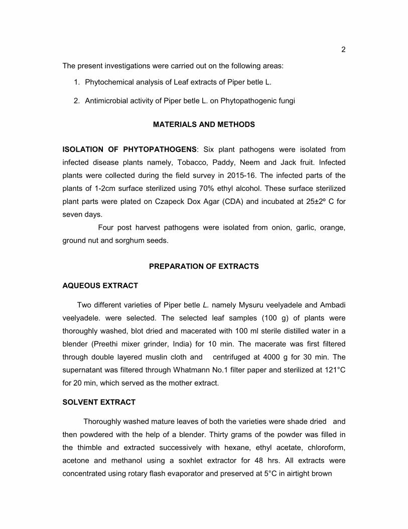
The present investigations were carried out on the following areas:
1. Phytochemical analysis of Leaf extracts of Piper betle L.
2. Antimicrobial activity of Piper betle L. on Phytopathogenic fungi
MATERIALS AND METHODS
ISOLATION OF PHYTOPATHOGENS: Six plant pathogens were isolated from
infected disease plants namely, Tobacco, Paddy, Neem and Jack fruit. Infected
plants were collected during the field survey in 2015-16. The infected parts of the
plants of 1-2cm surface sterilized using 70% ethyl alcohol. These surface sterilized
plant parts were plated on Czapeck Dox Agar (CDA) and incubated at 25±2º C for
seven days.
Four post harvest pathogens were isolated from onion, garlic, orange,
ground nut and sorghum seeds.
PREPARATION OF EXTRACTS
AQUEOUS EXTRACT
Two different varieties of Piper betle L. namely Mysuru veelyadele and Ambadi
veelyadele. were selected. The selected leaf samples (100 g) of plants were
thoroughly washed, blot dried and macerated with 100 ml sterile distilled water in a
blender (Preethi mixer grinder, India) for 10 min. The macerate was first filtered
through double layered muslin cloth and centrifuged at 4000 g for 30 min. The
supernatant was filtered through Whatmann No.1 filter paper and sterilized at 121°C
for 20 min, which served as the mother extract.
SOLVENT EXTRACT
Thoroughly washed mature leaves of both the varieties were shade dried and
then powdered with the help of a blender. Thirty grams of the powder was filled in
the thimble and extracted successively with hexane, ethyl acetate, chloroform,
acetone and methanol using a soxhlet extractor for 48 hrs. All extracts were
concentrated using rotary flash evaporator and preserved at 5°C in airtight brown

3
bottle until further use.All extracts were subjected to antifungal activity against the
test fungi.
PHYTOCHEMICAL ANALYSIS OF LEAF EXTRACT
The leaf extract and solvent extracts obtained from methanol, hexane,
acetone, ethyl acetate, chloroform and aqueous extract of both the varieties of Piper
betle L. were subjected to preliminary qualitative tests for the presence of
carbohydrates, proteins, steroids, flavonoids, tannins ,saponins, alkaloids,
terpionoids, steroids as described by Sofowara (1993), Trease and Evans (1989)
and Harborne (1973).
Test for Carbohydrates:
a. Molisch Test: To 2 ml extract few drops of α-naphthal (20% in ethyl alcohol)
were added. Then 1 ml of conc. H2SO4 was added along the side of the test
tube. Reddish violet ring at the junction of the two layers indicates the
presence of carbohydrates (Telrandhe et al., 2010).
b. Fehling’s Test : 10 ml of Fehling solution (copper sulphate in alkaline
condition) was added to the concentrated extracts and heated on a steam
bath. Brick-red precipitate indicates the presence of carbohydrate.
Test for Proteins:
a. Biuret Test: To 3 ml of extract was added 4% NaOH and few drops of 1%
CuSO4 solution. Violet or pink colour indicates the presence of proteins.
(Telrandhe et al., 2010).
b. Ninhydrin Test: To 1 ml of extract 1% Ninhydrin reagent was added and
heated on a steam bath. Violet colour indicates the presence of proteins.
Test for Alkaloids:
To 2 ml of extract 2 ml Conc. HCl and few drops of Mayer’s reagent was
added. A green or white precipitate indicates the presence of alkaloids (Culki, 1994).
4
Test for Tannins:
About 0.5 ml of the leaves extract was boiled in 20 ml of water in a test
tube and then filtered. A few drops of 0.1% ferric chloride was added and observed
for brownish green or a blue-black colouration.
Test for Saponin:
About 2ml of the leaf extract was boiled in 20 ml of distilled water in a
water bath and then filtered. 10 ml of the filtrate was mixed with 5 ml of distilled water
and shaken vigorously for a stable persistent froth. The frothing was mixed with 3
drops of olive oil and shaken vigorously, then observed for the formation of emulsion.
Test for Flavonoids :
To determine the presence of flavonoids in the plant sample (Sofowara,
1993; Harbrone, 1973), A 5 ml of dilute ammonia solution were added to a portion of
the aqueous filtrate of the plant extract followed by addition of con. H2SO4. A yellow
colouration in extract indicates the presence of flavonoids. The yellow colouration
disappeared on standing.
Test for steroids:
Two ml of acetic anhydride was added to 0.5 g of extract with 2 m H2SO4.
The colour changed from violet to blue or green in samples indicating the presence
of steroids.
Test for terpenoids (Salkowski test) :
Five ml of the extract was mixed in 2 ml of chloroform and con.H2SO4 (3
ml ) was carefully added to form a layer. A reddish brown colouration of the interface
was formed to show positive results for the presence of terpenoids.
Estimation of Total Phenolics, Carbohydrate , Flavanoids , α-Tocopherols and
ascorbic acid was carried out

5
Estimation of total phenolic compounds
A total phenolic content of the extracts was determined according to the method
of Kujala(kujala TS et al., 2000) with minor modifications, using gallic acid as
standard. The extracts ranging from 0-100 µl and 0-100µg of gallic acid were
dissolved in 0.5 ml of water and were mixed with 500µl of 50% Folin-Ciocalteau
reagent. The mixtures were then allowed to stand for 10min followed by the addition
of 1ml of 20% Na2CO3. After 10min of incubation at ambient temperature, the
absorbance of the supernatant was measured at 730nm. The total phenolics content
was expressed as gallic acid equivalents (GAE) in milligrams per gram powder.
Estimation of total flavonoids content
The basic principle of Aluminium chloride colorimetric method is that Aluminium
chloride forms acid stable complexes with the C-4 keto group and either the C-3 or
C-5 hydroxyl group of flavones and flavonols. In addition it also it also forms acid
labile complexes with the ortho-dihydroxyl groups in the A- or B-ring of flavonoids.
Quercetin is reported to be suitable of various concentrations were used to build up
the calibration curve.
The aluminium chloride colorimetric method was modified from the procedure
reported by Woisky and Saltino (Woisky R, Saltino A, 1998). Standard calibration
curve was prepared using quercetin. The differents extracts ranging from 0-100 µl
and 0-100 µg of quercetin were dissolved in 100 ml of 80% ethanol and then graded
concentrations of the above solution were mixed with 1.5ml of 95% ethanol, 0.1ml of
10% aluminium chloride, 0.1ml of 1 M potassium acetate and 2.8ml of distilled water.
After incubation at room temperature for 30 min, the absorbance of the reaction
mixture was measured at 415nm. Proper controls were also done. The amount of
flavonoids was calculated in the different extracts of the plant using the standard
calibration curve for quercetin.
Estimation of α- tocopherol
Vitamin E was determined according to the method of Kivcak and Mert
(Kivcak B, Mert T, 2001). It is based on the reduction of ferric iron to ferrous iron by
tocopherol, which forms a red colour with 2, 2 dipyridyl. 1g of each extract was taken

6
in 10ml of hexane and transferred to conical flask which was further extracted with
200ml of hexane with stirring for 24 hrs. The contents of the flasks were later filtered
through a whtmann No.1 filter paper . The hexane extract was further distilled in
vacuum to get dry extract and stored at -20ºC. 10 mg of dry hexane extract was
dissolved in 10ml of chloroform.
Aliquots of hexane extract solution (20-100µl) and α- tocopherol solution
(20-100µl) (10mg of α- tocopherol (sigma) was dissolved in 10ml of absolute alcohol)
was transferred to separate volumetric flask and the volume was made up to 3ml
with chloroform. Then 1ml of 2, 2’ dipyridyl dissolved in 25ml of absolute ethanol and
stored in dark bottle at 4C) and 1ml of FeCl3 solution (200mg of FeCl3.6H2O in 100ml
ethanol and stored in brown bottle and kept in refrigerator until used) was added and
mixed well. After 15min, read against the blank at 520nm. A blank was run, using
3ml of chloroform, 1ml of 2,2’ dipyridyl reagent and 1ml of FeCl3 solution. The
amount of α- tocopherol was calculated using the calibration curve for α-tocopherol.
Estimation of total carbohydrates
The total sugar concentrations of the extracts was estimated by Dubois method
(Dubois M et al., 1956). Different aliquots of extract along with the glucose (0-100µg)
where made up to 1ml with distilled water. To this 1ml of 5% phenol and 5ml of
concentrated 36N H2SO4 were added. Orange colour developed was read at 520nm
immediately. The total sugar concentration was calculated according to this standard
glucose calibration curve.
Estimation of ascorbic acid
The total ascorbic content was determined by Das guptha et al., (Das guptha GC
and Guha BC, 1941) method, taking pure ascorbic acid as standard (0-16µgm). 50µl
of different solvent extract were taken and the volume was made up to 1ml using 5%
TCA. This was followed by the addition of 1ml DNPH (Di Nitro Phenyl Hydrazine).
The reaction mixture was then incubated in boiling water both for 10min and allowed
to stand for 15min at room temperature. 60% ice cold sulphuric acid was then added
and the absorbance was read at 540nm. The total ascorbic acid was calculated
using a standard ascorbic acid calibration curve.

7
THIN LAYER CHROMATOGRAPHY (TLC)
Thin layer chromatographic (TLC) analyses were made on 0.25mm thick
silica gel 60G (Merk,7731) prepared on glass plates. The extracts obtained from
hexane , chloroform , ethyl acetate , acetone, methanol and aqueous extracts were
loaded on activated analytical TLC 0.25mm (20cm x 20cm) and separated using
solvent system hexane , ethyl acetate, acetone, di ethyl ether ( 50:50:50:20).
As the ethyl acetate extract was showing better antimicrobial activity and was
rich in tannins as found out by chemical analysis, we thought of purifying the
compound from the same. The ethyl acetate extract was loaded on to the
preparative TLC (20X20cm, 2mm thickness) and separated using the solvent system
hexane.
HPLC ANALYSIS OF PHENOLIC COMPOUNDS
Phenolic compounds was analysed using HPLC (Shimadzu) consisting of an
CR- 4A chromatopack data integrator, rheodyne injector, a SCL- 6A system
controller, LC 6A pump and a SPD 6AV UV visible spectroscopic detector. A
reversed- phase high- performance liquid chromatographic method has been
developed and validated for estimation. A C18 column was used with a gradient
elution of methanol and 0.1% (v/v) acetic acid in HPLC- grade water as mobile
phase at a flow rate of 0.9 ml per minute. UV detection was performed at 278 nm.
LC-MS
LC-MS analysis was performed by 1100 SL trap model with thermo stated
column compartment, diode array and standard auto sampler. Mass analysis was
carried out with 0.5 mL/min flow rate and negative ion mode. The mobile phase and
the solvent gradient were the same with that which was used in HPLC. The sample
injection volume was 10 µl. The UVVIS spectra were recorded in the range of 200–
700nm and chromatograms were acquired at 280 and 340nm. All of the analyses
used the ion-spray source in negative mode with the following settings: nebulizer gas
(N2) 40.0 psi, drying gas 12 L/min and drying gas temperature 350 °C. Full scan
data was acquired by scanning from m/z 50 to 800.
8
ANTIFUNGAL ACTIVITY ASSAY
Aqueous extract
Antifungal activity of the plant extract was carried out by poison food technique
(Nene and Thaplyal, 1979). Czapek Dox Agar medium (CDA) with 10, 25, 50, 75,
100% concentration of aqueous extracts of test plants namely, Mysuru veelyadele
and Ambadi veelyadele were prepared. About 15 ml of the medium was poured into
each petriplate and allowed to solidify. 5 mm disc of seven-day-old culture of
Rhizoctonia solani, Fusarium oxysporum, Rhizopus artocarpi, Phomoposis
azardirachta ,Sclerotium rolfsii and Pyricularia oryzae and Four postharvest
pathogens Aspergillus niger, Fusarium moniliforme Penicillium sp. and Aspergillus
flavus were placed at the center of the petriplate and incubated at 25±2°C for seven
days. After incubation period, radial colony growth (mm) were measured and
recorded in each treatment. For each treatment three replicates were maintained.
CDA medium without the aqueous extract served as control. The fungal toxicity of
the extracts in terms of % inhibition of mycelial growth was calculated using the
following formula:
% inhibition = dc – dt /dc x 100, where dc = average increase in mycelial growth in
control, dt = average increase in mycelial growth in treatment.
Solvent extract
One gram of each of the dried evaporated solvent extract of Mysuru
veelyadele and Ambadi veelyadele was separately dissolved in 10 ml of respective
solvents i.e., hexane, chloroform, ethyl acetate, acetone and methanol. Antifungal
activity of the solvent extract was carried out by poisoned food technique (Nene and
Thaplyal, 1987). The Czapek Dox Agar (CDA) containing 100, 200, 300, 400, 500
and 1000 ppm concentration of each solvent extract was prepared. The CDA
medium amended with 100 µl of the solvent without any extract served as control.
The solvent extract amended medium was poured into sterile 90 mm diameter
petriplates (15 ml per plate).The mycelia disc (5 mm) obtained from the margin of
seven-day-old culture was inoculated at the centre of the petriplate to both control
and solvent extract amended CDA medium. The petriplates were incubated at 25 ±

9
2°C for seven days. The experiment was replicated three times. The diameter of the
fungal colonies and growth characteristics in each petridish were recorded. The
antifungal activity was expressed as percentage of mycelial growth inhibition with
respect to control was computed using Srivatsava and Singh (2001) method.
RESULTS
Isolation of phytopathogens
Six phytopathogens were isolated from the disease plants namely,
Rhizoctonia solani, Fusarium oxysporum, Rhizopus artocarpi, Phomoposis
azardirachtae ,Sclerotium rolfsii and Pyricularia oryzae and Four postharvest
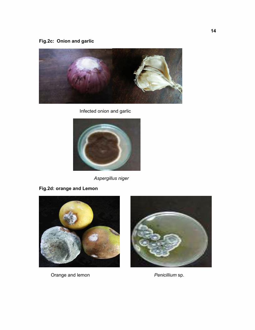
pathogens were isolated from ground nut seeds, onion & garlic, Sorghum seeds,
orange and lemon viz, Aspergillus niger ,Fusarium moniliforme ,Penicillium sp. and
Aspergillus flavus. All these cultures were maintained at 4°C.
Table 1a: Phytopathogens isolated from diseased plants
Table 1b: Post harvest pathogens
Sl.n
o.
Plant material organism
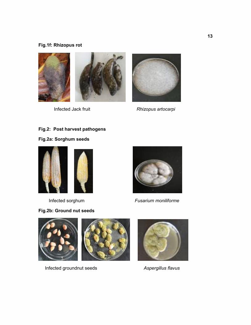
1 Sorgum seeds Aspergillus niger and Fusarium moniliforme
2 Groundnut seeds Aspergillus flavus
3 Onion and Garlic Aspergillus niger
4 Orange and lemon Penicillium sp.
Sl. No. Disease Host Pathogen isolated
1 Sore shin Tobacco Rhizoctonia solani
2 wilt Tobacco Fusarium oxysporum
3 Rhizopus rot Jack fruit Rhizopus artocarpi
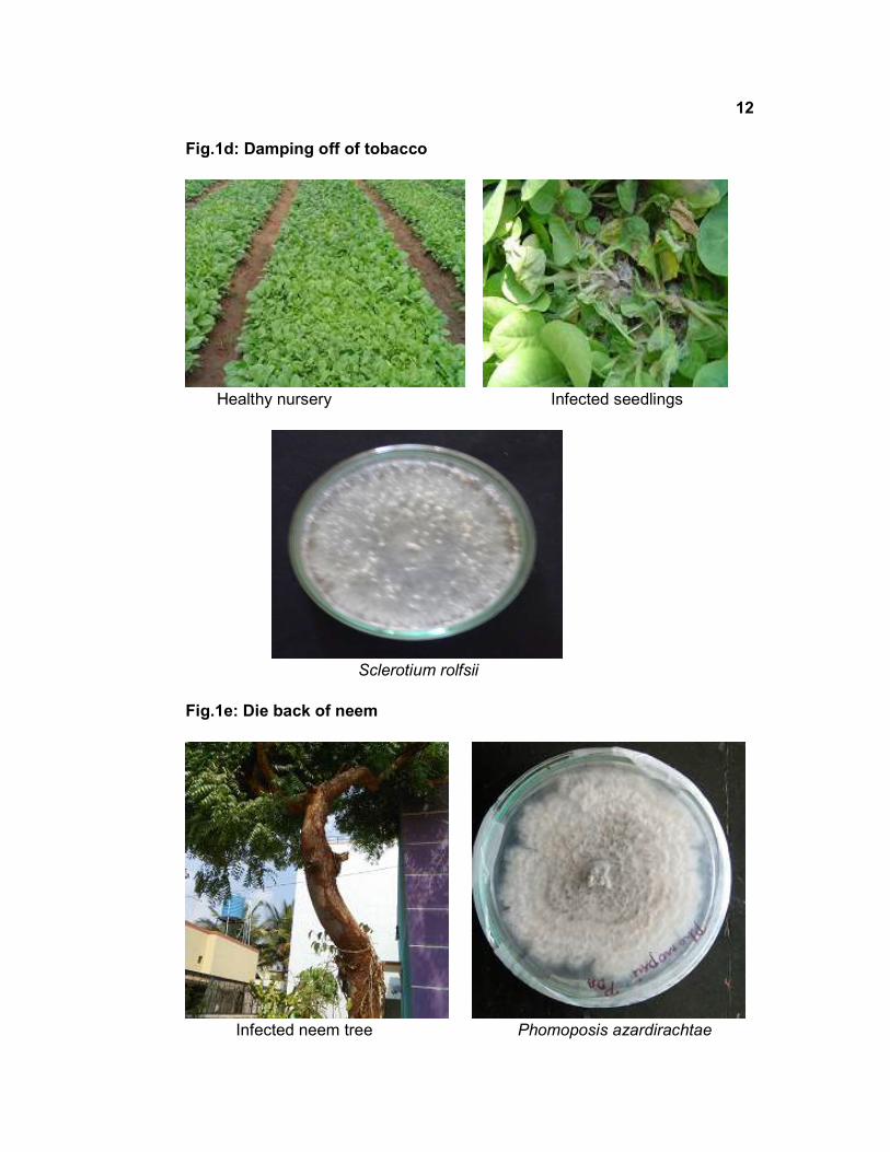
4 Damping off Tobacco Sclerotium rolfsii
4 Die back of neem Neem Phomoposis azardirachtae
5 Blast of paddy Paddy Pyricularia oryzae
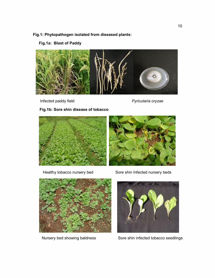
10
Fig.1: Phytopathogen isolated from diseased plants:
Fig.1a: Blast of Paddy
Infected paddy field Pyricularia oryzae
Fig.1b: Sore shin disease of tobacco
Healthy tobacco nursery bed Sore shin Infected nursery beds
Nursery bed showing baldness Sore shin infected tobacco seedlings
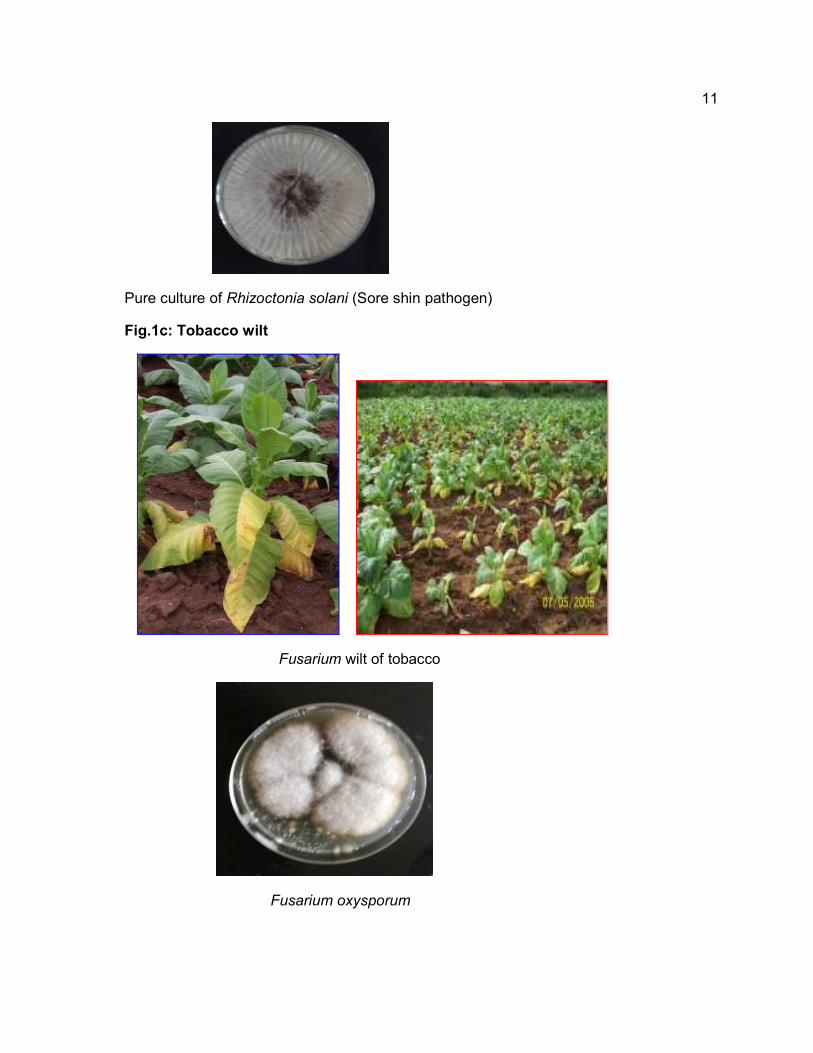
11
Pure culture of Rhizoctonia solani (Sore shin pathogen)
Fig.1c: Tobacco wilt
Fusarium wilt of tobacco
Fusarium oxysporum
12
Fig.1d: Damping off of tobacco
Healthy nursery Infected seedlings
Sclerotium rolfsii
Fig.1e: Die back of neem
Infected neem tree Phomoposis azardirachtae
13
Fig.1f: Rhizopus rot
Infected Jack fruit Rhizopus artocarpi
Fig.2: Post harvest pathogens
Fig.2a: Sorghum seeds
Infected sorghum Fusarium moniliforme
Fig.2b: Ground nut seeds
Infected groundnut seeds Aspergillus flavus
14
Fig.2c: Onion and garlic
Infected onion and garlic
Aspergillus niger
Fig.2d: orange and Lemon
Orange and lemon Penicillium sp.

15
QUALITATIVE ANALYSIS OF PHYTOCHEMICALS IN MYSORE AND AMBADI
VEELYADELE
Qualitative analysis revealed that carbohydrate, proteins, flavanoids,
alkaloids, tannins, Terpinoids, saponins and steroids were present in aqueous
extract. Whereas, carbohydrate, proteins, flavanoids, tannins and saponins were
absent in Hexane and chloroform extract but were present in other solvent extracts.
Terpenoids and steroids were absent in ethyl acetate, acetone and methanol extract
but present in hexane and chloroform.
Table 2: Qualitative analysis of phyto-constituents of aqueous and solvent
extracts of both the varieties of Piper betle L.
Phytochemical analysis also revealed that both Mysore and Ambadi
veelyadele are having high amount of vitamin E in hexane extract, Mysore
veelyadele is rich in flavonoids, total carbohydrates, phenolics, ascorbic acid and
alpha tocopherol when compared to Ambadi veelyadele. Both varieties are rich in
flavonoids, compared to other contents.
Phytochemicals Aqueous extract
Hexane Chloroform Ethyl acetate
acetone methanol Observation
Carbohydrate + - - + + + Brick-red precipitate
Proteins + - - + + + Violet colour
Flavanoids + - - + + + Yellow colouration
Alkaloids + + +
+ + + green precipitate
Tannins + - - + + + Blue black colouration
Terpenoids + + + - - - Reddish brown colouration
Saponins + - - + + + Stable persistant froth
Steroids + + + - - - Blue/ Green colouration

16
Table 3: Proximate Composition of various extracts in soxhlet
Table 3a: Ambadi veelyadele
Table 3b :Mysuru veelyadele
Extracts of Piper betle L. Mysore
Total carbohydrates (mg/gm)
Total phenolics (mg/gm)
Flavonoids (mg/gm)
Ascorbic acid (mg/gm)
α – tocopherol (mg/gm)
Hexane -- - - - 0.02
Chloroform - - - - 7.7 0.01
Ethyl acetate 0.04 0.01 0.32 -
Acetone 0.02 0.03 0.03 0 0.01 -
Methanol 0.04 0.05 0.02 0.02 -
Aqueous 0.01 0.05 0.01 -
TOTAL 17.7 22.1 32 2.64 15.7
THIN LAYER CHROMATOGRAPHY (TLC)
As the ethyl acetate extract was showing better antimicrobial activity and was
rich in tannins as found out by chemical analysis, we thought of purifying the
compound from the same. The ethyl acetate extract was loaded on to the
preparative TLC(20X20cm,2mm thickness) and separated using the solvent system
hexane , Ethyl acetate, acetone , diethyl ether (50:50:50:20) ethyl acetate extract are
loaded to the preparatory TLC separated using solvent system three bands were
obtained ,named as upper band, middle band and lower band ,these 3 bands were
scraped into the beaker acetone were added and filtered , the filtrate was again
Extracts of Piper betle L. Ambadi
Total carbohydrates (mg/gm)
Total phenolics (mg/gm)
Flavonoids (mg/gm)
Ascorbic acid (mg/gm)
α – tocopherol (mg/gm)
Hexane -- - - - 6.7 0.02
Chloroform - - - - 8.0 .001
Ethyl acetate 2.7 2.1 0.04 4.0 0.01 -
Acetone 3.3 0.02 5.1 0.03 6.0 0.03 -
Methanol 4.0 0.04 5.5 0.05 8.0 0.02 0.62 0.03 -
Aqueous 6.0 0.00 2.0 0.05 4.0 0.01 -
TOTAL 16 14.7 22 14.7

17
loaded on to the analytical TLC to check the purity of molecule, single spot was
obtained in all the three different bands and they were subjected further HPLC and
LC MS
Mysuru veelyadele Ambadi veelyadele
Fig 3: TLC profile of Mysuru veelyadele and Ambadi veelyadele
Mysuru veelyadele Ambadi veelyadele
Fig.4:TLC profile of Mysuru veelyadele and Ambadi veelyadele under UV

18
Fig.5a: Preparatory TLC of Mysuru veelyadele
Fig.5b: Preparatory TLC of Ambadi veelyadele

19
HIGH PERFORMANCE LIQUID CHROMATOGRAPHY
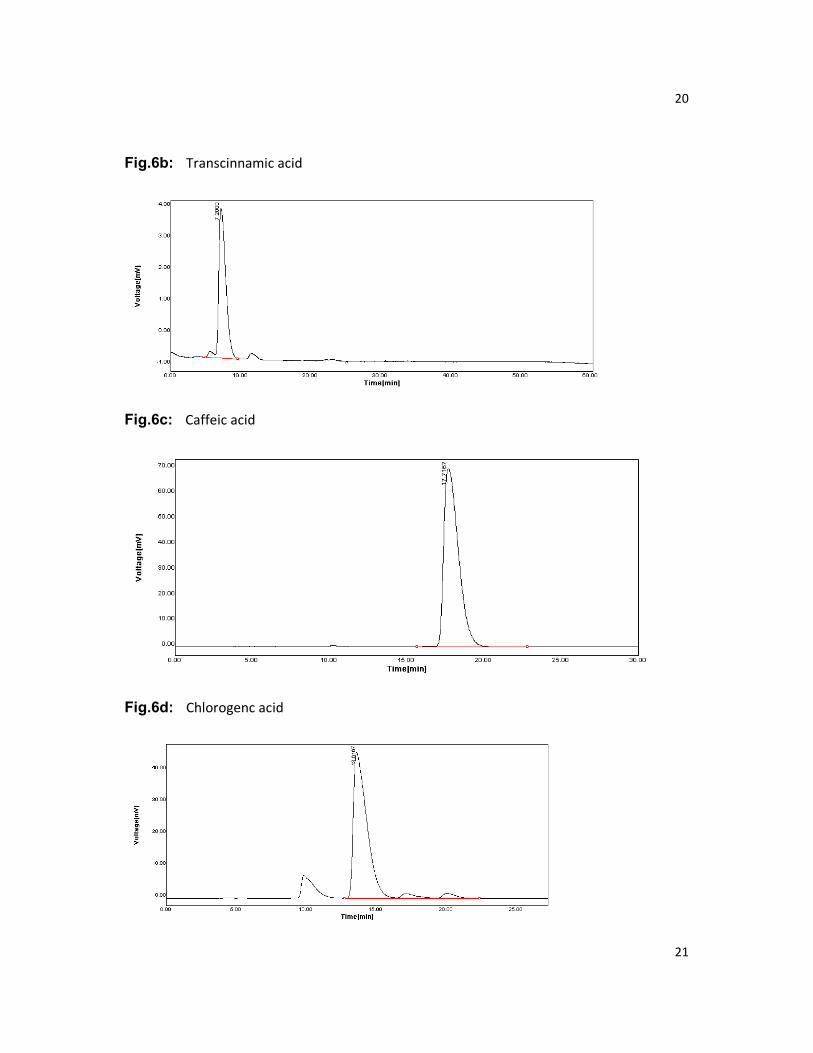
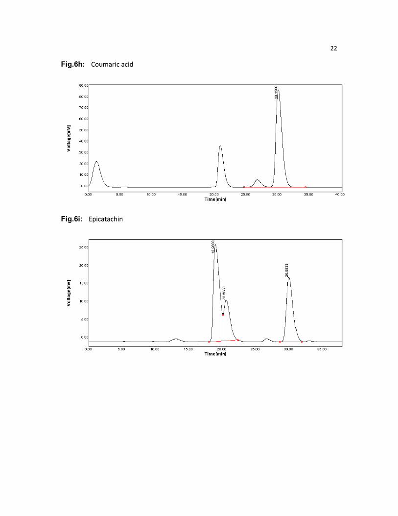
The phenolic standards used were Coumaric acid, Caffeic acid, Synergic acid,
Chlorogenic acid, Procalactin, Gallic acid, Vanillin, Transcinamic acid, Calcihin and
Epicatichin
Fig.6: HPLC Chromatogram of standard phenolics
Fig.6a: Gallic acid
20
Fig.6b: Transcinnamic acid
Fig.6c: Caffeic acid
Fig.6d: Chlorogenc acid
21
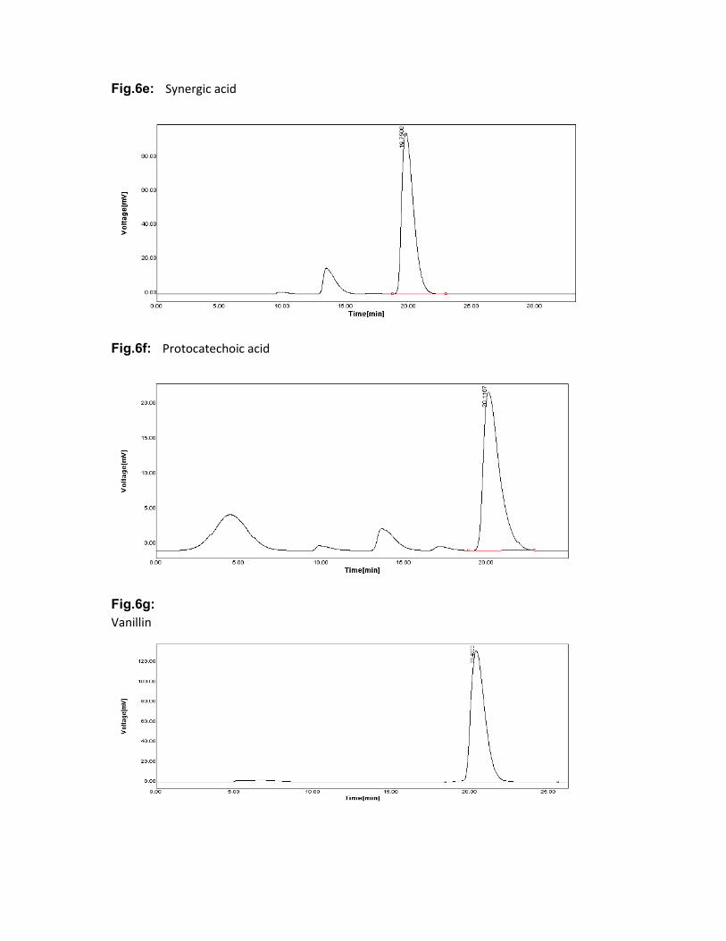
Fig.6e: Synergic acid
Fig.6f: Protocatechoic acid
Fig.6g:
Vanillin
22
Fig.6h: Coumaric acid
Fig.6i: Epicatachin
23
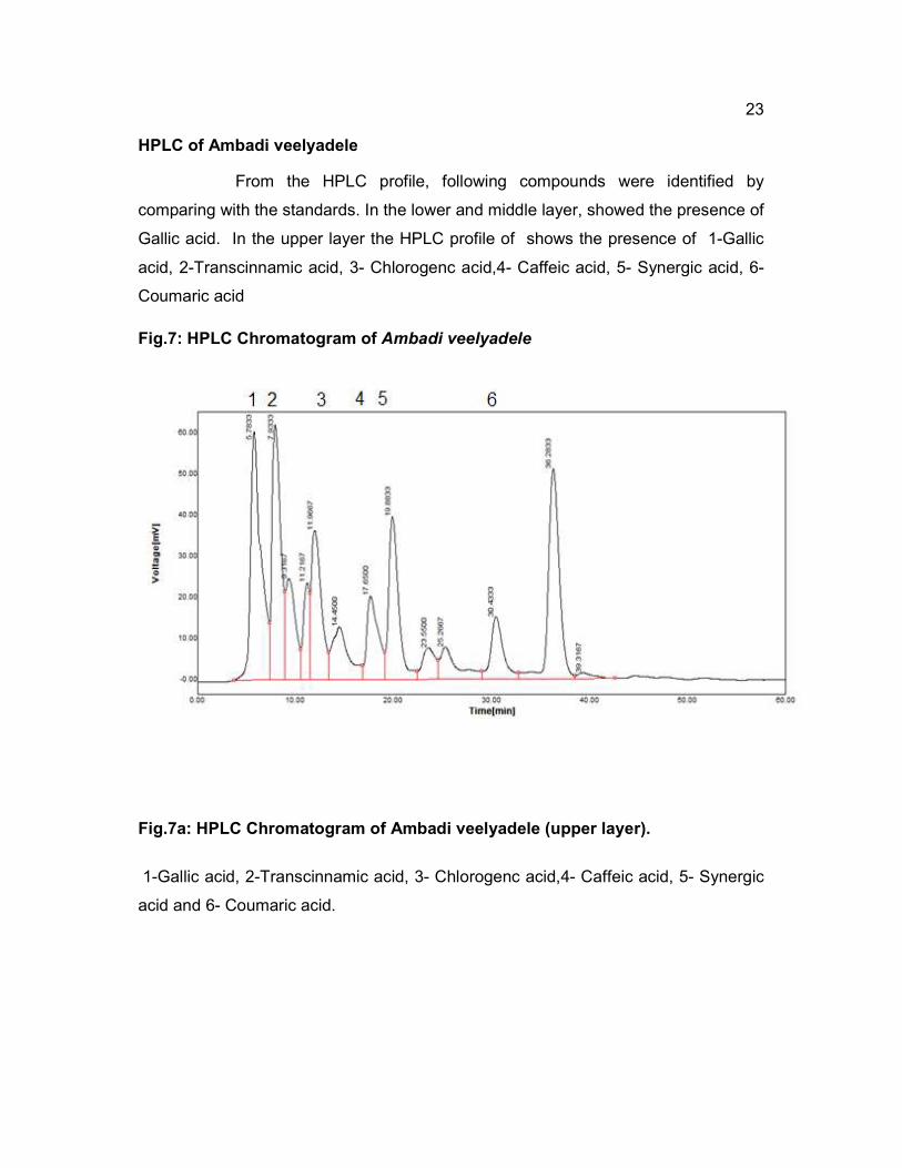
HPLC of Ambadi veelyadele
From the HPLC profile, following compounds were identified by
comparing with the standards. In the lower and middle layer, showed the presence of
Gallic acid. In the upper layer the HPLC profile of shows the presence of 1-Gallic
acid, 2-Transcinnamic acid, 3- Chlorogenc acid,4- Caffeic acid, 5- Synergic acid, 6-
Coumaric acid
Fig.7: HPLC Chromatogram of Ambadi veelyadele
Fig.7a: HPLC Chromatogram of Ambadi veelyadele (upper layer).
1-Gallic acid, 2-Transcinnamic acid, 3- Chlorogenc acid,4- Caffeic acid, 5- Synergic
acid and 6- Coumaric acid.

24
Fig.7b: HPLC Chromatogram of ambadi veelyadele (middle layer)
Fig.7c: HPLC Chromatogram of ambadi veelyadele(lower layer)
HPLC of Mysuru veelyadele
From the HPLC profile of Mysuru veelyadele following compounds were
identified by comparing with the standards. In HPLC profile of upper band which
shows the presence of 1-Gallic acid. HPLC profile of middle and lower band
showed the presence of 1-Chlorogenc acid and 1- Transcinnamic acid

25
Fig.8: HPLC Chromatogram of Mysuru veelyadele
Fig.8a: HPLC Chromatogram of Mysuru veelyadele (upper layer)
Fig.8b: HPLC Chromatogram of Mysuru veelyadele ( middle layer)
Fig.8c:HPLC Chromatogram of Mysuru veelyadele ( Lower layer)

26
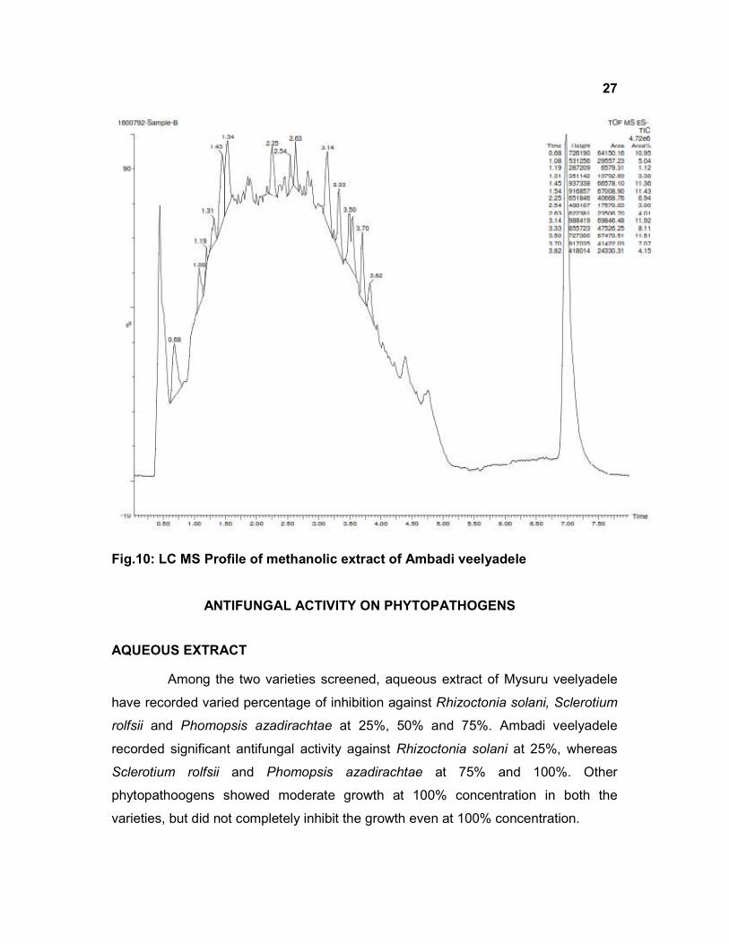
LCMS
In addition to HPLC, LCMS was also carried out for Methanol extract to
characterise the molecules which is present in both the varieties
In the Ambadi veelyadele (Negative mode)
1. Retention time 1.485 molecular mass 371.061
2. Retention time 0.68 molecular mass 290.035
In the Mysuru veelyadele (Negative mode)
1. Retention time 3.141 molecular mass 445.16
2. Retention time 3.534 molecular mass 593.265
3. Retention time 1.536 molecular mass 593.201
4. Retention time 1.436 molecular mass 341.048
5. Retention time 0.68 molecular mass 290.032
These are the abundant molecules present in these samples. One molecule
having a molecular mass of 290.035 and retention time 0.68 is common in both
Mysuru and ambadi veelyadele. Further characterisation is in progress.
Fig.9: LC MS Profile of methanolic extract of Ambadi veelyadele
27
Fig.10: LC MS Profile of methanolic extract of Ambadi veelyadele
ANTIFUNGAL ACTIVITY ON PHYTOPATHOGENS
AQUEOUS EXTRACT
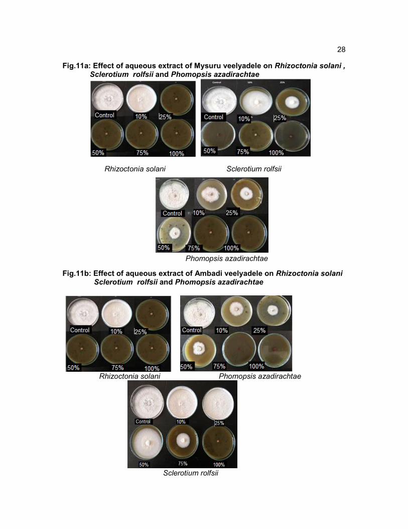
Among the two varieties screened, aqueous extract of Mysuru veelyadele
have recorded varied percentage of inhibition against Rhizoctonia solani, Sclerotium
rolfsii and Phomopsis azadirachtae at 25%, 50% and 75%. Ambadi veelyadele
recorded significant antifungal activity against Rhizoctonia solani at 25%, whereas
Sclerotium rolfsii and Phomopsis azadirachtae at 75% and 100%. Other
phytopathoogens showed moderate growth at 100% concentration in both the
varieties, but did not completely inhibit the growth even at 100% concentration.
28
Fig.11a: Effect of aqueous extract of Mysuru veelyadele on Rhizoctonia solani , Sclerotium rolfsii and Phomopsis azadirachtae
Rhizoctonia solani Sclerotium rolfsii
Phomopsis azadirachtae
Fig.11b: Effect of aqueous extract of Ambadi veelyadele on Rhizoctonia solani Sclerotium rolfsii and Phomopsis azadirachtae
Rhizoctonia solani Phomopsis azadirachtae
Sclerotium rolfsii

29 Fig.12a:Percentage inhibition of aqueous extract of Mysuru veelyadele
Fig.12b: Percentage inhibition of aqueous extract of Ambadi veelyadele
30 Table 4: Percentage inhibition (PI) of growth of plant pathogens by aqueous extract of
Mysuru veelyadele and Ambadi veelyadele
Sl. No.
Organism Mysuru veelyadele Ambadi veelyadele
Percentage of mycelia inhibition
Concentration range
10% 25% 50% 75% 100% 10% 25% 50% 75% 100%
1 Fusarium oxysporum 39 c 44
c 53
c 61
c 61
c 27
b 48
c 50
c 56
c 61
c
2 Rhizopus artocarpi 00 a 00
a 00
a 00
a 00
a 00
a 00
a 00
a 00
a 00
a
3. Phomopsis azadirachtae
39 c 44
c 47
c 100
d 100
d 56
c 47
c 58
c 100
d 100
d
4 Pyricularia oryzae 33 c 39
c 44
c 72
cd 72
cd 39
c 44
c 62
c 67
c 78
cd
5 Rhizoctonia solani 11b 100
d 100
d 100
d 100
d 33
b 100
d 100
d 100
d 100
d
6 Sclerotium rolfii 00 a 11
b 100
d 100
d 100
d 00
a 00
a 11
b 61
C 100
d
Post harvest pathogens
7 Fusarium moniliforme 28 b 41
c 44
c 50
c 67
c 28
b 44
c 56
c 62
c 77
cd
8. Penicillium sp. 00 a 39
c 50
c 50
c 67
c 00
a 33
c 44
c 50
c 64
c
9. Aspergillus niger 11 b 16
b 28
b 28
b 22
b 00
a 00
a 13
b 28
b 22
b
10. Aspergillus flavus 00 a 27
b 20
b 27
b 44
c 00
a 11
b 14
b 17
b 20
b
Figures having the same letters are not significantly different according to Ducan’s multiple range test (P<0.05).
SOLVENT EXTRACT
Among the solvent extracts tested, hexane and ethyl acetate extract of Mysuru
veelyadele has shown significant activity against Phomopsis azadirachtae,
Pyricularia oryzae, Rhizoctonia solani, Sclerotium rolfsii at 100 ppm and Ambadi
veelyadele was effective on only two phytopathogens viz., Rhizoctonia solani and
Sclerotium rolfsii at 100ppm.
The concentration of 100ppm of Hexane and ethyl acetate extract of Mysuru
veelyadele was effective in inhibition of mycelia growth of P. azadirachtae, P. oryzae,
R. solani and S. rolfsii at 100 ppm.
Ethyl acetate extract of Mysuru veelyadele has shown significant activity
against P. azadirachtae, P. oryzae, R. solani, S. rolfsii, F. oxysporum and R. artocari
at 100 ppm. Post harvest pathogens namely, F. moniliforme showed growth

31
inhibition at 400ppm, penicillium sp. at 500ppm and A. niger and A.flavus at
200ppm.
In hexane extract F.oxysporum was inhibited at 500ppm. Post harvest
pathogens namely, A. niger at 200ppm, Penicillium sp.at 400ppm, A. flavus, and
F.moniliforme was inhibited at 200ppm, 300ppm and 500ppm.
Methanol and Chloroform extracts showed significant growth inhibition of
Phomopsis azadirachtae, Pyricularia oryzae and Rhizoctonia solani at 1000 ppm.
Post harvest pathogens showed moderate growth at this concentration.
Total absence of inhibitory activities by Acetone extract of both Mysuru
veelyadele and Ambadi veelyadele on plant pathogens were observed
Rhizopus artocarpi was inhibited by ethyl acetate extract at 100ppm but not in
hexane extract.
Fig.13: Effect of Ethyl acetate extract of Mysuru veelyadele on Phytopathogens
Phomopsis azadirachtae Pyricularia oryzae Rhizoctonia solani
Sclerotium rolfsii Rhizopus artocari Fusarium oxysporum

32
Fusarium moniliforme Aspergillus niger
Penicillium digitatum Aspergillus flavus
Ambadi veelyadele extract was effective on only two phytopathogens viz.,
Rhizoctonia solani and Sclerotium rolfsii at 100ppm.
The concentration of 100ppm of hexane extract of Ambadi veelyadele
showed significant inhibition of mycelia growth of R. Solani, P. oryzae and S. rolfsii at
100ppm. P. azadirachtae at 200ppm and F. oxysporum at 500ppm. No growth
inhibition was seen in R. artocarpi. Among post harvest pathogens, A. niger at
100ppm, P. digitatum and F. moniliforme showed inhibiton at 500ppm and A. flavus
at 1000ppm. Whereas ethyl acetate extract showed significant inhibition of
P.azadirachtae, P. oryzae, R. solani, S. rolfsii and R. artocarpi at 300ppm and
F.oxysporum at 400ppm. Whereas Post harvest pathogen namely, Penicillium
sp.and F. moniliforme showed inhibition at 400ppm A. niger and A. flavus at
200ppm.
33
In methanol extract, only Phomopsis azadirachtae showed inhibition at 1000ppm.
where as other phytopathogens showed moderate growth at different concentration.
Rhizopus artocarpi was ineffective against methanol.
Chloroform extract was effective against Phomopsis azadirachtae, Pyricularia
oryzae and Rhizoctonia solani at 1000ppm but not against other pathogens
Total absence of inhibitory activities by Acetone extract of both Mysuru veelyadele
and Ambadi veelyadele on plant pathogens were observed.
Fig.14: Effect of Hexane extract of Mysuru veelyadele on Phytopathogens
Rhizoctonia solani Sclerotium rolfsii
Phomopsis azadirachtae Pyricularia oryzae
34
Fusarium moniliforme Aspergillus niger
Penicillium digitatum Aspergillus flavus
40
DISCUSSION
Biological control have attained importance in modern agriculture to curb
the hazards of intensive use of chemicals for pest and disease control (Baker and
Cook,1079). Phytochemical analysis and in vitro antifungal studies were carried out
to explore the fungicidal efficacy of two varieties of Piper betle L. viz., Mysuru and
Ambadi veelyadele aqueous and solvent extracts against plant pathogens.
In the current investigation both the varieties showed the presence of
carbohydrates, proteins, flavanoids, tannins, saponins, terpenoids, steroids and
alkaloids in aqueous extract. However carbohydrate, proteins, flavonoids ,tannins
and saponins were absent in hexane and chloroform extract but was present in ethyl
acetate, acetone and methanol. Steroids and terpinoids were absent in ethyl
acetate, acetone and methanol but present in chloroform and hexane extracts.
Similar findings were reported by Chakraborty and Shah, 2011 have reported that
several extracts of P. betle leaves using methanol, petroleum ether, aqueous and
ethyl acetate produced different results in which all the tested solvents, except for
water extract had indicated the presence of flavonoids, tannins, sterols and phenol,
but lack of alkaloids. Jayalakshmi et al.,2011 have reported the presence of
carbohydrate, proteins, flavonoids and tannins in methanolic extract. Prakash et.al.,
2014 have also reported the presence of tannins, flavonoids and terpenoids in
methanol extracts. Similarly Arani et al., 2011 also reported the presence of tannins,
flavanoids, carbohydrates and proteins.
Both mysore and ambadi varieties are having high amount of
vitamin E in hexane extract, Mysore variety is rich in flavonoids, total carbohydrates,
phenolics, ascorbic acid and alpha tocopherol when compared to Ambadi variety.
Both varieties are rich in flavonoids, compared to other contents. Similar findings
have been reported in Piper betle L. by Dwivedi and Tripathi 2014; Divyalashmi and
Aruna Sharmili 2017.
41
In preparatory and analytical TLC purity of the molecule was tested and the
band obtained were subjected further HPLC and LC MS. HPLC was carried out for
ethyl acetate extract to characterize the phenolic profile of the two varieties of Piper
betle L.. During the present investigation six phenolic compounds, comprising of
Gallic acid, Transcinnamic acid, Chlorogenc acid, Caffeic acid, Synergic acid and
Coumaric acid was present in Ambadi veelyadele. However, in Mysuru veelyadele
three phenolic compounds, namely gallic acid, Chlorogenic acid and Transcinnamic
acid were present. Gallic acid was the major compound in both the extract. Ferreres
et.al., 2014 have reported hydroxychavicol as the major compound in Piper betle L..
Similarly, Hydroxychavicol and chavibetol were detected in ethanol extracts and
aquous extracts the higher percentage of hydroxychavicol has been reported by
Nurul et al., 2013. In LC MS study revealed the presence of abundant molecules in
both Mysuru and Ambadi veelyadele These molecules should we further
characterised. One molecule having a molecular mass of 290.035 and retention time
0.68 was common in both varieties.
Among the two varieties studied, aqueous extract of Mysuru veelyadele have
recorded varied percentage of inhibition against Rhizoctonia solani, Sclerotium rolfsii
and Phomopsis azadirachtae and Ambadi veelyadele recorded significant antifungal
activity against Rhizoctonia solani . Other phytopathogens showed moderate growth
in both the varieties. Seema et al., 2011, has reported the antifungal activity of Piper
betle L. on sore shin pathogen, Rhizoctonia solani. The effect of these extracts was
ineffective against R. artocarpi. Prince and prabakaran, 2011 have reported that
aqueous extract of Piper betle L. was not effective against Colletotrichum falcatum
which is a soil borne fungus.
Among the solvent extracts tested, hexane and ethyl acetate extract of Mysuru
veelyadele has shown significant antifungal activity against Phomopsis azadirachtae,
Pyricularia oryzae, Rhizoctonia solani, Sclerotium rolfsii. Seema et al.., 2011 have
also reported that ethyl acetate extract of Piper betle L. possess inhibitory effect
against Rhizoctonia solani causing sore shin of tobacco. Ambadi veelyadele was
effective on only two phytopathogens viz., Rhizoctonia solani and Sclerotium rolfsii.
Mrinoy et al., 2017 has reported that methanol extract of Piper betle L. have shown
42
significant antifungal activity on Rhizoctonia solani. The hexane extract have shown
significant activity against Aspergillus niger Penicillium digitatum ,Aspergillus flavus
and Fusarium moniliforme . Ethyl acetate extract, completely inhibited the growth of
Fusarium moniliforme at very low concentration. Similar results have been reported
on Piper betle L. leaves have significant potential in the integrated pest management
of common contaminants in storage grains and soil borne pathogens by Seetha
lakshmi and Naidu, 2013. Rhizopus artocarpi was inhibited by ethyl acetate extract
at 100ppm but not in hexane extract. Shitut et al., have reported that ethyl acetate
extract of Piper betle L. have shown significant activity against human pathogenic
bacteria and phytopathogenic fungi.
Previous research literature on antifungal properties of Piper betle L. have
shown that they have varying degrees of growth inhibitory effect on soil borne
pathogens and post harvest pathogens due to their chemical compositon. Several
workers have identified the chemical compounds of these plants and showed that
those fractions are very efficient in suppressing the growth of fungi. The fungicidal
property of Piper betle L. might be due to the presence of hydrochavicol in the
extracts as reported by Ali et al.,2010; Nalina and Rahim,2007. The higher activity of
ethyl acetate extract could be because of the fractions containing phenolic
compounds. The results confirmed that these aqueous plant extracts and solvent
extracts have antifungal properties on plant pathogenic fungi. Further research work
is warranted to identify the bioactive compounds having antifungal activity in these
two varieties. This study is an experimental evaluation and offers a possibility to
make use of Piper betle L. in biopesicidal drug development to control the post
harvest pathogens and soil borne pathogens.
43
SUMMARY AND CONCLUSION
Piper betle L. (Piperaceae) is a tropical shade-loving perennial evergreen
climber. It is an important medicinal plant and its use is mentioned in our vedic
literature such as, Charaka Samhita, Sushruta Samhita and Astanga Hridayam. It is
known to have medicinal properties such as antiseptic, analgesic, antibacterial,
carminative and stimulant etc. This plant is having geographical indication tag. There
are many varieties and difference among these is not well documented. Hence, two
varieties of betel vine commonly cultivated in Mysuru viz., ‘Mysuru veelyadele’ and
‘Ambadi veelyadele’ were considered for the present study for comparing the
chemical components. Preliminary quantitative phytochemical analysis for
Carbohydrates, Phenolics, Flavonoids, Ascorbic acid and α-tocopherol were made
by following standard procedures using aqueous and solvent leaf extracts. Variation
in the quantity of phytochemicals was observed in both the varieties. Significant
biological activity was showed by ethyl acetate extract and hence this was partially
purified using TLC, HPLC and LCMS. The separated molecules from TLC were
subjected to HPLC for the confirmation of purity. Studies revealed the presence of
Gallic acid, Transcinnamic acid, Caffeic acid, Chlorogenic acid, Synergic acid and
Coumaric acid. In LCMS a molecule having molecular mass of 290.035 and retention
time of 0.68 was found in both the varieties. The four compounds identified in
Mysuru veelyadele were absent in Ambadi veelyadele. Further work on
characterization of active principles is in progress.
The in vitro antimicrobial activity of aqueous and solvent leaf extracts of two
different varieties of Piper betle L. namely Mysuru veelyadele and Ambadi
veelyadele leaf extracts were studied against phytopathogenic fungi such as,
Fusarium oxysporum, Rhizopus artocarpi, Phomopsis azadirachtae, Pyricularia
oryzae, Rhizoctonia solani, Sclerotium rolfsii and post harvest pathogens Fusarium
moniliforme, Penicillium digitatum, Aspergillus niger and Aspergillus flavus by
poisoned food technique. Aqueous extract of both the varieties showed significant
inhibition of Rhizoctonia solani, Phomopsis azadirachtae and Sclerotium rolfsii
when compared to other pathogens. Among the solvent extracts tested, hexane and
ethyl acetate extract of Mysuru veelyadele has shown significant activity against
Phomopsis azadirachtae, Pyricularia oryzae, Rhizoctonia solani, Sclerotium rolfsii at

100 ppm and Ambadi veelyadele was effective on only two phytopathogens viz.,
Rhizoctonia solani and Sclerotium rolfsii at 100ppm. The remaining pathogens listed
above were inhibited at 500ppm except Rhizopus artocarpi. Other solvent extracts
of these two plant varieties have shown a range of activity against most of the
phytopathogens tested.
This study has revealed the presence of many bioactive molecules in
both the varieties of Piper betle L. It has further confirmed that the plant extracts
could be used for control of phytopathogenic fungi and shows that Piper betle L.
could be exploited for new potent antimicrobial agent against plant pathogen. This
study forms a basis for the characterization of the phytochemicals of the two
varieties. The presence of various bioactive compounds justifies the usage of betel
leaf as an herbal choice for controlling various plant diseases. Aqueous and solvent
extracts
are found effective, it will help in the formulation of ecofriendly control measure,
which is cheap and can be recommended to the farmers.

45
BIBLIOGRAPHY
Ali, I., Khan, G. F., Suri, K. A., Gupta, B.D., Satti, N.K., Dutt, P., Afrin, F., Qazi ,G.
A., and Khan, I..A. 2010. In vitro antifungal activity of hydroxychavicol isolated
from Piper betel L. Annals of Clinical Microbiology and Antimicrobials 9:7.
Anonymous, 2007. The country report on the state of plant genetic resources
for food and agriculture in India. National Bureau of plant genetic
resources. ICAR, New Delhi, India, 55pp.
Arani Datta., Shreya Ghoshdastidar., and Mukesh Singh. 2011. Antimicrobial
Property of Piper betel Leaf against Clinical Isolates of Bacteria; Journal of
Pharma Sciences and Research (IJPSR) 2(3): 104-109.
Baker, K.F. and Cook, J.R. 1979. Biological control of plant pathogens. S. Chand
and Co.Limited, New Delhi, India, 433pp.
Chakraborty, D., and Shah, B. 2011. Antimicrobial, anti-oxidative and anti-
hemolytic activity of Piper betel leaf extracts. International Journal of
Pharmacy and Pharmaceutical Sciences 3: 192-199.
Culki, I.1994, Methodology of Analysis of Vegetable drug. Chemical Industries
branch, UNIDO, Romania: 24: 26 -27.
Das, J., Goswami, S., Gupta, R., and Begam, M. 2006. In vitro sensitivity of
phytopathogenic fungi against Indian Piper, International Journal of Herbal
Medicine J Curr Sci 9:721-725.
Divyalashmi, l. and Aruna sharmili, S. 2017. Phytochemical analysis and
antibacterial activity of mangifera indica L. and piper betle . International
Journal of pharma and bio sciences 8(2): (p) 84-91.
Dubois, M., K. A. Gilles, J. K., Hamilton, P. A., Rebers, F., and Smith. 1956.
Colorimetric method for determination of sugars and related substances.
Analytical Chemistry. 28, 350–356.
Dwivedi, V. and Tripathi, S. 2014. Review study on potential activity of Piper betle.
46
Journal of Pharmacognosy and Phytochemistry 3(4): 93-98.
Evans, W.C., Trease ., and Evans.2009. Pharmacognosy. Elsevier Health Sciences;
May 27.
Ferrers, F., Oliveria. A. P., Gil-Izquierdo, A., Valentao, P., and Andrade ,P.B. 2014.
Piper betle leaves: Profiling phenolic compounds by HPLC/DAD-ESI/MS and
Anti-Cholinesterasee activity. Phytochemical analysis 25(5):453-60.
Harborne, J.B, 1973. Photochemical Methods: A Guide to Modern Techniques of
Plant Analysis. Chapman A. and Hall. London, UK, pp 279.
Harborne, J.B.1998. Phytochemical methods: a guide to modern techniques of plant
analysis. 3rd Ed. Chapmann Hall, London, UK, 41-42.
Jayalakshmi, B., Raveesha, K. A., and Amruthesh, K.N.2011. Phytochemical
investigations and antibacterial activity of some medicinal plants against
pathogenic bacteria. Journal of Applied Pharmaceutical Science Jul
1;1(5):124.
Kivcak, B. and Mert, T, 2001. Quantitative determination of a-tocopherol in
Arbutus Ledodensitometry and colorimetry. Fitoterapia 72: 656 – 661.
Kujala, T.S., Loponen, J.M., Klika, K.D., and Pihlaja, K. 2000. Phenolics and
betacyanins in red beetroot (Beta Vulgaris) root: distribution and effect of
cold storage on the content of total phenolics and three individual
compounds. Journal of Agricultural and Food Chemistry. 48, 5338-
5342.
Kumbhar, P.P., Salnkhe, D.H., Borse, M.B., Hiwale, M.S., Nikam, L.B., Bendre, R.S.,
Kulkarni, M.V. and Dewang, P.M. 2000. Pesticidal potency of some common
plant extracts. Pestology 26: 51-53.
Mahesh, M. and Satish, S. 2008.Antimicrobial activity of some important medicinal
plant against plant and human pathogens. World Journal of Agricultural
Science 4(S):839-843.
47
Mrinoy Das, Sandhya chhetri, Mahima arora , Aneesha, G., Jayashree, D., and
Priya,V. 2017. In vitro evaluation of Piper betle L. Methanol extract and its
fractions aginst Rhizoctona solani. International Journal of herbal Medicine
5(2):35-37.
Nalina, T. and Rahim, Z.H.A. 2007.The crude aqueous extract of Piper betle L. and
its antibacterial effect towards Streptococcus mutant. American Journal of
Biotechnology and Biochemistry 3:10-15.
Nene, V.L. and Thaplyal, P.N. 1987. Fungicides in Plant Disease Control. Oxford
& IBH Publ. Co. Pvt. Limited, New Delhi. India, 507pp.
Nurul, N. M., Abdul, F.A. A. and Zhari, I. 2013.A validated reverse phase HPLC for
determination of hydroxychavicol and chavibetol in Piper betle extracts with
different extraction times and locations. The open conference proceedings.
2013.
Prajapati, R.C. 2010.Biodiversity of Karnataka- At a glance. Karnataka
biodiversity board, Bangalore, 96pp
Prakash, U, N., Smila, K.H., Priyanka, J.D., Srinithya. B., and Sripriya, N.2014.
Studies on phytochemistry and bioefficancy of cultivars of Piper Betle Linn.
International Journal of Research in Pharmaceutical Science; 5(2):94-8.
Prince, L. and Prabakaran, P.2011. Antifungal activity of medicinal plants against
plant pathogenic fungus Colletotrichum falcatum. Asian Journal of Plant
Science and Research 1 (1):84-87.
Sasidharan, S., Chen, Y., Saravanan, D., Sundram, K.M. and Yoga Latha,
L.2011. Extraction, Isolation and Characterization of Bioactive compounds
from plant extracts. African Journal of Traditional, Complementary and
Alternative medicines 8(1):1-10.
Seema, M., Sreenivas,S.S., Rekha, N.D. and Devaki, N.S.2011. Invitro studies of
some plant extracts against Rhizoctonia solani Kuhn infecting FCV tobacco in

48
Karnataka Light Soil, Karnataka, India. Journal of Agricultural Technology
7(5):1321-1329.
Seetha lakshmi and Naidu, 2013.Antifungal activity of Piper betle(L) leaf extracts
against pathogens of cereal crops. JPR: BioMedRx : An International
Journal 1(7), 712-714
Shitut, S., Pandit, V., and Mehta, B,K.1999. The antimicrobial efficiency of Piper
betle Linn leaf (stalk) against human pathogenic bacteria and
phytopathogenic fungi. Central European journal of public health 7(3):137-
9.
Sofowora, A. 1993. Screening Plants for Bioactive Agents. In: Medicinal Plants
and Traditional Medicine in Africa, Sofowora, A. (Ed.). 2nd Edn., Spectrum
Books Ltd., Ibadan, Nigeria, pp: 134-156.
Sofowora, A. 1993. Medicinal Plants and Traditional Medicines in Africa. Chichester
John Wiley & Sons, New York, USA, pp 97-145.
Srivatsava, S. and Singh, R.P. 2001. Antifungal activity of the essential oil of
Murraya koenigii (L.)Spreng. Indian Perfumer 45: 49 – 51.
Telrandhe, U,,B., Hemalatha, S.,and Modi, A.2010. Pharmacognostic and
phytochemical investigation on root of Cadaba farinosa Forsk. International
Journal of Pharma and Biological Sciences;1(2):1-13.
Woisky, R. and Salatino, A. 1998. Analysis of propolis: some parameters and
procedures for chemical quality control. Journal Apiculture Research. 37:
99-105.
