elucidating tumor necrosis factor signaling pathway using a functional gene ddentification approach
TRANSCRIPT

Introduction
Sequence information of all human geneswill soon become available because of therapid progression of the human genome pro-jectas well as the accumulation of expressedsequence tags (ESTs). However, a difficulttask we are still facing is to understand thebiologic functions of these genes. Among all
the genes cloned today, at least 50% have totallyunknown functions (1). Efficient use of thesequence information to uncover, confirm, andevaluate gene functions will become a big chal-lenge to biomedical scientists, and, as a result,a switch of focus from gene cloning to func-tional identification will be necessary for bio-logic research in the twenty-first century.
Genetic mutation has been proven to be anefficient way to functionally identify genes in
AbstractFunctional identification of genes is an efficient way to study manybiological processes in lower eukaryotes. However, an effectiveapproach in mammalian cells is still under development. We designeda functional gene identification procedure and applied it in a studyof tumor necrosis factor (TNF)-induced cell killing. This procedureemployed a specially designed retroviral vector that allows randomtruncation of genes, efficiently selecting clones in which a gene wasdisrupted and quickly identifying disrupted genes. We have identi-fied several novel genes by a preliminary test of this approach andconfirmed by reconstitution that the genes we identified are requiredfor TNF cytotoxicity in L929 cells. Because of the efficient identi-fication of these components in TNF-induced cell killing, we havealready been able to outline the killing pathway of TNF in L929cells. Application of this method could be widespread because itcan be used in studying any cellular responses if a specific selec-tion assay can be set up.
Key WordsRetroviral vectorGene identificationTumor necrosis factorCell death
Jiahuai HanDepartment of ImmunologyThe Scripps Research Institute10550 N. Torrey Pines RoadLa Jolla, CA 92037E-mail: [email protected]
55© 2000Humana Press Inc.0257–277X/00/21/2–3:55–61/$11.75
Elucidating Tumor Necrosis FactorSignaling Pathway Using a FunctionalGene Identification Approach
Immunologic Research 2000;21/2–3:55–61
Xiaofei WangJiahuai Han
Department of ImmunologyThe Scripps Research InstituteLa Jolla, CA
prokaryotes as well as lower eukaryotes. How-ever, similar approaches in mammalian cellsare not very successful, which is mostly owingto the big and complex genome. In addition,the mutagens—chemical or radiation—usedin lower eukaryotes are not effective forgenetic study in mammalian cell culturesbecause of the difficulty in controlling themutation frequency and identifying themutated gene (2). Nevertheless, there are afew successful cases using this approach. Thecomplexity of the human genome may soonno longer be an obstacle to genetic approachbecause of the genome project and theadvanced analyzing system benefiting frommodern computer science. An efficientmethod(s) to mutate genes and identify themutated genes in mammalian cells is also indevelopment.
It has long been found that retroviral inser-tion could generate genomic mutations.Because the insertion appears not to prefer anysite, random disruption of genes could beachieved by retroviral infection (3). Speciallydesigned retroviral vectors are made to facili-tate the identification of genes that are dis-rupted. Viral vectors containing promoterlesslacZ or antibiotic-resistant genes work quitesuccessfully in identifying promoters in embry-onic stem cells that facilitate the tracing of thepromoter activities and expression patternsduring embryonic development (4). This pro-moter trapping strategy has also been appliedto study genes repressed by epidermal growthfactor (5), genes involved in antiestrogen resis-tance of breast cancer cells (6), and genesinduced during interleukin-3 withdrawal-induced apoptosis (7). However, identifyingthe disrupted gene according to the sequenceat the vicinity of the insertion site is not effi-cient because of the chance of viral insertioninto the intron region or near the promoter.Improvement was made by adding a splicingacceptor sequence in front of the marker gene
(8,9). It has been shown that the splicing accep-tor significantly enhances the instances of genedisruption and the identification of the geneinto which it was inserted (10).
We adopted the principle of the gene-trapping strategy and further designed anapproach to study signal transduction path-ways of tumor necrosis factor (TNF)-inducedcell death. The rationale is that retroviral inser-tion generates a null allele, which may lead toreduced or diminished expression of the dis-rupted endogenous gene. If such a gene prod-uct is a required component of TNF signalingpathway in inducing cell death, disruption ofthis gene may generate a TNF-resistant cellline. The disrupted genes can be identifiedfrom these cell lines. The causal relationshipbetween gene disruption and cell resistance toTNF can be easily verified by restoring thegene expression ectopically and testing theTNF sensitivity.
Procedure for Identifying GenesRequired for TNF-Induced L929 Killing
Design of Gene Disruption Retroviral VectorFigure 1 shows our pDisrup vector. It con-
tains two functional domains. The first oneincludes a splicing acceptor (SA) sequence,an internal ribosomal entry site (IRES) fol-lowed by green fluorescent protein (GFP)cDNA, and a poly-A signal. The second func-tional domain of pDisrup contains a cyto-megalovirus (CMV) promoter followed bya Neo gene. There is no poly-Asignal after theNeo sequence, but a splicing donor instead.This design has several features that allow usto efficiently disrupt and identify genes. First,both domains are not intact, i.e., either with-out promoter or without poly A signal. If theinsertion does not occur in a gene, neithermarker will be expressed. Second, if the inser-tion occurs in either the intron or exon of agene, the expression of GFP will rely on
Wang and Han56
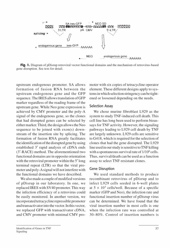
upstream endogenous promoter. SA allowsformation of fusion RNA between theupstream endogenous gene and the GFPsequence. The IRES allows translation of GFPmarker regardless of the reading frame of theupstream gene. While Neo gene expression isachieved by CMV promoter and the poly-Asignal of the endogenous gene, so the clonesthat had disrupted genes can be selected byeither marker. Third, the design allows the Neosequence to be joined with exon(s) down-stream of the insertion site by splicing. Theformation of fusion RNA greatly facilitatesthe identification of the disrupted gene by usingestablished 3′ rapid analysis of cDNA ends(3′-RACE) method. The aforementioned twofunctional domains are in opposite orientationwith the retroviral promoter within the 5′ longterminal repeat (LTR) so that the viral pro-moter and poly-Asignal will not interfere withthe functional domains we have described.
We also made a couple of modified versionsof pDisrup in our laboratory. In one, wereplaced IRES with SV40 promoter. This waythe infection efficiency of a retrovirus couldbe easily monitored. In another version, weincorporated tetracycline repressible promoterand transactivator into the vector. In this vector,we replaced GFP with transactivator cDNA,and CMV promoter with minimal CMV pro-
moter with six copies of tetracycline operatorelement. These different designs apply to sys-tems in which selection stringency can be tight-ened or loosened depending on the needs.
Selection AssayWe chose murine fibroblast L929 as the
system to study TNF-induced cell death. Thiscell line has long been used to perform bioas-says for TNF activity. However, the signalingpathways leading to L929 cell death by TNFare largely unknown. L929 cells are sensitiveto G418, which is required for the selection ofclones that had the gene disrupted. The L929line used in our study is sensitive to TNF killingwith a spontaneous survival rate of 1/106 cells.Thus, survival/death can be used as a functionassay to select TNF-resistant clones.
Gene DisruptionWe used standard methods to produce
recombinant retrovirus of pDisrup and toinfect L929 cells seeded in 6-well platesat 5 × 10⁵ cells/well. Because of a specificmarker (GFP and Neo), the infection rate andfunctional insertion number of pDisrup viruscan be determined. We have found that theviral insertion number in most cells is onewhen the infection rate was controlled at30–80%. Control of insertion numbers is
Identification of Genes in TNFSignaling
57
Fig. 1. Diagram of pDisrup retroviral vector functional domains and the mechanism of retrovirus-basedgene disruption. See text for detail.
important because multiple insertion willcause difficulty in determining which dis-rupted gene is responsible for the alteration offunction.
Selection of ClonesThat Had Gene Disruption
Cells in 6-well plates were polled andreseeded in 15-cm dishes and treated with G418to select the clones where the gene had beendisrupted. We normally obtained 1 G418-resistant clone from 500 cells to start with. Thisratio is a rough estimation of efficacy of viralDNAinsertion into the chromosomally encodedgene sequences with right orientation, eitherin exon or in intron. Because there are about1 × 105 genes in humans and mice, disruptingeach of these genes once by viral random inser-tion is feasible in cultured cells.
Functional selection in our studies is the TNFtreatment. As expected, some G418-resistantclones were almost intact after TNF treatment.Because each of the clones should maintain agood genetic homogeneity under the G418selection, the response of the cells in each cloneto TNF assault should be about the same. Thisfeature was taken to distinguish mutated linesfrom spontaneously resistant cells; for exam-ple, the resistant line should survive as a wholeclone. We have isolated 10 TNF-resistant clonesfrom 24 million cells that were initially infectedwith pDisrup. The time spent from the start ofretroviral infection to the isolation and expan-sion of resistant L929 clones is 6 wk.
Gene Identificationfrom TNF-Resistant Clones
Because the pDisrup design allows the Neo-resistant gene to be fused with endogenousexons downstream of the retroviral insertionsite, identification of the disrupted gene canbe achieved by determining the sequence ofthe cDNA fused with Neo. Determination of
the size of the fused mRNA using Neo probein Northern blotting would give useful infor-mation for the size of the fused mRNA. ThecDNA sequence downstream of Neo gene canbe amplified by 3′-RACE. Thus, the PCRfragment generated by 3′-RACE was thencloned into TA cloning vector and sequenced,and the sequence information was used tosearch the database.
There was only one functional insertion inall the clones we had isolated thus far, indi-cating that the concern with multiple gene dis-ruption in one cell may not be necessary. Thisfeature makes further functional analysis mucheasier. The 10 genes isolated included geneshomologous to EST sequences, and totallynew sequences. In all cases, the splicing junc-tion could be found, indicating the insertioninto an endogenous gene exon/intron complex.
Determination of Requirementof Disrupted Genes in TNF-InducedL929 Cell Death
Three of the 10 genes identified were clonedbefore, but without clear biological function.To determine whether the genes identified bythis approach are indeed important for TNF-induced L929 cell death, we initially expressedthe three known genes into their correspond-ing disruption lines. As expected, the TNF sen-sitivity was restored in all cases. Thus, thesegenes are required for TNF-induced L929 celldeath.
Characterization of Identified GenesTo characterize the identified genes, we used
one of the genes, metaxin 1, as an example todescribe our work. The metaxin is a mito-chondria outmembrane protein. This proteinis ubiquitously expressed with unclear func-tion. Based on sequence similarity to a yeastprotein, researchers proposed metaxin to be a
Wang and Han58

transport protein of mitochondria (11). pDis-rup insertion is mapped at the 5′ portion of thegene and thus causes a truncation. Northernblotting with metaxin probe shows that themRNAlevel of metaxin is reduced in metaxin-disrupted cells (data not shown). As shown inFig. 2, metaxin-disrupted L929 cells areextremely resistant to TNF treatment. Themetaxin-disrupted L929 cells ectopicallyexpressing metaxin cDNA regain TNF sensi-tivity to almost the same extent as wild-typeL929 (Fig. 2). These data clearly establish thecausal relationship between metaxin gene dis-ruption and TNF resistance of the cell. Furthercharacterization of metaxin defect lines indi-cated that metaxin may control free-radicaltransport. It appears that a deficiency in free-radical transport from mitochondria to othersubcellular component(s) prevented TNF-induced cell death. Because TNF-inducedL929 cell death is caspase independent, thefinding of metaxin as a controller of free-rad-ical transport in regulating cell death provided
very important information in understandingthe caspase-independent death pathway.
Discussion
Advantages and Limitationsof Retrovirus-Based Gene DisruptionTechnique for Functional Gene Identification
The idea of using a mammalian cell culturesystem to identify genes functionally is sharedamong many researchers. The fundamentalsare the same: disrupt endogenous gene func-tion and select for the phenotype in somedefined assay system. Approaches to disruptgene function can be different. Besides intro-ducing chromosomal insertions used by us,identification of genes by an antisense libraryhas drawn attention in recent years.
Antisense is widely used to downregulategene expression. cDNA library cloned intoantisense expression vector can randomlyinactivate endogenous genes. This “technicalknockout” technique has led to the isolation
Identification of Genes in TNFSignaling
59
Fig. 2. Metaxin gene is required for TNF-induced L929 cell killing. L929 cells with retroviral insertionin the metaxin gene (Mutant) were stable transfected with either metaxin cDNA (Mutant+Metaxin) or emptyvector (Mutant+Vector) and tested for TNF sensitivity. (A) Cells were treated with nothing or 100 ng/mL ofrecombinant mouse TNF for 24 h; (B) quantitative data of the same experiment using propidium iodidestaining and fluorescence activated cell sorting (FACS) analysis. Percentage of surviving cells are those withlow PI staining relative to total population.
of several genes that are related to specificfunctions. The thioredoxin gene, which isimportant for interferon-γ-induced growthinhibition (12), and ALG-2 and ALG-3, whichassociate with apoptosis in a T cell line (13),are examples of the success of this approach.In comparison to the gene disruption methodwe used, an antisense cDNA library has a fewdisadvantages. First, identification of antisensefragment needs several rounds of enrichments(14) whereas gene disruption does not. Second,an antisense library does not create a deficientline for functional study whereas gene dis-ruption does. Third, a high-quality antisenselibrary that covers different regions of genesis needed because in many cases only target-ing the right region can inhibit a gene (15).Thus, it is quite difficult to assess how manygenes can be inhibited by an antisense library.By contrast, the quality of the disruption virusis easy to control. Fourth, in certain circum-stances, cell type–specific antisense librariesare needed for different studies, whereas onekind of recombinant retrovirus can be used inall different projects. Finally, the efficiency ofinhibiting a given gene by an antisense libraryis dependent on the expression level of thegene. The highly expressed gene is difficultto inhibit by the antisense technique becausea higher expression level of antisense isrequired. On the other hand, less abundantmRNA would have fewer copies in the anti-sense library, which would lead to less chanceof being identified in the screening. By con-trast, retroviral insertion in theory has the sameefficiency for all genes.
However, the retroviral-based gene dis-ruption method is not free of limitations. First,because the randomness of the retroviral inser-tion site is crucial to the success of pDisrupscreening, caution is necessary in viewing thisissue. It has been well studied that retroviralinsertion sites are widely distributed in thegenome; however, a certain level of site pref-
erence has been proposed (16). It was sug-gested that local structural features wouldaffect viral insertion specificity, but mostresearchers believe there is no site that is inac-cessible for retroviral integration. Thus,although the overall view of the random inser-tion is optimistic, absolute randomness is notguaranteed. Second, mammalian cells arediploid. Retroviral insertion most likely willmake only one null allele while leaving theother one intact. It is well known that the het-erozygote of many genes such as p53 and trans-forming growth factor-β shows phenotype,termed haploid insufficiency. However, formany other genes, one copy could be justenough to carry normal function. Thus, manygenes can be missed in this gene disruptionapproach. By contrast, the antisense cDNAlibrary does not appear to have this limitation.
One can consider the pros and cons of thepDisrup method and the other techniques wehave described to determine which way is bestfor addressing the biologic question understudy. In our opinion, retroviral insertion-mediated gene disruption is a simple, rapid,and platform-independent method that is mostproductive when one is looking for missinglinks in a particular signaling pathway orprocess. Acombination of different techniquessuch as antisense and the yeast two-hybridsystem, as well as the pDisrup method, maycomplement each other to fulfill a givenresearch goal (17).
Potential Applications of the GeneDisruption Approach in Answeringa Broad Range of Biological Questions
Similar to other methods of somatic genedisruption, the retrovirus-based gene disrup-tion technique will also be versatile regardingaddressing various kinds of biologic questions.The antisense approach has been employed tostudy functionally important genes involvedin tumor generation, growth factor depen-
Wang and Han60
dence, drug resistance, cell cycle regulation,senescence, and so on (14). The pDisrupmethod can be employed in these studies aswell. Appropriate assay systems based on sur-vival/death, proliferation/arrest, adhesion/detachment, and so on, can be established tostudy many physiologic processes. The screen-ing procedure can also be established by intro-ducing a reporter gene that either will allowcells to grow, survive under certain restrictedconditions, or facilitate separation of positivecells by cell sorting. Because of the potentiallywide applications, the gene disruption tech-nique should facilitate studies in many differ-ent fields. In addition to basic research,retrovirus-based gene disruption technologymay also be useful in pharmaceutical researchseeking novel drug targets. This approach
offers an opportunity to identify many candi-date genes that are required for a given bio-logic response in a relatively short time.
Conclusion
We improved a retroviral insertion-mediated gene disruption technique andapplied it in a study of TNF-induced cell death.Using this approach, we identified severalnovel proteins that are required for TNF-induced L929 cell killing. Our results indi-cated that gene disruption is a simple andefficient method to functionally identify genesand may apply to many different studies. Thesetypes of functional identifications of gene willbe greatly facilitated by the growing infor-mation from the genome project. The combi-
Identification of Genes in TNFSignaling
61
should have great value inunderstanding many compli-cated biologic processes.
References1 Oliver SG: From DNA sequence
to biological function. Nature1996;379:597–600.
2 Nohturfft A, et al.: Recurrent G-to-A substitution in a single codon ofSREBPcleavage-activating proteincauses sterol resistance in threemutant Chinese hamster ovary celllines. Proc Natl Acad Sci USA1996;93:13,709–13,714.
3 Habets GGM, et al.: Identificationof an invasion-inducing gene,Tiam-1, that encodes a protein withhomology to GDP-GTP exchang-ers for Rho-like proteins. Cell 1994;77:537–549.
4 Hicks GG, et al.: Functionalgenomics in mice by taggedsequence mutagenesis. Nat Genet-ics 1997;16:338–344.
5 Andreu T, et al.: Gene trappingidentifies inhibitors of oncogenic
transformation. J Biol Chem1998;273:13,848–13,854.
6 Agthoven TV: Identification ofBCAR3 by a random search forgenes involved in antiestrogenresistance of human breast cancercells. EMBO J1998;17:2799–2808.
7 Russ AP, et al.: Identification ofgenes induced by factor depriva-tion in hematopoietic cells under-going apoptosis using gene-trapmutagenesis and site-specificrecombination. Proc Natl Acad SciUSA 1996;93:15,279–15,284.
8 Friedrich G, Soriano P: Promotertraps in embryonic stem cells:a genetic screen to identifyand mutate developmental genesin mice. Genes Dev 1991;5:1513–1523.
9 Skarnes WC, Auerbach BA, JoynerAL: Agene trap approach in mouseembryonic stem cells: the lac Zreporter is activated by splicing,reflects endogenous gene expres-sion, and is mutagenic in mice.Genes Dev 1992;6:903–918.
10 Evans MJ, Carlton MBL, Russ
AP: Gene trapping and func-tional genomics. TIG 1997;13:370–374.
11 Bornstein P, et al.: Metaxin, a genecontiguous to both thrombospondin3 and glucocerebrosidase, isrequired for embryonic develop-ment in the mouse: implications forGaucher disease. Proc Natl AcadSci USA 1995;92:4547–4551.
12 Deiss LP, Kimchi A: Agenetic toolused to identify thioredoxin as amediator of a growth inhibitorysignal. Science 1991;235:117–120.
13 Vito P, Lacana E, D’Adamio L:Interfering with apoptosis: Ca2+-binding protein ALG-2 andAlzheimer’s disease gene ALG-3.Science 1996;271:521–525.
14 Hannon GJ et al.: MaRX: anapproach to genetics in mam-malian cells. Science 1999;283:1129, 1130.
15 Krol ARVD, Mol JNM, StuitjeAR: Modulation of eukaryoticgene expression by complemen-tary RNA or DNA sequences.BioTechniques 1988;6:958–976.
16 Withers-Ward ES, et al.: Distribu-
nation of information on genome structure and efficient approaches to function identification