elisa kit catalog #kpc0061 - thermo fisher...
TRANSCRIPT

1
ELISA Kit Catalog #KPC0061
Monkey IL-6
www.invitrogen.com Invitrogen Corporation
542 Flynn Road, Camarillo, CA 93012 Tel: 800-955-6288
E-mail: [email protected]

2

3
TABLE OF CONTENTS
Purpose........................................................................................ 4 Introduction................................................................................. 4 Principle of the Method............................................................... 5 Reagents Provided....................................................................... 6 Storage Instructions..................................................................... 7 Specimen Collection ................................................................... 7 Supplies Required but not Provided ............................................ 8 Procedural Notes/Lab Quality Control ........................................ 8 Preparation of Reagents .............................................................. 10 Assay Method: Procedure and Calculations ................................ 14 Calculation of Results ................................................................. 18 Limitations of the Procedure ....................................................... 21 Performance Characteristics........................................................ 21 Test Protocol Summary............................................................... 24 Reagent Preparation Summary .................................................... 25 References................................................................................... 26
Rev. B 10/25/10 PR160

4
PURPOSE
The monkey IL-6 ELISA is an enzyme-linked immunosorbent assay for quantitative detection of monkey Interleukin-6 in cell culture supernatants, monkey serum, plasma or other body fluids.
INTRODUCTION
Interleukin-6 (IL-6) is a multi-functional cytokine that regulates immune responses, acute phase reactions and hematopoiesis and may play a central role in host defense mechanisms (3,7).
IL-6 is usually not produced constitutively by normal cells, but its expression is readily induced by a variety of cytokines (6), lipopolysaccharide (5) or viral infections (1).
IL-6 is a pleiotropic cytokine produced by a variety of cells. It acts on a wide range of tissues, exerting growth-induction, growth-inhibition, and differentiation respectively, depending on the nature of the target cells.
IL-6 is involved in the induction of B-cell differentiation, the induction of acute phase proteins in liver cells, growth promotion of myeloma/plasmacytoma/hybridoma cells, induction of IL-2 and IL-2 receptor expression, proliferation and differentiation of T cells, inhibition of cell growth of certain myeloid leukemic cell lines and induction of their differentiation to macrophages, enhancement of IL-3 induced multipotential colony cell formation in hematopoietic stem cells and induction of maturation of megakaryocytes as a thrombopoietic factor, induction of mesangial cell growth, induction of neural differentiation of PC 12 cells and induction of keratinocyte growth (4).
The abnormal production of IL-6 was first suggested to be related to polyclonal B-cell activation with autoantibody production in patients

5
with cardiac myxoma (2). Since then, IL-6 has been suggested to be involved in the pathogenesis of a variety of diseases. Measurement of IL-6 levels in serum and other body fluids thus provides more detailed insights into various pathological situations.
For Research Use Only. CAUTION: Not for human or animal therapeutic or diagnostic use.
Read entire protocol before use.
PRINCIPLE OF THE METHOD
An anti-monkey-IL-6 monoclonal coating antibody is adsorbed onto microwells. Monkey IL–6 present in the sample or standard binds to antibodies adsorbed to the microwells; a biotin-conjugated monoclonal anti-monkey-IL-6 antibody is added and binds to monkey IL-6 captured by the first antibody. Following incubation, unbound biotin-conjugated anti-monkey-IL-6 is removed during a wash step. Streptavidin-HRP is added and binds to the biotin-conjugated anti-monkey-IL-6. Following incubation, unbound Streptavidin-HRP is removed during a wash step, and substrate solution reactive with HRP is added to the wells. A colored product is formed in proportion to the amount of monkey IL-6 present in the sample. The reaction is terminated by addition of acid and absorbance is measured at 450 nm. A standard curve is prepared from seven monkey IL-6 standard dilutions and monkey IL-6 sample concentration is determined.

6
REAGENTS PROVIDED
Reagent
96 Test Kit
Aluminium pouch with microwell plate coated with monoclonal antibody (murine) to monkey IL-6.
1 plate
Biotin-Conjugate anti-IL-6 monoclonal (murine) antibody. 100 L per vial.*
1 vial
Monkey IL-6 Standard, lyophilized. 240 U/mL upon reconstitution.*
2 vials
Streptavidin-HRP. 150 L per vial.* 1 vial
Wash Buffer Concentrate 20x (PBS with 1% Tween 20). 50 mL per bottle.*
1 bottle
Assay Buffer Concentrate 20x (PBS with 1% Tween 20 and 10% BSA). 5 mL per vial.*
1 vial
Substrate Solution (tetramethylbenzidine). 15 mL per vial. 1 vial
Stop Solution (1 M phosphoric acid). 15 mL per vial. 1 vial
Blue-Dye, Green-Dye, Red-Dye. 0.4 mL per vial.* 3 vials
Adhesive Plate Covers. 4
* reagents contain preservative (0.01% Proclin® 300).

7
STORAGE INSTRUCTIONS
Store kit reagents at 2 to 8°C. Immediately after use, remaining reagents should be returned to cold storage (2 to 8°C). Expiration date of the kit and reagents is stated on labels.
The expiration date of the kit components can only be guaranteed if the components are stored properly, and if, in case of repeated use of one component, the reagent is not contaminated by the first handling.
SPECIMEN COLLECTION
Cell culture supernatants, monkey serum, plasma, or other biological samples are suitable for use in the assay. Remove the serum from the clot or red cells, respectively, as soon as possible after clotting and separation.
Samples containing a visible precipitate must be clarified prior to use in the assay. Do not use grossly hemolyzed or lipemic specimens.
Clinical samples must be stored at 2 to 8°C and separated rapidly before storing at -20°C to avoid loss of bioactive monkey IL-6. If samples are to be run within 24 hours, they may be stored at 2 to 8°C. Avoid repeated freeze-thaw cycles. Prior to assay, frozen serum or plasma should be brought to room temperature slowly, mixed gently and properly diluted with Sample Diluent.
For sample stability and suitability refer to 13. E. and F.

8
SUPPLIES REQUIRED BUT NOT PROVIDED
1. 5 mL and 10 mL graduated pipettes. 2. 10 L to 1000 L adjustable single channel micropipettes with
disposable tips. 3. 50 L to 300 L adjustable multi-channel micropipette with
disposable tips. 4. Multi-channel micropipette reservoir. 5. Beakers, flasks, cylinders necessary for preparation of reagents. 6. Device for delivery of wash solution (multi-channel micropipette,
wash bottle or automatic wash system). 7. Microwell strip reader capable of reading at 450 nm (620 nm as
optional reference wave length). 8. Glass-distilled or deionized water. 9. Statistical calculator with program to perform linear regression
analysis. PROCEDURAL NOTES/LAB QUALITY CONTROL
1. Reagents are intended for research use only and are not for use in diagnostic or therapeutic procedures.
2. Do not mix or substitute reagents with those from other lots or other sources.
3. Do not use kit reagents beyond expiration date on label. 4. Do not expose kit reagents to strong light during storage or
incubation. 5. Do not pipette by mouth. 6. Do not eat or smoke in areas where kit reagents or samples are
handled.

9
7. Avoid contact of skin or mucous membranes with kit reagents or specimens.
8. Gloves should be worn while handling kit reagents or specimens. 9. Avoid contact of substrate solutions with oxidizing agents and
metal. 10. Avoid splashing or generation of aerosols. 11. In order to avoid microbial contamination or cross-contamination
of reagents or specimens which may invalidate the test, use disposable pipette tips and/or pipettes.
12. Use clean, dedicated reagent trays for dispensing the conjugate and substrate reagents.
13. Exposure to acids will inactivate the conjugate. 14. Glass-distilled water or deionized water must be used for reagent
preparation. 15. Substrate solution must be at room temperature prior to use. 16. Decontaminate and dispose of specimens and all potentially
contaminated materials as if they could contain infectious agents. The preferred method of decontamination is autoclaving for a minimum of 1 hour at 121.5°C.
17. Liquid wastes not containing acid and neutralized waste may be mixed with sodium hypochlorite in volumes such that the final mixture contains 1.0% sodium hypochlorite. Allow 30 minutes for effective decontamination. Liquid waste containing acid must be neutralized prior to the addition of sodium hypochlorite.

10
PREPARATION OF REAGENTS
Prepare Wash Buffer (reagent A) and Assay Buffer (reagent B) before starting with the test procedure.
A. Wash Buffer
If crystals have formed in the Wash Buffer Concentrate, warm it gently until they have completely dissolved.
Pour entire contents (50 mL) of the Wash Buffer Concentrate into a clean 1,000 mL graduated cylinder. Bring final volume to 1,000 mL with glass-distilled or deionized water. Mix gently to avoid foaming. Adjust the final pH of the solution to 7.4.
Transfer to a clean wash bottle and store at 2 to 25°C. Please note that the Wash Buffer is stable for 30 days. Wash Buffer may be prepared as needed according to the following table:
Number Wash Buffer Distilled of Strips Concentrate (mL) Water (mL) 1 - 6 25 475 1 - 12 50 950

11
B. Assay Buffer
Mix the contents of the bottle well. Add contents of Assay Buffer Concentrate (5.0 mL) to 95 mL distilled or deionized water and mix gently to avoid foaming. Store at 2 to 8°C. Please note that the Assay Buffer is stable for 30 days. Assay Buffer may be prepared as needed according to the following table:
C. Preparation of Biotin-Conjugate
The Biotin-Conjugate must be diluted 1:100 with Assay Buffer in a clean plastic tube just prior to use.
Please note that the Biotin-Conjugate should be used within 30 minutes after dilution. The Biotin-Conjugate may be prepared as needed according to the following table:
Number Assay Buffer Distilled of Strips Concentrate (mL) Water (mL) 1 - 6 2.5 47.5 1 - 12 5.0 95.0
Number Assay of Strips Biotin-Conjugate (mL) Buffer (mL) 1 - 6 0.03 2.97 1 - 12 0.06 5.94

12
D. Preparation of Streptavidin-HRP
Make a 1:200 dilution of the concentrated Streptavidin-HRP solution, as needed, according to the following table:
E. Preparation of monkey IL-6 Standard
Reconstitute monkey IL-6 Standard by addition of distilled water. Reconstitution volume is stated on the label of the standard vial. Swirl or mix gently to ensure complete and homogenous solubilization.
F. Addition of color-giving reagents: Blue-Dye, Green-Dye, Red-Dye
In order to help our customers to avoid any mistakes in pipetting, this kit offers a new tool that helps to monitor the addition of even very small volumes of a solution to the reaction well by giving distinctive colors to each step of the ELISA procedure.
This procedure is optional, does not in any way interfere with the test results, and is designed to help the customer with the performance of the test, but can also be omitted, just following the instruction booklet.
Number Assay of Strips Streptavidin-HRP (L) Buffer (mL)
1 - 6 30 6 1 - 12 60 12

13
Alternatively, the dye solutions from the stocks provided (Blue-Dye, Green-Dye, Red-Dye) can be added to the reagents according to the following guidelines:
1. Diluent: Before sample dilution, add the Blue-Dye at a dilution of 1:250 (see table below) to the appropriate diluent (1x) according to the test protocol. After addition of Blue-Dye, proceed according to the instruction booklet.
2. Biotin-Conjugate: Before dilution of the concentrated conjugate, add the Green-Dye at a dilution of 1:100 (see table below) to the Assay Buffer used for the final conjugate dilution. Proceed after addition of Green-Dye according to the instruction booklet, preparation of Biotin-conjugate.
5 mL Assay Buffer 20 L Blue-Dye
12 mL Assay Buffer 48 L Blue-Dye
50 mL Assay Buffer 200 L Blue-Dye
60 mL Assay Buffer 240 L Blue-Dye
3 mL Assay Buffer 30 L Green-Dye
6 mL Assay Buffer 60 L Green-Dye
12 mL Assay Buffer 120 L Green-Dye

14
3. Streptavidin-HRP: Before dilution of the concentrated Streptavidin-HRP; add the Red-Dye at a dilution of 1:250 (see table below) to the Assay Buffer used for the final Streptavidin-HRP dilution. Proceed after addition of Red-Dye according to the instruction booklet, preparation of Streptavidin-HRP.
ASSAY METHOD: PROCEDURE AND CALCULATIONS
Be sure to read the Procedural Notes/Lab Quality Control section before carrying out the assay.
Allow all reagents to reach room temperature before use. Gently mix all liquid reagents prior to use.
1. Determine the number of Microwell Strips required to test the desired number of samples plus appropriate number of wells needed for running blanks and standards. Each sample, standard, blank and optional control sample should be assayed in duplicate. Remove extra Microwell Strips coated with Monoclonal Antibody (murine) to monkey IL-6 from holder and store in foil bag with the desiccant provided at 2 to 8°C sealed tightly.
2. Wash the microwell strips twice with approximately 300 L Wash Buffer per well with thorough aspiration of microwell contents between washes. Take care not to scratch the surface of the microwells. After the last wash, empty wells and tap microwell
6 mL Assay Buffer 24 L Red-Dye
12 mL Assay Buffer 48 L Red-Dye

15
strips on absorbent pad or paper towel to remove excess Wash Buffer. Use the microwell strips immediately after washing or place upside down on a wet absorbent paper for not longer than 15 minutes. Do not allow wells to dry.
3. Add 100 L of Assay Buffer in duplicate, to all standard wells. Prepare standard dilutions by pipetting 100 L of solubilized monkey IL-6 Standard, in duplicate, into wells A1 and A2 (see Figures 1 and 2). Mix the contents of wells A1 and A2 by repeated aspiration and ejection, and transfer 100 L to wells B1 and B2, respectively. Take care not to scratch the inner surface of the microwells. Continue this procedure five times, creating two rows of monkey IL-6 standard dilutions ranging from 120 to 1.9 U/mL. Discard 100 L of the contents from the last microwells (G1, G2) used.
Figure 1. Preparation of monkey IL-6 standard dilutions:
monkey IL-6Standard 200 µl
transfer 100 µl
100 µl Assay Buffer
discard100 µl
A1 B1 C1 D1 G1-
Transfer 100 L
100 L Assay Buffer
monkey IL-6 Standard 100 L
discard100 L

16
be read immediately after the Stop Solution is added or within one hour if the microwell strips are stored at 2 - 8°C in the dark.
18. Read absorbance of each microwell on a spectrophotometer using 450 nm as the primary wavelength (optionally 620 nm as the reference wavelength; 610 nm to 650 nm is acceptable). Blank the plate reader according to the manufacturer's instructions by using the blank wells. Determine the absorbance of both the samples and the monkey IL-6 standards.
Note: In case of incubation without shaking, the obtained O.D. values may be lower than indicated below. Nevertheless the results are still valid.
CALCULATION OF RESULTS
1. Calculate the average absorbance values for each set of duplicate standards and samples. Duplicates should be within 20 per cent of the mean.
2. Create a standard curve by plotting the mean absorbance for each standard concentration on the ordinate against the monkey IL-6 concentration on the abscissa. Draw a best fit curve through the points of the graph.
3. To determine the concentration of circulating monkey IL-6 for each sample, first find the mean absorbance value on the ordinate and extend a horizontal line to the standard curve. At the point of intersection, extend a vertical line to the abscissa and read the corresponding monkey IL-6 concentration.
4. For samples which have been diluted according to the instructions given in this manual, 1:2, the concentration has to be multiplied by the dilution factor (x2).
Note: Calculation of samples with an O.D. exceeding 2.0 may result in incorrect, low monkey IL-6 levels. Such samples

17
require further dilution of 1:4 - 1:8 or even higher with Assay Buffer in order to precisely quantitate the actual monkey IL-6 level. 5. It is suggested that each testing facility establish a control sample
of known monkey IL-6 concentration and run this additional control with each assay. If the values obtained are not within the expected range of this control, the assay results may be invalid.
6. A representative standard curve is shown in Figure 3. This curve cannot be used to derive test results. Every laboratory must prepare a standard curve for each group of microwell strips assayed.
Figure 3. Representative standard curve for monkey IL-6 ELISA. Monkey IL–6 was diluted in serial two-fold steps in Assay Buffer. Symbols represent the mean of three parallel titrations. Do not use this standard curve to derive test results. A standard curve must be run for each group of microwell strips assayed.
0
0.5
1
1.5
2
2.5
0 25 50 75 100
monkey IL-6 CONCENTRATION [U/ml]
AB
SO
RP
TIO
N 4
50 n
m
monkey IL-6 CONCENTRATION

18
Figure 2. Diagram depicting an example of the arrangement of blanks, standards and samples in the microwell strips:
4. Add 100 L of Assay Buffer, in duplicate, to the blank wells. 5. Add 50 L of Assay Buffer to the sample wells. 6. Add 50 L of each Sample, in duplicate, to the designated wells. 7. Prepare Biotin-Conjugate (refer to preparation of reagents). 8. Add 50 L of diluted Biotin-Conjugate to all wells, including the blank wells.
1 2 3 4 A Standard 1 Standard 1 Sample 1 Sample 1 (120 U/mL) (120 U/mL) B Standard 2 Standard 2 Sample 2 Sample 2 (60 U/mL) (60 U/mL) C Standard 3 Standard 3 Sample 3 Sample 3 (30 U/mL) (30 U/mL) D Standard 4 Standard 4 Sample 4 Sample 4 (15 U/mL) (15 U/mL) E Standard 5 Standard 5 Sample 5 Sample 5 (7.5 U/mL) (7.5 U/mL) F Standard 6 Standard 6 Sample 6 Sample 6 (3.75 U/mL) (3.75 U/mL) G Standard 7 Standard 7 Sample 7 Sample 7 (1.9 U/mL) (1.9 U/mL) H Blank Blank Sample 8 Sample 8

19
9. Cover with a Plate Cover and incubate at room temperature (18 to 25°C) for 2 hours, on a rotator set at 100 rpm, if available. 10. Remove Plate Cover and empty wells. Wash microwell strips 4 times according to point c. of the test protocol. Proceed immediately to the next step. 11. Prepare Streptavidin-HRP (refer to preparation of reagents). 12. Add 100 L of diluted Streptavidin-HRP to all wells, including the blank wells. 13. Cover with a Plate Cover and incubate at room temperature (18 to 25°C) for 1 hour, on a rotator set at 100 rpm, if available. 14. Remove Plate Cover and empty wells. Wash microwell strips 4 times according to point c. of the test protocol. Proceed immediately to the next step. 15. Pipette 100 L of TMB Substrate Solution to all wells, including the blank wells. 16. Incubate the microwell strips at room temperature (18 to 25°C) for about 10 to 20 minutes, on a rotator set at 100 rpm, if available. Avoid direct exposure to intense light. The point at which the substrate reaction is stopped is often determined by the ELISA reader being used. Many ELISA readers record absorbance only up to 2.0 O.D. Therefore the color development within individual microwells must be watched by the person running the assay and the substrate reaction stopped before positive wells are no longer properly recordable. 17. Stop the enzyme reaction by quickly pipetting 100 L of Stop
Solution into each well, including the blank wells. It is important that the Stop Solution is spread quickly and uniformly throughout the microwells to completely inactivate the enzyme. Results must

20
Typical data using the monkey IL-6 ELISA
Measuring wavelength: 450 nm Reference wavelength: 620 nm
Standard
Monkey IL-6 concentration
(U/mL)
O.D. Mean
C.V. (%)
1 120 2.350 1.1
120
2 60 1.358 1.5
60
3 30 0.705 0.6
30
4 15 0.355 0.9
15
5 7.50 0.205 2.3
7.50
6 3.75 0.115 3.1
3.75
7 1.90 0.086 2.8
1.90
Blank 0 0.030
0

21
LIMITATIONS OF THE PROCEDURE
1. Since exact conditions may vary from assay to assay, a standard curve must be established for every run.
2. Bacterial or fungal contamination of either samples or reagents or cross-contamination between reagents may cause erroneous results.
3. Disposable pipette tips, flasks or glassware are preferred. Reusable glassware must be washed and thoroughly rinsed of all detergents before use.
4. Improper or insufficient washing at any stage of the procedure will result in either false positive or false negative results. Completely empty wells before dispensing fresh Wash Buffer; fill with Wash Buffer as indicated for each wash cycle and do not allow wells to sit uncovered or dry for extended periods.
For Research Use Only. CAUTION: Not for human or animal therapeutic or diagnostic use. PERFORMANCE CHARACTERISTICS
SENSITIVITY
The limit of detection of monkey IL-6 defined as the analyte concentration resulting in an absorption significantly higher than that of the dilution medium (mean plus two standard deviations) was determined to be less than 1.25 U/mL (mean of 6 independent assays).
22
REPRODUCIBILITY
1. Intra-assay
Reproducibility within the assay was evaluated in independent experiments. The overall intra-assay coefficient of variation has been calculated to be <5%.
2. Inter-assay
Assay to assay reproducibility within one laboratory was evaluated in independent experiments. The overall inter-assay coefficient of variation has been calculated to be <10%.
SPIKE RECOVERY
The spike recovery was evaluated by spiking four levels of monkey IL-6 into pooled monkey serum. The mean recovery was 90%.
DILUTION PARALLELISM
Serum samples with different levels of monkey IL-6 were assayed at serial two-fold dilutions (1:2 - 1:16) with 4 replicates each. The overall mean recovery was 105%.

23
SAMPLE STABILITY
1. Freeze-Thaw Stability
Aliquots of serum samples (unspiked or spiked) were stored at -20°C and thawed up to 5 times, and monkey IL-6 levels determined. There was no significant loss of monkey IL-6 by freezing and thawing
2. Storage Stability
Aliquots of serum samples (unspiked or spiked) were stored at -20°C, 2 - 8°C, room temperature (RT) and at 37°C, and the monkey IL-6 levels determined after 24 hours. There was no loss of monkey IL-6 immunoreactivity during storage under these conditions.
COMPARISON OF SERUM AND PLASMA
The interference of circulating factors of the immune systems was evaluated by spiking these proteins at physiologically relevant concentrations into a monkey IL-6 positive serum. There was no detectable cross-reactivity.

24
TEST PROTOCOL SUMMARY
1. Wash microwell strips twice with Wash Buffer. 2. Add 100 L Assay Buffer, in duplicate, to standard wells. 3. Pipette 100 L diluted monkey IL-6 Standard into the first wells and create standard dilutions ranging from 120 to 1.9 U/mL by transferring 100 L from well to well. Discard 100 L from the last wells. 4. Add 100 L Assay Buffer, in duplicate, to the blank wells. 5. Add 50 L Assay Buffer to sample wells. 6. Add 50 L Sample, in duplicate, to designated wells. 7. Prepare Biotin-Conjugate. 8. Add 50 L of diluted Biotin-Conjugate to all wells. 9. Cover microwell strips and incubate 2 hours at room temperature (18 to 25°C). 10. Prepare Streptavidin-HRP. 11. Empty and wash microwell strips 4 times with Wash Buffer. 12. Add 100 L diluted Streptavidin-HRP to all wells. 13. Cover microwell strips and incubate 1 hour at room temperature (18 to 25°C). 14. Empty and wash microwell strips 4 times with Wash Buffer. 15. Add 100 L of TMB Substrate Solution to all wells including
blank wells. 16. Incubate the microwell strips for about 10 to 20 minutes at room
temperature (18 to 25°C). 17. Add 100 L Stop Solution to all wells including blank wells. 18. Blank microwell reader and measure color intensity at 450 nm.
25
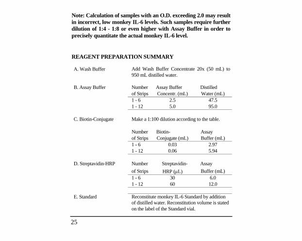
Note: Calculation of samples with an O.D. exceeding 2.0 may result in incorrect, low monkey IL-6 levels. Such samples require further dilution of 1:4 - 1:8 or even higher with Assay Buffer in order to precisely quantitate the actual monkey IL-6 level.
REAGENT PREPARATION SUMMARY
A. Wash Buffer Add Wash Buffer Concentrate 20x (50 mL) to 950 mL distilled water.
B. Assay Buffer Number Assay Buffer Distilled of Strips Concentr. (mL) Water (mL) 1 - 6 2.5 47.5 1 - 12 5.0 95.0 C. Biotin-Conjugate Make a 1:100 dilution according to the table. Number Biotin- Assay of Strips Conjugate (mL) Buffer (mL) 1 - 6 0.03 2.97 1 - 12 0.06 5.94 D. Streptavidin-HRP Number Streptavidin- Assay
of Strips HRP (L) Buffer (mL) 1 - 6 30 6.0 1 - 12 60 12.0 E. Standard Reconstitute monkey IL-6 Standard by addition
of distilled water. Reconstitution volume is stated on the label of the Standard vial.

26
REFERENCES
1. Cayphas S., J. Van Damme, A. Vink, R.J. Simpson, A. Billiau, and J. Van Snick. (1987). Identification of an interleukin HPI-like plasmacytoma growth factor produced by L cells in response to viral infection. J. Immunol. 139:2965-2969.
2. Hirano T., T. Taga, N. Nakano, K. Yasukawa, S. Kashiwamura, K. Shimizu, K. Nakajima, K.H. Pyun, and T. Kishimoto. (1985). Purification to homogeneity and characterization of human B-cell differentiation factor (BCDF or BSFp-2). PNAS 82:5490-5494.
3. Hirano T., and T. Kishimoto. (1990). Interleukin-6. In: Handbook of Experimental Pharmacology, Peptide Growth Factors and Their Receptors, edited by M.B. Sporn, A.B. Roberts, Berlin, Springer-Verlag, pp 633-665.
4. Hirano T., A. Shizuo, T. Taga, and T. Kishimoto. (1990). Biological and clinical aspects of interleukin-6. Immunology Today 11:443-449.
5. Nordan R., and M. Potter. (1986). A macrophage-derived factor required by plasmacytomas for survival and proliferation in vitro. Science 233:566-569.
6. Ray A., S.B. Tatter, U. Santhanam, D.C. Helfgott, L.T. May, and P.B. Sehgal. (1989). Regulation of expression of interleukin-6: Molecular and clinical studies. Ann. NY Acad. Sci. 557:353-362.
7. Sehgal P.B., G. Greininger, and G. Tosato. (1989). Regulation of the acute phase and immune responses: Interleukin-6. Ann. NY Acad. Sci. 557:1-583.

27
Important Licensing Information - These products may be covered by one or more Limited Use Label Licenses (see the Invitrogen Catalog or our website, www.invitrogen.com). By use of these products you accept the terms and conditions of all applicable Limited Use Label Licenses. Unless otherwise indicated, these products are for research use only and are not intended for human or animal diagnostic, therapeutic or commercial use.
Explanation of symbolsSymbol Description Symbol Description
Catalogue Number Batch code
Research Use Only In vitro diagnostic medical device
Use by
Temperature limitation
Manufacturer European Community authorised representative
[-] Without, does not contain [+] With, contains
Protect from light Consult accompanying documents
Directs the user to consult instructions for use (IFU), accompanying the product.
Copyright © Invitrogen Corporation. 25 October 2010
28