elisa immuno exlorer tm : using antibodies for diagnosis and detection
DESCRIPTION
ELISA Immuno Exlorer TM : Using Antibodies for Diagnosis and Detection. ELISA Immuno Explorer TM Instructors. Sherri Andrews, Ph.D. Curriculum and Training Specialist Bio-Rad Laboratories Damon Tighe , Ph.D. Curriculum and Training Specialist Bio-Rad Laboratories Bill Woodruff - PowerPoint PPT PresentationTRANSCRIPT


ELISA Immuno ExlorerTM : Using Antibodies for Diagnosis and Detection

Sherri Andrews, Ph.D.Curriculum and Training SpecialistBio-Rad Laboratories
Damon Tighe, Ph.D.Curriculum and Training SpecialistBio-Rad Laboratories
Bill WoodruffDepartment Head, Biotechnology Alamance Community College
ELISA Immuno ExplorerTM
Instructors

Why Teach ELISA?
• Hands-on Immunology
• Tangible, visual results
• Laboratory extensions
• Real-world connections
• Link to careers and industry
• Standards-based: One lesson integrates multiple standards
–Health sciences –Immunology–Biodefense –Immune response – antibody/antigen interactions
–Disease – infection, detection, transmission


ELISA Immuno Explorer Kit Advantages
• Lab completed in a 45 min period
• Supplies for 48 students (12 workstations)
• Comprehensive and flexible curriculum
• Compelling real-world links
• Striking results
• Cost effective
• Classroom Safe

WorkshopTime Line
• Introduction
• Antigen Detection by ELISA–Antibody detection if time allows
• Ways the ELISA-Immuno Explorer Kit can be used
• Real-World Examples

ELISA
Enzyme-Linked ImmunosorbantAssay
• Mammalian immune system
• Antibody specificity
• Biology’s “magic bullet”
• Evolved over millions of years
• Harness nature’s tool kit
• Imagine the applications!

Links to the Real World
• Mad Cow Disease, SARS, HIV
• GMO
• Drug and steroid testing
• Pregnancy / Reproduction
• Biodefense
• Cancer treatment

Overview• The Immune System
–Those mechanisms by which the body protects itself from often damaging (allergy, death) environmental contaminants foreign to the body (antigen – Ag)–Without the immune system we could not
survive on earth
• Mechanisms –Innate immunity
•Born with these elements•Non-specific cellular and molecular•Equally active against all types of foreign molecules•“First line of defense”

–Acquired immunity•Specific attack against an invader•Requires activation through contact, hence,
acquired•Present only in vertebrates•Includes cells and proteins•Basis of vaccination and immunity
–Resistant to subsequent attack–As a child get chicken pox, as a parent care
for our own children without a repeat episode•Immunity to one disease does not impart
immunity to other, unrelated diseases–Cowpox vs smallpox

• Characteristics of acquired immunity–Self/non-self discrimination
•Major aspect to recognize and attack foreigness•Recognize and not attack self (autoimmunity)•Based on Ag-specific receptors
–Memory•Anamnestic response•Ability to quickly respond to a foreign molecule
that has been seen before •Faster, stronger, longer•Protects from pathogenic (often lethal)
organisms •Also, basis of allergic response
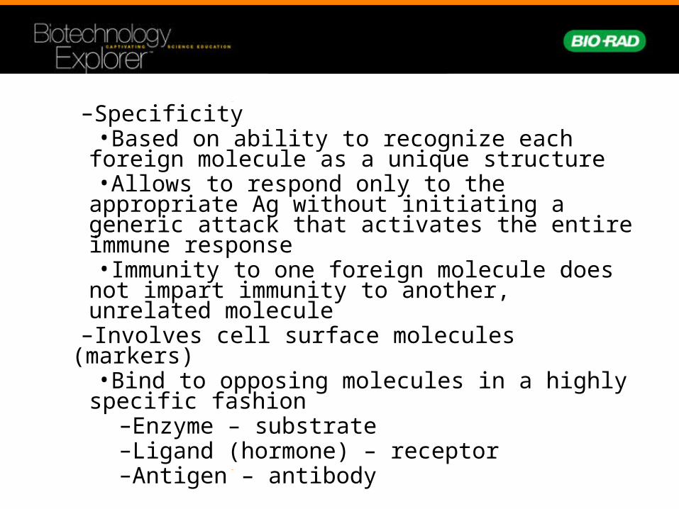
–Specificity•Based on ability to recognize each foreign
molecule as a unique structure•Allows to respond only to the appropriate Ag
without initiating a generic attack that activates the entire immune response•Immunity to one foreign molecule does not
impart immunity to another, unrelated molecule–Involves cell surface molecules (markers)
•Bind to opposing molecules in a highly specific fashion
–Enzyme – substrate–Ligand (hormone) – receptor–Antigen – antibody

• Cells of the Acquired Immune Response–Lymphocytes – cells that exhibit specificity
•T cells–Develop in the thymus
•B cells–Develop in the bone marrow–When B cells proliferate they differentiate
into plasma cells that produce and secrete large amounts of Ag specific antibodies (Ab), an immune protein of high specificity

FIGURE 1.1. Clonal selection theory of B cells leading to antibody productionInc.

•Each B cell expresses ~ 1 X 10⁵ surface Ab of the exact same specificity•Surface Ab binds specific Ag => B cell
activation => proliferation => plasma cell => secrete Ab to attack and destroy specific triggering Ag•Called an Immune Response (IR)
–The antibody•All Abs share two common elements
–specific Ag binding sites–Class specific biological functions
•Structure–2 identical light chains–2 identical heavy chains


Immune Response
A. Pathogen
C. Macrophage
D. Macrophage
E. MacrophageF. T cell
B. B cells
G. B cell
H. Memory B cellsI. Plasma cells
J. Antibodies attach to pathogen

ELISA Antibody Structure
Light chain
Heavy chain
Disulfide bonds
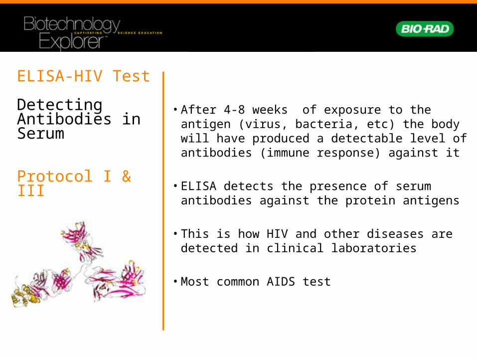
ELISA-HIV Test
Detecting Antibodies in Serum
Protocol I & III
• After 4-8 weeks of exposure to the antigen (virus, bacteria, etc) the body will have produced a detectable level of antibodies (immune response) against it
• ELISA detects the presence of serum antibodies against the protein antigens
• This is how HIV and other diseases are detected in clinical laboratories
• Most common AIDS test

ELISA Animation
http://www.sumanasinc.com/webcontent/animations/molecularbiology.html
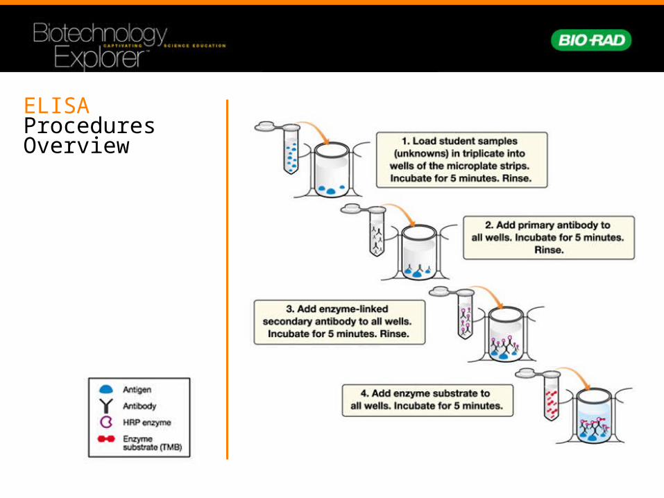
ELISA ProceduresOverview

ELISA Kit Workstation Inventory
Reagents:Yellow tubes Test samples 2Violet tube (+) Positive control 1Blue tube (-) Negative control 1Green tube (PA) Primary antibody 1Orange tube (SA) Secondary antibody 1
Lab Equipment and Supplies:Microplate strips, pipettor, pipette tips, transfer pipette, wash buffer, paper towels, marking pen

Laboratory Quick Guide

Step One
Label and add controls
• Obtain a test-sample
• Label the 12-well strip:–First 3 wells: positive controls “+”–Next 3 wells: negative controls “-”–Remaining wells to identify test-samples
• Add 50 ul of positive control to 1st 3 wells
• Add 50 ul of negative control to 2nd 3 wells
• Add 50ul of the student samples to the appropriately labeled wells
• Wait 5 minutes for the antigen to bind

Microplate Strips
• Microplate strips are made of polystyrene
• Hydrophobic side chains in amino acids bind to the polystyrene wells
• No coating is needed

Step Two
WASH • Remove samples from wells by firmly tapping them on a paper towel
• Discard the top paper towel
• Using a disposable transfer pipette wash wells with wash buffer
• Remove wash buffer by firmly tapping the wells on a paper towel
• Discard the top paper towel
• Repeat wash step

• Add 50 ul of the primary antibody (PA) to all 12 wells
• Samples are left in wells for 5 minutes
• After 5 minutes WASH 2X
Step Three
Add (PA)Primary Antibody
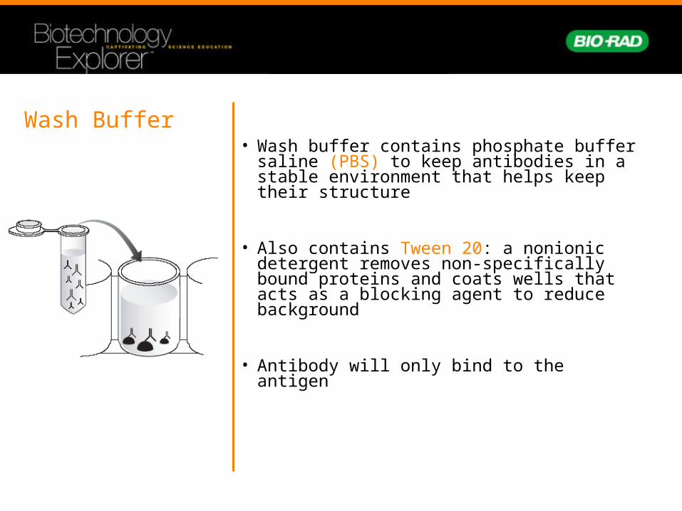
Wash Buffer• Wash buffer contains phosphate buffer
saline (PBS) to keep antibodies in a stable environment that helps keep their structure
• Also contains Tween 20: a nonionic detergent removes non-specifically bound proteins and coats wells that acts as a blocking agent to reduce background
• Antibody will only bind to the antigen

Step Four
Wash antibody and add enzyme-linked secondary antibody (SA)
• Wash the primary antibody from polystyrene wells as before
• WASH 2X
• Add 50ul of the enzyme-linked secondary antibody to each well
• Wait 5 minutes

Antibody Specificity • Secondary antibody (enzyme-linked
antibody) will only bind to the primary antibody (serum antibody)
• Secondary antibody specifically recognizes the constant region of the primary antibody
• In which wells do you predict this is happening?

Step Five
Add enzyme substrate(SUB)
• Wash the enzyme-linked secondary antibody from polystyrene wells as before
• Using a disposable transfer pipette wash wells with wash buffer
• WASH 3X
• Add 50ul of the enzyme substrate to each well
• Wait 5 minutes
• positive samples will begin to turn blue

What are the reagents?
Antigen: Chicken gamma globulin
Primary antibody (PA): Polyclonal anti-chicken antibody made by rabbits
Secondary antibody (enzyme-linked) SA: Polyclonal anti-rabbit antibody made by goats linked (conjugated) to horseradish peroxidase (HRP)
Enzyme substrate (SUB): 3,3’,5,5’ – tetramethylbenzidine (TMB) – a colorless solution that when oxidized by HRP turns blue

ELISA Kit Results
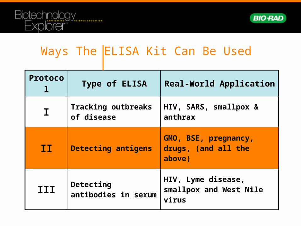
Ways The ELISA Kit Can Be Used
Protocol Type of ELISA Real-World Application
I Tracking outbreaks of disease
HIV, SARS, smallpox & anthrax
II Detecting antigensGMO, BSE, pregnancy, drugs, (and all the above)
III Detecting antibodies in serum
HIV, Lyme disease, smallpox and West Nile virus

ELISA test for Transmissible Spongiform Encephalopathies (TSEs)
a
PrPres
• Proteinase K resistant
• Aggregates in detergent
PrPsens
• Proteinase K sensitive
• Soluble in detergent
b
Prion Proteins (PrPres and PrPsens)
• Uses differences in diseased prions vs. normal prions to prepare sample.
• Proteinase K only digests normal, not diseased, prions .
• ELISA tests for any prion protein

1. Sample brain tissue
2. Homogenize brain tissue
3. Digest with Proteinase K (normal prions are digested, diseased prions are resistant)
4. Concentrate
5. Denature Proteinase K
6. Perform ELISA
TSE test sample preparation

Protocol II: Antigen Detection ELISA
Protocol - ELISA on simulated animal brain samples
Tube Description
Actual Tube Contents
Simulated Tube Contents
Student samples
Antigen or PBS Processed brain
Primary antibody
Primary antibody Antibody against prion protein
Secondary antibody
Secondary antibody
HRP-linked antibody against primary antibody
Positive control Antigen Synthesized peptide with prion sequence
Negative control
PBS Buffer
Real-World Application – TSE Test

Real-world Applications of Antibodies
Agricultural Uses– Crop-specific disease diagnosis– Animal disease diagnosis– Detection of GM crops– Basic research
Applications– Dipstick tests/ELISA– Immunostaining– Western blotting
Bio-Rad’s TSE ELISA Kit

ELISA to test for GMOs
• ELISA can help farmers separate their GMO grain lots from non-GMO grain lots.
• ELISA tests are used to identify specific proteins
- Delta-endotoxin Cry1Ab from Bt11 - glyphosate from Round-up (RR)
“Genetically Modified Organism (GMO)"
an organism in which the genetic material has been altered in a way that does not occur naturally by mating and/or natural recombination
DNA RNA Protein

How to test for GMOs ELISA:
Test for presence of proteins expressed from genetic modifications
Pro: Quick, inexpensive, low tech
Con: Crop specific, protein stability
PCR:
Test for presence of inserted foreign DNA
Pro: ID different GM crops, DNA stability
Con: Expensive, timely

Example: Pregnancy Test