elevated prevalence of helicobacter species and … · 2 opisthorchiasis and associated...
TRANSCRIPT

1
Elevated prevalence of Helicobacter species and virulence factors in 1 opisthorchiasis and associated hepatobiliary disease 2 3 4 Raksawan Deenonpoe1,2, Eimorn Mairiang3, Pisaln Mairiang4, Chawalit Pairojkul2, 5 Yaovalux Chamgramol2, Gabriel Rinaldi 5, ♯, Alex Loukas6, Paul J. Brindley5, Banchob 6 Sripa1,2 7
8
9 1
Tropical Disease Research Laboratory, Faculty of Medicine, Khon Kaen University, Khon Kaen 10 40002, Thailand 11 Departments of 2Pathology, 3Radiology and 4Medicine, Faculty of Medicine, Khon Kaen 12 University, Khon Kaen, 40002, Thailand 13
5 Department of Microbiology, Immunology and Tropical Medicine, and Research Center for 14 Neglected Tropical Diseases of Poverty, School of Medicine & Health Sciences, The George 15 Washington University, Washington DC, 20037, USA 16
6 Centre for Biodiscovery and Molecular Development of Therapeutics, Australian Institute of 17 Tropical Health & Medicine, James Cook University, Cairns, Queensland 4878, Australia 18 19
♯ Current address: Sanger Institute, Wellcome Trust Genome Campus, Cambridge CB10 1SA, 20 United Kingdom 21 22 23 24 25 26 27 Correspondence: Banchob Sripa, Tropical Disease Research Laboratory, Department of 28 Pathology, Faculty of Medicine, Khon Kaen University, Khon Kaen 40002, Thailand, email 29 [email protected]; or Paul J. Brindley, Department of Microbiology, Immunology and 30 Tropical Medicine, and Research Center for Neglected Tropical Diseases of Poverty, School of 31 Medicine & Health Sciences, The George Washington University, Washington DC, 20037, USA, 32 email, [email protected] 33 34 35 36 37 38 39 40 41 42 43
2
Recent reports suggest that Opisthorchis viverrini serves as a reservoir of Helicobacter and 44 implicate Helicobacter in pathogenesis of opisthorchiasis-associated cholangiocarcinoma (CCA). 45 Here, 553 age-sex matched cases and controls, 293 and 260 positive and negative for liver fluke 46 O. viverrini eggs, of residents in Northeastern Thailand were investigated for associations among 47 infection with liver fluke, Helicobacter and hepatobiliary fibrosis. The prevalence of H. pylori 48 infection was higher in O. viverrini-infected than uninfected participants. H. pylori bacterial 49 load correlated positively with intensity of O. viverrini infection, and participants with 50 opisthorchiasis exhibited higher frequency of virulent cagA-positive H. pylori than those free of 51 fluke infection. Genotyping of cagA from feces of both infected and uninfected participants 52 revealed that the AB genotype accounted for 78 % and Western type 22 %. Participants infected 53 with O. viverrini exhibited higher prevalence of typical Western type (EPIYA ABC) and variant 54 AB’C type (EPIYT B) CagA. Multivariate analyses among H. pylori virulence genes and 55 severity of hepatobiliary disease revealed positive correlations between biliary periductal fibrosis 56 during opisthorchiasis and CagA and CagA with CagA multimerization (CM) sequence-positive 57 H. pylori. These findings support the hypothesis that H. pylori contributes to the pathogenesis of 58 chronic opisthorchiasis and specifically to opisthorchiasis-associated CCA. 59 60 61 62 63 64 65 66 67 68 69 70 71 72 73 74 75 76 77 78 79 80 81 82 83 84 85 86 87 88 89
3
Introduction 90 91 Infection with the fish-borne liver fluke Opisthorchis viverrini is endemic in Southeast Asia 92 including regions of the Lao People’s Democratic Republic, Thailand, Cambodia and Vietnam 1-93 3. Opisthorchiasis is associated with hepatobiliary morbidity including chronic cholangitis, 94 cholelithiasis, periductal fibrosis and bile duct cancer, or cholangiocarcinoma (CCA) 4-7. Khon 95 Kaen province in Northeast Thailand has reported the highest incidence of CCA in the world, 96 greater than 100 cases per 100,000 residents 8. Chronic inflammation in response to metabolites 97 and growth factors released by this parasitic worm and related phenomena are implicated in the 98 pathogenesis of liver fluke infection-associated hepatobiliary diseases 7,9-12. However, the biliary 99 morbidity in the setting of opisthorchiasis may not be solely linked with liver fluke infection; 100 other factors including carriage of Helicobacter and other microbiome changes within the biliary 101 tract might participate 13. 102 103 More than 30 species of Helicobacter have been described 14 and H. pylori was the first bacterial 104 pathogen confirmed to cause gastric disease including peptic ulcer, gastric lymphoma and gastric 105 adenocarcinoma 15-19. On the other hand, carriage of H. pylori occurs in at least half the human 106 population with transmission from mother to child and other routes. Indeed the human-H. pylori 107 association likely is at least 100,000 years old 20, an association that appears to be beneficial in 108 early life, including contributions to a healthy microbiome and reduced early-onset asthma 21,22. 109 Infection with species of Helicobacter has been implicated in other malignant and benign 110 diseases of the biliary tract 23-28. Virulence factors of H. pylori including cytotoxin-associated 111 gene A (cagA), cagE and vacuolating cytotoxin A (vacA) participate in the pathogenesis of these 112 conditions 29. The related species H. hepaticus and H. bilis also associate with hepatobiliary 113 diseases 30-32. 114 115 Opisthorchiasis may enhance colonization of the biliary tree by species of Helicobacter in like 116 fashion to other changes in the biliary microbiome 33. The influence of opisthorchiasis on 117 cholestasis as a consequence of the liver fluke migration and establishment within the bile ducts 118 provide explanations for bacterial colonization leading to bacterial cholangitis 34. In addition, the 119 migration of the flukes themselves from the external environmental through the alimentary tract 120 and into the biliary tract might convey bacterial passengers, both on the external surface of the 121 trematode and within the gut of the parasite 35-38. 122 123 We recently reported, in a hamster model of liver fluke infection-induced biliary disease, higher 124 prevalence and intensity of co-infection with H. pylori and H. bilis in O. viverrini-infected 125 compared to uninfected hamsters, suggesting that this liver fluke serves as a reservoir for H. 126 pylori37. Here we undertook a human study with more than 500 residents in villages of four 127 provinces of northeastern Thailand endemic for opisthorchiasis3. Liver fluke infection was 128 associated with a higher frequency of cagA-positive H. pylori. Moreover, the presence of H. 129 pylori cagA gene as well as alleles of was associated with increased morbidity, specifically 130 periductal fibrosis of the biliary tree. These findings support the hypothesis that H. pylori 131 contributes to the pathogenesis of chronic opisthorchiasis and specifically to opisthorchiasis-132 associated cholangiocarcinoma. 133 134 135

4
Results 136 137 Liver fluke burden positively correlated with Helicobacter infection 138 139 The distribution of infection with Helicobacter spp. in regions endemic for opisthorchiasis was 140 established according to age, gender, burden of liver fluke, as diagnosed fecal EPG and 141 ultrasonography score for hepatobiliary disease including fibrosis. A total of 553 residents from 142 four provinces of Thailand participated in the study; samples of feces from 260 participants were 143 egg negative whereas 293 were positive for eggs of O. viverrini (Table 1). 144 145 In addition to infection with O. viverrini, analysis of feces by PCR was used to investigate the 146 presence of Helicobacter spp. A total of 267 residents of four Isaan provinces of Thailand were 147 positive for H. pylori, 99 for H. bilis and 18 for H. hepaticus. Gender did not correlate with 148 presence of species of Helicobacter, P > 0·05 (Table 1). 149 150 The prevalence of infection with H. pylori assigned as ureA gene-positive by stool PCR was 151 64·6% vs. 29·6% in O .viverrini-infected and uninfected participants, respectively; P < 0·01. The 152 prevalence of infection with H. bilis, but not H. hepaticus, was also significantly higher in O. 153 viverrini-infected vs. uninfected individuals, 29·3 vs 5·4%, P < 0·01. In addition, mixed H. 154 pylori/H. bilis infection was significantly higher during infection with O .viverrini: 26·9% 155 compared to participants who were stool-negative for O. viverrini, 4·2%, P < 0·01 (Figure 1). 156 157 Increased prevalence and load of H. pylori and H. bilis during opisthorchiasis 158 159 The mass of H. pylori in one-gram of feces correlated positively with the intensity of liver fluke 160 infection (one-way ANOVA, P < 0·001) (Supplementary Figure S1). In general, participants 161 with higher intensity infection (>1,000 EPG) had ~15 times the total cell counts of H. pylori 162 compared those who were negative for infection with O. viverrini (EPG = 0). Load of H. pylori 163 increased according to intensity of liver fluke infection; P < 0·001 for each sequential 164 comparison (Supplementary Figure S1). 165 166 The positive relationship between Helicobacter and O. viverrini infection was substantiated by 167 positive correlations between 16S rRNA and intensity of liver fluke infection (χ2 = 006), ureA 168 (H. pylori) and intensity of liver fluke infection (χ2 trend < 0001), cagA and intensity of liver 169 fluke infection (χ2 trend < 0001), cagE and intensity of liver fluke infection (χ2 trend < 0001), 170 and H. bilis and intensity of liver fluke infection (χ2 trend < 0001); but not for H. hepaticus. 171 Generally, the presence of H. pylori and H. bilis was far higher during elevated levels of 172 infection with O. viverrini than during low intensity infections or in the uninfected participants. 173 Table 2 details the findings. 174 175 Helicobacter spp. associated with grade of biliary peridcutal fibrosis 176 177 The presence of cagA was associated with an elevated risk of both grade 2 and grade 3 biliary 178 periductal fibrosis. The relative risk ratio (RRR) for grade 2 versus grade 1 or 0 hepatobiliary 179 disease was 338 (95% C151-758, P = 0003) comparing individuals with and without cagA, in 180 the model adjusted for age and sex (Table 3). The analogous RRR was 915 for grade 3 vs. grade 181
5
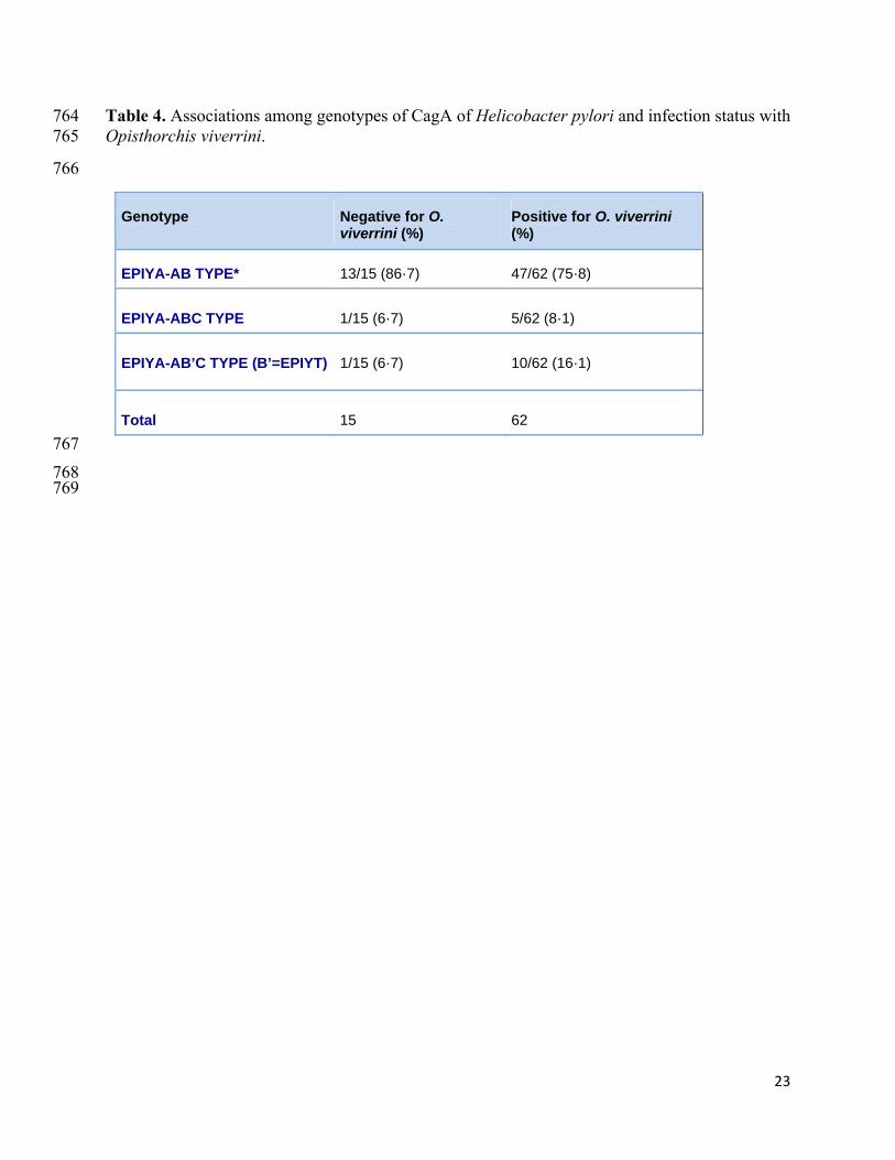
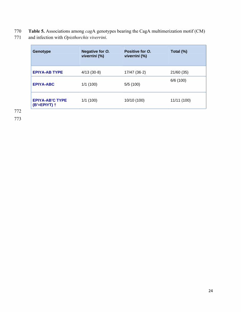
1 or 0 hepatobiliary disease (95% CI 174 - 4797, P = 0009) (Table 3). After confirming the 182 proportional odds assumption, we determined and overall odds ratio of 424 for each subsequent 183 grade of hepatobiliary disease comparing individuals with and without cagA, controlling for age 184 and sex; P < 0001. 185 186 Also, a strong, positive association was evident between the presence of mixed cagA and cagE 187 and marked hepatobiliary disease; RRR = 4·96 for grade 3 vs. grade 1 or 0, 95% CI =1·50-188 16·34, P = 0·009. Association was not evident between positivity for mixed cagA and cagE and 189 grade 2-biliary periductal fibrosis. Associations between the presence of H. pylori, H. bilis, H. 190 hepaticus alone or in combination with hepatobiliary disease were not significant 191 192 cagA genotypes associated with biliary periductal fibrosis 193 194 In order to categorize the cagA genotypes, sequence analysis was undertaken on cagA-positive 195 samples. Seventy-seven cagA strains were Western CagA type and unclassified type, AB type. 196 The predominant CagA types were EPIYA-AB type, EPIYA-ABC type and EPIYA-AB’C type 197 (B’=EPIYT)39. Participants who were not infected with O. viverrini showed higher frequency of 198 EPIYA-AB type than did the infected participants, 86·7 % vs. 75·8%, respectively (Table 4). On 199 the other hand, O. viverrini-infected participants carried a marginally higher frequency of EPYA-200 ABC type (8·1 vs. 6·7%) and twice as high frequency of EPIYA-AB’C (16·1 vs. 6·7%) (Table 201 4). In overview, the Western type CagA with EPIYA-AB’C showed higher frequency in the liver 202 fluke-infected cases. 203 204 In addition, some cagA genotypes included the CagA multimerization (CM) motif. CM is 205 comprised of 16 amino acids, FPLKRYDKFDDLSKVG or FPLKRHDKFDDLSKVG and is 206 highly conserved for Western and Eastern CagA 40,41. Whereas the prevalence of CagA with CM 207 in EPIYA-AB type was 30·8% – 36·2% in liver fluke infection-negative and -positive 208 participants, respectively, CM was present in all (100%) of the EPIYA-ABC and EPIYA-AB’C 209 (EPIYT) genotypes detected (Table 5). 210 211 Concerning associations between CagA types and grade of biliary periductal fibrosis, significant 212 associations between AB’C type versus AB type and both grade 2 (RRR = 23·12, 95% CI =2·31-213 23·50, P = 0·007, and grade 3 (RRR= 24·36, 95% CI = 1·71-347·09, P = 0·018) were apparent 214 (Table 6). There was no association between ABC type versus AB type and hepatobiliary 215 disease. In addition, regarding CagA types with or without the CM sequence, significant 216 association between CagA with CM sequence and grade 2 was evident (RRR = 30·74, 95% CI = 217 5·25-180·08, P < 0·001). After confirming the proportional odds assumption, we determined an 218 overall odds ratio of 30·8 for each subsequent grade of biliary periductal fibrosis comparing 219 individuals carrying CagA with and without the CM sequence, and controlling for age and sex (P 220 < 0·001). Similarly, after grouping the degree of hepatobiliary disease as either negative (grades 221 0+1) or positive (grades 2+3), as described 42,43, a significant association was apparent between 222 positive for hepatobiliary disease and CagA with CM sequence with an odds ratio of 38·21 (95% 223 CI = 6·85 - 213·03, P < 0·001). On the other hand, the wide range for CI in this analysis 224 reflected the limited number of cases in the dataset due to this uncommon genotype and, in turn, 225 the limited power of this analysis. 226 227
6
Phylogram analysis of CagA EPIYA motifs revealed novel genotypes during liver fluke 228 infection 229
The phylogenetic relationships of CagA genotypes among 75 samples from this cohort of 230 participants from northeastern Thailand, specifically 13 negative and 62 positive for infection 231 with O. viverrini, were compared with two Western cagA and two Eastern cagA reference 232 strains detected in gastro-duodenal disease in Thailand, and three CagA sequences isolated from 233 bile from Thai cholangiocarcinoma (CCA) cases, as reported33,44. The relationships were 234 determined using maximum parsimony. Representative cagA-encoded sequences of our Thai 235 cohort mainly grouped into main clusters: 1) Unclassified type, EPIYA AB without CM 236 sequence, e.g. samples FPNS105, FNK3; 2) Unclassified type, EPIYA AB with CM sequence, 237 e.g. FNK9, FNK10; 3) ‘Western-like’ type EPIYA ABC, e.g. FPK3, FPNS5; and 4) ‘Western-238 like’ type EPIYA AB’C, e.g. FPK4, FPNS8. By contrast, the sequences of Western, Eastern and 239 CCA cases grouped together, generally divergent from sequences in the present cohort (Figure 240 2). The association between the ‘Western like’ genotypes, including EPIYA AB’C, and the 241 hepatobiliary pathology is evident for the sequences analyzed in the phylogenetic tree shown in 242 Figure 2A. Figure 2B depicts representative sequences that belong to the main clusters of CagA 243 described above, indicating the EPIYA motives and CM sequences. Whereas the prevalence of 244 cagA-encoding the CM sequence in EPIYA-AB type was 35 %, CM was present in all (100%) 245 of the EPIYA-ABC and EPIYA-AB’C (EPIYT) genotypes characterized here (Table 5). 246 247 Discussion 248 249 County-wide sampling indicates a prevalence of carriage of H. pylori by asymptomatic Thais of 250 ~ 44%, based on fecal examination 41, with marginally higher sero-prevalence 45. This report 251 describes an association between infection with the fish-borne liver fluke O. viverrini and 252 carriage of species of Helicobacter in opisthorchiasis-endemic northeastern Thailand. H. pylori 253 represented the major species of Helicobacter but, in addition, H. bilis and mixed H. pylori/H. 254 bilis infection occurred more often during active opisthorchiasis than in uninfected or lightly 255 infected persons, in turn confirming earlier reports 46,47. Mixed infection with H. pylori and H. 256 bilis may be associated with more severe hepatobiliary disease. Prevalence of H. pylori and H. 257 bilis also was elevated in participants who were heavily infected with O. viverrini. 258 Opisthorchiasis appeared to exacerbate severity of H. pylori/ H. bilis-associated disease in like 259 fashion to infections in hamsters 35,37, confirming an association between intensity of H. pylori/ 260 H. bilis infection and presence of the liver fluke. 261 262 Prevalence of both cagA-and cagE-positive H. pylori positively correlated with increasing levels 263 of liver fluke infection, and prevalence of cagA-positive strains of H. pylori correlated positively 264 with increased biliary periductal fibrosis as diagnosed by abdominal ultrasound. The presence of 265 cagA- and cagE-positive H. pylori strains associated with severe fibrosis, findings that suggested 266 that H. pylori, and in particular cagA-positive strains, reached the biliary tract, and induced 267 hepatic inflammation that exacerbated periductal fibrosis. Discrete genotypes of cagA associate 268 with severity of gastrointestinal diseases 48. Unclassified type (AB type) represented the major 269 cagA genotype in this study, in contrast with earlier reports indicating that AB represents only a 270 minority genotype carried by otherwise healthy Thais 41,49. Here, 22% of the cagA-encoded 271 sequences were Western type (ABC type) with no East Asian type (ABD type), lower than 272 reported for liver fluke infection-induced CCA 44. A meta-analysis of cagA status in Southeast 273

7
Asia has revealed 51% vs. 49% of Western type and East Asian type, respectively 48. There was 274 a higher prevalence of typical Western type (EPIYA ABC) and variant AB’C type (EPIYT B) 275 cagA genotypes in O. viverrini-infected compared to uninfected participants. 276 277 The present findings also demonstrated that polymorphisms in cagA of H. pylori circulate among 278 Thais with opisthorchiasis. For the ABC and AB’C type CagA, there was a higher frequency of 279 the deduced 16-amino-acid CagA multimerization (CM) types during liver fluke infection. CM is 280 conserved between Western CagA and East Asian CagA 50, although Western type CagA 281 invariably exhibits the CM sequence 39. The CM sequence represents a membrane-targeting 282 signal 50, which interacts with PAR1b, thus inducing junctional and polarity defects 29,50,51. 283 Notably, the PCR primers employed here spanned the entire 3’-region of cagA encoding the 284 multimers of the tyrosine phosphorylation motifs52,53. Structural polymorphism in the CM 285 reflects the degree of virulence of CagA 54. Here infection with any CagA type H. pylori bearing 286 CM sequences was associated with severe hepatobiliary disease, with an odds ratio up as high as 287 38. This characterization of sequences with both EPIYA-C/D motif and CM sequence suggested 288 increased phosphorylation motifs capable of provoking pronounced disease 54. Phylogenetic 289 analysis revealed four discrete clades, and all four differed from the from typical Western and 290 East Asian CagA types including those associating with Western CCA sequences 44. Although 291 the Thai CagA sequences were separated from the pathogenic reference sequences, 292 opisthorchiasis might be involved in the various novel types of CagA (with CM sequence), 293 which associates with severe disease. Accordingly, we hypothesize that not only is the liver fluke 294 O. viverrini a reservoir of Helicobacter but also a selector for pathogenic strains of this ɛ-295 proteobacterium. Given the elevated presence of H. pylori, and CagA including its 296 polymorphisms with increasing intensity of liver fluke infection and biliary tract fibrosis, these 297 new variants may, at least partly, underlie progression of hepatobiliary disease in opisthorchiasis-298 endemic regions. 299 300 The International Agency for Research on Cancer of the World Health Organization classifies 301 infection with the liver flukes O. viverrini and Clonorchis sinensis and with H. pylori as Group 1 302 carcinogens4. In northern and northeastern Thailand and Laos, infection with O. viverrini is the 303 major risk for CCA 4,8,55,56. Following initiation, oncogenesis appears to be promoted by 304 cholestasis and chronic inflammation. Increased mutation rates of the tumor suppressor genes 305 p53 and CDKN2A, and of genes encoding protein tyrosine phosphatases, SMAD4 and others 306 sustain cholangio-carcinogenesis, with differences between CCA induced by opisthorchiasis 307 compared to other risks factors57,58. As reviewed 59, the release and interaction of interleukin-6, 308 transforming growth factor beta, tumor necrosis factor alpha, and platelet-derived growth factor 309 are pivotal to the proliferation of cholangiocytes, while evasion of apoptosis, autonomous 310 proliferation, and angiogenesis sustain incipient neoplasia. In parallel, infection with cagA-311 positive H. pylori is the major risk for gastric adenocarcinoma and mucosa associated 312 lymphoid tissue (MALT) lymphoma. Cellular changes following the injection of the CagA 313 oncoprotein include epithelial to mesenchymal transition and the hummingbird phenotype60,61, 314 along with genetic mutations in E-cadherin and epigenetic changes. Genome sequencing has 315 identified driver mutations TP53, ARID1A, CDH1, MUC6, CTNNA2, GLI3, RNF43 and others 316 in gastric cancer 62. Loss of epithelial cadherin expression from CDH1 alterations is a primary 317 carcinogenetic incident. Cytogenetic abnormalities including the t(11;18)(q21;q21) translocation 318 are frequently acquired during H. pylori-associated gastric MALT lymphoma 63. 319

8
320 The association between opisthorchiasis and the presence of H. pylori in feces was statistically 321 significant. Nonetheless, direct evidence of a causal relationship where H. pylori and liver fluke 322 infection jointly prime the pathogenesis of hepatobiliary disease including CCA has not been 323 obtained. It is relevant to note the outcome of a recent study using a rodent model of human 324 opisthorchiasis, which provides support for the association among O. viverrini, H. pylori and 325 biliary periductal fibrosis 37,64. Liver fluke-infected hamsters were treated with antibiotics and the 326 anthelmintic, praziquantel. Quantitative PRC analysis of tissue and organs from the hamsters 327 indicated that the majority of the H. pylori emanated from the same sites as the liver flukes in the 328 biliary tract given that antibiotics failed to reduce the load of H. pylori to the baseline achieved 329 with dual treatment with antibiotics and praziquantel. H. pylori load in the stomach was 330 unaffected. In addition, immunohistochemical approaches detected H. pylori within the gut of 331 liver flukes recovered from the hamsters. 332 333 Hepatobiliary disorders caused by Helicobacter33,44,65 can resemble opisthorchiasis 42,66. Chronic 334 lesions ascribed to liver fluke infection, including cholangitis, biliary hyperplasia and metaplasia, 335 periductal fibrosis and CCA, may be due in part to Helicobacter-associated hepatobiliary 336 disease. H. pylori DNA has been isolated from tissues from CCA and from 337 cholecystitis/cholelithiasis in regions endemic for opisthorchiasis 33,44. Moreover, serological 338 findings indicate infection with H. pylori in Thais at high risk for CCA 65. An explanation for 339 why infection with the liver fluke induces bile duct cancer 10 might now be clearer – involvement 340 by H. pylori and its virulence factors. The spiral bacilli of H. pylori attach to biliary cells, which 341 internalize in similar fashion to their behavior on gastric epithelium 29,67. Helicobacter likely 342 passes from the stomach to the duodenum and enters the biliary tree through the duodenal papilla 343 and ampulla of Vater 40,67. How the microbe tolerates the neutral to alkaline pH of the small 344 intestine and biliary tree remains unclear 17. However, an association with the migrating liver 345 flukes offers a plausible explanation: given that Helicobacter-like curved rods occur in the gut of 346 O. viverrini 37, and given that the micro-environment of the O. viverrini gut is acidic, the microbe 347 might hitchhike within the migrating juvenile trematode. Intriguingly, glycoprotein gylcans 348 expressed on the gut epithelium of O. viverrini 68 resemble receptors of gastric epithelial cells to 349 which H. pylori binds 69. Helicobacter may have evolved a commensalism with O. viverrini, 350 with conveyance into the biliary tract during the migration of the parasite following ingestion of 351 the metacercaria with undercooked freshwater fish 35,37. 352 353 Given the elevated prevalence of CCA in regions where infection with liver fluke prevails, and 354 given the increasing evidence of linkage between carriage of Helicobacter during 355 opisthorchiasis, these two biological carcinogens together may orchestrate the pathogenesis of 356 opisthorchiasis and bile duct cancer. The association of Helicobacter and its virulence factors, 357 together with chronic opisthorchiasis, may underlie biliary tract disease including CCA in liver 358 fluke-endemic regions 70. Whereas additional studies are needed to clarify this association, at 359 present detection of H. pylori in feces provides a non-invasive approach to investigate its 360 association with biliary tract disease during opisthorchiasis. 361 362 Materials and Methods 363 364

9
Ethics statement 365 366 The Institutional Human Ethics Committee of Khon Kaen University approved the study, 367 approval number HE 551332. All methods were performed in accordance with the relevant 368 guidelines and regulations of the committee. The participants provided written informed 369 consent following discussion with the researchers that included information on fecal samples for 370 laboratory analyses. All participants were adults; children were not enrolled (Table 1). 371 372 Study participants 373 374 Participants were asked to refrain for up to 10 days from consumption of fatty foods, antacid 375 medication, antibiotics, anti-parasitic agents, barium, mineral oil, bismuth, or non-absorbable 376 anti-diarrheal agents. Patients with history of digestive-tract diseases (gastritis, gastric ulcer, 377 cholecystitis, cholangitis, cholecystectomy, others) were excluded from the study. A total of 553 378 participants provided stool samples; 260 were parasitologically negative for fecal eggs of O. 379 viverrini and 293 were egg-positive for O. viverrini from age-sex matched residents of villages 380 in four provinces of the opisthorchiasis-endemic Isaan region of northeastern Thailand1,8,42,71. In 381 particular, those enrolled included 273, 107, 93 and 80 people from the provinces of Khon Kaen, 382 Roi-et, Mahasarakham and Kalasin, respectively (Supplementary Figure S2). The participants 383 included 288 females and 265 males, aged 30 to 70 years (Table 1). 384 385 Parasitological diagnosis of infection with the liver fluke Opisthorchis viverrini 386 387 Parasitological diagnosis of opisthorchiasis was accomplished using formalin-ethyl acetate 388 concentration of one gram of feces, followed by light microscopy examination of the concentrate 389 72. The method is suitable for diagnosis of O. viverrini eggs and widely employed for diagnosis 390 of opisthorchiasis 72. Thereafter, participants were grouped according to fecal egg count, i.e. 391 intensity of infection into five categories: 1) EPG (eggs per gram of feces) = 0 [i.e. uninfected]; 392 2) 1-100 EPG; 3) 101-500 EPG; 4) 501-1,000 EPG; and 5) >1,000 EPG. There were 260, 193, 393 73, 12 and 15 participants in these five categories, respectively (Table 2). 394 395 Detection by PCR of Helicobacter species and virulence genes 396 397 DNA was isolated from about one gram of feces, stored in 70% ethanol, using a QIAamp DNA 398 Stool Mini Kit (Qiagen, Germany) with concentrations ranging from 50 to 500 ng/µl, and total 399 yields of 2 to 15 µg. Subsequently, 50 ng DNA from the samples served as the template for PCR 400 performed in a GeneAmp PCR system 9700, Applied Biosystems thermal cycler; the reaction 401 mixture included 1x Gotaq Colorless Master Mix (Promega) containing 0·2 mM dNTP, 1·5 mM 402 MgCl2, 1·25 U Tag DNA polymerase, with primers at 0·2 mM each. Supplementary Table S1 403 provides the gene specific primers for Helicobacter species, specifically for 16S rRNA, ureA, 404 cagA, cagE of H. pylori, and for H. bilis and H. hepaticus. Amplicons were sized by 405 electrophoresis through 1·0 % agarose, stained with ethidium bromide and visualized under UV 406 light. The expected sizes of amplicons for the 16S rRNA, ureA (H. pylori), H. bilis, H. 407 hepaticus, cagA sequencing and cagE were 480, 350, 418, 405, 550-800, and 508 bp, 408 respectively (Supplementary Figure S3). 409 410

10
Abdominal ultrasonography to visualize hepatobiliary fibrosis 411 412 Abdominal scans were performed using a mobile high-resolution ultrasound-imaging appliance 413 (GE model LOGIQ Book XP), as described 43,73. Hepatobiliary abnormalities including 414 periductal fibrosis in liver parenchyma, gallbladder wall, gallbladder size, sludge, and suspected 415 CCA (dilated intra or extrahepatic bile duct and/or liver mass) were graded and recorded 4234. 416 Based on the ultrasonography, grading of periductal biliary fibrosis was assigned as follows: 417 grade 0 = absence of periportal echo(s) from all segments of liver; grade 1 = presence of 418 periportal echo(s) in one segment of liver; grade 2 = periportal echo(s) in two to three segments; 419 grade 3 = periportal echo(s) in more than three segments. Status of infection with liver fluke or 420 presence of species of Helicobacter was not known by the radiologist during the abdominal 421 ultrasonography. 422 423 Quantitative real-time PCR 424 425 Fecal samples from the H. pylori infected (conventional PCR ureA-positive) participants (n = 426 267) used in this study were assigned to one of five groups based on fecal EPG for O. viverrini 427 (above): O. viverrini EPG = 0 (n = 77), EPG = 1-100 (n = 110), EPG = 101- 500 (n = 57), EPG = 428 501-1,000 (n = 10) and EPG > 1,000 (n = 13). In addition, feces free of H. pylori were included 429 as a negative control 37. Presence of H. pylori was established and quantified by real time PCR 430 using primers HpyF1: GGGTATTGAAGCGATGTTTCCT and HpyR1: GCTTTTTTGC-431 CTTCGTTGATAGT44. The quantitative real-time analysis targeted the species-specific gene 432 ureA of H. pylori 74. DNA samples were diluted to employ equivalent template concentrations in 433 the qPCR reactions that included 10 µl SYBR master mix (Thermo Fisher), 1 µl template-DNA, 434 0·5 µl of each primer (625 nM), and 9 µl nuclease-free water. PCR was performed in triplicate 435 (technical triplicates) in a thermal cycler (Light Cycler 1·5, Roche), using initial denaturation at 436 95ºC for 9 min, followed by 40 cycles of 95ºC, 15 s, 60ºC, 60 s for the annealing and elongation 437 steps, respectively. A 10-fold serial dilution of H. pylori DNA was included to establish a 438 standard curve, from 108 cells/ml to 101 cells/ml; bacterial cells were counted in a Thoma-439 counting-chamber, plated and incubated for subsequent extraction of DNA. E. coli DNA served 440 as the negative control 74. 441 442 Phylogenetic analysis of cagA gene partial sequences 443 444 To establish phylogenetic relationships among the H. pylori genotypes, 62 participants infected 445 and 13 uninfected with O. viverrini were investigated. Partial sequences of cagA genes amplified 446 by PCR were sequenced by the Sanger approach (First BASE Laboratories, Malaysia). In 447 addition, sequences of Western-like CagA from four references were analyzed: Thailand 448 (GenBank accession BAB87427 75) Western, Thailand (BAB87428) Western, Thailand 449 (BAB87429) eastern, and Thailand (BAB87430) eastern. Partial, deduced amino acid sequences 450 of CagA were searched for EPIYA motifs 39,44 using the ExPASy-Translate software followed by 451 multiple sequence alignment using ClustalW (Bioedit)76 with further editing using GeneDoc 452 (http://www.nrbsc.org/gfx/genedoc/ebinet.htm). Evolutionary history was inferred using 453 Neighbor-Joining 77. A bootstrapped consensus tree inferred from 500 replicates was taken to 454 represent the evolutionary history of the taxa analyzed. Branches corresponding to partitions 455 reproduced in less than 50% bootstrap replicates were collapsed. Evolutionary distances were 456

11
computed using the JTT matrix-based method 78 and the units represent the number of amino 457 acid substitutions per site. The analysis of cagA included 82 deduced amino acid residues; 458 positions containing gaps or missing data were eliminated, leaving 61 positions in the final 459 dataset. Phylogenetic analyses were conducted with MEGA5 79. 460
461 Statistical analysis 462 463 Both univariate and multivariate analyses were employed. Participants were categorized 464 according to the intensity of infection with O. viverrini, i.e. EPG = 0, 1 to 100, 101 to 500, 501 to 465 1,000, and >1,000. The findings are presented in a box and whisker plot, and means of total 466 bacterial cell counts per gram of feces according to intensity of infection with O. viverrini were 467 compared using a one-way analysis of variance (ANOVA) post hoc test. 468 469 χ2 tests were performed to determine the relationship between intensity of infection with O. 470 viverrini and prevalence of Helicobacter. Measures of Helicobacter infection included PCR-471 positivity for the presence of the 16S rRNA gene, ureA, cagA, cagA genotype, cagE, mixed 472 cagA and cagE, H. bilis, H. hepaticus, and H. pylori + H. hepaticus. χ2 tests for trend were used 473 to investigate the effect of increasing level of liver fluke infection and each parameter of 474 infection with species of Helicobacter. 475 476 Age and sex adjusted relative risk ratios (RRR) and 95% confidence intervals (CIs) for presence 477 or absence of Helicobacter infection, and association with hepatobiliary disease were determined 478 using age and sex adjusted multinomial logistic regression analyses. Ordinal logistic regression 479 was performed to determine overall odds ratios for each model; these were only presented if the 480 proportional odds assumption was met for a given mode. Statistical tests were two-sided, and 481 were performed using IBM SPSS Statistics, IBM Corp., NY, 2x2 Contingency Table online 482 calculator, VassarStats, and STATA version 10, College Station, TX. P ≤ 0·05 was considered 483 statistically significant. 484 485 Competing Financial Interests 486 487 The authors declare there were no competing financial interests. 488 489 Acknowledgments 490 491 We thank Dr. Apiporn Thinkhamrop, Khon Kaen University for assistance in preparation of map 492 of the study sites, and Dr. Makedonka Mitreva and laboratory colleagues for comments on the 493 study findings. We acknowledge the advice of Dr. Supot Kamsa-ard, Khon Kaen University and 494 Dr. Isha Agarwal, Harvard University for statistical analysis. R.D. acknowledges support as a 495 PhD research scholar from the Commission on Higher Education, Thailand, under the program 496 Strategic Scholarships for Frontier Research Network for the Joint PhD Program Thai Doctoral 497 Degree; B.S. acknowledges support from Thailand Research Fund Senior Research Scholar; and 498 A.L. acknowledges support from his NHMRC Principal Research Fellowship. This work was 499 supported by the National Health Security Office, Thailand, the Higher Education Research 500 Promotion and National Research University Project of Thailand, Office of the Higher Education 501 Commission, through the Health Cluster (SHeP-GMS), the Faculty of Medicine, Khon Kaen 502
12
University, Thailand (award number I56110), and the Thailand Research Fund under the TRF 503 Senior Research Scholar (RTA 5680006); the National Research Council of Thailand. The 504 National Institute of Allergy and Infectious Diseases (NIAID), Tropical Medicine Research 505 Center award number P50AI098639, The National Cancer Institute, award number 506 R01CA164719, and the United States Army Medical Research and Materiel Command 507 (USAMRMC), contract number W81XWH-12-C-0267 also provided support. The content is 508 solely the responsibility of the authors and does not necessarily represent the official views of the 509 funders including USAMRMC, NIAID, NCI or the NIH. 510 511 Author Contributions Statement 512 513 B.S., C.C., R.D., C.P., Y.C., and P.J.B. conceived and designed the study. B.S., E.M., and P.M. 514 collected stool samples, demographic and ultrasonographic data. R.D., B.S., and C.C. performed 515 the experiments. G.R., P.J.B., R.D. and B.S. analyzed and interpreted the phylogenetic findings. 516 B.S., R.D., C.C., A.L. and P.J.B. analyzed and interpreted overall data. B.S., R.D., G.R., C.C., 517 A.L., and P.J.B. wrote the manuscript. All authors read and approved the final version of the 518 paper. 519 520 References 521 522 1 Petney, T. N., Andrews, R. H., Saijuntha, W., Wenz-Mucke, A. & Sithithaworn, P. The 523
zoonotic, fish-borne liver flukes Clonorchis sinensis, Opisthorchis felineus and 524 Opisthorchis viverrini. Int J Parasitol 43, 1031-1046, doi:10.1016/j.ijpara.2013.07.007 525 (2013). 526
2 Sripa, B., Kaewkes, S., Intapan, P. M., Maleewong, W. & Brindley, P. J. Food-borne 527 trematodiases in Southeast Asia epidemiology, pathology, clinical manifestation and 528 control. Adv Parasitol 72, 305-350, doi:10.1016/S0065-308X(10)72011-X (2010). 529
3 Sithithaworn, P. et al. The current status of opisthorchiasis and clonorchiasis in the 530 Mekong Basin. Parasitol Int 61, 10-16, doi:10.1016/j.parint.2011.08.014 (2012). 531
4 Humans, I. W. G. o. t. E. o. C. R. t. Biological agents. Volume 100 B. A review of human 532 carcinogens. IARC Monogr Eval Carcinog Risks Hum 100, 1-441 (2012). 533
5 Sripa, B. et al. Liver fluke induces cholangiocarcinoma. PLoS Med 4, e201, 534 doi:10.1371/journal.pmed.0040201 (2007). 535
6 Bouvard, V. et al. A review of human carcinogens--Part B: biological agents. Lancet 536 Oncol 10, 321-322 (2009). 537
7 Sripa, B. et al. The tumorigenic liver fluke Opisthorchis viverrini--multiple pathways to 538 cancer. Trends Parasitol 28, 395-407, doi:10.1016/j.pt.2012.07.006 (2012). 539
8 Khuntikeo, N., Loilome, W., Thinkhamrop, B., Chamadol, N. & Yongvanit, P. A 540 Comprehensive Public Health Conceptual Framework and Strategy to Effectively 541 Combat Cholangiocarcinoma in Thailand. PLoS Negl Trop Dis 10, e0004293, 542 doi:10.1371/journal.pntd.0004293 (2016). 543
9 Sripa, B. Pathobiology of opisthorchiasis: an update. Acta Trop 88, 209-220 (2003). 544 10 Jurberg, A. D. & Brindley, P. J. Gene function in schistosomes: recent advances toward a 545
cure. Front Genet 6, 144, doi:10.3389/fgene.2015.00144 (2015). 546

13
11 Jusakul, A., Yongvanit, P., Loilome, W., Namwat, N. & Kuver, R. Mechanisms of 547 oxysterol-induced carcinogenesis. Lipids Health Dis 10, 44, doi:10.1186/1476-511X-10-548 44 (2011). 549
12 Correia da Costa, J. M. et al. Schistosome and liver fluke derived catechol-estrogens and 550 helminth associated cancers. Front Genet 5, 444, doi:10.3389/fgene.2014.00444 (2014). 551
13 Abu Al-Soud, W. et al. DNA of Helicobacter spp. and common gut bacteria in primary 552 liver carcinoma. Dig Liver Dis 40, 126-131, doi:10.1016/j.dld.2007.09.011 (2008). 553
14 Flahou, B., Rimbara, E., Mori, S., Haesebrouck, F. & Shibayama, K. The Other 554 Helicobacters. Helicobacter 20 Suppl 1, 62-67, doi:10.1111/hel.12259 (2015). 555
15 Cover, T. L. Helicobacter pylori Diversity and Gastric Cancer Risk. MBio 7, e01869-556 01815, doi:10.1128/mBio.01869-15 (2016). 557
16 Marshall, B. J. The pathogenesis of non-ulcer dyspepsia. Med J Aust 143, 319 (1985). 558 17 Gaynor, E. C. & Szymanski, C. M. The 30th anniversary of Campylobacter, 559
Helicobacter, and Related Organisms workshops-what have we learned in three decades? 560 Front Cell Infect Microbiol 2, 20, doi:10.3389/fcimb.2012.00020 (2012). 561
18 Sheh, A. & Fox, J. G. The role of the gastrointestinal microbiome in Helicobacter pylori 562 pathogenesis. Gut Microbes 4, 505-531, doi:10.4161/gmic.26205 (2013). 563
19 Marshall, B. J. & Warren, J. R. Unidentified curved bacilli in the stomach of patients 564 with gastritis and peptic ulceration. Lancet 1, 1311-1315 (1984). 565
20 Moodley, Y. et al. Age of the association between Helicobacter pylori and man. PLoS 566 Pathog 8, e1002693, doi:10.1371/journal.ppat.1002693 (2012). 567
21 Kienesberger, S. et al. Gastric Helicobacter pylori Infection Affects Local and Distant 568 Microbial Populations and Host Responses. Cell Rep 14, 1395-1407, 569 doi:10.1016/j.celrep.2016.01.017 (2016). 570
22 Cover, T. L. & Blaser, M. J. Helicobacter pylori in health and disease. Gastroenterology 571 136, 1863-1873, doi:10.1053/j.gastro.2009.01.073 (2009). 572
23 de Martel, C., Plummer, M., Parsonnet, J., van Doorn, L. J. & Franceschi, S. Helicobacter 573 species in cancers of the gallbladder and extrahepatic biliary tract. Br J Cancer 100, 194-574 199, doi:10.1038/sj.bjc.6604780 (2009). 575
24 Fallone, C. A. et al. Helicobacter DNA in bile: correlation with hepato-biliary diseases. 576 Aliment Pharmacol Ther 17, 453-458 (2003). 577
25 Apostolov, E. et al. Helicobacter pylori and other Helicobacter species in gallbladder and 578 liver of patients with chronic cholecystitis detected by immunological and molecular 579 methods. Scand J Gastroenterol 40, 96-102 (2005). 580
26 Kobayashi, T., Harada, K., Miwa, K. & Nakanuma, Y. Helicobacter genus DNA 581 fragments are commonly detectable in bile from patients with extrahepatic biliary 582 diseases and associated with their pathogenesis. Dig Dis Sci 50, 862-867 (2005). 583
27 Kosaka, T. et al. Helicobacter bilis colonization of the biliary system in patients with 584 pancreaticobiliary maljunction. Br J Surg 97, 544-549, doi:10.1002/bjs.6907 (2010). 585
28 Aviles-Jimenez, F. et al. Microbiota studies in the bile duct strongly suggest a role for 586 Helicobacter pylori in extrahepatic cholangiocarcinoma. Clin Microbiol Infect 22, 178 587 e111-122, doi:10.1016/j.cmi.2015.10.008 (2016). 588
29 Hatakeyama, M. Helicobacter pylori CagA and gastric cancer: a paradigm for hit-and-run 589 carcinogenesis. Cell Host Microbe 15, 306-316, doi:10.1016/j.chom.2014.02.008 (2014). 590
30 Mateos-Munoz, B. et al. Enterohepatic Helicobacter other than Helicobacter pylori. Rev 591 Esp Enferm Dig 105, 477-484 (2013). 592

14
31 Zhou, D. et al. Infections of Helicobacter spp. in the biliary system are associated with 593 biliary tract cancer: a meta-analysis. Eur J Gastroenterol Hepatol 25, 447-454, 594 doi:10.1097/MEG.0b013e32835c0362 (2013). 595
32 Murphy, G. et al. Association of seropositivity to Helicobacter species and biliary tract 596 cancer in the ATBC study. Hepatology 60, 1963-1971, doi:10.1002/hep.27193 (2014). 597
33 Boonyanugomol, W. et al. Helicobacter pylori in Thai patients with cholangiocarcinoma 598 and its association with biliary inflammation and proliferation. HPB (Oxford) 14, 177-599 184, doi:10.1111/j.1477-2574.2011.00423.x (2012). 600
34 Carpenter, H. A. Bacterial and parasitic cholangitis. Mayo Clin Proc 73, 473-478, 601 doi:10.1016/S0025-6196(11)63734-8 (1998). 602
35 Plieskatt, J. L. et al. Infection with the carcinogenic liver fluke Opisthorchis viverrini 603 modifies intestinal and biliary microbiome. FASEB J 27, 4572-4584, doi:10.1096/fj.13-604 232751 (2013). 605
36 Greiman, S. E., Rikihisa, Y., Cain, J., Vaughan, J. A. & Tkach, V. V. Germs within 606 Worms: Localization of Neorickettsia sp. within Life Cycle Stages of the Digenean 607 Plagiorchis elegans. Appl Environ Microbiol 82, 2356-2362, doi:10.1128/AEM.04098-15 608 (2016). 609
37 Deenonpoe, R. et al. The carcinogenic liver fluke Opisthorchis viverrini is a reservoir for 610 species of Helicobacter. Asian Pac J Cancer Prev 16, 1751-1758 (2015). 611
38 Saltykova, I. V. et al. Biliary Microbiota, Gallstone Disease and Infection with 612 Opisthorchis felineus. PLoS Negl Trop Dis 10, e0004809, 613 doi:10.1371/journal.pntd.0004809 (2016). 614
39 Xia, Y., Yamaoka, Y., Zhu, Q., Matha, I. & Gao, X. A comprehensive sequence and 615 disease correlation analyses for the C-terminal region of CagA protein of Helicobacter 616 pylori. PloS one 4, e7736, doi:10.1371/journal.pone.0007736 (2009). 617
40 Pellicano, R., Menard, A., Rizzetto, M. & Megraud, F. Helicobacter species and liver 618 diseases: association or causation? Lancet Infect Dis 8, 254-260, doi:10.1016/S1473-619 3099(08)70066-5 (2008). 620
41 Hirai, I. et al. Infection of less virulent Helicobacter pylori strains in asymptomatic 621 healthy individuals in Thailand as a potential contributing factor to the Asian enigma. 622 Microbes Infect 12, 227-230, doi:10.1016/j.micinf.2009.12.007 (2010). 623
42 Mairiang, E. et al. Ultrasonography assessment of hepatobiliary abnormalities in 3359 624 subjects with Opisthorchis viverrini infection in endemic areas of Thailand. Parasitol Int 625 61, 208-211, doi:10.1016/j.parint.2011.07.009 (2012). 626
43 Sripa, B. et al. Advanced periductal fibrosis from infection with the carcinogenic human 627 liver fluke Opisthorchis viverrini correlates with elevated levels of interleukin-6. 628 Hepatology 50, 1273-1281, doi:10.1002/hep.23134 (2009). 629
44 Boonyanugomol, W. et al. Molecular analysis of Helicobacter pylori virulent-associated 630 genes in hepatobiliary patients. HPB (Oxford) 14, 754-763, doi:10.1111/j.1477-631 2574.2012.00533.x (2012). 632
45 Fock, K. M. & Ang, T. L. Epidemiology of Helicobacter pylori infection and gastric 633 cancer in Asia. J Gastroenterol Hepatol 25, 479-486, doi:10.1111/j.1440-634 1746.2009.06188.x (2010). 635
46 Bulajic, M. et al. Helicobacter pylori and the risk of benign and malignant biliary tract 636 disease. Cancer 95, 1946-1953, doi:10.1002/cncr.10893 (2002). 637
15
47 Matsukura, N. et al. Association between Helicobacter bilis in bile and biliary tract 638 malignancies: H. bilis in bile from Japanese and Thai patients with benign and malignant 639 diseases in the biliary tract. Jpn J Cancer Res 93, 842-847 (2002). 640
48 Sahara, S. et al. Role of Helicobacter pylori cagA EPIYA motif and vacA genotypes for 641 the development of gastrointestinal diseases in Southeast Asian countries: a meta-642 analysis. BMC Infect Dis 12, 223, doi:10.1186/1471-2334-12-223 (2012). 643
49 Hirai, I., Yoshinaga, A., Kimoto, A., Sasaki, T. & Yamamoto, Y. Sequence analysis of 644 East Asian cagA of Helicobacter pylori isolated from asymptomatic healthy Japanese and 645 Thai individuals. Curr Microbiol 62, 855-860, doi:10.1007/s00284-010-9797-9 (2011). 646
50 Murata-Kamiya, N. Pathophysiological functions of the CagA oncoprotein during 647 infection by Helicobacter pylori. Microbes Infect 13, 799-807, 648 doi:10.1016/j.micinf.2011.03.011 (2011). 649
51 Hashi, K. et al. Natural variant of the Helicobacter pylori CagA oncoprotein that lost the 650 ability to interact with PAR1. Cancer Sci 105, 245-251, doi:10.1111/cas.12342 (2014). 651
52 Rudi, J. et al. Diversity of Helicobacter pylori vacA and cagA genes and relationship to 652 VacA and CagA protein expression, cytotoxin production, and associated diseases. J Clin 653 Microbiol 36, 944-948 (1998). 654
53 Argent, R. H., Zhang, Y. & Atherton, J. C. Simple method for determination of the 655 number of Helicobacter pylori CagA variable-region EPIYA tyrosine phosphorylation 656 motifs by PCR. J Clin Microbiol 43, 791-795, doi:10.1128/JCM.43.2.791-795.2005 657 (2005). 658
54 Lu, H. S. et al. Structural and functional diversity in the PAR1b/MARK2-binding region 659 of Helicobacter pylori CagA. Cancer Sci 99, 2004-2011, doi:10.1111/j.1349-660 7006.2008.00950.x (2008). 661
55 Sungkasubun, P. et al. Ultrasound screening for cholangiocarcinoma could detect 662 premalignant lesions and early-stage diseases with survival benefits: a population-based 663 prospective study of 4,225 subjects in an endemic area. BMC Cancer 16, 346, 664 doi:10.1186/s12885-016-2390-2 (2016). 665
56 Aye Soukhathammavong, P. et al. Subtle to severe hepatobiliary morbidity in 666 Opisthorchis viverrini endemic settings in southern Laos. Acta Trop 141, 303-309, 667 doi:10.1016/j.actatropica.2014.09.014 (2015). 668
57 Chan-On, W. et al. Exome sequencing identifies distinct mutational patterns in liver 669 fluke-related and non-infection-related bile duct cancers. Nat Genet 45, 1474-1478, 670 doi:10.1038/ng.2806 (2013). 671
58 Gao, Q. et al. Activating mutations in PTPN3 promote cholangiocarcinoma cell 672 proliferation and migration and are associated with tumor recurrence in patients. 673 Gastroenterology 146, 1397-1407, doi:10.1053/j.gastro.2014.01.062 (2014). 674
59 Al-Bahrani, R., Abuetabh, Y., Zeitouni, N. & Sergi, C. Cholangiocarcinoma: risk factors, 675 environmental influences and oncogenesis. Ann Clin Lab Sci 43, 195-210 (2013). 676
60 Segal, E. D., Cha, J., Lo, J., Falkow, S. & Tompkins, L. S. Altered states: involvement of 677 phosphorylated CagA in the induction of host cellular growth changes by Helicobacter 678 pylori. Proc Natl Acad Sci U S A 96, 14559-14564 (1999). 679
61 Saadat, I. et al. Helicobacter pylori CagA targets PAR1/MARK kinase to disrupt 680 epithelial cell polarity. Nature 447, 330-333, doi:10.1038/nature05765 (2007). 681

16
62 Wang, K. et al. Whole-genome sequencing and comprehensive molecular profiling 682 identify new driver mutations in gastric cancer. Nat Genet 46, 573-582, 683 doi:10.1038/ng.2983 (2014). 684
63 Nie, Z. et al. Conversion of the LIMA1 tumour suppressor into an oncogenic LMO-like 685 protein by API2-MALT1 in MALT lymphoma. Nat Commun 6, 5908, 686 doi:10.1038/ncomms6908 (2015). 687
64 Sripa, B., Deenonpoe, R. & Brindley, P. J. Co-infections with liver fluke and 688 Helicobacter species: A paradigm change in pathogenesis of opisthorchiasis and 689 cholangiocarcinoma? Parasitol Int, doi:10.1016/j.parint.2016.11.016 (2016). 690
65 Pisani, P. et al. Cross-reactivity between immune responses to Helicobacter bilis and 691 Helicobacter pylori in a population in Thailand at high risk of developing 692 cholangiocarcinoma. Clin Vaccine Immunol 15, 1363-1368, doi:10.1128/CVI.00132-08 693 (2008). 694
66 Lvova, M. N. et al. Comparative histopathology of Opisthorchis felineus and 695 Opisthorchis viverrini in a hamster model: an implication of high pathogenicity of the 696 European liver fluke. Parasitol Int 61, 167-172, doi:10.1016/j.parint.2011.08.005 (2012). 697
67 Boonyanugomol, W. et al. Helicobacter pylori cag pathogenicity island (cagPAI) 698 involved in bacterial internalization and IL-8 induced responses via NOD1- and MyD88-699 dependent mechanisms in human biliary epithelial cells. PLoS One 8, e77358, 700 doi:10.1371/journal.pone.0077358 (2013). 701
68 Talabnin, K. et al. Stage-specific expression and antigenicity of glycoprotein glycans 702 isolated from the human liver fluke, Opisthorchis viverrini. Int J Parasitol 43, 37-50, 703 doi:10.1016/j.ijpara.2012.10.013 (2013). 704
69 Hanisch, F. G., Bonar, D., Schloerer, N. & Schroten, H. Human trefoil factor 2 is a lectin 705 that binds alpha-GlcNAc-capped mucin glycans with antibiotic activity against 706 Helicobacter pylori. J Biol Chem 289, 27363-27375, doi:10.1074/jbc.M114.597757 707 (2014). 708
70 Segura-Lopez, F. K., Guitron-Cantu, A. & Torres, J. Association between Helicobacter 709 spp. infections and hepatobiliary malignancies: a review. World J Gastroenterol 21, 710 1414-1423, doi:10.3748/wjg.v21.i5.1414 (2015). 711
71 Grundy-Warr, C. et al. Raw attitudes, wetland cultures, life-cycles: socio-cultural 712 dynamics relating to Opisthorchis viverrini in the Mekong Basin. Parasitol Int 61, 65-70, 713 doi:10.1016/j.parint.2011.06.015 (2012). 714
72 Elkins, D. B., Haswell-Elkins, M. & Anderson, R. M. The epidemiology and control of 715 intestinal helminths in the Pulicat Lake region of Southern India. I. Study design and pre- 716 and post-treatment observations on Ascaris lumbricoides infection. Trans R Soc Trop 717 Med Hyg 80, 774-792 (1986). 718
73 Sripa, B. et al. Elevated plasma IL-6 associates with increased risk of advanced fibrosis 719 and cholangiocarcinoma in individuals infected by Opisthorchis viverrini. PLoS Negl 720 Trop Dis 6, e1654, doi:10.1371/journal.pntd.0001654 (2012). 721
74 Linke, S., Lenz, J., Gemein, S., Exner, M. & Gebel, J. Detection of Helicobacter pylori in 722 biofilms by real-time PCR. Int J Hyg Environ Health 213, 176-182, 723 doi:10.1016/j.ijheh.2010.03.006 (2010). 724
75 Yamaoka, Y. et al. Helicobacter pylori in North and South America before Columbus. 725 FEBS Lett 517, 180-184 (2002). 726

17
76 Thompson, J. D., Higgins, D. G. & Gibson, T. J. CLUSTAL W: improving the sensitivity 727 of progressive multiple sequence alignment through sequence weighting, position-728 specific gap penalties and weight matrix choice. Nucleic Acids Res 22, 4673-4680 (1994). 729
77 Saitou, N. & Nei, M. The neighbor-joining method: a new method for reconstructing 730 phylogenetic trees. Mol Biol Evol 4, 406-425 (1987). 731
78 Jones, D. T., Taylor, W. R. & Thornton, J. M. The rapid generation of mutation data 732 matrices from protein sequences. Comput Appl Biosci 8, 275-282 (1992). 733
79 Tamura, K., Stecher, G., Peterson, D., Filipski, A. & Kumar, S. MEGA6: Molecular 734 Evolutionary Genetics Analysis version 6.0. Mol Biol Evol 30, 2725-2729, 735 doi:10.1093/molbev/mst197 (2013). 736
737
18
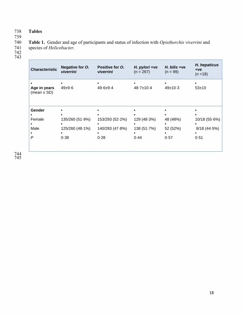
Tables 738 739 Table 1. Gender and age of participants and status of infection with Opisthorchis viverrini and 740 species of Helicobacter. 741 742 743
Characteristic Negative for O. viverrini
Positive for O. viverrini
H. pylori +ve (n = 267)
H. bilis +ve (n = 99)
H. hepaticus +ve (n =18)
• Age in years (mean ± SD)
• 49±9·6
•49·6±9·4
•48·7±10·4
•49±10·3
•53±10
Gender • Female • Male • P
• • 135/260 (51·9%) • 125/260 (48·1%) • 0·38
••153/293 (52·2%) •140/293 (47·8%) •0·28
••129 (48·3%) •138 (51·7%) •0·44
••48 (48%) •52 (52%) •0·57
••10/18 (55·6%)• 8/18 (44·5%) •0·51
744 745
19
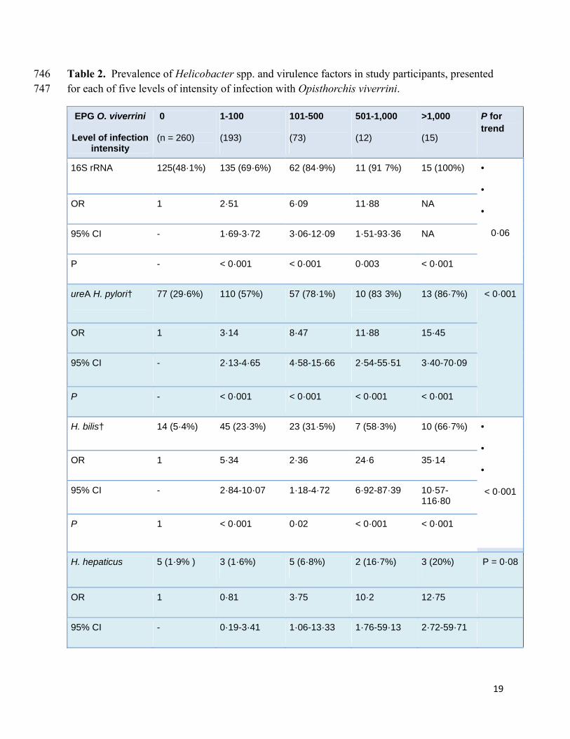
Table 2. Prevalence of Helicobacter spp. and virulence factors in study participants, presented 746 for each of five levels of intensity of infection with Opisthorchis viverrini. 747
EPG O. viverrini
Level of infection intensity
0
(n = 260)
1-100
(193)
101-500
(73)
501-1,000
(12)
>1,000
(15)
P for trend
16S rRNA 125(48·1%) 135 (69·6%) 62 (84·9%) 11 (91·7%) 15 (100%) •
•
•
0·06
OR 1 2·51 6·09 11·88 NA
95% CI - 1·69-3·72 3·06-12·09 1·51-93·36 NA
P - < 0·001 < 0·001 0·003 < 0·001
ureA H. pylori† 77 (29·6%) 110 (57%) 57 (78·1%) 10 (83·3%) 13 (86·7%) < 0·001
OR 1 3·14 8·47 11·88 15·45
95% CI - 2·13-4·65 4·58-15·66 2·54-55·51 3·40-70·09
P - < 0·001 < 0·001 < 0·001 < 0·001
H. bilis† 14 (5·4%) 45 (23·3%) 23 (31·5%) 7 (58·3%) 10 (66·7%) •
•
•
< 0·001
OR 1 5·34 2·36 24·6 35·14
95% CI - 2·84-10·07 1·18-4·72 6·92-87·39 10·57-116·80
P 1 < 0·001 0·02 < 0·001 < 0·001
H. hepaticus 5 (1·9% ) 3 (1·6%) 5 (6·8%) 2 (16·7%) 3 (20%) P = 0·08
OR 1 0·81 3·75 10·2 12·75
95% CI - 0·19-3·41 1·06-13·33 1·76-59·13 2·72-59·71

20
P - 0·53 · 0·03 0·006
H. pylori + H. bilis† 11 (4·2%) 40 (20·7%) 23(31·5%) 6 (50%) 10 (66·7%) < 0·001
OR 1 5·92 10·41 22·63 45·27
95% CI - 2·95-11·88 4·77-22·71 6·27-81·63 13·21-155·16
P - < 0·001 < 0·001 < 0·001 < 0·001
cagA† 13 (5·0%) 21 (10·9%) 26 (35·6%) 6 (50%) 9 (60%)
< 0·001 OR 1 2·32 10·51 19·0 28·5
95% CI - 1·13-4·76 5·04-21·92 15·38-67·09 8·81-92·19
P - 0·02 < 0·001 < 0·001 < 0·001
cagE† 7 (2·7%) 13 (6·7%) 16 (21·9%) 3 (25%) 5 (33%)
< 0·001 OR 1 2·61 10·15 12·05 18·07
95% CI - 1·02-6·67 3·99-25·80 2·67-54·38 4·87-66·9
P - 0·04 < 0·001 0·007 < 0·001
cagA + cagE† 1 (0·4%) 9 (4·7%) 12 (16·4%) 3 (25%) 5 (33%)
< 0·001
OR 1 12·7 50·95 86·33 129·5
95% CI - 1·59-100·86 6·5-399·37 8·16-913·23 13·81-1214·18
P - 0·003 < 0·001 < 0·001 < 0·001
748
P = P-value, OR = Odds Ratio, CI = Confidence Interval. The reference group for the 749 analysis and to estimate P-values and RRR is the uninfected i.e. group, EPG O. viverrini 750 = 0, where the OR is 1. 751
752

21
Table 3. Prevalence of Helicobacter species and virulence genes during infection with 753 Opisthorchis viverrini, and relationships with status (grade) of hepatobiliary disease as 754 established by abdominal ultrasonography for degree of periportal echoes. Bold type letters 755 denote significant differences. 756 757 Helicobacter Grade 0 +1 Grade +2 Grade +3
(n = 241) (n = 36) (n = 16)
16S rRNA Helicobacter species
179 (74·3%) •
30 (83·3%) 13 (81·3%)
RRR 1 1·62 1·63
95% CI - 0·64-4·11 0·43-6·24
P - 0·307 0·476
ureA H. pylori 153 (90%) 28 (90·32%) 8 (100%)
RRR 1 1·03 NA
95% CI - 0·28-3·75 NA
P - 0·969 0·991
cagA 39 (25·32%) 16 (53·33%) 6 (75%)
RRR 1 3·38 9·15
95% CI - 1·51-7·58 1·75-47·97
P - 0·003 0·009
cagE 26 (61·9%) 7 (43·75% ) 5 (83·33%)
RRR 1 0·44 3·30
95% CI - 0·13-1·52 0·33-32·89
P - 0·195 0·311
cagA + cagE 21 (8·71%) 5 (13·89%) 5 (31·25%)
RRR 1 1·69 4·96
95% CI - 0·59-4·83 1·50-16·35
P - 0·331 0·009

22
H. bilis 64 (35·75%) 14 (45·16%) 7 (53·85 %)
RRR 1 1·48 2·15
95% CI - 0·68-3·21 0·68-6·75
P - 0·319 0·191
H. hepaticus 12 (7·23%) 2 (7·41%) 1(9·09%)
RRR 1 1·20 1·09
95% CI - 0·25-5·82 0·12-9·70
P - 0·823 0·937
H. pylori + H. bilis 59 (24·38%) 14(38·89%) 6(37·50%)
RRR 1 1·95 1·96
95% CI - 0·93-4·06 0·66-5·79
P - 0·076 0·223
758 NA = not applicable, RRR = relative risk ratio, P = P-value, CI = Confidence Interval 759 760 The reference group for RRR is Grade 0+1 as grade 0 is baseline negative periductal 761 fibrosis. Some participants were grade 1. 762 763
23
Table 4. Associations among genotypes of CagA of Helicobacter pylori and infection status with 764 Opisthorchis viverrini. 765
766
Genotype Negative for O. viverrini (%)
Positive for O. viverrini (%)
EPIYA-AB TYPE* 13/15 (86·7) 47/62 (75·8)
EPIYA-ABC TYPE 1/15 (6·7) 5/62 (8·1)
EPIYA-AB’C TYPE (B’=EPIYT) 1/15 (6·7) 10/62 (16·1)
Total 15 62
767
768 769
24
Table 5. Associations among cagA genotypes bearing the CagA multimerization motif (CM) 770 and infection with Opisthorchis viverrini. 771
Genotype Negative for O. viverrini (%)
Positive for O. viverrini (%)
Total (%)
EPIYA-AB TYPE 4/13 (30·8) 17/47 (36·2) 21/60 (35)
EPIYA-ABC 1/1 (100) 5/5 (100) 6/6 (100)
EPIYA-AB’C TYPE (B’=EPIYT) †
1/1 (100) 10/10 (100) 11/11 (100)
772
773

25
Table 6. CagA genotypes in participants positive for liver fluke infection, and relationships with 774 status (grade) of biliary periductal fibrosis as established by abdominal ultrasonography. 775
Helicobacter Grade 0 +1 Grade +2 Grade +3 n = 242 n = 36 n = 16
cagA genotype 40 (16·6%) 12 (40%) 3 (23·1%)
RRR 1 1·27*
23·12**
2·31*
24·36**
95% CI - 0·21-7·61
2·32-230·5
0
1·71-347·09
P - 0·794*
0·007**
0·994*
0·018**
cagA CM sequence 11 (26·8%) 16 (88·9%) 6 (100%)
RRR 1 30·74 1·41
95% CI - 5·25-180·08 0
P - <0·001 0·99
776 *, ABC type; **, AB’C type; RRR, relative risk ratio; P = P-value; 777 CI, confidence interval. Boldface type highlights significant differences. 778

26
Figure legends 779 780 Figure 1. Prevalence of Helicobacter species, H. pylori, H. bilis, H. hepaticus and mixed H. 781 pylori and H. bilis in participants who were either uninfected or infected with Opisthorchis 782 viverrini. 783 784 Figure 2. Phylogenetic relationship among partial CagA sequences amplified from 785 representative samples. Panel A. Bootstrap consensus phylogenetic tree inferred from 500 786 replicates revealing four major clusters; EPIYA AB type without CagA multimerization domain 787 (CM) (blue); EPIYA AB type containing CM domain (red); EPIYA ABC type ‘Western-like’ 788 (green), and EPIYA AB’C type ‘Western-like’ (purple). Two Western CagA (W) and two 789 Eastern CagA (E) reference strains detected in gastro-duodenal disease in the Thailand cohorts, 790 and three CagA sequences isolated from bile from Thai cholangiocarcinoma (CCA) cases 42 were 791 included (black). Branches corresponding to partitions reproduced in less than 50% bootstrap 792 replicates were collapsed; bootstrap numbers higher than 60% are shown. Hepatobiliary disease 793 status and O. viverrini infection status are shown for each sample following the indicated color 794 code, * for egg-negative O. viverrini samples no ultrasound study was performed, EPG: eggs per 795 gram of feces. Panel B. Multiple sequence alignment of representative partial CagA sequences 796 belonging to four major clusters comprising the phylogram. Two representative sequences of 797 each cluster are color-squared following the same color code as in Panel A. EPIYA domains are 798 indicated as A, B and C, and CagA multimerization domains (CM) are highlighted (yellow). 799

