electrophysiologic approaches in delirium apl · 11/28/2016 1 electrophysiologic approaches to...
TRANSCRIPT

11/28/2016
1
Electrophysiologic approaches to delirium
Alvaro Pascual‐Leone, MD, PhD
Mouhsin Shafi, MD, PhD
Overview
• Overview of the problem and techniques
• Electrophysiological studies in delirium
• Electrophysiological studies in related conditions– Alzheimer’s Disease
– Minimal Hepatic Encephalopathy
• Conceptual Model: Delirium is due to breakdown of normal brain function reflecting impairments in brain connectivity and plasticity
• Clinical Significance

11/28/2016
2
The Problem
• Delirium is
– Common and costly
– Associated with cognitive decline, loss of independence and increased mortality
• However, the neurobiological basis for and effects of delirium are NOT well understood
– No way to identify individual subjects at risk for delirium
– No brain‐based interventions to prevent delirium, accelerate recovery, or minimize long‐term effects
Modern concept of brain function
• Normal brain function involves flexible interactions between different brain regions to produce behavior and cognition.
• Key concepts:– Connectivity: Structured and dynamic interactions between different brain regions depending on the task at hand. “Human Connectome Project”.
– Neuroplasticity: The brain’s ability to reorganize itself by forming new connections and modifying old ones to adapt to changing environmental demands

11/28/2016
3
Tools to study brain function
• Electroencephalography (EEG)
• Magnetic Resonance Imaging (MRI)
• Transcranial Magnetic Stimulation (TMS)
• And many others …
MRI
• Magnetic Resonance Imaging
– High Resolution Brain Structure
– Task‐related functional MRI
– Resting‐state functional connectivity MRI
• Measure changes in blood oxygenation over time while subjects rest quietly in the scanner
• Correlations between brain regions indicative of connectivity between them

11/28/2016
4
EEG
• EEG signals are the result of the synchronous activity of cortical neurons
• EEG signals described in terms of
Amplitude (or Power)
Frequency
# of Cycles/Second (Hz)
Strength(µV or µV2)
10Hz
20Hz
How to Analyze EEG
Local Response
‐ Amplitude/Power‐ Frequency
Spontaneous EEG: Spectral Power
123
Functional Connectivity
1 2 3
Direction of Information FlowDirected Transfer FunctionDirected Partial Coherence
Correlation (time)Coherence (frequency)Synchrony (phase‐locking)
Θ

11/28/2016
5
Transcranial Magnetic Stimulation
9Wagner 2007 Ridding & Rothwell, 2007
Measure evoked brain responses
10Farzan et al, 2016

11/28/2016
6
And brain plasticity!• TMS applied at a fixed frequency or in particular patterns can change brain excitability – a measure of brain plasticity
Studies duringDelirium
• Most extensive data is for EEG
– Slowing of background EEG rhythms (Romano & Engel 1944)
– Background EEG rhythm speeds up as delirium resolves
Changes in EEG rhythms over time in a patient with alcoholism presenting with Wernicke’s encephalopathy; EEG speeds up as encephalopathy resolves (Romano & Engel 1944)

11/28/2016
7
EEG changes during Delirium
• Numerous subsequent studies have replicated finding of slowing of normal background rhythms during delirium
– More recent studies applying quantitative measures have confirmed these findings, and also noted changes in measures of EEG connectivity (van Dellen 2014 Anesthesiology), EEG complexity (van der Kooi 2014 Clin Neurophys), and EEG brain dynamics (Sarkis 2013 J. Clin Neurophys).
EEG as diagnostic test
EEG features such as posterior dominant rhythm and visual analysis of EEG features have approximately 80% accuracy in differentiating delirious from non‐delirious subjects (Trzepacz1988 Biological Psychiatry )
11/28/2016
8
Utility of EEG in delirium
• EEG features correlate with MMSE scores in patients with delirium, and changes in EEG features correlate with changes in MMSE over time (Jacobson 1993 Biological Psychiatry)
• Rule out non‐convulsive status
• Quantitative EEG measures can be used to differentiate delirious from non‐delirious subjects with up to 100% sensitivity and 96% specificity in some (retrospective) studies (van der Kooi 2015 Chest)
• However, no studies evaluating utility of EEG features in predicting delirium
Utility of EEG in delirium
• Bispectral Index (BIS) Monitoring
– Weighted sum of several EEG parameters considering time and frequency domain
– Adjustment of anesthetic depth associated with a marked reduction in postoperative delirium(Whitlock 2014; Chan 2013)
– Large, block‐randomized, double‐blinded, effectiveness trial (Wildes 2016)

11/28/2016
9
EEG in delirium vs dementia• Rule out non‐convulsive status• Quantitative EEG changes helped differentiate dementia plus delirium from dementia alone with 83% accuracy (Thomas 2007 British Medical Journal)
Resting‐state fMRI changes in delirium
Choi 2012 Am J Psych

11/28/2016
10
How about related conditions?
• A number of studies have been conducted in conditions that predispose to delirium
– Alzheimer’s Disease (AD)
• Patients with AD at particularly high risk for delirium
• Delirium accelerates cognitive decline in AD
– Minimal Hepatic Encephalopathy (MHE)
• Earliest form of hepatic encephalopathy
• Cognitive impairments in attention, vigilance and integrative function without clinical deficits
Slowing in AD• Patients with mild cognitive impairment (MCI) and Alzheimer’s Disease show increased slowing relative to normal elderly
Babiloni 2006 Clin Neurophys
Changes in EEG power correlate with and predict changes in cognition over time (Huang 2000 Clin Neurophys; Jelic 2000 Neurobiol Aging; Rossini 2006 Neuroscience; Babiloni 2010 Hum Br Map)
11/28/2016
11
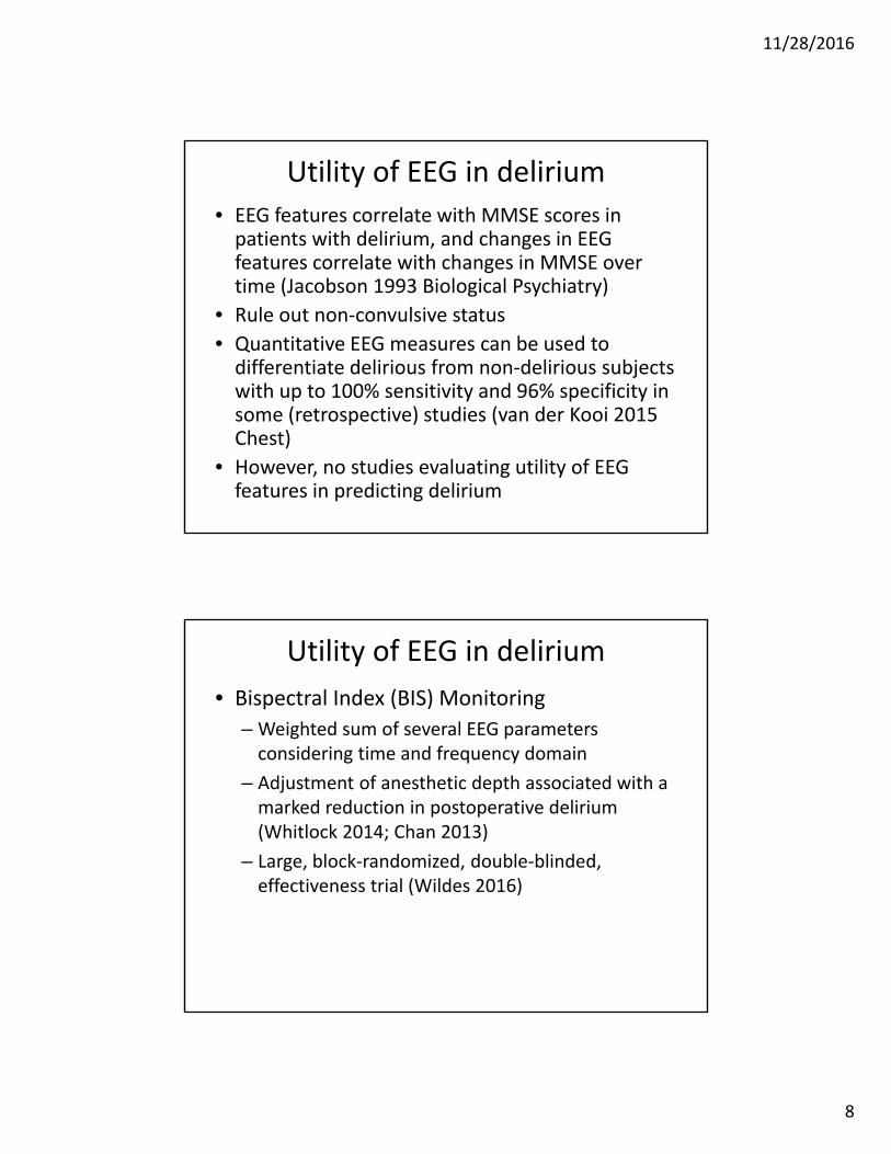
Decreased connectivity in AD
• Patients with AD and MCI also have decreased frontoparietal EEG connectivity, particularly in the alpha band
Babiloni 2006 Brain Research Bulletin
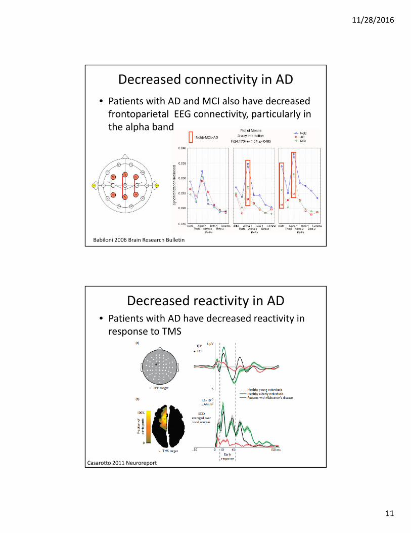
Decreased reactivity in AD• Patients with AD have decreased reactivity in response to TMS
Casarotto 2011 Neuroreport

11/28/2016
12
And decreased plasticity• Patients with AD have decreased plasticity in response to excitatory patterned TMS protocols than healthy subjects
– Cognitive progression at 18 months is correlated with TMS plasticity changes (Lorenzo 2016 Ann Neurol)
Minimal Hepatic Encephalopathy• Patients initially w/o overt hepatic encephalopathy but with EEG slowing have an increased risk of progression to overt hepatic encephalopathy over time
Amodio 2001 J Hepatol

11/28/2016
13
Decreased plasticity in MHE• Patients with MHE have decreased plasticity in response to patterned TMS protocols in comparison to healthy controls
Golaszewski 2016 Brain Research Bulletin
Conceptual Model of Delirium
• We propose that Delirium is due to a breakdown of normal brain network function in response to external stressors in patients with impaired baseline brain connectivity and/or impaired baseline brain plasticity
– Baseline deficits e.g. due to AD or MHE
– Inability to accommodate to stressors results in clinical syndrome of delirium

11/28/2016
14
Conceptual Model of Delirium
Shafi 2016 JAGS (accepted)