electromyography (emg) - universitydocs.neu.edu.tr/staff/asli.aykac/5 -...
TRANSCRIPT
ELECTROMYOGRAPHY (EMG)
Dr. Aslı AYKAÇ

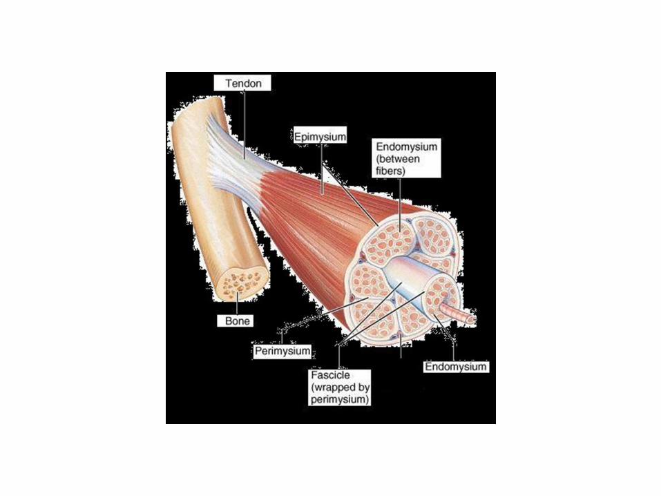
"Electromyography (EMG) is an experimental technique concerned with the recording and analysis of myoelectric signals. Classical Neurological EMG, : an artificial muscle response due to external electrical stimulation is analyzed in static conditions, Kinesiological EMG can be described as the study of the neuromuscular activation of muscles within postural tasks, functional movements, work conditions and treatment/training regimes. Myoelectric signals are formed by physiological variations in the state of muscle fiber membranes."

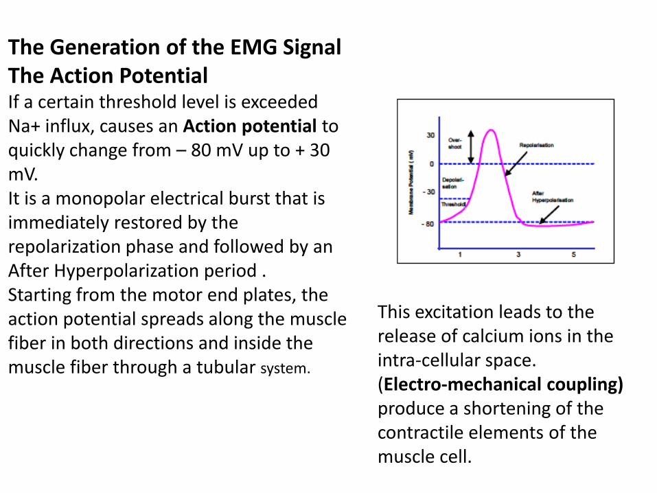
The Generation of the EMG Signal The Action Potential If a certain threshold level is exceeded Na+ influx, causes an Action potential to quickly change from – 80 mV up to + 30 mV. It is a monopolar electrical burst that is immediately restored by the repolarization phase and followed by an After Hyperpolarization period . Starting from the motor end plates, the action potential spreads along the muscle fiber in both directions and inside the muscle fiber through a tubular system.
This excitation leads to the release of calcium ions in the intra-cellular space. (Electro-mechanical coupling) produce a shortening of the contractile elements of the muscle cell.
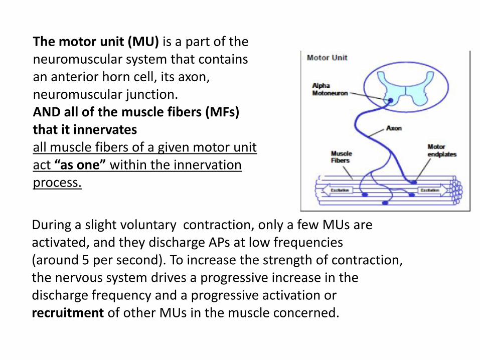
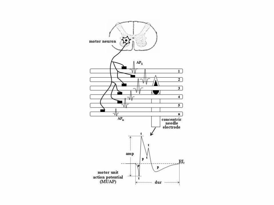
The motor unit (MU) is a part of the neuromuscular system that contains an anterior horn cell, its axon, neuromuscular junction. AND all of the muscle fibers (MFs) that it innervates all muscle fibers of a given motor unit act “as one” within the innervation process.
During a slight voluntary contraction, only a few MUs are activated, and they discharge APs at low frequencies (around 5 per second). To increase the strength of contraction, the nervous system drives a progressive increase in the discharge frequency and a progressive activation or recruitment of other MUs in the muscle concerned.
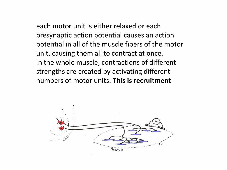
each motor unit is either relaxed or each presynaptic action potential causes an action potential in all of the muscle fibers of the motor unit, causing them all to contract at once. In the whole muscle, contractions of different strengths are created by activating different numbers of motor units. This is recruitment


In general, normal MUAPs show mean peak-to-peak amplitudes of around 0.5 mV and a duration from 8 to 14 ms, depending on the size of the MUs. The size and shape of MUAPs is determined by certain structural and functional aspects of MUs.
Pathologic processes of the peripheral nervous system (neurogenic processes) and of muscles (myopathic pathologies) lead to abnormal deviations in MUAP parameters;
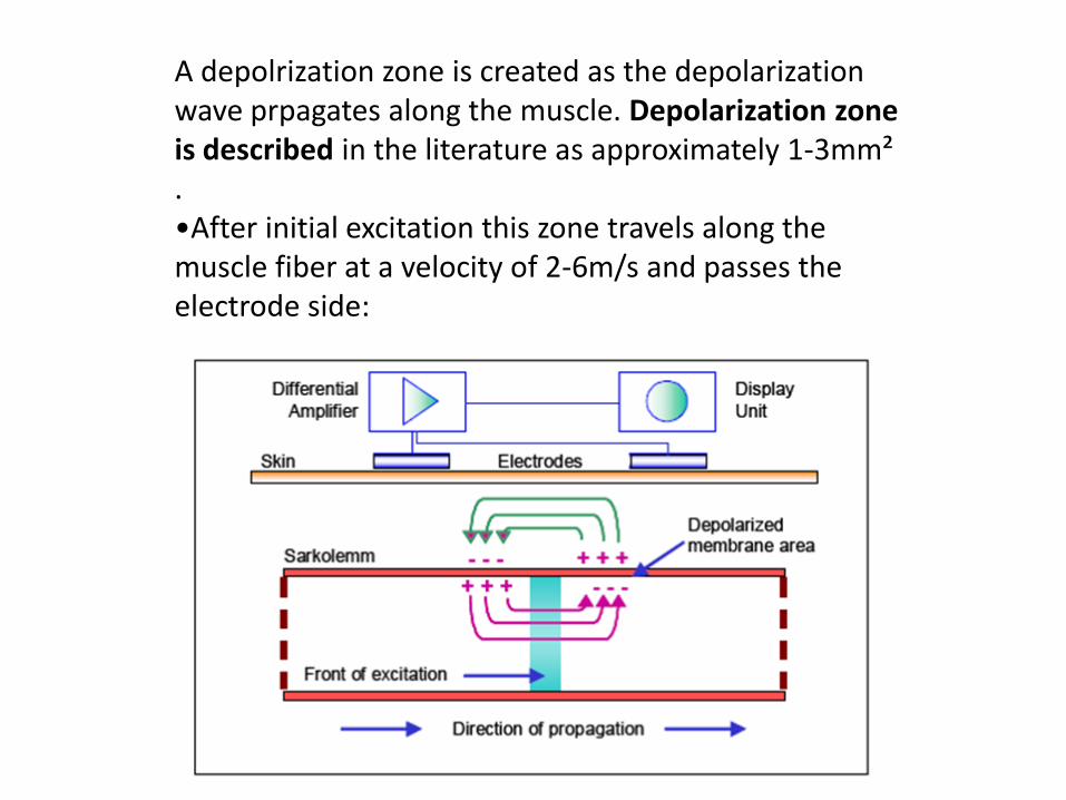
A depolrization zone is created as the depolarization wave prpagates along the muscle. Depolarization zone is described in the literature as approximately 1-3mm² . •After initial excitation this zone travels along the muscle fiber at a velocity of 2-6m/s and passes the electrode side:
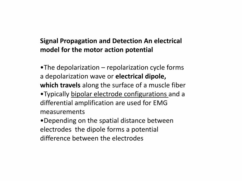
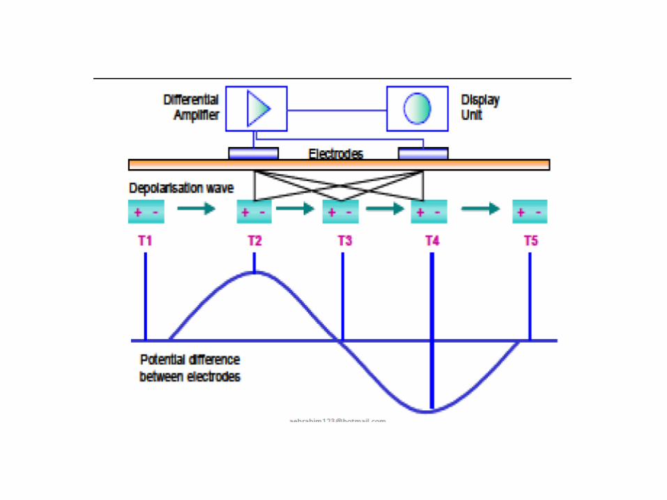
Signal Propagation and Detection An electrical model for the motor action potential •The depolarization – repolarization cycle forms a depolarization wave or electrical dipole, which travels along the surface of a muscle fiber •Typically bipolar electrode configurations and a differential amplification are used for EMG measurements •Depending on the spatial distance between electrodes the dipole forms a potential difference between the electrodes

Generation of the triphasic motor unit action potential •Because a motor unit consists of many muscle fibers, the electrode pair “sees” the magnitude of all innervated fibers within this motor unit - depending on their spatial distance and resolution. •Typically, they sum up to a triphasic Motor unit action potential which differs in form and size depending on the geometrical fiber orientation in ratio to the electrode site
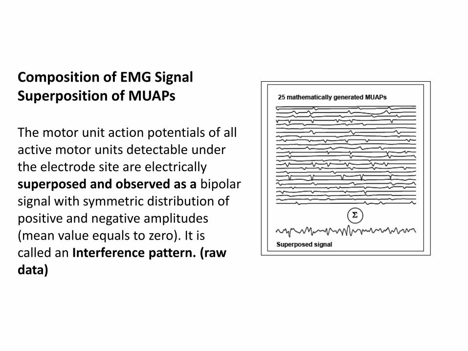
Composition of EMG Signal Superposition of MUAPs The motor unit action potentials of all active motor units detectable under the electrode site are electrically superposed and observed as a bipolar signal with symmetric distribution of positive and negative amplitudes (mean value equals to zero). It is called an Interference pattern. (raw data)

Parameters of the MUAP
1- Size parameters are related to the size (diameter), number and density of the MFs that generate the MU. These parameters include duration, amplitude, area and indices
2- MUAP waveform shape parameters : the temporal synchrony/ / dispersion of the activation times of the MFs and their conduction velocities. These parameters include the number of phases, the number of turns, and indices such as the coefficient of irregularity.
Stability parameters or jiggle parameters : the degree of variability in MUAP shape at consecutive discharges
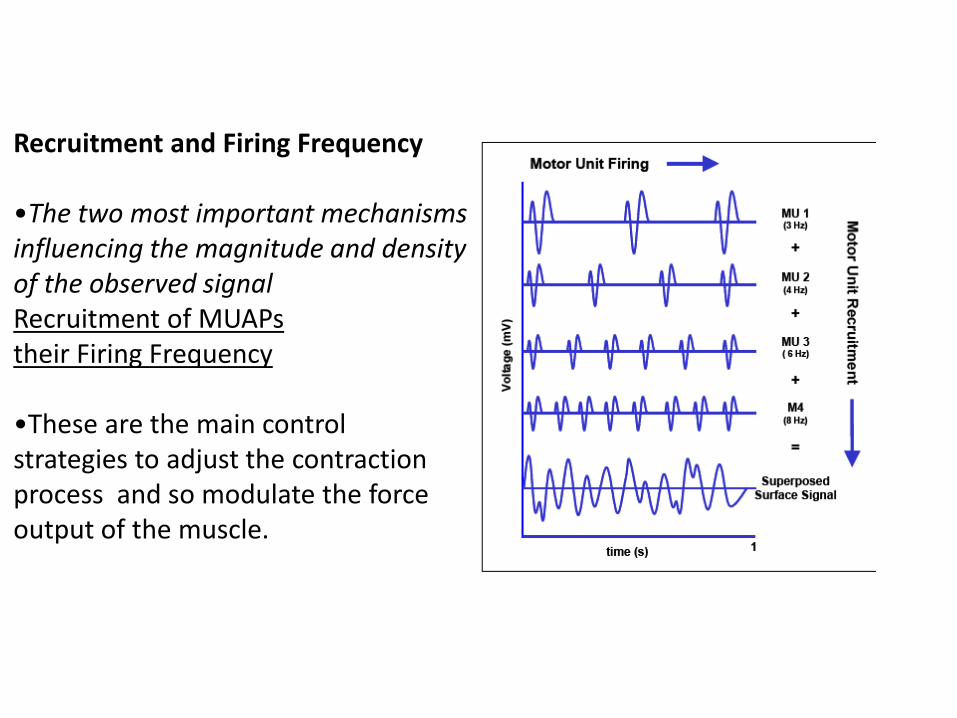
Recruitment and Firing Frequency •The two most important mechanisms influencing the magnitude and density of the observed signal Recruitment of MUAPs their Firing Frequency •These are the main control strategies to adjust the contraction process and so modulate the force output of the muscle.

Nature the of EMG Signal The “raw” EMG signal
•An unfiltered and unprocessed signal detecting the superposed MUAPs is called a raw EMG Signal.
When the muscle is relaxed, a more or less noise-free EMG Baseline can be seen. The raw EMG baseline

In EMG Amplitude range: 0– 10 mV (+5 to -5) prior to amplification • EMG frequency: Range of 10 - 500 Hz
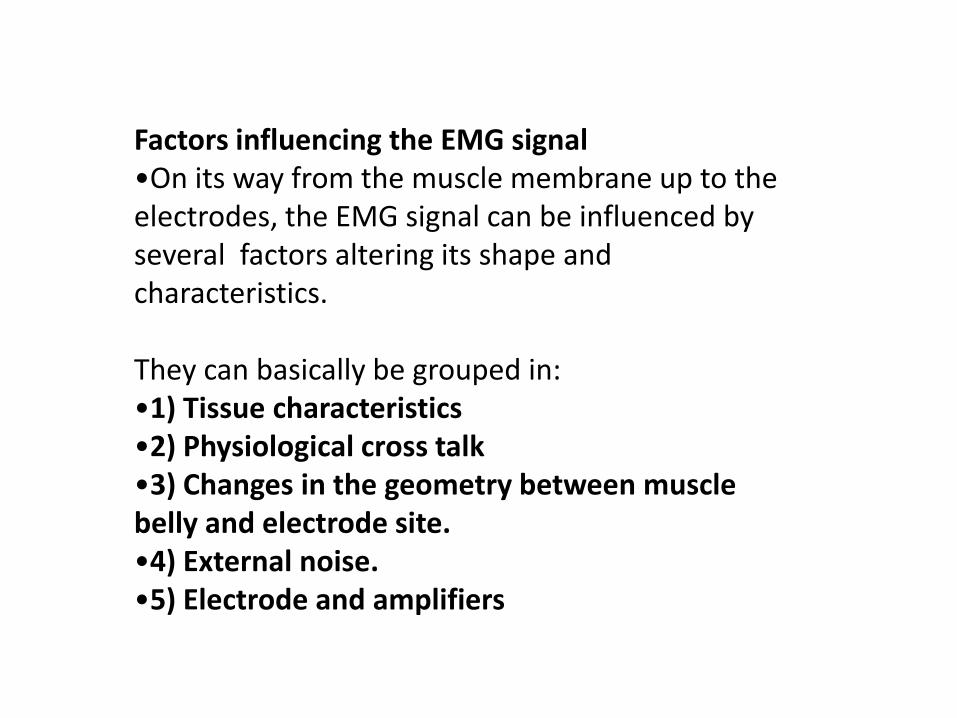
Factors influencing the EMG signal •On its way from the muscle membrane up to the electrodes, the EMG signal can be influenced by several factors altering its shape and characteristics. They can basically be grouped in: •1) Tissue characteristics •2) Physiological cross talk •3) Changes in the geometry between muscle belly and electrode site. •4) External noise. •5) Electrode and amplifiers

1) Tissue characteristics •the electrical conductivity varies with : - tissue type - thickness - physiological changes - Temperature •These conditions can greatly vary from subject to subject (and even within subject) And so prohibit a direct quantitative comparison of EMG amplitude parameters calculated on the unprocessed EMG signal.

2) Physiological cross talk •Neighboring muscles may produce a significant amount of EMG that is detected by the local electrode site. •Typically this “Cross Talk” does not exceed 10%-15% of the overall signal contents or isn’t available at all. •ECG spikes can interfere with the EMG recording, especially when performed on the upper trunk / shoulder muscles. They are easy to see and new algorithms are developed to eliminate them
3) Changes in the geometry between muscle belly and electrode site •Any change of distance between signal origin and detection site will alter the EMG reading. •It is an inherent problem of all dynamic movement studies and can also be caused by external pressure.
4) External noise •Due to very noisy electrical environments. e.g. the direct interference of power hum, typically produced by incorrect grounding of other external devices.
5) Electrode and amplifiers •The selection/quality of electrodes and internal amplifier noise may add signal contents to the EMG baseline. •Most of these factors can be minimized or controlled by accurate preparation and lab conditions

EMG consist of: Amplifier , or preamplifier 3 electrodes ( 2 active, 1 reference –ground-) Cables and wires or wireless

Skin surface electrodes •Advantages: non- invasive character. easy handling their main limitation only surface muscles can be detected. Fine-wire or needle electrodes For deeper muscles •The selection of an electrode type strongly depends on the given investigation condition
An example of How the Test is Performed in needle electrode EMG •a needle electrode is inserted through the skin into the muscle. •The electrical activity detected by this electrode is displayed on an oscilloscope, and may be heard through a speaker. •After placement of the electrodes, ask the subject to contract the muscle (for example, by bending his arm). •The presence, size, and shape of the wave form -- the action potential -- produced on the oscilloscope provide information about the ability of the muscle to respond when the nerves are stimulated.

Electrode connections for recording from the abductor pollicis brevis muscle, and stimulation of the median nerve at the wrist and elbow.

Signal Processing •The raw EMG recording already contains very important information and may serve as a first objective information of the muscle innervation. •Qualitative assessments can directly be derived and give an important first understanding of the neuromuscular control within tests and exercises •If a quantitative amplitude analysis is required some EMG specific signal processing steps are applied to increase the reliability and validity of findings

Converting EMG from time domain to frequency domain Fourier !!!

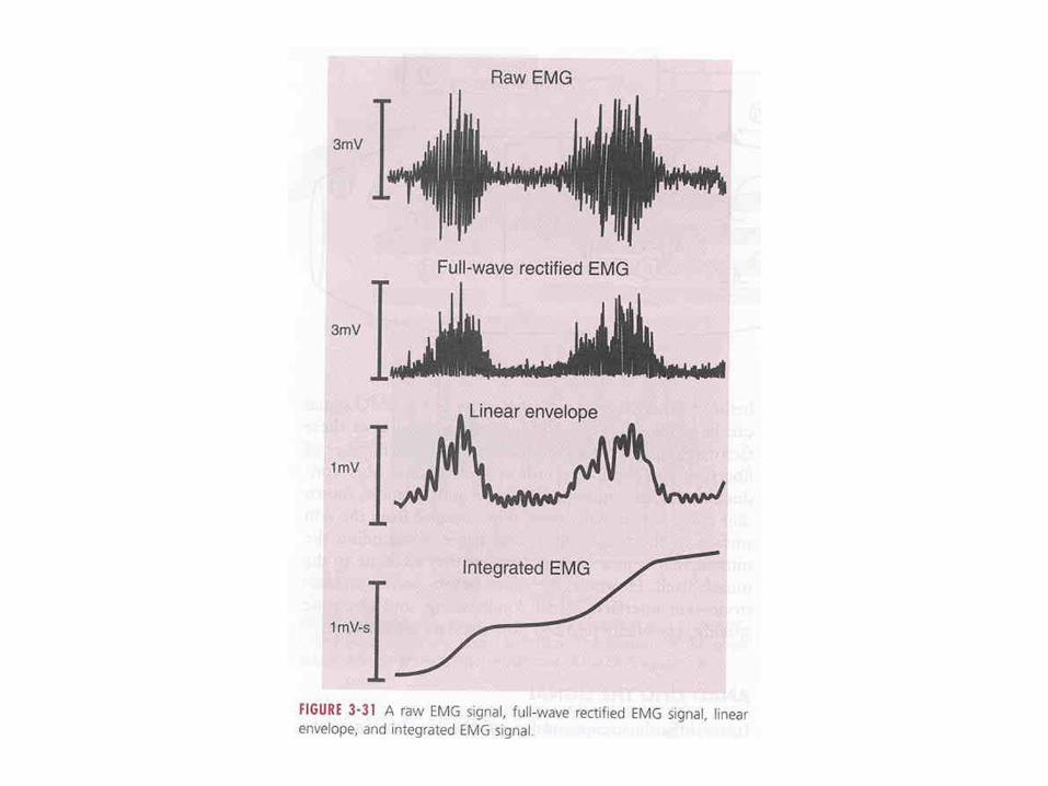
Some of the signal processing methods •Full wave rectification •Smoothing •Digital filtering •Amplitude normalization •ECG reduction
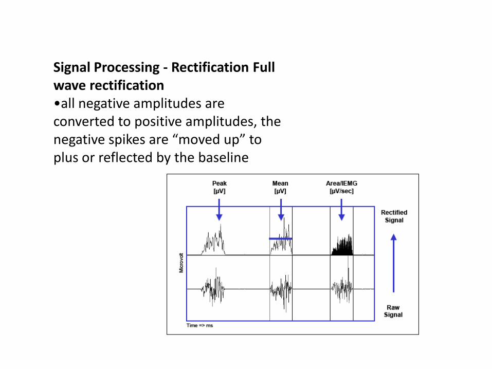
Signal Processing - Rectification Full wave rectification •all negative amplitudes are converted to positive amplitudes, the negative spikes are “moved up” to plus or reflected by the baseline
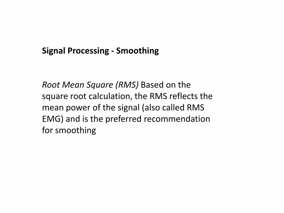
Signal Processing - Smoothing Root Mean Square (RMS) Based on the square root calculation, the RMS reflects the mean power of the signal (also called RMS EMG) and is the preferred recommendation for smoothing

Signal Processing – Amplitude normalization •One big drawback of any EMG analysis is that the amplitude (microvolt scaled) data are strongly influenced by the given detection condition: it can strongly vary between electrode sites, subjects and even day to day measures of the same muscle site. •One solution is the normalization to reference value, e.g. the maximum voluntary contraction (MVC) value of a reference contraction. •The basic idea is to “calibrate the microvolts value to a unique calibration unit with physiological relevance, the “percent of maximum innervation capacity” in that particular sense. •The main effect of all normalization methods is that the influence of the given detection condition is eliminated -data are rescaled from microvolt to percent of selected reference value.
EMG parameters The EMG signal is analyzed by - Amplitude variables (mV) - frequency variables (Hz)
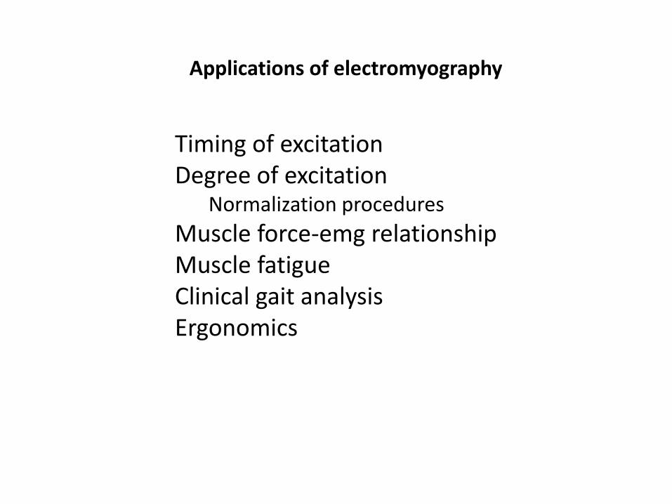
Applications of electromyography
Timing of excitation Degree of excitation
Normalization procedures
Muscle force-emg relationship Muscle fatigue Clinical gait analysis Ergonomics
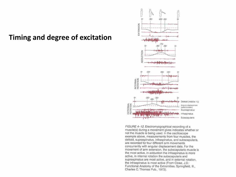
Timing and degree of excitation

EMG-force relationship
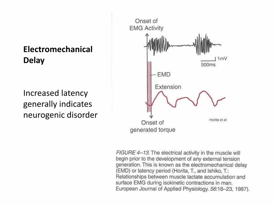
Electromechanical Delay Increased latency generally indicates neurogenic disorder

The fatigue index from EMG
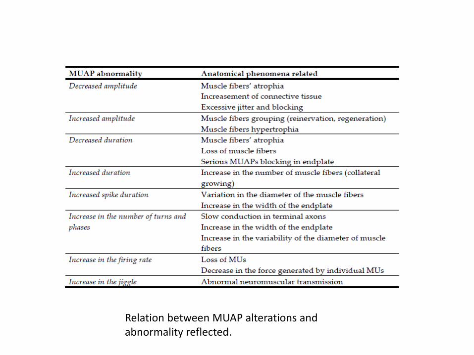
Relation between MUAP alterations and abnormality reflected.


Thank you