electrocorticography-based brain computer interface—the...
TRANSCRIPT

194 IEEE TRANSACTIONS ON NEURAL SYSTEMS AND REHABILITATION ENGINEERING, VOL. 14, NO. 2, JUNE 2006
Electrocorticography-Based Brain ComputerInterface—The Seattle Experience
Eric C. Leuthardt, Kai J. Miller, Gerwin Schalk,Rajesh P. N. Rao, and Jeffrey G. Ojemann
Abstract—Electrocorticography (ECoG) has been demonstrated to bean effective modality as a platform for brain–computer interfaces (BCIs).Through our experience with ten subjects, we further demonstrate evi-dence to support the power and flexibility of this signal for BCI usage. In asubset of four patients, closed-loop BCI experiments were attempted withthe patient receiving online feedback that consisted of one-dimensionalcursor movement controlled by ECoG features that had shown correlationwith various real and imagined motor and speech tasks. All four achievedcontrol, with final target accuracies between 73%–100%. We assess themethods for achieving control and the manner in which enhancing onlinecontrol can be accomplished by rescreening during online tasks. Addition-ally, we assess the relevant issues of the current experimental paradigm inlight of their clinical constraints.
Index Terms—Brain–computer interface (BCI), brain–machine interface(BMI), neuroprosthetics.
I. INTRODUCTION
Various signal modalities have been utilized for brain–computerinterfaces (BCIs). BCIs can use noninvasive or invasive methods.Noninvasive BCIs mainly use electroencephalographic activity (EEG)recorded from the scalp [1]–[6]. They are easy to use, but they haverelatively low spatial resolution [7], [8], are susceptible to artifactssuch as electromyographic (EMG) signals, and often require extensiveuser training. Invasive BCIs use single-neuron activity recorded withinthe brain [9]–[12]. While they have higher spatial resolution and mightallow for many degrees of freedom, they require that tens or hundredsof small electrodes be implanted in the brain and are, as a result,subject to tissue response which can impair their long-term stability[13].
An alternate BCI methodology has been studied in epilepsy patientsundergoing invasive monitoring for seizure localization. Electrocor-ticography (ECoG), which is recorded from electrodes placed on thesurface of the brain, has been shown to be a powerful and practical al-ternative to these other modalities. ECoG has higher spatial resolutionthan EEG [8], broader bandwidth, higher amplitude, and far less vulner-ability to artifacts such as EMG [7], [8], [14]. At the same time, becauseECoG is recorded by subdural electrode arrays and thus does not re-quire cortical penetration, it entails less clinical risk and is likely to havegreater long-term stability than single-neuron recording. Leuthardt etal. demonstrated that a high level of control was achieved with minimaltraining using various real and imagined motor and speech tasks [15].
Manuscript received August 15, 2005; revised March 24, 2006. This workwas supported by the National Institute of Health under Grant NIH NS007144and by JGO-NS41272.
E. C. Leuthardt and J. G. Ojemann are with the Department of Neurolog-ical Surgery, School of Medicine, University of Washington, Seattle, WA 98104USA (e-mail: [email protected]).
K. J. Miller is with the Department of Physics, University of Washington,Seattle, WA 98195 USA.
G. Schalk is with the Wadsworth Center, Albany, NY 12201 USA and alsowith the Electrical, Computer, and Systems Engineering Department, Rensse-laer Polytechnic Institute, Troy, NY 12202 USA.
R. P. N. Rao is with the Department of Computer Science, University ofWashington, Seattle, WA 98195 USA.
Digital Object Identifier 10.1109/TNSRE.2006.875536
At the University of Washington, our group has further explored theuse of ECoG as a signal modality for a BCI platform. We studied pa-tients in whom subdural electrode arrays were implanted in preparationfor surgery to remove an epileptic focus. We identified the locationsand frequency bands of ECoG sensorimotor rhythms associated withspecific movements or speech or with imagery of those actions. Sub-jects could quickly learn to use the ECoG activity associated with eitherovert motor movement or motor imagery to control a cursor. Becausebrain signals associated with these tasks often changed once the sub-ject used them for cursor control, we further improved the translationof the subjects’ control over those signals into cursor movement byreadjusting locations and frequencies following initial cursor controlattempts. These results indicate that by using ECoG, accurate one-di-mensional (1-D) BCI control can be rapidly achieved by patients withepilepsy.
II. METHODS
A. Subjects and Experimental Paradigms
The subjects in this study were ten patients with intractable epilepsywho underwent temporary placement of intracranial electrode arraysto localize seizure foci prior to surgical resection. All gave informedconsent. The study was approved by the Human Studies Committee ofthe University of Washington Medical Center. Prior to this study, thesepatients had not been trained on a BCI system.
Each patient had a grid and/or strip electrodes placed subdurally onthe cortical surface. In some subjects, electrode coverage included sen-sorimotor or speech cortex areas. The electrodes had an exposed di-ameter of 2 mm and an interelectrode distance of 1 cm. Grid and stripplacements were based solely on the requirements of the clinical eval-uation, without any consideration of this study. Following placementof the subdural grid/strips, each patient had postoperative radiographsto verify the location of the electrodes.
B. Data Collection
Each patient studied was in a sitting position (semi-recumbent),approximately 75 cm from a video screen. In all experiments, werecorded ECoG from up to 64 electrodes from a combination of gridsand strips using the general-purpose BCI system BCI2000 [16]. Allelectrodes were referenced to a scalp electrode, amplified, bandpassfiltered (0.1–220 Hz), digitized at 1000 Hz, and stored. The amountof data obtained varied from patient to patient and depended on thepatient’s physical state and willingness to continue.
C. Signal Identification
In order to identify brain signals that might be used for BCI control(i.e., screening procedure), we first asked subjects to perform pairedblocks of repetitive hand/tongue, foot/shoulder movements, or repeti-tive speaking of the word “move.” The imagined execution was per-formed in the same manner as the motor task execution. From spec-tral analysis of the data gathered with each of the various tasks, weidentified the locations and frequency bands in which amplitude wasdifferent between the task and rest. For these analyses, the time-seriesECoG data were converted into the frequency domain using an autore-gressive model. Those electrodes and frequency bins with the most sig-nificant task-related amplitude changes (i.e., the highest values of r2)were identified as features to be used to control cursor movement in thesubsequent online BCI experiments. Please see Leuthardt et al. [15] fora more in-depth description of the technique.
1534-4320/$20.00 © 2006 IEEE

IEEE TRANSACTIONS ON NEURAL SYSTEMS AND REHABILITATION ENGINEERING, VOL. 14, NO. 2, JUNE 2006 195
TABLE IACTIONS AND IMAGINED ACTIONS, ECoG FREQUENCY BANDS, AND ANATOMIC LOCATIONS USED FOR ECoG CONTROL OF 1-D CURSOR MOVEMENT AND FINAL
ACCURACIES OF THAT CONTROL. ACCURACY IS CALCULATED AS NUMBER OF CORRECT TARGETS HIT WITH CURSOR FROM TOTAL NUMBER OF TARGETS
PRESENTED DURING FINAL RUN OF SESSION INVOLVING PARTICULAR ACTION
D. ECoG Control of Vertical Cursor Movement Online
In a subset of patients, closed-loop BCI experiments were attemptedwith the patient receiving online feedback that consisted of 1-D cursormovement controlled by ECoG features that had shown correlationwith tasks during the screening procedure. The accuracy expected inthe absence of any control was 50%.
The cursor moved vertically every 40 ms, controlled by a translationalgorithm based on a weighted linear summation of the amplitudes inthe identified frequency bands from the identified electrodes for theprevious 280 ms (as developed for EEG-based control [17], [18]). Theweights were chosen so that this translation algorithm moved the cursorup with task execution (e.g., imagining tongue protrusion) and downwith rest. This relationship was explained to the patient prior to theexperiments. Please see Leuthardt et al. [15] for a more in-depth de-scription of the technique.
E. Adjustment of Signal Features
Subsequent to initial real-time experiments, data were subject to thesame analysis procedure that was performed during the screening pro-cedure. Although we used signal features that we identified during thescreening procedure in response to particular tasks, and we advisedthe subject to use the same tasks (i.e., imagined motor or speech) tocontrol the cursor, those signal features might change in response tothe online feedback that was provided to the subject. In one patient,in whom anatomic location and frequencies changed compared to thescreening procedure, BCI2000 parameters were updated to take advan-tage of those signal features in subsequent closed-loop experiments.
F. Anatomical and Functional Mapping
Radiographs were used to identify the stereotactic coordinates ofeach grid electrode [19], and cortical areas were defined using Ta-lairach’s Co-Planar Stereotaxic Atlas of the Human Brain [20].
III. RESULTS
A. Patient Characteristics
Four patients attempted closed-loop BCI control and all rapidly ac-quired 1-D control. Logistical or technical reasons prevented BCI ex-periments in other patients. The remaining six did not participate in theactual closed-loop portion of the experiment due to: 1) technical issues
(one patient); 2) clinical constraints (two patients); or 3) the subjectsdeclined further participation (three patients).
B. Screening Procedure
In all patients, we identified signal features that strongly changedwith task execution. Motor and language tasks (real and imagined) andeven sensory tasks produced focal responses in different locations andfrequencies. Transformation to stereotactic coordinates demonstratedcolocalization of prominent features with expected areas of the brain(e.g., motor activity in motor cortex). Covert tasks generally colocal-ized with overt tasks with less prominent, but otherwise similar, signals.
C. Control
Those subjects that performed BCI control completed one to eight3-min runs separated by 1-min breaks. Over these short training pe-riods (3–24 min), reliable cursor control was achieved (73%–100% ac-curacy; see Table I).
Notably, in one case (Patient D, see Table I), control was achievedfrom an electrode overlying the dura using an 80-Hz signal. In thiscase, the dura was adherent to the brain during surgery and a portionof the electrode array was directly over cortex, with another portionepidural. Online BCI operation was achieved using signal features froman electrode in the epidural space.
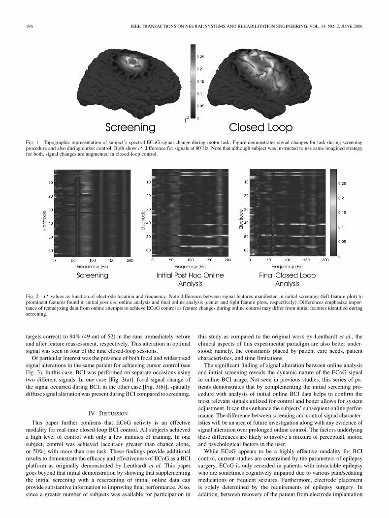
Consistently, signal features during closed-loop BCI control differedfrom those identified during screening. This was noted on all four sub-jects in each independent online session for which there was a total ofnine. The manner in which these signals differed, however, was vari-able. In one scenario, when compared against the screening task, therewas a general anatomic extension of the signal alteration in whichthe cortical region demonstrating signal change was of a higher sig-nificance following online attempts and more spatially spread out inadjacent cortex (see Fig. 1). This occurred in five of the nine onlineclosed-loop sessions. Alternatively, a markedly different set of signalsthat might have overlapped only somewhat with the screening featureswas seen in another case (see Fig. 2). Although the subject was ableto acquire BCI control using the originally selected electrode and fre-quency, many other features changed prominently with the task butwere not present during the initial screening. In this case, reassessingearly control attempts for better features was critical for success. Ac-curacy for this subject immediately improved from 71% (i.e., 30 of 42
196 IEEE TRANSACTIONS ON NEURAL SYSTEMS AND REHABILITATION ENGINEERING, VOL. 14, NO. 2, JUNE 2006
Fig. 1. Topographic representation of subject’s spectral ECoG signal change during motor task. Figure demonstrates signal changes for task during screeningprocedure and also during cursor control. Both show r difference for signals at 80 Hz. Note that although subject was instructed to use same imagined strategyfor both, signal changes are augmented in closed-loop control.
Fig. 2. r values as function of electrode location and frequency. Note difference between signal features manifested in initial screening (left feature plot) toprominent features found in initial post hoc online analysis and final online analysis (center and right feature plots, respectively). Differences emphasize impor-tance of reanalyzing data from online attempts to achieve ECoG control as feature changes during online control may differ from initial features identified duringscreening.
targets correct) to 94% (49 out of 52) in the runs immediately beforeand after feature reassessment, respectively. This alteration in optimalsignal was seen in four of the nine closed-loop sessions.
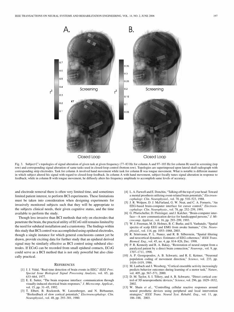
Of particular interest was the presence of both focal and widespreadsignal alterations in the same patient for achieving cursor control (seeFig. 3). In this case, BCI was performed on separate occasions usingtwo different signals. In one case [Fig. 3(a)], focal signal change ofthe signal occurred during BCI; in the other case [Fig. 3(b)], spatiallydiffuse signal alteration was present during BCI compared to screening.
IV. DISCUSSION
This paper further confirms that ECoG activity is an effectivemodality for real-time closed-loop BCI control. All subjects achieveda high level of control with only a few minutes of training. In onesubject, control was achieved (accuracy greater than chance alone,or 50%) with more than one task. These findings provide additionalresults to demonstrate the efficacy and effectiveness of ECoG as a BCIplatform as originally demonstrated by Leuthardt et al. This papergoes beyond that initial demonstration by showing that supplementingthe initial screening with a rescreening of initial online data canprovide substantive information to improving final performance. Also,since a greater number of subjects was available for participation in
this study as compared to the original work by Leuthardt et al., theclinical aspects of this experimental paradigm are also better under-stood; namely, the constraints placed by patient care needs, patientcharacteristics, and time limitations.
The significant finding of signal alteration between online analysisand initial screening reveals the dynamic nature of the ECoG signalin online BCI usage. Not seen in previous studies, this series of pa-tients demonstrates that by complementing the initial screening pro-cedure with analysis of initial online BCI data helps to confirm themost relevant signals utilized for control and better allows for systemadjustment. It can thus enhance the subjects’ subsequent online perfor-mance. The difference between screening and control signal character-istics will be an area of future investigation along with any evidence ofsignal alteration over prolonged online control. The factors underlyingthese differences are likely to involve a mixture of perceptual, motor,and psychological factors in the user.
While ECoG appears to be a highly effective modality for BCIcontrol, current studies are constrained by the parameters of epilepsysurgery. ECoG is only recorded in patients with intractable epilepsywho are sometimes cognitively impaired due to various pain/sedatingmedications or frequent seizures. Furthermore, electrode placementis solely determined by the requirements of epilepsy surgery. Inaddition, between recovery of the patient from electrode implantation
IEEE TRANSACTIONS ON NEURAL SYSTEMS AND REHABILITATION ENGINEERING, VOL. 14, NO. 2, JUNE 2006 197
Fig. 3. Subject C’s topologies of signal alteration of given task at given frequency (77–83 Hz for column A and 97–103 Hz for column B) used in screening (toprow) and corresponding signal alteration of same tasks used in closed-loop control (bottom row). Topologies are superimposed upon lateral skull radiograph withcorresponding strip electrodes. Task for column A involved hand movement while task for column B was tongue movement. What is notable is different mannerin which subject altered his signal with regard to closed-loop feedback. In column A with hand movement, subject focally tunes signal alteration in response tofeedback; while in column B with tongue movement, he diffusely alters his frequency amplitude to accomplish same levels of accuracy.
and electrode removal there is often very limited time, and sometimeslimited patient interest, to perform BCI experiments. These limitationsmust be taken into consideration when designing experiments forinvasively monitored subjects such that they will be appropriate tothe subjects clinical needs, their given cognitive status, and the timeavailable to perform the study.
Though less invasive than BCI methods that rely on electrodes thatpenetrate the brain, the practical utility of ECoG still remains limited bythe need for subdural installation and a craniotomy. The findings withinthis study that BCI control was accomplished using epidural electrodes,though a single instance for which general conclusions cannot yet bedrawn, provide exciting data for further study that an epidural derivedsignal may be similarly effective as BCI control using subdural elec-trodes. If ECoG can be recorded from small epidural contacts, ECoGcould serve as a BCI method that is not only powerful but also clini-cally practical.
REFERENCES
[1] J. J. Vidal, “Real-time detection of brain events in EEG,” IEEE Proc.Special Issue Biological Signal Processing Analysis, vol. 65, pp.633–664, 1977.
[2] E. E. Sutter, “The brain response interface: communication throughvisually-induced electrical brain responses,” J. Microcomp. Applicat.,vol. 15, pp. 31–45, 1992.
[3] T. Elbert, B. Rockstroh, W. Lutzenberger, and N. Birbaumer,“Biofeedback of slow cortical potentials,” Electroencephalogr. Clin.Neurophysiol., vol. 48, pp. 293–301, 1980.
[4] L. A. Farwell and E. Donchin, “Talking off the top of your head: Towarda mental prosthesis utilizing event-related brain potentials,” Electroen-cephalogr. Clin. Neurophysiol., vol. 70, pp. 510–523, 1988.
[5] J. R. Wolpaw, D. J. McFarland, G. W. Neat, and C. A. Forneris, “AnEEG-based brain-computer interface for cursor control,” Electroen-cephalogr. Clin. Neurophysio., vol. 78, pp. 252–259, 1991.
[6] G. Pfurtscheller, D. Flotzinger, and J. Kalcher, “Brain-computer inter-face—A new communication device for handicapped persons,” J. Mi-crocomp. Applicat., vol. 16, pp. 293–299, 1993.
[7] W. J. Freeman, M. D. Holmes, B. C. Burke, and S. Vanhatalo, “Spatialspectra of scalp EEG and EMG from awake humans,” Clin. Neuro-physiol., vol. 114, pp. 1053–1068, 2003.
[8] R. Srinivasan, P. L. Nunez, and R. B. Silberstein, “Spatial filteringand neocortical dynamics: Estimates of EEG coherence,” IEEE Trans.Biomed. Eng., vol. 45, no. 4, pp. 814–826, Dec. 1998.
[9] P. R. Kennedy and R. A. Bakay, “Restoration of neural output from aparalyzed patient by a direct brain connection,” Neurorep., vol. 9, pp.1707–1711, 1998.
[10] A. P. Georgopoulos, A. B. Schwartz, and R. E. Kettner, “Neuronalpopulation coding of movement direction,” Science, vol. 233, pp.1416–1419, 1986.
[11] M. Laubach and J. Wessberg, “Cortical ensemble activity increasinglypredicts behavior outcomes during learning of a motor task,” Nature,vol. 405, pp. 567–571, 2000.
[12] D. M. Taylor, S. I. Tillery, and A. B. Schwartz, “Direct cortical con-trol of 3D neuroprosthetic devices,” Science, vol. 296, pp. 1829–1832,2002.
[13] W. Shain et al., “Controlling cellular reactive responses aroundneural prosthetic devices using peripheral and local interventionstrategies,” IEEE Trans. Neural Syst. Rehabil. Eng., vol. 11, pp.186–188, 2003.
198 IEEE TRANSACTIONS ON NEURAL SYSTEMS AND REHABILITATION ENGINEERING, VOL. 14, NO. 2, JUNE 2006
[14] Boulton, G. B. Baker, and C. H. Vanderwolf, Neurophysiological Tech-niques, II: Applications to Neural Systems. Totowa, NJ: Humana,1990, pp. 277–312.
[15] E. C. Leuthardt, G. Schalk, J. W. Wolpaw, J. G. Ojemann, and D. W.Moran, “A brain computer interface using electrocorticographic sig-nals in humans,” J. Neural Eng., vol. 1, no. 2, pp. 63–62, 2004.
[16] G. Schalk, D. J. McFarland, T. Hinterberger, N. Birbaumer, and J. R.Wolpaw, “BCI2000: Development of a general purpose brain-computerinterface (BCI) system,” IEEE Trans. Biomed. Eng., vol. 51, no. 8, pp.1034–1043, Aug. 2001.
[17] H. Ramoser, J. R. Wolpaw, and G. Pfurtscheller, “EEG-based commu-nication: Evaluation of alternative signal prediction methods,” Biomed.Tech. (Berl), vol. 42, pp. 226–233, 1997.
[18] D. J. McFarland and J. R. Wolpaw, “EEG-based communication andcontrol: speed-accuracy relationships,” Appl. Psychophysiol Biofeed-back, vol. 28, pp. 217–223, 2003.
[19] P. T. Fox, J. S. Perlmutter, and M. E. Raichle, “A stereotactic method ofanatomical localization for positron emission tomography,” J. Comput.Assist. Tomogr., vol. 9, pp. 141–153, 1985.
[20] J. Talairach and P. Tournoux, Co-Planar Sterotaxic Atlas of the HumanBrain. New York: Thieme, 1988.
Could Cortical Signals Control Intraspinal Stimulators?A Theoretical Evaluation
Vivian K. Mushahwar, Lisa Guevremont, and Rajiv Saigal
Abstract—In this paper, we examine the control signals that are requiredto generate stepping using two different intraspinal microstimulation(ISMS) paradigms and discuss the theoretical feasibility of controllingISMS-evoked stepping using a brain computer interface. Tonic (constantamplitude) and phasic (modulated amplitude) ISMS protocols were usedto produce stepping in the hind limbs of paralyzed cats. Low-amplitudetonic ISMS activated a spinal locomotor-like network that resulted inbilateral stepping of the hind limbs. Phasic ISMS generated coordinatedstepping by simultaneously activating flexor synergies in one limb coupledwith extensor synergies in the other. Using these ISMS paradigms, wepropose that one or two independent cortical signals will be adequate forcontrolling ISMS-induced stepping after SCI.
Index Terms—Functional electrical stimulation, intraspinal microstimu-lation, locomotion, spinal cord injury.
I. INTRODUCTION
Spinal cord injury (SCI), stroke, and neurodegenerative diseasessuch as amyotrophic lateral sclerosis (ALS) often result in paralysis,leaving an individual without the ability to perform normal motor func-tions. The development of brain–computer interface (BCI) systems hasallowed otherwise incapacitated individuals to control external devicesthrough the use of volitionally-generated cortical signals [1]–[9]. Asingle isolated cortical signal has been shown to be sufficient for basictasks such as controlling a light switch [1], and even more complextasks including moving a computer cursor [2], spelling out messages[3], or controlling an upper extremity grasping neuroprosthesis [4].
Wolpaw and McFarland [5] demonstrated that two significantly dif-ferent control signals could be extracted from EEG activity recordedfrom human subjects. In a more recent study, they were able to achieveelectroencephalogram (EEG)-based two-dimensional control of a com-puter cursor, with results comparable to those obtained from monkeysimplanted with multiple cortical recording electrodes [10]. The abilityto extract two independent cortical control signals could conceivablylead to the use of EEG to control advanced systems with multiple de-grees-of-freedom. If a single control signal can currently be used togenerate a grasping movement, a more complete reaching task (in-cluding elbow and wrist movements) may be achieved by using as fewas six independent control signals [2] obtained through a high-resolu-tion BCI system.
In this paper, we discuss the feasibility of extending the use of BCIsto include the control of lower-limb functional electrical stimulation(FES) systems aimed at restoring locomotion in individuals with com-plete SCI. Intraspinal microstimulation (ISMS) has been suggested asa potential FES technique for restoring standing and walking after SCI[11], [12]. The lumbar enlargement (5 cm long in humans), is the targetregion for implantation and contains motoneurons innervating all lower
Manuscript received October 17, 2005; revised March 15, 2006; March 20,2006. This work was supported in part by the Alberta Heritage Foundation forMedical Research, in part by the Canadian Institutes for Health Research and inpart by the National Institutes of Health.
V. K. Mushahwar is with the Department of Biomedical Engineering andCentre for Neuroscience, University of Alberta, Edmonton, AB, T6G 2S2Canada (e-mail: [email protected]).
L. Guevremont is with the Department of Biomedical Engineering, Universityof Alberta, Edmonton, AB, T6G 2S2 Canada (e-mail: [email protected]).
R. Saigal is with the Division of Health Sciences and Technology, Harvard-MIT, Cambridge, MA 201390 USA (e-mail: [email protected]).
Digital Object Identifier 10.1109/TNSRE.2006.875532
1534-4320/$20.00 © 2006 IEEE