electrical stimulation for neuromuscular testing and ... · electrical stimulation for...
TRANSCRIPT
EDITORIAL
Electrical stimulation for neuromuscular testing and training:state-of-the art and unresolved issues
Nicola A. Maffiuletti • Marco A. Minetto •
Dario Farina • Roberto Bottinelli
Received: 2 August 2011 / Accepted: 15 August 2011 / Published online: 25 August 2011
� Springer-Verlag 2011
Introduction
Contrary to other widespread forms of electrical stimula-
tion, such as transcutaneous electrical nerve stimulation
(TENS) and functional electrical stimulation (FES), neu-
romuscular electrical stimulation (NMES) is generally
delivered to the muscle in static conditions (without func-
tional movement occurring) and at sufficiently high current
intensities to evoke visible muscle contractions (beyond
motor threshold). NMES has received increasing attention
in the last few years, because it has the potential to serve
as:
• a strength training tool for healthy subjects and
athletes, since its chronic use may induce neuromus-
cular adaptations similar/complementary to voluntary
strength training;
• a rehabilitation and preventive tool for partially- or
totally immobilized patients, since its chronic use may
preserve muscle mass and function during prolonged
periods of reduced muscular use;
• a testing tool for evaluating the neural and/or muscular
function in vivo, since it is able to induce standardized
muscle contractions whose electrical (EMG) and
mechanical (torque) properties could be easily quanti-
fied; and
• a post-exercise recovery tool for athletes, since its acute
application may increase muscle blood flow and
therefore metabolite washout which could in turn
accelerate recovery kinetics during and after exercise.
Portable NMES units are widely available to the general
population. However, commercial claims regarding NMES
use often go far beyond the existing scientific evidence.
Moreover, due to the lack of general consensus in the
scientific community about the main physiological and
methodological features of NMES, end users are faced with
confusion regarding its usage and effectiveness, so that
they often prefer not to apply NMES or to apply it with
exaggerated caution.
Communicated by Susan A. Ward.
This article is published as part of the Special Issue Cluster on the
XVIII Congress of the International Society of Electrophysiology and
Kinesiology (ISEK 2010) that took place in Aalborg, Denmark on
16–19 June 2010.
N. A. Maffiuletti (&)
Neuromuscular Research Laboratory, Schulthess Clinic,
Lengghalde 2, 8008 Zurich, Switzerland
e-mail: [email protected]
M. A. Minetto
Division of Endocrinology, Diabetology and Metabolism,
Department of Internal Medicine, University of Turin, Turin,
Italy
M. A. Minetto
Laboratory for Engineering of the Neuromuscular System
(LISiN), Department of Electronics, Politecnico di Torino,
Turin, Italy
D. Farina
Department of Neurorehabilitation Engineering, Bernstein
Center for Computational Neuroscience, University Medical
Center Gottingen, Georg-August University, Gottingen,
Germany
R. Bottinelli
Department of Physiology and Interuniversity Institute
of Myology, University of Pavia, Pavia, Italy
R. Bottinelli
Fondazione Salvatore Maugeri (IRCCS), Scientific Institute
of Pavia, Pavia, Italy
123
Eur J Appl Physiol (2011) 111:2391–2397
DOI 10.1007/s00421-011-2133-7
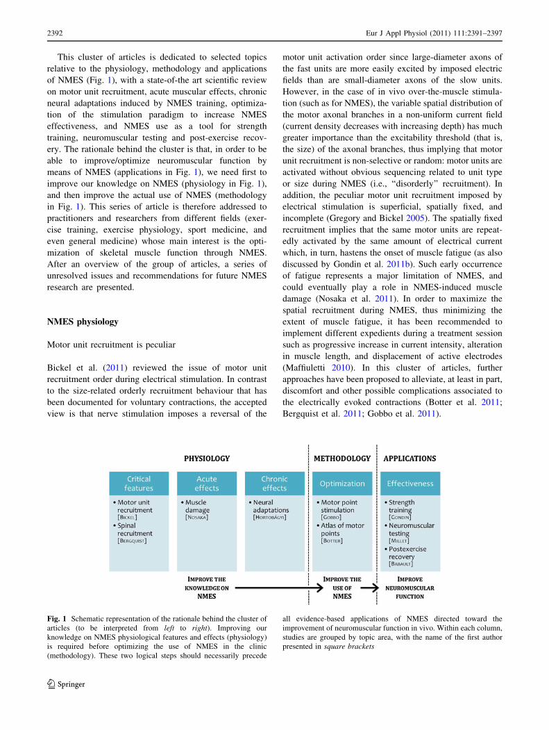
This cluster of articles is dedicated to selected topics
relative to the physiology, methodology and applications
of NMES (Fig. 1), with a state-of-the art scientific review
on motor unit recruitment, acute muscular effects, chronic
neural adaptations induced by NMES training, optimiza-
tion of the stimulation paradigm to increase NMES
effectiveness, and NMES use as a tool for strength
training, neuromuscular testing and post-exercise recov-
ery. The rationale behind the cluster is that, in order to be
able to improve/optimize neuromuscular function by
means of NMES (applications in Fig. 1), we need first to
improve our knowledge on NMES (physiology in Fig. 1),
and then improve the actual use of NMES (methodology
in Fig. 1). This series of article is therefore addressed to
practitioners and researchers from different fields (exer-
cise training, exercise physiology, sport medicine, and
even general medicine) whose main interest is the opti-
mization of skeletal muscle function through NMES.
After an overview of the group of articles, a series of
unresolved issues and recommendations for future NMES
research are presented.
NMES physiology
Motor unit recruitment is peculiar
Bickel et al. (2011) reviewed the issue of motor unit
recruitment order during electrical stimulation. In contrast
to the size-related orderly recruitment behaviour that has
been documented for voluntary contractions, the accepted
view is that nerve stimulation imposes a reversal of the
motor unit activation order since large-diameter axons of
the fast units are more easily excited by imposed electric
fields than are small-diameter axons of the slow units.
However, in the case of in vivo over-the-muscle stimula-
tion (such as for NMES), the variable spatial distribution of
the motor axonal branches in a non-uniform current field
(current density decreases with increasing depth) has much
greater importance than the excitability threshold (that is,
the size) of the axonal branches, thus implying that motor
unit recruitment is non-selective or random: motor units are
activated without obvious sequencing related to unit type
or size during NMES (i.e., ‘‘disorderly’’ recruitment). In
addition, the peculiar motor unit recruitment imposed by
electrical stimulation is superficial, spatially fixed, and
incomplete (Gregory and Bickel 2005). The spatially fixed
recruitment implies that the same motor units are repeat-
edly activated by the same amount of electrical current
which, in turn, hastens the onset of muscle fatigue (as also
discussed by Gondin et al. 2011b). Such early occurrence
of fatigue represents a major limitation of NMES, and
could eventually play a role in NMES-induced muscle
damage (Nosaka et al. 2011). In order to maximize the
spatial recruitment during NMES, thus minimizing the
extent of muscle fatigue, it has been recommended to
implement different expedients during a treatment session
such as progressive increase in current intensity, alteration
in muscle length, and displacement of active electrodes
(Maffiuletti 2010). In this cluster of articles, further
approaches have been proposed to alleviate, at least in part,
discomfort and other possible complications associated to
the electrically evoked contractions (Botter et al. 2011;
Bergquist et al. 2011; Gobbo et al. 2011).
Fig. 1 Schematic representation of the rationale behind the cluster of
articles (to be interpreted from left to right). Improving our
knowledge on NMES physiological features and effects (physiology)
is required before optimizing the use of NMES in the clinic
(methodology). These two logical steps should necessarily precede
all evidence-based applications of NMES directed toward the
improvement of neuromuscular function in vivo. Within each column,
studies are grouped by topic area, with the name of the first author
presented in square brackets
2392 Eur J Appl Physiol (2011) 111:2391–2397
123

Bergquist et al. (2011) provided an overview of how
peripheral (direct activation of motor axon branches) and
central (reflexive recruitment of spinal motor neurons by
the electrically evoked afferent volley) pathways contribute
to electrically evoked contractions and suggested that some
of the limitations of NMES (particularly discomfort and
random recruitment) could be minimized by increasing the
contribution through central pathways. In fact, motor unit
recruitment through central pathways may be more orderly,
less synchronous and more spatially diffuse throughout the
muscle, than recruitment through purely peripheral path-
ways. Enhancing central recruitment during NMES
requires the stimulation to be delivered at low pulse
amplitudes (to minimize the antidromic block, that is the
collision between the action potentials travelling anti-
dromically along motor axons and those generated fol-
lowing the reflexive recruitment of motor neurons), pulse
durations in the range 0.2–1 ms (to maximize the excita-
tion of afferent axons that present a longer strength-dura-
tion time constant and lower rheobase than motor axons),
stimulation train durations shorter than 2 s for stimulation
over the nerve and longer than 2 s for stimulation over the
muscle, and high (100 Hz) pulse frequencies (to increase
the rate at which the sensory volley is sent to the spinal
cord and supra-spinal centres). Based on these character-
istics of the stimulation burst, this stimulation paradigm has
been referred to as ‘‘wide-pulse high-frequency NMES’’.
Acute NMES use may cause profound muscle damage
Nosaka et al. (2011) reviewed the most recent evidence
available for the muscle damage induced by NMES-evoked
contractions, which resembles the type of damage pro-
duced by voluntary eccentric contractions, resulting in
decreased maximal voluntary contraction (MVC) strength,
increased circulating levels of muscle proteins and delayed
onset muscle soreness. Muscle damage profile following
NMES-evoked isometric contractions of the knee extensors
is similar between different stimulation paradigms (pulsed
vs. alternating current) for similar force outputs. Moreover,
its magnitude is reduced following a second session of
NMES performed two weeks after a first session: this
phenomenon, known as ‘‘repeated bout effect’’, has origi-
nally been described for voluntary eccentric contractions
and could be useful to limit the damage associated to
NMES if the training or rehabilitation program includes
‘‘conditioning’’ sessions.
Strength training by NMES leads to considerable neural
adaptations
Hortobagyi and Maffiuletti (2011) reviewed the changes in
the function of the central nervous system (neural
adaptations) that may occur with NMES strength training,
which include spinal and supraspinal mechanisms of
adaptation. Interestingly, the increases in MVC strength
induced by NMES training seem to be mainly mediated by
supraspinal rather than spinal changes, although no training
study has yet documented the former adaptations. The fact
that NMES and voluntary strength training may have dif-
ferent supraspinal effects in the contralateral homologous
muscle should be used as a key point motivating the
combination of the two forms of exercise in the context of
neuromuscular retraining. Somatosensory and nociceptive
inputs associated to NMES use may lead to changes in
motor cortical excitability, which in turn can cause func-
tional improvements to occur. Moreover NMES seems to
be able to modify the excitability of interhemispheric
connections and possibly the balance between interhemi-
spheric excitation and inhibition. These facts lead us to the
conjecture that NMES training can be viewed as a ‘‘neural’’
rather than a ‘‘muscular’’ treatment technique, particularly
for neurological patients.
NMES methodology
Motor point stimulation as a sine qua non-condition
to optimize NMES use
Gobbo et al. (2011) provided convincing evidence on the
importance of motor point determination to minimize
current intensity and discomfort, and to maximize the
muscular tension evoked by NMES. They also used near
infrared spectroscopy, which is a technique particularly
suitable for investigating local metabolic changes of
stimulated muscles (Muthalib et al. 2009), and were able to
demonstrate that, besides contractile activity, muscle oxy-
gen consumption and hyperaemia were significantly
increased when motor point location was carefully deter-
mined with a pen electrode rather than being inferred from
several legally marketed motor point charts. We recom-
mend that the procedure described by Gobbo et al. (2011),
which is completed in less than a minute, should be con-
sistently incorporated in both clinical and research contexts
to optimize NMES application.
Botter et al. (2011) investigated the uniformity of the
muscle motor point location in the lower limb of healthy
subjects and found different motor points innervating dif-
ferent portions of the quadriceps, posterior thigh, and tib-
ialis anterior muscles. On this basis, they suggested that a
maximization of the spatial recruitment during NMES
could also be obtained through a multi-channel stimula-
tion technique that involves a non-synchronous activation
of different muscle volumes. Interestingly, it has recently
been demonstrated that asynchronous low-frequency
Eur J Appl Physiol (2011) 111:2391–2397 2393
123
(16 Hz) stimulation of the quadriceps muscle using a
multi-pad electrode (four channels) can elicit a strong,
fused contraction, though producing less muscle fatigue
compared to single-channel high-frequency (30 Hz) stim-
ulation (Malesevic et al. 2010).
NMES applications
NMES for optimizing muscle function: is that really
useful?
Gondin et al. (2011b) provided an overview of the main
training studies in which NMES has been applied to
healthy subjects or even to competitive athletes with the
objective to improve muscle function. As already pointed
out in a recent review published in this journal (Maffiuletti
2010), the so-called training intensity (i.e., the level of
force evoked by NMES, expressed as a fraction of the
MVC force) seems to be the main determinant of NMES
training effectiveness. In other words, muscles should
produce the greatest relative tension when stimulated by
NMES in order to maximize the training-induced strength
gains (Lai et al. 1988; Selkowitz 1985). Interestingly, we
plotted the MVC strength gains collected from different
NMES training studies completed in our laboratory against
respective training intensities, and confirmed that a dose-
response relation does exist between these two variables
(Maffiuletti NA, unpublished observations). This would
imply that the improvement in MVC strength induced by
NMES training could be predicted from the average
training intensity; for example, a training intensity of 40%
MVC being predicted to improve MVC strength by 20%.
Such predictive equation may even be improved (and
eventually extended to unhealthy populations) by adding
the initial MVC strength and NMES training volume as
independent variables.
Millet et al. (2011) presented the potential interests and
fundamentals of electrical stimulation for the evaluation of
voluntary activation, muscle contractility (with and without
fatigue), as well as respiratory muscle function. Contrary to
MVC assessment, contractions evoked by electrical stim-
ulation are not or little influenced by motivational factors
and can be easily standardized; however, many researchers
still adopt electrical stimulation in a simplistic way, with
scarce or no attention to physiological and methodological
issues that may invalidate the test. The heterogeneity of
electrical stimulation procedures already discussed for
NMES training also applies to the evaluation of neuro-
muscular features, as different laboratories adopt different
stimulation units, current characteristics, electrode arrange-
ments, etc. We believe the increasing use of magnetic
stimulation would necessarily reduce such heterogeneity
even though, as discussed by Millet et al. (2011), the
maximal power output of modern magnetic stimulators
does not seem appropriate enough for subjects with excess
subcutaneous adipose tissue.
Babault et al. (2011) reviewed the most recent evidence
available for the effectiveness of non-tetanic low-intensity
NMES as a post-exercise recovery tool. The idea that this
exercise modality may increase blood flow thus favouring
metabolite washout is appealing and at least in part con-
ceivable. It is however hard to imagine how this would
accelerate the time course of post-exercise recovery in
sportsmen, as muscle recruitment with electrical stimula-
tion is extremely limited and superficial. Moreover, none of
the most common recovery strategies (such as massage,
active exercise and stretching) have proved effective in
accelerating recovery kinetics in sportsmen (Barnett 2006),
so perhaps the question we should ask ourselves is ‘‘Aren’t
we wasting our time?’’. This is a clear example of how
commercial claims (http://www.shopcompex.com/testimon
ials) often go beyond scientific evidence. Regarding the
management of exercise-induced pain and psychological
benefits for recovery, additional evidence should be pro-
vided before considering NMES as a valid tool for post-
exercise recovery.
Unresolved physiological and clinical issues, and future
directions
The purpose of this section is to outline elements of con-
fusion and controversy that still plague the use of NMES as
a testing and training modality in healthy and pathological
subjects, and to examine some limitations in our under-
standing of NMES effects. With such an approach, we aim
to provide a conceptual foundation for subsequent research
studies in this area, but also for further implementation of
NMES in the clinic. The same logical progression outlined
in Fig. 1 is followed (from physiology to methodology to
applications).
Critical features
The ‘‘wide-pulse high-frequency NMES’’ modality remains
to be explored more in depth: for example, it is presently not
defined whether inter-individual variability exists for the
generation of the electrically elicited ‘‘extra’’ force that
results from the reflexive recruitment of motor neurons and
could also be amplified by an involuntary descending drive
(Frigon et al. 2011). In addition, it has never been demon-
strated that a stimulation paradigm aimed to produce a
reflexive recruitment of motor neurons rather than to max-
imize the muscle tension can trigger positive neural and/or
muscular adaptations favourable to muscle performance.
2394 Eur J Appl Physiol (2011) 111:2391–2397
123
As soon as the effectiveness of reflexive motor neuron
recruitment can be demonstrated in controlled physiological
studies, clinical investigations will be required to assess the
feasibility of this new stimulation paradigm in clinical
settings.
Acute effects
Mackey et al. (2011) recently showed that NMES-induced
damage of the human medial gastrocnemius muscle results
in de-adhesive, disassembly, and disorganization responses
in the contractile connective tissue, followed by a delayed
anabolic response leading to extracellular matrix remod-
elling. Besides these responses, the other mechanisms
underlying NMES-induced muscle damage remain to be
clarified. In particular, it remains to be established whether
the damage provoked by NMES is specific for this type of
stimulus or whether it resembles that produced by length-
ening contractions, which represents the traditional exper-
imental model for the study of muscle damage and
regeneration. In addition, it remains to be elucidated
whether the training effects of NMES on muscle and neural
plasticity are reduced when muscle damage is minimized.
In fact, it may be hypothesized that some damage is nec-
essary to maximize muscle hypertrophy and strength gains
that could be achieved by NMES.
Guarascio et al. (2004) reported a case of rhabdomyol-
ysis in a young male subject presenting severe asthenia and
increased levels of circulating muscle proteins induced by
excessive NMES use. Even if routine measurements of
circulating muscle proteins such as creatine kinase and
myoglobin are not required during NMES training pro-
grams, it should be recommended to perform pre-training
measurements and assessment of thyroid and kidney
function (to rule out, at least in pathological subjects, the
existence of an overt/subclinical hypothyroidism and renal
insufficiency that may increase the risk of rhabdomyolysis)
so as to facilitate the evaluation of subsequent muscle
complaints. In addition, special attention is required if
individuals subjected to NMES are under therapy with
drugs that may produce muscle complaints (i.e., statins) or
in the case of particular patient populations (e.g., critically
ill patients). Finally, all subjects performing repeated ses-
sions of NMES should be warned to stop the training and to
contact their physician if they become symptomatic for
severe muscle weakness and myalgias and notice dark
discoloration of their urine.
Chronic effects
Strength training by NMES does promote neural and mus-
cular adaptations that are complementary to the well-known
effects of voluntary resistance training (Vanderthommen
and Duchateau 2007). Future efforts should be directed at
investigating more precisely the locus/loci of neural adap-
tations induced by NMES; in particular, supraspinal
mechanisms/sites such as cortical excitability and cortical
maps could be explored using transcranial magnetic stim-
ulation and functional magnetic resonance imaging, tech-
niques that are nowadays relatively accessible. The
determinants of phenotypic variability in response to NMES
strength training also require greater attention (Gondin et al.
2011a), with particular emphasis on factors that should be
controlled, such as training intensity. Once the physiologi-
cal mechanisms underlying neural and/or muscular changes
to NMES training are substantiated by research evidence,
one or both forms of adaptation (or even specific mecha-
nisms) could be specifically targeted according to the
individual needs of the end user. This would necessarily
result in a more scientific and profitable use of NMES in
clinical settings.
Optimization
The two main limitations associated to the use of NMES
are the considerable discomfort and the relatively incom-
plete muscle recruitment, both imposed by the electrical
field. Alternative techniques, stratagems and tools that
might be able to minimize the impact of these limitations
on NMES use should receive more attention. For example,
the use of ‘‘distributed’’ NMES (Malesevic et al. 2010),
multi-path stimulation with large electrodes (Feil et al.
2011), and magnetic stimulation (Bustamante et al. 2010)
should be encouraged as opposed to the classical NMES
set-up, particularly for commonly stimulated large mus-
cles. Additionally, a world-wide consensus on standardi-
zation of NMES terminology, which should include current
and contraction characteristics, is considered necessary to
allow a more uniform use of NMES both in research and in
clinical settings.
Effectiveness
With regard to strength training, although NMES was
originally introduced to treat muscle atrophy consequent to
immobilization or denervation (Jackson and Seddon 1945),
its use has traditionally been confined to exercise physi-
ology, particularly for research purposes, while the clinical
use of NMES has been relatively limited. As a result of
this, the origin (neural vs. muscular) and the time course of
adaptations to NMES strength training have been exten-
sively investigated in healthy but not in pathological pop-
ulations. It is now time to fill this gap! The experience
gained from exercise physiology studies should now be
redirected to pathological applications; good examples
Eur J Appl Physiol (2011) 111:2391–2397 2395
123
come from cardiorespiratory and internal care medicine
where chronic obstructive pulmonary disease patients and
critically ill patients are increasingly stimulated to preserve
their muscle mass and function (Vivodtzev et al. 2008;
Routsi et al. 2010). Additional efforts are needed to iden-
tify the most relevant applications of NMES training in the
clinical setting, and also to discern its effectiveness and the
time-course of neuromuscular adaptations for specific
patient populations.
As far as muscle testing is concerned, future studies
using in vitro stimulation patterns that are closer to the in
vivo situation are required to further validate the existing
methodologies (such as the twitch interpolation technique,
see Place et al. 2008) or to conceive new ones. It would
also be worth validating a quick, simple, standardized, and
comprehensive testing procedure for the assessment of in
vivo neuromuscular function in clinical settings, whose
parameters could be easily manipulated according to
patient characteristics and prefixed goals. This would
provide a clear picture of the central (muscle activation)
and peripheral (muscle contractility) components of muscle
function, and certainly minimize the heterogeneity of in
vivo neuromuscular evaluation between different centres.
Regarding post-exercise recovery optimization, the
focus of future research studies should definitely be on
acute physiological effects of non-tetanic low-intensity
NMES (e.g., increased blood flow and metabolic altera-
tions) rather than on global physical outcomes. Since there
is no scientific evidence that this form of NMES could
provide some physiological benefits for sportsmen, its
application cannot currently be recommended to enhance
post-exercise recovery, at least from a physiological
perspective.
Acknowledgments The work reported in this article was supported
by bank foundation ‘‘Compagnia di San Paolo’’ (Project ‘‘Neuro-
muscular Investigation and Conditioning in Endocrine Myopathy’’)
(MAM), by the ERC grant DEMOVE and the Bernstein Focus
Neurotechnology Gottingen (DF), and by MYOAGE Grant
HEALTH-F2-2009-223576 (‘‘Understanding and combating age-
related muscle weakness’’) (RB). The authors are grateful to
Prof. R. Merletti (LISiN, Politecnico di Torino, Italy) for his careful
review of the final version of the manuscript.
References
Babault N, Cometti C, Maffiuletti NA, Deley G (2011) Does electrical
stimulation enhance post-exercise performance recovery? Eur J
Appl Physiol. doi:10.1007/s00421-011-2117-7
Barnett A (2006) Using recovery modalities between training sessions
in elite athletes: does it help? Sports Med 36:781–796
Bergquist AJ, Clair JM, Lagerquist O, Mang CS, Okuma Y, Collins
DF (2011) Neuromuscular electrical stimulation: implications of
the electrically-evoked sensory volley. Eur J Appl Physiol. doi:
10.1007/s00421-011-2087-9
Bickel SC, Gregory CM, Dean JC (2011) Motor unit recruitment
during neuromuscular electrical stimulation: a critical appraisal.
Eur J Appl Physiol (in press)
Botter A, Oprandi G, Lanfranco F, Allasia S, Maffiuletti NA, Minetto
MA (2011) Atlas of the muscle motor points for the lower limb:
implications for electrical stimulation procedures and electrode
positioning. Eur J Appl Physiol. doi:10.1007/s00421-011-2093-y
Bustamante V, Lopez de Santa Maria E, Gorostiza A, Jimenez U,
Galdiz JB (2010) Muscle training with repetitive magnetic
stimulation of the quadriceps in severe COPD patients. Respir
Med 104:237–245
Feil S, Newell J, Minogue C, Paessler HH (2011) The effectiveness of
supplementing a standard rehabilitation program with superim-
posed neuromuscular electrical stimulation after anterior cruciate
ligament reconstruction: a prospective, randomized, single-blind
study. Am J Sports Med 39:1238–1247
Frigon A, Thompson CK, Johnson MD, Manuel M, Hornby TG,
Heckman CJ (2011) Extra forces evoked during electrical
stimulation of the muscle or its nerve are generated and
modulated by a length-dependent intrinsic property of muscle
in humans and cats. J Neurosci 31:5579–5588
Gobbo M, Gaffurini P, Bissolotti L, Esposito F, Orizio C (2011)
Trascutaneous neuromuscular electrical stimulation: influence of
electrode positioning and stimulus amplitude settings on muscle
response. Eur J Appl Physiol. doi:10.1007/s00421-011-2047-4
Gondin J, Brocca L, Bellinzona E, D’Antona G, Maffiuletti NA,
Miotti D, Pellegrino MA, Bottinelli R (2011a) Neuromuscular
electrical stimulation training induces atypical adaptations of the
human skeletal muscle phenotype: a functional and proteomic
analysis. J Appl Physiol 110:433–450
Gondin J, Cozzone PJ, Bendahan D (2011b) Is high frequency
neuromuscular electrical stimulation a suitable tool for muscle
performance improvement in both healthy humans and athletes?
Eur J Appl Physiol. doi:10.1007/s00421-011-2101-2
Gregory CM, Bickel CS (2005) Recruitment patterns in human
skeletal muscle during electrical stimulation. Phys Ther
85:358–364
Guarascio P, Lusi EA, Soccorsi F (2004) Electronic muscular
stimulators: a novel unsuspected cause of rhabdomyolysis. Br J
Sports Med 38:505
Hortobagyi T, Maffiuletti NA (2011) Neural adaptations to electrical
stimulation strength training. Eur J Appl Physiol. doi:
10.1007/s00421-011-2012-2
Jackson EC, Seddon HJ (1945) Galvanism and denervated muscle
atrophy. Br Med J 2:485–486
Lai HS, De Domenico G, Strauss GR (1988) The effect of different
electro-motor stimulation training intensities on strength
improvement. Aust J Physiother 34:151–164
Mackey AL, Brandstetter S, Schjerling P, Bojsen-Moller J, Qvortrup
K, Pedersen MM, Doessing S, Kjaer M, Magnusson SP,
Langberg H (2011) Sequenced response of extracellular matrix
deadhesion and fibrotic regulators after muscle damage is
involved in protection against future injury in human skeletal
muscle. FASEB J 25:1943–1959
Maffiuletti NA (2010) Physiological and methodological consider-
ations for the use of neuromuscular electrical stimulation. Eur J
Appl Physiol 110:223–234
Malesevic NM, Popovic LZ, Schwirtlich L, Popovic DB (2010)
Distributed low-frequency functional electrical stimulation
delays muscle fatigue compared to conventional stimulation.
Muscle Nerve 42:556–562
Millet GY, Martin V, Martin A, Verges S (2011) Electrical
stimulation for testing neuromuscular function: from sport to
pathology. Eur J Appl Physiol. doi:10.1007/s00421-011-1996-y
Muthalib M, Jubeau M, Millet GY, Maffiuletti NA, Nosaka K (2009)
Comparison between electrically evoked and voluntary isometric
2396 Eur J Appl Physiol (2011) 111:2391–2397
123
contractions for biceps brachii muscle oxidative metabolism
using near-infrared spectroscopy. Eur J Appl Physiol 107:
235–241
Nosaka K, Aldayel A, Jubeau M, Chen TC (2011) Muscle damage
induced by electrical stimulation. Eur J Appl Physiol. doi:
10.1007/s004021-011-2086-x
Place N, Yamada T, Bruton JD, Westerblad H (2008) Interpolated
twitches in fatiguing single mouse muscle fibres: implications
for the assessment of central fatigue. J Physiol 586:2799–2805
Routsi C, Gerovasili V, Vasileiadis I, Karatzanos E, Pitsolis T,
Tripodaki E, Markaki V, Zervakis D, Nanas S (2010) Electrical
muscle stimulation prevents critical illness polyneuromyopathy:
a randomized parallel intervention trial. Crit Care 14:R74
Selkowitz DM (1985) Improvement in isometric strength of the
quadriceps femoris muscle after training with electrical stimu-
lation. Phys Ther 65:186–196
Vanderthommen M, Duchateau J (2007) Electrical stimulation as a
modality to improve performance of the neuromuscular system.
Exerc Sport Sci Rev 35:180–185
Vivodtzev I, Lacasse Y, Maltais F (2008) Neuromuscular electrical
stimulation of the lower limbs in patients with chronic obstruc-
tive pulmonary disease. J Cardiopulm Rehabil Prev 28:79–91
Eur J Appl Physiol (2011) 111:2391–2397 2397
123