eimd module 1 copy
TRANSCRIPT
Dr Claire Minshull
E-mail: [email protected] Twitter: @getbacktosport
Web: www.getbacktosport.com
Training-Induced Muscle Damage & DOMS;
What; why & how to use it
Module 1 © Get Back To Sport 2016
1
Welcome!
Training-Induced Muscle Damage & DOMS;What; why & how to use it
Welcome to this online course:
From this point onwards we’re going to use the ‘proper’ term of Exercise-Induced Muscle Damage. It’s essentially the same thing as that induced by training, however, this is the most widely accepted term.
In this course you will learn:- what what muscle damage actually is, - how muscle damage occurs, - what the consequences of muscle damage are to performance - if and how you can manage muscle damage - how to use muscle damage to our advantage for injury prevention and to
maximise training gains
2
Scheme of workMODULE FOCUS AIMS
MODULE 1 Muscle Structure & Function - Recap Recap of the structure & function of skeletal muscle; the functional unit of skeletal muscle (sarcomere); sliding filament theory & types of muscle contraction/action
MODULE 2 Types and properties of contractile & non-contractile tissue
Properties of contractile & non-contractile tissue; Role of connective tissue in muscle force transmission; Loading patterns of contractile & non-contractile tissue during concentric and eccentric muscle actions
MODULE 3 Exercise-induced muscle damage (EIMD); mechanisms, processes & effects on performance
What exercise-induced muscle damage (EIMD) is and isn't. How does muscle damage happen and what are the effects on muscle function & physical performance; symptoms & causes of EIMD, including DOMS; length-tension relationship of muscle
MODULE 4 Recovery from EIMD The time course of recovery of performance and symptoms of EIMD; muscle recovery & adaptation following EIMD; the repeated-bout effect
MODULE 5 Management and utility of EIMD in exercise, training & injury prevention
Myth-busting: common strategies for reducing DOMS & EIMD; how to use eccentric exercise & EIMD to benefit training & injury prevention
3
MODULE 1:Muscle Structure & Function - Recap
In this module we will cover:
- Structure & function of skeletal muscle- The functional unit of skeletal muscle (sarcomere)- Sliding filament theory & types of muscle contractions
4

Myology is the scientific study of Muscles
MYO = muscle &
OLOGY = study
The study of muscle structure and function
What is Myology?
www.funnieststuffover.com
5
Smooth:Involuntary muscle; controlled unconsciouslyIn the walls of blood vessels and internal organs
Cardiac: Controls itself with help from nervous and endocrine systemsOnly in the heart
Skeletal Voluntary muscle; controlled consciouslyOver 600 throughout the body
Types of Muscle:
Focus of this
course!
6
Quick Quiz…
Here’s a quick quiz before we start…
Skeletal muscle tissue has many functions. Take a few minutes and list as many as you can think of before you go
to the next slide …
7
Answer:The Functions of Muscle Include:
How did you do?Pretty easy?
Ever wondered why you shiver…?
The repeated muscle contractions generate small amounts of heat
• Movement
• Posture
• Support of soft tissues (e.g. ligaments)
• Guard entrances and exits (sphincters)
• Body temperature (contraction of muscle results in production of heat; the body is only 25% efficient, therefore you lose approximately 75% of energy as heat)
• Absorb stress to protect joints
• Act as a protein reserve
• Act as muscle pump to help venous return
8
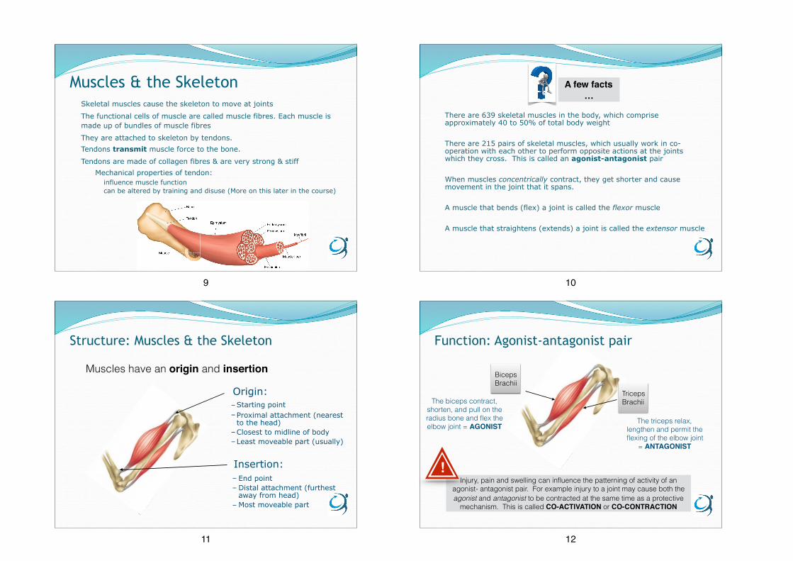
Muscles & the SkeletonSkeletal muscles cause the skeleton to move at joints
The functional cells of muscle are called muscle fibres. Each muscle is made up of bundles of muscle fibres
They are attached to skeleton by tendons. Tendons transmit muscle force to the bone.
Tendons are made of collagen fibres & are very strong & stiff Mechanical properties of tendon:
influence muscle function can be altered by training and disuse (More on this later in the course)
9
There are 639 skeletal muscles in the body, which comprise approximately 40 to 50% of total body weight
There are 215 pairs of skeletal muscles, which usually work in co-operation with each other to perform opposite actions at the joints which they cross. This is called an agonist-antagonist pair
When muscles concentrically contract, they get shorter and cause movement in the joint that it spans.
A muscle that bends (flex) a joint is called the flexor muscle
A muscle that straightens (extends) a joint is called the extensor muscle
A few facts…
10
Origin: –Starting point –Proximal attachment (nearest
to the head) –Closest to midline of body –Least moveable part (usually)
Insertion: – End point – Distal attachment (furthest
away from head) – Most moveable part
Structure: Muscles & the Skeleton
Muscles have an origin and insertion
11
Biceps Brachii
Triceps BrachiiThe biceps contract,
shorten, and pull on the radius bone and flex the elbow joint = AGONIST
The triceps relax, lengthen and permit the flexing of the elbow joint
= ANTAGONIST
Injury, pain and swelling can influence the patterning of activity of an agonist- antagonist pair. For example injury to a joint may cause both the agonist and antagonist to be contracted at the same time as a protective
mechanism. This is called CO-ACTIVATION or CO-CONTRACTION
!
Function: Agonist-antagonist pair
12

Stabilisers
– surround joint or body part – contract to fixate or stabilise the area to
enable another limb or body segment to exert force & move
– also known as fixators – essential in establishing a relatively firm base
for the more distal joints to work from when carrying out movements
e.g. supraspinatus helps to stabilise the shoulder joint by keeping the head of the humerus firmly pressed medially against the glenoid fossa of the scapula
Function: Role of Muscles
Fig. 409. Gray, Henry. 1918. Anatomy of the Human Body.
13
Synergist
– assist in action of agonists – not necessarily prime movers for the action – known as guiding muscles – assist in refined movement & rule out
undesired motions
e.g. Teres major is called “the latissimus dorsi’s little helper” (sometimes referred to “handcuff muscles” – since action collectively bring arms into the arresting position..!)
Function: Role of Muscles
en.wikipedia.org. Gray, Henry. 1918. Anatomy of the Human Body.
14
A muscle that accomplishes a certain movement.
A muscle acting in opposition to an agonist.
A muscle or group of muscles that assist theagonist to produce a movement.
Muscles that hold one bone in place relative to the body while a more distal bone is moved.
Stabiliser/
Fixator
Antagonist
Agonist
Synergist
Recap:
15
Function: Muscle Size
Longer muscles can contract over a greater distance and develop higher velocities of shortening (i.e quicker speed of contraction)
Muscles with long tendons can form pulley arrangements that allow large external movement (e.g., grasping by the fingers) with relatively small movement of the muscles and tendons
A few facts…
Thicker muscles with a larger cross-sectional area can produce greater amounts of force
Hence why big muscles gives the
appearance of strength!
(There is some limitation to this relationship, however)
vs.
16
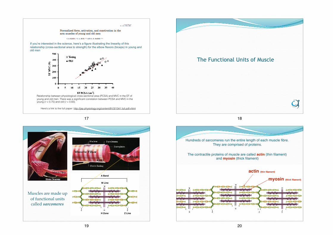
If you’re interested in the science, here’s a figure illustrating the linearity of this relationship (cross-sectional area to strength) for the elbow flexors (biceps) in young and old men
Relationship between physiological cross-sectional area (PCSA) and MVC in the EF of young and old men. There was a significant correlation between PCSA and MVC in the young (r = 0.75) and old (r = 0.83).
Here’s a link to the full paper: http://jap.physiology.org/content/91/3/1341.full.pdf+html
17
The Functional Units of Muscle
18
Muscles are made up of functional units called sarcomeres
M Line
H Zone Z Line
A Band
19
The contractile proteins of muscle are called actin (thin filament) and myosin (thick filament)
Hundreds of sarcomeres run the entire length of each muscle fibre. They are comprised of proteins.
actin (thin filament)
myosin (thick filament)
20

The sliding filament theory of muscle contraction explains how these muscle proteins slide past each other to generate movement
Okay, we’re going to skip over a lot of the biochemical parts of muscle activation in favour of making sure we understand how the actin and myosin filaments interact with each other during a contraction
1 A nervous impulse arrives at the neuromuscular junction, which eventually causes calcium (Ca+) to be released, which is essential to muscle activation.
2 Ca+ causes a few changes in the muscle cell that importantly enables Myosin to attach to the Actin. This attachment is called a cross-bridge.
3 The breakdown of ATP releases energy which enables the Myosin to pull the Actin filaments inwards and so shortening the muscle. This occurs along the entire length of every myofibril in the muscle cell.
actin myosin
Understanding the sliding filament theory is really important to be able to distinguish
between concentric and eccentric muscle contractions
This also provides the foundation to the
understanding of how muscle damage occurs
21
4 The Myosin detaches from the Actin and the cross-bridge is broken when an ATP molecule binds to the Myosin head. When the ATP is then broken down the Myosin head can again attach to an Actin binding site further along the Actin filament and repeat the 'power stroke'. This repeated pulling of the Actin over the myosin is often known as the ratchet mechanism.
5 This process of muscular contraction can last for as long as there is adequate ATP and Ca+ stores (and as long as there’s a nervous impulse)
A single power stroke of the myosin heads results in only a shortening of approximately 1% of the entire muscle.
Therefore to achieve a significant shortening the process must be repeated many times.
actin myosin
22
https://youtu.be/7O_ZHyPeIIA
Watch this brilliant animation of muscle contraction on YouTube. There’s a little more detailed than we’ve covered here, but the animation should really help to bring the theory to life! It’s important to understand this when we come on to eccentric activation and muscle damage
23
Types of muscle contraction (action)*
24

Types of muscle contraction (action)*How’s it going so far? Has it been an easy refresher, or a tough grind? Bear the sliding filament theory in mind, we’re going to come back to it in a second. First let’s just remind ourselves of the types of muscle contraction (action):
• Isometric: No change in muscle length
• Isotonic: Change in length
– Concentric: – muscle length shortens whilst developing force
– Eccentric: – muscle length increases whilst developing force*
• Isokinetic: Angular speed is constant during contraction
– Concentric – Eccentric
* From now on, we’re going to avoid the term ‘contraction’ as much as possible because the muscle lengthens under eccentric conditions, yet
contraction implies a shortening - it doesn’t make sense!!!
25
Muscle Action/contraction (under tension)
Isometric Isotonic
EccentricConcentric
Knee Extension: Controlled by concentric activation of knee extensors (quadriceps)
Knee Flexion: Controlled by eccentric activation of knee extensors (quadriceps)
26
Types of muscle contraction (action)*
So, here’s a challenge for you…
We understand the sliding filament theory, which involves a concentric contraction , or action(!) and a shortening of the muscle …
…can you use the sliding filament theory to describe eccentric muscle actions?? What happens to the filaments, how is force produced?
Pause for a while and spend a moment trying to describe this, write it down if you need to, before you advance to the next slide.
…
27
Okay, so in an eccentric action (or ‘contraction’) the muscle is lengthening whilst developing force, right?
Eccentric activation of the quadriceps muscles above is needed to control the fall of the weight at the foot under the effects of gravity.
The tension generated by the quadriceps is insufficient to overcome the external load on the muscle and the muscle fibres lengthen as they ‘contract’.
What does this mean…?
…Eccentric
Knee Flexion: Controlled by eccentric activation of knee extensors (quadriceps)
Ever wondered why these
exercises are called
‘negatives’? Next slide…
We’ll cover this in more detail in
Module 3
The myosin heads are thus are forcefully detached from the actin as the muscle is being ‘pulled’ and lengthened under load. They myosin heads re-attach, but the process of forced detachment is repeated
28
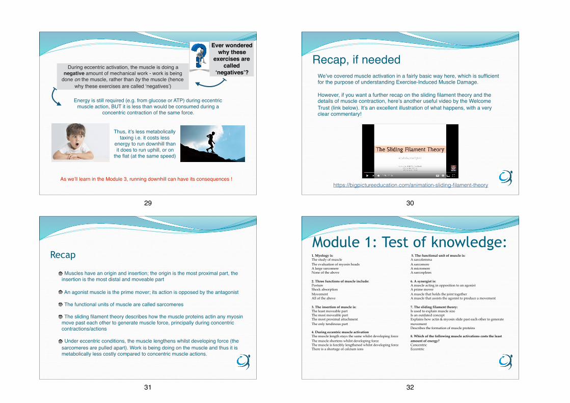
During eccentric activation, the muscle is doing a negative amount of mechanical work - work is being
done on the muscle, rather than by the muscle (hence why these exercises are called ‘negatives’)
Thus, it’s less metabolically taxing i.e. it costs less
energy to run downhill than it does to run uphill, or on
the flat (at the same speed)
As we’ll learn in the Module 3, running downhill can have its consequences !
Ever wondered why these
exercises are called
‘negatives’?
Energy is still required (e.g. from glucose or ATP) during eccentric muscle action, BUT it is less than would be consumed during a
concentric contraction of the same force.
29
Recap, if needed
https://bigpictureeducation.com/animation-sliding-filament-theory
We’ve covered muscle activation in a fairly basic way here, which is sufficient for the purpose of understanding Exercise-Induced Muscle Damage.
However, if you want a further recap on the sliding filament theory and the details of muscle contraction, here’s another useful video by the Welcome Trust (link below). It’s an excellent illustration of what happens, with a very clear commentary!
30
Recap
Muscles have an origin and insertion; the origin is the most proximal part, the insertion is the most distal and moveable part
An agonist muscle is the prime mover; its action is opposed by the antagonist
The functional units of muscle are called sarcomeres
The sliding filament theory describes how the muscle proteins actin any myosin move past each other to generate muscle force, principally during concentric contractions/actions
Under eccentric conditions, the muscle lengthens whilst developing force (the sarcomeres are pulled apart). Work is being doing on the muscle and thus it is metabolically less costly compared to concentric muscle actions.
31
Module 1: Test of knowledge: 1. Myology is: The study of muscleThe evaluation of myosin headsA large sarcomereNone of the above 2. Three functions of muscle include:PostureShock absorptionMovementAll of the above 3. The insertion of muscle is: The least moveable partThe most moveable partThe most proximal attachmentThe only tendinous part 4. During eccentric muscle activation The muscle length stays the same whilst developing forceThe muscle shortens whilst developing forceThe muscle is forcibly lengthened whilst developing forceThere is a shortage of calcium ions
5. The functional unit of muscle is:A sarcolemmaA sarcomereA micromereA sarcospleen
6. A synergist is:A muscle acting in opposition to an agonistA prime moverA muscle that holds the joint togetherA muscle that assists the agonist to produce a movement
7. The sliding filament theory:Is used to explain muscle sizeIs an outdated conceptExplains how actin & myosin slide past each other to generate movementDescribes the formation of muscle proteins
8. Which of the following muscle activations costs the least amount of energy?ConcentricEccentric
32