efficient genome editing of human natural killer cells by ... · t cell recognizing tumor antigens...
TRANSCRIPT

Efficient genome editing of human natural killer cells by CRISPR RNP Jai Rautela1,2*, Elliot Surgenor1,2, Nicholas D. Huntington1,2*
1 Molecular Immunology Division, Walter and Eliza Hall Institute of Medical Research.
Parkville, Victoria 3052, Australia
2 Department of Medical Biology, Faculty of Medicine, Dentistry and Health Sciences,
University of Melbourne, Parkville, Victoria 3010, Australia
* Corresponding Authors
Key words: natural killer cell, CRISPR, immunotherapy, drug target validation, human
immunology
Corresponding author contact details:
Prof. Nicholas Huntington
Dr. Jai Rautela
Abstract The ability to genetically modify CD8 T cells using viral gene delivery has facilitated the
development of next generation of cancer immunotherapies such as chimeric antigen receptor
(CAR) T cells engineered to specifically kill tumor cells. Development of immunotherapies
targeting natural killer (NK) cells have stalled in part by their resistance to viral gene delivery.
Here, we describe an efficient approach to genetically edit human NK cells by electroporation
and CRISPR-Cas9 ribonucleoprotein (RNP) complexes. We detail electroporation pulse
codes and buffer optimization for protein uptake by human NK cells and viability, and the
efficiency of this approach over other methods. To highlight the transformative step this
technique will have for NK cell immunotherapy drug discovery, we deleted NKp46 and CIS in
primary human NK cells and validated murine findings on their key roles in regulating NK cell
anti-tumor function.
.CC-BY-NC-ND 4.0 International licensewas not certified by peer review) is the author/funder. It is made available under aThe copyright holder for this preprint (whichthis version posted September 6, 2018. . https://doi.org/10.1101/406934doi: bioRxiv preprint

Introduction Immunotherapies are emerging treatment options for several cancer types with CD8 T
cells being the primary therapeutic target. The efficiency of viral gene delivery to activated
CD8 T cells in vitro has facilitated the clinical development of chimeric antigen receptor (CAR)
T cell recognizing tumor antigens such as CD19 on B cell leukemia and have recently been
approved by regulatory agencies. Natural killer (NK) cells are an additional anti-tumor effector
population with a clear role in limiting tumor metastasis1, 2 and an emerging role in
orchestrating inflammation and immune infiltration within solid tumors3, 4. NK cells are
dependent on the cytokine IL-15 for their development and maturation5, and are educated in
the periphery through the signals transduced through an array of germline activating and
inhibitory receptors6. In addition to numerous soluble factors, it is the balance of signals
transduced through these receptors that also dictates whether an NK cell engages in a
cytotoxic attack against a target cell. We previously described CIS (encoded by Cish) as a
potent checkpoint in NK cell activation by limiting IL-15 signal transduction7, while CBL-B
(Cblb) has also been proposed as an intracellular repressor of NK cell activation8. In addition,
the group of Mantovani described IL-1R8 (Il1r8) acting as a negative regulator of IL-18
responsiveness in NK cells9 while more recently, TIGIT (Tigit) was shown to be a major
inhibitory receptor on NK cells10 with deletion of Cish, Cblb, Il1r8 or Tigit resulting in
dramatically improved NK cell-dependent anti-tumor immunity.
These pre-clinical findings highlight that NK cell anti-tumor immunity can be
augmented in vivo and as such, development of NK cell immunotherapy drugs that target such
pathways should be investigated. In contrast to CD8 T cells, NK cells are yet to be specifically
or efficiently targeted by immunotherapy drugs in human cancer6, 11. A major hurdle to NK cell
drug discovery has been target validation in human NK cells and this is indeed an important
step given the clear disparities in NK cell phenotypes and frequencies between species. The
field has therefore awaited the development of rapid and efficient gene-editing technologies
such as CRISPR-Cas9 in order to perform targeted gene-editing to understand the biology on
primary human NK cells. Since the initial description of CRISPR-Cas9 in mammalian cells,
this technology has been used to great effect to edit genes in several immune cell lines and
more recently, primary human T cells12, 13, 14. Editing immune cell lines has been relatively
simple through lentiviral delivery of Cas9 and a gene-specific guide RNA, and has allowed
genome-wide screening studies to be performed15. The efficacy of using similar viral delivery
systems in primary immune cells has been highly cell-type specific. NK cells are notoriously
difficult to infect with retro/lentiviral particles meaning that tried and tested genetic modification
approaches for human T cells are not appropriate for human NK cells. For example, while IL-
2 or IL-15 results in robust activation and proliferation of human NK cells, typically only around
.CC-BY-NC-ND 4.0 International licensewas not certified by peer review) is the author/funder. It is made available under aThe copyright holder for this preprint (whichthis version posted September 6, 2018. . https://doi.org/10.1101/406934doi: bioRxiv preprint

2-3% of these cells will be infected with a lentivirus. When lentiviral multiplicity of infection
(MOI) was increased from 1 to 20, the efficiency of lentivirus transduction on human NK92
cells was unchanged16, while other groups have increased the MOI to 150 and still only
achieve ~10% transduction of human NK cells17. Electroporation of NK cells with mRNA or
plasmid DNA is efficient for transient expression (such as anti-CD19 CAR) yet there is no
evidence that transient Cas9 and sgRNA expression can lead to efficient gene editing in NK
cells17, 18. While viral delivery of the Cas9 system to primary cells is the most utilized approach
at present, delivery of recombinant Cas9 in complex with a gene-specific gRNA (together
termed a ribonucleoprotein, RNP) is an emerging alternative. Reports have demonstrated that
delivery of this complex can result in gene-editing in vivo19 and highly efficient in primary
immune cells, including human T cells12, 13, 14. With our extensive experience with primary
human NK cells, we set out to firstly establish a robust and efficient DNA and viral-free
CRISPR-RNP gene-editing strategy for primary human NK cells, and secondly, use this
technical breakthrough to validate our recent discovery of an NK cell checkpoint in primary
human NK cells. The efficiency and simplicity of our CRISPR-RNP gene-editing strategy will
expedite target validation in NK cells and be transformative for immunotherapy drug
development.
Methods Cell culture & lentivirus production Primary human NK cells were isolated from peripheral blood samples using an
immunomagnetic negative selection kit (Stemcell Technologies, #17955) and expanded in G-
Rex plates (Wilson Wolf, 80240M) using NK-MACS media (Miltenyi, 130-114-429)
supplemented with 5% AB human serum (Sigma-Aldrich, H6914), 1000 IU.mL of human IL-2
and 20 ng.mL human IL-15 unless otherwise stated. The SNK6 and SNK10 cell lines were a
gift from (Professor Maher Ghandi, The University of Queensland) were grown in NK MACS
media supplemented with 10 ng.mL human IL-15. The Daudi Burkitt’s lymphoma cell line were
maintained in RPMI containing 10% FBS. Cas9-containing lentivirus was produced according
standard procedures using the FUCas9-mCherry plasmid described in20. Fresh lentivirus was
used to spin-infect human T and NK cell lines on two consecutive days.
CRISPR protocols, reagents & sequencing Optimization of electroporation conditions:
Expanded primary human NK cells (500,000 cells per well of a 16-well Nucleocuvette strip)
were used to sequentially optimize the pulse code and buffer conditions for maximum viability
and uptake of 70kDa FITC-labelled dextran (Sigma #46945). A range of custom buffers were
.CC-BY-NC-ND 4.0 International licensewas not certified by peer review) is the author/funder. It is made available under aThe copyright holder for this preprint (whichthis version posted September 6, 2018. . https://doi.org/10.1101/406934doi: bioRxiv preprint

created and tested, with Solution 2 + mannitol (5mM KCl, 15mM MgCl2, 15mM HEPES,
150mM Na2HPO4/NaH2PO4 (phosphate buffer), 50mM mannitol, pH 7.2) resulting in the best
combination of viability and dextran-uptake (see RNP protocol below for electroporation
handling technique). All electroporation experiments were carried out using the 4D
Nucleofector system (Lonza).
Evaluation of the transient Cas9 expression system:
Primary human NK cells (isolated and rested overnight) and human NK cell lines were
electroporated using the optimized electroporation conditions (CM137 & solution 2+mannitol)
with a positive control plasmid (pmaxGFP, Lonza), dextran-FITC or a px458 vector encoding
spCas9 and a sgRNA against human CD45 (vector and cloning described by21, sgRNA
sequence in table below). Transfection efficiency (dextran), transcription/translation efficiency
(pmaxGFP) and CD45-deletion efficiency (px458-CD45) were assessed by FACS 60 and 144
hours after electroporation (see RNP protocol below for electroporation handling technique).
Evaluation of Cas9-RNP system:
Primary human NK cells (isolated and rested overnight) and human NK cell lines were
electroporated using the optimized electroporation conditions (CM137 & solution 2+mannitol)
with Cas9-RNP complexes targeting human CD45. Deletion efficiency was assessed 120
hours post-electroporation, and various SNK10-CD45 populations were FACS sorted and
cultured for 14 days to show stability of the phenotype. The optimized RNP workflow follows:
1. Prepare cells for Electroporation
a. Pre-warm culture media to 37°C
b. Pellet 500,000 cells primary NK/NK cell lines by centrifuging at 400 x g for 5
minutes at room temp
2. Form the Cas9/gRNA complex
a. Cas9 nuclease provided at concentration of 61µM (IDT, 1081059)
b. Resuspend electroporation enhancer to 100µM in TE buffer (IDT, 1075916)
c. Resuspend sgRNA in to 100µM in TE buffer (Synthego)
d. Mix components as follows (for one well of a 16-well Nucleocuvette strip)
i. 5µl 100µM sgRNA (or molar equivalent if using crRNA:tracrRNA 2-part
system)
ii. 1.7µl 61µM Cas9
iii. 1µl 100µM enhancer
.CC-BY-NC-ND 4.0 International licensewas not certified by peer review) is the author/funder. It is made available under aThe copyright holder for this preprint (whichthis version posted September 6, 2018. . https://doi.org/10.1101/406934doi: bioRxiv preprint

e. Incubate at room temperature for 10 minutes
3. Electroporation
a. Aspirate supernatant from centrifuged cells
b. Gently resuspend cells in 13µl electroporation buffer (Sol2 + mannitol for human
NK) and mix with 6.7µl of the RNP mixture (total of approx. 20µl), avoid getting
bubbles
c. Transfer entire mixture to one well of 16-well Nucleocuvette strip
d. Gently tap the 16-well strip to ensure no air bubbles (or use needle/pipette to
remove any big bubbles)
e. Select wells for electroporation and specify pulse code (CM137) and buffer used
(set buffer ‘P3’ as a surrogate for solution 2 + mannitol)
4. Post-electroporation handling
a. Very gently add 100µl of pre-warmed complete media to each well of the 16-well
strip and incubate at 37°C/5% CO2 for 10 minutes
b. Very gently transfer cells to appropriate flask/plate, avoiding excessive pipetting
Application of Cas9-RNP system:
Primary human NK cells (isolated and rested overnight) and human NK cell lines were
electroporated using the optimized electroporation conditions above with Cas9-RNP
complexes targeting NCR1, or CISH. Cells were allowed to recover and expand for 5 days
after electroporation in media containing 5ng.ml IL-15 and deletion efficiency was analyzed by
western blotting, FACS or next-generation sequencing. For studies targeting NCR1, the
NCR1+ and NCR1- populations were subsequently FACS sorted and used in cytotoxicity
assays. Next generation sequencing of CIS indel frequencies was performed using standard
NGS protocols, briefly; PCR reactions containing sample gDNA and primers (listed below)
flanking the CRISPR binding site were used to produce amplicons that were subjected to a
second round of PCR to introduce indexes and sequencing adaptors. These ‘indel libraries’
were then purified (AMPure XP, Beckman Coulter), pooled for multiplexing, and sequenced
on an miSeq instrument (illumina) with a target of 10-20,000 reads.
sgRNA and primer sequences used in this study:
Target: Guide# Guide sequence (5'->3')
PTPRC 61 AGTGCTGGTGTTGGGCGCAC
NCR1 543 TGGGGCTCGGCCCAGATGAA
.CC-BY-NC-ND 4.0 International licensewas not certified by peer review) is the author/funder. It is made available under aThe copyright holder for this preprint (whichthis version posted September 6, 2018. . https://doi.org/10.1101/406934doi: bioRxiv preprint

NCR1 544 TCTCCCAAAACCGTTCATCT
CISH 26 CTCACCAGATTCCCGAAGGT
CISH 57 CCGCCTTGTCATCAACCGTC
CISH NGS primers: Fwd: AGAGAGTGAGCCAAAGGTGC, Rev:
GGGCTATGCCTCCCAAATCA
Protein assays & Flow cytometery Flow cytometry antibodies and reagents: CD335 (NKp46; clone REA808 Miltenyi Biotec),
CD45 (clone REA747 Miltenyi Biotec), human NKp30/NKp46-Fc reagents were a kind gift from
Prof. Ofer Mandelboim (Hebrew University, Israel).
Western blotting reagents: JAK1 (B-3, #sc-376996 Mouse monoclonal, Santa Cruz, 130kDa),
Phospho-JAK1 (Tyr1022, Tyr1023, #44-422G, Rabbit polyclonal, Thermo Fisher, 130kDa),
Phospho-STAT5A/B (Tyr694/699, 8-5-2, #05-495, Mouse monoclonal, Merck, 90kDa),
STAT5A (ST5a-2H2, #13-3600, Mouse monoclonal, Thermo Fisher, 90kDa), CISH (D4D9,
#8731S, Rabbit monoclonal, Cell Signaling Technology, 32kDa, 38kDa), Actin (I-19, #sc-1616,
Goat polyclonal, Santa Cruz 45kDa). Samples were processed using RIPA lysis buffer (50mM
Tris pH 7.4, 150mM NaCl, 0.25% deoxycholic acid, 1% NP-40, 1mM EDTA, Millipore)
containing cOmplete protease inhibitors (Roche), PMSF (#8553, Cell Signaling Technology),
sodium orthovanadate (NEB) and PhosSTOP (Roche). Protocol: cell pellets (1.2 million) were
lysed in 60 µl lysis buffer with protease inhibitors for 30m ice and centrifuged at 13,000 x g for
5 minutes to clear cell debris. 10 µl of 6X sample buffer was added to 50 µl of clarified lysate,
incubated at 85°C for 5 minutes and 30 µl was run on a 4-12% gel using MOPS buffer. Primary
antibodies were used at a 1/1000 dilution in PBS containing 5% BSA, 0.1% Tween-20 and
incubated overnight at 4°C. Secondary antibodies were used at 1/3000 in the same diluent
and incubated for 1 hour at room temperature.
Cytotoxicity & proliferation assays NK cell cytotoxicity assays were performed as previously described22. Briefly, target cells were
loaded with calcein-AM, mixed with NK cells at various ratios, incubated for 4 hours at 37°C
and analyzed by transferring 100 µl of cell-free supernatant into opaque 96-well plates and
comparing the fluorescent intensity of test wells to positive (target cells + 2% triton) and
negative (target cells alone) controls. For proliferation assays, cells were plated at the
indicated starting densities and absolute live cell number was quantified daily using FACS by
adding enumeration beads (123count eBeads, Thermo Fisher) and a viability dye (propidium
iodide).
.CC-BY-NC-ND 4.0 International licensewas not certified by peer review) is the author/funder. It is made available under aThe copyright holder for this preprint (whichthis version posted September 6, 2018. . https://doi.org/10.1101/406934doi: bioRxiv preprint
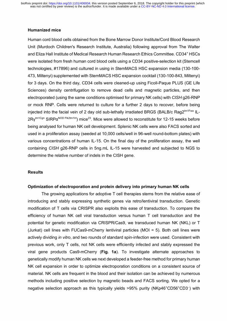
Humanized mice
Human cord blood cells obtained from the Bone Marrow Donor Institute/Cord Blood Research
Unit (Murdoch Children's Research Institute, Australia) following approval from The Walter
and Eliza Hall Institute of Medical Research Human Research Ethics Committee. CD34+ HSCs
were isolated from fresh human cord blood cells using a CD34 positive-selection kit (Stemcell
technologies, #17896) and cultured in using in StemMACS HSC expansion media (130-100-
473, Miltenyi) supplemented with StemMACS HSC expansion cocktail (130-100-843, Miltenyi)
for 3 days. On the third day, CD34 cells were cleaned-up using Ficoll-Paque PLUS (GE Life
Sciences) density centrifugation to remove dead cells and magnetic particles, and then
electroporated (using the same conditions optimised for primary NK cells) with CISH g26-RNP
or mock RNP. Cells were returned to culture for a further 2 days to recover, before being
injected into the facial vein of 2 day old sub-lethally irradiated BRGS (BALB/c Rag2tm1Fwa IL-
2Rγtm1Cgn SIRPαNOD Flk2tm1Irl) mice23. Mice were allowed to reconstitute for 12-15 weeks before
being analysed for human NK cell development. Splenic NK cells were also FACS sorted and
used in a proliferation assay (seeded at 10,000 cells/well in 96-well round-bottom plates) with
various concentrations of human IL-15. On the final day of the proliferation assay, the well
containing CISH g26-RNP cells in 5ng.mL IL-15 were harvested and subjected to NGS to
determine the relative number of indels in the CISH gene.
Results
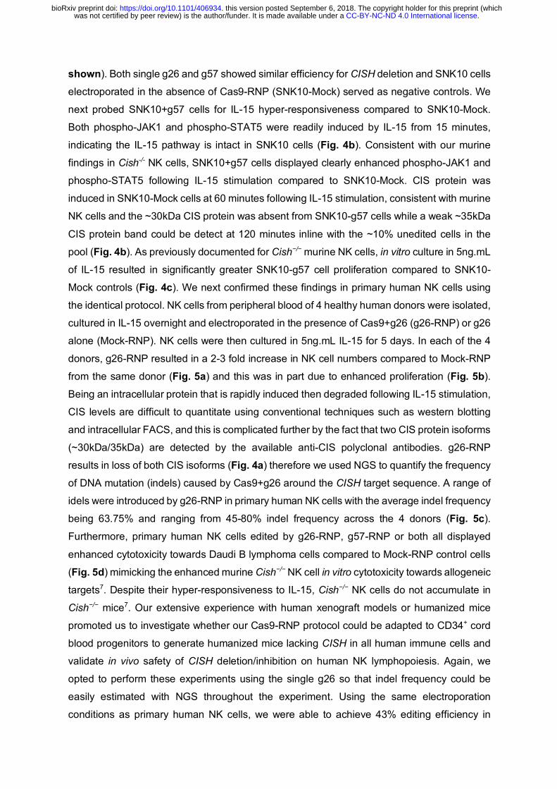
Optimization of electroporation and protein delivery into primary human NK cells The growing applications for adoptive T cell therapies stems from the relative ease of
introducing and stably expressing synthetic genes via retro/lentiviral transduction. Genetic
modification of T cells via CRISPR also exploits this ease of transduction. To compare the
efficiency of human NK cell viral transduction versus human T cell transduction and the
potential for genetic modification via CRISPR/Cas9, we transduced human NK (NKL) or T
(Jurkat) cell lines with FUCas9-mCherry lentiviral particles (MOI = 5). Both cell lines were
actively dividing in vitro, and two rounds of standard spin-infection were used. Consistent with
previous work, only T cells, not NK cells were efficiently infected and stably expressed the
viral gene products Cas9-mCherry (Fig. 1a). To investigate alternate approaches to
genetically modify human NK cells we next developed a feeder-free method for primary human
NK cell expansion in order to optimize electroporation conditions on a consistent source of
material. NK cells are frequent in the blood and their isolation can be achieved by numerous
methods including positive selection by magnetic beads and FACS sorting. We opted for a
negative selection approach as this typically yields >95% purity (NKp46+CD56+CD3-) with
.CC-BY-NC-ND 4.0 International licensewas not certified by peer review) is the author/funder. It is made available under aThe copyright holder for this preprint (whichthis version posted September 6, 2018. . https://doi.org/10.1101/406934doi: bioRxiv preprint

minimal loss of viability as is often associated with FACS sorting. NK cells were then seeded
at a density of 2e6 cells.cm2 in either G-rex 24 or 6 well plates using NK-MACS media
(Miltenyi) supplemented with 1000 IU.mL IL-2, 20ng.mL IL-15 and 5% AB+ human serum. Half
the culture media was removed and replaced with fresh media every third day until desired
number of NK cells was achieved (Fig. 1b). Numerous methods exist to deliver small
molecules and nucleic acids into cells, however delivery of large protein complexes is most
efficient when the cell surface is physically disrupted to allow the passive diffusion of such
molecules into the cells. Both mechanical disruption and electroporation-based technologies
have been adapted to introduce CRISPR RNPs into mammalian cells, though electroporation
has emerged as the predominant platform due to the widespread availability of these devices
and adaptability for use in in-vivo settings19. However, one of the major hurdles in the NK cell
field has been the relatively poor survival of NK cells post-electroporation. We therefore next
establish electroporation conditions for human NK cells that would maximize their viability and
uptake of large molecules using the widely-available Lonza 4D nucleofector device (Fig. 1b).
Beginning with the manufacture’s recommendations, we set out to optimize the electrical pulse
applied to the cells, or “pulse-code”, using the manufacturer’s own P3 nucleofection buffer
(Fig. 1c). The recommended read-out for such optimizations is the nucleofection of a GFP-
encoding plasmid to determine the efficacy of nucleofection. However, given the size of
recombinant Cas9 (approximately 160kDa) and the fact that RNP-mediated editing does not
require transcription from a plasmid template we decided to optimize with a fluorescently-
labelled high molecular weight dextran. Pulse code DN100 resulted in the maximum up-take
of FITC-dextran (~95%), yet viability was only ~40% (Fig. 1c). We next tested whether
alternate buffers could further improve NK cell viability and therefore created a panel of home-
made buffers to compare to the P3 standard. Our home-made buffer “Sol. 2 + mannitol”
improved NK cell viability (>60%) without compromising FITC-Dextran uptake and was
therefore adopted as our standard buffer to for primary NK cell electroporation (Fig. 1d). We
then re-optimized the pulse-codes using our Sol. 2 + mannitol buffer and found that pulse-
code CM137 was optimal resulting in >80% NK cell viability and FITC-Dextran uptake and
Sol. 2 + mannitol/pulse-code CM137 was adopted as our human NK cell electroporation
conditions (Fig. 1d).
Transient transfection of Cas9 RNP but not Cas9-expressing plasmid results in efficient gene editing in human NK cells
A recent report demonstrated that T cells could be efficiently gene-edited by simply
introducing plasmids that contain Cas9 and the gRNA (transient transfection)24, and if
applicable to NK cells, would represent the most rapid and cost-effective method of gene-
editing. We therefore cloned one of our validated guides against human CD45 (guide #61)
.CC-BY-NC-ND 4.0 International licensewas not certified by peer review) is the author/funder. It is made available under aThe copyright holder for this preprint (whichthis version posted September 6, 2018. . https://doi.org/10.1101/406934doi: bioRxiv preprint

into the Px458 ‘all-in-one’ vector21 that encodes Cas9 gene, eGFP and the sgRNA. A vector
encoding eGFP alone (pmaxGFP) was used as a positive control for transient gene
expression. 60 and 144 hours following electroporation of primary human NK cells with these
plasmids, we measured NK cell expression of GFP and CD45 (targeted for deletion by
PX458). While primary human NK cells were found to expressed GFP (25-35%) following
electroporation of pmaxGFP, we failed to detect GFP expression using Px458 and therefore
unsurprisingly no deletion of CD45 was observed (Fig. 2a). The fraction of GFP expressing
cells was significantly higher (50-80%) when human NK cell lines (SNK6, SNK10) where
electroporated with pmaxGFP, yet still no Cas9-GFP nor CD45 deletion was observed in these
lines (Fig. 2a). Thus, we concluded that primary human NK cells poorly transcribe genes from
plasmid templates, which is further diminished when required to transcribe large genes such
as Cas9 in the case of Px458.
Considering the difficulty of inducing Cas9 expression within NK cells, we opted for the delivery
of the fully assembled “ready-to-go” ribonucleoprotein (RNP) complexes as this has recently
been shown to efficiently edit primary human T cells (Fig. 2b)13, 14. RNP complexes were
created by incubating fully-synthetic sgRNA guides (identical in sequence to those used in the
Px458 plasmid) with recombinant Cas9 prior to electroporation into in vitro expanded primary
human NK cells as in Fig. 1. NK cells were then rested in culture for 3-5 days to allow sufficient
time for protein turnover. By flow cytometry, the extent of gene editing after this single-step,
single-guide, manipulation appeared to be remarkably efficient. In both primary human NK
cells and NK cell lines (SNK10 and SNK6) we observed loss of both copies of PTPRC (CD45-
/-) in 55-75% of the cells, whereas between 3-20% of the NK cells remained unedited (CD45+/+;
Fig. 2c). We also detected a population of cells (20-40%) which expressed intermediate levels
of CD45 and likely represents heterozygous deletion PTPRC (CD45+/-). We next FACS sorted
SNK10 cells based on CD45 expression (high, intermediate, negative) 5 days after Cas9-RNP
was performed. SNK10 CD45 variants were maintained in culture for 2 weeks and CD45
expression was found to be stable (Fig. 2c) suggesting homo- and heterozygous human NK
cells can be generated and that Cas9-RNP is an efficient method to genetically modify large
numbers of primary human NK cells.
Using Cas9 RNP to validate genetically-modified murine NK cell data in human NK cells The NCR-family of NK cell activating receptors includes NKp46, NKp44, NKp30 (encoded by
Ncr1, Ncr2, Ncr3) in humans but only NKp46 exists in mice25. NKp46 is ubiquitously expressed
on mature NK cells where it defines the lineage in both species and can trigger IFNg production
when cross-linked7. NKp46-null murine NK cells (Ncr1gfp/gfp) display impaired ability to kill
NKp46-ligand expressing tumor cells in vitro 26, 27. To investigate if optimal human NK cell
.CC-BY-NC-ND 4.0 International licensewas not certified by peer review) is the author/funder. It is made available under aThe copyright holder for this preprint (whichthis version posted September 6, 2018. . https://doi.org/10.1101/406934doi: bioRxiv preprint

cytotoxicity against tumor cells also requires NKp46, we deleted NCR1 from freshly isolated
primary human NK cells from two independent donors using Cas9-RNP with two distinct gRNA
against NCR1. Deletion efficiency ranged from 8-23%, with both gRNA having similar NCR1
deletion efficiency (loss of NKp46 expression; Fig. 3a). We next FACS sorted NKp46+ and
NKp46− NK cells from each donor and directly compared their cytotoxicity against Daudi cells
(human Burkitt B cell lymphoma). Daudi cells were chosen due to their susceptibility to lysis
by allo-NK cells and because they stained highly for human NKp46-Fc indicating they express
putative NKp46 ligands (Fig. 3b). Consistent with the murine data, we found human NKp46-
NK cells were clearly inferior to NKp46+ counterparts from the same donor at lysing Daudi
cells in vitro (Fig. 3c). Given the low deletion efficiency of NCR1 (% NKp46-) in freshly isolated
primary human NK cells, we next investigated if highly metabolic, activated and proliferating
primary human NK cells were a more efficient target population for NCR1 deletion. NK cells
isolated from donor 2 (Fig. 3a) were expanded in G-Rex flasks for 14 days as in Fig. 1. Using
the identical gNCR1/g543 as in Fig. 3a, we observed significantly higher NCR1-deletion using
expanded primary human NK cells with 85% NKp46- NK cells (5µl gRNA; Fig. 3d) compared
to 23.7% NKp46- NK cells using freshly NK cells from the same donor (Fig. 3a). We also
observed that NCR1 gene-editing efficiency could be modulated by titrating the amount of
gRNA used in the RNP complex and including a non-homologous DNA enhancer as
previously shown28 (Fig. 3d). Taken together, these data support a conserved role for NKp46
in augmenting NK cell cytotoxicity upon detection of NKp46-ligands on tumor cells and
highlight the utility of Cas9-RNP in validating murine NK cell biology in humans.
Validating CISH as an immune checkpoint in human NK cells using Cas9-RNP Work from our group and others over the past 10 years has established the cytokine IL-15 is
essential for most facets of NK cell biology including survival, homeostasis, differentiation and
activation1, 7, 29. We recently discovered that CIS (encoded by Cish) acts as a checkpoint in
mouse NK cell activation by negatively regulating IL-15 signaling7. The heightened IL-15
responsiveness of Cish−/− NK cells rendered Cish−/− mice resistant to melanoma, prostate and
TNBC metastasis. A hallmark of Cish loss-of-function in murine NK cells is hyper-
responsiveness to IL-15 in vitro which is read-out as enhanced proliferation, enhanced
phospho-JAK1 and phospho-STAT5 thus we next generated Cish-null human NK cells to
investigate these parameters. Firstly, we targeted CISH for deletion in SNK10 and SNK6
human NK cell lines to test the efficiency of the gRNA. Using two independent Cish gRNA
(g26 and g57) or pooled CISH gRNA (g26 + g57) resulted in a loss of CIS protein in both
SNK6 and SNK10 with effiency being 80-90% based of frequency of insertion/deletion
mutations (indels) in CISH determined by Next-Gen Sequencing (NGS; Fig. 4a and data not
.CC-BY-NC-ND 4.0 International licensewas not certified by peer review) is the author/funder. It is made available under aThe copyright holder for this preprint (whichthis version posted September 6, 2018. . https://doi.org/10.1101/406934doi: bioRxiv preprint
shown). Both single g26 and g57 showed similar efficiency for CISH deletion and SNK10 cells
electroporated in the absence of Cas9-RNP (SNK10-Mock) served as negative controls. We
next probed SNK10+g57 cells for IL-15 hyper-responsiveness compared to SNK10-Mock.
Both phospho-JAK1 and phospho-STAT5 were readily induced by IL-15 from 15 minutes,
indicating the IL-15 pathway is intact in SNK10 cells (Fig. 4b). Consistent with our murine
findings in Cish-/- NK cells, SNK10+g57 cells displayed clearly enhanced phospho-JAK1 and
phospho-STAT5 following IL-15 stimulation compared to SNK10-Mock. CIS protein was
induced in SNK10-Mock cells at 60 minutes following IL-15 stimulation, consistent with murine
NK cells and the ~30kDa CIS protein was absent from SNK10-g57 cells while a weak ~35kDa
CIS protein band could be detect at 120 minutes inline with the ~10% unedited cells in the
pool (Fig. 4b). As previously documented for Cish−/− murine NK cells, in vitro culture in 5ng.mL
of IL-15 resulted in significantly greater SNK10-g57 cell proliferation compared to SNK10-
Mock controls (Fig. 4c). We next confirmed these findings in primary human NK cells using
the identical protocol. NK cells from peripheral blood of 4 healthy human donors were isolated,
cultured in IL-15 overnight and electroporated in the presence of Cas9+g26 (g26-RNP) or g26
alone (Mock-RNP). NK cells were then cultured in 5ng.mL IL-15 for 5 days. In each of the 4
donors, g26-RNP resulted in a 2-3 fold increase in NK cell numbers compared to Mock-RNP
from the same donor (Fig. 5a) and this was in part due to enhanced proliferation (Fig. 5b).
Being an intracellular protein that is rapidly induced then degraded following IL-15 stimulation,
CIS levels are difficult to quantitate using conventional techniques such as western blotting
and intracellular FACS, and this is complicated further by the fact that two CIS protein isoforms
(~30kDa/35kDa) are detected by the available anti-CIS polyclonal antibodies. g26-RNP
results in loss of both CIS isoforms (Fig. 4a) therefore we used NGS to quantify the frequency
of DNA mutation (indels) caused by Cas9+g26 around the CISH target sequence. A range of
idels were introduced by g26-RNP in primary human NK cells with the average indel frequency
being 63.75% and ranging from 45-80% indel frequency across the 4 donors (Fig. 5c).
Furthermore, primary human NK cells edited by g26-RNP, g57-RNP or both all displayed
enhanced cytotoxicity towards Daudi B lymphoma cells compared to Mock-RNP control cells
(Fig. 5d) mimicking the enhanced murine Cish−/− NK cell in vitro cytotoxicity towards allogeneic
targets7. Despite their hyper-responsiveness to IL-15, Cish−/− NK cells do not accumulate in
Cish−/− mice7. Our extensive experience with human xenograft models or humanized mice
promoted us to investigate whether our Cas9-RNP protocol could be adapted to CD34+ cord
blood progenitors to generate humanized mice lacking CISH in all human immune cells and
validate in vivo safety of CISH deletion/inhibition on human NK lymphopoiesis. Again, we
opted to perform these experiments using the single g26 so that indel frequency could be
easily estimated with NGS throughout the experiment. Using the same electroporation
conditions as primary human NK cells, we were able to achieve 43% editing efficiency in
.CC-BY-NC-ND 4.0 International licensewas not certified by peer review) is the author/funder. It is made available under aThe copyright holder for this preprint (whichthis version posted September 6, 2018. . https://doi.org/10.1101/406934doi: bioRxiv preprint

freshly-isolated human cord blood CD34+ cells. This result was encouraging for only a single
guide and the fact that CIS is not highly expressed in CD34 cells, an obvious impediment to
efficient gene editing in cells30. These pool of partially CISH-deleted/g26-RNP cells or Mock-
RNP cells were then injected into the facial vein of lethally-irradiated 3-day old BRGS mice23
and allowed to reconstitute for 12 weeks (Fig. 5e). Similar to our data from Cish−/− mice, there
was no obvious accumulation of NK cells in mice that received g26-RNP CD34+ cells (Fig.5f), and NGS sequencing of the resulting NK cells showed a similar indel frequency to the starting
g26-RNP CD34+ cord blood progenitors (data not shown) suggesting CIS is not playing a
major role in human NK cell development and homeostasis. The NK cell population
reconstituted from g26-RNP edited CD34+ cord blood progenitors phenocopied that of human
peripheral blood NK cells as we have previously shown31 and did not differ from Mock-RNP
controls (data not shown). We next interrogated the IL-15 receptor signaling of g26-RNP
CD34+ cord blood progenitor-derived NK cells in vitro. We isolated human NK cells from the
spleens of two g26-RNP and two Mock-RNP humanized mice and subjected them to an IL-15
proliferation assay in vitro. Similar to the data observed in g26-RNP primary human NK cells
and g57-RNP SNK10 cells, the NK cells derived from g26-RNP humanized mice were able to
proliferate significantly greater in response to IL-15 compare to their Mock-RNP counterparts
(Fig. 5g). Furthermore, the indel frequency increased from 43% upon NK cell isolation from
the g26-RNP humanized mouse spleen to 75% following the 7-day proliferation assay (data not shown). Taken together, these data demonstrate the efficiency of gene targeting in
human NK cells and the validates the role of CIS as a potent negative regulator of IL-15
signaling in human NK cells.
Discussion Here we describe, for the first time, an effective approach for genetic manipulation of primary
human NK cells using CRISPR-Cas9 RNP and demonstrate that our recent discovery of an
NK cell checkpoint in mice is also a conserved and valid target in human NK cells. To date,
there have been few examples of genetically-engineered primary human NK cells largely due
to poor infection rates with viral gene-transfer systems and difficulties in culturing primary
human cells to significant numbers. Recent advances in viral pseudotyping and antagonizing
intracellular anti-viral pathways have improved infection rates and opened the door for
application such as CAR-NK cell products16. A concern from lentiviral modification of human
NK cells is mutagenesis caused by random insertion of viral genes into the genome given the
high viral titers (>150 MOI) required for infection17. In addition, viral delivery and expression of
gene-editing machinery require the target cells to be cultured for several days before the
desired gene is edited, potentially losing the naïve phenotype of the target cell. A recent study
showed that simply delivering plasmids that encode the gene editing machinery could
.CC-BY-NC-ND 4.0 International licensewas not certified by peer review) is the author/funder. It is made available under aThe copyright holder for this preprint (whichthis version posted September 6, 2018. . https://doi.org/10.1101/406934doi: bioRxiv preprint

efficiently target genes in T cells24, however our data suggest that even highly-activated NK
cells are inefficient at transcribing the required genes from large plasmid templates to result
in Cas9-mediated editing. It is worth noting that pMAX-GFP vector uses a CMV promoter and
at 3486bp is considerably smaller than the Px458 (9300bp) which uses a Cbh promoter.
We therefore set out to establish whether direct delivery of Cas9-RNP complexes directly into
freshly isolated human NK cells could rapidly and efficiently edit certain genes. An additional
benefit of using NLS-tagged Cas9 RNP is that the delivery method does not depend upon
successful nuclear delivery, as the complex can be easily transported into the nucleus19. Upon
entry into the nucleus, the Cas9 RNP rapidly binds DNA and begins scanning for the regions
complementary to the guide RNA. The kinetics of this editing process have been described to
take between 2-12 hours32, meaning that primary human NK cells could be easily edited within
a day to study their function.
The cancer immunotherapy revolution over the past 10 years has seen rising interest in
targeting NK cells given their spontaneous ability to detect and kill stressed/transformed cells6.
Furthermore, NK cells are emerging as key drivers of dendritic cell infiltration and optimal
priming of IFN+ and TNFa+ CD8 T cells3, 4, 10. Yet, despite our best efforts, cancer
immunotherapy drugs that exploit the innate anti-tumor functions of NK cells are yet to have
a positive impact on cancer outcomes. This is in part due to the vast differences between
mouse and human NK cell frequencies and phenotypes in vivo and due to the difficulty in
validating if therapeutic targets derived from murine genetic knockout studies hold true for
human NK cells. To this end, our CRISPR-Cas9 RNP approach detailed here will be
transformative for NK cell immunotherapy drug development and basic understanding of
human NK cell biology. CRISPR-Cas9 RNP now makes ease of rapid gene editing in primary
human NK cells overcoming this current barrier to target pathway validation. To this end, we
investigated two pathways known to regulate murine NK cell anti-tumor function in human NK
cell lines and primary human NK cells. NKp46 is a transmembrane receptor that signaling via
CD3ζ and FcεRIγ to recruit multiple ITAM-containing proteins. Cross-linking NKp46 with
antibodies results in IFNg production, which is augmented by IL-15 signaling7. NKp46-null
mice display impair anti-metastatic responses in vivo and impaired cytotoxicity in vitro to tumor
cells that bind NKp46-Fc and presumable express putative NKp46 ligands27, 33, 34. Using Cas9-
RNP+gNcr1, we were able to delete NKp46 on a subset of primary human NK cells and
validate that NKp46 expression confers optimal NK cell cytotoxicity towards NKp46-ligand
expressing Daudi B lymphoma cells. Deletion of genes for cell-surface proteins was thus
clearly feasible, and the ability to FACS sort for gene-edited cells makes cell surface proteins
.CC-BY-NC-ND 4.0 International licensewas not certified by peer review) is the author/funder. It is made available under aThe copyright holder for this preprint (whichthis version posted September 6, 2018. . https://doi.org/10.1101/406934doi: bioRxiv preprint

practical targets for validation using this method. We next addressed the feasibility to delete
the intracellular immunotherapy drug target CIS using Cas9-RNP+gCish. Cish has been
published by multiple groups to limit IL-15 and TCR signaling in murine NK and T cells,
ultimately dampening anti-tumor responses7, 35, 36. Cish was efficiently deleted in human NK
cells (45-80% indels in primary cells; 85-93% indels in NK cell lines) yet the inability to sort on
Cish-null cells meant functional assays were run on pools of deleted cells. Despite the
incomplete absence of CIS protein, Cas9-RNP+gCish edited cells behaved as predicted from
mouse studies in that they were hyper-sensitive to IL-15 in vitro and had augmented
cytotoxicity towards an allogeneic tumor line in vitro. Much resources have been placed on
optimizing NK cells as an adoptive off-the-shelf cellular immunotherapy. These products
derive from numerous precursor cell populations, including induced pluripotent stem cells,
placental NK cells and cord-blood progenitors37. Given that deletion of Cish in mature human
NK cells phenocopies the IL-15 hyper-responsiveness data from mouse7, it is expected that
CRISPR deletion of Cish in human adoptive NK cell therapy products would augment their
anti-tumor function as previously shown for murine Cish-/- NK cells.
Thus, while primary human T cells can be genetically modified by several approaches
including lenti/retrovirus transduction, transient transfection and Cas9-RNP13, 24, the latter
approach is the only viable approach for large-scale genetic modification of primary human
NK cells. With further optimization of guide sequences, recombinant Cas9 source and
multiplexing of distinct guides, it is likely that most genes will be able to be deleted at a high
enough efficiency to concinving conclude their role in human NK cells. Our detailed technical
report of Cas9-RNP in human NK cells lends itself to immunotherapy drug discovery including
rapid gene-editing of patient-derived or allo-NK cellular therapy products meaning
undruggable pathways can be efficiently targeted for personalized immunotherapy.
Acknowledgements
This work was supported by project grants from the National Health and Medical Research
Council (NHMRC) of Australia (1124907, 1124784, 1049407, 1066770, 1057852, 1027472)
and a fellowship (1124788) to N.D.H as well as an NHMRC Independent Research Institute
Infrastructure Support scheme grant and a Victorian State Government Operational
Infrastructure Scheme grant. N.D.H. is a recipient of Research Grants from the Harry J Lloyd
Charitable Trust (USA), Melanoma Research Alliance (USA), Tour de Cure (AUS), the Ian
Potter Foundation (AUS), Cancer Council of Victoria (1145730) and a CLIP grant from Cancer
Research Institute (USA).
.CC-BY-NC-ND 4.0 International licensewas not certified by peer review) is the author/funder. It is made available under aThe copyright holder for this preprint (whichthis version posted September 6, 2018. . https://doi.org/10.1101/406934doi: bioRxiv preprint

Authorship Contributions JR, ES and NDH designed and performed experiments and interpreted all data. JR and NDH
wrote the paper.
Disclosure of Conflicts of Interest JR and NDH are co-founders and share-holders of oNKo-Innate Pty Ltd. NDH has a
collaborative research agreement with Servier.
Figures
Figure 1. The unmet need of efficient gene editing of human NK cells. (a) Human T cell
(Jurkat) and NK cell (NKL) lines were spin-infected with FUCas9Cherry lentiviral particles
(MOI = 5) and Cas9-mCherry monitored by FACS at 72 hours. Data are representative of 3
independent experiments. (b) Flow chart of human NK cell isolation, expansion and
electroporation optimization. Lonza 4D electroporation optimization for primary human NK
cells; (c) Pulse code optimization for using buffer P3, (d) Buffer optimization using DN100
pulse code and (e) Pulse code re-optimization using Sol. 2 + Mannitol buffer. Black bars
indicate NK cell viability, green bars indicate uptake of FITC-dextran (electroporation
efficiency). Data are representative of 2 independent experiments.
Figure 2. Cas9-RNP results in rapid, efficient gene deletion in primary human NK cells. (a) A transient Cas9 and sgRNA expression system (Px458) was tested for the ability to delete
PTPRC (CD45) from NK cell lines (SNK6/SNK10) and primary human NK cells (dextran-FITC
and pmaxGFP served as transfection and transcription controls, respectively). All data are
shown. (b) Schematic of workflow for isolating and electroporating primary human NK cells.
(c) Electroporation of Cas9 RNP complexes (carrying the same sgPTPRC sequence used in
the px458 vector in (a) results in highly efficient deletion of CD45 from SNK10 and SNK6
human NK cell lines and primary human cells after 3-5 days in culture. Histograms show the
stability of FACS-sorted CD45-expression variants (high, medium and low) in the SNK10 line
after 2 weeks in culture. Data are representative of 2 independent experiments for
SNK10/SNK6 and 4 individual healthy adult donors for primary NK cells.
Figure 3. Deletion of NCR1 using Cas9 RNP. (a) Deletion efficiency of NCR1 5-days post
electroporation using two different guides shows some variability between donors. Data are
representative of 4 healthy adult donors. (b) FACS-sorted NCR1+ and NCR1- primary human
NK cells were used in a cytotoxicity assay against Daudi target cells. All data are shown. (c)
.CC-BY-NC-ND 4.0 International licensewas not certified by peer review) is the author/funder. It is made available under aThe copyright holder for this preprint (whichthis version posted September 6, 2018. . https://doi.org/10.1101/406934doi: bioRxiv preprint

Daudi target cells express high levels of putative NKp46 ligands as revealed by staining with
an hNKp46-Fc. hNKp30-Fc served as a negative control. Data are representative of 2
experiments. (d) Deletion efficiency of NCR1 on highly activated primary human NK cells (14
days post-expansion; donor 2 from 3a) using a titration g543/NCR1. NKp46 staining was
performed 60 hours post-electroporation. Percentages indicated NCR1-deleted (NKp46-)
fraction. All data are shown from donor 2. Mock-RNP (electroporation of g543 alone) served
as a negative control in all experiments.
Figure 4. Deletion of CISH enhances cellular responses to IL-15. (a) Cas9 RNP-mediated
deletion of CISH from SNK6/SNK10 NK cell lines using various guides alone or in combination.
(b) CIS-deficient SNK10 cells show enhanced activation of the IL-15 signaling pathway
relative to parental controls. (c) Proliferative response to IL-15 is enhanced in CIS-deficient
SNK10 cells. Data are representative of 2 experiments.
Figure 5. Deletion of CISH in primary human NK cells. NK cells were isolated from 4
healthy donors and electroporated with Cas9 RNP+g26(CISH) or Mock RNP (g26 alone).
Proliferation was determined 5 days after electroporation by (a) seeding 50,000 cells/well in a
96-well plate and enumerating the number of live cells or (b) labeling cells with a division-
tracking dye (CTV). CISH deletion efficiency was quantified for all donors and ranged from 45-
80%, representative NGS results shown in (c). (d) CISH-edited primary human NK cells (g26,
g57 or g26+g57) and Mock-RNP control NK cells from the same donor were tested for their
cytotoxicity against Daudi B lymphoma cells in vitro. Data are representative of 2 donors (e)
Workflow used to generate gene-edited human immune system (HIS) mice. (f) Frequency of
human NK cells in the bone marrow (BM), spleen, liver and blood of HIS mice 12 weeks after
reconstitution with WT (Mock RNP) or CISH-g26 edited CD34+ hematopoietic stem cells. (g)
Proliferation of NK cells derived from the spleen of HIS mice (e, f) in various concentrations
of IL-15, NGS revealed that CISH-indels were enriched from 43% to 75% after 7 days in
culture.
References 1. Sathe, P. et al. Innate immunodeficiency following genetic ablation of Mcl1 in natural killer
cells. Nat Commun 5, 4539 (2014). 2. Kim, S., Iizuka, K., Aguila, H.L., Weissman, I.L. & Yokoyama, W.M. In vivo natural killer
cell activities revealed by natural killer cell-deficient mice. Proc Natl Acad Sci U S A 97, 2731-2736 (2000).
.CC-BY-NC-ND 4.0 International licensewas not certified by peer review) is the author/funder. It is made available under aThe copyright holder for this preprint (whichthis version posted September 6, 2018. . https://doi.org/10.1101/406934doi: bioRxiv preprint

3. Bottcher, J.P. et al. NK Cells Stimulate Recruitment of cDC1 into the Tumor Microenvironment Promoting Cancer Immune Control. Cell 172, 1022-1037 e1014 (2018).
4. Barry, K.C. et al. A natural killer-dendritic cell axis defines checkpoint therapy-responsive
tumor microenvironments. Nat Med 24, 1178-1191 (2018). 5. Rautela, J. & Huntington, N.D. IL-15 signaling in NK cell cancer immunotherapy. Curr Opin
Immunol 44, 1-6 (2017). 6. Guillerey, C., Huntington, N.D. & Smyth, M.J. Targeting natural killer cells in cancer
immunotherapy. Nat Immunol 17, 1025-1036 (2016). 7. Delconte, R.B. et al. CIS is a potent checkpoint in NK cell-mediated tumor immunity. Nat
Immunol 17, 816-824 (2016). 8. Paolino, M. et al. The E3 ligase Cbl-b and TAM receptors regulate cancer metastasis via
natural killer cells. Nature 507, 508-512 (2014). 9. Molgora, M. et al. IL-1R8 is a checkpoint in NK cells regulating anti-tumour and anti-viral
activity. Nature 551, 110-114 (2017). 10. Zhang, Q. et al. Blockade of the checkpoint receptor TIGIT prevents NK cell exhaustion and
elicits potent anti-tumor immunity. Nat Immunol (2018). 11. Rezvani, K. & Rouce, R.H. The Application of Natural Killer Cell Immunotherapy for the
Treatment of Cancer. Front Immunol 6, 578 (2015). 12. Schumann, K. et al. Generation of knock-in primary human T cells using Cas9
ribonucleoproteins. Proc Natl Acad Sci U S A 112, 10437-10442 (2015). 13. Seki, A. & Rutz, S. Optimized RNP transfection for highly efficient CRISPR/Cas9-mediated
gene knockout in primary T cells. J Exp Med 215, 985-997 (2018). 14. Roth, T.L. et al. Reprogramming human T cell function and specificity with non-viral
genome targeting. Nature 559, 405-409 (2018). 15. Wucherpfennig, K.W. & Cartwright, A.N. Genetic screens to study the immune system in
cancer. Curr Opin Immunol 41, 55-61 (2016). 16. Sutlu, T. et al. Inhibition of intracellular antiviral defense mechanisms augments lentiviral
transduction of human natural killer cells: implications for gene therapy. Hum Gene Ther 23, 1090-1100 (2012).
17. Boissel, L. et al. Comparison of mRNA and lentiviral based transfection of natural killer cells
with chimeric antigen receptors recognizing lymphoid antigens. Leuk Lymphoma 53, 958-965 (2012).
18. Shimasaki, N. et al. A clinically adaptable method to enhance the cytotoxicity of natural killer
cells against B-cell malignancies. Cytotherapy 14, 830-840 (2012). 19. Staahl, B.T. et al. Efficient genome editing in the mouse brain by local delivery of engineered
Cas9 ribonucleoprotein complexes. Nat Biotechnol 35, 431-434 (2017).
.CC-BY-NC-ND 4.0 International licensewas not certified by peer review) is the author/funder. It is made available under aThe copyright holder for this preprint (whichthis version posted September 6, 2018. . https://doi.org/10.1101/406934doi: bioRxiv preprint

20. Aubrey, B.J. et al. An inducible lentiviral guide RNA platform enables the identification of tumor-essential genes and tumor-promoting mutations in vivo. Cell Rep 10, 1422-1432 (2015).
21. Ran, F.A. et al. Genome engineering using the CRISPR-Cas9 system. Nat Protoc 8, 2281-
2308 (2013). 22. Neri, S., Mariani, E., Meneghetti, A., Cattini, L. & Facchini, A. Calcein-acetyoxymethyl
cytotoxicity assay: standardization of a method allowing additional analyses on recovered effector cells and supernatants. Clinical and diagnostic laboratory immunology 8, 1131-1135 (2001).
23. Legrand, N. et al. Functional CD47/signal regulatory protein alpha (SIRP(alpha)) interaction
is required for optimal human T- and natural killer- (NK) cell homeostasis in vivo. Proc Natl Acad Sci U S A 108, 13224-13229 (2011).
24. Kornete, M., Marone, R. & Jeker, L.T. Highly Efficient and Versatile Plasmid-Based Gene
Editing in Primary T Cells. J Immunol 200, 2489-2501 (2018). 25. Koch, J., Steinle, A., Watzl, C. & Mandelboim, O. Activating natural cytotoxicity receptors
of natural killer cells in cancer and infection. Trends Immunol 34, 182-191 (2013). 26. Glasner, A. et al. NKp46 Receptor-Mediated Interferon-gamma Production by Natural Killer
Cells Increases Fibronectin 1 to Alter Tumor Architecture and Control Metastasis. Immunity 48, 107-119 e104 (2018).
27. Glasner, A. et al. Recognition and prevention of tumor metastasis by the NK receptor
NKp46/NCR1. J Immunol 188, 2509-2515 (2012). 28. Richardson, C.D., Ray, G.J., Bray, N.L. & Corn, J.E. Non-homologous DNA increases gene
disruption efficiency by altering DNA repair outcomes. Nat Commun 7, 12463 (2016). 29. Delconte, R.B. et al. The Helix-Loop-Helix Protein ID2 Governs NK Cell Fate by Tuning
Their Sensitivity to Interleukin-15. Immunity 44, 103-115 (2016). 30. Daer, R.M., Cutts, J.P., Brafman, D.A. & Haynes, K.A. The Impact of Chromatin Dynamics
on Cas9-Mediated Genome Editing in Human Cells. ACS Synth Biol 6, 428-438 (2017). 31. Huntington, N.D. et al. IL-15 trans-presentation promotes human NK cell development and
differentiation in vivo. J Exp Med 206, 25-34 (2009). 32. Brinkman, E.K. et al. Kinetics and Fidelity of the Repair of Cas9-Induced Double-Strand
DNA Breaks. Mol Cell 70, 801-813 e806 (2018). 33. Elboim, M. et al. Tumor immunoediting by NKp46. J Immunol 184, 5637-5644 (2010). 34. Halfteck, G.G. et al. Enhanced in vivo growth of lymphoma tumors in the absence of the NK-
activating receptor NKp46/NCR1. J Immunol 182, 2221-2230 (2009). 35. Palmer, D.C. et al. Cish actively silences TCR signaling in CD8+ T cells to maintain tumor
tolerance. J Exp Med 212, 2095-2113 (2015). 36. Putz, E.M. et al. Targeting cytokine signaling checkpoint CIS activates NK cells to protect
from tumor initiation and metastasis. Oncoimmunology 6, e1267892 (2017).
.CC-BY-NC-ND 4.0 International licensewas not certified by peer review) is the author/funder. It is made available under aThe copyright holder for this preprint (whichthis version posted September 6, 2018. . https://doi.org/10.1101/406934doi: bioRxiv preprint

37. Veluchamy, J.P. et al. The Rise of Allogeneic Natural Killer Cells As a Platform for Cancer
Immunotherapy: Recent Innovations and Future Developments. Front Immunol 8, 631 (2017).
.CC-BY-NC-ND 4.0 International licensewas not certified by peer review) is the author/funder. It is made available under aThe copyright holder for this preprint (whichthis version posted September 6, 2018. . https://doi.org/10.1101/406934doi: bioRxiv preprint

Figure 1
a
100ml human blood
50% media change every 3 days and resuspend cells until desired
cell number achieved
EasySepTM Human NK cell Isolation Kit
2 x10⁶ NK cells/cm2 in G-Rex flaskNK-MACS media+ 1000IU/mL IL-2 + 20ng/mL hIL-15+ 5% human AB+ serum
Electroporation optimization
500,000 NK cells/well
Pulse codes ------->
Bu
ffers
--------
>
b
No pulseEO100
CA137CN114
EH100
EW113DS120
CM150
CM138EN150
DS138DS150
DS130DS137
CM137
DN1000
20
40
60
80
100Pulse Code Optimization (Buffer P3)
P3 + no pulse contro
l
Sol 2 + Na su
ccinate
Sol 3 + HEPES/TRIS
Sol 1 + Na su
ccinate
Sol 1 + NaCl
Sol 2 + NaCl
Sol 4 + m
annitol
Sol 2 + Na la
ctobionate
Sol 4 + Na su
ccinate
Sol 1 + Na la
ctobionate
Sol 2 + m
annitol P3
Sol 3 + Phosp
hate
Sol 1 + m
annitol
Viab
ility (
% o
f no
pulse
cont
rol)
Dext
ran-
FITC
+ (%
of l
ive
cells
)
0
20
40
60
80
100
Buffer Optimization (Pulse Code DN100)
0
20
40
60
80
100
0
20
40
60
80
100Vi
abilit
y (%
of n
o pu
lse co
ntro
l)
Pulse Code Re-optimization (Sol 2 + Mannitol)
0
20
40
60
80
100
Viab
ility (
% o
f no
pulse
cont
rol)
c
d
0
20
40
60
80
100
44.5%
0.01%
0.03%
0.2%
Cas9-mCherry
Non-transduced FUCas9CherryLenti-transduced
e
T cells
NK cells
Dext
ran-
FITC
+ (%
of l
ive
cells
)De
xtra
n-FI
TC+
(% o
f liv
e ce
lls)
.CC-BY-NC-ND 4.0 International licensewas not certified by peer review) is the author/funder. It is made available under aThe copyright holder for this preprint (whichthis version posted September 6, 2018. . https://doi.org/10.1101/406934doi: bioRxiv preprint

Ctrl.
Px458-PTPRC
pmaxGFP
Dextran-FITC
60hrs post-electroporation 144hrs post-electroporation
Prim
ary
NK
cells
SNK6
SNK1
0
0.25%
35%
95% 74%
27%
0.05%
0% GFP+ 0.05% GFP+ 0% CD45+
100%
100%
100%
0% CD45+
100%
100%
100%
0.1% GFP+
4%
81%
99%
0.01% GFP+
2%
74%
99%
0% CD45+
99%
99%
99%
0% CD45+
100%
100%
100%
0.1% GFP+
4%
58%
89%
0.9% GFP+
1.4%
53%
93%
0% CD45+
99%
100%
99%
0% CD45+
100%
100%
100%
Ctrl.
Px458-PTPRC
pmaxGFP
Dextran-FITC
Ctrl.
Px458-PTPRC
pmaxGFP
Dextran-FITC
a
GFP CD45
GFP CD45
GFP CD45
GFP CD45
GFP CD45
GFP CD45
Electroporation - Transient Transfection with Px458 (pCas9-2A-GFP-gPTPRC)
bElectroporation - Cas9 RNP
FSC-A
CD
45
CD45+/+
CD45+/−
CD45−/−
0%
0%
99%
96%
2%
0%
4%
20%
73%
Unstained RNP (-gRNA) RNP (+gRNA)
Mix &Electroporate
Isolate NK cells
Rest overnight in2ng/mL IL-15
Assemble RNP (incubate for 20’)
Add 100uL of completeNK cell medium to each well& immediately placein incubator for 10min.
Return to culture plate
NK cells
C
0 103
104
105
Primary Human NK cells
RNP (+gRNA)
FSC-A
CD
45
16%
20%
62%
CD45+/+
CD45+/−
CD45−/−
UnstainedSNK10
CD45 Figure 2
SNK6
59%
3%
36%
RNP (+gRNA) FACS sorted SNK10 PTPRC variants
.CC-BY-NC-ND 4.0 International licensewas not certified by peer review) is the author/funder. It is made available under aThe copyright holder for this preprint (whichthis version posted September 6, 2018. . https://doi.org/10.1101/406934doi: bioRxiv preprint

NCR1-28.4
0 10 3 10 4 10 5
NKp46
Unstained
Mock
Donor 1. NCR1-RNP (g543)
Donor 1. NCR1-RNP (g544)
60hrs post-electroporation
Donor 2. NCR1-RNP (g544)
Donor 2. NCR1-RNP (g543)
100%
0.47%
9.01%
8.31%
23.7%
19.7%
Sort
0 1 2 3 4 50
10
20
30
40
E:T Ratio
% L
ysis
of D
audi
cel
ls
Donor 1
NKp46 deletion on primary human NK cells by Cas9 RNP
NKp46+
NKp46−
% NKp46−
Figure 3
a
0 1 2 3 4 50
10
20
30
E:T Ratio
Donor 2
NKp46+
NKp46−
% L
ysis
of D
audi
cel
lsb
cDaudi Burkitt’s Lymphoma
hNKp46-Fc
hNKp30-FcSecondary Ab
Seconday Ab
0 10 3 10 4 10 5
100%
2.25%
89.7%
84.7%
83.9%
85%
76.6%
72.9%
Unstained
Mock
(no enhancer) 5ul sgRNA
5ul sgRNA
4ul sgRNA
3ul sgRNA
2ul sgRNA
1ul sgRNA
d NKp46 deletion on day 14 in vitro expanded primary human NK cells (Donor 2) by Cas9 RNP
sgRNA titration (g543/NCR1)
NKp46
.CC-BY-NC-ND 4.0 International licensewas not certified by peer review) is the author/funder. It is made available under aThe copyright holder for this preprint (whichthis version posted September 6, 2018. . https://doi.org/10.1101/406934doi: bioRxiv preprint

Figure 4
a
b
c
*
*
* *
Vinculin
37kDa
120kDa
SNK6
+ M
ock
SNK6
+ g2
6SN
K6 +
g57
SNK6
+ g2
6+g5
7SN
K10 +
Moc
kSN
K10 +
g26
SNK1
0 + g5
7SN
K10 +
g26+
g57
SNK10 + g57SNK10-Mock
SNK108
6
4
2
01 2 3 4 5 6 7
Days
Live c
ells (
x105 )
Mock (0.5ng.mL)Mock (5ng.mL)g57 (0.5ng.mL)g57 (5ng.mL)
CIS
37kDaCIS
130kDa pJAK1
130kDa JAK1
90kDa pSTAT5
90kDa STAT5
45kDa Actin
0 15 30 60 120 0 15 30 60 120
.CC-BY-NC-ND 4.0 International licensewas not certified by peer review) is the author/funder. It is made available under aThe copyright holder for this preprint (whichthis version posted September 6, 2018. . https://doi.org/10.1101/406934doi: bioRxiv preprint

Assemble RNP
Isolate CD34+ cells
Expand for 3days
Mix &Electroporate
Recover for 2days
100,000 cells into facial vein of 2 day old irradiatedBRGS pups
a
b
d
CTV
g26-RNP (68% indels)Mock-RNP
Primary human NK cells in IL-15
Mock-RNP g26-RNP0
1
2
3
4To
tal c
ell n
umbe
rLiv
e cell
s (x1
05 )
*
10
8
2NKp4
6+ (%
of hC
D45)
0
6
4
BM Blood Spleen Liver
Mock RNP CD34+g26-RNP CD34+ (43% indels)
g26 (CISH)-RNP CD34+ humanized mice
6
4
2
1 2 3 4 5 6 7Days
Live c
ells (
x105 )
0Mock RNP (0.5ng.mL)
Mock RNP (5ng.mL)
g26-RNP (0.5ng.mL)
g26-RNP (5ng.mL)(75% indels)
Mock RNP (0ng.mL)g26-RNP (0ng.mL)
g26 (CISH)-RNP NK cells from humanized mice
d.0
Primary human NK cells in IL-15c
e
f
Delet
ion co
unts
= 76
48
Wildtype counts = 5175Insertion counts = 556
NGS of primary human NK cells g26-RNP (61.3% indels)
Figure 5
g
g57-RNPg26+g57-RNP
g26-RNP
1 100.1
40
80
0
Lysis
of D
audi
cel
ls (%
)
20
60
Mock-RNP
NK cell : Daudi (E:T)
g26 (CISH)-RNP humanized miceCISH-RNP primary human cytotoxicity.CC-BY-NC-ND 4.0 International licensewas not certified by peer review) is the author/funder. It is made available under a
The copyright holder for this preprint (whichthis version posted September 6, 2018. . https://doi.org/10.1101/406934doi: bioRxiv preprint