efficacy of glaucoma drainage devices in uveitic glaucoma and a … · 2019-01-07 · categories...
TRANSCRIPT

GLAUCOMA
Efficacy of glaucoma drainage devices in uveitic glaucomaand a meta-analysis of the literature
Wishal D. Ramdas1 & Jan Pals1 & Aniki Rothova1 & Roger C. W. Wolfs1
Received: 1 August 2018 /Revised: 24 September 2018 /Accepted: 2 October 2018 /Published online: 11 October 2018# The Author(s) 2018
AbstractPurpose To assess the efficacy of glaucoma drainage devices (GDD) in uveitic glaucoma and non-uveitic glaucoma, and toperform a meta-analysis of previously published results to compare with our data.Methods Retrospective case-control study, in which all eyes that underwent GDD surgery were included from 2015 onwards.Cases were defined as patients with uveitic glaucoma. Patients with non-uveitic glaucoma served as controls. To compare ourresults, a review of the literature was performed using PubMed database.Results A total of 99 eyes were included (38 with uveitic glaucoma). The preoperative IOP was 25.9 ± 7.7 mmHg and 27.9 ±9.6 mmHg for patients with and without uveitis (p = 0.277). No significant differences were found between patients with andwithout uveitis in the final IOP or reduction in IOP (44.9% vs. 42.8%, respectively). Within the first year after surgery, 13.2% ofcases developed macular edema (vs. 6.6%; p = 0.267) and 15.8% a transient hypotony (vs. 8.2%; p = 0.242). A meta-analysis of24 studies showed a postoperative weighted mean difference of − 17.8 mmHg and 2.2 lower number of IOP-lowering medica-tions in uveitic glaucoma (compared to − 13.2 mmHg and 3.5 in the current study, respectively).Conclusion GDD surgery in patients with uveitis has a similar effect on IOP as in patients without uveitis. The risks of developingmacular edema and hypotony were slightly higher in patients with uveitis, but the results were not statistically significant. Thesefindings are in line with previous reports, though data on the efficacy of GDD surgery and macular edema in uveitic glaucoma isscarce.
Keywords Glaucoma . Intraocular pressure . Glaucoma drainage device . Ahmed . Baerveldt . Uveitis
Introduction
Glaucoma, aneurodegenerative eyedisease, is themost com-mon cause of irreversible blindness worldwide. Of all differ-ent forms of glaucoma, about 20–30% are caused by an un-derlying systemic or ophthalmic disorder: secondary glauco-ma [1]. One of the most serious causes is uveitis, especiallyanterior uveitis [2]. The incidence of secondary glaucomacaused by uveitis (uveitic glaucoma) is reported to be 10–
20% [3]. Patientswith both glaucoma and uveitis have a highrisk of severe visual impairment [2, 4]. Uveitic glaucomapresents a two-pronged problem: inflammatory damage tothe trabecular meshwork and uvea (e.g., synechiae) coupledwith steroid-induced increase in intraocular pressure (IOP).It is one of the most difficult forms of glaucoma to manage,because the ophthalmologist must simultaneously addressinflammation and elevated IOP. Among the current surgicaltreatment options are trabeculectomy, deep sclerectomy,minimally invasive glaucoma surgery (MIGS; e.g.,trabectome), cyclodestructive procedures, and several typesof glaucoma drainage devices (GDD), with comparable effi-cacy [5–7].However, the long-term roleof trabeculectomy inpatients with uveitis is compromised by young age,inflammation-induced fibrosis and conjunctival scarring,and scleral thinning, making trabeculectomy in many casesnot the optimal choice. Moreover, uveitic glaucoma usuallydevelops at a younger age (having a thick Tenon’s capsuleand more robust healing response, often resulting in
Electronic supplementary material The online version of this article(https://doi.org/10.1007/s00417-018-4156-9) contains supplementarymaterial, which is available to authorized users.
* Wishal D. [email protected]
1 Department of Ophthalmology, Erasmus Medical Center, ‘sGravendijkwal 230, 3000 CA Rotterdam, The Netherlands
Graefe's Archive for Clinical and Experimental Ophthalmology (2019) 257:143–151https://doi.org/10.1007/s00417-018-4156-9

progressive subconjunctival fibrosis) than primary glauco-ma. Cyclodestructive procedures are irreversible; hence, thistreatment option is often reserved for eyeswith aworsevisualprognosis. Therefore, GDD are currently considered a betterchoice in the treatment of uveitic glaucoma. The most com-monly used GDD are the valved Ahmed and the non-valvedBaerveldt implant. Nonetheless, reports on the performanceof these GDD in uveitis eyes are scarce. Therefore, we com-pared the efficacy and safety of the most common GDD(Ahmed and Baerveldt implant) in eyes with uveitic glauco-ma to glaucoma eyes without uveitis. Next, we assessed thedifferences in complications after GDD surgery. Finally, weperformed a review of the literature on these GDD (AhmedFP7 andBaerveldt-350) in eyeswith uveitic glaucoma and ineyes with other forms of glaucoma. Meta-analyses of theretrieved studies were performed and compared to ourresults.
Methods
Studies population
The first part of the study was designed as a retrospectivestudy and the second part as a review with several meta-analyses (see further). For the retrospective study, we selectedall patients that underwent at least one GDD surgery in theperiod from January 2015 to January 2018 at the Departmentof Ophthalmology of the Erasmus University Medical Center,Rotterdam, The Netherlands.
All patients underwent extensive ophthalmic examinationat each visit. The medical records of all patients werereviewed, and clinically relevant data were entered in a data-base. Data of the last preoperative visit, postoperative follow-up data at 1 day, 1 week, 1 month (monthly till 6 months),1 year, and their last visit were collected. The following datawere recorded: age, gender, ethnicity, medical history, visualacuity, refraction, intraocular lens status, IOP, type of uveitis,complications of surgery, use of medication, date of surgery,operated eye, and type of GDD. Eyes that had previouslyfailed GDD surgery were excluded.
Surgical technique
All GDD surgeries were performed by the same surgeon (RW)using the following surgical technique. A limbal-based con-junctival flap was made in the superotemporal quadrant. AnAhmed FP7 (New World Medical, Rancho Cucamonga, CA,USA) or a Baerveldt BG101-350 (Abbott Medical Optics Inc.,Santa Ana, CA, USA) GDD was placed 10 mm from thelimbus, the Baerveldt with its wings underneath the lateraland superior rectus muscles. The plate was secured to thesclera with two nylon 9-0 sutures. The anterior chamber was
entered using a 23 g knife after which the tube was inserted,with a preferred intraocular tube length of 3 mm. Next, thetube was sutured to the sclera with one nylon 9-0 suture and,in case of a Baerveldt GDD, tied off with a vicryl 6-0 suture.The extraocular part of the tube was patched with fascia lata(Tutoplast; Tutogen Medical, USA). Finally, the conjunctivawas closed with a running vicryl 8-0 suture.
The main reason for preferring an Ahmed GDD to aBaerveldt GDD were cases in which a quick postoperativeIOP-lowering effect was required. At the time of surgery, therewas no active inflammation. Postoperative topical medication(steroids and antibiotics) was similar for both GDD. Patientsthat were on preoperative immunosuppressive medicationsand/or antibiotic or antiviral systemic medications continuedtheir use postoperatively at the discretion of the uveitis spe-cialist (AR), who controlled the uveitis regularly during fol-low-up. If postoperative necessary glaucoma medication wasadded to reach target IOP.
Assessment of main outcomes
The IOP was measured using Goldmann applanation tonom-etry (Haag-Streit, Köniz, Switzerland). The device had beencalibrated according to manufacturer’s recommendations. Thenumber of IOP-lowering medications was calculated byadding the number of different categories of medication. Thecategories were beta-blockers, prostaglandin-analogues, car-bonic anhydrase inhibitors, alfa2-agonists, and oral acetazol-amide. Fixed combinations of eye drops were calculated astwo separate drugs. Patients with an unexplained decrease ofvisual acuity postoperatively underwent optical coherence to-mography (OCT) of the macular region using Spectralis OCT(Heidelberg Engineering, Dossenheim, Germany) to detectunderlying macular edema. Hypotony was defined as an IOPbelow 5 mmHg at two or more consecutive visits during thefirst year of follow-up.
Search strategy and data extraction of studies
For the second part of the study, a review of the literature wasperformed by searching the PubMed database for studies onthe Ahmed FP7 or Baerveldt-350 GDD up to April 2018.Studies had to assess the performance of at least one or bothGDD on the IOP in either uveitic glaucoma or a mixture ofdifferent types of glaucoma. If a study used another type ofAhmed or Baerveldt GDD, the study was excluded from themeta-analyses. Extracted data included the first author, publi-cation year, study design, sample size, diagnosis, type ofGDD, follow-up duration, pre- and post-operative IOP, num-ber of IOP-lowering medications pre- and postoperative, num-ber of patients without any IOP-lowering medications at fol-low-up, number of secondary surgeries, and occurrence ofmacular edema or hypotony. From the research groups that
144 Graefes Arch Clin Exp Ophthalmol (2019) 257:143–151

published the results of the same study population more thanonce (e.g., 3-year follow-up and 5-year follow-up), only onestudy was included.
Statistical analyses
Differences in general baseline characteristics of the patientswere analyzed using independent t tests for continuous vari-ables and chi-square tests for categorical variables. Withingroup analyses (e.g., between pre- and postoperative IOP)were analyzed using paired t tests.
Linear mixed models were applied to assess differences inIOP and IOP-lowering medication over time. Thus, twomodels were created in which one of these variables was fittedas the dependent variable with (fixed) visit as a factor, assum-ing a random effects intercept, and an unstructured correlationmatrix. The models were adjusted for age, gender, type ofGDD, and presence of uveitis. Follow-up time was calculatedas the time between the date of surgery and the date of the lastvisit. If an eye required a second surgery during follow-up,follow-up was counted until the date of the second surgery tominimize confounding. All statistical analyses were per-formed using SPSS v22.0 for Windows (SPSS Inc.,Chicago, IL, USA) and R statistical package version 2.11.1for Mac (http://www.r-project.org). A p value of < 0.05 wasconsidered statistically significant.
Data analysis of the literature review was performed usingRevMan 5 for Windows (The Cochrane Collaboration,Oxford, UK). The mean and standard deviation were used tocalculate the weighted mean differences (WMDs) with corre-sponding 95% confidence interval (CI). Heterogeneity wasevaluated by calculating the I2-statistics and p values [8]. Ifheterogeneity was high (I2 > 50%), the studies that created theheterogeneity were excluded. This was done by sequentiallyomitting one study and reanalyzing the estimates of the re-maining studies [9]. Results were pooled using the random-effects model in a meta-analysis. Using this method, theWMD of IOP across the retrieved studies was calculated forfour groups: Ahmed GDD, Baerveldt GDD, both GDDin uveitic glaucoma, and in a mixture of different types ofglaucoma. This approachwas also applied to analyze the num-ber of IOP-lowering medications.
Results
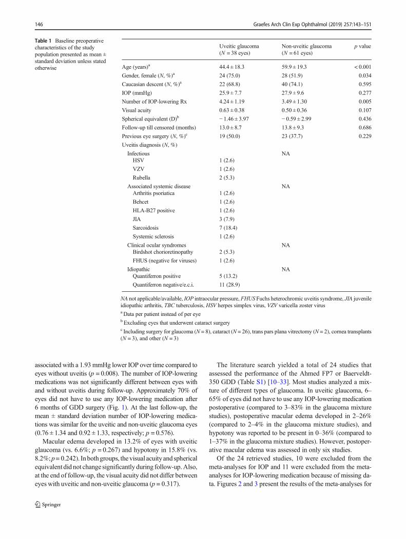
A total of 99 eyes (86 patients) were included (38 with uveiticglaucoma). Of the patients with uveitic glaucoma, 44.7% hadanterior uveitis, 2.6% had intermediate uveitis, 13.2% hadposterior uveitis, and the remaining 39.4% had panuveitis.Table 1 shows the baseline characteristics of the study popu-lation and the underlying cause of the uveitis. Patients withuveitic glaucoma were significantly younger, more often
female, and had a higher number of IOP-loweringmedicationscompared to patients with non-uveitic glaucoma. Almost halfof the patients had a history of intraocular surgery, most oftencataract surgery (52.6% of the uveitic glaucoma eyes and69.6% of the non-uveitic glaucoma eyes). The duration be-tween the first episode of the uveitis and the first glaucomasurgery was median (interquartile range) 4.0 (2–7) years.
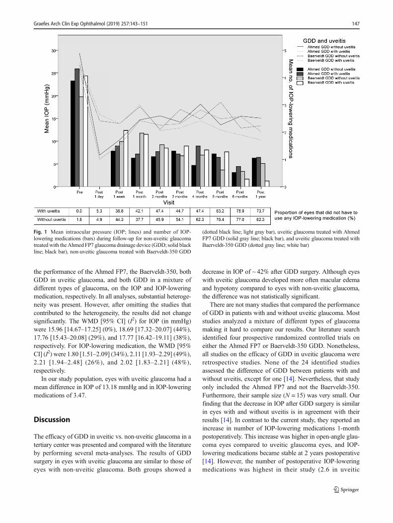
Figure 1 displays the IOP levels (separately for AhmedGDD and Baerveldt GDD) and number of IOP-lowering med-ications for the whole study population during follow-up.Nine eyes underwent a second surgery within the first yearafter the GDD surgery. Of these, one had uveitic glaucomaand underwent regular cataract extraction. The remaining(eight) had non-uveitic glaucoma of which one underwentregular trans pars plana vitrectomy, one developedneovascular glaucoma for which cyclocryotherapy was ap-plied, and six required GDD-related surgery because of tubemalposition (N = 2), tube protrusion through the conjunctiva(N = 1), tube removal (persistent hypotony; N = 2), and tubeflushing (N = 1). As we included two different GDD (Ahmedand Baerveldt), we first assessed whether there were differ-ences in the results between both GDD. None of the in Table 1presented baseline characteristics were significantly differentbetween both GDD, except for the preoperative number ofIOP-lowering medications (4.14 ± 1.36 and 3.50 ± 1.21 [p =0.015] for the Ahmed GDD and Baerveldt GDD, respective-ly). The IOP in the AhmedGDD group decreased from 27.5 ±9.1 mmHg preoperative to 14.5 ± 4.5 mmHg (p < 0.001; N =43) at the last follow-up, and in the Baerveldt GDD groupfrom 26.9 ± 8.9 mmHg to 12.1 ± 3.8 mmHg (p < 0.001; N =56). Figure 1 also shows that in the very early postoperativephase, the IOP was significantly lower with an Ahmed GDDcompared to a Baerveldt GDD; however, at 6 months, thischanged to a significantly higher IOP (p = 0.031). Also atthe last visit (> 1 year postoperative), eyes with an AhmedGDD had still a significantly higher IOP compared to theBaerveldt GDD (p = 0.005). The only significant differencein number of IOP-lowering medications during follow-up wasat 1 month postoperative (1.14 ± 1.25 and 1.96 ± 1.74 [p =0.010] for the Ahmed GDD and Baerveldt GDD, respective-ly). Because of the comparable performance of both GDD onthe IOP, we combined both GDD into one group with uveiticglaucoma and one with non-uveitic glaucoma. As expected,the IOP decreased significantly after GDD surgery in eyeswith uveitic glaucoma from 25.9 ± 7.7 to 12.7 ± 4.4 mmHg(44.9% decrease; p < 0.001), and in eyes with non-uveiticglaucoma from 27.9 ± 9.6 to 13.3 ± 4.2 mmHg (42.8% de-crease; p < 0.001). The postoperative decrease in IOP in eyeswith uveitic glaucoma did not differ with the decrease in IOP ineyes with non-uveitic glaucoma (p = 0.729). If we take the IOPof the whole follow-up period into account, the linear mixedmodel showed that age and uveitis were significantly associatedwith IOP during follow-up. The presence of uveitis was
Graefes Arch Clin Exp Ophthalmol (2019) 257:143–151 145
associated with a 1.93mmHg lower IOP over time compared toeyes without uveitis (p = 0.008). The number of IOP-loweringmedications was not significantly different between eyes withand without uveitis during follow-up. Approximately 70% ofeyes did not have to use any IOP-lowering medication after6 months of GDD surgery (Fig. 1). At the last follow-up, themean ± standard deviation number of IOP-lowering medica-tions was similar for the uveitic and non-uveitic glaucoma eyes(0.76 ± 1.34 and 0.92 ± 1.33, respectively; p = 0.576).
Macular edema developed in 13.2% of eyes with uveiticglaucoma (vs. 6.6%; p = 0.267) and hypotony in 15.8% (vs.8.2%;p= 0.242). Inbothgroups, thevisual acuityand sphericalequivalent did not change significantly during follow-up.Also,at the end of follow-up, the visual acuity did not differ betweeneyes with uveitic and non-uveitic glaucoma (p = 0.317).
The literature search yielded a total of 24 studies thatassessed the performance of the Ahmed FP7 or Baerveldt-350 GDD (Table S1) [10–33]. Most studies analyzed a mix-ture of different types of glaucoma. In uveitic glaucoma, 6–65% of eyes did not have to use any IOP-lowering medicationpostoperative (compared to 3–83% in the glaucoma mixturestudies), postoperative macular edema developed in 2–26%(compared to 2–4% in the glaucoma mixture studies), andhypotony was reported to be present in 0–36% (compared to1–37% in the glaucoma mixture studies). However, postoper-ative macular edema was assessed in only six studies.
Of the 24 retrieved studies, 10 were excluded from themeta-analyses for IOP and 11 were excluded from the meta-analyses for IOP-lowering medication because of missing da-ta. Figures 2 and 3 present the results of the meta-analyses for
Table 1 Baseline preoperativecharacteristics of the studypopulation presented as mean ±standard deviation unless statedotherwise
Uveitic glaucoma(N = 38 eyes)
Non-uveitic glaucoma(N = 61 eyes)
p value
Age (years)a 44.4 ± 18.3 59.9 ± 19.3 < 0.001
Gender, female (N, %)a 24 (75.0) 28 (51.9) 0.034
Caucasian descent (N, %)a 22 (68.8) 40 (74.1) 0.595
IOP (mmHg) 25.9 ± 7.7 27.9 ± 9.6 0.277
Number of IOP-lowering Rx 4.24 ± 1.19 3.49 ± 1.30 0.005
Visual acuity 0.63 ± 0.38 0.50 ± 0.36 0.107
Spherical equivalent (D)b − 1.46 ± 3.97 − 0.59 ± 2.99 0.436
Follow-up till censored (months) 13.0 ± 8.7 13.8 ± 9.3 0.686
Previous eye surgery (N, %)c 19 (50.0) 23 (37.7) 0.229
Uveitis diagnosis (N, %)
Infectious NAHSV 1 (2.6)
VZV 1 (2.6)
Rubella 2 (5.3)
Associated systemic disease NAArthritis psoriatica 1 (2.6)
Behcet 1 (2.6)
HLA-B27 positive 1 (2.6)
JIA 3 (7.9)
Sarcoidosis 7 (18.4)
Systemic sclerosis 1 (2.6)
Clinical ocular syndromes NABirdshot chorioretinopathy 2 (5.3)
FHUS (negative for viruses) 1 (2.6)
Idiopathic NAQuantiferron positive 5 (13.2)
Quantiferron negative/e.c.i. 11 (28.9)
NA not applicable/available, IOP intraocular pressure, FHUS Fuchs heterochromic uveitis syndrome, JIA juvenileidiopathic arthritis, TBC tuberculosis, HSV herpes simplex virus, VZV varicella zoster virusa Data per patient instead of per eyeb Excluding eyes that underwent cataract surgeryc Including surgery for glaucoma (N = 8), cataract (N = 26), trans pars plana vitrectomy (N = 2), cornea transplants(N = 3), and other (N = 3)
146 Graefes Arch Clin Exp Ophthalmol (2019) 257:143–151
the performance of the Ahmed FP7, the Baerveldt-350, bothGDD in uveitic glaucoma, and both GDD in a mixture ofdifferent types of glaucoma, on the IOP and IOP-loweringmedication, respectively. In all analyses, substantial heteroge-neity was present. However, after omitting the studies thatcontributed to the heterogeneity, the results did not changesignificantly. The WMD [95% CI] (I2) for IOP (in mmHg)were 15.96 [14.67–17.25] (0%), 18.69 [17.32–20.07] (44%),17.76 [15.43–20.08] (29%), and 17.77 [16.42–19.11] (38%),respectively. For IOP-lowering medication, the WMD [95%CI] (I2) were 1.80 [1.51–2.09] (34%), 2.11 [1.93–2.29] (49%),2.21 [1.94–2.48] (26%), and 2.02 [1.83–2.21] (48%),respectively.
In our study population, eyes with uveitic glaucoma had amean difference in IOP of 13.18 mmHg and in IOP-loweringmedications of 3.47.
Discussion
The efficacy of GDD in uveitic vs. non-uveitic glaucoma in atertiary center was presented and compared with the literatureby performing several meta-analyses. The results of GDDsurgery in eyes with uveitic glaucoma are similar to those ofeyes with non-uveitic glaucoma. Both groups showed a
decrease in IOP of ~ 42% after GDD surgery. Although eyeswith uveitic glaucoma developed more often macular edemaand hypotony compared to eyes with non-uveitic glaucoma,the difference was not statistically significant.
There are not many studies that compared the performanceof GDD in patients with and without uveitic glaucoma. Moststudies analyzed a mixture of different types of glaucomamaking it hard to compare our results. Our literature searchidentified four prospective randomized controlled trials oneither the Ahmed FP7 or Baerveldt-350 GDD. Nonetheless,all studies on the efficacy of GDD in uveitic glaucoma wereretrospective studies. None of the 24 identified studiesassessed the difference of GDD between patients with andwithout uveitis, except for one [14]. Nevertheless, that studyonly included the Ahmed FP7 and not the Baerveldt-350.Furthermore, their sample size (N = 15) was very small. Ourfinding that the decrease in IOP after GDD surgery is similarin eyes with and without uveitis is in agreement with theirresults [14]. In contrast to the current study, they reported anincrease in number of IOP-lowering medications 1-monthpostoperatively. This increase was higher in open-angle glau-coma eyes compared to uveitic glaucoma eyes, and IOP-lowering medications became stable at 2 years postoperative[14]. However, the number of postoperative IOP-loweringmedications was highest in their study (2.6 in uveitic
Fig. 1 Mean intraocular pressure (IOP; lines) and number of IOP-lowering medications (bars) during follow-up for non-uveitic glaucomatreated with the Ahmed FP7 glaucoma drainage device (GDD; solid blackline; black bar), non-uveitic glaucoma treated with Baerveldt-350 GDD
(dotted black line; light gray bar), uveitic glaucoma treated with AhmedFP7 GDD (solid gray line; black bar), and uveitic glaucoma treated withBaerveldt-350 GDD (dotted gray line; white bar)
Graefes Arch Clin Exp Ophthalmol (2019) 257:143–151 147

glaucoma and 2.8 in non-uveitic glaucoma) among all re-trieved studies. It should be noted that in the current study,the mean IOP at baseline was lower compared to other studies,but this might be related to the higher number of IOP-loweringmedications used by our patients (Fig. 3C, D). The non-significant difference in IOP between uveitic glaucoma andother forms of glaucoma may also be observed from the re-sults of the meta-analyses (Fig. 2C, D).
Our proportion of eyes requiring secondary surgery related tothe GDD (6.1%) is similar to other studies reporting 0–13%(Table S1). Due to the retrospective design of most studies, thedevelopment of macular edema may have been underreported.
Nevertheless, according to our results and the previous reports(Table S1), it is likely that the risk of developing macular edemaafter GDD surgery is higher in uveitic glaucoma compared tonon-uveitic glaucoma. The development of (transient) hypotonyin our study (15.8% in uveitic glaucoma and 8.2% in non-uveiticglaucoma) is in agreement with others; however, the reportedrange in other studies is extremely wide: 0–37.1% (Table S1).
One of the strengths of the current study is that the perfor-mance of the GDD did not depend on the great variability inhow glaucoma specialists perform GDD surgery, because allsurgeries were performed by the same surgeon using the sametechnique each time. Also, the indication for surgery was set
Fig. 2 Meta-analyses for the postoperative change in IOP after the Ahmed FP7 (A), Baerveldt-350 (B), both GDD in uveitic glaucoma (C), and bothGDD in a mixture of different types of glaucoma (D)
148 Graefes Arch Clin Exp Ophthalmol (2019) 257:143–151

by the same surgeon. To minimize the effect of different GDDon the IOP, only two types of GDD were included.
Furthermore, the IOP was measured at each visit using thesame method.
Fig. 3 Meta-analyses for the postoperative change in number of IOP-lowering medications after the Ahmed FP7 (A), Baerveldt-350 (B), both GDD inuveitic glaucoma (C), and both GDD in a mixture of different types of glaucoma (D)
Graefes Arch Clin Exp Ophthalmol (2019) 257:143–151 149

Due to the retrospective design of the current study, thereare several limitations. First, the follow-up of our study wasrelatively short. Nevertheless, according to the tube versustrabeculectomy study, the IOP of the Baerveldt GDD did notchange between 1-year follow-up and 5-year follow-up [31].Moreover, the Ahmed Baerveldt Comparison study andAhmed versus Baerveldt study did not show significant dif-ferences in IOP after 6 months [28, 30]. The study that com-pared the Ahmed GDD efficacy in uveitic glaucoma to open-angle glaucoma patients had a longer follow-up than the cur-rent study, but their results on IOP did not change significantlyafter 3 months of follow-up [14]. Second, our differences be-tween Ahmed GDD and Baerveldt GDD may suffer fromselection bias, because the surgeon may have a preferencefor a certain GDD in specific situations. For example, thehigher drop in IOP in the first week after AhmedGDD surgery(compared to Baerveldt GDD; Fig. 1) may result from thisselection bias next to the valve mechanism. However, thesignificantly higher IOP after Ahmed GDD surgery comparedto Baerveldt GDD at the end of follow-up is in agreement withtwo other studies that compared the Ahmed FP7 with theBaerveldt-350 GDD [28, 30]. Moreover, the preoperativeIOP level did not differ between Ahmed GDD and BaerveldtGDD (p = 0.660; Fig. 1), suggesting that selection bias basedon the preoperative IOP played a minor role. Regarding themeta-analyses, if different types of non-uveitic glaucomawould result in different outcomes of GDD surgery, cautionshould be taken when interpreting the studies including a mix-ture of types of glaucoma. The heterogeneity between studiesmay also be explained by the many different factors that mayaffect postoperative GDD results (e.g., experience of surgeon,surgical technique, follow-up duration, IOP measurementmethods, method used to calculate number of IOP-loweringmedications, severity of glaucoma, etc.).
The risk of developing secondary glaucoma due to uveitisdepends on various factors such as the age, ethnicity, durationof inflammation, the clinical manifestations of uveitis, and itstherapy. High risk for glaucoma development was especiallynoted for viral causes and anterior location of uveitis [34]. Ourresults are consistent with these findings, as the majority ofour patients exhibited anterior segment involvement ofuveitis.
In conclusion, both the Ahmed FP7 and the Baerveldt-350GDD are effective in lowering IOP in uveitic glaucoma. Theirperformance in terms of IOP and IOP-lowering medications issimilar to eyes with non-uveitic glaucoma. Also, the risk ofdeveloping macular edema or hypotony after GDD was notdifferent in uveitic glaucoma compared to non-uveitic glauco-ma. Unfortunately, prospective studies that assessed the clin-ical manifestations of uveitis in relation to GDD surgery arecurrently lacking. Future prospective randomized controlledtrials on uveitic glaucoma are required to verify the currentresults.
Funding The study did not receive any funding.
Compliance with ethical standards
Conflict of interest The authors declare that they have no conflict ofinterest.
Ethical approval All procedures performed in studies involving humanparticipants were in accordance with the ethical standards of the institu-tional and/or national research committee and with the 1964 Helsinkideclaration and its later amendments or comparable ethical standards.
For this type of study, formal consent is not required.
Open Access This article is distributed under the terms of the CreativeCommons At t r ibut ion 4 .0 In te rna t ional License (h t tp : / /creativecommons.org/licenses/by/4.0/), which permits unrestricted use,distribution, and reproduction in any medium, provided you giveappropriate credit to the original author(s) and the source, provide a linkto the Creative Commons license, and indicate if changes were made.
References
1. Baskaran M, Foo RC, Cheng C-Y et al (2015) The prevalence andtypes of glaucoma in an urban Chinese population: the SingaporeChinese eye study. JAMA Ophthalmol 133:874–880. https://doi.org/10.1001/jamaophthalmol.2015.1110
2. Merayo-Lloves J, Power WJ, Rodriguez A et al (1999) Secondaryglaucoma in patients with uveitis. Ophthalmologica 213:300–304.https://doi.org/10.1159/000027443
3. Takahashi T, Ohtani S, Miyata K et al (2002) A clinical evaluationof uveitis-associated secondary glaucoma. Jpn J Ophthalmol 46:556–562
4. Groen F, Ramdas W, de Hoog J et al (2016) Visual outcomes andocular morbidity of patients with uveitis referred to a tertiary centerduring first year of follow-up. Eye (Lond) 30:473–480. https://doi.org/10.1038/eye.2015.269
5. Minckler DS, Francis BA, Hodapp EA et al (2008) Aqueous shuntsin glaucoma. A Report by the American Academy ofOphthalmology. Ophthalmology 115:1089–1098. https://doi.org/10.1016/j.ophtha.2008.03.031
6. Mercieca K, Steeples L, Anand N, Medscape (2017) Deepsclerectomy for uveitic glaucoma: long-term outcomes. Eye(Lond) 31:1008–1019. https://doi.org/10.1038/eye.2017.80
7. Anton A, Heinzelmann S, Neß T et al (2015) Trabeculectomy abinterno with the Trabectome® as a therapeutic option for uveiticsecondary glaucoma. Graefes Arch Clin Exp Ophthalmol 253:1973–1978. https://doi.org/10.1007/s00417-015-3102-3
8. Higgins JPT, Thompson SG, Deeks JJ, Altman DG (2003)Measuring inconsistency in meta-analyses. BMJ 327:557–560.https://doi.org/10.1136/bmj.327.7414.557
9. Tobias A (1999) sbe26: assessing the influence of a single study inthe meta-analysis estimate. Stata Tech Bull STB-47:15–17
10. Aung T, Seah SKL (1998) Glaucoma drainage implants in Asianeyes. Ophthalmology 105:2117–2122. https://doi.org/10.1016/S0161-6420(98)91136-8
11. Bettis DI, Morshedi RG, Chaya C et al (2015) Trabeculectomy withmitomycin c or ahmed valve implantation in eyes with uveitic glau-coma. J Glaucoma 24:591–599. https://doi.org/10.1097/IJG.0000000000000195
150 Graefes Arch Clin Exp Ophthalmol (2019) 257:143–151
12. Kwon HJ, Kong YXG, Tao LW et al (2017) Surgical outcomes oftrabeculectomy and glaucoma drainage implant for uveitic glauco-ma and relationship with uveitis activity. Clin Exp Ophthalmol 45:472–480. https://doi.org/10.1111/ceo.12916
13. Mackenzie PJ, Schertzer RM, Isbister CM (2007) Comparison ofsilicone and polypropylene Ahmed glaucoma valves: two-year fol-low-up. Can J Ophthalmol 42:227–232. https://doi.org/10.3129/can.j.ophthalmol.i07-032
14. Rachmiel R, Trope GE, Buys YM et al (2008) Ahmed glaucomavalve implantation in uveitic glaucoma versus open-angle glauco-ma patients. Can J Ophthalmol 43:462–467. https://doi.org/10.3129/I08-082
15. Roy S, Ravinet E, Mermoud A (2001) Baerveldt implant in refrac-tory glaucoma: long-term results and factors influencing outcome.Int Ophthalmol 24:93–100. https://doi.org/10.1023/A:1016335313035
16. Seah SKL, Gazzard G, Aung T (2003) Intermediate-term outcomeof Baerveldt glaucoma implants in Asian eyes. Ophthalmology110:888–894. https://doi.org/10.1016/S0161-6420(03)00088-5
17. Sevgi D (2017) A retrospective study on the outcomes of Ahmedvalve versus Ahmed valve combined with fluocinolone implant inuveitic glaucoma. Digit J Ophthalmol 23:63–70. https://doi.org/10.5693/djo.01.2017.06.001
18. Siegner SW, Netland PA, Urban RC et al (1995) Clinical experiencewith the Baerveldt glaucoma drainage implant. Ophthalmology102:1298–1307. https://doi.org/10.1016/S0161-6420(95)30871-8
19. Smith MF, Doyle JW, Sherwood MB (1995) Comparison of theBaerveldt glaucoma implant with the double-plate Molteno drain-age implant. Arch Ophthalmol 113:444–447. https://doi.org/10.1001/archopht.1995.01100040060027
20. Sungur G, Yakin M, Eksioglu U et al (2017) Assessment of condi-tions affecting surgical success of Ahmed glaucoma valve implantsin glaucoma secondary to different uveitis etiologies in adults. Eye31:1435–1442. https://doi.org/10.1038/eye.2017.84
21. Syed HM, Law SK, Nam SH et al (2004) Baerveldt-350 implantversus Ahmed valve for refractory glaucoma: a case-controlledcomparison. J Glaucoma 13:38–45. https://doi.org/10.1097/00061198-200402000-00008
22. Brasil MVOM, Rockwood EJ, Smith SD (2007) Comparison ofsilicone and polypropylene ahmed glaucoma valve implants. JGlaucoma 16:36–41. https://doi.org/10.1097/01.ijg.0000243477.82779.31
23. Tan AN, Cornelissen MF, Webers CAB et al (2018) Outcomes ofsevere uveitic glaucoma treated with Baerveldt implant: can
blindness be prevented? Acta Ophthalmol 96:24–30. https://doi.org/10.1111/aos.13489
24. Tsai JC, Johnson CC, Kammer JA, DietrichMS (2006) The Ahmedshunt versus the Baerveldt shunt for refractory glaucoma II.Longer-term Outcomes from a Single Surgeon. Ophthalmology113:913–917. https://doi.org/10.1016/j.ophtha.2006.02.029
25. Wang JC, See JLS, Chew PTK (2004) Experience with the use ofBaerveldt and Ahmed glaucoma drainage implants in an Asianpopulation. Ophthalmology 111:1383–1388. https://doi.org/10.1016/j.ophtha.2003.11.005
26. Law SK, Nguyen A, ColemanAL, Caprioli J (2005) Comparison ofsafety and efficacy between silicone and polypropylene ahmedglaucoma valves in refractory glaucoma. Ophthalmology 112:1514–1520. https://doi.org/10.1016/j.ophtha.2005.04.012
27. Britt MT, Labree LD, Lloyd MA et al (1999) Randomized clinicaltrial of the 350-mm 2 versus the 500-mm 2 Baerveldt implant:longer term results is bigger better? Ophthalmology 106:2312–2318
28. Budenz DL, Barton K, Feuer WJ et al (2011) Treatment outcomesin the Ahmed baerveldt comparison study after 1 year of follow-up.Ophthalmology 118:443–452. https://doi.org/10.1016/j.ophtha.2010.07.016
29. Ceballos EM, Parrish RK, Schiffman JC (2002) Outcome ofBaerveldt glaucoma drainage implants for the treatment of uveiticglaucoma. Ophthalmology 109:2256–2260. https://doi.org/10.1016/S0161-6420(02)01294-0
30. Christakis PG, Tsai JC, Kalenak JWet al (2013) The Ahmed versusBaerveldt study: three-year treatment outcomes. Ophthalmology120:2232–2240. https://doi.org/10.1016/j.ophtha.2013.04.018
31. Gedde SJ, Schiffman JC, Feuer WJ et al (2012) Treatment out-comes in the tube versus trabeculectomy (TVT) study after fiveyears of follow-up. Am J Ophthalmol 153:789–803.e2. https://doi.org/10.1016/j.ajo.2011.10.026
32. Hoffman KB, Feldman RM, Budenz DL et al (2002) Combinedcataract extraction and baerveldt indications and outcomes. 6420:1916–1920
33. Iverson SM, Bhardwaj N, Shi W et al (2015) Surgical outcomes ofinflammatory glaucoma: a comparison of trabeculectomy andglaucoma-drainage-device implantation. Jpn J Ophthalmol 59:179–186. https://doi.org/10.1007/s10384-015-0372-6
34. Siddique SS, Suelves AM, Baheti U, Foster CS (2013) Glaucomaand uveitis. Surv Ophthalmol 58:1–10. https://doi.org/10.1016/j.survophthal.2012.04.006
Graefes Arch Clin Exp Ophthalmol (2019) 257:143–151 151