effects of surgically assisted rapid maxillary expansion
TRANSCRIPT

RESEARCH Open Access
Effects of surgically assisted rapid maxillaryexpansion on mandibular position: a three-dimensional studyTalles Fernando Medeiros Oliveira1*, Valfrido Antônio Pereira-Filho2, Mario Francisco Real Gabrielli2,Eduardo Sanches Gonçales3 and Ary Santos-Pinto1
Abstract
Background: This study aimed to evaluate three-dimensional changes in mandibular position after surgicallyassisted rapid maxillary expansion (SARME).
Methods: A retrospective study was carried out with tomographic records of 30 adult patients with maxillary transversedeficiency who underwent SARME. Cone beam computed tomography scans were obtained preoperatively (T1), afterexpansion (T2) and 6 months after expansion (T3). Mandibular landmarks were measured with respect to axial, sagittal,and coronal planes. Repeated measures ANOVA was used for statistical analysis.
Results: Clockwise rotation and lateral displacement of the mandible were observed immediately after SARME. However,mandibular displacements tended to return close to their initial values at T3.
Conclusions: Clockwise rotation and lateral shift of the mandible are transient effects of SARME.
Keywords: Malocclusion, Palatal expansion technique, Cone beam computed tomography
BackgroundSurgically assisted rapid maxillary expansion (SARME)has been widely used to treat the maxillary transversedeficiency in adult patients [1–5]. The main effects ofSARME occur transversally; however, skeletal changes insagittal and vertical planes as a result of expansion havealso been reported in the literature [1, 3, 4, 6, 7].Despite the effectiveness of expansion in the treat-
ment of maxillary transverse deficiencies, the possibilityof causing adverse changes in patient’s profile as aresult of mandibular displacement still causes concernin the indication of this procedure, mainly in hyperdi-vergent patients [8]. The clockwise rotation of the man-dible has been reported as one of the main effects ofSARME on the mandibular positioning; however, thereis no consensus about the extent and stability of thesechanges [4, 6, 9, 10].
A possible explanation for mandibular rotation afterSARME is the occlusal change due to extrusion andtipping of maxillary segments and cuspal interferencesas result of expansion [9]. Previous studies that assessedchanges in mandibular position after SARME have limi-tations since the cephalometric analysis used does notallow the three-dimensional evaluation of the mandibu-lar positioning, consequently lateral displacement of themandible due to expansion cannot be assessed. The useof cone beam computed tomography (CBCT) has advan-tages because it allows three-dimensional assessment ofbilateral structures without superimposition and withminimal distortion [11–13].This study aimed to evaluate the three-dimensional
changes in mandibular positioning after SARME.
MethodsThis retrospective study assessed the CBCT records of 30adult patients (mean age, 27.5 years; range 18.7–39.7 years;19 females and 11 males) with maxillary transversedeficiency greater than 5 mm and unilateral or bilateralposterior crossbite. Patients with cleft lip and palate or
* Correspondence: [email protected] of Orthodontics, School of Dentistry, São Paulo State University(UNESP), Rua Humaitá, 1680, Centro, Araraquara, São Paulo 14801-903, BrazilFull list of author information is available at the end of the article
© The Author(s). 2017 Open Access This article is distributed under the terms of the Creative Commons Attribution 4.0International License (http://creativecommons.org/licenses/by/4.0/), which permits unrestricted use, distribution, andreproduction in any medium, provided you give appropriate credit to the original author(s) and the source, provide a link tothe Creative Commons license, and indicate if changes were made.
Oliveira et al. Progress in Orthodontics (2017) 18:22 DOI 10.1186/s40510-017-0179-8

congenital craniofacial syndromes were excluded. Thisstudy was approved by the Ethics Committee of theAraraquara School of Dentistry, UNESP, (protocol14484713.1.0000.5416).
Surgery and treatment protocolSurgery was carried out under general anesthesia inhospital environment by two surgeons (V.A.P-F. andE.S.G.). SARME was performed with Subtotal LeFort Iosteotomy, midpalatal suture separation, and pterygo-maxillary disjunction. Patients were treated with Hyraxtype appliance and activation rate of one quarter turn(0.2 mm) three times a day until the crossbite correc-tion. The appliance activation was initiated 7 days post-operatively. After achieving the intended expansion ofthe maxilla width, the appliance was blocked and left inplace for about 4 months. Afterward, it was removedand replaced by a transpalatal arch.
CBCT analysisCBCT scans were acquired preoperatively (T1), immedi-ately after expansion (T2) and 6 months after expansion(T3) using an iCAT CBCT scanner (Imaging SciencesInternational, Hatfield, PA, USA) set up at 120 kVp,36 mA, 0.3 mm voxel, and FOV of 17 × 23 cm. Thepatients were positioned sitting upright in the naturalhead position, and they were instructed to occlude inmaximum habitual intercuspation during the CBCTscanning. The DICOM files were imported into Dolphin3D (version 11.5, Dolphin Imaging, Chatsworth, CA,USA) for further analysis. In order to maintain the samereference planes in all time points, head orientation ofeach data set was standardized using orientation tool inDolphin 3D software. The 3D orientation was performedaccording to three reference planes obtained from stable
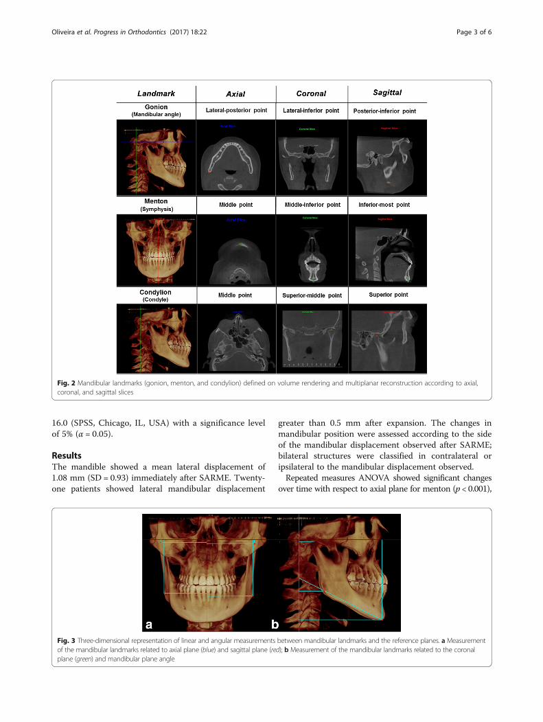
landmarks such as porion, orbitale, and nasion. TheFrankfurt horizontal plane was defined by the right andleft orbitale and the right and left porion landmarks. Thetransporionic plane was defined by the right and leftporion landmarks, perpendicular to Frankfurt horizontalplane. The midsagittal plane was defined as the planeorthogonal to axial and coronal planes passing throughnasion landmark [14]. Then, the head was moved so thatthe previously defined planes were coincident with thereference planes. The Frankfurt horizontal plane wasoriented to match the axial plane, the transporionicplane was oriented to match the coronal plane, and themidsagittal plane was moved to match the sagittal plane(Fig. 1). Afterward, the mandibular landmarks (Menton,the right and left condylion and the right and leftgonion) were defined using volume rendering and multi-planar reconstruction (Fig. 2). In order to assess thechanges in mandibular position at the three time points,linear and angular measurements were performedbetween the mandibular landmarks and the referenceplanes (Fig. 3).
Data analysisEighteen CBCT images were randomly chosen andassessed twice by the same calibrated examiner, with aminimum interval of 30 days. Reliability was confirmedby the intra-class correlation coefficient (ICC), whichranged from 0.929 to 0.996. The Shapiro-Wilk test wasused to investigate assumptions of normality. Longitu-dinal changes were evaluated using repeated measuresANOVA, Greenhouse-Geisser corrections were appliedfor data that violated sphericity assumptions. In statisti-cally significant results, the Bonferroni multiple com-parison test was used to assess differences betweentime points. Data analysis were performed using SPSS
Fig. 1 Reference planes: axial plane coincident with Frankfurt horizontal plane (blue), coronal plane coincident with transporionic plane (green),and sagittal plane coincident with midsagittal plane (red)
Oliveira et al. Progress in Orthodontics (2017) 18:22 Page 2 of 6
16.0 (SPSS, Chicago, IL, USA) with a significance levelof 5% (α = 0.05).
ResultsThe mandible showed a mean lateral displacement of1.08 mm (SD = 0.93) immediately after SARME. Twenty-one patients showed lateral mandibular displacement
greater than 0.5 mm after expansion. The changes inmandibular position were assessed according to the sideof the mandibular displacement observed after SARME;bilateral structures were classified in contralateral oripsilateral to the mandibular displacement observed.Repeated measures ANOVA showed significant changes
over time with respect to axial plane for menton (p < 0.001),
Fig. 2 Mandibular landmarks (gonion, menton, and condylion) defined on volume rendering and multiplanar reconstruction according to axial,coronal, and sagittal slices
Fig. 3 Three-dimensional representation of linear and angular measurements between mandibular landmarks and the reference planes. a Measurementof the mandibular landmarks related to axial plane (blue) and sagittal plane (red); b Measurement of the mandibular landmarks related to the coronalplane (green) and mandibular plane angle
Oliveira et al. Progress in Orthodontics (2017) 18:22 Page 3 of 6

and for contralateral gonion (p = 0.025) (Table 1). Inrelation to the coronal plane, only the menton measure-ment had significant changes (p < 0.001) (Table 2). How-ever, with respect to the sagittal plane, there were changesover time for ipsilateral condylion (p = 0.024), contralateralcondylion (p = 0.001), ipsilateral gonion (p = 0.018), andcontralateral gonion (p = 0.029) (Table 3). Measurementsof the mandibular plane angle (FMA) also changed signifi-cantly over the time of this study (p < 0.01) (Table 4).Multiple comparison test revealed differences in the
menton measures between T1 and T2 with respect tothe axial plane (1.35 mm) and to the coronal plane(−1.53 mm), showing downward and backward move-ment of this landmark immediately after SARME(Tables 1 and 2). However, the assessment at T3 revealeda relapse of these movements (T3-T2, p < 0.05). Similarchanges were found for measures of mandibular planeangle (FMA), indicating a transitional clockwise rotationof the mandible after expansion.Changes in mandibular landmark measures with respect
to sagittal plane confirm the lateral movement of themandible immediately after SARME (T2-T1, p < 0.05);however, no significant change was observed between T2and T3 neither between T1 and T3 (Table 3).
DiscussionThe possibility of causing adverse changes in patient’s pro-file as a result of mandibular displacement still causes
concern in indicating maxillary expansions [8]. Clockwiserotation of the mandible with an increase in lower facialheight has been reported as a side effect of SARME [6, 9].In fact, our study found a clockwise rotation of the man-dible immediately after SARME. This movement was rep-resented by an increase in the values of the FMA as wellas downward and backward displacements of the menton.However, according to our results, the mandibular
rotation seems to be a transient movement as the valuesobserved 6 months after SARME (T3) tended to returnclose to their initial values (T1). Altug-Atac et al. [6] andGunbay et al. [9] reported clockwise rotation of themandible after SARME whereas Parhiz et al. [4] andIodice et al. [10] did not observe significant rotationalmovement of the mandible. Methodological differencesamong these studies and assessment in different timepoints justify the divergence in their findings on mandibu-lar rotation. The first authors carried out the assessmentafter a short period following SARME whereas the otherauthors conducted a later evaluation. Our findings agreewith the studies found in the literature since a transientincrease in the mandibular plane angle was observed.Our findings showed that besides the clockwise rota-
tion, previously reported in literature, there is also alateral displacement of the mandible immediately afterSARME. However, it was not related with the type ofcrossbite presented previously. Variations on mandibulardisplacement could be observed among the patients,
Table 1 Mean and standard deviation of distances (millimeter) between the mandibular landmarks and the axial plane observed atthe three time-point evaluations. Results for repeated measures ANOVA
Mandibular landmarksAxial plane
T1 T2 T3 p
Mean SD Mean SD Mean SD
Ipsilateral condylion 0.79a 2.10 0.82a 1.97 0.72a 2.17 0.805
Contralateral condylion 0.75a 2.29 0.76a 2.00 0.87a 2.35 0.615
Ipsilateral gonion 58.34a 6.68 58.61a 6.79 58.47a 6.30 0.610
Contralateral gonion 58.49a 6.82 59.21b 6.68 59.98a,b 6.95 0.025
Menton 86.66a 8.00 88.01b 8.05 86.98a 7.92 <0.001
Different superscript letters show statistically significant differencesT1 preoperatively, T2 immediately after expansion, T3 6 months after expansion, SD standard deviation
Table 2 Mean and standard deviation of distances (millimeter) between the mandibular landmarks and the coronal plane observedat the three time-point evaluations. Results for repeated measures ANOVA
Mandibular landmarksCoronal plane
T1 T2 T3 p
Mean SD Mean SD Mean SD
Ipsilateral condylion 15.93a 1.69 15.98a 1.70 15.76a 1.58 0.392
Contralateral condylion 15.91a 1.40 15.87a 1.44 15.83a 1.48 0.929
Ipsilateral gonion 26.09a 4.69 25.62a 5.18 26.22a 4.92 0.257
Contralateral gonion 25.75a 4.68 25.18a 5.70 25.92a 5.59 0.226
Menton 90.88a 8.77 89.35b 8.79 90.31a 9.13 <0.001
Different superscript letters show statistically significant differencesT1 preoperatively, T2 immediately after expansion, T3 6 months after expansion, SD standard deviation
Oliveira et al. Progress in Orthodontics (2017) 18:22 Page 4 of 6

even in those with unilateral posterior crossbite. Thelack of a pattern for mandibular displacement can beexplained by individual changes in the pattern of occlu-sion following the expansion, such as in asymmetricexpansion [15]. Thus, the direction to which the man-dible will move after SARME becomes unpredictable inadult patients, in contrast to the correction of posturalasymmetry found in children with functional unilateralposterior crossbites [16].Changes observed in condylion and gonion landmarks
with respect to the sagittal plane occurred because theanalysis was performed considering the mandibulardisplacement. So, one would expect an increase in thedistance from the landmarks ipsilateral to mandibulardisplacement to the midsagittal plane, as well as adecrease in the distance from the contralateral structuresto the same plane. Thus, even though an average dis-placement of 1.08 mm had been observed in mentonbetween T1 and T2, it was not possible to predict thedirection of this change since this landmark can moveaway or closer to the midsagittal plane as a result of themandibular movement after SARME. Despite thischanges occur at T2, there was a tendency to return tooriginal position 6 months after expansion, so that nosignificant difference was observed between T3 and T1.Additionally, mandibular lateral movements were smalland showed no clinical relevance.Mandibular movements take place in three dimen-
sions; thereby, bilateral mandibular structures may showdistinct behaviors during SARME. Such fact was
observed in vertical changes of the gonion, which wassignificant only to the contralateral side to the mandibu-lar displacement. This resulted in different values of themandibular plane angle between the ipsilateral andcontralateral sides, although both have shown a signifi-cant increase.
ConclusionsThis study suggests the presence of mandibular displace-ment in most patients after SARME; however, the direc-tion of this displacement cannot be predicted. Clockwiserotation and mandibular lateral displacement are transi-ent effects of SARME.
AbbreviationsCBCT: Cone beam computed tomography; SARME: Surgically assisted rapidmaxillary expansion
FundingNone.
Authors’ contributionsTFMO has contributed with acquisition and statistical analysis of data anddrafted the manuscript. VAPF and ESG have undertaken the surgical part ofthe study and acquisition of CBCT. MFRG and ASP have contributed to thedesign of the study and revised the manuscript. All authors have read andapproved the final manuscript.
Ethics approvalThis study was approved by the Ethics Committee of Araraquara School ofDentistry, under protocol number - CAAE: 14484713.1.0000.5416.
Competing interestsThe authors declare that they have no competing interests.
Publisher’s NoteSpringer Nature remains neutral with regard to jurisdictional claims inpublished maps and institutional affiliations.
Author details1Department of Orthodontics, School of Dentistry, São Paulo State University(UNESP), Rua Humaitá, 1680, Centro, Araraquara, São Paulo 14801-903, Brazil.2Department of Oral and Maxillofacial Surgery, School of Dentistry, SãoPaulo State University (UNESP), Araraquara, São Paulo, Brazil. 3Departmentof Stomatology, School of Dentistry, São Paulo University, Bauru, SãoPaulo, Brazil.
Table 3 Mean and standard deviation of distances (millimeter) between the mandibular landmarks and the sagittal plane observedat the three time-point evaluations. Results for repeated measures ANOVA
Mandibular landmarkssagittal plane
T1 T2 T3 p
Mean SD Mean SD Mean SD
Ipsilateral condylion 48.63a 3.22 49.13b 2.87 48.85a,b 2.87 0.024
Contralateral condylion 48.73a 3.07 47.93b 3.18 48.26a,b 46.05 0.001
Ipsilateral gonion 46.05a 3.05 46.67b 3.11 46.40a,b 3.05 0.018
Contralateral gonion 45.70a 3.81 45.17b 3.68 45.59a,b 3.58 0.029
Menton 2.23a 1.92 1.91a 1.49 2.04a 1.52 0.297
Different superscript letters show statistically significant differencesT1 preoperatively, T2 immediately after expansion, T3 6 months after expansion, SD standard deviation
Table 4 Mean and standard deviation of mandibular angleobserved at the three time-point evaluations. Results for repeatedmeasures ANOVA
Mandibular angle T1 T2 T3 p
Mean SD Mean SD Mean SD
Ipsilateral FMA 19.74a 4.73 20.95b 4.79 19.99a 4.60 <0.001
Contralateral FMA 19.57a 4.55 20.30b 4.76 19.67a 4.80 0.003
Different superscript letters show statistically significant differencesFMA mandibular plane angle, T1 preoperatively, T2 immediately afterexpansion, T3 6 months after expansion, SD standard deviation
Oliveira et al. Progress in Orthodontics (2017) 18:22 Page 5 of 6

Received: 22 May 2017 Accepted: 27 June 2017
References1. Chung CH, Woo A, Zagarinsky J, Vanarsdall RL, Fonseca RJ. Maxillary sagittal
and vertical displacement induced by surgically assisted rapid palatalexpansion. Am J Orthod Dentofacial Orthop. 2001;120:144–8.
2. Anttila A, Finne K, Keski-Nisula K, et al. Feasibility and long-term stability ofsurgically assisted rapid maxillary expansion with lateral osteotomy. Eur JOrthod. 2004;26:391–5.
3. Lagravere MO, Major PW, Flores-Mir C. Dental and skeletal changesfollowing surgically assisted rapid maxillary expansion. Int J Oral MaxillofacSurg. 2006;35:481–7.
4. Parhiz A, Schepers S, Lambrichts I, et al. Lateral cephalometry changes afterSARPE. Int J Oral Maxillofac Surg. 2011;40:662–71.
5. Prado GP, Furtado F, Aloise AC, et al. Stability of surgically assisted rapidpalatal expansion with and without retention analyzed by 3-dimensionalimaging. Am J Orthod Dentofacial Orthop. 2014;145:610–6.
6. Altug Atac AT, Karasu HA, Aytac D. Surgically assisted rapid maxillaryexpansion compared with orthopedic rapid maxillary expansion. AngleOrthod. 2006;76:353–9.
7. Bretos JL, Pereira MD, Gomes HC, Toyama Hino C, Ferreira LM. Sagittal andvertical maxillary effects after surgically assisted rapid maxillary expansion(SARME) using Haas and Hyrax expanders. J Craniofac Surg. 2007;18:1322–6.
8. Lineberger MW, McNamara JA, Baccetti T, Herberger T, Franchi L. Effects ofrapid maxillary expansion in hyperdivergent patients. Am J OrthodDentofacial Orthop. 2012;142:60–9.
9. Gunbay T, Akay MC, Gunbay S, et al. Transpalatal distraction using bone-borne distractor: clinical observations and dental and skeletal changes. JOral Maxillofac Surg. 2008;66:2503–14.
10. Iodice G, Bocchino T, Casadei M, Baldi D, Robiony M. Evaluations of sagittaland vertical changes induced by surgically assisted rapid palatal expansion.J Craniofac Surg. 2013;24:1210–4.
11. Hilgers ML, Scarfe WC, Scheetz JP, Farman AG. Accuracy of lineartemporomandibular joint measurements with cone beam computedtomography and digital cephalometric radiography. Am J OrthodDentofacial Orthop. 2005;128:803–11.
12. Ikeda K, Kawamura A. Assessment of optimal condylar position with limitedcone-beam computed tomography. Am J Orthod Dentofacial Orthop. 2009;135:495–501.
13. Sanders DA, Rigali PH, Neace WP, Uribe F, Nanda R. Skeletal and dentalasymmetries in class II subdivision malocclusions using cone-beam computedtomography. Am J Orthod Dentofacial Orthop. 2010;138(542):e1–20.
14. Cevidanes L, Oliveira AEF, Motta A, Phillips C, Burke B, Tyndall D. Headorientation in CBCT-generated cephalograms. Angle Orthod. 2009;79:971–7.
15. Koudstaal MJ, Smeets JB, Kleinrensink GJ, Schulten AJ, van der Wal KG.Relapse and stability of surgically assisted rapid maxillary expansion: ananatomic biomechanical study. J Oral Maxillofac Surg. 2009;67:10–4.
16. Pinto AS, Buschang PH, Throckmorton GS, Chen P. Morphological andpositional asymmetries of young children with functional unilateralposterior crossbite. Am J Orthod Dentofacial Orthop. 2001;120:513–20.
Oliveira et al. Progress in Orthodontics (2017) 18:22 Page 6 of 6