effects of sulphydryl reagents on the structure...
TRANSCRIPT
J. Cell Sci. 73, 245-260 (1985) 245Printed in Great Britain © The Company of Biologists Limited 1985
EFFECTS OF SULPHYDRYL REAGENTS ON THE
STRUCTURE OF DEHISTONIZED METAPHASE
CHROMOSOMES
PETER JEPPESEN AND HELEN MORTENMRC Clinical and Population Cvtogenelics Unit, Western General Hospital, Crewe Road,Edinburgh EH4 2XU, U.K.
SUMMARY
Dehistonized metaphase chromosomes lose their apparent axial organization (the 'scaffold') andsediment more slowly following exposure to )3-mercaptoethanol (BME). We have subsequentlytreated BME chromosomes with reagents that oxidize protein sulphydryls to disulphides, andfound that if calcium is also present during the oxidation an apparently similar axial structure isrestored following dehistonization, as seen by microscopic examination. In general, however, wedo not find that oxidation restores the higher sedimentation rate of dehistonized controlchromosomes. Analysis of residual core protein in dehistonized chromosomes by sodium dodecylsulphate/polyacrylamide gel electrophoresis fails to detect any differences in polypeptidecomposition related to the state of oxidation or to the presence or absence of visible axialorganization. Combining our results with those of other workers, we conclude that the axialstructure evident in dehistonized metaphase chromosomes is maintained, at least partially, byinter-protein cross-linking, although in vivo this may not be via simple disulphide bridges.Additional factors, which we have not yet characterized, but which possibly include heavy metalions, appear to be involved in the axial organization existing in vivo.
INTRODUCTION
The organization of chromatin in eukaryotic chromosomes is not static. Aparticularly striking change in the degree of chromatin condensation occurs duringcell division, at both mitosis and meiosis, and it has been suggested that a possiblemechanism for achieving this might involve the formation of interprotein disulphidebridges (e.g. see Dounce, Chanda & Townes, 1973). There is a body of biochemicaland cytochemical evidence to show that the -SS-/-SH ratio is higher in condensedchromatin, whether in interphase or at mitosis, although there are a number ofconflicting reports in the literature also (for a review, see Sumner, 1983). Thedifficulty is in determining the in vivo oxidation state - sulphydryl groups maybecome oxidized, or disulphide bridges reduced, in the process of samplepreparation. During the course of our work on the structure of metaphasechromosomes, we have examined the effects of adding disulphide reducing agents,such as /5-mercaptoethanol (BME) and dithiothreitol (DTT) to chromosomesisolated by a variety of procedures (e.g. see Jeppesen, Bankier & Sanders, 1978;Gooderham & Jeppesen, 1983), and have been unable to detect any alteration in
Key words: chromosome, dehistonized, metaphase, sulphydryl reagents.
246 P. Jeppesen and H. Morten
morphology or other properties of native chromosomes, following exposure to theseagents (unpublished observations). It has been reported that reduction of disulphidebonds leads to swelling in methanol: acetic-fixed chromosomes (Dounce et al. 1973;Sumner, 1973), but acid fixation can be expected to have considerably altered thestate of the chromosomes under these conditions.
When metaphase chromosomes that have not been exposed to BME or DTT aretreated with 2ivi-NaCl to remove histones, as described before (Gooderham &Jeppesen, 1983; Paulson & Laemmli, 1977), many of the resulting structuresappear, by both light and electron microscopy, as a 'halo' of DNA surrounding anaxial filament or 'scaffold', although some structures have a much more expandedappearance (Gooderham & Jeppesen, 1983; Hadlaczky, Sumner & Ross, 1981). WehaVe shown that these axial regions not only have an apparently high concentrationof DNA, as indicated by their bright fluorescence with DNA fluorochromes such as.Hoechst '33258', but that they also contain the bulk of the non-histone protein(NHP) that is resistant to extraction by 2M-NaCl (Gooderham & Jeppesen, 1983).The NHP may be freed of over 99 % of its associated DNA by nuclease digestion,resulting in the structures we have called chromosome 'cores' (Jeppesen & Bankier,1979). There is evidence from a number of sources of axial organization ofchromatin in metaphase chromosomes (Satya-Prakash, Hsu & Pathak, 1980;Adolph, 1980), and we think it likely that the axial filaments in dehistonizedchromosomes, and hence chromosome cores, might retain the elements of thishigher-order structure, which we envisage as an interaction between NHP andspecific DNA sequences (Jeppesen et al. 1978). As we shall show later, dehis-tonization of metaphase chromosomes that have been exposed to either BME orDTT leads to structures that are all of a highly expanded form, with littleresemblance to chromosomes, and having no visibly identifiable axial organization.It has also been reported that there is a reduction in sedimentation coefficient with aloss of associated proteins in chromosome scaffolds prepared from BME-treatedchromosomes (Lewis & Laemmli, 1982).
These observations of the effects of apparently reducing disulphide bonds indehistonized mitotic chromosomes are remarkably similar to what would bepredicted by earlier models of chromosome structure, involving a central proteincore maintained by disulphide bridging (Dounce et al. 1973; Sobell, 1973).However, Lewis & Laemmli (1982), based on observations with the chelating agentso-phenanthroline (OP) and neocuproine, have proposed that BME and DTT act notby reducing disulphide bridges, but by chelating a metal ion contained as ametalloprotein component of the chromosome scaffold, although their results doimplicate the involvement of protein sulphydryl groups. They have furthersuggested that the metal chelated is copper, since they show that cupric ions arecapable of restoring a high sedimentation rate to dehistonized BME-treatedchromosomes. We report here the results of treating Chinese hamster metaphasechromosomes isolated in the presence of BME with agents specifically selected tooxidize free protein sulphydryls to disulphide bonds, and compare these with theeffects of copper. We shall demonstrate that in the presence of calcium, both cupric
Sulphydryl reagents and chromosome structure 247
ions and oxidizing agents are indistinguishable in reproducing axial filaments clearlyvisible in the light microscope following dehistonization, and morphologicallyclosely resembling those observed in non-BME-treated dehistonized chromosomes.Since we detected no significant differences in polypeptide composition, weconclude that axial filaments are a consequence of protein cross-linking. Of theprocedures we have investigated, however, only cupric ions lead to an apparentpartial restoration of high sedimentation rate in dehistonized structures, althoughthis is also produced in the absence of calcium, when axial filaments are not evident.The implications of these observations with reference to chromosome structure invivo will be discussed.
MATERIALS AND METHODS
Metaphase chromosome isolation
CH0-K1 Chinese hamster ovary cells (ATCC no. CCL 61) were adapted for spinner culture,and grown in modified Eagle's Minimum Essential Medium for Suspension Cultures (FlowLaboratories), supplemented with 10% foetal calf serum, 30/xg/ml i.-proline, lOOi.u./mlpenicillin and 100/xg/ml streptomycin. Mitotic enrichment was achieved by partial synchron-ization of cultures as follows: cells were grown to stationary phase, then diluted twofold with freshmedium; after 8h incubation, colcemid (Fluka) was added to 0 1 /u.g/ml, and growth continued fora further 16 h. In this way, up to SO % of cells in metaphase were obtained. For tritium labelling ofchromosomal DNA, [me(/ry/-3H]thymidine (5 Ci/mmol, Amersham International) was added tothe culture medium to a final activity of OS/xCi/ml at the time of twofold dilution.
Metaphase chromosomes were isolated by the 'physiological' procedure described previously(Gooderham & Jeppesen, 1983), with the following modifications. Approx. 2x 10⁸ total cells wereharvested by centrifugation at 1000 rev./min and resuspended in 2x 10 ml samples of fresh culturemedium. After 30min on ice the cells were pelleted by centrifugation again, and trypsinized for5 min at 37 °C (Gooderham & Jeppesen, 1983). After washing each sample with a further 10 ml offresh culture medium to inactivate trypsin activity, the cells were pelleted once more, and eachsample was resuspended in 10ml 50m.M-KCl, which for one sample contained 7mM-/3-mercaptoethanol (BME). After 10min of hypotonic treatment at 37°C, the swollen cells werecooled, collected by centrifugation at 1000 rev./min and resuspended in cold 120niM-KCl,20 mM-NaCl, 10 niM-TrisHCl (pH 8), 2 mM-CaCl2, again with the inclusion of 7 m.\i-BME to thepreviously similarly treated sample. Cell lysis was carried out after the addition of 0 1 % (v/v)Triton X-100 by passing the cells through a hypodermic needle several times (Gooderham &Jeppesen, 1983). Excess calcium was removed with EDTA, and the chromosomes purified by theone-step 10% to 50% glycerol gradient centrifugation described before (Gooderham & Jeppesen,1983). For this and all subsequent procedures, both samples ('BME chromosomes', and non-BMEor 'standard' chromosomes) were treated identically in parallel, with no further exposure to BME.The chromosome peaks were collected from the glycerol gradients and dialysed against120mM-KCl, 20niM-NaCl, 10niM-TrisHCl (pH8), 0-1% (v/v) Triton X-100, (KCM). Tominimize chance oxidation, all solutions were degassed under reduced pressure before use.
Although the proportion of interphase nuclei in the crude lysatc from suspension cultures wasmuch higher than obtained previously from monolayer 'wash off mitotic cells (Gooderham &Jeppesen, 1983), nuclei pelleted at the bottom of the glycerol gradient, and the chromosomepreparations described here appeared by both microscopic examination and polyacrylamide gelpolypeptide analysis to be equally clean.
Treatment ivith oxidizing and other agentsWhere required, after removing glycerol by dialysis, samples of standard and/or BME
chromosomes were variously treated before dehistonization, as follows. The chromosomes
248 P. Jeppeson and H. Morten
suspended in KCM were incubated for 2h on ice with the appropriate agent, with occasionalgentle agitation to prevent chromosomes settling and aggregating. The final concentrations ofagents used for these treatments were: 7m.\i-/3-mercaptoethanol; 3 m.M-dithiothreitol; 10 ITIM-EDTA; 3 mM-o-phenanthroline; (H % (w/v) ammonium persulphate; 3 rmi-oxidized glutathione(GSSG); 0-1 mM-CuS04; 0-01 ITIM-0-1 mM-copper-OP complex (Cu(OP)2). In addition, whereindicated in the text, 1 m:\1-MgCl2 or 1 m:\1-CaCI2 were also included.
Dehistonization and preparation of core proteins
Following treatment, if appropriate, as described above, the chromosome suspension containingapproximately 10/ig/ml DNA was extracted with 2M-NaCl to remove histones essentially asdescribed previously (Gooderham & Jeppesen, 1983). After adding 2/3 vol. of 5 M-NaCl, gentlymixing, and standing for 30min on ice, the dehistonized chromosomes were separated fromdissociated protein by centrifugation through a solution containing 2M-NaCl, 2-5% (w/v)sucrose, 10mM-TrisHCl (pH8), 0 1 % (v/v) Triton X-100 (NaCl/sucrose). To prepare coreproteins for sodium dodecyl sulphate (SDS)/polyacrylamide gel electrophoresis, 20 ml of2M-NaCl chromosome suspension was layered onto 10 ml of NaCl/sucrose supported on 0-3 ml of0-6M-Metrizamide (Nyegaard & Co., Oslo), 2M-NaCl, lOmM-TrisHCl (pH8), 0 1 % (v/v)Triton X-100, centrifuged for 4h at 3000 rev./min in a Sorvall AH 627 swingout rotor, and thedehistonized chromosomes collected from the Metrizamide/sucrose boundary with a Pasteurpipette (Gooderham & Jeppesen, 1983). After dialysis at 4°C for 16h against 10mM-Tris-HCl(pH8), lmM-MgCl2, 0-1% (v/v) Triton X-100 to remove salt, DNA was liberated fromchromosome core protein by digesting on ice for 30min with 10/xg/ml DNase 1 (Sigma). Enzymeactivity was terminated by addition of EDTA to 10 HIM, and core protein collected bycentrifugation at 25 000 rev./min (approx. 50000#,v), in a Sorvall AH 650 swingout rotor.SDS/polyacrylamide gel electrophoresis and sample preparation were as previously described(Gooderham & Jeppesen, 1983). Low molecular weight marker proteins were obtained fromPharmacia^.
To follow the sedimentation properties of dehistonized "'H-labelled chromosomes, asmaller-scale version of the centrifugation procedure described above was used; 1 ml of 2M-NaClchromosome suspension (activity 1x10 to 2xlO4 disints/min) was layered onto 3'5 mlNaCl/sucrose, supported on 0-1 ml 0-6.M-Metrizamide solution, contained in a 5-5 ml pblyallomercentrifuge tube. The tube was then centrifuged at 3000 rev./min for 3 h at 4°C in a Sorvall AH 650rotor, after which 1-ml fractions were carefully removed from the top using a wide-bore Pasteurpipette. (Other methods of collecting fractions containing the viscous dehistonized material provedunsatisfactory.) Each total fraction was added to 2-5 ml Aquasol (New England Nuclear), shakenbriefly to obtain a clear gel, and the radioactivity was measured by scintillation counting.
For preparation of microscopic specimens, lml of NaCl/sucrose containing 4% (w/v)formaldehyde was added to a 15 mmX50 mm flat-bottom glass vial holding a 13 mm circular glasscoverslip; 0-1 ml of 2M-NaCl chromosome suspension was gently layered on top, and thedehistonized chromosomes were centrifuged onto the coverslip at 3000 rev./min for 15min in aswing-out bench centrifuge. After allowing to fix by standing 30min at room temperature, thecoverslip was removed, rinsed in distilled water and stained with Hoechst 33258 as describedbefore (Jeppesen et al. 1978).
RESULTS
The axial organization in non-BME-treated Chinese hamster metaphasechromosomes ('standard' chromosomes), following dissociation of histones with2M-NaCl, is most clearly shown in the light microscope by using a DNAfluorochrome such as Hoechst 33258, as illustrated in Fig. 1A. The majority of theresulting structures have a typical appearance of a diffuse 'halo' of DNA surroundinga more brightly fluorescent axial filament. The protein-specific fluorochromefluorescein isothiocyanate (FITC) also stains the NHP-rich axial filaments

Sulphydryl reagents and chromosome structure Z4f
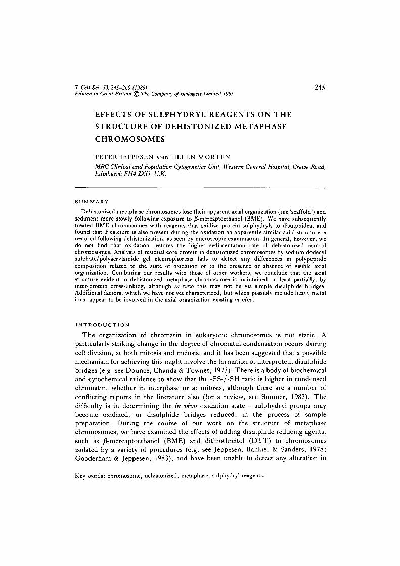
Fig. 1. Abolition of visible chromosome 'cores' by )3-mercaptoethanol. DehistonizedChinese hamster metaphase chromosomes were stained with Hoechst 33258 andobserved by blue-light fluorescence. \. Standard chromosome isolation procedure; H,chromosomes isolated in the presence of 7niM-BME. approx. X3000.
250 P. Jeppesen and H. Morten
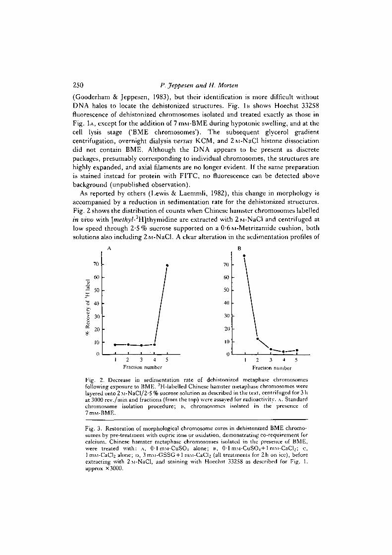
(Gooderham & Jeppesen, 1983), but their identification is more difficult withoutDNA halos to locate the dehistonized structures. Fig. 1B shows Hoechst 33258fluorescence of dehistonized chromosomes isolated and treated exactly as those inFig. LA, except for the addition of 7 ITIM-BME during hypotonic swelling, and at thecell lysis stage ('BME chromosomes'). The subsequent glycerol gradientcentrifugation, overnight dialysis versus KCM, and 2M-NaCl histone dissociationdid not contain BME. Although the DNA appears to be present as discretepackages, presumably corresponding to individual chromosomes, the structures arehighly expanded, and axial filaments are no longer evident. If the same preparationis stained instead for protein with FITC, no fluorescence can be detected abovebackground (unpublished observation).
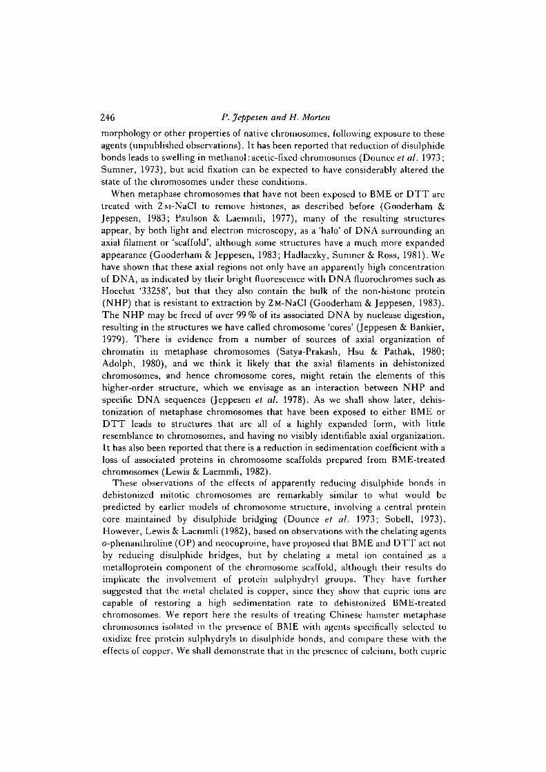
As reported by others (Lewis & Laemmli, 1982), this change in morphology isaccompanied by a reduction in sedimentation rate for the dehistonized structures.Fig. 2 shows the distribution of counts when Chinese hamster chromosomes labelledin vivo with [/ne</ry/-3H]thymidine are extracted with 2M-NaCl and centrifuged atlow speed through 2-5% sucrose supported on a 0-6M-Metrizamide cushion, bothsolutions also including 2ivi-NaCl. A clear alteration in the sedimentation profiles of
70
60
•2 50X
"o 40
30
20
10
0
70
60
50
40
30
20
10
0
*
1 2 3 4 5Fraction number
1 2 3 4 5
Fraction number
Fig. 2. Decrease in sedimentation rate of dehistonized metaphase chromosomesfollowing exposure to BME. 3H-labelled Chinese hamster metaphase chromosomes werelayered onto 2 M-NaCl/2-5 % sucrose solution as described in the text, centrifuged for 3 hat 3000 rev./min and fractions (from the top) were assayed for radioactivity. ,\. Standardchromosome isolation procedure; B, chromosomes isolated in the presence of7mM-BME.
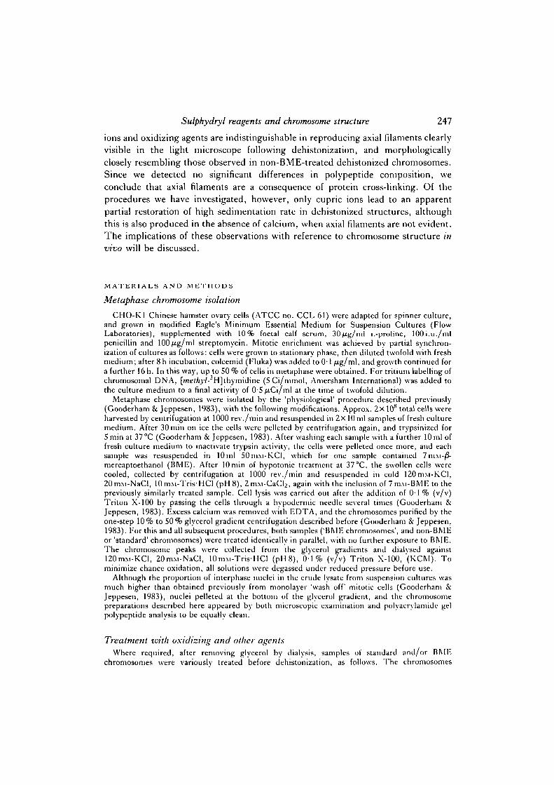
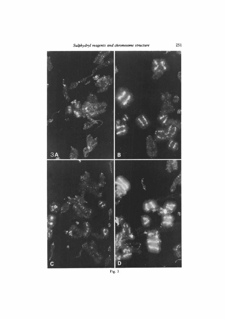
Fig. 3. Restoration of morphological chromosome cores in dehistonized BME chromo-somes by pre-treatment with cupric ions or oxidation, demonstrating co-requirement forcalcium. Chinese hamster metaphase chromosomes isolated in the presence of BME,were treated with: A, 0-lniM-CuSO4 alone; n, 0 1 niM-CuSO4+1 m:\1-CaCl2; c,1 mM-CaCl2 alone; D, 3mM-GSSG + l m:\1-CaCl2 (all treatments for 2h on ice), beforeextracting with 2M-NaCl, and staining with Hoechst 33258 as described for Fig. 1.approx X3000.
Sulphydryl reagents and chromosome structure 251
Fig. 3
252 P. Jeppesen and H. Morten
dehistonized chromosomes is evident following exposure to BME: standardchromosomes (Fig. 2A) sediment to the Metrizamide cushion, whereas BMEchromosomes (Fig. 2B) fail to penetrate far into the 2-5 % sucrose solution. Similareffects on the morphology and sedimentation rate of dehistonized chromosomes areproduced if BME is added to standard chromosomes at any stage up to andincluding the 2ivi-NaCl extraction step; 3 mru-DTT and 3 miu-OP were also foundto produce identical results to BME (not shown), but lOmiu-EDTA has no effect.
BME chromosomes were treated in a number of ways before extracting histoneswith 2M-NaCl, and the effects on the resulting dehistonized structures observed.Although we were primarily interested in alterations to the NHP axial filaments, wechose the DNA fluorochrome Hoechst 33258 for the morphological assay for thereasons noted above. Fig. 3 compares four pre-treatments. Incubation with0-1 mM-CuSO4 alone for 2 h (Lewis & Laemmli, 1982) has no apparent effect on ourpreparations of BME chromosomes (Fig. 3A), but if 1 niM-CaCl2 is also included(Fig. 3B), a high proportion of dehistonized chromosomes exhibit brightlyfluorescent axial filaments, similar to those seen after dehistonization of standardchromosomes (Fig. 1A); 1 mM-CaCl2 by itself has no effect (Fig. 3c). Magnesiumions may be substituted for calcium, but a lower proportion of dehistonizedstructures with axial filaments is subsequently obtained (not shown here).
If BME chromosomes are treated for 2h with oxidized glutathione (GSSG) tooxidize free protein sulphydryls to disulphide bonds, axial filaments are also seenfollowing extraction of histones with 2i\i-NaCl (Fig. 3D). AS in the case of CuSC>4treatment described above, the presence of calcium (or less efficiently, magnesium)is also required. In addition to using GSSG, we have also oxidized BMEchromosomes with the less-specific agent ammonium persulphate, and the complexformed between cupric ions and OP (Cu(OP)2), which, in the presence of freeoxygen, is specific for oxidizing sulphydryls to disulphide bonds (Kobashi, 1968).Both of these procedures, in the presence of calcium, also lead to dehistonizedstructures with axial filaments, similar to those of Fig. 3B and D (not illustrated). Ofthe procedures we have used to reproduce axial filaments following dehistonizationof BME chromosomes, the most efficient are CuSO4/Ca, and Cu(OP)2/O2/Ca, thelatter effective at a concentration of Cu(OP)2 as low as 0-01 niM. Interestingly, ifexcess OP is used in the mixture with CUSO4, then axial filaments are not observed,which is consistent with the observation of Lewis & Laemmli (1982), and our ownresults, that OP treatment of chromosomes itself destroys evidence of axial structurefollowing dehistonization. It should be stressed that the treatments described here toreverse the effects of BME are only effective if applied to BME chromosomes beforedehistonization takes place. The loss of axial structure induced by BME appearsnon-reversible following dehistonization.
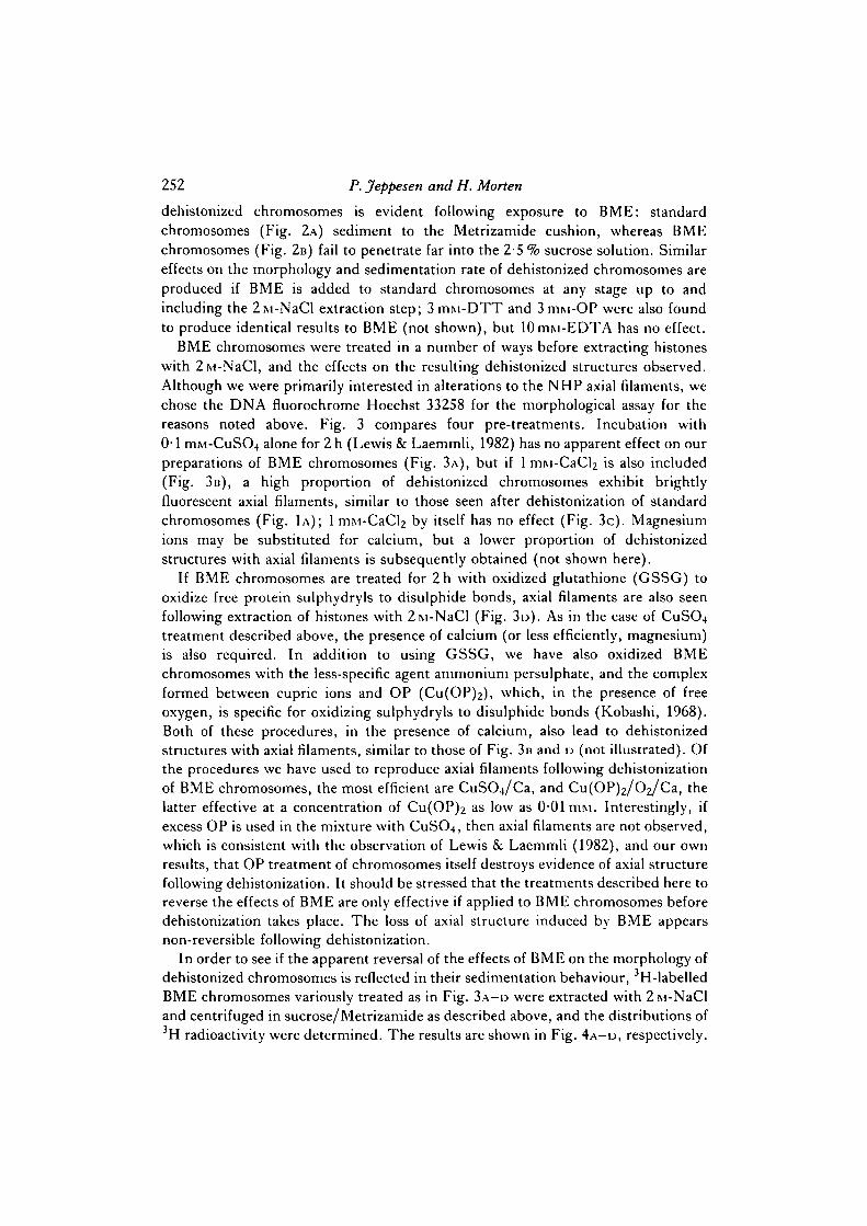
In order to see if the apparent reversal of the effects of BME on the morphology ofdehistonized chromosomes is reflected in their sedimentation behaviour, 3H-labelledBME chromosomes variously treated as in Fig. 3A-D were extracted with 2M-NaCland centrifuged in sucrose/Metrizamide as described above, and the distributions of3H radioactivity were determined. The results are shown in Fig. 4A-D, respectively.
Sulphydryl reagents and chromosome structure 253
70
60
1 50
Z. 40
830
20
10
01 2 3 4 5
Fraction number
70
60
50
40
30
20
10
01 2 3 4 5
Fraction number
70
—X 50
o
V
40
8 30
20
10
02 3 4 5
Fraction number
70
60
50
40
30
20
10
D
1 2 3 4 5
Fraction number
Fig. 4. Non-correlation of sedimentation rates with restoration of morphological cores inBME-treated dehistonized chromosomes, A - D . 3H-labelled Chinese hamster metaphasechromosomes prepared in the presence of BME were treated identically to thoseillustrated in Fig. 3 A - D , and the sedimentation rates after dehistonization were com-pared, as described in the legend to Fig. 2.
Pretreatment with 01mM-CuSO4, in both the absence and presence of calcium,leads to significantly changed sedimentation profiles (Fig. 4A and B, respectively)compared to untreated BME chromosomes (Fig. 2B), with large fractions of the 3Hradioactivity being recovered from the Metrizamide cushion. Although the propor-tion sedimenting is somewhat variable from experiment to experiment, it is in
254 P. Jeppesen and H. Morten
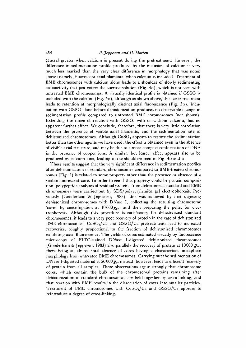
general greater when calcium is present during the pretreatment. However, thedifference in sedimentation profile produced by the inclusion of calcium is verymuch less marked than the very clear difference in morphology that was notedabove: namely, fluorescent axial filaments, when calcium is included. Treatment ofBME chromosomes with calcium alone leads to a shoulder of slowly sedimentingradioactivity that just enters the sucrose solution (Fig. 4c), which is not seen withuntreated BME chromosomes. A virtually identical profile is obtained if GSSG isincluded with the calcium (Fig. 4D), although as shown above, this latter treatmentleads to retention of morphologically distinct axial fluorescence (Fig. 3D). Incu-bation with GSSG alone before dehistonization produces no observable change insedimentation profile compared to untreated BME chromosomes (not shown).Extending the times of reaction with GSSG, with or without calcium, has noapparent further effect. We conclude, therefore, that there is very little correlationbetween the presence of visible axial filaments, and the sedimentation rate ofdehistonized chromosomes. Although CuSO4 appears to restore the sedimentationbetter than the other agents we have used, the effect is obtained even in the absenceof visible axial structure, and may be due to a more compact conformation of DNAin the presence of copper ions. A similar, but lesser, effect appears also to beproduced by calcium ions, leading to the shoulders seen in Fig. 4c and D.
These results suggest that the very significant difference in sedimentation profilesafter dehistonization of standard chromosomes compared to BME-treated chromo-somes (Fig. 2) is related to some property other than the presence or absence of avisible fluorescent core. In order to see if this property could be protein composi-tion, polypeptide analyses of residual proteins from dehistonized standard and BMEchromosomes were carried out by SDS/polyacrylamide gel electrophoresis. Pre-viously (Gooderham & Jeppesen, 1983), this was achieved by first digestingdehistonized chromosomes with DNase I, collecting the resulting chromosome'cores' by centrifugation at 10000g1
av, and then preparing the pellet for elec-trophoresis. Although this procedure is satisfactory for dehistonized standardchromosomes, it leads to a very poor Recovery of protein in the case of dehistonizedBME chromosomes. CuSO^/Ca and GSSG/Ca pretreatments lead to increasedrecoveries, roughly proportional to the fraction of dehistonized chromosomesexhibiting axial fluorescence. The yields of cores estimated visually by fluorescencemicroscopy of FITC-stained DNase I-digested dehistonized chromosomes(Gooderham & Jeppesen, 1983) also parallels the recovery of protein at 10000 gav,there being an almost total absence of cores having a characteristic metaphasemorphology from untreated BME chromosomes. Carrying out the sedimentation ofDNase I-digested material at 50 000^av instead, however, leads to efficient recoveryof protein from all samples. These observations argue strongly that chromosomecores, which contain the bulk of the chromosomal proteins remaining afterdehistonization of standard chromosomes, are held together by cross-linking, andthat reaction with BME results in the dissociation of cores into smaller particles.Treatment of BME chromosomes with CuSCt/Ca and GSSG/Ca appears toreintroduce a degree of cross-linking.
Sulphydryl reagents and chromosome structure MS$
^ 8 4 5 6 7 8r r r ff
i-94
-67
^ ^ MB IP XI ^ V ^ O
-30
-21
-144
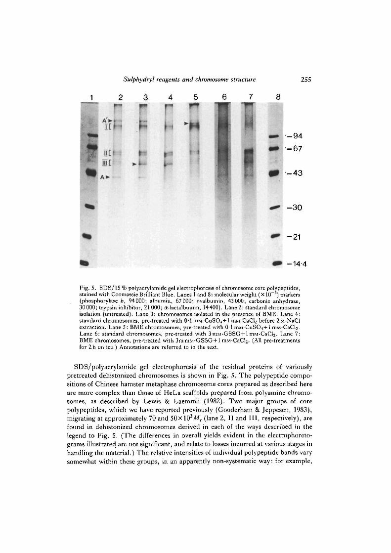
Fig. 5. SDS/15 % polyacrylamide gel electrophoresis of chromosome core polypeptides,stained with Coomassie Brilliant Blue. Lanes 1 and 8: molecular weight (X 10 ) markers(phosphorylase b, 94000; albumin, 67 000; ovalbumin, 43 000; carbonic anhydrase,30000; trypsin inhibitor, 21 000; O'-lactalbumin, 14400). Lane 2: standard chromosomeisolation (untreated). Lane 3: chromosomes isolated in the presence of BME. Lane 4:standard chromosomes, pre-treated with 0-1 ITIM-CUSO^+I mM-CaCl2 before 2M-NaClextraction. Lane 5: BME chromosomes, pre-treated with 0-1 mM-CuS04+l mM-CaCl^.Lane 6: standard chromosomes, pre-treated with 3 mM-GSSG + 1 mM-CaCl2. Lane 7:BME chromosomes, pre-treated with 3m mM-GSSG + 1 mM-CaCl2. (All pre-treatmentsfor 2h on ice.) Annotations are referred to in the text.
SDS/polyacrylamide gel electrophoresis of the residual proteins of variouslypretreated dehistonized chromosomes is shown in Fig. 5. The polypeptide compo-sitions of Chinese hamster metaphase chromosome cores prepared as described hereare more complex than those of HeLa scaffolds prepared from polyamine chromo-somes, as described by Lewis & Laemmli (1982). Two major groups of corepolypeptides, which we have reported previously (Gooderham & Jeppesen, 1983),migrating at approximately 70 and 50xl03Mr (lane 2, II and III, respectively), arefound in dehistonized chromosomes derived in each of the ways described in thelegend to Fig. 5. (The differences in overall yields evident in the electrophoreto-grams illustrated are not significant, and relate to losses incurred at various stages inhandling the material.) The relative intensities of individual polypeptide bands varysomewhat within these groups, in an apparently non-systematic way: for example,
256 P. Jeppesen and H. Morten
the band arrowed in lane 3 is relatively strong in lanes 3, 4 and 7, but weaker in lanes2, 5 and 6. In somes samples, a relatively strong actin band is found (A, lane 2)(Gooderham & Jeppesen, 1983), which seems to correlate with the presence of aband close to the origin (A', lane 2). It is possible that this latter band is myosin,which migrates in a similar position on one-dimension gels (unpublished obser-vation), but further characterization is necessary to establish its identity. Actin, andthe putative myosin, are strong contaminants of unpurified chromosomes, andcollect mainly in fractions 1-3 during the glycerol gradient chromosome purificationstep. Presumably therefore, the presence of these bands in dehistonized chromo-some preparations is mostly, if not wholly, accounted for by contamination. Forreasons we cannot explain, in all cases where chromosomes were treated with CuSO4
or GSSG in the presence of calcium before dehistonization (lanes 4-7) the yields ofactin and the putative myosin are lower than from untreated chromosomes (lanes 2and 3). This observation does indicate, however, that such treatments do not merelycause a random cross-linking of proteins present in the chromosome preparation toproduce an artefactual insoluble protein core. Just below the position of the putativemyosin is a group of polypeptides (I, lane 2), which is common to all samples ofdehistonized chromosomes — again, the relative intensities of the individual compo-nents are somewhat variable. This region of the 15 % gel is not well resolved, andmolecular weights are not easy to estimate, but we would expect the majorchromosome scaffold protein components Scl (170Xl03Mr) and Sc2 (135xlO3Mr)described by Lewis & Laemmli (1982) to migrate in this area, although we have notidentified whether any of the group I polypeptides do indeed correspond to Scl orSc2. Lewis & Laemmli (1982) reported a loss of these polypeptides in dehistonizedchromosomes exposed to BME, which however are retained if the chromosomes aretreated with copper before dehistonization. Fig. 5 fails to show exactly similarbehaviour by any of the group I polypeptides, although we have noted onereproducible difference, comparing the protein profiles of differently prepareddehistonized chromosomes over several experiments, which may be related. Anextra band is apparent just below the position of the putative myosin in BMEchromosomes treated with CuSO^Ca before dehistonization (arrowed in lane 5).No similar bands are apparent in dehistonized standard choromosomes (lane 2),standard chromosomes treated with CuSO4/Ca before dehistonization (lane 4), or instandard or BME chromosomes treated with GSSG/Ca (lanes 6 and 7).
DISCUSSION
Axial structure in dehistonized chwmosomes
The most direct and striking evidence for the 'scaffold' model of chromosomestructure is the visualization by both optical and electron microscopy of a residualNHP axial filament or core in dehistonized metaphase chromosomes (Paulson &Laemmli, 1977). As reported by others (Lewis & Laemmli, 1982), and clearlyshown here, exposure to BME abolishes the morphological evidence for axialstructure. We also show that nuclease digestion of dehistonized BME chromosomes
Sulphydryl reagents and chromosome structure 257
does not liberate protein cores, recognizably derived from metaphase chromosomes,as we have described previously (Gooderham & Jeppesen, 1983). Instead, muchsmaller NHP particles requiring higher centrifugation speeds for sedimentation areobtained. Our interpretation of these results is that the NHP core in standarddehistonized chromosomes is held together by BME-labile cross-linking. Supportfor this interpretation is provided by our demonstration that axial structure, evidentafter dehistonization, and morphologically closely resembling that apparent instandard dehistonized chromosomes, can be simulated by oxidation of BMEchromosomes in the presence of calcium ions, using conditions in which one wouldexpect to convert free sulphydryl groups to disulphide bridges. These observationssuggest that the chromosome core in standard dehistonized chromosomes might alsobe maintained by cross-linking between protein sulphydryls. We do not know whycalcium or, less effectively, magnesium ions are required during oxidation toreproduce axial NHP filaments in dehistonized chromosomes, but to create thepossibility of a disulphide bond forming two sulphydryl groups need to be in closeproximity, and it may be that divalent cations play a role in aligning adjacent proteinmolecules so that intermolecular disulphide bonds can form.
Our results clearly fail to demonstrate a specific requirement for copper, or forany other heavy metal, in regaining morphological chromosome cores by oxidationof BME chromosomes. We have not found it practicable to eliminate atmosphericoxygen completely when treating BME chromosomes with copper (Lewis &Laemmli, 1982), and have therefore not attempted to do so. The morphologicaleffects of copper appear very similar to the effects of the sulphydryl oxidizing agentswe have used. In particular, free CuSO4 is indistinguishable in its effects from thecopper—OP complex, which specifically catalyses the oxidation of sulphydryls todisulphide bonds by O2 (Kobashi, 1968), and it therefore seems likely that in theexperiments described here copper is also exerting its effect on the morphology ofdehistonized BME chromosomes by catalysing the oxidation of sulphydryls. Lewis& Laemmli (1982) have reported oxidation by copper in the presence of O2, andfurther support for this interpretation is provided by the finding that, in commonwith the other oxidative procedures we have used, the presence of calcium isnecessary for the recovery of NHP axial filaments.
Sedimentation properties
Although oxidation in the presence of calcium is apparently sufficient to reversethe effects of BME on the morphology of dehistonized chromosomes, in general weare unable to demonstrate a corresponding reversion in the sedimentation propertiesto those of standard dehistonized chromosomes. We have found that sedimentationrate is no indication of whether a particular pretreatment leads to a recovery ofvisible axial structure in dehistonized BME chromosomes. Thus although treatmentwith CuSO4 appears to give a sizeable fraction of dehistonized material reverting tohigh sedimentation rate, there is no significant difference whether axial filaments areevident (CuS04+calcium) or not (CUSO4 alone). On the other hand, whereas axialstructure is clearly evident after oxidation by GSSG in the presence of calciumfollowed by dehistonization, only a marginal increase in sedimentation rate isachieved, which also occurs with calcium ions alone when no axial structure can be
258 P. jfeppesen and H. Morten
detected. These changes in sedimentation rate seem to be influenced by the nature ofany divalent cations that may be present, rather than by the recovery of visible axialstructure. The protein compositions following dehistonization of oxidized andnon-oxidized BME chromosomes are not significantly different from standarddehistonized chromosomes, and our inability to reverse the change in sedimentationrate cannot be accounted for by an irreversible loss of protein. We conclude thatthere is some additional factor present in standard dehistonized chromosomes thatwe cannot detect by fluorescence microscopy.
77ie NHP chromosome core
It has been argued that chromosome cores or scaffolds observed in dehistonizedchromosomes are artefacts produced by the random deposition of insoluble or poorlysoluble NHP along the chromatid axes, which are regions of high DNAconcentration (Okada & Comings, 1980). Against this view is the evidence for aspecific subset of chromosomal proteins retained after 2M-NaCl extraction(Gooderham & Jeppesen, 1983; Lewis & Laemmli, 1982). In addition, theconclusion drawn here that interprotein cross-linking is required to maintain theintegrity of NHP axial cores makes the explanation that such structure has anentirely artefactual origin less likely. Moreover, we can find no evidence that,following dehistonization, either the total amount of residual NHP or the pattern ofpolypeptide bands differ significantly whether chromosomes are in an oxidized orreduced state, exhibiting identifiable axial cores or not. If apparent axial structurewere a result of non-specific deposition and cross-linking of chromosomal proteins,we would expect an increase in the total amount of protein left unsolubilized after2M-NaCl extraction of oxidized chromosomes compared with BME-treatedchromosomes. Loss of visible core structure is not due to solubilization of protein,but rather to a breakage of cross-linking between protein molecules, whichnevertheless remain associated with the DNA. The demonstrated requirement forcalcium during oxidation of BME chromosomes also suggests that a non-randomcross-linking of sulphydryls is necessary in order to regain axial cores followingdehistonization. As the solubilization of chromosome cores for electrophoresisrequires the presence of sulphydryl reducing agents such as BME or DTT, we areunable to investigate the degree of cross-linking directly on gels.
Cross-linking in vivo
It seems clear that the NHP axial structure observed following dehistonization ofoxidized BME chromosomes is held together by disulphide cross-linking. Thesimplest extrapolation is that NHP cores evident in non-BME-treated dehistonizedchromosomes are also maintained by disulphide bridging, and that this reflects invivo metaphase chromosome structure. This interpretation is attractive in that itagrees with previous models of chromatin condensation and chromosome structure,as discussed in the Introduction. However, it does not easily explain why theheavy-metal chelating agents OP and the closely related neocuproine(2,9-dimethyl-o-phenanthroline) abolish axial structure (Lewis & Laemmli, 1982).Similar exposure to the much stronger chelator EDTA (as an example, the log

Sulphydryl reagents and chromosome structure 259
stability constants for EDTA and OP complexes with cupric ion are 18-8 and 6-3,respectively) has no effect, and it could be argued that OP (and neocuproine) mightfunction as a reducing agent. We are unaware of this activity having been reportedpreviously, although OP appears to act as a redox intermediate in a number ofcatalytic oxidations (Kobashi, 1968). Further doubt is cast on disulphide bridging invivo, since NaBH4, which would be expected to reduce disulphides to sulphydryls,does not abolish scaffolds (Lewis & Laemmli, 1982). An alternative model, which isentirely speculative but is consistent with most of the available data, is that in vivocross-linking between NHP is via a dimercaptide of the form:
protein—S—M—S-protein,
where M is a metal ion. This link would be labile to chelators (including sulphydrylreagents) through removal of M, but not susceptible to pure reducing agents such asNaBH4. Cross-linking could be re-introduced either by replacing M, or a metal withsimilar properties, or by oxidizing the now free sulphydryl pair, suitably alignedunder the influence of calcium ions, as we have demonstrated. If this model iscorrect, replacing M should lead to complete reversal of the effects of BME both onthe morphology and on the sedimentation rate of dehistonized chromosomes.Although, in the presence of calcium, copper (Lewis & Laemmli, 1982) appears tofulfil the requirements for M, the evidence is complicated by two observations.First, cupric ions can catalyse the oxidation of sulphydryls, and it is difficult toeliminate this possibility completely, even when precautions are taken. Second, weshow that cupric ions have an effect on the sedimentation rate of dehistonizedchromosomes in the absence of evident axial structure. Further clarification of thesepoints is necessary.
To summarize our conclusions, we believe that the axial cores visible byfluorescence and electron microscopy in dehistonized chromosomes, are maintainedby cross-linking between NHP. The cross-linking is labile to sulphydryl reagentsand certain chelators, such as OP, leading to a loss of visible structure.Morphologically similar axial cores may be reproduced in BME-treatedchromosomes if the free sulphydryls are oxidized to disulphides in the presence ofcalcium before dehistonization, although in general this does not lead to a return tothe high sedimentation rate of non-BME-treated dehistonized chromosomes.Neither exposure of chromosomes to BME, nor subsequent oxidation, leads tosignificantly different polypeptide profiles following dehistonization. Taking intoaccount the results of Lewis & Laemmli (1982), we suggest that cross-linking in vivomay occur via heavy-metal dimercaptide bridges linking the same sulphydrylgroups. The metal involved could be copper (Lewis & Laemmli, 1982), although wehave no direct evidence to support this. We hope in the future to determine whichproteins present in the core are involved in cross-linking.
We are grateful to Dr A. T. Sumner for reading the manuscript, and for his helpful comments.We also thank Professor H. J. Evans for his encouragement of this work.

260 P. Jeppesen and H. Morten
REFERENCES
ADOLPII, K. W. (1980). Isolation and structural organization of human mitotic chromosomes.Chromosoma 76, 23-33.
DOUNCE, A. L., CIIANDA, S. K. & TOWNES, P. L. (1973). The structure of higher eukaryoticchromosomes. J. theor. Biol. 42, 275-285.
GOODERIIAM, K. & JEPPESEN, P. (1983). Chinese hamster metaphase chromosomes isolatedunder physiological conditions: a partial characterization of associated nonhistone proteins andprotein cores. Expl Cell Res. 144, 1-14.
HADLACZKY, G., SUMNER, A. T. & Ross, A. (1981). Protein-depleted chromosomes. Chromo-soma 81, 557-567.
JEPPESEN, P. G. N. & BANKIER, A. T. (1979). A partial characterization of DNA fragmentsprotected from nuclease degradation in histone depleted metaphase chromosomes of the Chinesehamster. Nucl. Acids Res. 7, 49-67.
JEPPESEN, P. G. N., BANKIER, A. T. & SANDERS, L. (1978). Nonhistone proteins and thestructure of metaphase chromosomes. Expl Cell Res. 115, 293-302.
KOBASIII, K. (1968). Catalytic oxidation of sulfhydryl groups by o-phenanthroline coppercomplex. Biochim. biophys. Ada 158, 239-245.
LEWIS, C. D. & LAEMMLI, U. K. (1982). Higher order metaphase chromosome structure:evidence for metalloprotein interactions. Cell 29, 171-181.
OKADA, T. A. & COMINGS, D. E. (1980). A search for protein cores in chromosomes: is thescaffold an artifact? Aw. J . hunt. Genet. 32, 814-832.
PAULSON, J. R. & LAEMMLI, U. K. (1977). The structure of histone-depleted metaphasechromosomes. Cell 12, 817-828.
SATYA-PRAKASII, K. L., HSU, T. C. & PATHAK, S. (1980). Behavior of the chromosome core inmitosis and meiosis. Chromosoma 81, 1-8.
SOBELL, H. M. (1973). The stereochemistry of actinomycin binding to DNA and its implicationsin molecular biology. In Progress in Nucleic Acid Research and Molecular Biohgy, vol. 13 (ed.J. N. Davidson & W. E. Cohn), pp. 153-190. New York: Academic Press.
SUMNER, A. T. (1973). Involvement of protein disulphides and sulphydryls in chromosomebanding. Expl Cell Res. 83, 438-442.
SUMNER, A. T . (1983). The role of protein sulphydryls and disulphides in chromosome structureand condensation. In Kew Chromosome Conference II (ed. P. E. Brandham & M. D. Bennett),pp. 1-9. London: George Allen & Unwin.
{Received 21 May 1984 - Accepted 13 August 1984)